


Simple Summary
The health of llama crias is strongly affected by intestinal diseases, which are a major cause of mortality in Andean livestock systems. These diseases are often linked to changes in the microorganisms that live in the digestive system, known as the gut microbiota. This study aimed to compare the fecal microbiota of healthy and diseased llama crias to better understand how these microbial communities are affected by disease. We found that diseased animals showed important changes in their microbial composition, including a higher presence of bacteria commonly associated with infections, as well as greater variability in their microbial communities. In contrast, healthy llamas had more stable microbial profiles, with bacteria that contribute to digestion and gut health. These findings suggest that intestinal disease in llamas is associated with an imbalance in gut microorganisms. Understanding these changes can help develop new strategies to improve animal health, prevent disease, and enhance productivity in llama farming systems, which are essential for rural livelihoods in high-altitude regions.
Abstract
The gastrointestinal microbiota plays a key role in the health and productivity of llama crias, yet its dynamics under disease conditions remain poorly understood. This study aimed to characterize and compare the fecal microbiota of healthy and diseased llama crias. Fecal samples collected from nine llama crias (six with diarrhea and three clinically healthy controls) were analyzed using 16S rRNA gene sequencing targeting the V4 region, followed by bioinformatic processing and statistical analyses, including diversity metrics and generalized linear mixed models. Results showed that microbial communities were dominated by Bacillota and Bacteroidota in both groups; however, diseased animals exhibited increased abundance of Pseudomonadota, particularly Gammaproteobacteria and Enterobacteriaceae. Beta diversity analysis revealed significant differences in community structure between groups, with diseased samples showing higher dispersion. Additionally, a greater number of unique ASVs was observed in diseased individuals, indicating increased microbial heterogeneity. Differential abundance analysis identified distinct microbial signatures associated with health status, with opportunistic taxa enriched in diseased animals and fiber-degrading taxa more prevalent in healthy individuals. Overall, the findings demonstrate an association between diarrhea and differences in the fecal microbiota of llama crias. Because this was a cross-sectional observational study, causal relationships cannot be inferred. These results provide a foundation for future investigations into host–microbiota interactions and gastrointestinal health in South American camelids.
1. Introduction
The gastrointestinal microbiota constitutes a highly complex and dynamic ecosystem that plays a central role in digestion, metabolism, and immune regulation in ruminants and pseudo-ruminants such as llamas (Lama glama). These microbial communities are essential for the degradation of plant polysaccharides, synthesis of short-chain fatty acids, and maintenance of intestinal homeostasis [1,2,3,4]. Fecal microbiota has been widely used as a non-invasive proxy of the distal gut microbial ecosystem, as it reflects the composition and functional potential of intestinal microbial communities involved in nutrient absorption and host health [3,5].
In llamas, gut microbiota composition is influenced by multiple factors, including diet, altitude, age, and environmental conditions, which shape microbial diversity and structure [5,6,7]. Previous studies have consistently reported that the fecal microbiota of llamas is dominated by Bacillota (Firmicutes) and Bacteroidota, which together account for the majority of microbial abundance and are closely associated with fiber degradation and host energy metabolism [6]. These dominant phyla have also been widely described in other ruminants, highlighting a conserved microbial structure linked to herbivorous digestion [2,4]. However, despite their importance in Andean livestock systems, there remains limited knowledge regarding microbiota dynamics in llama crias, particularly under disease conditions.
Gastrointestinal disorders, especially diarrhea, are among the leading causes of morbidity and mortality in llama crias and are increasingly associated with alterations in microbial diversity and composition, commonly referred to as dysbiosis [1,8]. Dysbiosis is characterized by instability in microbial communities, reduced diversity, and the proliferation of opportunistic or pathogenic taxa, which may compromise intestinal homeostasis and immune function [4,8]. In llamas, disease conditions have been linked to shifts in key bacterial groups, including an increase in Pseudomonadota (Proteobacteria), particularly Gammaproteobacteria and Enterobacteriaceae, and a reduction in beneficial taxa such as Bacteroidota and Clostridia [1,8]. These microbial imbalances are consistent with patterns observed in other livestock species and may reflect inflammatory processes and altered gut functionality.
Microbial diversity analyses provide essential tools to evaluate these ecological changes. Alpha diversity metrics allow the assessment of microbial richness and evenness, whereas beta diversity approaches—such as Bray–Curtis dissimilarity and ordination techniques like Principal Coordinates Analysis (PCoA)—enable the comparison of microbial community structure between groups [3,9]. Previous studies have demonstrated that diet, environmental conditions, and disease status significantly influence microbial community composition, often resulting in distinct clustering patterns and increased dispersion in diseased animals, reflecting microbiome instability [5,7,9]. Furthermore, the evaluation of shared and unique operational taxonomic units (OTUs) provides insights into the core microbiota and condition-specific taxa, revealing ecological shifts associated with health status and microbial resilience.
In addition, differential abundance analyses have identified specific microbial taxa associated with health or disease conditions across multiple taxonomic levels, supporting the use of microbiota as differentially abundant taxa for disease detection and management [6,8,10]. The enrichment of opportunistic taxa in diseased animals, together with the reduction in fiber-degrading and metabolically beneficial microorganisms, suggests a functional disruption of the gut ecosystem that may directly impact host physiology and productivity. Similar microbial shifts have been described across ruminant gastrointestinal systems, where alterations in dominant fermentative taxa are associated with impaired metabolic stability and intestinal dysbiosis [11,12].
In this context, the characterization of fecal microbiota in llama crias provides a valuable framework for understanding host–microbiota interactions under contrasting health conditions. Based on current evidence, diseased microbiota are expected to exhibit higher variability, increased abundance of opportunistic taxa, and a greater proportion of unique microbial communities, whereas healthy individuals tend to show more stable and functionally coherent microbial profiles, consistent with the concept of a conserved core microbiota described in ruminants [13]. Therefore, the aim of this study was to compare the fecal microbiota of healthy and diseased llamas, focusing on microbial diversity, community structure, and taxonomic composition. Additionally, we evaluated shared and unique ASVs, as well as differentially abundant taxa associated with health status, in order to identify microbial signatures linked to disease and contribute to a better understanding of intestinal ecosystem dynamics in llamas.
2. Materials and Methods
2.1. Animal and Sample Collection
Samples were collected from a total of nine llama crias (three healthy and six presenting with diarrhea), aged between 3 and 4 months, all sourced from the La Raya Experimental Center of the Universidad Nacional del Altiplano (UNAP) (Table S1). The crias were maintained under a single extensive rearing system alongside their dams at an altitude of 4200 m above sea level (m a.s.l.). The study was conducted during March and April, corresponding to the late rainy season, under the same climatic conditions for all animals. At the study site, the climate is characterized by cold high-altitude conditions, with temperatures typically ranging from approximately −12 °C to 19.75 °C, annual mean temperatures of about 7.5 °C, and relative humidity generally ranging from 65% to 85%. The animals were predominantly nourished with maternal milk and grazed on native pastures, mainly Festuca dolichophila, from 07:00 to 17:00 h, with free access to the same water source. Healthy and diarrheic crias were maintained under identical environmental and management conditions throughout the study. Both groups were kept in the same housing system, grazed in the same grazing area under the same daily grazing schedule, and received the same feeding regimen, veterinary management, sanitary management, and handling by the same personnel. Therefore, environmental and management conditions were consistent between groups, minimizing potential environmental confounding factors [14]. The diseased llama crias were identified by a veterinarian based exclusively on a clinical examination performed at the time of sampling. The evaluation included anamnesis obtained from the animal caretakers, assessment of rectal temperature, respiratory rate, hydration status, general behavior, and fecal consistency. Animals were classified as diarrheic according to conventional veterinary clinical criteria, including profuse watery feces with loss of normal pellet formation, increased frequency and forceful evacuation of feces, fecal contamination of the perineal region, mild dehydration, mild lethargy, and a fecal consistency score of 7, corresponding to watery diarrhea without visible blood. Rectal temperature and respiratory rate remained within the normal physiological range. Healthy control animals presented normally formed fecal pellets, normal defecation frequency, and no clinical evidence of gastrointestinal disease at the time of sampling. Following the clinical diagnosis of diarrhea, coproparasitological examinations were performed as a complementary diagnostic procedure to exclude enteric protozoan infections as potential causes of diarrhea and thereby minimize confounding factors in the microbiota analysis. Diagnostic procedures followed previously established methodologies [8]. Fecal samples from affected animals were initially examined by direct microscopic observation using Lugol’s iodine staining (Sigma-Aldrich, St. Louis, MO, USA) to detect protozoan cysts and oocysts [15]. Subsequently, qualitative flotation techniques using saturated sucrose solution (Sigma-Aldrich, St. Louis, MO, USA) were applied to improve parasite detection [16]. Quantitative analysis was performed using the modified McMaster method, adapted for fecal samples from llamas with clinical conditions [17]. These parasitological methods were used exclusively to detect and quantify gastrointestinal protozoa and were not employed as diagnostic criteria for diarrhea. No parasitic structures were detected in the control group.
2.2. DNA Extraction and Sequencing
Microbial genomic DNA was extracted from 500 mg of each fecal sample using the PureLink™ Microbiome DNA Purification Kit (Invitrogen, Thermo Fisher Scientific, Waltham, MA, USA), following the manufacturer’s instructions. DNA concentration and quality were assessed using a Qubit® fluorometer and electrophoresis on 1% agarose gels (Sigma-Aldrich, St. Louis, MO, USA). The V4 region of the bacterial 16S rRNA gene was amplified using the universal primers 515F (5′-GTGYCAGCMGCCGCGGTAA-3′) and 806R (5′-GGACTACNVGGGTWTCTAAT-3′) [18]. Primers were adapted to include Ion Torrent-specific sequencing adapters. Amplicon libraries were prepared according to the Ion 16S™ Metagenomics Kit (Ion Torrent, Thermo Fisher Scientific, Waltham, MA, USA) Ion Torrent protocol, including adapter ligation and emulsion PCR amplification. Sequencing was performed on the Ion S5 (Ion Torrent, Thermo Fisher Scientific, Waltham, MA, USA) platform following the manufacturer’s guidelines [19]. Library sequencing was performed on the Ion Torrent S5 platform using an Ion 530™ Chip, generating single-end reads with a read length of approximately 400 bp.
2.3. Bioinformatic Analysis
Raw sequencing reads in FASTQ format were processed in R v4.1.1. using the DADA2 pipeline (v1.38.0) [20]. The microbiome analysis workflow followed current recommendations for standardized microbiome data processing and downstream statistical analyses [21]. Initially, all sequence files were imported and organized, and sample names were assigned based on file identifiers. Quality filtering and trimming were performed using the filterAndTrim function, applying the following parameters: no truncation of read length (truncLen = 0), maximum expected errors of 3 (maxEE = 3), and a truncation quality threshold of 2 (truncQ = 2). Filtered reads were stored for downstream analyses. Error rates were learned from the filtered reads using the learnErrors function, allowing accurate modeling of sequencing errors. Dereplication of sequences was conducted using the derepFastq function, followed by inference of amplicon sequence variants (ASVs) using the dada algorithm. A sequence table was then constructed using makeSequenceTable, and chimeric sequences were removed using the consensus method implemented in removeBimeraDenovo. The resulting non-chimeric ASV table was exported for downstream analyses. Taxonomic assignment was conducted using the SILVA reference database (version 138.2) [22]. A naïve Bayesian classifier was applied using the training dataset for genus-level classification, followed by species-level assignment using the corresponding reference file. Phylogenetic relationships among ASVs were inferred using multiple sequence alignment and maximum likelihood approaches. Sequences were aligned using the DECIPHER package (v3.8.0) [23], and a phylogenetic tree was constructed using the phangorn package (v2.12.1). Genetic distances were calculated, and a neighbor-joining tree was initially generated and subsequently optimized under the Jukes–Cantor model using nearest-neighbor interchange (NNI) rearrangements. All bioinformatic analyses were performed in R, ensuring reproducibility and integration with downstream ecological and statistical analyses.
2.4. Data Processing and Normalization
Raw taxonomic data were filtered to retain only ASVs classified within the domain Bacteria for downstream analyses, whereas archaeal and non-target sequences were excluded. Additional filtering steps were applied to eliminate potential contaminants, including mitochondrial and chloroplast sequences, ensuring that downstream analyses reflected true microbial communities. Following taxonomic filtering, the dataset was standardized using the microeco framework. To control for sequencing depth variability, samples with low sequencing coverage were excluded. The total number of reads per sample was calculated, and samples with fewer than 7000 reads were removed from the dataset. All nine samples exceeded this threshold; therefore, no samples were removed during quality filtering. After filtering, the dataset was restructured to ensure consistency between ASV tables and sample metadata. Normalization of sequencing data was performed using the total sum scaling (TSS) method implemented in the microeco package v.2.3.0. This approach converts raw counts into relative abundances by dividing each taxon count by the total number of reads per sample, allowing for direct comparison of microbial communities across samples with different sequencing depths.
2.5. Statistical Analysis
Statistical analyses were performed using the R software environment (v4.1.1). Beta diversity was assessed using Bray–Curtis dissimilarity and visualized through Principal Coordinates Analysis (PCoA). Statistical significance of community structure differences between groups was tested using permutational multivariate analysis of variance (PERMANOVA) with 9999 permutations, as implemented in the vegan package v.2.7.5. For differential abundance analysis, a generalized linear mixed model (GLMM) based on beta distribution was applied using the microeco package v.2.3.0. and the glmmTMB backend v.1.1.14. Beta-distributed GLMMs provide an appropriate statistical framework for modeling proportional microbiome data and identifying differentially abundant taxa across taxonomic levels. Prior to analysis, low-abundance taxa were filtered using a threshold of 0.05 to reduce noise and improve model stability. The model was specified with health status as the main explanatory variable (Group), allowing the identification of taxa significantly associated with healthy and diseased llama crias. Model outputs included estimated coefficients and statistical significance for each taxon. Differentially abundant taxa were visualized using bar plots and heatmaps representing model coefficients and significance levels. Graphical outputs were further customized using the ggplot2 package v.4.0.3. to improve visualization clarity. Statistical significance was set at p < 0.05 for all analyses.
3. Results
3.1. Relative Abundance of Microbial Communities at Phylum and Class Levels
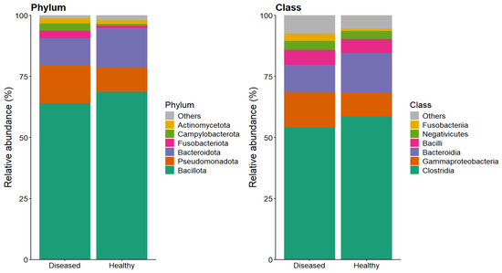
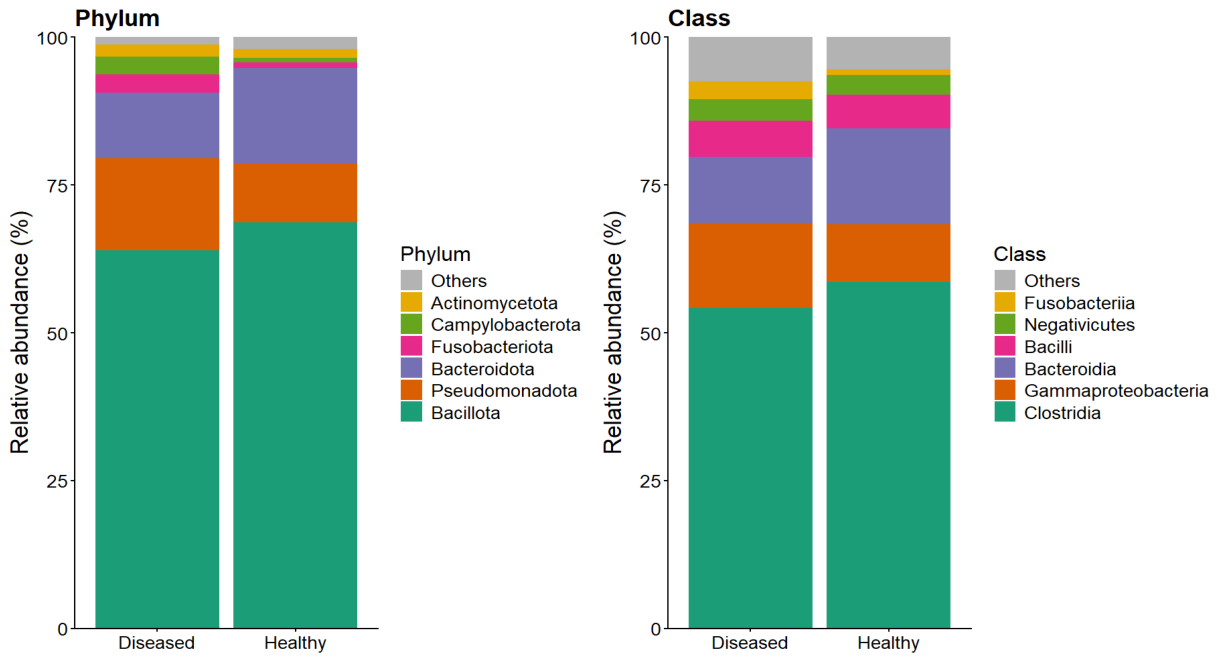
At the phylum level, the microbial communities in both groups were predominantly composed of Bacillota, accounting for 64% in diseased animals and 68.7% in healthy individuals, indicating a slightly higher prevalence in the latter. Pseudomonadota was more abundant in diseased animals (15.5%) compared to healthy ones (9.8%), whereas Bacteroidota showed the opposite pattern, being more represented in healthy animals (16.2%) than in diseased counterparts (11.1%). Minor phyla, including Campylobacterota and Fusobacteriota, remained below 5% in both groups. At the class level, Clostridia dominated the microbiota, with higher relative abundance in healthy animals (58.6%) compared to diseased ones (54.2%). In contrast, Gammaproteobacteria was enriched in diseased animals (14.4% vs. 9.8%), while Bacteroidia was more prevalent in healthy individuals (16.2% vs. 11.1%). Less abundant classes such as Bacilli and Negativicutes showed relatively minor contributions across both conditions (Figure 1). Overall, these patterns suggest a shift in microbial composition associated with health status, characterized by an increase in Pseudomonadota/Gammaproteobacteria in diseased animals and a relative enrichment of Bacteroidota/Bacteroidia in healthy individuals.
Figure 1.
Relative abundance of microbial communities at phylum and class levels in healthy and diseased llamas.
3.2. Differences in Core and Condition-Specific Microbiota Based on ASV Distribution
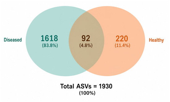
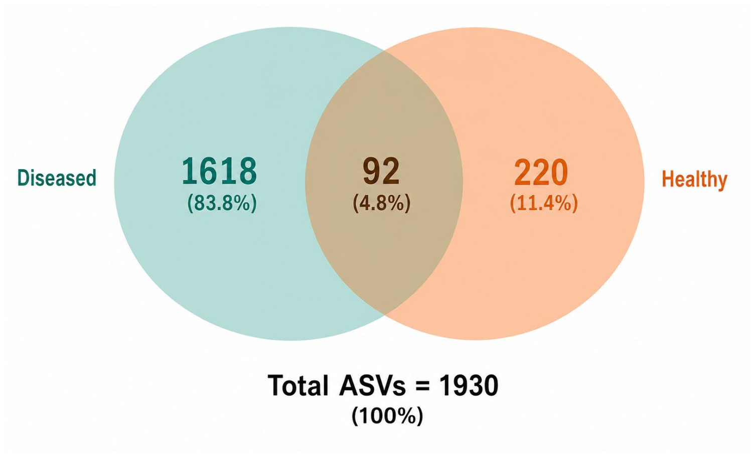
A Venn diagram analysis identified a total of 1930 ASVs in the fecal microbiota of healthy and diseased llama crias (Figure 2). Of these, 1618 ASVs (83.8%) were shared between both groups, representing the core microbiota. Diseased animals harbored 220 unique ASVs (11.4%), whereas healthy animals contained 92 unique ASVs (4.8%).
Figure 2.
Shared and unique ASVs in fecal microbiota of healthy and diseased llama crias.
3.3. Beta Diversity and Community Structure Differences Between Healthy and Diseased Llamas
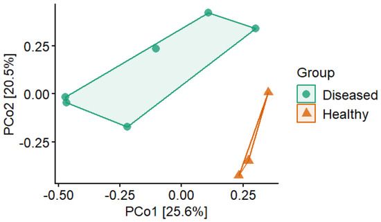
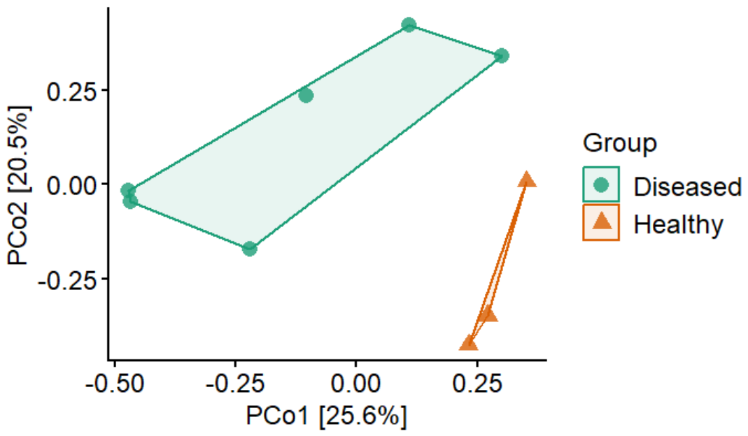
Principal Coordinates Analysis (PCoA) based on Bray–Curtis dissimilarity revealed a clear separation between the fecal microbial communities of healthy and diseased llama crias (Figure 3). Samples from healthy animals formed a relatively compact cluster, indicating greater similarity in microbial community composition, whereas samples from diseased animals were more widely dispersed, reflecting increased inter-individual variability and microbial instability. Although partial overlap between groups was observed, the overall ordination pattern demonstrated a distinct shift in microbial community structure associated with disease status. These differences were statistically supported by PERMANOVA (R2 = 0.208, F = 1.839, p = 0.018) (Table 1), indicating that health status significantly explained 20.8% of the variation in microbial community composition.
Figure 3.
PCoA of Bray–Curtis dissimilarity showing beta diversity differences between healthy and diseased llama crias.
Table 1.
Beta diversity analysis of fecal microbiota in healthy and diseased llama crias based on Bray–Curtis dissimilarity.
3.4. Differentially Abundant Taxa Associated with Health Status Identified by GLMM
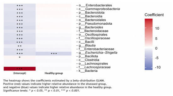
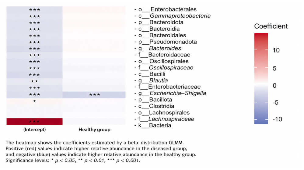
Differential abundance analysis using a GLMM identified significant candidate microbial signatures with health status across multiple taxonomic ranks, including phylum, class, order, family, and genus. At the phylum level, Pseudomonadota was significantly enriched in diseased llama crias, whereas Bacteroidota and Bacillota were relatively more abundant in healthy animals. Similar patterns were observed at the class level, where Gammaproteobacteria predominated in diseased animals, while Bacteroidia and Clostridia were enriched in healthy individuals. At finer taxonomic resolution, Enterobacterales and Enterobacteriaceae were significantly associated with diseased animals, whereas Bacteroidales, Bacteroidaceae, and Lachnospiraceae were enriched in healthy animals. At the genus level, Escherichia–Shigella showed significantly greater relative abundance in diseased llama crias, while Bacteroides and Blautia were significantly associated with healthy animals, suggesting that these genera may serve as potential microbial indicators of gastrointestinal health status (Figure 4).
Figure 4.
Differentially abundant microbial taxa associated with health status identified using a generalized linear mixed model (GLMM). Colors indicate the direction and magnitude of the standardized effect size, with positive values (red) representing taxa enriched in diseased llama crias and negative values (blue) representing taxa enriched in healthy llama crias. Taxa are shown at the phylum, class, order, family, and genus levels. Statistical significance is indicated as follows: p < 0.05 (*), p < 0.01 (**), and p < 0.001 (***).
Additionally, taxa within the phylum Bacillota, including Clostridia, Lachnospirales, and Lachnospiraceae, were more abundant in healthy llama crias, as indicated by positive coefficients in the GroupHealthy comparison. Other groups such as Oscillospirales and Oscillospiraceae showed moderate but consistent associations, while Bacilli and Blautia displayed less pronounced differences. Overall, the GLMM results revealed a clear differential distribution of microbial taxa between groups, with a higher number of significantly enriched taxa in diseased animals and a distinct set of taxa associated with healthy individuals (Figure 4).
4. Discussion
The present study revealed clear differences in the fecal microbiota of healthy and diseased llama crias, highlighting the role of microbial community structure in health status. At the phylum level, both groups were dominated by Bacillota and Bacteroidota, which is consistent with previous reports in ruminants and camelids, where these taxa are associated with fiber degradation and energy metabolism [12,24,25]. However, diseased animals exhibited a relative increase in Pseudomonadota, particularly Gammaproteobacteria, which has been widely recognized as a hallmark of microbial dysbiosis and intestinal inflammation across multiple host species [26,27,28,29]. This shift suggests a disruption of gut homeostasis, potentially linked to altered metabolic functions and host–microbe interactions.
The enrichment of Pseudomonadota and Enterobacteriaceae in diseased llamas, as observed in the GLMM analysis, is consistent with findings in other livestock and mammalian systems, where these taxa are associated with opportunistic pathogenic behavior and inflammatory responses [28,30,31]. Members of the genus Escherichia–Shigella have been particularly implicated in gastrointestinal disorders due to their ability to exploit disturbed gut environments and outcompete commensal bacteria [29,32]. Their increased abundance in diseased individuals supports the hypothesis that microbial imbalance contributes to disease progression in llama crias. In contrast, healthy llamas showed higher relative abundance of Bacteroidota and Bacillota-associated taxa, including Bacteroidia and Clostridia. These groups are known to play key roles in polysaccharide degradation and short-chain fatty acid production, contributing to gut stability and immune modulation [25,33,34]. The enrichment of Lachnospiraceae and other Clostridia-related taxa in healthy animals further supports their role in maintaining intestinal homeostasis, as these bacteria are often associated with anti-inflammatory effects and epithelial integrity [34,35,36].
The Venn diagram analysis revealed a markedly higher number of unique ASVs in diseased llamas compared to healthy individuals. This pattern suggests increased microbial heterogeneity under disease conditions, which has been described as a characteristic of dysbiotic ecosystems [37,38]. In unstable microbial environments, opportunistic taxa may colonize and expand, leading to a higher proportion of unique or transient taxa. Conversely, healthy microbiota tends to be more stable and functionally redundant, resulting in a smaller number of exclusive ASVs and a more consistent core microbiota [37,39]. The presence of shared ASVs between groups likely represents core microbial taxa essential for basic gut functions. Beta diversity analysis further supported these findings, showing clear separation between healthy and diseased groups and greater dispersion among diseased samples. Increased dispersion has been associated with microbiome instability and reduced ecological resilience [38,40]. The PERMANOVA results indicated that health status explained a significant portion of the variation in microbial community structure, although a substantial proportion remained unexplained. This suggests that additional factors, such as diet, environmental exposure, host genetics, and early-life microbial colonization, may also contribute to microbiota variability in llamas [12,26,41,42].
Differential abundance analysis using GLMM provided further evidence of distinct microbial signatures associated with health status. Diseased animals exhibited a higher number of significantly enriched taxa, particularly within Pseudomonadota, whereas healthy animals showed enrichment of taxa involved in fiber degradation and metabolic stability. Similar patterns have been reported in other ruminants, where disease states are associated with an expansion of facultative anaerobes and a reduction in obligate anaerobic fermenters [29,33]. This shift may lead to altered metabolic outputs, including reduced production of beneficial metabolites such as butyrate, which is essential for intestinal health [35,43]. Interestingly, taxa such as Oscillospirales and Oscillospiraceae showed moderate but consistent associations with health status. These groups have been linked to fiber fermentation and host metabolic regulation, although their ecological roles remain incompletely understood [44,45]. Their presence in both groups suggests that they may contribute to core microbiota functions, with subtle variations depending on host condition. One limitation of the present study is the relatively small sample size and the unequal distribution of animals between the healthy (n = 3) and diseased (n = 6) groups. Although the statistical analyses identified significant differences in microbial community composition, these findings should be interpreted with caution. Future studies including larger and more balanced cohorts will be necessary to validate the observed microbial patterns and improve the robustness and generalizability of the results. Additionally, the cross-sectional design of this study precludes causal inference between fecal microbiota composition and diarrhea. Although all animals originated from the same herd and were maintained under the same environmental, dietary, and management conditions, residual host-related factors may have influenced the microbiota. Furthermore, pathogen-specific molecular confirmation and functional validation were not performed; therefore, the observed taxonomic differences should be interpreted as associations rather than direct evidence of microbial function or disease causation. Another limitation of the present study is that the etiological agents responsible for diarrhea were not specifically identified. Although coproparasitological examinations were performed to characterize the parasitological status of the animals, molecular or microbiological screening for viral and bacterial enteropathogens, such as Rotavirus, Coronavirus, enterotoxigenic Escherichia coli, and Salmonella spp., was not conducted. Consequently, the observed microbiota alterations should be interpreted as being associated with the diarrheic condition rather than attributed to a specific infectious agent. Future studies integrating pathogen-specific diagnostics with microbiome analyses will be necessary to clarify the etiology of diarrhea in llama crias.
Overall, the results of this study support the concept that gastrointestinal disease in llama crias is associated with microbial dysbiosis characterized by increased abundance of opportunistic taxa, higher microbial variability, and disruption of beneficial microbial communities. These findings are consistent with broader ecological theories of microbiome disturbance, where disease is associated with loss of stability, increased heterogeneity, and shifts toward opportunistic dominance [38,45]. Despite these insights, several limitations should be considered. A limitation of the present study is that microbial community characterization was based exclusively on 16S rRNA gene amplicon sequencing, which provides taxonomic profiles but does not directly measure microbial functional activity. Consequently, functional interpretations regarding fiber degradation, short-chain fatty acid production, epithelial barrier integrity, and metabolic homeostasis should be considered as hypotheses supported by previous studies rather than direct evidence from the present dataset. The relatively small sample size may limit the generalizability of the results, and the use of 16S rRNA gene sequencing restricts functional interpretation of microbial communities. Future studies should incorporate metagenomic or metabolomic approaches to better understand functional changes associated with disease. Additionally, longitudinal studies are needed to determine whether observed microbial shifts are a cause or consequence of disease.
5. Conclusions
This study demonstrated that the fecal microbiota of llama crias differed significantly between healthy and diseased animals, revealing an association between disease status and changes in microbial community composition. Diseased animals exhibited a higher relative abundance of Pseudomonadota, particularly Gammaproteobacteria and Enterobacteriaceae, together with greater microbial dispersion and a higher number of unique ASVs. Conversely, healthy llama crias exhibited a higher relative abundance of taxa that have previously been associated with fiber degradation and intestinal homeostasis in ruminants, including members of the phyla Bacteroidota and Bacillota. However, the functional roles of these taxa were not directly evaluated in the present study. Because this was an observational study based on 16S rRNA gene sequencing, causal relationships cannot be established, and the potential influence of other factors, including diet, environmental conditions, host genetics, and management practices, cannot be excluded. Differential abundance analysis using GLMM identified distinct microbial taxa associated with health status, providing additional insight into microbial community differences between healthy and diseased llama crias. Overall, these findings contribute to a better understanding of the relationship between gastrointestinal disease and the fecal microbiota of South American camelids. Because this study was based on a cross-sectional observational design and 16S rRNA gene sequencing, the observed differences should be interpreted as associations rather than causal relationships. Future longitudinal and functional studies will be necessary to determine whether these microbial alterations represent causes, consequences, or correlates of diarrhea in llama crias. Future studies incorporating larger sample sizes, longitudinal sampling, and functional approaches, such as metagenomics and metabolomics, will be necessary to validate these microbial signatures and further elucidate microbiota–host interactions.
Supplementary Materials
The following supporting information can be downloaded at: https://www.mdpi.com/article/10.3390/ruminants6030067/s1, Table S1: Clinical and parasitological characteristics of the healthy and diarrheic llama crias included in this study.
Author Contributions
Author Contributions: Conceptualization, J.C.A., F.C., C.T.M. and C.Z.; Methodology, J.C.A.; Formal analysis, J.C.A., F.C. and C.T.M.; Investigation, J.C.A., F.C., C.T.M., E.F. and C.S.T.; Validation, E.F. and C.S.T.; Resources, F.C., C.T.M., E.F. and C.S.T.; Data curation, J.C.A. and R.E.; Writing—original draft preparation, J.C.A. and R.E.; Writing—review and editing, J.C.A., R.E., C.Z., E.F. and C.S.T.; Supervision, F.C.; Funding acquisition, F.C. All authors have read and agreed to the published version of the manuscript.
Funding
This research was funded by Vicerrectorado de Investigación, Universidad Nacional de San Antonio Abad del Cusco, Contract No. 003-2021–UNSAAC Research Projects Program Yachayninchis Wiñariñanpaq.
Institutional Review Board Statement
This study did not require prior approval from a specific Institutional Animal Care and Use Committee because fecal sample collection was entirely non-invasive and conducted as part of the research activities of the Universidad Nacional de San Antonio Abad del Cusco (UNSAAC). All procedures complied with Peruvian National Law No. 30407 (Animal Protection and Welfare Law) and were performed by trained veterinary personnel to minimize animal stress and ensure animal welfare.
Informed Consent Statement
Not applicable.
Data Availability Statement
The original contributions presented in this study are included in the article/Supplementary Materials. Further inquiries can be directed to the corresponding authors.
Acknowledgments
The authors would like to thank the La Raya Experimental Center of the Universidad Nacional del Altiplano.
Conflicts of Interest
The authors declare no conflicts of interest.
References
- Zapata, C.; Estrada, R.; Oros, O.; Sánchez, D.; Maicelo, J.L.; Arbizu, C.I.; Coila, P. Alterations in gut microbial composition and diversity associated with diarrhea in neonatal Peruvian alpacas. Small Rumin. Res. 2024, 230, 107273. [Google Scholar] [CrossRef] [Scilit]
- Boughey, I.; Samsing, F.; Hall, E.; Rodney, R.; Bush, R. Characterisation of the faecal microbiome of alpacas raised in south-eastern Australia. Animals 2025, 15, 1748. [Google Scholar] [CrossRef] [Scilit] [PubMed]
- Guo, W.; Liu, T.; Wang, W.; Hao, F.; Wang, Y.; He, Y.; Zhang, H.; Liu, G. Survey of fecal microbiota and its association with host health in ruminants. Front. Microbiol. 2024, 14, 1415230. [Google Scholar] [CrossRef] [Scilit] [PubMed]
- Hooks, K.B.; O’Malley, M.A. Dysbiosis and its discontents. mBio 2017, 8, e01492-17. [Google Scholar] [CrossRef] [Scilit] [PubMed]
- Flores-Huarco, N.H.; Quispe, E.C.; Arbizu, C.I.; Coila, P.; Estrada, R.; Zapata, C. Effects of Diet and Altitude on the Microbiota of the First Compartment of the Stomach in Peruvian Alpacas in Highland Puna Regions and Their Correlations with Blood Parameters. Microorganisms 2026, 14, 138. [Google Scholar] [CrossRef] [Scilit] [PubMed]
- Bedenice, D.; Resnick-Sousa, J.; Bookbinder, L.; Trautwein, V.; Creasey, H.N.; Widmer, G. The association between fecal microbiota, age, and parasitism in alpacas. PLoS ONE 2022, 17, e0272556. [Google Scholar] [CrossRef] [Scilit] [PubMed]
- Carroll, C.S.; Martinez, I.; Wallace, R.S.; Song, Y.; Yu, Z.; Loor, J.J.; Fernando, S.C. Bacterial Communities in the Alpaca Gastrointestinal Tract Vary With Diet and Body Site. Front. Microbiol. 2019, 9, 3334. [Google Scholar] [CrossRef] [Scilit] [PubMed]
- Sanchez, D.; Zapata, C.; Romero, Y.; Flores-Huarco, N.H.; Oros, O.; Alvarado, W.; Quilcate, C.; Guevara-Alvarado, H.M.; Estrada, R.; Coila, P. Parasitism-Induced Changes in Microbial Eukaryotes of Peruvian Alpaca Gastrointestinal Tract. Life 2024, 14, 187. [Google Scholar] [CrossRef] [Scilit] [PubMed]
- Campbell, B.E.; Hassan, M.M.; Moore, R.J.; Olchowy, T.; Soust, M.; Al Jassim, R.; Alawneh, J.I. Temporal changes in ruminal microbiota composition and diversity in dairy cows supplemented with a lactobacilli-based DFM. Front. Vet. Sci. 2025, 12, 1584959. [Google Scholar] [CrossRef] [Scilit] [PubMed]
- Zhao, C.; Hu, X.; Zhang, N.; Fu, Y. Emerging role of ruminal microbiota in the development of perinatal bovine diseases. Anim. Zoonoses 2025, 1, 86–98. [Google Scholar] [CrossRef] [Scilit]
- Mao, S.; Zhang, M.; Liu, J.; Zhu, W. Characterising the bacterial microbiota across the gastrointestinal tracts of dairy cattle: Membership and potential function. Sci. Rep. 2015, 5, 16116. [Google Scholar] [CrossRef] [Scilit] [PubMed]
- Henderson, G.; Cox, F.; Ganesh, S.; Jonker, A.; Young, W.; Global Rumen Census Collaborators; Janssen, P.H. Rumen microbial community composition varies with diet and host, but a core microbiome is found across a wide geographical range. Sci. Rep. 2015, 5, 14567. [Google Scholar] [CrossRef] [Scilit] [PubMed]
- Jami, E.; Mizrahi, I. Composition and similarity of bovine rumen microbiota across individuals. PLoS ONE 2012, 7, e33306. [Google Scholar] [CrossRef] [Scilit] [PubMed]
- Huanca Mamani, T.; Apaza Castillo, N.; Sapana Valdivia, R. Defectos Congénitos y Hereditarios Visibles en Alpacas de dos Zonas Representativas de la Región Puno; Instituto Nacional de Innovación Agraria—INIA: Lima, Peru, 2007. [Google Scholar]
- Bowman, D.D. Georgis’ Parasitology for Veterinarians, 11th ed.; Elsevier: St. Louis, MO, USA, 2020. [Google Scholar]
- Barriga, O.O. Las Enfermedades Parasitarias de los Animales Domésticos en América Latina; Editorial Germinal: Santiago, Chile, 2002. [Google Scholar]
- Cebra, C.K.; Stang, B.V. Comparison of methods to detect gastrointestinal parasites in llamas and alpacas. J. Am. Vet. Med. Assoc. 2008, 232, 733–741. [Google Scholar] [CrossRef] [Scilit] [PubMed]
- Walters, W.; Hyde, E.R.; Berg-Lyons, D.; Ackermann, G.; Humphrey, G.; Parada, A.; Gilbert, J.A.; Jansson, J.K.; Caporaso, J.G.; Fuhrman, J.A.; et al. Improved bacterial 16S rRNA gene (V4 and V4–V5) primers for microbial community surveys. mSystems 2016, 1, e00009-15. [Google Scholar] [CrossRef] [Scilit] [PubMed]
- Thermo Fisher Scientific. Ion 16S™ Metagenomics Kit User Guide; Thermo Fisher Scientific: Waltham, MA, USA, 2017. [Google Scholar]
- Callahan, B.J.; McMurdie, P.J.; Rosen, M.J.; Han, A.W.; Johnson, A.J.A.; Holmes, S.P. DADA2: High-resolution sample inference from Illumina amplicon data. Nat. Methods 2016, 13, 581–583. [Google Scholar] [CrossRef] [Scilit] [PubMed]
- Zhou, J.; Deng, Y.; Zhang, P.; Xue, K.; Liang, Y.; Van Nostrand, J.D.; Yang, Y.; He, Z.; Wu, L.; Stahl, D.A.; et al. Stochasticity, succession, and environmental perturbations in a fluidic ecosystem. Proc. Natl. Acad. Sci. USA 2014, 111, E836–E845. [Google Scholar] [CrossRef] [Scilit] [PubMed]
- Quast, C.; Pruesse, E.; Yilmaz, P.; Gerken, J.; Schweer, T.; Yarza, P.; Peplies, J.; Glöckner, F.O. The SILVA ribosomal RNA gene database project: Improved data processing and web-based tools. Nucleic Acids Res. 2013, 41, D590–D596. [Google Scholar] [CrossRef] [Scilit] [PubMed]
- Wright, E.S. DECIPHER: Harnessing local sequence context to improve protein multiple sequence alignment. BMC Bioinform. 2015, 16, 322. [Google Scholar] [CrossRef] [Scilit] [PubMed]
- Flint, H.J.; Scott, K.P.; Duncan, S.H.; Louis, P.; Forano, E. Microbial degradation of complex carbohydrates in the gut. Gut Microbes 2012, 3, 289–306. [Google Scholar] [CrossRef] [Scilit] [PubMed]
- Ríos-Covián, D.; Ruas-Madiedo, P.; Margolles, A.; Gueimonde, M.; de los Reyes-Gavilán, C.G.; Salazar, N. Intestinal short-chain fatty acids and their link with diet and health. Front. Microbiol. 2016, 7, 185. [Google Scholar] [CrossRef] [Scilit] [PubMed]
- Li, F.; Li, C.; Chen, Y.; Liu, J.; Zhang, C.; Irving, B.; Fitzsimmons, C.; Plastow, G.; Guan, L.L. Host genetics influence the rumen microbiota and heritable rumen microbial features associate with feed efficiency in cattle. Microbiome 2019, 7, 92. [Google Scholar] [CrossRef] [Scilit] [PubMed]
- Shin, N.R.; Whon, T.W.; Bae, J.W. Proteobacteria: Microbial signature of dysbiosis in gut microbiota. Trends Biotechnol. 2015, 33, 496–503. [Google Scholar] [CrossRef] [Scilit] [PubMed]
- Litvak, Y.; Byndloss, M.X.; Tsolis, R.M.; Bäumler, A.J. Dysbiotic microbiota and disease. Nat. Rev. Microbiol. 2017, 15, 55–63. [Google Scholar]
- Zeng, M.Y.; Inohara, N.; Nuñez, G. Mechanisms of inflammation-driven dysbiosis. Nat. Immunol. 2017, 18, 367–380. [Google Scholar]
- Kaper, J.B.; Nataro, J.P.; Mobley, H.L.T. Pathogenic Escherichia coli. Nat. Rev. Microbiol. 2004, 2, 123–140. [Google Scholar] [CrossRef] [Scilit] [PubMed]
- Baumler, A.J.; Sperandio, V. Interactions between microbiota and pathogens. Nature 2016, 535, 85–93. [Google Scholar] [CrossRef] [Scilit] [PubMed]
- Croxen, M.A.; Law, R.J.; Scholz, R.; Keeney, K.M.; Wlodarska, M.; Finlay, B.B. Recent advances in understanding enteric pathogenic E. coli. Nat. Rev. Microbiol. 2013, 11, 361–374. [Google Scholar]
- Louis, P.; Flint, H.J. Diversity, metabolism and microbial ecology of butyrate-producing bacteria from the human large intestine. FEMS Microbiol. Lett. 2009, 294, 1–8. [Google Scholar] [CrossRef] [Scilit] [PubMed]
- Koh, A.; De Vadder, F.; Kovatcheva-Datchary, P.; Bäckhed, F. From Dietary Fiber to Host Physiology: Short-Chain Fatty Acids as Key Bacterial Metabolites. Cell 2016, 165, 1332–1345. [Google Scholar] [CrossRef] [Scilit] [PubMed]
- Canani, R.B.; Di Costanzo, M.; Leone, L. The epigenetic effects of butyrate: Potential therapeutic implications for clinical practice. World J. Gastroenterol. 2012, 18, 4985–4992. [Google Scholar]
- Vacca, M.; Celano, G.; Calabrese, F.M.; Portincasa, P.; Gobbetti, M.; De Angelis, M. The Controversial Role of Human Gut Lachnospiraceae. Microorganisms 2020, 8, 573. [Google Scholar] [CrossRef] [Scilit] [PubMed]
- Lozupone, C.A.; Stombaugh, J.I.; Gordon, J.I.; Jansson, J.K. Knight, Diversity, stability and resilience of the human gut microbiota. Nature 2012, 489, 220–230. [Google Scholar] [CrossRef] [Scilit] [PubMed]
- Costello, E.K.; Lauber, C.L.; Hamady, M.; Fierer, N.; Gordon, J.I.; Knight, R. Bacterial community variation in human body habitats across space and time. Science 2009, 326, 1694–1697. [Google Scholar] [CrossRef] [Scilit] [PubMed]
- Shade, A.; Handelsman, J. Beyond the Venn diagram: The hunt for a core microbiome. ISME J. 2012, 6, 343–351. [Google Scholar]
- Yatsunenko, T.; Rey, F.E.; Manary, M.J.; Trehan, I.; Dominguez-Bello, M.G.; Contreras, M.; Magris, M.; Hidalgo, G.; Baldassano, R.N.; Anokhin, A.P.; et al. Human gut microbiome viewed across age and geography. Nature 2012, 486, 222–227. [Google Scholar] [CrossRef] [Scilit] [PubMed]
- Tamburini, S.; Shen, N.; Wu, H.C.; Clemente, J.C. The microbiome in early life: Implications for health outcomes. Nat. Med. 2016, 22, 713–722. [Google Scholar] [CrossRef] [Scilit] [PubMed]
- den Besten, G.; van Eunen, K.; Groen, A.K.; Venema, K.; Reijngoud, D.J.; Bakker, B.M. The role of short-chain fatty acids in the interplay between diet, gut microbiota, and host energy metabolism. J. Lipid Res. 2013, 54, 2325–2340. [Google Scholar] [CrossRef] [Scilit] [PubMed]
- Konikoff, T.; Gophna, U. Oscillospira: A central, enigmatic component of the human gut microbiota. Trends Microbiol. 2016, 24, 523–524. [Google Scholar] [CrossRef] [Scilit] [PubMed]
- Waters, J.L.; Ley, R.E. The human gut bacteria Christensenellaceae are widespread, heritable, and associated with health. BMC Biol. 2019, 17, 83. [Google Scholar] [CrossRef] [Scilit] [PubMed]
- Coyte, K.Z.; Schluter, J.; Foster, K.R. The ecology of the microbiome: Networks, competition, and stability. Science 2015, 350, 663–666. [Google Scholar] [CrossRef] [Scilit] [PubMed]

Disclaimer/Publisher’s Note: The statements, opinions and data contained in all publications are solely those of the individual author(s) and contributor(s) and not of MDPI and/or the editor(s). MDPI and/or the editor(s) disclaim responsibility for any injury to people or property resulting from any ideas, methods, instructions or products referred to in the content. |
© 2026 by the authors. Licensee MDPI, Basel, Switzerland. This article is an open access article distributed under the terms and conditions of the Creative Commons Attribution (CC BY) license.