


Abstract
Background: Melatonin concentrations in follicular fluid exceed serum levels, suggesting local ovarian accumulation and a potential role in the follicular microenvironment. Previous studies reporting associations between intrafollicular melatonin and IVF outcomes have largely relied on dominant-follicle sampling. This study evaluated pooled follicular-fluid melatonin concentrations across clinical ovarian-response phenotypes in women undergoing standardized melatonin-supplemented IVF-ICSI cycles. Methods: Seventy-one women undergoing controlled ovarian stimulation with a GnRH antagonist protocol were prospectively enrolled. All participants received oral melatonin at 3 mg/day) from gonadotropin initiation until oocyte retrieval. Participants were classified as poor (n = 24), normal (n = 22), or high responders (n = 25) using predefined clinical ovarian-response criteria. Pooled follicular fluid was collected during oocyte retrieval and melatonin was measured by ELISA. Age-adjusted and multivariable models were used to re-evaluate between-group melatonin differences, the AMH–melatonin association, and the relationship between melatonin and retrieved oocyte number. Results: In the unadjusted analysis, pooled follicular-fluid melatonin concentrations differed across ovarian -response groups (p = 0.0132), with lower levels in poor responders than in normal and high responders. This group effect remained after age adjustment (overall p = 0.0315), with higher concentrations in normal versus poor responders (β = 0.758, p = 0.0278) and high versus poor responders (β = 0.589, p = 0.0070). The age-adjusted AMH–melatonin association remained weakly positive (β = 0.174, 95% CI 0.022–0.327, p = 0.0250), but was no longer evident after conditioning on ovarian-response group (β = 0.011, 95% CI −0.350–0.372, p = 0.9534). In a negative-binomial model, melatonin was associated with retrieved oocyte number after age adjustment (IRR = 1.435, 95% CI 1.066–1.931, p = 0.0173), whereas the association was not retained in the exploratory group-conditioned model (IRR = 1.027, 95% CI 0.893–1.180, p = 0.7103). No consistent association was observed with downstream embryological outcomes. Conclusions: Under standardized melatonin supplementation, lower pooled follicular-fluid melatonin remained associated with the poor-response phenotype after age adjustment. However, the AMH–melatonin and melatonin–oocyte-yield associations were sensitive to the structure of the ovarian-response categories. These findings are observational and hypothesis-generating and do not establish a causal treatment effect, an independent ovarian-reserve biomarker relationship, or clinical utility.
1. Introduction
The World Health Organization defines infertility as failure to achieve pregnancy after 12 months of regular unprotected intercourse, affecting nearly one in six adults worldwide (~17% of couples) [1]. Infertility is a disease characterized by impaired reproductive function. Evaluation is recommended after 12 months of unsuccessful attempts to conceive in women under 35 years of age, after 6 months in women aged 35 years or older, and earlier in women over 40 years or in the presence of known risk factors [2]. Beyond medical consequences, female infertility carries significant psychological and social burdens. In high-income countries, postponement of childbearing due to career or financial priorities contributes to increasing prevalence [3]. The etiology is multifactorial and often overlapping, with ovulatory disorders constituting 25–40% of cases [4].
Oocyte developmental competence, a key determinant of fertilization success and embryo viability in assisted reproductive technologies (ART), depends on complex and interrelated cellular mechanisms. Among these, oxidative stress plays a central role in regulating oocyte quality. Reactive oxygen species (ROS), mainly produced by mitochondrial electron transport chain activity, exhibit a dual function: physiological levels support signaling pathways and germ cell functions, whereas excessive accumulation impairs developmental potential [5,6].
The mitochondrial origin of oxidative burden is particularly critical because oocytes possess metabolic adaptations that suppress Complex I activity to limit ROS generation [7]. Complex I is also a major ROS source, and elevated ROS levels are directly associated with chromosomal errors and developmental abnormalities [8]. Oxidative stress further compromises genomic integrity by inducing base modifications and double-strand DNA breaks, potentially leading to aneuploidy, embryonic lethality, and clinical infertility [9]. Advanced maternal age exacerbates these vulnerabilities and is associated with mitochondrial dysfunction, telomere shortening, persistent oxidative stress, DNA damage accumulation, impaired proteostasis, increased apoptosis, and reduced autophagy [10].
In addition to intrinsic mechanisms, bidirectional communication between the oocyte and surrounding cumulus cells is essential for developmental competence. Cumulus cells provide metabolic substrates and regulatory signals required for maturation [11]. Disruption of cumulus–oocyte communication due to oxidative stress, metabolic impairment, or premature separation diminishes oocyte quality. Aging accelerates these processes: mitochondrial dysfunction increases ROS production, which further damages mitochondria and nuclear DNA, disrupts protein quality control, and compromises cellular structure [12,13].
Collectively, oxidative stress-induced mitochondrial dysfunction leading to ATP depletion and spindle abnormalities, DNA damage causing chromosome segregation errors and aneuploidy, and impaired cumulus–oocyte interaction producing metabolic insufficiency—each exacerbated by aging—represent fundamental mechanisms that must be understood to improve ART outcomes.
Melatonin (N-acetyl-5-methoxytryptamine) is an indolamine primarily synthesized by the pineal gland but also produced in peripheral tissues including bone marrow, cumulus–oocyte complexes, thymus, and testis [14,15,16]. Beyond circadian regulation, melatonin has emerged as a multifunctional therapeutic molecule in reproductive medicine due to potent antioxidant properties. It acts both as a direct free-radical scavenger and indirectly by upregulating antioxidant enzymes and preserving mitochondrial function [17,18,19].
Importantly, melatonin concentrations in follicular fluid exceed serum levels, suggesting local synthesis and accumulation within the ovarian follicular microenvironment [20,21,22,23]. Given the central role of oxidative stress in oocyte deterioration via mitochondrial dysfunction, DNA damage, and impaired cumulus–oocyte signaling, melatonin represents a promising intervention capable of targeting multiple pathological pathways simultaneously.
The relationship between oxidative stress and oocyte quality has been demonstrated in clinical and experimental studies. In follicular fluid collected during IVF-ET, women with higher rates of degenerated oocytes exhibited significantly elevated levels of the oxidative DNA damage marker 8-hydroxy-2′-deoxyguanosine (8-OHdG). A negative correlation between intrafollicular 8-OHdG and melatonin levels suggests a protective role of melatonin against oxidative damage. Antioxidant treatment with melatonin and/or vitamin E significantly reduced both 8-OHdG and the lipid peroxidation marker hexanoyl-lysine adduct. Clinically, melatonin administration improved fertilization rates in patients who previously failed to achieve pregnancy due to low fertilization in IVF-ET cycles, whereas no improvement occurred without treatment. Similarly, in animal models, hydrogen peroxide-induced oxidative stress impaired maturation of preovulatory follicle-derived oocytes, and this inhibition was abolished by concurrent melatonin exposure [24].
Recent evidence indicates that melatonin exerts protective effects primarily through improving mitochondrial function and activating specific molecular pathways. In a randomized controlled trial involving 68 women with diminished ovarian reserve undergoing ART, Sadeghpour et al. (2025) administered 3 mg/day melatonin starting from day 5 of the stimulation cycle, resulting in increased retrieved oocyte number, fertilization rates, and embryo quality. Follicular fluid analysis demonstrated significant elevations in reduced glutathione (rGSH) and total antioxidant capacity (TAC) [25].
The clinical efficacy of melatonin supplementation in improving ART outcomes has been confirmed by multiple randomized trials and meta-analyses. A systematic review and meta-analysis including randomized studies up to March 2025 showed that melatonin increases clinical pregnancy rates, mature (MII) oocyte numbers, fertilization outcomes, and embryo quality [26]. Together, these findings support melatonin as a therapeutic agent capable of targeting key oxidative stress mechanisms that impair oocyte developmental competence and with demonstrated clinical efficacy across different patient populations.
In light of these findings, the present prospective observational study was designed to characterize pooled follicular-fluid melatonin concentrations under standardized oral melatonin supplementation in women undergoing IVF-ICSI. Participants were classified according to clinical ovarian-response phenotype, and the study examined the age-adjusted associations of pooled follicular-fluid melatonin with ovarian-response group, serum AMH, retrieved oocyte number, and exploratory embryological outcomes. Because all participants received melatonin and no unsupplemented control group was included, the study was not designed to estimate the causal effect or treatment efficacy of melatonin supplementation.
2. Materials and Methods
2.1. Participants and Eligibility Criteria
Women were enrolled consecutively between April and September 2024 at the Private Fertijin Women’s Health and Assisted Reproduction Center. The investigation was performed in accordance with the ethical standards of the Republic of Türkiye Ministry of Health. Written informed consent was obtained from all participants prior to inclusion, and the study protocol received approval from the Istanbul Medipol University Non-Interventional Clinical Research Ethics Committee (Decision No: 305; Date: 30 March 2023).
Seventy-one women undergoing controlled ovarian stimulation followed by intracytoplasmic sperm injection (ICSI) were prospectively evaluated. Eligible participants were lifelong non-smokers (verified by structured self-report at enrollment; cotinine measurement was not performed) and had no history of systemic or chronic medical disorders. Individuals with congenital uterine malformations, ovarian neoplasms, or continuous medication use were excluded to avoid potential confounding effects on ovarian physiology.
Infertility etiologies among the couples consisted exclusively of tubal factor, male factor, or combined causes. For each participant, demographic and reproductive characteristics were documented, including age, infertility classification, duration of infertility, and body mass index (BMI). Genetic testing, genetic ancestry, family history of diminished ovarian reserve, self-identified race/ethnicity, and skin-color information were not collected as study variables. Prior to initiation of ovarian stimulation, early follicular phase hormonal assessment was performed. Serum concentrations of basal follicle-stimulating hormone (bFSH), basal luteinizing hormone (bLH), estradiol (E2), and anti-Müllerian hormone (AMH) were measured between menstrual cycle days 2 and 5 of the cycle preceding treatment.
2.2. Clinical Management of Melatonin-Supported Controlled Ovarian Stimulation and Trigger Strategy
All IVF cycles were performed using a GnRH antagonist controlled ovarian hyperstimulation protocol. During the initial clinical evaluation, all patients were started on oral melatonin supplementation at a fixed daily dose of 3 mg (Melatonina, LEK-AM, Zakroczym, Poland), which was continued for 40 days until the day of oocyte retrieval. Initial gonadotropin dosing was determined individually according to patient age, ovarian reserve parameters, and previous treatment history. Dose adjustments were made during follow-up visits based on follicular growth and serum hormone levels. Ovarian stimulation was carried out using recombinant FSH (Fostimon, IBSA Farmaceutici Italia S.r.l., Lodi, Italy; GONAL-f, Merck Serono S.p.A., Modugno [Bari], Italy; or Puregon, N.V. Organon, Oss, The Netherlands) or human menopausal gonadotropin (Menopur, Ferring GmbH, Kiel, Germany), according to clinician preference and ovarian response. Follicular development was monitored by serial transvaginal ultrasonography beginning on the first stimulation day and repeated every 2–3 days. Final oocyte maturation was triggered when at least one leading follicle reached approximately 20 mm in mean diameter and serum estradiol levels were compatible with mature follicle count (200–300 pg/mL per follicle >14 mm). Ovulation triggering was performed using either recombinant hCG (Ovitrelle [Merck Serono S.p.A., Modugno (Bari), Italy] or Lizhu [Livzon Pharmaceutical Group Inc., Zhuhai, Guangdong, China]) or a GnRH agonist (Decapeptyl; Ferring GmbH, Kiel, Germany).
The choice of trigger agent was made according to estradiol levels and risk of ovarian hyperstimulation syndrome. Transvaginal ultrasound-guided oocyte retrieval was carried out 35–36 h after triggering.
2.3. Laboratory Fertilization Strategy and Embryo Grading Approach
Due to clinical indications, intracytoplasmic sperm injection (ICSI) was performed in all treatment cycles. Oocyte retrieval day was defined as day 0. Fertilization was assessed 16–18 h after microinjection, and the presence of two distinct pronuclei was accepted as normal fertilization.
Embryo development was monitored daily. Cleavage-stage embryos were morphologically evaluated based on blastomere number, symmetry, and degree of cytoplasmic fragmentation, and categorized into four quality grades according to established morphological criteria [27]. On day 3, up to four high-quality embryos (good or excellent grade) were selected in accordance with laboratory policy; two embryos were transferred in fresh cycles, and the remaining embryos were cryopreserved. If fresh embryo transfer was not performed, all suitable embryos were vitrified for subsequent use. No more than two embryos were transferred per cycle.
Remaining embryos were cultured to the blastocyst stage (day 5–6). Blastocysts were graded according to expansion status, inner cell mass integrity, and trophectoderm quality [28]. Only adequately expanded blastocysts demonstrating identifiable inner cell mass and trophectoderm structures were vitrified.
2.4. Classification of Ovarian Response
Participants were categorized according to their ovarian response to stimulation into poor, normal, and high responder groups. Poor ovarian response was determined in accordance with the Bologna consensus and required the presence of at least two of the following conditions: advanced maternal age (≥40 years) or a recognized risk factor for diminished response, a prior stimulation cycle yielding ≤3 oocytes, or impaired ovarian reserve testing defined by a low antral follicle count (<5–7) or reduced anti-Müllerian hormone (AMH) levels (<0.5–1.1 ng/mL) [29]. Criteria for normal and high response were adapted from previously published clinical consensus definitions [30,31]. A normal ovarian response was defined by age below 35 years, ovarian reserve parameters within expected limits (basal FSH <10 IU/mL and AMH approximately within the 1–4 ng/mL range), and absence of previous cycle cancelation due to inadequate or exaggerated stimulation. High ovarian response was diagnosed when at least one of the following was observed: retrieval of more than 15 oocytes, development of an excessive number of growing follicles (>20 follicles measuring >12–14 mm), markedly elevated estradiol levels (>5000 pg/mL) during stimulation, or clinical evidence of ovarian hyperstimulation syndrome requiring cycle modification or cancelation. Age was not used as a defining criterion for high response, and no 35–40-year age interval was prespecified for this category. Accordingly, the age distribution shown in Table 1 represents an observed baseline characteristic of the cohort and not a diagnostic threshold for high response. These categories represented clinical ovarian-response phenotypes based on ovarian reserve and stimulation/retrieval characteristics and were not intended to represent genetic or ancestry-based groups.
Table 1.
Clinical baseline features of the study population and follicular fluid melatonin levels at oocyte retrieval. a: poor response group; b: normal response group; c: high response group; FF: follicular fluid; IQR: interquartile range; Ns: not statistically significant; p: Kruskal–Wallis test; *p: multiple comparisons; d: Chi-square test; e: Fisher’s exact test.
2.5. Collection and Processing of Follicular Fluid
Follicular fluid samples were obtained during transvaginal ultrasound-guided oocyte retrieval following controlled ovarian stimulation. Fluids aspirated from all visible follicles of each patient were collected into a single sterile polypropylene tube to obtain a representative pooled follicular fluid sample.
During aspiration, special attention was paid to avoid contamination with flushing medium and samples showing visible blood contamination were excluded. Immediately after retrieval, cumulus–oocyte complexes were identified and removed under stereomicroscopy, and the remaining follicular fluid was transferred to pre-cooled tubes for processing.
Samples were centrifuged at 1000× g for 20 min at 4 °C to remove cellular debris, erythrocytes and granulosa cell remnants. The resulting supernatant was carefully separated, aliquoted into light-protected microtubes and stored at −20 °C until analysis. All handling procedures were performed under reduced light exposure to prevent melatonin degradation.
Melatonin levels were measured from pooled follicular fluid samples and expressed as intrafollicular concentration (pg/mL) for each patient.
2.6. Biochemical Analysis of Melatonin
Following oocyte retrieval, pooled follicular fluid samples were centrifuged at 4 °C and 1000× g for 20 min to remove cellular debris. The supernatant was transferred into Eppendorf tubes and immediately stored at −20 °C until biochemical analysis. Prior to analysis, samples were thawed only once and gently mixed; repeated freeze–thaw cycles were avoided. All procedures were carried out under reduced light exposure to prevent melatonin degradation. Melatonin concentrations were determined using a commercially available Melatonin ELISA Kit (ab285251, Abcam plc, Cambridge, UK) according to the manufacturer’s instructions. The assay had an analytical sensitivity of 4.688 pg/mL with a measurement range of 7.813–500 pg/mL, intra-assay precision <5% and an inter-assay coefficient of variation <13%. Samples yielding values above the upper calibration limit were appropriately diluted with assay buffer and re-measured to obtain concentrations within the standard curve range. Final concentrations were calculated by multiplying the measured value by the dilution factor. All samples were analyzed in duplicate, and the mean value of the two measurements was accepted as the final concentration for each patient. Results were expressed as pg/mL.
2.7. Statistical Analysis
All statistical analyses were performed using GraphPad Prism software version 11.1.0 (GraphPad Software, San Diego, CA, USA). The distribution of continuous variables was assessed using the Shapiro–Wilk and Kolmogorov–Smirnov normality tests. Because most variables did not follow a Gaussian distribution, continuous data were summarized as the median and interquartile range (IQR), with mean ± standard deviation reported where appropriate. Non-parametric methods were used for the primary unadjusted comparisons. Comparisons among the poor-, normal-, and high-response groups were performed using the Kruskal–Wallis test, followed by Dunn’s multiple-comparison test when the overall comparison was statistically significant. Independent two-group comparisons were performed using the Mann–Whitney U test. Categorical variables were compared using the chi-square test or Fisher’s exact test, as appropriate. Relationships between pooled follicular fluid melatonin concentrations and clinical, hormonal, ovarian stimulation, or embryological parameters were assessed using two-sided Spearman rank correlation analyses. The association between serum anti-Müllerian hormone (AMH) and pooled follicular fluid melatonin concentration was assessed in the overall cohort and separately within the poor-, normal-, and high-response groups. The overall-cohort analysis was considered the main cohort-level estimate for this association, whereas the response-group analyses were considered exploratory because of the limited sample size within each subgroup. Spearman’s correlation coefficient (ρ), its 95% confidence interval (CI), and the two-sided p value were reported. Melatonin concentrations measured in pooled follicular fluid were analyzed as absolute concentrations (pg/mL); therefore, no normalization according to follicle number or retrieved oocyte count was performed. Additional multivariable analyses were performed to evaluate the central associations while accounting for potential confounding by age. Because pooled follicular-fluid melatonin and serum AMH concentrations were right-skewed, natural-log transformations of both variables were used in the regression models. Between-group differences in log-transformed pooled follicular-fluid melatonin were evaluated using linear regression with ovarian-response group and age as predictors. Poor responders were used as the reference group. Heteroskedasticity-consistent HC3 standard errors were used for the linear regression models. A secondary model additionally included body mass index (BMI) and infertility duration as covariates. The association between serum AMH and pooled follicular-fluid melatonin was evaluated using a linear regression model including log-transformed AMH and age. Sensitivity analyses additionally included ovarian-response group, BMI, and infertility duration. The model including ovarian-response group was interpreted cautiously because AMH contributed to the definitions of the poor- and normal-response categories. Retrieved oocyte number was analyzed using negative-binomial regression because the count outcome showed overdispersion relative to a Poisson distribution. The primary count model included log-transformed pooled follicular-fluid melatonin and age as predictors. A secondary model additionally included BMI and infertility duration. An exploratory model additionally included ovarian-response group; this model was interpreted cautiously because retrieved oocyte number contributed to the definition of high response. For the additional regression analyses, effect estimates, 95% CIs, and two-sided p values were reported. Linear-regression coefficients (β) represent differences on the log-melatonin scale, whereas negative-binomial regression results are presented as incidence–rate ratios (IRRs) with 95% CIs. All regression models included 71 participants with complete data. A p value < 0.05 was considered statistically significant. Because the study was exploratory and included multiple clinical, laboratory, stimulation, embryological, subgroup, and sensitivity analyses, no formal global correction for multiple comparisons was applied; therefore, p values from exploratory analyses should be interpreted as nominal and hypothesis-generating. No formal a priori power calculation was performed for the correlation or multivariable analyses.
3. Results
3.1. Baseline Reproductive Parameters Across Ovarian Response Categories and Melatonin Levels in Follicular Fluid
Participants were categorized according to ovarian response as poor (n = 24), normal (n = 22), and high responders (n = 25). Several baseline clinical and endocrine parameters differed significantly among the groups (Table 1).
Women in the poor response group were older than those in the normal and high response groups. Basal FSH concentrations followed the same distribution pattern and were highest in poor responders (poor vs. normal p = 0.0001, poor vs. high p < 0.0001, normal vs. high ns). In contrast, AMH levels increased progressively from poor to high responders (poor vs. normal p < 0.0001, normal vs. high p = 0.0326, poor vs. high p < 0.0001). Baseline estradiol levels were elevated in the poor response group compared with the other groups (poor vs. normal p < 0.0001, normal vs. high p = 0.0041, poor vs. high ns). Although apparently counterintuitive given the early follicular phase timing of measurement, elevated basal E2 in poor responders is a recognized phenomenon reflecting premature follicular recruitment in the setting of diminished ovarian reserve, consistent with the known relationship between accelerated follicular aging, elevated early-cycle estradiol, and reduced oocyte yield. Basal LH concentrations were markedly higher in high responders compared with both normal and poor responders (normal vs. high p = 0.0001, poor vs. high p < 0.0001, poor vs. normal ns).
Follicular fluid melatonin concentrations differed significantly between ovarian response categories (p = 0.0132). The lowest levels were observed in poor responders, whereas higher concentrations were detected in normal and high responders (poor vs. normal p = 0.0416, poor vs. high p = 0.0265, normal vs. high ns).
Body mass index was comparable across groups (ns). Duration of infertility showed a difference only between normal and high responders (p = 0.0290; normal vs. high p = 0.0252). All patients presented with primary infertility (ns), and the distribution of infertility etiology varied among groups (p = 0.0183). Notably, tubal-factor infertility was absent from the high responder group (0%), whereas male factor predominated in poor responders (54%) and tubal factor was present in 16% of poor responders. This imbalance likely reflects patient selection patterns at a tertiary fertility center where high responders are enriched for ovulatory and unexplained causes, rather than a systematic recruitment artifact; nonetheless, it represents a potential confounding factor for embryological outcome comparisons and should be considered when interpreting inter-group differences. Because age differed substantially across the ovarian-response groups, the between-group difference in pooled follicular-fluid melatonin was subsequently re-evaluated using age-adjusted and multivariable models, as described in Section 3.8.
3.2. IVF Outcomes According to Ovarian Response Category
Embryological and clinical outcomes varied significantly according to ovarian response status (Table 2). The number of retrieved oocytes increased stepwise across the groups, with the lowest values in poor responders and the highest in high responders (poor vs. normal p = 0.0371, normal vs. high p < 0.0001, poor vs. high p < 0.0001). The same trend was observed for the number of fertilized oocytes and cleaved zygotes, both of which were markedly higher in high responders compared with the other groups (p < 0.0001 for both; poor vs. normal ns, normal vs. high p < 0.0001, poor vs. high p < 0.0001).
Table 2.
Association between ovarian response status and IVF outcomes. a: poor response group; b: normal response group; c: high response group; IQR: interquartile range; Ns: not statistically significant; p: Kruskal–Wallis test; *p: multiple comparisons; d: Chi-square test; THBR: take-home baby rate (live birth rate per initiated oocyte retrieval cycle, including fresh and subsequent frozen-thawed transfers from the same stimulation).
The number of top-quality embryos on day 3 increased with ovarian response (p < 0.0001), driven by significantly higher counts in high responders (normal vs. high p = 0.0002, poor vs. high p < 0.0001), whereas no difference was detected between poor and normal responders. In contrast, the proportion of top-quality embryos differed in the opposite direction, being lower in high responders (normal vs. high p = 0.0021, poor vs. high p = 0.0386, poor vs. normal ns).
The absolute number of blastocysts obtained was highest in the high response group (normal vs. high p < 0.0001, poor vs. high p < 0.0001, poor vs. normal ns). However, the blastocyst formation rate decreased with increasing ovarian response (normal vs. high p = 0.0331, poor vs. high p < 0.0001, poor vs. normal ns). The rate of blastocysts with ≥3AA quality did not differ among the groups (ns).
The number of embryos suitable for transfer was significantly greater in high responders (normal vs. high p < 0.0001, poor vs. high p = 0.0060, poor vs. normal ns). Finally, the take-home baby rate (defined here as live birth rate per initiated oocyte retrieval cycle, encompassing both fresh and subsequent frozen-thawed embryo transfer cycles derived from the same stimulation) showed a significant difference between groups (p = 0.00033), being lowest in poor responders and highest in normal responders.
3.3. Correlation Between Follicular Fluid Melatonin Levels and Ovarian Stimulation Parameters According to Ovarian Response Category
The relationship between follicular fluid melatonin concentrations and stimulation characteristics was evaluated separately within each ovarian response group treated with the antagonist protocol.
In the poor responder group, melatonin levels showed no significant association with serum hormone concentrations on the day of hCG administration, including estradiol, progesterone, LH, and FSH (all p > 0.05). Likewise, stimulation duration, total gonadotropin consumption, starting gonadotropin dose, and endometrial thickness were not correlated with intrafollicular melatonin levels (Table 3).
Table 3.
Correlation between follicular fluid melatonin levels (pg/mL) and ovarian stimulation parameters in poor ovarian responders treated with the antagonist protocol. r represents the Spearman rank correlation coefficient; ns indicates non-significant differences (p > 0.05); n = 24.
Similarly, in the normal responder group, no meaningful correlations were observed between melatonin levels and E2, progesterone, LH, or FSH levels on the trigger day. Stimulation duration, total or initial gonadotropin dose, and endometrial thickness were also unrelated to melatonin concentrations (all p > 0.05) (Table 4).
Table 4.
Correlation between follicular fluid melatonin levels (pg/mL) and ovarian stimulation parameters in normal ovarian responders treated with the antagonist protocol. r represents the Spearman rank correlation coefficient; ns indicates non-significant differences (p > 0.05); n = 22.
In contrast, in the high responder group, most stimulation parameters again showed no relationship with follicular fluid melatonin levels. However, a weak negative correlation was detected between melatonin concentration and endometrial thickness (r = −0.3967, p = 0.0496). No associations were found with hormonal trigger-day values, gonadotropin doses, or stimulation duration (Table 5).
Table 5.
Correlation between follicular fluid melatonin levels (pg/mL) and ovarian stimulation parameters in high ovarian responders treated with the antagonist protocol. r represents the Spearman rank correlation coefficient; ns indicates non-significant differences (p > 0.05); n = 25.
Overall, pooled follicular-fluid melatonin concentrations were not significantly correlated with most measured controlled ovarian stimulation characteristics in the unadjusted analyses; the only nominally significant finding was a modest inverse correlation with endometrial thickness in high responders.
3.4. Correlation Between Follicular Fluid Melatonin Levels and IVF Outcomes According to Ovarian Response Category
The associations between intrafollicular melatonin concentrations and IVF laboratory outcomes were analyzed separately within each ovarian response group.
In the poor responder group, melatonin levels were not correlated with the number of retrieved oocytes, fertilized oocytes, cleaved zygotes, day-3 top-quality embryos, blastocysts obtained, embryos suitable for transfer, blastocyst rate, proportion of top-quality embryos, or ≥3AA blastocyst rate (all p > 0.05) (Table 6).
Table 6.
Correlation between follicular fluid melatonin levels and IVF treatment outcomes in the poor ovarian response group. r represents the Spearman rank correlation coefficient; ns indicates non-significant differences (p > 0.05); n = 24.
In the normal responder group, most IVF outcome parameters also showed no association with melatonin concentrations. However, a moderate inverse correlation was detected between melatonin levels and the number of embryos suitable for transfer (r = −0.5017, p = 0.0174). No relationships were observed with oocyte yield, fertilization, cleavage, embryo quality indices, blastocyst formation, or blastocyst quality rates (all p > 0.05) (Table 7).
Table 7.
Correlation between follicular fluid melatonin levels and IVF treatment outcomes in the normal ovarian response group. r represents the Spearman rank correlation coefficient; ns indicates non-significant differences (p > 0.05); n = 22.
In the high responder group, melatonin levels were not related to any evaluated embryological outcome, including oocyte retrieval, fertilization, cleavage, embryo development, blastocyst formation, embryo quality parameters, or transfer-eligible embryo number (all p > 0.05) (Table 8).
Table 8.
Correlation between follicular fluid melatonin levels and IVF treatment outcomes in the high ovarian response group. r represents the Spearman rank correlation coefficient; ns indicates non-significant differences (p > 0.05); n = 25.
Overall, intrafollicular melatonin concentrations demonstrated no consistent association with IVF outcome parameters across ovarian response categories, except for a modest negative relationship with the number of transferable embryos observed only in normal responders.
3.5. Correlation Between Follicular Fluid Melatonin Levels and Ovarian Stimulation Parameters in the Overall Study Population
When all ovarian response categories were analyzed together, intrafollicular melatonin concentrations showed no significant association with controlled ovarian stimulation characteristics. Melatonin levels were not correlated with serum estradiol, progesterone, LH, or FSH concentrations on the day of trigger (all p > 0.05). Likewise, stimulation duration, total and initial gonadotropin dose, and endometrial thickness were unrelated to follicular fluid melatonin levels (all p > 0.05) (Table 9). These findings indicate that follicular fluid melatonin does not reflect stimulation-related hormonal exposure or treatment intensity within the study cohort.
Table 9.
Correlation between follicular fluid melatonin levels (pg/mL) and ovarian stimulation parameters among poor, normal, and high ovarian responders undergoing the antagonist protocol. r represents the Spearman rank correlation coefficient; ns indicates non-significant differences (p > 0.05); n = 71.
3.6. Relationship Between Follicular Fluid Melatonin Levels and IVF Outcomes in the Overall Cohort
Across the entire study population, follicular fluid melatonin concentration demonstrated a weak positive correlation with the number of retrieved oocytes (r = 0.2473, p = 0.0376). No significant associations were observed between melatonin levels and fertilization count, cleavage-stage embryo number, day-3 embryo quality, blastocyst formation, number of transferable embryos, blastocyst rate, proportion of top-quality embryos, or ≥3AA blastocyst rate (all p > 0.05) (Table 10). These unadjusted data suggest a modest association between pooled follicular-fluid melatonin and oocyte yield, whereas no consistent association was observed with subsequent embryological parameters. Because the above association was unadjusted, and because retrieved oocyte number contributes to the definition of high response, it was further evaluated using age-adjusted negative-binomial regression and an exploratory group-conditioned sensitivity analysis, as described in Section 3.8.
Table 10.
IVF outcomes in relation to follicular fluid melatonin levels among poor, normal, and high ovarian responders receiving antagonist protocol. r represents the Spearman rank correlation coefficient; ns indicates non-significant differences (p > 0.05); n = 71.
3.7. Association Between Serum AMH and Pooled Follicular Fluid Melatonin Concentrations According to Ovarian Response Category
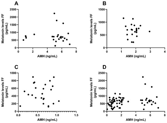
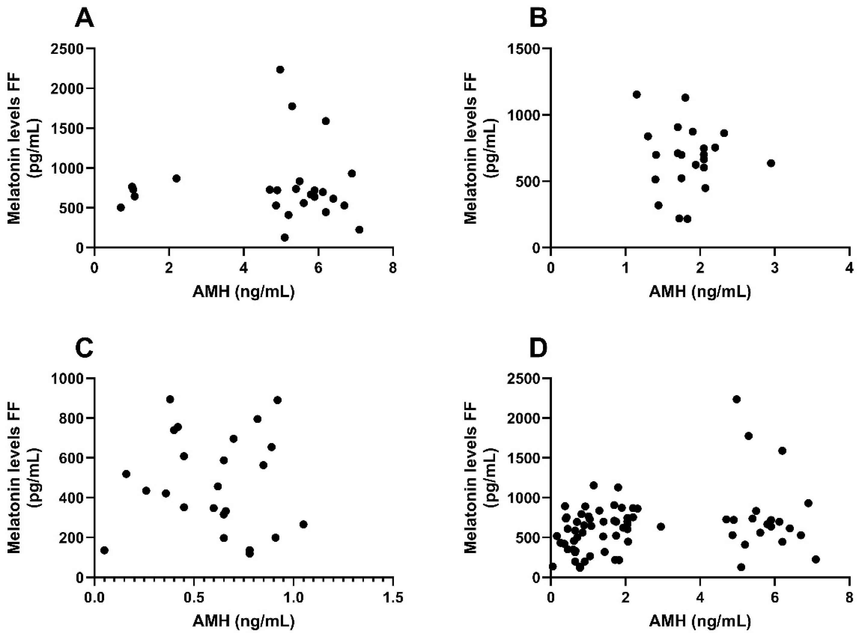
To explore the association between serum AMH and pooled follicular-fluid melatonin, two-sided Spearman rank correlation analyses were performed between serum AMH and pooled follicular fluid melatonin concentrations in the overall cohort and within each ovarian response category. In the overall cohort (n = 71), serum AMH concentration showed a weak positive correlation with pooled follicular fluid melatonin concentration (Spearman’s ρ = 0.2721, 95% CI 0.0344–0.4806, p = 0.0217; Figure 1, panel D). This association was not observed within the individual response categories. Among high responders (n = 25), the correlation was weakly negative and not statistically significant (ρ = −0.1543, 95% CI −0.5268 to 0.2680, p = 0.4615; Figure 1, panel A). Similarly, no significant correlation was observed among normal responders (n = 22; ρ = −0.0655, 95% CI −0.4842 to 0.3777, p = 0.7723; Figure 1, panel B) or poor responders (n = 24; ρ = −0.0579, 95% CI −0.4608 to 0.3648, p = 0.7881; Figure 1, panel C). Thus, the overall cohort demonstrated a weak positive monotonic association between serum AMH and pooled follicular fluid melatonin, whereas no statistically significant within-group associations were detected. Because AMH contributed to the definition of the poor- and normal-response categories, the overall AMH–melatonin association was subsequently examined in age-adjusted and response-group-conditioned models, as described in Section 3.8.
Figure 1.
Association between serum AMH and pooled follicular fluid melatonin concentrations according to ovarian response category. Scatter plots show serum AMH concentration versus pooled follicular fluid (FF) melatonin concentration. Each point represents one participant. (A) High responders (n = 25), (B) normal responders (n = 22), (C) poor responders (n = 24), and (D) the overall cohort (n = 71). Two-sided Spearman rank correlation analyses showed no statistically significant association in high responders (ρ = −0.1543, 95% CI −0.5268 to 0.2680, p = 0.4615), normal responders (ρ = −0.0655, 95% CI −0.4842 to 0.3777, p = 0.7723), or poor responders (ρ = −0.0579, 95% CI −0.4608 to 0.3648, p = 0.7881). In the overall cohort, serum AMH showed a weak positive correlation with pooled FF melatonin concentration (ρ = 0.2721, 95% CI 0.0344–0.4806, p = 0.0217).
3.8. Multivariable Reanalysis of the Central Associations
Because age differed substantially across the ovarian-response categories, and because AMH and retrieved oocyte number contributed to the structure of the response-group definitions, the three central associations were re-evaluated using prespecified multivariable models. In the age-adjusted linear regression model, pooled follicular-fluid melatonin concentrations differed across ovarian-response groups (overall p = 0.0315). Compared with poor responders, melatonin concentrations were higher in normal responders (β = 0.758, 95% CI 0.083–1.434, p = 0.0278) and high responders (β = 0.589, 95% CI 0.161–1.018, p = 0.0070), whereas the high-versus-normal contrast was not significant (p = 0.4440). These group differences remained in a sensitivity model additionally adjusted for BMI and infertility duration (overall p = 0.0363).
The positive association between serum AMH and pooled follicular-fluid melatonin remained in the age-adjusted model (β = 0.174 per log-unit increase in AMH, 95% CI 0.022–0.327, p = 0.0250). However, the association was no longer evident after additionally conditioning on ovarian-response group (β = 0.011, 95% CI −0.350–0.372, p = 0.9534).
Because retrieved oocyte number showed substantial overdispersion, negative binomial regression was used. In the age-adjusted model, higher log-transformed follicular-fluid melatonin was associated with a higher expected retrieved oocyte count (IRR = 1.435, 95% CI 1.066–1.931, p = 0.0173). This association remained in the model additionally adjusted for BMI and infertility duration (IRR = 1.421, 95% CI 1.072–1.883, p = 0.0146). In the exploratory model additionally conditioned on ovarian-response group, the association was attenuated and no longer significant (IRR = 1.027, 95% CI 0.893–1.180, p = 0.7103). Because retrieved oocyte number contributes to the definition of high response, this group-conditioned model was interpreted as a sensitivity analysis.
The results of the multivariable analyses are summarized in Table 11.
Table 11.
Multivariable analyses of pooled follicular-fluid melatonin and retrieved oocyte number.
4. Discussion
This prospective observational study examined pooled follicular-fluid melatonin concentrations across three clinical ovarian-response phenotypes—poor, normal, and high responders—in women undergoing IVF-ICSI cycles in which all participants received oral melatonin supplementation at a fixed dose of 3 mg/day throughout the stimulation period, from the initiation of gonadotropin administration until oocyte retrieval. This corresponded to approximately 40 days of supplementation. The ovarian-response categories represented clinical phenotypes defined by ovarian-reserve and stimulation/retrieval characteristics and were not genetic classifications. The supplementation protocol was aligned with that of Espino et al., who administered 3 mg/day over a similar interval and reported rebalancing of the intrafollicular oxidative environment, improvements in oocyte quality, and modest improvements in IVF outcomes, while also reporting that 3 mg/day was as effective as 6 mg/day in ameliorating the follicular-fluid microenvironment [32].
In the present study, pooled follicular-fluid melatonin concentrations were lower in poor responders than in normal and high responders in the unadjusted analysis. This difference remained detectable after adjustment for age and in a sensitivity model additionally including BMI and infertility duration. In the age-adjusted model, the overall response-group effect was significant (p = 0.0315), with higher melatonin concentrations in normal versus poor responders and high versus poor responders, whereas the high-versus-normal contrast was not significant. The weak positive AMH–melatonin association observed in the overall cohort remained in the age-adjusted model but was no longer evident after conditioning on ovarian-response group. Similarly, the age-adjusted association between pooled follicular-fluid melatonin and retrieved oocyte number was attenuated and no longer significant in the exploratory response-group-conditioned model. No consistent association was observed between pooled follicular-fluid melatonin and downstream embryological parameters. These findings suggest that pooled follicular-fluid melatonin is associated with the clinical ovarian-response phenotype, whereas the overall AMH–melatonin and melatonin–oocyte-yield associations may partly reflect the between-group structure of the cohort.
A central methodological distinction of the present study is the use of pooled follicular-fluid sampling. All visible follicles were aspirated into a single collection tube, rather than measuring melatonin exclusively in the dominant or lead follicle, as has been done in most previous investigations. Tong et al. first systematically examined endogenous melatonin in follicular fluid across ovarian-response categories and reported strong positive correlations between intrafollicular melatonin and age, AMH, and basal FSH, as well as with several IVF outcomes, including oocyte yield, fertilization, cleavage, top-quality embryos on day 3, and blastocyst formation rate [33]. Zheng et al. similarly reported positive correlations between follicular-fluid melatonin, antral follicle count (AFC), and IVF outcomes in women classified according to low, intermediate, and high AFC [34]. More recently, Majeed Shareef et al. conducted a prospective cross-sectional study in which follicular-fluid melatonin was strongly correlated with oocyte yield, fertilized oocytes, cleaved zygotes, high-quality embryos, and embryos suitable for transfer, with correlation coefficients substantially higher than those observed in the present study [35].
In the present cohort, serum AMH also showed a weak positive correlation with pooled follicular-fluid melatonin in the overall unadjusted analysis (Spearman’s ρ = 0.2721), consistent in direction but considerably weaker in magnitude than the associations reported in studies using individual or dominant-follicle samples. The association remained weakly positive after adjustment for age. However, it was not observed within the poor-, normal-, or high-response groups, and it was no longer evident in the model additionally conditioned on ovarian-response group. This pattern suggests that the overall AMH–melatonin association may partly reflect between-group differences rather than a uniform within-group relationship. Therefore, the present findings do not establish an independent relationship between serum AMH and pooled follicular-fluid melatonin.
In the studies by Tong et al., Zheng et al., and Majeed Shareef et al., follicular fluid was collected from individual follicles, typically the dominant preovulatory follicle or follicles of comparable maturity, rather than from a pooled sample reflecting the full aspirated cohort. In stimulated IVF cycles, the oocyte cohort is derived from follicles spanning a range of sizes and developmental stages, and melatonin concentrations in smaller or less mature follicles may differ from those in the lead follicle. Pooled sampling should therefore be regarded as a methodological limitation rather than an inherent advantage. The strong correlations with IVF outcomes reported in the individual- or dominant-follicle studies contrast with the largely weak or absent correlations observed in the present study using pooled samples. If dominant-follicle melatonin is the clinically informative signal, pooling follicles of heterogeneous maturity could dilute follicle-specific associations, and the weaker correlations observed here would be compatible with that possibility. Pooled sampling may nevertheless provide a broader patient-level estimate of the melatonin milieu across the retrieved follicular cohort rather than a measurement from a single dominant follicle. This broader representation may be useful for population-level characterization, but it may attenuate follicle-specific signals that are most predictive of individual oocyte quality. Future studies should include parallel dominant-follicle and pooled sampling from the same cycle to quantify this potential dilution effect directly.
The finding that poor responders had lower pooled follicular-fluid melatonin concentrations is compatible with the oxidative and mitochondrial burden associated with diminished ovarian reserve. Women with poor ovarian response may exhibit impaired antioxidant defenses, increased reactive oxygen species, and compromised granulosa-cell function, which could reduce the capacity of the follicular microenvironment to maintain oxidative homeostasis [36]. Reiter et al. reviewed evidence that melatonin produced locally within oocytes, granulosa cells, and cumulus cells declines with ovarian aging, in parallel with increased oxidative stress and impaired follicular development [37]. The local biosynthetic contribution of granulosa cells has also been investigated using transcriptomic approaches demonstrating that melatonin supplementation modulates gene-expression profiles related to steroidogenesis, apoptosis suppression, and angiogenesis in women with poor embryo development [38]. These observations provide biological context for the lower melatonin concentrations observed in poor responders, although the present observational study cannot determine whether the group difference reflects reduced local synthesis, altered follicular composition, differences in exogenous melatonin distribution, or other age- and reserve-related factors. In a randomized controlled trial, Sadeghpour et al. reported that 3 mg/day melatonin in women with diminished ovarian reserve increased follicular-fluid reduced glutathione and total antioxidant capacity and was accompanied by improvements in oocyte yield, fertilization rate, and embryo quality [25]. Those findings indicate that the follicular antioxidant environment in this population may be pharmacologically modifiable, but they do not establish that the between-group melatonin differences observed in the present study are causal or that supplementation produces the same effect in all ovarian-response phenotypes.
The differences in early follicular-phase serum estradiol concentrations should also be considered when interpreting the ovarian-response findings. In the present cohort, serum estradiol was higher in poor responders than in normal responders (p < 0.0001) and higher in normal responders than in high responders (p = 0.0041), whereas the difference between poor and high responders was not statistically significant. This pattern is compatible with early follicular hormonal changes associated with diminished ovarian reserve and premature follicular recruitment, rather than providing evidence that estradiol directly alters follicular-fluid melatonin concentrations [7]. Serum estradiol was measured in peripheral blood on cycle days 2–5 of the cycle preceding treatment, whereas melatonin was measured in pooled follicular fluid obtained at oocyte retrieval. Serum estradiol would therefore not be expected to interfere directly with the follicular-fluid melatonin assay. A separate potential issue is blood-derived interference in follicular-fluid samples. Samples with visible blood contamination were excluded, and samples were centrifuged at 1000× g for 20 min at 4 °C to remove erythrocytes and cellular debris, as described in the Methods. Nevertheless, no objective hemolysis index or hemoglobin measurement was performed, and microscopic blood contamination cannot be completely excluded. Residual blood-derived matrix effects may therefore have contributed to measurement variability and should be considered when interpreting between-group differences in follicular-fluid melatonin.
With respect to clinical and embryological outcomes, the present cohort demonstrated the expected ovarian-response-dependent hierarchy across several ART parameters. Poor responders had the lowest oocyte yield, the fewest top-quality embryos, and the lowest take-home baby rate, whereas high responders achieved the highest absolute embryo numbers despite lower proportional quality indices. These findings are consistent with the established clinical significance of ovarian-response classification and ART prognosis [29]. Within the individual response categories, pooled follicular-fluid melatonin was not consistently associated with fertilization, cleavage, embryo quality, or blastocyst formation rate. The sole exception was a moderate inverse correlation between melatonin and the number of transferable embryos in normal responders, a finding of uncertain biological significance that may be attributable to the limited sample size and the number of analyses performed in that subgroup.
Across the entire cohort, the unadjusted association between pooled follicular-fluid melatonin and retrieved oocyte number was weakly positive, and the association remained detectable in the age-adjusted negative-binomial model and in a model additionally adjusted for BMI and infertility duration. However, it was no longer significant in the exploratory model conditioned on ovarian-response group. Retrieved oocyte number contributes to the definition of high response, and this structural overlap makes the group-conditioned estimate difficult to interpret as an independent biological association. Accordingly, the present results do not establish that higher follicular-fluid melatonin independently increases oocyte yield. They instead indicate that the apparent melatonin–oocyte-yield association may partly reflect the clinical response-group structure.
The absence of consistent associations with post-fertilization embryological outcomes should not be interpreted as evidence that follicular melatonin has no biological relevance. Rather, pooled follicular-fluid melatonin may represent a patient-level measure of the overall follicular microenvironment rather than a direct predictor of the developmental potential of an individual embryo. Because pooled sampling integrates signals from follicles with heterogeneous maturity and size, it cannot resolve the melatonin status of a specific oocyte–follicle unit. This sampling characteristic may reduce the strength of associations with individual embryo outcomes. The absence of consistent post-fertilization associations may also reflect dilution of follicle-specific signals or may indicate that the relevant role of melatonin occurs primarily at the level of follicular environment and oocyte availability rather than later embryo development. However, the present findings do not establish either mechanism.
It is important to address the take-home baby rate (THBR) in the poor-responder group directly. Poor responders had a THBR of 4.2% (1 out of 24 patients), which is consistent with the unfavorable prognosis generally observed in this population but remains clinically important to report transparently. This rate reflects the cumulative outcome of fresh and frozen-thawed transfer cycles derived from the same stimulation and should be interpreted in the context of diminished ovarian reserve, advanced median age of 41 years, and high basal FSH. Because all participants received melatonin and there was no unsupplemented control arm, no causal inference can be made regarding the effect, or lack of effect, of melatonin on THBR in poor responders. The data are compatible with the possibility that 3 mg/day melatonin for approximately 40 days may not overcome the prognostic disadvantage associated with poor ovarian reserve, but they do not establish this possibility. Randomized controlled trials with unsupplemented control arms are required to evaluate this question definitively.
The broader clinical literature increasingly supports melatonin as a potential adjuvant therapy in IVF. A meta-analysis by Wu et al., including eleven randomized controlled trials and 1481 participants, reported that melatonin supplementation significantly improved clinical pregnancy rates (OR = 1.59, 95% CI 1.22–2.07), increased the number of MII oocytes (SMD = 0.99), increased the number of high-quality embryos (MD = 0.43), and improved fertilization rates (OR = 1.32) [26]. The subgroup analysis indicated that doses at or below 3 mg/day were associated with the greatest improvements in clinical pregnancy, providing external context for the supplementation protocol used in the present study. An earlier meta-analysis by Hu et al. reported improved clinical pregnancy rates across ten randomized trials (OR = 1.43, 95% CI 1.11–1.86) together with increases in retrieved oocytes, mature oocytes, and good-quality embryos [39]. These findings were also supported by the systematic review of Mejlhede et al. [40]. Recent evidence has extended the proposed therapeutic mechanisms of melatonin beyond its classical antioxidant function. Tsui et al. reported in a prospective cohort study that melatonin supplementation at 2 mg/day for at least eight weeks improved clinical pregnancy and live birth rates in women aged 38 years or older undergoing IVF-FET cycles. Their mechanistic findings implicated suppression of cuproptosis and ferroptosis in cumulus and granulosa cells together with mitochondrial metabolic reprogramming [41]. These pathways broaden the mechanistic framework through which melatonin may support follicular function and oocyte developmental competence, but findings from supplementation trials should not be interpreted as evidence that pooled follicular-fluid melatonin is an independent treatment-response biomarker.
The observation that pooled follicular-fluid melatonin was not significantly correlated with most controlled ovarian stimulation parameters in the unadjusted analyses is compatible with the possibility that local ovarian factors contribute to measured follicular-fluid melatonin concentrations. However, these observational data do not establish intrinsic ovarian biosynthesis as the primary determinant of follicular-fluid melatonin, and endogenous and supplemented contributions could not be separated. Granulosa cells, cumulus cells, and oocytes have been reported to express the rate-limiting melatonin-synthesizing enzyme arylalkylamine N-acetyltransferase (AANAT), providing a biological basis for local melatonin production within the follicular environment [42].
The higher melatonin concentrations observed in high responders than in poor responders should nevertheless be interpreted cautiously. Zheng et al. reported that intrafollicular melatonin was elevated in women who developed ovarian hyperstimulation syndrome compared with women who did not develop the syndrome, and that these concentrations correlated with AFC, AMH, and embryological outcomes [43]. Higher follicular-fluid melatonin in high responders may therefore reflect physiological changes accompanying a larger and more rapidly developing follicular cohort rather than a direct improvement in the quality of individual oocytes. Pooled sampling across follicles of heterogeneous maturity makes this distinction difficult to resolve. The absence of a significant high-versus-normal difference after age adjustment also indicates that the apparent hierarchy should not be interpreted as a simple linear relation between ovarian response and follicular-fluid melatonin.
Several limitations of the present study merit consideration. First, the observational design precludes causal inference. Because all participants received the same supplementation protocol—3 mg/day for approximately 40 days—the endogenous and exogenous contributions to measured intrafollicular melatonin could not be disentangled. Although the regimen mirrors the protocol of Espino et al. [32], the absence of a non-supplemented control group restricts interpretation of absolute melatonin concentrations and prevents attribution of outcome differences specifically to melatonin supplementation. Second, pooled sampling precludes follicle-specific analysis and may attenuate signals from dominant follicles that are most relevant to individual oocyte quality. Third, the modest sample size within each ovarian-response subgroup limits statistical power, and single-center recruitment may restrict generalizability. Fourth, additional multivariable analyses adjusted for age, and sensitivity models additionally included BMI and infertility duration. The poor-versus-normal and poor-versus-high melatonin differences remained detectable after age adjustment. Nevertheless, residual confounding cannot be excluded because age was strongly imbalanced across groups, the common age range across groups was limited, and age is closely related to ovarian reserve and response characteristics. The observed response categories also incorporated clinical features that overlap with the covariates and outcomes examined in the regression models. In particular, AMH contributed to the definitions of poor and normal response, whereas retrieved oocyte number contributed to the definition of high response. The disappearance of the AMH–melatonin association and the attenuation of the melatonin–oocyte-yield association in the response-group-conditioned models should therefore be interpreted as evidence of sensitivity to group structure, not as definitive proof of the absence or presence of an independent biological relationship. Infertility etiology was not included in the reported multivariable models because its distribution was imbalanced across groups and the sample size was limited. Fifth, genetic determinants of ovarian response and follicular-fluid melatonin concentrations could not be evaluated because genotyping, genetic ancestry, and family history of diminished ovarian reserve were not collected. Self-identified race/ethnicity and skin-color information were also not recorded; therefore, the representativeness of these findings across ancestry or ethnocultural groups cannot be assessed. The poor-, normal-, and high-response categories should consequently be interpreted as clinical phenotypes rather than genetic or ancestry-based categories. Sixth, the absence of concurrent oxidative-stress biomarkers, such as total antioxidant capacity, 8-hydroxy-2′-deoxyguanosine, or glutathione, limits the ability to place the melatonin findings within the broader follicular oxidative milieu. Seventh, the circadian timing of follicular-fluid aspiration was not recorded. Because melatonin follows a pronounced circadian rhythm and oocyte-retrieval times may vary by several hours between patients, uncontrolled variation in aspiration timing represents a potential source of measurement variability. Patient-reported adherence was used to assess melatonin supplementation, but no objective compliance measure, such as pill counts, a medication diary, or a pharmacological biomarker, was collected. Variable adherence across participants and response groups therefore cannot be excluded. Finally, the large number of correlation, subgroup, and sensitivity analyses increases the risk of spurious nominally significant findings. No formal correction for multiple testing was applied; accordingly, the subgroup correlations, including the associations with endometrial thickness in high responders and transferable-embryo number in normal responders, should be regarded as exploratory and hypothesis-generating rather than confirmatory.
Future well-designed randomized controlled trials should stratify participants by ovarian-response phenotype and compare melatonin-supplemented cycles with unsupplemented control cycles to determine whether the intrafollicular melatonin differences observed here translate into clinically meaningful ART outcomes. Parallel sampling of dominant and smaller follicles within the same cycle would allow direct comparison of pooled and dominant-follicle approaches and would characterize within-cycle melatonin heterogeneity. Incorporating oxidative-stress biomarkers and mitochondrial-function assessments in cumulus cells together with intrafollicular melatonin measurement would provide a more complete mechanistic assessment of how local melatonin availability relates to the oxidative landscape and oocyte quality.
Given that extended melatonin pretreatment was associated with improved clinical pregnancy and live birth rates in women of advanced maternal age [41], future studies may also investigate the dose, timing, and duration of supplementation specifically in poor ovarian responders, the subgroup with the lowest pooled follicular-fluid melatonin concentrations in the present cohort. The integration of follicular-fluid biomarkers into multivariable prognostic models may eventually improve patient stratification, but such models should first demonstrate incremental prognostic value beyond age, AMH, and conventional ovarian-response measures. The present study does not establish a clinically validated threshold for pooled follicular-fluid melatonin, support routine biomarker-guided supplementation, or justify incorporation of melatonin measurements into routine IVF management algorithms. Randomized trials comparing standard and biomarker-guided supplementation strategies are required before the clinical utility of such approaches can be determined.
5. Conclusions
In conclusion, pooled follicular-fluid melatonin concentrations were lower in poor ovarian responders than in normal and high responders in the unadjusted analysis, and this difference remained detectable after adjustment for age and in a sensitivity model additionally including BMI and infertility duration. The age-adjusted association between serum AMH and pooled follicular-fluid melatonin was weak and was no longer evident after conditioning on ovarian-response group. Likewise, the age-adjusted association between pooled follicular-fluid melatonin and retrieved oocyte number was attenuated and was not retained in the exploratory response-group-conditioned model. No consistent association was observed between pooled follicular-fluid melatonin and downstream embryological outcomes or most ovarian stimulation parameters. These findings support an association between pooled follicular-fluid melatonin and the clinical ovarian-response phenotype under standardized supplementation, but they do not establish an independent ovarian-reserve or oocyte-yield biomarker relationship, causality, treatment efficacy, or clinical utility. Future studies should combine prospectively specified multivariable analyses with parallel follicle-specific and pooled sampling, comprehensive oxidative-stress profiling, and randomized comparisons of supplementation strategies.
Author Contributions
Conceptualization, S.T. (Seval Tasdemir); Methodology, M.V.B. and S.N.P.; Validation, M.S.S. and T.Y.; Formal analysis, M.V.B., S.N.P. and S.U.K.; Investigation, S.T. (Seval Tasdemir), B.K., S.T. (Safak Tavukcuoglu) and F.A.; Data curation, M.S.S., T.Y. and S.U.K.; Writing—original draft, S.T. (Seval Tasdemir); Writing—review and editing, S.T. (Seval Tasdemir), M.V.B., B.K., S.T. (Safak Tavukcuoglu) and E.C.S.; Supervision, E.C.S.; Project administration, S.T. (Seval Tasdemir). All authors have read and agreed to the published version of the manuscript.
Funding
This research received no external funding.
Institutional Review Board Statement
This study was approved by the Istanbul Medipol University Non-Interventional Clinical Research Ethics Committee (Decision No: 305, Date: 30 March 2023). The investigation was conducted in accordance with the ethical standards of the Republic of Türkiye Ministry of Health and the principles of the Declaration of Helsinki.
Informed Consent Statement
Written informed consent was obtained from all participants prior to inclusion in the study.
Data Availability Statement
The datasets used and/or analyzed during the current study are available from the corresponding authors on reasonable request.
Conflicts of Interest
Authors Seval Tasdemir, Betul Kalay, Safak Tavukcuoglu, and Fevzi Akcil were employed by Private Fertijin Women’s Health and Assisted Reproduction Center. The remaining authors declare that the research was conducted in the absence of any commercial or financial relationships that could be construed as a potential conflict of interest.
References
- World Health Organization. Infertility Prevalence Estimates, 1990–2021; World Health Organization: Geneva, Switzerland, 2023. [Google Scholar]
- Zegers-Hochschild, F.; Adamson, G.D.; Dyer, S.; Racowsky, C.; de Mouzon, J.; Sokol, R.; Rienzi, L.; Sunde, A.; Schmidt, L.; Cooke, I.D.; et al. The International Glossary on Infertility and Fertility Care, 2017. Fertil. Steril. 2017, 108, 393–406. [Google Scholar] [CrossRef] [Scilit] [PubMed]
- Inhorn, M.C.; Patrizio, P. Infertility around the globe: New thinking on gender, reproductive technologies and global movements in the 21st century. Hum. Reprod. Update 2015, 21, 411–426. [Google Scholar] [CrossRef] [Scilit] [PubMed]
- Rotterdam ESHRE/ASRM-Sponsored PCOS Consensus Workshop Group. Revised 2003 consensus on diagnostic criteria and long-term health risks related to polycystic ovary syndrome (PCOS). Hum. Reprod. 2004, 19, 41–47. [Google Scholar] [CrossRef] [Scilit] [PubMed]
- Keane, J.A.; Ealy, A.D. An overview of reactive oxygen species damage occurring during in vitro bovine oocyte and embryo development and the efficacy of antioxidant use to limit these adverse effects. Animals 2024, 14, 330. [Google Scholar] [CrossRef] [Scilit] [PubMed]
- Lin, J.; Wang, L. Oxidative stress in oocytes and embryo development: Implications for in vitro systems. Antioxid. Redox Signal. 2021, 34, 1394–1406. [Google Scholar] [CrossRef] [Scilit] [PubMed]
- May-Panloup, P.; Boguenet, M.; El Hachem, H.; Bouet, P.-E.; Reynier, P. Embryo and its mitochondria. Antioxidants 2021, 10, 139. [Google Scholar] [CrossRef] [Scilit] [PubMed]
- Bozdemir, N.; Cakir, C.; Topcu, U.; Uysal, F. A comprehensive review of mitochondrial complex I during mammalian oocyte maturation. Genesis 2025, 63, e70017. [Google Scholar] [CrossRef] [Scilit] [PubMed]
- Munisha, M.; Schimenti, J.C. Genome maintenance during embryogenesis. DNA Repair 2021, 106, 103195. [Google Scholar] [CrossRef] [Scilit] [PubMed]
- Mikwar, M.; MacFarlane, A.J.; Marchetti, F. Mechanisms of oocyte aneuploidy associated with advanced maternal age. Mutat. Res. Mutat. Res. 2020, 785, 108320. [Google Scholar] [CrossRef] [Scilit] [PubMed]
- Xie, J.; Xu, X.; Liu, S. Intercellular communication in the cumulus–oocyte complex during folliculogenesis: A review. Front. Cell Dev. Biol. 2023, 11, 1087612. [Google Scholar] [CrossRef] [Scilit] [PubMed]
- Bebbere, D.; Coticchio, G.; Borini, A.; Ledda, S. Oocyte aging: Looking beyond chromosome segregation errors. J. Assist. Reprod. Genet. 2022, 39, 793–800. [Google Scholar] [CrossRef] [Scilit] [PubMed]
- Mogessie, B.; Scheffler, K.; Schuh, M. Assembly and positioning of the oocyte meiotic spindle. Annu. Rev. Cell Dev. Biol. 2018, 34, 381–403. [Google Scholar] [CrossRef] [Scilit] [PubMed]
- El-Raey, M.; Geshi, M.; Somfai, T.; Kaneda, M.; Hirako, M.; Abdel-Ghaffar, A.E.; Sosa, G.A.; Abou El-Roos, M.E.A.; Nagai, T. Evidence of melatonin synthesis in the cumulus oocyte complexes and its role in enhancing oocyte maturation in vitro in cattle. Mol. Reprod. Dev. 2011, 78, 250–262. [Google Scholar] [CrossRef] [Scilit] [PubMed]
- Reiter, R.J.; Sharma, R.; Tan, D.-X.; Chuffa, L.G.d.A.; da Silva, D.G.H.; Slominski, A.T.; Steinbrink, K.; Kleszczynski, K. Dual sources of melatonin and evidence for different primary functions. Front. Endocrinol. 2024, 15, 1414463. [Google Scholar] [CrossRef] [Scilit] [PubMed]
- Sakaguchi, K.; Itoh, M.T.; Takahashi, N.; Tarumi, W.; Ishizuka, B. The rat oocyte synthesises melatonin. Reprod. Fertil. Dev. 2013, 25, 674–682. [Google Scholar] [CrossRef] [Scilit] [PubMed]
- Reiter, R.J.; Mayo, J.C.; Tan, D.-X.; Sainz, R.M.; Alatorre-Jimenez, M.; Qin, L. Melatonin as an antioxidant: Under promises but over delivers. J. Pineal Res. 2016, 61, 253–278. [Google Scholar] [CrossRef] [Scilit] [PubMed]
- Tamura, H.; Takasaki, A.; Taketani, T.; Tanabe, M.; Lee, L.; Tamura, I.; Maekawa, R.; Asada, H.; Yamagata, Y.; Sugino, N. Melatonin and female reproduction. J. Obstet. Gynaecol. Res. 2014, 40, 1–11. [Google Scholar] [CrossRef] [Scilit] [PubMed]
- Tan, D.-X.; Reiter, R.J. Mitochondria: The birth place, battle ground and the site of melatonin metabolism in cells. Melat. Res. 2019, 2, 44–66. [Google Scholar] [CrossRef] [Scilit]
- Brzezinski, A.; Seibel, M.M.; Lynch, H.J.; Deng, M.-H.; Wurtman, R.J. Melatonin in human preovulatory follicular fluid. J. Clin. Endocrinol. Metab. 1987, 64, 865–867. [Google Scholar] [CrossRef] [Scilit] [PubMed]
- Nakamura, Y.; Tamura, H.; Takayama, H.; Kato, H. Increased endogenous level of melatonin in preovulatory human follicles does not directly influence progesterone production. Fertil. Steril. 2003, 80, 1012–1016. [Google Scholar] [CrossRef] [Scilit] [PubMed]
- Rönnberg, L.; Kauppila, A.; Leppäluoto, J.; Martikainen, H.; Vakkuri, O. Circadian and seasonal variation in human preovulatory follicular fluid melatonin concentration. J. Clin. Endocrinol. Metab. 1990, 71, 493–496. [Google Scholar] [CrossRef] [Scilit] [PubMed]
- Tamura, H.; Nakamura, Y.; Korkmaz, A.; Manchester, L.C.; Tan, D.-X.; Sugino, N.; Reiter, R.J. Melatonin and the ovary: Physiological and pathophysiological implications. Fertil. Steril. 2009, 92, 328–343. [Google Scholar] [CrossRef] [Scilit] [PubMed]
- Tamura, H.; Takasaki, A.; Miwa, I.; Taniguchi, K.; Maekawa, R.; Asada, H.; Taketani, T.; Matsuoka, A.; Yamagata, Y.; Shimamura, K.; et al. Oxidative stress impairs oocyte quality and melatonin protects oocytes from free radical damage and improves fertilization rate. J. Pineal Res. 2008, 44, 280–287. [Google Scholar] [CrossRef] [Scilit] [PubMed]
- Sadeghpour, S.; Ghasemnejad-Berenji, M.; Maleki, F.; Behroozi-Lak, T.; Bahadori, R.; Ghasemnejad-Berenji, H. The effects of melatonin on follicular oxidative stress and art outcomes in women with diminished ovarian reserve: A randomized controlled trial. J. Ovarian Res. 2025, 18, 5. [Google Scholar] [CrossRef] [Scilit] [PubMed]
- Wu, Y.; Huang, W.; Tang, L.; Feng, Y.; Chen, H.; Pan, M.; Peng, J.; Li, C.; Wang, H. Melatonin improved the outcomes of women with ART: A systematic review and meta-analysis of randomized trials. Front. Reprod. Health 2025, 7, 1680984. [Google Scholar] [CrossRef] [Scilit] [PubMed]
- Balaban, B.; Urman, B.; Alatas, C.; Mercan, R.; Aksoy, S.; Isiklar, A. Blastocyst-stage transfer of poor-quality cleavage-stage embryos results in higher implantation rates. Fertil. Steril. 2001, 75, 514–518. [Google Scholar] [CrossRef] [Scilit] [PubMed]
- Gardner, D.K.; Lane, M.; Stevens, J.; Schlenker, T.; Schoolcraft, W.B. Blastocyst score affects implantation and pregnancy outcome: Towards a single blastocyst transfer. Fertil. Steril. 2000, 73, 1155–1158. [Google Scholar] [CrossRef] [Scilit] [PubMed]
- Ferraretti, A.P.; La Marca, A.; Fauser, B.C.J.M.; Tarlatzis, B.; Nargund, G.; Gianaroli, L.; ESHRE Working Group on Poor Ovarian Response Definition. ESHRE consensus on the definition of ‘poor response’to ovarian stimulation for in vitro fertilization: The Bologna criteria. Hum. Reprod. 2011, 26, 1616–1624. [Google Scholar] [CrossRef] [Scilit] [PubMed]
- Feferkorn, I.; Ata, B.; Esteves, S.C.; La Marca, A.; Paulson, R.; Blockeel, C.; Conforti, A.; Fatemi, H.M.; Humaidan, P.; Lainas, G.T.; et al. The HERA (Hyper-response Risk Assessment) Delphi consensus definition of hyper-responders for in-vitro fertilization. J. Assist. Reprod. Genet. 2023, 40, 1071–1081. [Google Scholar] [CrossRef] [Scilit] [PubMed]
- Qiao, J.; Ma, C.H.; Liu, J.Y.; Ma, X.; Li, S.W.; Yang, Y.Z.; Zhu, Y.M. A consensus of poor ovarian response. Chin. J. Reprod. Contracept. 2015, 35, 211–223. [Google Scholar]
- Espino, J.; Macedo, M.; Lozano, G.; Ortiz, A.; Rodríguez, C.; Rodríguez, A.B.; Bejarano, I. Impact of Melatonin Supplementation in Women with Unexplained Infertility Undergoing Fertility Treatment. Antioxidants 2019, 8, 338. [Google Scholar] [CrossRef] [Scilit] [PubMed]
- Tong, J.; Sheng, S.; Sun, Y.; Li, H.; Li, W.-P.; Zhang, C.; Chen, Z.-J. Melatonin levels in follicular fluid as markers for IVF outcomes and predicting ovarian reserve. Reproduction 2017, 153, 443–451. [Google Scholar] [CrossRef] [Scilit] [PubMed]
- Zheng, M.; Tong, J.; Li, W.-P.; Chen, Z.-J.; Zhang, C. Melatonin concentration in follicular fluid is correlated with antral follicle count (AFC) and in vitro fertilization (IVF) outcomes in women undergoing assisted reproductive technology (ART) procedures. Gynecol. Endocrinol. 2018, 34, 446–450. [Google Scholar] [CrossRef] [Scilit] [PubMed]
- Majeed Shareef, R.; Yassen, N.S.; Alkhudair, S.H. IVF Technique and its Relationship with Ovarian Reserve Based on Melatonin Level in Follicular Fluid. J. Obstet. Gynecol. Cancer Res. 2025, 10, 293–299. [Google Scholar] [CrossRef] [Scilit]
- Liu, S.; Jia, Y.; Meng, S.; Luo, Y.; Yang, Q.; Pan, Z. Mechanisms of and potential medications for oxidative stress in ovarian granulosa cells: A review. Int. J. Mol. Sci. 2023, 24, 9205. [Google Scholar] [CrossRef] [Scilit] [PubMed]
- Reiter, R.J.; Sharma, R.; Romero, A.; Manucha, W.; Tan, D.-X.; Zuccari, D.A.P.D.C.; Chuffa, L.G.D.A. Aging-related ovarian failure and infertility: Melatonin to the rescue. Antioxidants 2023, 12, 695. [Google Scholar] [CrossRef] [Scilit] [PubMed]
- Tamura, H.; Jozaki, M.; Tanabe, M.; Shirafuta, Y.; Mihara, Y.; Shinagawa, M.; Tamura, I.; Maekawa, R.; Sato, S.; Taketani, T.; et al. Importance of melatonin in assisted reproductive technology and ovarian aging. Int. J. Mol. Sci. 2020, 21, 1135. [Google Scholar] [CrossRef] [Scilit] [PubMed]
- Hu, K.-L.; Ye, X.; Wang, S.; Zhang, D. Melatonin Application in Assisted Reproductive Technology: A Systematic Review and Meta-Analysis of Randomized Trials. Front Endocrinol. 2020, 11, 160, Correction in Front. Endocrinol. 2020, 11, 333. [Google Scholar] [CrossRef] [Scilit] [PubMed]
- Mejlhede, M.A.B.; Jepsen, J.B.; Knudsen, U.B. Oral melatonin supplementation during in vitro fertilization treatment: A systematic PRISMA review and meta-analysis of randomized controlled trials. Gynecol. Endocrinol. 2021, 37, 1079–1085. [Google Scholar] [CrossRef] [Scilit] [PubMed]
- Tsui, K.-H.; Li, C.-J.; Lin, L.-T. Melatonin supplementation attenuates cuproptosis and ferroptosis in aging cumulus and granulosa cells: Potential for improving IVF outcomes in advanced maternal age. Reprod. Biol. Endocrinol. 2024, 22, 138. [Google Scholar] [CrossRef] [Scilit] [PubMed]
- Zhong, J.; Lu, Z.; Zhou, Z.; Ma, N.; Li, Y.; Hu, J.; Wan, B.; Lu, W. Melatonin biosynthesis and regulation in reproduction. Front. Endocrinol. 2025, 16, 1630164. [Google Scholar] [CrossRef] [Scilit] [PubMed]
- Zheng, M.; Zuo, G.; Tong, J.; Chen, Z.-J.; Li, W.-P.; Zhang, C. Intrafollicular melatonin concentration is elevated in patients with ovarian hyperstimulation syndrome (OHSS) and can serve as an important predictor of OHSS. Arch. Gynecol. Obstet. 2019, 299, 1151–1158. [Google Scholar] [CrossRef] [Scilit] [PubMed]

Disclaimer/Publisher’s Note: The statements, opinions and data contained in all publications are solely those of the individual author(s) and contributor(s) and not of MDPI and/or the editor(s). MDPI and/or the editor(s) disclaim responsibility for any injury to people or property resulting from any ideas, methods, instructions or products referred to in the content. |
© 2026 by the authors. Licensee MDPI, Basel, Switzerland. This article is an open access article distributed under the terms and conditions of the Creative Commons Attribution (CC BY) license.