


Abstract
The widespread presence of microplastics (MPs) and associated plastic-derived compounds, bisphenols (BPs), in the environment—originating from plastic production or adsorption of environmental pollutants—results in unavoidable human exposure. Therefore, assessing their occurrence in human biological samples is essential for understanding potential health risks. In this study, pyrolysis gas chromatography–mass spectrometry was used to analyse human intraocular fluids—aqueous humour (AH) and vitreous humour (VH)—to identify 11 microplastic polymer clusters. A method based on dansyl chloride derivatisation combined with ultra-performance liquid chromatography–tandem mass spectrometry was employed to screen for five bisphenols (BPs) in blood serum collected from the same patients. Potential MPs isolated from ocular samples were qualitatively assessed by stereomicroscopy. The number of particles ranged from 14 to 69 per gram in AH and from 21 to 34 per gram in VH. Among AH samples, the most frequently detected polymer clusters were C-PVC, C-PMMA, C-PET, and C-PP, with average levels ranging from 0.41 to 0.84 µg/g. The three VH samples contained only C-PMMA, with an average concentration of 0.55 ± 0.12 µg/g. Polymers such as C-PA66 (0.30 ± 0.21 µg/g) and C-PA6 (0.15 ± 0.44 µg/g) were occasionally observed in AH. Analysis of 13 human serum samples demonstrated exposure to bisphenol A (BPA; 4.05–5.18 ng/mL) and bisphenol AF (BPAF; 2.13–4.54 ng/mL), but no clear correlation was observed between serum BP levels and intraocular MPs within the limitations of the present dataset. These findings suggest the presence of MPs and BPs in human biological samples; however, the results should be interpreted with caution owing to potential background contamination and the pilot nature of the study.
1. Introduction
Human exposure to microplastics (MPs) occurs through multiple pathways, including ingestion, inhalation, and dermal contact, raising increasing concerns regarding their potential impacts on health [1,2]. It has increased markedly and its toxicity depends on material type, size, dose, and chemical additives [3,4]. Zhao and You [5] reported a six-fold rise in MPs ingestion and inhalation since 1990, especially in hotspots such as the USA, China, parts of the Middle East, North Africa, and Scandinavia. Although assessing health impacts remains challenging, plastic particles can absorb environmental toxins, bind heavy metals, and release chemical additives capable of interacting with endocrine pathways [6,7,8]. MPs enter the body through ingestion, inhalation, dermal contact, and medical procedures [9,10,11].
However, current knowledge of human exposure and toxicity remains limited [12], yet micro- and nanoplastics can translocate to several organs, including the liver, spleen, lungs, kidneys, reproductive organs, and even the brain. The World Health Organization [13] states that no conclusive evidence of harm exists to date, but stresses the need for further research. Potential health effects include respiratory and gastrointestinal disturbances, oxidative stress, and carcinogenesis [14].
MPs have been detected in a wide range of human tissues and body fluids, including the bone, brain, colon, heart, liver, kidney, the vascular system, lung, placenta, alveolar lavage fluid, breast milk, saliva, hands of young children, sputum, semen, testes, faeces, and urine indicating their ability to translocate within the human body and raising concerns about potential systemic effects [15,16,17,18,19,20,21,22,23,24,25,26,27,28,29,30,31,32].
Studies examining MPs in the human eye remain limited, although interest in this area is rapidly increasing. The growing body of research reporting MPs in various human tissues has prompted investigations into their presence in ocular tissues, particularly within the intraocular compartments, including the aqueous humour (AH) and vitreous humour (VH) [33]. Despite the eye’s high sensitivity and susceptibility to environmental exposure, it has received comparatively little attention in MP research. MPs may reach ocular tissues through airborne exposure [34] or direct contact with contaminated personal care products, such as detergents and cosmetics [35,36]. Increasing evidence suggests that MPs can induce ocular surface inflammation, tissue damage, and cell death [37]; however, their long-term effects and the underlying mechanisms remain poorly understood.
To investigate potential intraocular exposure, researchers have begun analysing metabolically active intraocular fluids, including AH and VH, which are derived from blood ultrafiltration. The AH plays a crucial role in maintaining ocular homeostasis by supplying nutrients to avascular tissues and regulating intraocular pressure. Two recent studies have confirmed the presence of MPs in both AH and VH. In the vitreous body, 1745 particles smaller than 50 μm were identified, with polyamide 66 (C-PA 66), polyvinyl chloride (C-PVC), and polystyrene (C-PS) being the predominant polymer clusters. In the AH, five polymer types were detected: polyethylene (C-PE), polyvinyl chloride (C-PVC), polypropylene (C-PP), polyamide 66 (C-PA 66), and polystyrene (C-PS), with C-PE and C-PVC being the most abundant [38,39,40].
Despite these initial findings, several important aspects of ocular MPs exposure remain insufficiently understood. Current evidence is based on a limited number of studies with relatively small sample sizes, and there is little information regarding the sources and pathways of MPs into intraocular tissues, their accumulation over time, and the factors influencing their distribution within different ocular compartments. Furthermore, the biological consequences of intraocular MPs, including their potential to induce oxidative stress, chronic inflammation, retinal or optic nerve damage, and their possible contribution to the development or progression of ocular diseases, remain largely unexplored. Standardised analytical methods and larger clinical studies are therefore needed to clarify the prevalence, characteristics, and clinical significance of MPs in the human eye. Flieger et al. [41] additionally detected bisphenol A (BPA) as an MP-associated plastic-derived compound in ocular fluid during cataract surgery.
Bisphenols (BPs) have attracted considerable scientific attention because of their endocrine-disrupting properties [42,43]. These compounds are widely used in the production of polycarbonate plastics and epoxy resins [44]. Human exposure to BPA has been extensively investigated, and an increasing number of biomonitoring studies have also reported the occurrence of BPA analogues, including bisphenol F (BPF), bisphenol S (BPS), bisphenol AF (BPAF), and other substitutes, in a variety of biological matrices. Nevertheless, continued biomonitoring is required to better characterise exposure patterns across different populations, assess temporal trends associated with BPA replacement, and evaluate exposure to emerging bisphenol analogues. BP exposure occurs primarily through food consumption but also via inhalation and dermal contact [45]. Serum is considered a valuable biomatrix for assessing recent internal exposure because it reflects systemic distribution and is in contact with all body tissues [46]. Accordingly, a previously established ultra-performance liquid chromatography–tandem mass spectrometry (UPLC–MS/MS) method based on dansyl chloride (Dns-Cl) derivatisation has been successfully applied to quantify BPA, bisphenol AF (BPAF), bisphenol B (BPB), bisphenol F (BPF), and bisphenol S (BPS) in human serum.
This pilot study investigated potential MPs in AH and VH collected during routine ophthalmic surgery using complementary stereomicroscopy and pyrolysis gas chroma-tography–mass spectrometry (Py-GC/MS), and screened five BPs in serum from the same patients by ultra-performance liquid chromatography–tandem mass spectrometry (UPLC–MS/MS). Surgical-condition and procedural blanks and the surgical environment were monitored to assess potential contamination. Exploratory relationships with participant characteristics and between ocular MPs and serum BPs were also examined. The study was designed as an analytical feasibility and hypothesis-generating investigation rather than a population-level assessment.
2. Materials and Methods
2.1. Study Population and Clinical Sample Collection
This pilot study was conducted during one surgical week over three consecutive working days at University Hospital Sestre Milosrdnice (UHSM), Zagreb, Croatia. Twenty adults aged 18–85 years (7 females, 13 males) undergoing routine ophthalmic surgery were recruited. Nineteen AH and three VH samples were collected; 13 of 20 serum samples were suitable for BP analysis after exclusion of seven haemolysed samples. AH was aspirated at the start of surgery using a 27-gauge paracentesis syringe before viscoelastic injection. VH was obtained during procedures requiring vitrectomy after a 25-gauge scleral puncture. Samples were transferred to pre-cleaned glass vials and stored frozen until analysis. Venous blood was collected into additive-free 10 mL glass tubes, centrifuged at 2500 rpm for 15 min, and serum was transferred with a glass Pasteur pipette to glass vials with PP caps and PTFE septa. Participant and surgical characteristics are reported in Table S1.
2.2. Contamination Control and Surgical-Environment Monitoring
Contamination prevention relied on glass equipment, metal tools, pre-cleaned containers, and laboratory blanks. Daily surgical-condition blanks were collected under the same operating-theatre conditions as clinical samples. Airborne deposition was monitored in the operating theatre (OT) and anaesthetic room (AR) using pre-combusted 400 mL glass beakers placed at eye level for alternating 12-h working and non-working periods over three days (n = 12), following Field et al. [9]. Deposited material was rinsed with pre-filtered distilled water and filtered through 25 mm, 1.2 µm glass-fibre filters (Whatman GF/C™, Cytiva/Whatman, Marlborough, MA, USA). Detailed environmental-sampling calculations are provided in Supplementary Methods SA-S1.
2.3. Method Development and Ocular-Fluid Preparation
Because human AH/VH volumes were limited, method development and validation used bovine ocular fluids from five adult Simmental cows. Samples were homogenised with a Vortex 3 mixer (IKA-Werke GmbH and Co. KG, Staufen, Germany). Human AH/VH samples were thawed, vortexed and weighed in glass-stoppered tubes (Šurlan, Medulin, Croatia) using a RADWAG AS 82/220.R2 PLUS analytical balance (RADWAG Wagi Elektroniczne, Radom, Poland). Wet oxidative digestion used 30% H2O2 (Gram-Mol d.o.o., Zagreb, Croatia) at 65 °C for 12 h [12,47], followed by vacuum filtration through 25 mm, 1.2-µm Whatman GF/C™ glass-fibre filters pre-treated at 500 °C. The central 9-mm filter area was excised, dried, and used for stereomicroscopy and Py-GC/MS. Full bovine method development, digestion, transfer, and filtration procedures are given in Supplementary Methods SA-S2.
2.4. Stereomicroscopy and Py-GC/MS Analyses
Potential particles retained on filters were examined using a SteREO Discovery V8 stereomicroscope with an Axiocam 105 R2 colour camera (Zeiss Microscopy GmbH, Jena, Germany). For each particle, length (defined as the longest dimension), shape (according to Markley et al. 2024) [48], and colour were recorded. Particle size was measured as the maximum dimension (length) following Abidli et al. (2019) [49]. Polymer identity and mass fraction were determined using an EGA/PY-3030D multishot pyrolyser with AS-1020E autosampler (Frontier Laboratories Ltd., Koriyama, Fukushima, Japan) coupled to a GC-8890/MSD 5977C system (Agilent, Santa Clara, CA, USA) and an Ultra Alloy-5 column (Frontier Laboratories Ltd., Koriyama, Fukushima, Japan). Eleven polymer clusters were targeted: PE, PP, PS, ABS, SBR, PMMA, PC, PVC, PET, PA6, and PA66. Thermochemolysis used tetramethylammonium hydroxide (TMAH, 10% in methanol; Sigma-Aldrich Chemie GmbH, Taufkirchen, Germany). Mixed polymer standards were obtained from Frontier Laboratories Ltd. (Frontier Laboratories Ltd., Koriyama, Fukushima, Japan ), and d5-PS/poly(4-fluorostyrene) internal standards from Polymer Source (Polymer Source Inc., Dorval, QC, Canada). Detailed Py-GC/MS conditions and indicator compounds are provided in Tables S2–S5 and Figure S1.
2.5. Serum Bisphenol Analysis by UPLC–MS/MS
Serum BPs were extracted by liquid–liquid extraction based on Owczarek et al. [50]. The internal standard 24BPS13C12 was obtained from Toronto Research Chemicals (Toronto Research Chemicals, Toronto, ON, Canada), acetonitrile from Biosolve Chimie SARL (Biosolve Chimie SARL, Dieuze, France), and Na2SO4 and dansyl chloride (Dns-Cl) from Merck KGaA (Merck KGaA, Darmstadt, Germany). After protein precipitation and concentration, extracts were derivatised with Dns-Cl according to Liu et al. [51,52]. BPA, BPAF, BPB, BPF, and BPS were separated using a 1290 Infinity UPLC system (Agilent Technologies Singapore (Sales) Pte Ltd, Singapore) coupled to a G6460 ESI triple quadrupole mass spectrometer (Agilent Technologies Deutschland GmbH, Waldbronn, Germany) with a Poroshell 120 EC-C18 column. MRM transitions and detailed extraction, derivatisation, chromatographic, and MS conditions are provided in Supplementary Methods SA-S3 and Table S8.
2.6. Method Validation and Quality Assurance
The Py-GC/MS and serum BP methods were validated in accordance with EC Regulation No. 333/2007 [53]. Performance characteristics included specificity, linearity, LOD, LOQ, repeatability, reproducibility/recovery, and matrix effects. Py-GC/MS recovery and matrix effects were assessed in bovine AH/VH, with internal quality-control samples included in each analytical batch. Serum validation used matrix-matched calibration and negative-control serum spiked at defined concentrations; LODs for BPs ranged from 0.1 to 1.0 ng/mL and LOQs from 1 to 5 ng/mL [53]. Full calibration ranges, equations, spiking levels, matrix-effect calculations, and validation results are reported in Supplementary Tables S6, S7 and S9.
2.7. Statistical Analysis and Blank Treatment
Chromatographic data were processed with MassHunter WorkStation Quantitative Analysis for GC/MS (v12.0). Statistical analyses were performed in XLSTAT version 2019.2.2.59614 (Addinsoft, New York, NY, USA) Normality was assessed using the Shapiro–Wilk test; non-normal data were summarised as median (IQR), and group comparisons used the Kruskal–Wallis test as appropriate. Surgical-environment comparisons used the Fisher F-test, and associations among particle counts, polymer mass fractions, serum BPs, and age were examined by Spearman correlation. Statistical significance was set at p < 0.05. Values below the LOD were treated as non-detects and assigned a value of zero for descriptive and exploratory analyses; C-PP and C-PA6 were excluded from subgroup analyses because of extensive censoring. No numerical blank correction was applied because of the heterogeneous nature of MP contamination; sample and blank results are therefore reported separately to preserve transparency.
3. Results and Discussion
3.1. Stereomicroscopy of Potential MPs in AH and VH
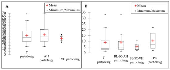
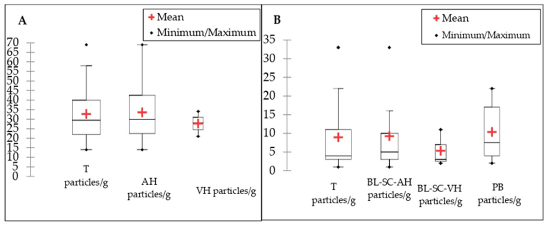
All analysed human intraocular fluid samples contained potential MP particles, with an overall average of 32.7 ± 13.5 particles/g. Notably, the abundance of potential MPs detected in intraocular fluid samples was comparable to that observed in procedural and surgical blanks, indicating a possible contribution of background contamination despite the strict quality control measures applied. A more detailed analysis of the two intraocular fluid types and corresponding quality assurance/quality control (QA/QC) samples revealed average MPs concentrations of 33.5 ± 14.2 particles/g in AH and 27.7 ± 5.31 particles/g in VH. In comparison, blanks simulating sampling and analytical procedures, namely, the surgical condition blanks for AH and VH, as well as the procedural blank, showed average values of 9.25 ± 9.27 particles/g, 5.33 ± 4.03 particles/g, and 10.32 ± 7.76 particles/g, respectively. The lowest number of potential MPs observed in an ocular fluid sample was 14 particles/g (AH-5), while the highest was 69 particles/g (AH-7). The distribution of MPs abundance in ocular fluid samples and QA/QC environmental controls is presented in Figure 1A,B.
Figure 1.
Box plots of potential microplastic (MPs) particles per gram in ocular fluids and blank samples. (A): T, potential MPs recovered from both ocular fluids combined; AH, aqueous humour; VH, vitreous humour; (B): T, potential MPs recovered from all blanks combined; BL-SC-AH, blank of surgical conditions obtained during AH sampling; BL-SC-VH, blank of surgical conditions obtained during VH sampling; PB, procedural blank. Boxes represent the interquartile range (IQR), with the median indicated by the horizontal line; whiskers denote the minimum and maximum values.
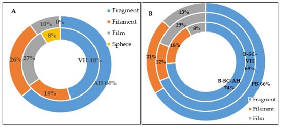
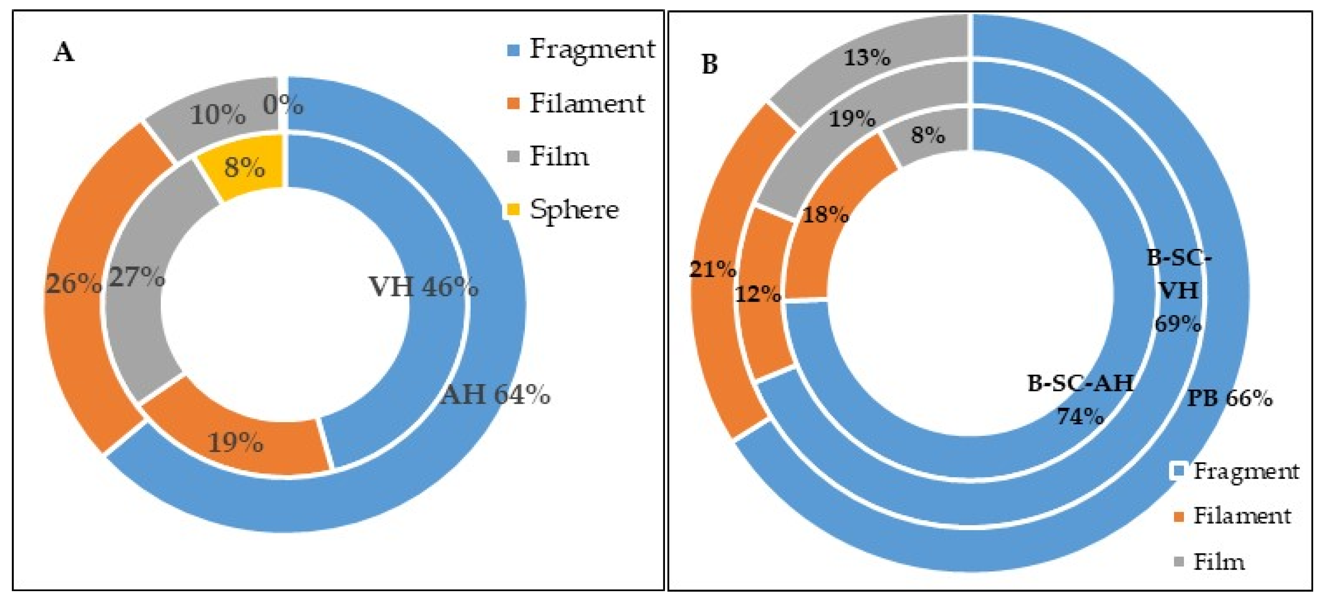
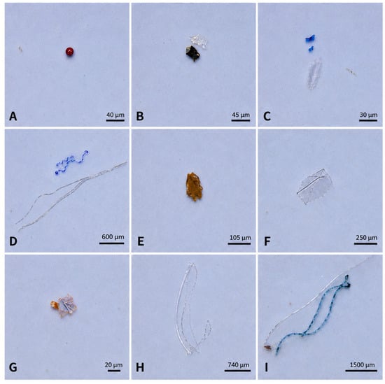
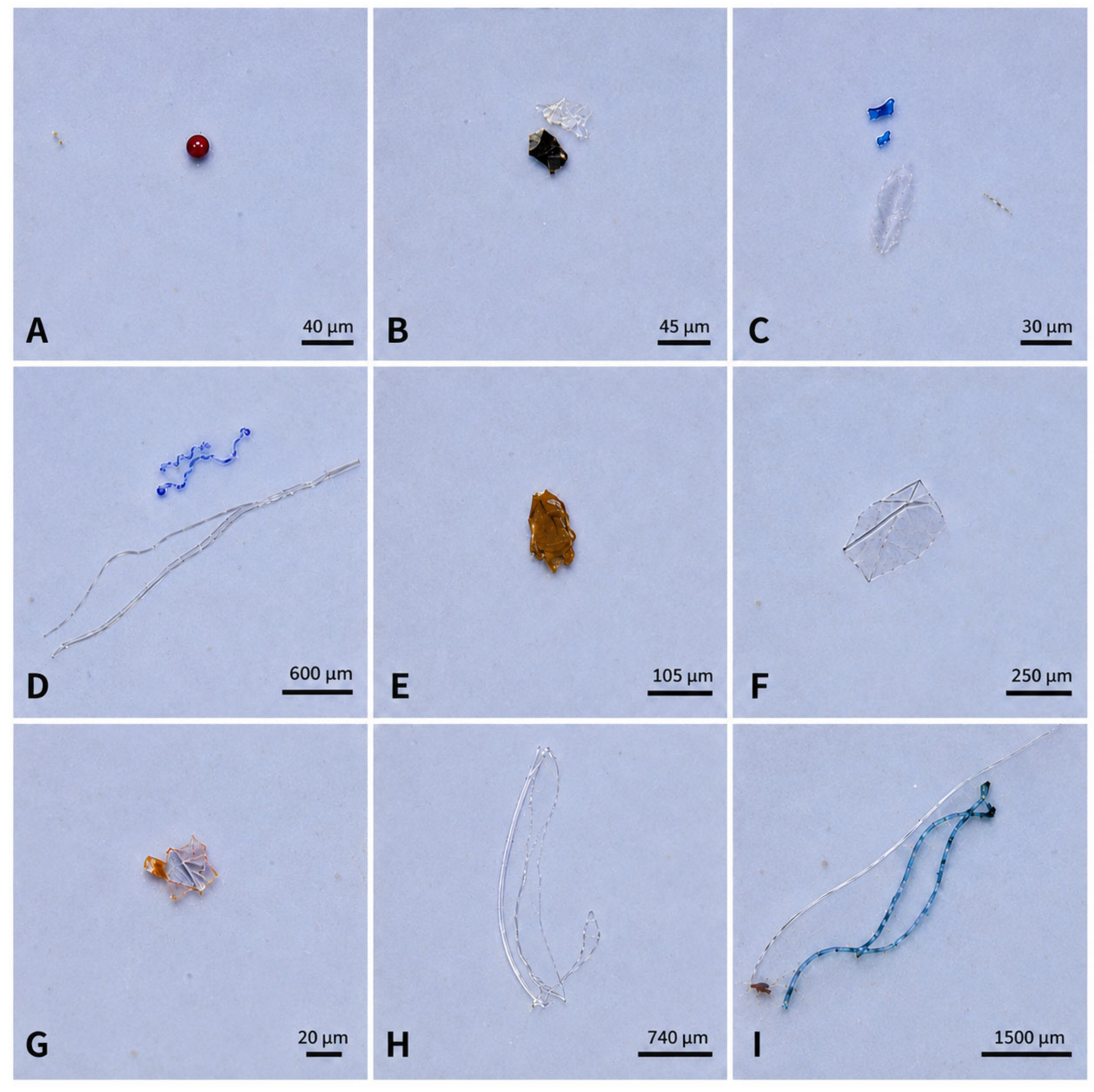
Of the total 719 potential MP identified, 442 were classified as fragments, 184 as filaments, 84 as fibres, and 9 as spheres. The proportional distribution of MPs shapes is presented in Figure 2A,B, whereas major ocular fluids shapes on Figure 3.
Figure 2.
Percentage of potential microplastic (MPs) particles in ocular fluids and control-blank samples by shape category (A): AH aqueous humour (outer doughnut chart); VH, vitreous humour (inner doughnut chart); (B): BL-SC-AH, blank of surgical conditions obtained during AH sampling; BL-SC-VH, blank of surgical conditions obtained during VH sampling (middle doughnut chart); PB, procedural blank (outer doughnut chart).
Figure 3.
Images of representative potential MP morphologies observed by stereomicroscopy in ocular fluids: (A) AH2 sample, red sphere; (B) AH2 sample, black fragment; (C) VH2 sample, blue fragments; (D) AH2 sample, blue filament and grey filament; (E) AH4 sample, brown fragment; (F) AH4 sample, grey films; (G) AH19 sample, orange fragment; (H) AH9 sample, transparent filament; (I) AH5 sample, blue filament. Scale bars are shown in the lower right corner of each panel and range from 20 µm to 1500 µm.
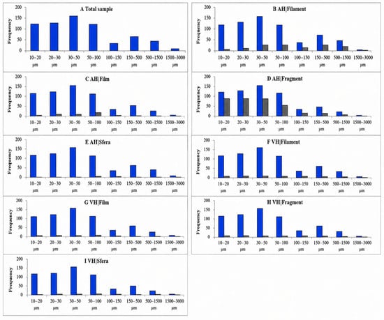
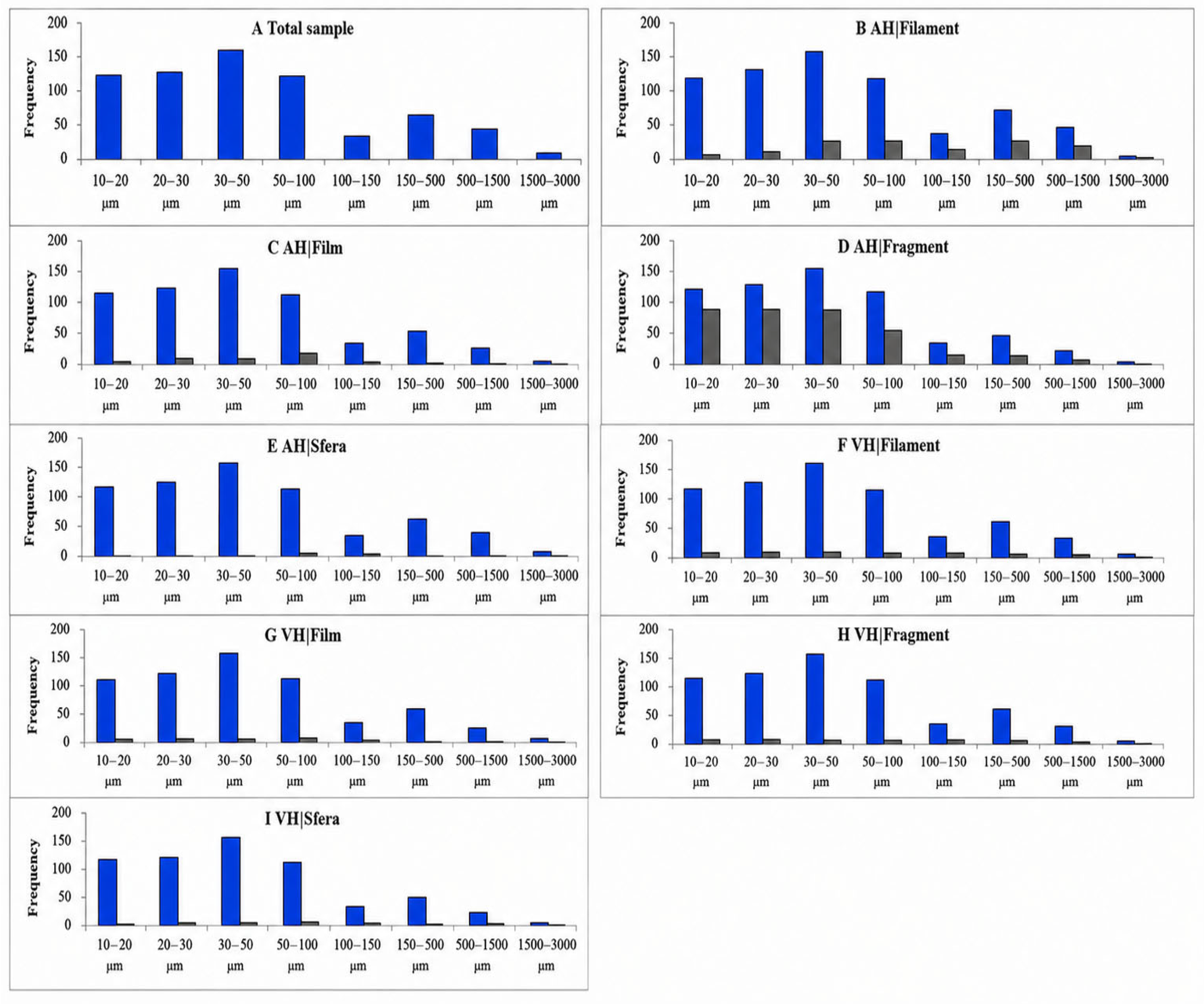
The size of potential MPs ranged from 7.0 µm to 2981.7 µm in length, with mean ± standard deviation values of 199.6 ± 382.3 µm and 113.2 ± 234.1 µm for aqueous AH and VH, respectively. Morphometric distribution across the nine size classes showed that particles were predominantly fragments, followed by filaments, fibres, and spheres. The frequency distribution of potential MPs across size ranges is presented in Figure 4.
Figure 4.
Frequency distribution of potential microplastic (MP) shapes across particle-size ranges. (A) Total sample; (B) aqueous humour (AH), filaments; (C) AH, films; (D) AH, fragments; (E) AH, spheres; (F) vitreous humour (VH), filaments; (G) VH, films; (H) VH, fragments; and (I) VH, spheres. Blue bars represent the total frequency of each shape within the indicated size range, whereas grey bars represent the corresponding frequency detected in AH or VH samples, as applicable.
In both investigated intraocular fluids, a substantial proportion (often exceeding 50%) of detected potential MP particles fell within the smaller size ranges (e.g., <100 µm or <50 µm). The size of potential MPs in blanks collected under surgical conditions and during analytical procedures ranged from 11.8 µm to 737.2 µm in length. The mean ± standard deviation values were 42.3 ± 31.3 µm for BL-SC-AH, 40.39 ± 37.7 µm for BL-SC-VH, and 74.2 ± 140.8 µm for procedural blanks. Stereomicroscopic observations may indicate that fragments were the most prevalent potential MPs shape across both intraocular fluids, laboratory blanks, and surgical condition blanks (Figure 2), supporting the applied quality control measures.
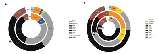
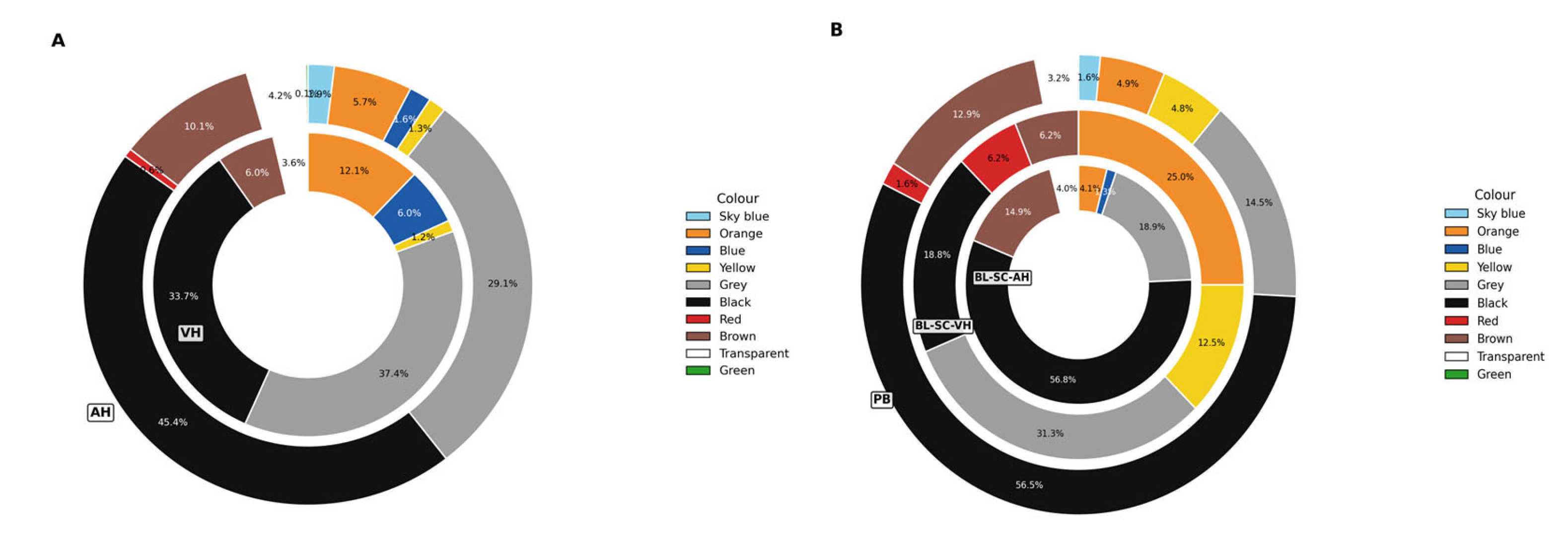
Filaments and films were also detected in the majority of samples (with films absent in some AH samples), whereas spheres were observed in all VH samples and in one AH sample. The 719 potential MP particles were classified into 10 colour categories, with the most prevalent in ocular samples being black (44.1%), grey (30.0%), brown (9.6%), orange (6.4%), transparent (4.17%), and blue/sky blue (3.76%) (Figure 5A). Black and grey were the predominant colours in both aqueous humour (AH) and vitreous humour (VH) samples. Blank samples exhibited a similar colour distribution; of the 152 potential MPparticles detected across all sampling and procedural blanks, most were black (52.6%), grey (18.4%), brown (13.2%), orange (6.6%), transparent (3.29%), and blue/sky blue (3.76%) (Figure 5B).
Figure 5.
Percentage of potential microplastic (MP) particles in ocular fluids and control-blank samples by colour (A): Abundance of potential microplastic particles in aqueous humour (AH) (outer doughnut chart) and vitreous humour (inner doughnut chart) based on colour; (B): Abundance of potential microplastic particles in method blanks: PB, procedural blank (outer doughnut chart); sampling blanks: BL-SC-AH (inner doughnut chart), blank of surgical conditions obtained at AH sampling; BL-SC-VH (middle doughnut chart), blank of surgical conditions obtained at VH sampling, all based on colour.
In both investigated intraocular fluids, a substantial proportion (often exceeding 50%) of detected potential MP particles fell within the smaller size ranges (e.g., <100 µm or <50 µm).
The predominance of fragments and the broad size distribution are consistent with the heterogeneous morphologies reported in human and surgical-environment studies [9,17,19,38,54,55,56]. However, stereomicroscopy cannot establish polymer identity. The similarity in particle abundance, colour, and morphology between ocular samples and blanks therefore requires particular caution: microscopy findings are best interpreted as indicative of potential MPs and considered together with chemical confirmation by Py-GC/MS.
3.2. Contamination Control and Surgical Environment
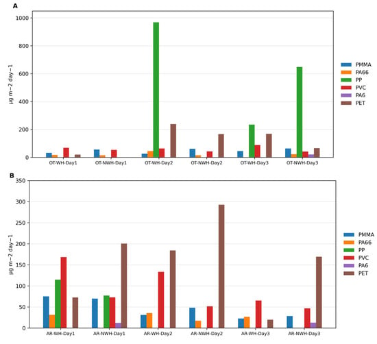
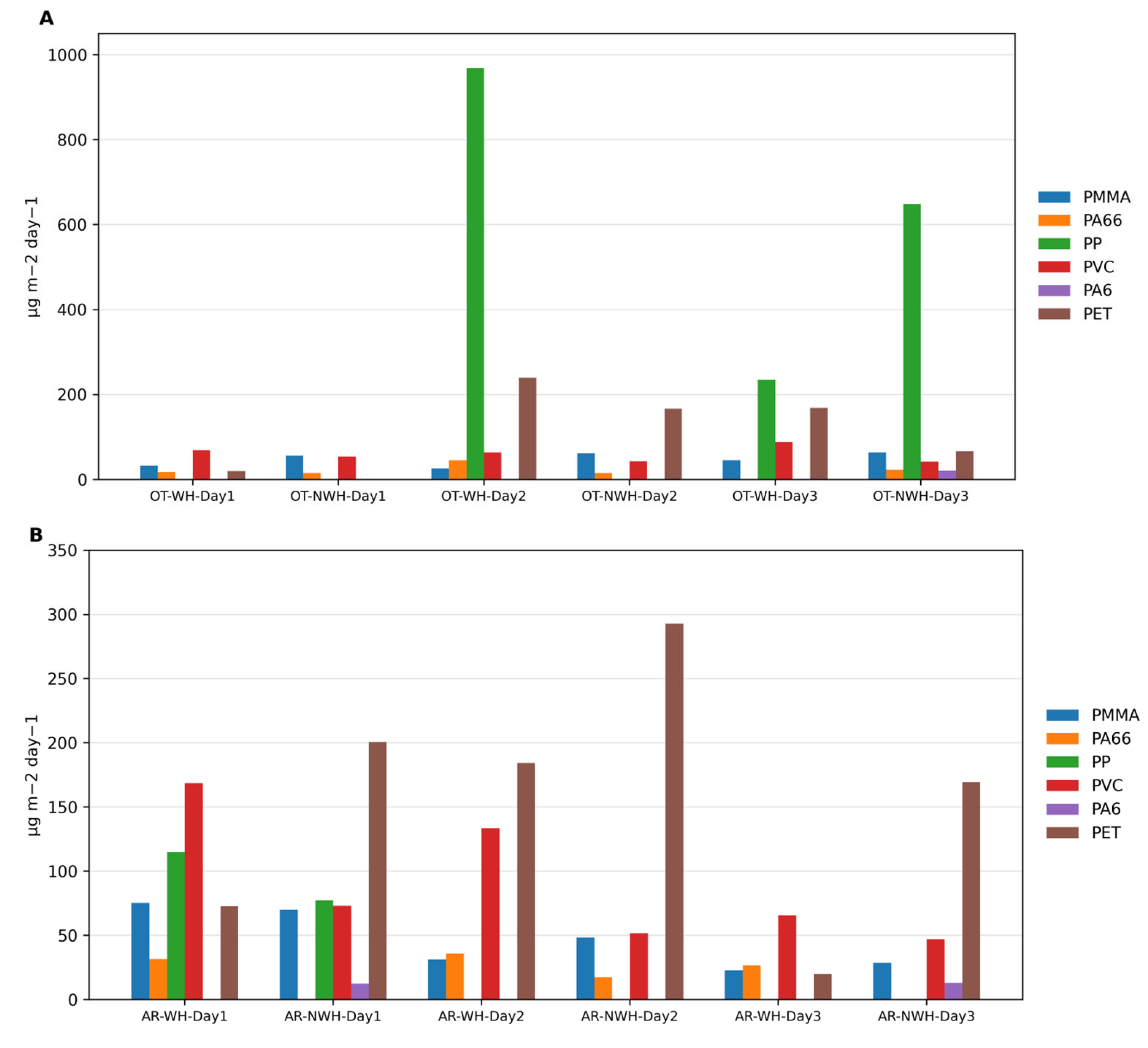
Quality control measures implemented throughout the entire sampling-to-detection workflow, including Py-GC/MS analysis and stereomicroscopy, comprised laboratory procedural blanks and blanks collected under surgical conditions. MP particles were detected in these control samples and surgical environmental samples as shown in Figure 6A,B.
Figure 6.
Bar chart displaying microplastic abundance per sampling period for both (A) operating theatre—OT and (B) the anaesthetic room—AR. Abbreviations: WH, working hours; NWH, non-working hours.
The presence of MPs across all analysed samples may reflect continuous exposure pathways; however, given the comparable levels observed in blank controls, contributions from external contamination cannot be excluded. In addition to contamination-related challenges, several methodological limitations should be considered when interpreting the results. The application of Py-GC/MS provides robust identification and quantification of polymer mass fractions; however, it does not allow direct determination of particle number, size distribution, or morphology. Therefore, complementary stereomicroscopic analysis was employed, although this approach is inherently limited by visual identification and may be subject to misclassification of non-plastic particles. Furthermore, the relatively small sample volumes of intraocular fluids and the limited number of analysed samples, particularly for vitreous humour, constrain the statistical power of the study. Future research should integrate complementary analytical techniques, such as μFTIR or Raman spectroscopy, to improve particle-level characterisation and enhance methodological reliability.
The surgical environment findings are consistent with the presence of MPs reported in an operating theatre by Field et al. [9], but methodological differences preclude direct quantitative comparison. Detection of related particle morphologies and polymer profiles in the environment, blanks and ocular samples identifies a plausible contamination pathway without demonstrating that the ocular findings originated intraoperatively. These results are therefore hypothesis-generating and emphasise the need for extensive field and procedural blanks in future ocular MP studies.
3.3. Py-GC/MS Polymer Profiles in Ocular Fluids and the Surgical Environment
Strict quality-control measures were applied to ensure precise quantification and accurate characterisation of potential background contamination. Given the ubiquitous presence of MPs in the environment, both procedural controls and surgical environmental samples were analysed, and the results were reported as part of routine QA/QC assessments of plastic contamination, in line with recommendations for clinical MPs research [57]. These findings provide new insights into the quantities and types of polymer clusters present in surgical environments, where the use of plastic materials is considered beneficial for clinical practice. The results may inform future cell-toxicity studies investigating the effects of human exposure to MPs and suggest a potential new exposure pathway associated with surgical procedures. Overall, the findings of this study should be considered preliminary and hypothesis-generating, warranting confirmation in larger and more comprehensive investigations.
Importantly, quality-control measures and procedural blanks showed no detectable MPs, whereas sampling blanks exhibited a total MPs contamination of 2.21 µg/g, composed of C-PMMA, C-PA66, and C-PVC in descending order (Table 1).
Table 1.
Microplastic polymer cluster concentrations in aqueous humor and vitreous humor samples. Results are expressed as mean value of two determinations calculated on two separate calibration curves.
MPs detected by Py-GC/MS in surgical blanks were not consistently present across all sampling days, and the concentrations observed were negligible. This finding highlights the minimal contribution of contamination relative to the total MPs detected by stereomicroscopy in both types of blanks. Furthermore, although eleven polymer types were targeted in the analysis, additional MPs may be associated with other polymer clusters not included in the current method.
In total, six out of the eleven investigated polymers were detected in ocular samples (Table 1). MPs were present in all 22 human samples analysed. The most prominent polymer clusters in AH samples were C-PMMA, C-PVC, C-PET, and C-PP, with average concentrations of 0.82 ± 0.32 µg/g, 0.84 ± 0.58 µg/g, 0.48 ± 0.70 µg/g, and 0.41 ± 0.82 µg/g, respectively. All three VH samples contained only C-PMMA, with an average concentration of 0.55 ± 0.12 µg/g. Additionally, C-PA66 (0.30 ± 0.21 µg/g) was frequently detected, whereas C-PA6 (0.15 ± 0.44 µg/g) was only occasionally detected in AH samples. Other polymer clusters, including C-ABS, C-PC, C-PE, C-PS, and C-SBR, were not detected in any of the analysed ocular samples.
MPs were detected in all monitored surgical environmental samples, including both the operating theatre (OT) and anaesthetic room (AR), during both working hours (WH) and non-working hours (NWH). Across all sampled environments and time periods, total MP concentrations ranged from 12.3 to 969 μg m−2 day−1, with a mean atmospheric abundance of 74.8 ± 143.6 μg m−2 day−1. In the OT environment, the total MPs mass fraction varied considerably during the sampling period, with a mean value of 91.6 ± 190.6 μg m−2 day−1 and a range of 15 to 969 μg m−2 day−1 (Figure 5A,B). Similarly, variation was observed in the AR, where the mean MP concentration was 57.9 ± 70.2 μg m−2 day−1, ranging from 12.3 to 292.9 μg m−2 day−1.
When comparing temporal differences, the mean MPs concentration in the AR during working hours was 54.6 ± 59.4 μg m−2 day−1, compared to 61.2 ± 81.2 μg m−2 day−1 during non-working hours. This difference was not statistically significant (p = 0.242). In the OT, the mean MP concentration during working hours was 112.3 ± 226.9 μg m−2 day−1, while during non-working hours it was lower, at 71.0 ± 149.7 μg m−2 day−1. Although MPs concentrations were higher during working hours in the OT—consistent with findings from previous studies—this difference was also not statistically significant.
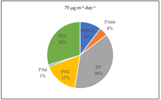
The most dominant polymer clusters identified were C-PP (38% of total MPs), C-PET (30%), C-PVC (17%), and C-PMMA (10%). In contrast, C-PP, C-PA66 (accounting for 4% of the total MPs mass), and C-PA6 (1%) were not consistently detected throughout the monitoring period. Notably, C-PET was absent only in the OT during non-working hours on the first sampling day (Figure 7).
Figure 7.
Total MPs composition. Data is for the operating theatre (OT) and anaesthetic room (AR) combined for the entire three-day sampling period, and for working and non-working hours.
When comparing working and non-working hours across both environments (OT and AR combined), the average MPs abundance during working hours was 92 ± 166 μg m−2 day−1, whereas during non-working hours it was 68 ± 119 μg m−2 day−1. This difference was not statistically significant (p = 0.247, Fisher’s F-test). The polymer profile partly overlaps with previous human studies in which PA, PE, PMMA, PP, PS, and PVC have been reported [12,38,39,58]. Py-GC/MS studies of AH have identified PE, PVC, PP, and PA66 [39], while VH analyses have shown method-dependent differences between LD-IR and Py-GC/MS [38]. Thus, cross-study comparisons should remain qualitative. Importantly, overlap between polymers detected in sampling blanks, the surgical environment, and ocular fluids precludes definitive attribution to pre-existing in vivo exposure.
3.4. Relationship Between MPs Mass Fraction Levels and Individual Participant Characteristics
Table 2 presents the presence of MPs in the AH of study participants, categorised according to individual characteristics outlined in (Table S1).
Table 2.
Comparison of the presence of microplastics in ocular fluids according to the individual characteristics of the participants.
The analysis was further stratified by four of the six detected polymer clusters: C-PMMA, C-PVC, C-PET, and C-PA66. Polymer clusters with a high proportion of values below the limit of detection, such as C-PP and C-PA6, were not interpreted further because of the limited robustness of subgroup comparisons. Overall, total MP levels were higher in female participants than in males; however, this difference was not statistically significant (p > 0.05), with the exception of C-PET, which showed a significantly higher concentration in females. This pattern was generally consistent across most polymer types, except for C-PA66, where levels were lower in females. No statistically significant differences in MP presence were observed based on occupation or operated eye laterality, regardless of MP polymer type (Table 2). When examining age groups, the pilot study population was divided into three classes. Notably, the very old subgroup (≥75 years) showed significantly higher concentrations of C-PA66.
These subgroup findings should be regarded as exploratory because of the small and uneven groups, and the high proportion of non-detects. Although sex- and age-related exposure differences have been proposed in relation to cosmetics, occupational exposure, and polymer-specific behaviour [39,59,60,61,62], the present data are insufficient to establish demographic determinants of intraocular MPs. Total MP abundance was not significantly associated with age.
3.5. UPLC–MS/MS Analysis of BPs in Blood Serum
Due to the limited availability of AH and VH samples, this study focused on BP detection in blood serum. Accepting the previously recommended sensitive detection method based on dansyl chloride derivatisation (Table S8), comprehensive data on patient bisphenol levels were collected (Table 3). BPAF was detected in five samples (2.13–4.54 ng/mL), and in three of these, BPA was co-detected (4.05–5.18 ng/mL).
Table 3.
Concentration of bisphenols in human-blood serum samples.
The detected BPA and BPAF concentrations fall within the broad ranges reported in human biomonitoring studies, although direct comparisons are limited by differences in matrix and analytical method [50,63,64,65,66,67]. BPA and BPAF have documented endocrine activity, and replacement bisphenols remain an area of toxicological concern [42,68,69,70,71,72,73]. Nevertheless, this pilot study was designed to characterise exposure analytically and cannot evaluate health effects.
3.6. Exploratory Relationships Between Serum BPs and Ocular MPs
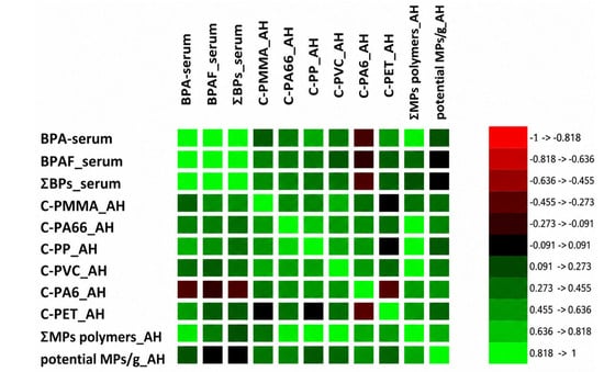
Analysis of interrelationships among MPs polymer types, systemic bisphenols (BPs), and visual particle counts in human samples revealed a strong, highly significant intra-ocular co-contamination cluster (Figure 8).
Figure 8.
Spearman correlogram between serum bisphenols and aqueous humour microplastics (n = 12) illustrating cross and inter-compartmental dynamics of MPs polymer mass fractions, bisphenols, and potential microplastic particles per gram. BPA, Bisphenol A, BPAF, Bisphenol AF, ƩBPs the sum of BPA and its four analogues (BPAF, BPB, BPF, BPS); MPs, microplastics; C-PMMA: polymethyl methacrylate cluster, C-PA6: polyamide 6 cluster, C-PA66: polyamide 66 cluster, C-PP: polypropylene cluster, C-PVC: polyvinyl chloride cluster, C-PET: polyethylene terephthalate cluster, ƩMPs polymers, the sum of MPs polymers clusters (C-PMMA, C-PA6, C-PA66, C-PP, C-PVC, C-PET).
Specifically, robust positive correlations were identified between C-PA66 and C-PVC (ρ= 0.816, p = 0.0012), as well as between C-PMMA and C-PVC (ρ = 0.649, p = 0.0224). Furthermore, C-PA66 and C-PP were significantly co-localised (ρ = 0.633, p = 0.0273). Regarding cross-compartment dynamics, serum BPA exhibited a statistically significant, moderately strong positive correlation with aqueous humour PA-66 (ρ = 0.601, p = 0.0388), alongside a strong positive trend with the total polymer mass sum ƩMPs polymers (ρ = 0.569, p = 0.05). Crucially, raw particle counts (potential MPs/g) and C-PA6 did not exhibit any statistically significant correlations with systemic bisphenols (p > 0.70). Given the moderate sample size and the high proportion of values below the limit of detection (<LOD) for several variables, these correlation analyses should be interpreted as exploratory trends that warrant validation in larger, adequately powered future cohorts.
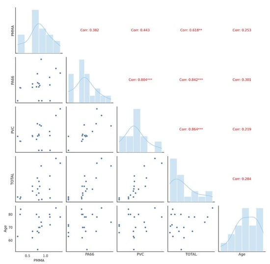
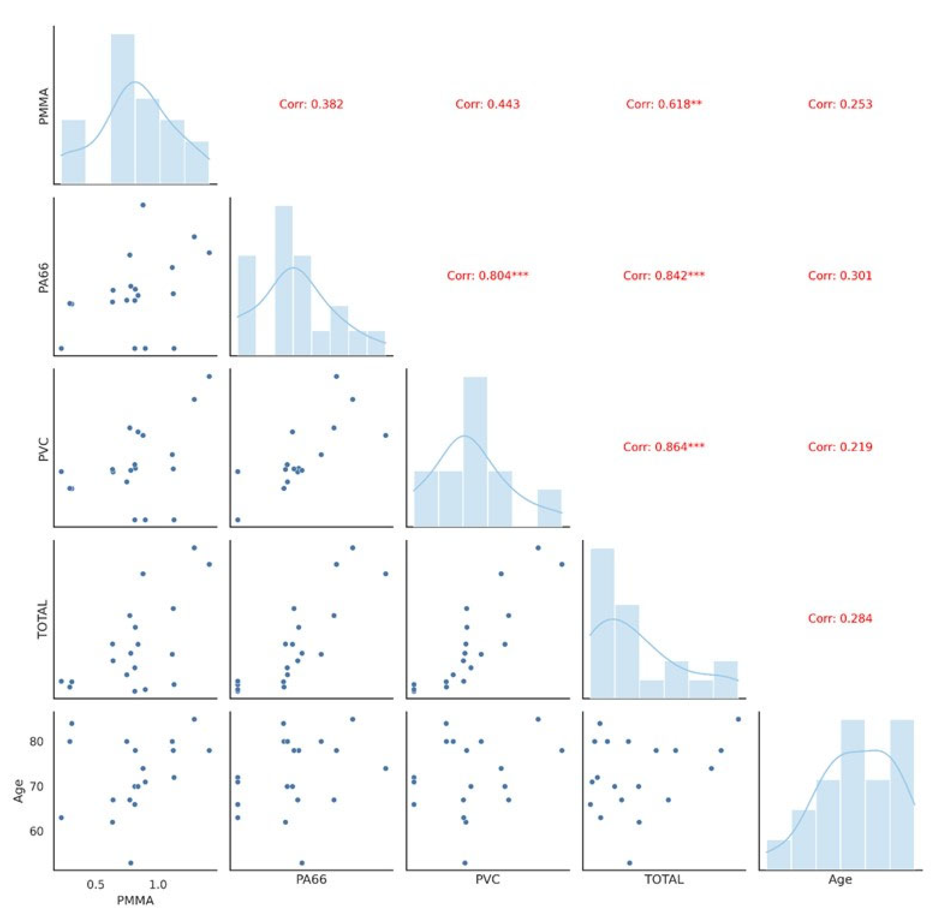
When evaluating MP accumulation patterns in ocular fluids in relation to individuals’ lifelong exposure, no significant correlation was observed between total MPs levels and age across the pilot-study population (Figure 9).
Figure 9.
Correlation analysis: relationships between MP concentrations and patients’ age. Diagonal plots illustrate the distribution of data in logarithmic ratios. Lower plots display the scatter plots of data, while the upper plots indicate the correlation coefficients (r). Significant correlations are denoted by red markers. Notes: MPs, microplastics; PMMA, polymethyl methacrylate; PA66, nylon 66; PVC, polyvinyl chloride, TOTAL, sum of polymer clusters.
The observed correlations are exploratory and should not be interpreted as evidence of a biological or causal relationship. The small number of paired observations, multiple comparisons, and frequent non-detects limit robustness. In addition, correlations involving ΣMPs may be inflated by mathematical coupling because the total is calculated from the individual polymer clusters. No significant relationship was observed between total MPs and age. According to [39], variations in MPs abundance in aqueous humour may be linked to differing daily exposures across age groups. Individuals employed in plastic manufacturing, recycling, or textile industries are exposed to high levels of airborne MPs. One study found thousands of MPs fibres on workers’ skin, hair, and even in saliva after shifts in plastic factories [62]. These workers are likely to experience continuous deposition of MPs on the lungs and eyes. Overall, the correlation analysis is best considered hypothesis-generating and requires confirmation in a larger, prospectively powered cohort with standardised contamination control.
3.7. Strengths and Limitations
A strength of this study is the complementary use of stereomicroscopy, polymer-specific Py-GC/MS, serum UPLC–MS/MS, surgical-environment monitoring, and multiple blank types. The principal limitations are the pilot design, small cohort, only three VH samples, 13 evaluable serum samples, one surgical week of sampling, frequent non-detects, and potential background contamination. Py-GC/MS provides polymer mass but not particle-level morphology, whereas stereomicroscopy is not chemically specific. Consequently, the study supports analytical feasibility and generates exposure hypotheses but does not establish population prevalence, accumulation, causality or clinical effects. Larger multicentre studies with standardised field/procedural blanks and complementary particle-level spectroscopy are required.
4. Conclusions
MPS were detected in all analysed ocular samples, while selected bisphenols (BPA and BPAF) were identified in human serum, indicating human exposure to plastic-associated contaminants. Serum BPA exhibited a statistically significant, moderately strong positive correlation with aqueous humour PA-66, alongside a strong positive trend with the total polymer mass ƩMPs polymers. Six polymer types (C-PMMA, C-PVC, C-PET, C-PP, C-PA66, and C-PA6) were identified, and exploratory subgroup analyses showed statistically significant differences according to age and gender, with higher MPs abundance and prevalence in the aqueous humour of female participants. Environmental contamination was identified through the implemented blank-control strategy; however, the contribution of the surgical environment to intraocular MPs exposure could not be conclusively determined. While the presented findings remain preliminary due to sample quantity and analytical limitations, the observed MP trends provide supportive knowledge relevant for further systemic assessments of the detected polymers in ocular fluids and tissues. The strategy presented in this pilot study enables direct quantitative assessment of MPs accumulation in the aqueous humour and vitreous humour using analytical techniques such as Py-GC/MS. Extending this approach to a cohort study could provide stronger evidence for the relationship between MP exposure and ocular pathology, while accelerating the development of diagnostic criteria. Future research should focus on developing standardised methods, exploring mechanisms in depth, and expanding translational applications to address the growing threat of plastic pollution to human health.
Supplementary Materials
The following supporting information can be downloaded at: https://www.mdpi.com/article/10.3390/microplastics5030176/s1, Table S1. Clinical characteristics of patients. Table S2. Py-GC/MS method. Table S3. Main pyrolysis products of the 11 polymer clusters. Table S4. Specific marker and ions used for quantification of the 11 polymer clusters; characteristic Py-GC/MS decomposition products used for identification and quantification of detected polymer clusters indicated by “C-" as pure polymers and potentially included polymer-related derivatives. Table S5. Polymer-Specific Mass Equivalents of MCS for Calibration Range in Py-GC/MS Analysis (MSC in SiO2, low level, Lot: 23031601) example. Table S6a. Recovery of microplastic polymer mass fractions spiked AH sample (AH 6). Table S6b. Recovery of microplastic polymer mass fractions spiked VH sample (VH 3). Table S7. Selected Py-GC/MS method performance indicators in animal ocular internal fluids (aqueous and vitreous humour, AH, VH): linearity range, limit of detection (LOD), limit of quantification (LOQ), recovery and precision (RSDr) for ocular tissues. Table S8. Optimised parameters for the mass spectrometry analysis of derivatised bisphenols. Table S9. Method performance criteria of UPLC–MS/MS method developed in human blood serum. Figure S1. The chromatograms (on the left side) and mass spectrograms (on the right side) through pyrolysis-gas chromatography/mass spectrometry (Py-GC/MS) analysis of spiked AH sample, including polymethyl methacrylate cluster (C-PMMA) with 0.075 μg, polyamide-6 cluster (C-PA-6) with 0.135 μg, polypropylene cluster (C-PP) at 1.06 μg, polyvinyl chloride cluster (C-PVC) with 0.88 μg, polyamide-66 (PA-66) with 0.213 μg, styrene-butadiene rubber (SBR) with 0.400 μg, polyethylene terephthalate cluster (C-PET) with 0.251 μg, polycarbonate cluster (C-PC) with 0.08 μg, poly(4-fluorostyrene) ISTD 4PFS at 0.2 μg, polystyrene cluster (C-PS) with 0.163 μg, polyethylene cluster (C-PE) at 3.29 μg (average sum of α-Alkenes (e.g., C14-C22)), and deuterated polystyrene (d5-PS) at 0.2 μg used in internal standard calibration with thermochemolysis (10% TMAH in MeOH).
Author Contributions
Conceptualisation, T.B., J.P., and A.G.; methodology, Z.V., G.M., V.M., S.Š., T.B., Z.J., F.D.G., C.P., S.P., E.L., and I.L.; validation, S.Š., G.M., Z.V., V.M., T.B., and Z.J.; formal analysis, A.G., V.M., C.P., S.P., E.L., and I.L.; investigation, T.B. Z.J., G.M., Z.V., S.Š., T.B., and F.D.G.; resources, Z.V., G.M., S.Š., J.P., S.P., and E.L.; data curation, G.M., S.Š., T.B., Z.J., and C.P.; writing—original draft preparation, T.B. and J.P.; writing—review and editing, J.P., G.M., S.Š., A.G., Z.V., V.M., F.D.G., S.P., and C.P.; visualisation, T.B., J.P., V.M., F.D.G., and C.P.; supervision, T.B. and E.L.; project administration, T.B. and J.P.; funding acquisition, T.B. and S.P. All authors have read and agreed to the published version of the manuscript.
Funding
This research was funded by the European Union NextGenerationEU and supported by the Ministry of Science and Education of the Republic of Croatia through the project No. NPOO 6 of Croatian Veterinary Institute titled ”Occurrence of Microplastics and Bisphenols in Shellfish Along the Croatian Coast of the Adriatic Sea” (PLASTICshell).
Institutional Review Board Statement
Institutional ethical approval was obtained from the University Hospital Centre Sestre milosrdnice, Zagreb (protocol code: 003-06/26-03/001/Urbroj: 251-29-11/3-26-1, date of approval: 12 January 2026).
Informed Consent Statement
Informed consent was obtained from all subjects involved in the study.
Data Availability Statement
The original contributions presented in this study are included in the article/Supplementary Material. Further inquiries can be directed to the corresponding authors.
Conflicts of Interest
The authors declare no conflicts of interest.
Abbreviations
The following abbreviations are used in this manuscript:
| ACN | acetonitrile |
| AH | aqueous humour |
| anhydrous Na2SO4 | anhydrous natrium sulphate |
| AR | anaesthetic room |
| BPA | bisphenol A |
| BPAF | bisphenol AF |
| BPB | bisphenol B |
| BPBP | bisphenol BP |
| BPC | bisphenol C |
| BPE | bisphenol E |
| BPF | bisphenol F |
| BPFL | bisphenol FL |
| BPG | bisphenol G |
| BPM | bisphenol M |
| BPP | bisphenol P |
| BPS | bisphenol S |
| BPs | bisphenols |
| BS | blood serum |
| BPZ | bisphenol Z |
| C-ABS | acrylonitrile butadiene cluster |
| C-PA6 | polyamide 6 cluster |
| C-PA66 | polyamide 66 cluster |
| C-PC | polycarbonate cluster |
| C-PE | polyethylene cluster |
| C-PET | polyethylene terephthalate cluster |
| C-PMMA | polymethyl methacrylate |
| C-PP | polypropylene cluster |
| C-P | polystyrene cluster |
| C-PVC | polyvinyl chloride cluster |
| C-SBR | styrene-butadiene rubber cluster |
| Dns-Cl | dansyl chloride |
| ESI | electrospray ionisation |
| GF/C | glass fibre filters |
| H2O2 | hydrogen peroxide |
| IOL | intraocular lens |
| IQR | interquartile range |
| LD-IR | laser direct infrared |
| LLE | liquid–liquid extraction |
| LOD | limit of detection |
| LOQ | limit of quantification |
| MPs | microplastics |
| MRM | transitions |
| ND | not detected |
| NWH | non-working hours |
| OD | oculus dexter |
| OS | oculus sinister |
| OT | operating theatre |
| PCI | percutaneous coronary intervention |
| PP | polypropylene |
| PTFE | polytetrafluoroethylene |
| Py-GC/MS | pyrolysis gas chromatography–mass spectrometry |
| RSDr | relative standard deviation |
| SEM | scanning electron microscopy |
| TMAH | tetramethylammonium hydroxide |
| UHSM | University Hospital Sestre Milosrdnice |
| UPLC–MS/MS | ultra-performance liquid chromatography–tandem mass spectrometry |
| VH | vitreous humour |
| WH | working hours |
| μFTIR | micro-Fourier transform infrared spectroscopy |
References
- Dey, T.; Trasande, L.; Altman, R.; Wang, Z.; Krieger, A.; Bergmann, M.; Allen, D.; Allen, S.; Walker, T.R.; Wagner, M.; et al. Global plastic treaty should address chemicals. Science 2022, 378, 841–842. [Google Scholar] [CrossRef] [Scilit]
- Bogdanović, T.; Pleadin, J. Microplastics as unexplored emerging contaminant: A challenge for food safety. In Advances in Food and Nutrition Research, 1st ed.; Toldrá, F., Ed.; Academic Press Elsevier: London, UK, 2026; Volume 18, pp. 1–352. [Google Scholar] [CrossRef] [Scilit]
- Schirinzi, G.F.; Pérez-Pomeda, I.; Sanchís, J.; Rossini, C.; Farré, M.; Barceló, D. Cytotoxic effects of commonly used nanomaterials and microplastics on cerebral and epithelial human cells. Environ. Res. 2017, 159, 579–587. [Google Scholar] [CrossRef] [Scilit]
- Thompson, R.C.; Courtene-Jones, W.; Boucher, J.; Pahl, S.; Raubenheimer, K.; Koelmans, A.A. Twenty years of microplastic pollution research-what have we learned? Science 2024, 386, eadl2746. [Google Scholar] [CrossRef] [Scilit]
- Zhao, X.; You, F. Microplastic Human Dietary Uptake from 1990 to 2018 Grew across 109 Major Developing and Industrialized Countries but Can Be Halved by Plastic Debris Removal. Environ. Sci. Technol. 2024, 58, 8709–8723. [Google Scholar] [CrossRef] [Scilit]
- Rafa, N.; Ahmed, B.; Zohora, F.; Bakya, J.; Ahmed, S.; Ahmed, S.F.; Mofijur, M.; Chowdhury, A.A.; Almomani, F. Microplastics as carriers of toxic pollutants: Source, transport, and toxicological effects. Environ. Pollut. 2024, 343, 123190. [Google Scholar] [CrossRef] [Scilit]
- Liu, S.; Huang, J.; Zhang, W.; Shi, L.; Yi, K.; Yu, H.; Zhang, C.; Li, S.; Li, J. Microplastics as a vehicle of heavy metals in aquatic environments: A review of adsorption factors, mechanisms, and biological effects. J. Environ. Manag. 2022, 302, 113995. [Google Scholar] [CrossRef] [Scilit]
- Ullah, S.; Ahmad, S.; Guo, X.; Ullah, S.; Ullah, S.; Nabi, G.; Wanghe, K. A review of the endocrine disrupting effects of micro and nano plastic and their associated chemicals in mammals. Front. Endocrinol. 2023, 13, 1084236. [Google Scholar] [CrossRef] [Scilit]
- Field, D.T.; Green, J.L.; Bennett, R.; Jenner, L.C.; Sadofsky, L.R.; Chapman, E.; Loubani, M.; Rotchell, J.M. Microplastics in the surgical environment. Environ. Int. 2022, 170, 107630. [Google Scholar] [CrossRef] [Scilit]
- Leslie, H.A.; van Velzen, M.J.M.; Brandsma, S.H.; Vethaak, A.D.; Garcia-Vallejo, J.J.; Lamoree, M.H. Discovery and quantification of plastic particle pollution in human blood. Environ. Int. 2022, 163, 107199. [Google Scholar] [CrossRef] [Scilit]
- Li, K.; Li, W.; Sun, Y.; Ma, T.; Yuan, L.; Rong, Y.; Liu, X.; Fu, Y.; Yu, X.; Xu, X. Medical Microplastics: Research Progress on Exposure Pathways, Toxic Effects, and Detection Methods. Microplastics 2026, 5, 61. [Google Scholar] [CrossRef] [Scilit]
- Barceló, D.; Picó, Y.; Alfarhan, A.H. Microplastics: Detection in human samples, cell line studies, and health impacts. Environ. Toxicol. Phar. 2023, 101, 104204. [Google Scholar] [CrossRef] [Scilit]
- World Health Organization (WHO). Dietary and Inhalation Exposure to Nano- and Microplastic Particles and Potential Implications for Human Health; WHO: Geneva, Switzerland, 2022; Available online: https://www.who.int/publications/i/item/9789240054608 (accessed on 15 September 2025).
- Snehamayee, N.; Somya, S.; Kumar, S.C.; Niranjan, M.; Ranjan, S.B.; Kumar, M.N. Microplastics and Human Health: A Comprehensive Review on Exposure Pathways, Toxicity, and Emerging Risks. Microplastics 2026, 5, 8. [Google Scholar] [CrossRef] [Scilit]
- Lu, S.; Wei, Y.; Xu, R.; Li, M.; Qv, Y.; Zha, Y.; Gong, M.; Wang, N.; Lu, X.; Jiang, X.; et al. New insights: Discovery of microplastics in human bone and skeletal muscle. Innov. Med. 2024, 2, 100100. [Google Scholar] [CrossRef] [Scilit]
- Nihart, A.J.; Garcia, M.A.; El Hayek, E.; Liu, R.; Olewine, M.; Kingston, J.D.; Castillo, E.F.; Gullapalli, R.R.; Howard, T.; Bleske, B.; et al. Bioaccumulation of microplastics in decedent human brains. Nat. Med. 2025, 31, 1114–1119. [Google Scholar] [CrossRef] [Scilit]
- Ibrahim, Y.S.; Tuan Anuar, S.; Azmi, A.A.; Wan Mohd Khalik, W.M.A.; Lehata, S.; Hamzah, S.R.; Ismail, D.; Ma, Z.F.; Dzulkarnaen, A.; Zakaria, Z.; et al. Detection of microplastics in human colectomy specimens. JGH Open 2020, 5, 116–121. [Google Scholar] [CrossRef] [Scilit]
- Yang, Y.; Xie, E.; Du, Z.; Peng, Z.; Han, Z.; Li, L.; Zhao, R.; Qin, Y.; Xue, M.; Li, F.; et al. Detection of Various Microplastics in Patients Undergoing Cardiac Surgery. Environ. Sci. Technol. 2023, 57, 10911–10918. [Google Scholar] [CrossRef] [Scilit]
- Horvatits, T.; Tamminga, M.; Liu, B.; Sebode, M.; Carambia, A.; Fischer, L.; Püschel, K.; Huber, S.; Fischer, E.K. Microplastics detected in cirrhotic liver tissue. eBioMedicine 2022, 82, 104147. [Google Scholar] [CrossRef] [Scilit]
- Jiang, N.; Zheng, X.; Zhang, N.; Cao, Y. The detrimental effects of microplastic exposure on kidney function. Front. Med. 2025, 12, 1620733. [Google Scholar] [CrossRef] [Scilit]
- Amato-Lourenço, L.F.; Carvalho-Oliveira, R.; Júnior, G.R.; Dos Santos Galvão, L.; Ando, R.A.; Mauad, T. Presence of airborne microplastics in human lung tissue. J. Hazard. Mater. 2021, 416, 126124. [Google Scholar] [CrossRef] [Scilit]
- Zhu, L.; Kang, Y.; Ma, M.; Wu, Z.; Zhang, L.; Hu, R.; Xu, Q.; Zhu, J.; Gu, X.; An, L. Tissue accumulation of microplastics and potential health risks in human. Sci. Total Environ. 2024, 915, 170004. [Google Scholar] [CrossRef] [Scilit]
- Ragusa, A.; Svelato, A.; Santacroce, C.; Catalano, P.; Notarstefano, V.; Carnevali, O.; Papa, F.; Rongioletti, M.C.A.; Baiocco, F.; Draghi, S.; et al. Plasticenta: First evidence of microplastics in human placenta. Environ. Int. 2021, 146, 106274. [Google Scholar] [CrossRef] [Scilit]
- Baeza-Martínez, C.; Olmos, S.; González-Pleiter, M.; López-Castellanos, J.; García-Pachón, E.; Masiá-Canuto, M.; Hernández-Blasco, L.; Bayo, J. First evidence of microplastics isolated in European citizens’ lower airway. J. Hazard. Mater. 2022, 438, 129439. [Google Scholar] [CrossRef] [Scilit]
- Amiri, H.; Moradalizadeh, S.; Jahani, Y.; Nasiri, A. Biomonitoring of microplastics in saliva and hands of young children in kindergartens: Identification, quantification, and exposure assessment. Environ. Monit. Assess. 2025, 197, 859. [Google Scholar] [CrossRef] [Scilit]
- Zhu, L.; Zhu, J.; Zuo, R.; Xu, Q.; Qian, Y.; An, L. Identification of microplastics in human placenta using laser direct infrared spectroscopy. Sci. Total Environ. 2023, 856, 159060. [Google Scholar] [CrossRef] [Scilit]
- Liu, S.; Guo, J.; Liu, X.; Yang, R.; Wang, H.; Sun, Y.; Chen, B.; Dong, R. Detection of various microplastics in placentas, meconium, infant feces, breastmilk and infant formula: A pilot prospective study. Sci. Total Environ. 2023, 854, 158699. [Google Scholar] [CrossRef] [Scilit]
- Quinzi, V.; Orilisi, G.; Vitiello, F.; Notarstefano, V.; Marzo, G.; Orsini, G. A spectroscopic study on orthodontic aligners: First evidence of secondary microplastic detachment after seven days of artificial saliva exposure. Sci. Total. Environ. 2023, 866, 161356. [Google Scholar] [CrossRef] [Scilit] [PubMed]
- Huang, S.; Huang, X.; Bi, R.; Guo, Q.; Yu, X.; Zeng, Q.; Huang, Z.; Liu, T.; Wu, H.; Chen, Y.; et al. Detection and Analysis of Microplastics in Human Sputum. Environ. Sci. Technol. 2022, 56, 2476–2486. [Google Scholar] [CrossRef] [Scilit]
- Schwabl, P.; Köppel, S.; Königshofer, P.; Bucsics, T.; Trauner, M.; Reiberger, T.; Liebmann, B. Detection of Various Microplastics in Human Stool: A Prospective Case Series. Ann. Intern. Med. 2019, 171, 453–457. [Google Scholar] [CrossRef] [Scilit]
- Yan, Z.; Liu, Y.; Zhang, T.; Zhang, F.; Ren, H.; Zhang, Y. Analysis of Microplastics in Human Feces Reveals a Correlation between Fecal Microplastics and Inflammatory Bowel Disease Status. Environ. Sci. Technol. 2022, 56, 414–421. [Google Scholar] [CrossRef] [Scilit]
- Pironti, C.; Notarstefano, V.; Ricciardi, M.; Motta, O.; Giorgini, E.; Montano, L. First Evidence of Microplastics in Human Urine, a Preliminary Study of Intake in the Human Body. Toxics 2022, 11, 40. [Google Scholar] [CrossRef] [Scilit]
- Verbraeken, H.; Verstraete, A.; Van de Velde, E.; Verschraegen, G. Penetration of gentamicin and ofloxacin in human vitreous after systemic administration. Graefes Arch. Clin. Exp. Ophthalmol. 1996, 234, 59–65. [Google Scholar] [CrossRef] [Scilit]
- Qi, Y.; Liu, X.; Chen, Y.; Wu, Y.; Sun, Y.; Liu, X.; Bao, Q.; Zhang, J.; Yuan, G.; Wang, T.; et al. Enhanced Intrusion of Exogenous Airborne Fine Particles toward Eyes in Humans and Animals: Where Damaged Blood–Ocular Barrier Plays a Crucial Role. Environ. Sci. Technol. 2024, 58, 13636–13647. [Google Scholar] [CrossRef] [Scilit]
- Wu, D.; Lim, B.X.H.; Seah, I.; Xie, S.; Jaeger, J.E.; Symons, R.K.; Heffernan, A.L.; Curren, E.E.M.; Leong, S.C.Y.; Riau, A.K.; et al. Impact of Microplastics on the Ocular Surface. Int. J. Mol. Sci. 2023, 24, 3928. [Google Scholar] [CrossRef] [Scilit]
- Zhou, X.; Wang, G.; An, X.; Wu, J.; Fan, K.; Xu, L.; Li, C.; Xue, Y. Polystyrene microplastic particles: In vivo and in vitro ocular surface toxicity assessment. Environ. Pollut. 2022, 303, 119126. [Google Scholar] [CrossRef] [Scilit]
- Upaphong, P.; Thonusin, C.; Wanichthanaolan, O.; Chattipakorn, N.; Chattipakorn, S.C. Consequences of exposure to particulate matter on the ocular surface: Mechanistic insights from cellular mechanisms to epidemiological findings. Environ. Pollut. 2024, 345, 123488. [Google Scholar] [CrossRef] [Scilit]
- Zhong, Y.; Yang, Y.; Zhang, L.; Ma, D.; Wen, K.; Cai, J.; Cai, Z.; Wang, C.; Chai, X.; Zhong, J.; et al. Revealing new insights: Two-centre evidence of microplastics in human vitreous humor and their implications for ocular health. Sci. Total Environ. 2024, 921, 171109. [Google Scholar] [CrossRef] [Scilit]
- Zhang, K.; Yu, L.; Qu, L.; Hui, N.; Chen, L.; Wang, J.; Yan, H. Identifying and analysing the microplastics in human aqueous humor by pyrolysis-gas chromatography/mass spectrometry. iScience 2025, 28, 112078. [Google Scholar] [CrossRef] [Scilit]
- He, L.; Zheng, J.; Han, X.K.; Tao, T.Y.; Zeng, J.; Luo, W.; Chen, X.; Wang, J.M.; Sha, X.Y. Micro/nanoplastics and eye health: A review. Int. J. Ophthalmol. 2026, 18, 405–413. [Google Scholar] [CrossRef] [Scilit] [PubMed] [PubMed Central]
- Flieger, J.; Śniegocki, T.; Dolar-Szczasny, J.; Załuska, W.; Rejdak, R. The First Evidence on the Occurrence of Bisphenol Analogues in the Aqueous Humor of Patients Undergoing Cataract Surgery. J. Clin. Med. 2022, 11, 6402. [Google Scholar] [CrossRef] [Scilit]
- Konieczna, A.; Rutkowska, A.; Rachoń, D. Health risk of exposure to Bisphenol A (BPA). Rocz. Panstw. Zakl. Hig. 2015, 66, 5–11. [Google Scholar]
- Abbas, G.; Ahmed, U.; Ahmad, M.A. Impact of Microplastics on Human Health: Risks, Diseases, and Affected Body Systems. Microplastics 2025, 4, 23. [Google Scholar] [CrossRef] [Scilit]
- Pelch, K.; Wignall, J.A.; Goldstone, A.E.; Ross, P.K.; Blain, R.B.; Shapiro, A.J.; Holmgren, S.D.; Hsieh, J.H.; Svoboda, D.; Auerbach, S.S.; et al. A scoping review of the health and toxicological activity of bisphenol A (BPA) structural analogues and functional alternatives. Toxicology 2019, 424, 152235. [Google Scholar] [CrossRef] [Scilit]
- Bogdanović, T.; Listeš, I.; Gjerde, J.; Petričević, S.; Jažo, Z.; Listeš, E.; Pleadin, J.; Sokolić, D.; Jadrešin, I.; di Giacinto, F. Microplastic Polymer Mass Fractions in Marine Bivalves: From Isolation to Hazard Risk. J. Xenobiotics 2025, 15, 186. [Google Scholar] [CrossRef] [Scilit]
- Wang, J.; Hong, X.; Liu, W.; Zhang, L.; Yan, S.; Li, Z.; Zha, J. Comprehensive assessment of the safety of bisphenol A and its analogs based on multi-toxicity tests in vitro. J. Hazard. Mater. 2025, 486, 136983. [Google Scholar] [CrossRef] [Scilit]
- Dzierżyński, E.; Gawlik, P.J.; Puźniak, D.; Flieger, W.; Jóźwik, K.; Teresiński, G.; Forma, A.; Wdowiak, P.; Baj, J.; Flieger, J. Microplastics in the Human Body: Exposure, Detection, and Risk of Carcinogenesis: A State-of-the-Art Review. Cancers 2024, 16, 3703. [Google Scholar] [CrossRef] [Scilit]
- Markley, L.A.T.; Driscoll, C.T.; Hartnett, B.; Mark, N.; Mateos Cárdenas, A.; Hapich, H. Guide for the Visual Identification & Classification of Plastic Particles. Head Neck 2024, 46, 1294–1303. [Google Scholar]
- Abidli, S.; Lahbib, Y.; El Menif, N.T. Microplastics in commercial molluscs from the lagoon of Bizerte (Northern Tunisia). Mar. Pollut. Bull. 2019, 142, 243–252. [Google Scholar] [CrossRef] [Scilit]
- Owczarek, K.; Kubica, P.; Kudłak, B.; Rutkowska, A.; Konieczna, A.; Rachoń, D.; Namieśnik, J.; Wasik, A. Determination of trace levels of eleven bisphenol A analogues in human blood serum by high performance liquid chromatography-tandem mass spectrometry. Sci. Total Environ. 2018, 628-629, 1362–1368. [Google Scholar] [CrossRef] [Scilit]
- Liu, X.; Lv, Q.; Song, X.; Chen, Y.; Zhao, L.; Yan, M.; Hu, B.; Chen, D. Screening for Bisphenol Chemicals: A Strategy Based on Dansyl Chloride Derivatizatio n Coupled with In-Source Fragmentation by High-Resolution Mass Spectrometry. Anal. Chem. 2023, 95, 6227–6234. [Google Scholar] [CrossRef] [Scilit]
- Schiano, M.E.; Sodano, F.; Cassiano, C.; Fiorino, F.; Seccia, S.; Rimoli, M.G.; Albrizio, S. Quantitative Determination of Bisphenol A and Its Congeners in Plant-Based Beverages by Liquid Chromatography Coupled to Tandem Mass Spectrometry. Foods 2022, 11, 3853. [Google Scholar] [CrossRef] [Scilit]
- European Union. Commission Regulation (EC) No 333/2007 of 28 March 2007 Laying Down the Methods of Sampling and Analysis for the Official Control of the Levels of Lead, Cadmium, Mercury, Inorganic tin, 3-MCPD and Benzo(a)pyrene in Foodstuffs; European Union: Brussels, Belgium, 2007. [Google Scholar]
- Minoda, Y.; Kobayashi, A.; Iwaki, H.; Miyaguchi, M.; Kadoya, Y.; Ohashi, H.; Yamano, Y.; Takaoka, K. Polyethylene Wear Particles in Synovial Fluid After Total Knee Arthroplasty. Clin. Orthop. Relat. Res. 2003, 410, 165–172. [Google Scholar] [CrossRef] [Scilit]
- Abbasi, S.; Turner, A. Human exposure to microplastics: A study in Iran. J. Hazard. Mater. 2021, 403, 123799. [Google Scholar] [CrossRef] [Scilit]
- Lamichhane, G.; Acharya, A.; Marahatha, R.; Modi, B.; Paudel, R.; Adhikari, A. Microplastics in environment: Global concern, challenges, and controlling measures. Int. J. Environ. Sci. Technol. 2023, 20, 4673–4694. [Google Scholar] [CrossRef] [Scilit]
- Liu, S.; Wang, C.; Yang, Y.; Du, Z.; Li, L.; Zhang, M.; Ni, S.; Yue, Z.; Yang, K.; Wang, Y.; et al. Microplastics in three types of human arteries detected by pyrolysis-gas chromatography/mass spectrometry (Py-GC/MS). J. Hazard. Mater. 2024, 469, 133855. [Google Scholar] [CrossRef] [Scilit]
- Vdovchenko, A.; Resmini, M. Mapping Microplastics in Humans: Analysis of Polymer Types, and Shapes in Food and Drinking Water—A Systematic Review. Int. J. Mol. Sci. 2024, 25, 7074. [Google Scholar] [CrossRef] [Scilit]
- Not, C.; Chan, K.; So, M.W.K.; Lau, W.; Tang, L.T.; Cheung, C.K.H. State of microbeads in facial scrubs: Persistence and the need for broader regulation. Environ. Sci. Pollut. Res. Int. 2025, 32, 11063–11071. [Google Scholar] [CrossRef] [Scilit]
- Zhu, S.; Gong, L.; Li, Y.; Xu, H.; Gu, Z.; Zhao, Y. Safety assessment of nanomaterials to eyes: An important but neglected issue. Adv. Sci. 2019, 6, 1802289. [Google Scholar] [CrossRef] [Scilit]
- Canellas, E.; Vera, P.; Song, X.C.; Nerin, C.; Goshawk, J.; Dreolin, N. The use of ion mobility time-of-flight mass spectrometry to assess the migration of polyamide 6 and polyamide 66 oligomers from kitchenware utensils to food. Food. Chem. 2021, 350, 129260. [Google Scholar] [CrossRef] [Scilit]
- Shahsavaripour, M.; Abbasi, S.; Mirzaee, M.; Amiri, H. Human occupational exposure to microplastics: A cross-sectional study in a plastic products manufacturing plant. Sci. Total Environ. 2023, 882, 163576. [Google Scholar] [CrossRef] [Scilit]
- Siracusa, J.S.; Yin, L.; Measel, E.; Liang, S.; Yu, X. Effects of bisphenol A and its analogs on reproductive health: A mini review. Reprod. Toxicol. 2018, 79, 96–123. [Google Scholar] [CrossRef] [Scilit]
- Asimakopoulos, A.G.; Xue, J.; De Carvalho, B.P.; Iyer, A.; Abualnaja, K.O.; Yaghmoor, S.S.; Kumosani, T.A.; Kannan, K. Urinary biomarkers of exposure to 57 xenobiotics and its association with oxidative stress in a population in Jeddah, Saudi Arabia. Environ. Res. 2016, 150, 573–581. [Google Scholar] [CrossRef] [Scilit]
- Rocha, B.A.; da Costa, B.R.; de Albuquerque, N.C.; de Oliveira, A.R.; Souza, J.M.; Al-Tameemi, M.; Campiglia, A.D.; Barbosa, F., Jr. A fast method for bisphenol A and six analogues (S, F, Z, P, AF, AP) determination in urine samples based on dispersive liquid-liquid microextraction and liquid chromatography-tandem mass spectrometry. Talanta 2016, 154, 511–519. [Google Scholar] [CrossRef] [Scilit]
- Cobellis, L.; Colacurci, N.; Trabucco, E.; Carpentiero, C.; Grumetto, L. Measurement of bisphenol A and bisphenol B levels in human blood sera from healthy and endometriotic women. Biomed. Chromatogr. 2009, 23, 1186–1190. [Google Scholar] [CrossRef] [Scilit]
- Deceuninck, Y.; Bichon, E.; Marchand, P.; Boquien, C.-Y.; Legrand, A.; Boscher, C.; Antignac, J.P.; Le Bizec, B. Determination of bisphenol A and related substitutes/analogues in human breast milk using gas chromatography-tandem mass spectrometry. Anal. Bioanal. Chem. 2015, 407, 2485–2497. [Google Scholar] [CrossRef] [Scilit]
- Rochester, J.R. Bisphenol A and human health: A review of the literature. Reprod. Toxicol. 2013, 42, 132–155. [Google Scholar] [CrossRef] [Scilit]
- Haq, M.E.U.; Akash, M.S.H.; Rehman, K.; Mahmood, M.H. Chronic exposure of bisphenol A impairs carbohydrate and lipid metabolism by altering corresponding enzymatic and metabolic pathways. Environ. Toxicol. Phar. 2020, 78, 103387. [Google Scholar] [CrossRef] [Scilit]
- Wang, H.; Liu, Z.H.; Zhang, J.; Huang, R.P.; Yin, H.; Dang, Z. Human exposure of bisphenol A and its analogues: Understandings from human urinary excretion data and wastewater-based epidemiology. Environ. Sci. Pollut. Res. 2020, 27, 3247–3256. [Google Scholar] [CrossRef] [Scilit]
- Wang, Y.; Wu, H.; Li, K.; Huang, R.; Liu, J.; Lu, Z.; Wang, Y.; Wang, J.; Du, Y.; Jin, X.; et al. Environmental triggers of autoimmunity: The association between bisphenol analogues and systemic lupus erythematosus. Ecotoxicol. Environ. Saf. 2024, 278, 116452. [Google Scholar] [CrossRef] [Scilit]
- Maćczak, A.; Cyrkler, M.; Bukowska, B.; Michałowicz, J. Bisphenol A, bisphenol S, bisphenol F and bisphenol AF induce different oxidative stress and damage in human red blood cells (in vitro study). Toxicol. In Vitro 2017, 41, 143–149. [Google Scholar] [CrossRef] [Scilit]
- Michałowicz, J. Bisphenol A--sources, toxicity and biotransformation. Environ. Toxicol. Pharmacol. 2014, 37, 738–758. [Google Scholar] [CrossRef] [Scilit]

Disclaimer/Publisher’s Note: The statements, opinions and data contained in all publications are solely those of the individual author(s) and contributor(s) and not of MDPI and/or the editor(s). MDPI and/or the editor(s) disclaim responsibility for any injury to people or property resulting from any ideas, methods, instructions or products referred to in the content. |
© 2026 by the authors. Licensee MDPI, Basel, Switzerland. This article is an open access article distributed under the terms and conditions of the Creative Commons Attribution (CC BY) license.