


Abstract
While various disturbances in organisms have been reported following long-term oral exposure to small plastic particles (microplastic particles, MPs), the effects of acute, short-term encounters remain underrepresented in scientific research. In this study, adult male Wistar rats were orally gavaged with MPs of three different sizes (~41 µm, 70 µm, or 106 µm; dose: 35 mg/kg), originating from poly(ethylene terephthalate) (PET) bottles. Twenty-four hours post-exposure, the impact on overall health indicators, including food and water intake, sensorimotor function and clinical signs of toxicity, in addition to serum biochemical markers related to organ function and oxidative stress, were assessed. Although no overt sensorimotor impairments or visible toxicity signs were observed in all MPs-treated groups, several investigated biochemical parameters were significantly altered. Water intake was also modified, whereas reduced food intake occurred only in the group treated with median-sized MPs, suggesting that acute exposure to MPs can lead to early physiological and biochemical responses. The obtained results, compared to the data extracted by using machine-learning (ML) tools and GPT-5 model within the available literature, highlighted the importance of investigating the acute effects of MPs, which may precede or contribute to long-term health consequences.
1. Introduction
The introduction of plastic, a “wonder material”, marked a revolutionary point in industry, particularly in the food and beverage sector. The convenience, durability and lightweight characteristics of plastic made it an ideal choice for packaging, enabling longer shelf lives for various products and improving transportation efficiency [1,2]. Currently, several types of plastics are being used, with poly(ethylene terephthalate) (PET) being one of the most prevalent due to its properties such as clarity, tensile strength, and recyclability [3,4].
The very attributes that once established it as a preferable material have also led to substantial environmental and health concerns. Increased production combined with insufficient recycling, reuse and transformation efforts, resulted in the accumulation of plastic in the environment, thus being exposed to physical, chemical and biological processes that fragment it into particles in the micro (MPs; ≤5 mm) and nano size range (NPs; <1 µm) [5]. Once these particles infiltrate ecosystems and enter the food chain, they can be ingested by a variety of organisms [6,7], including humans [7]. Studies have shown that MPs smaller than 150 µm can be found in various bodily fluids and tissues, including blood, lymph, the portal vein, breast milk and feces, and can penetrate organs, leading to systemic exposure [8,9,10,11]. In contrast, particles larger than 150 µm are generally considered to have limited ability of being absorbed, typically causing only localized immune responses and inflammation within the gastrointestinal tract (GIT) [8].
While the European Union recommends reducing plastic use, there are no established standards for food contamination, although systematic monitoring of food is advised by the European Food Safety Authority (EFSA) [7,8]. Besides their presence in many fruits, vegetables and various food products [7], MPs have also been found in tap and bottled water [7,12], beverages, sea and lake salt produced by the evaporation of original water [12]. For instance, 93% of 259 bottled water samples showed microplastic contamination, with particle sizes ranging from 6.5 to 100 µm and with the fragments being predominant in particle form, followed by fibers [13]. In drinking water, the particles display a similar size distribution [12,14].
Having previously observed changes in food and water intake and serum biochemical parameters in adult Wistar rats following acute oral exposure to MPs applied at different doses (1.4, 35, or 125 mg/kg, with a median diameter of 85 µm) [15], currently the research is aimed at exploring the size-dependent effects of MPs further. Therefore, the particles of three different sizes, 41.322 µm (~41 µm), 70.061 µm (~70 µm) or 105.959 µm (~106 µm) at a dose of 35 mg/kg, all derived from PET bottles of the same globally recognized soft drink were administered. The applied dose was selected based on our previous findings, where this intermediate dose initiated measurable but not overwhelming biological responses [15]. To facilitate the analysis and interpretation of the experimental results, machine learning (ML) tools and GPT-5 large language model (LLM) were employed to extract data of interest from the relevant articles.
2. Materials and Methods
2.1. Microplastic Particle Preparation and Physicochemical Characterization
In our previous study, we described the MPs preparation and their physicochemical characteristics [15].
Briefly, the PET bottles from a worldwide well-known soft drink brand were purchased from local stores, thoroughly washed with distilled water (dH2O) and Milli-Q water, and made more brittle through cycles of cooling to −50 °C and subsequent heating to 40 °C for easier manipulation. The bottles were then manually filed using a hand file. The filing process was performed intermittently, and the overall preparation procedure required approximately 72 h, under ambient laboratory conditions. The produced MPs were then sieved through different mesh sizes (160, 100, 63 and 50 µm) to ensure a more specific size range. The particle size distribution was determined after the sonication of suspended powders in 96% ethanol for 60 s using the laser diffraction (LD) particle-size analysis method (Malvern Instruments Mastersizer 2000, Malvern, Worcestershire, UK). The obtained results are presented as a frequency curve.
2.2. Machine Learning and GPT-5-Assisted Literature Data-Extraction and Analysis
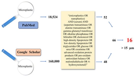
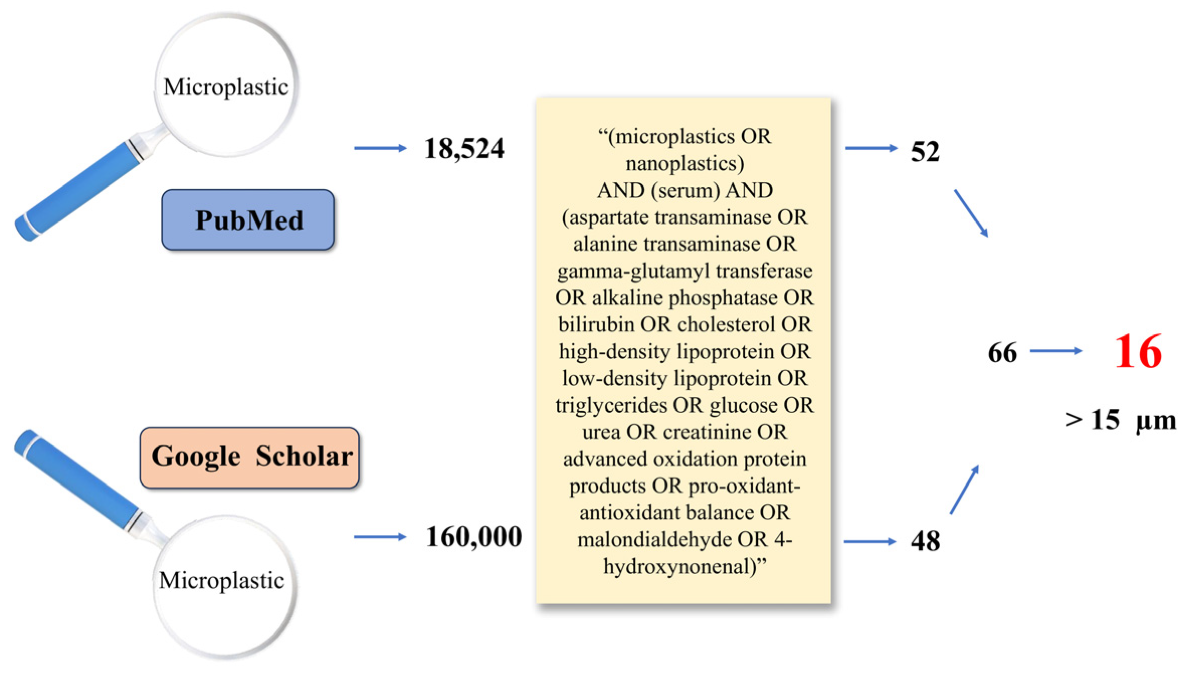
As an initial step of the research process, a comprehensive literature search was performed to identify relevant studies and establish the current state of knowledge. The search was conducted via PubMed and Google Scholar databases (both assessed on 28 July 2025). The initial query using the keyword “microplastic” retrieved 18,524 results on PubMed (https://pubmed.ncbi.nlm.nih.gov/?term=microplastic&size=200) and 160,000 on Google Scholar (https://scholar.google.com/scholar?hl=sr&as_sdt=0%2C5&q=microplastic&btnG=). To increase topic specificity and to reduce the number of irrelevant articles, the search was refined by using word combinations such as “(microplastics OR nanoplastics) AND (serum) AND (aspartate transaminase OR alanine transaminase OR gamma-glutamyl transferase OR alkaline phosphatase OR bilirubin OR cholesterol OR high-density lipoprotein OR low-density lipoprotein OR triglycerides OR glucose OR urea OR creatinine OR advanced oxidation protein products OR pro-oxidant-antioxidant balance OR malondialdehyde OR 4-hydroxynonenal)”, without additional filters applied. Overall, 52 articles from PubMed and 48 from Google Scholar were initially retrieved. Then, the titles and abstracts were screened, the duplicates removed and the irrelevant studies excluded, which yielded 66 articles in total in PDF that were selected for further analysis.
To extract information from original research articles, the GPT-5 model (version gpt-5-2025-07-15) was employed, at the time the latest available version of LLMs from OpenAI [https://openai.com/].
Downloaded articles in PDF were then parsed using the GROBID (version 0.6.2) service [https://github.com/kermitt2/grobid] with the Python library scipdf [https://github.com/titipata/scipdf_parser]. Parsed text was normalized using the textacy library [https://pypi.org/project/textacy/]. For textual data manipulation, the pandas library [https://pandas.pydata.org/] was used. Cleaned paragraphs were then joined to obtain the full publication text, which, in that form, was passed to the GPT prompt context as input. The prompt contained instructions specifying which information was relevant and how to format JSON [https://www.json.org/json-en.html] output. For interfacing with the model, the latest version of the OpenAI library [https://github.com/openai/openai-python] was used, all accessed on 1 March 2026. The best practices in prompt engineering were followed, starting with the simplest possible zero-shot versions and very soon ended with one-shot prompts, in which more data fields were gradually introduced intended for extraction. Prompts were optimized by manual examination of outputs of randomly selected articles comparing and verifying data extracted with one in PDF. The extracted data from articles was loaded into a pandas data frame, cleaned and aggregated to create the desired tabular representation.
As the study was designed with the intention of using MPs in the µm size range, the literature screening was limited to studies investigating particles larger than 15 µm, which yielded a total of 16 research articles (Figure 1).
Figure 1.
Schematic representation of the literature selection process.
2.3. Acute Oral Toxicity Study
2.3.1. Animal Handling
All research procedures that were performed on animals were approved by the Ethical Committee for the Use of Laboratory Animals of VINČA Institute of Nuclear Sciences–National Institute of the Republic of Serbia, University of Belgrade, Republic of Serbia (protocol authorization number 02/11) and Veterinary Directorate of the Ministry of Agriculture, Forestry and Water Management of the Republic of Serbia (protocol authorization number 323-07-03460/2021-05) and were in accordance with the European Communities Council Directive (2010/63/EU for animal experiments). The experiments were conducted on young adult male Wistar rats (n = 20), obtained from the local colony and separate litters. Briefly, animals were kept (2–3 per cage) under standard conditions, were randomly divided into four groups: the rats that received 2.5 mL of Milli-Q served as the control (Q); the rats that received MPs dispersed in 2.5 mL of Milli-Q in sizes of 41 µm (S), 70 µm (M), or 106 µm (L).
On the first day of the experiment, all animals were subjected to fasting for 4 h after which the treatments were administered once in a single dose using a reusable stainless steel feeding needle (16-G4”, 3 mm ball diameter, Cadence Inc., Staunton, VA, USA). Pre-weighed food and water (measured in grams and milliliters) were reintroduced 2 h following the treatments and animals were observed over the 24 h period.
2.3.2. Overall Health Indicators
Overall health indicators, such as food and water intake, neurological deficits and clinical signs of toxicity, are sensitive, non-invasive, routinely assessed parameters that provide information about an organism’s physiological status and can reveal early signs of toxic insult.
The amounts of water and food consumed were determined 24 h following the treatments by subtracting the remaining quantities from the initial amounts provided and normalized to body weight. The results are presented as a percentage of the mean ± SEM (Standard Error of the Mean) relative to the control (Q = 100%), while absolute values are provided in Table S2. For estimating the sensorimotor functions 24 h before and after the treatments, the Garcia neurological test, which included six parameters, was applied: spontaneous activity, forepaw outstretching, climbing, symmetry of four-limb movements, body proprioception, and response to vibrissae touch [16]. Each parameter was scored by a researcher blinded to the experimental setup, using a scale from 0 to 3 points, while the total score was calculated as the sum of the average scores for all assessed parameters (0 indicating the lowest, and 18 the highest score) [15]. The results are presented as a percentage of the mean ± SEM relative to the control (Q = 100%), while absolute values are provided in Table S3. In the current study, the clinical signs of toxicity were assessed, such as agitation, convulsions, ataxia, touch response, piloerection, sleepiness, lethargy, respiratory distress, and mortality and were closely observed throughout the study. These observations were made at multiple time points: prior to and following fasting, within the first 30 min post-treatment, three times during the 3–4 h after treatment, and two times on the following day. The final assessment was conducted 24 h post-treatment, just before the animals were sacrificed. The results are presented descriptively, using a scale to categorize the severity of the observed effects (the scale includes symbols such as − (for no effect), + (for mild effect), ++ (for moderate effect), and +++ (for major effect)).
Twenty-four hours following the treatments, the rats were sacrificed with a Harvard Apparatus guillotine (Holliston, MA, USA), the trunk blood was collected, centrifuged (3000 rpm for 15 min using Heraeus Labofuge GL (Heraeus, Hanau, Germany) and the resulting serum was stored at −80 °C for further analysis.
2.3.3. Estimation of Biochemical Parameters in Serum
All chemicals used for the assessment of oxidative-stress parameters were purchased from Sigma-Aldrich Co. (St. Louis, MO, USA) and were of analytical grade and of the highest purity.
Oxidative-stress parameters are important for identifying early biochemical disturbances, with prooxidant–antioxidant balance (PAB) reflecting the overall systemic redox challenge, while advanced oxidation protein products (AOPP), malondialdehyde (MDA) and 4-hydroxynonenal (HNE) indicate specific protein or lipid oxidative damage-hallmarks of an acute toxicological response. Levels of PAB, AOPP, MDA and HNE in serum were assessed according to the methodology described by Guševac Stojanović et al. (2024) [15]. All assays were performed in duplicate in 96-well microtiter plates using a Tecan Sunrise microplate reader (Tecan Group Ltd., Männedorf, Switzerland). The results are presented as a percentage of the mean ± SEM relative to the control (Q = 100%), while absolute values are provided in Table S4. The PAB was determined using a modified method of Alamdari et al. (2008) [17]. Briefly, 10 µL of standards (mixtures of hydrogen peroxide (H2O2) and uric acid in different ratios)/blank (dH2O)/samples were added to the wells of a microtiter plate, followed by 180 µL of freshly prepared working TMB solution. The plates were incubated in the dark for 12 min at 37 °C, and the reaction was stopped by adding 40 µL of 2 mol/L HCl. Absorbance was measured at 450 nm. A calibration curve was constructed from standard solutions, and PAB values were calculated accordingly. The results are expressed in arbitrary units as hydrogen peroxide complementary units (HKU) and normalized relative to the control.
For estimation of AOPP, the modified method of Witko-Sarsat et al. (1996) [18] was used. The absorbance of a mixture of standards/samples (10 µL) with an acetic acid/phosphate buffer solution pH 7.4 (90 µL) and 1.16 M potassium iodide (5 µL) was read at 340 nm using a microplate reader. AOPP concentrations were calculated from a calibration curve constructed using 10–100 µmol/L chloramine-T standards and expressed as chloramine-T equivalents. The results are presented as a percentage relative to the control.
The lipid peroxidation products (MDA and HNE) were assessed using manual assays based on the method of Gérard-Monnier et al. (1998) [19]. For MDA determination, standards (serial dilutions of 10 mM 1,1,3,3-tetramethoxypropane, TMOP), or blank (acetonitrile:methanol, 3:1, v/v), or samples (47 µL) were mixed with a freshly prepared working solution (3/4 volume of 20 mM 1- methyl-2-phenylindole and ¼ acetonitrile:methanol (3:1, v/v)) (152 µL). After gentle vortexing, 35 µL of hydrochloric acid was added, and the mixture was incubated for 60 min at 45 °C and centrifuged at 13,000× g for 15 min at 4 °C. The aliquots (100 µL) of the supernatant were transferred to a microtiter plate, and absorbance was measured at 580 nm.
For HNE determination, the assay was performed by mixing 47 µL of blank (acetonitrile:methanol, 3:1, v/v), or the samples with 152 µL of the freshly prepared working solution (1/4 volume of iron stock solution (34 µM FeCl3 in acetonitrile:methanol, 3:1, v/v) and ¾ volume of 20 mM 1-methyl-2-phenylindole), and 35 µL methanesulfonic acid. Subsequent steps were performed as described for the MDA assay. The concentrations of MDA and HNE were calculated from the calibration curve and expressed in µM. The results are presented as a percentage relative to the control.
The serum levels of the markers related to organ/tissue function such as aspartate transaminase (AST (U/L)), alanine transaminase (ALT (U/L)), bilirubin (mmol/L), gamma-glutamyl transferase (GGT (U/L)), alkaline phosphatase (ALP (U/L)), cholesterol (mmol/L), high-density lipoprotein (HDL (mmol/L)), low-density lipoprotein (LDL (mmol/L)), triglycerides (mmol/L), glucose (mmol/L), urea (mmol/L), and creatinine (µmol/L) were measured using a COBAS Integra 400 plus analyzer (Roche, Rotkreuz, Switzerland). All analyses were performed in an accredited clinical laboratory using original manufacturer’s reagents, following standardized enzymatic and colorimetric protocols. The measured serum parameters are indicators of liver function (AST, ALT, bilirubin, GGT, ALP), lipid metabolism (cholesterol, HDL, LDL, triglycerides), glucose homeostasis (glucose), and kidney function (urea, creatinine). The data are expressed as the mean of the values of the group ± SEM.
2.4. Data Analysis
The analysis of the experimental data was conducted using GraphPad Prism 8.02 software (GraphPad Software, Boston, MA, USA). The normality of the data was tested using the Shapiro–Wilk test. For the data that followed a Gaussian distribution, the one-way analysis of variance (ANOVA) was performed to examine the differences between the means of the groups, which was followed by Tukey’s post hoc test. For not normally distributed data, the Kruskal–Wallis test was employed, followed by Dunn’s post hoc test for multiple comparisons. The values of p < 0.05 were considered statistically significant.
3. Results and Discussion
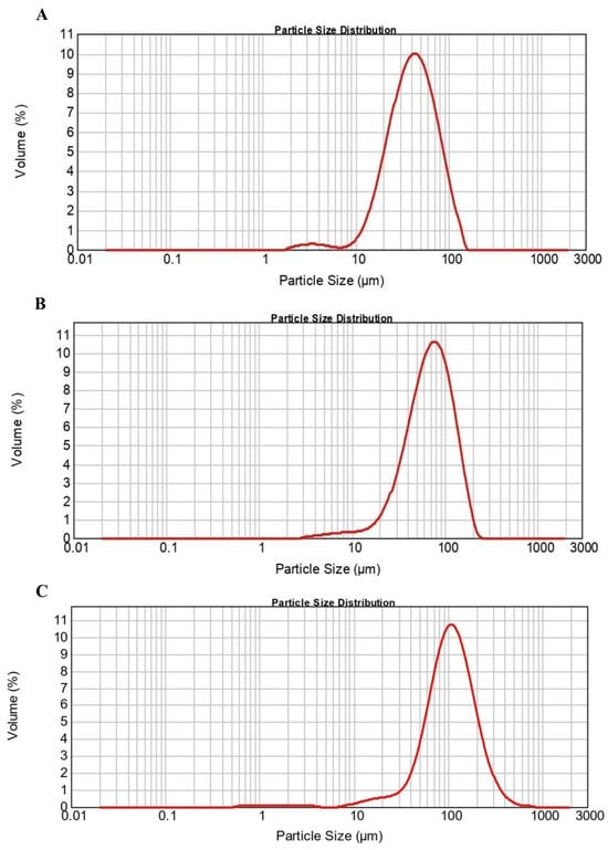
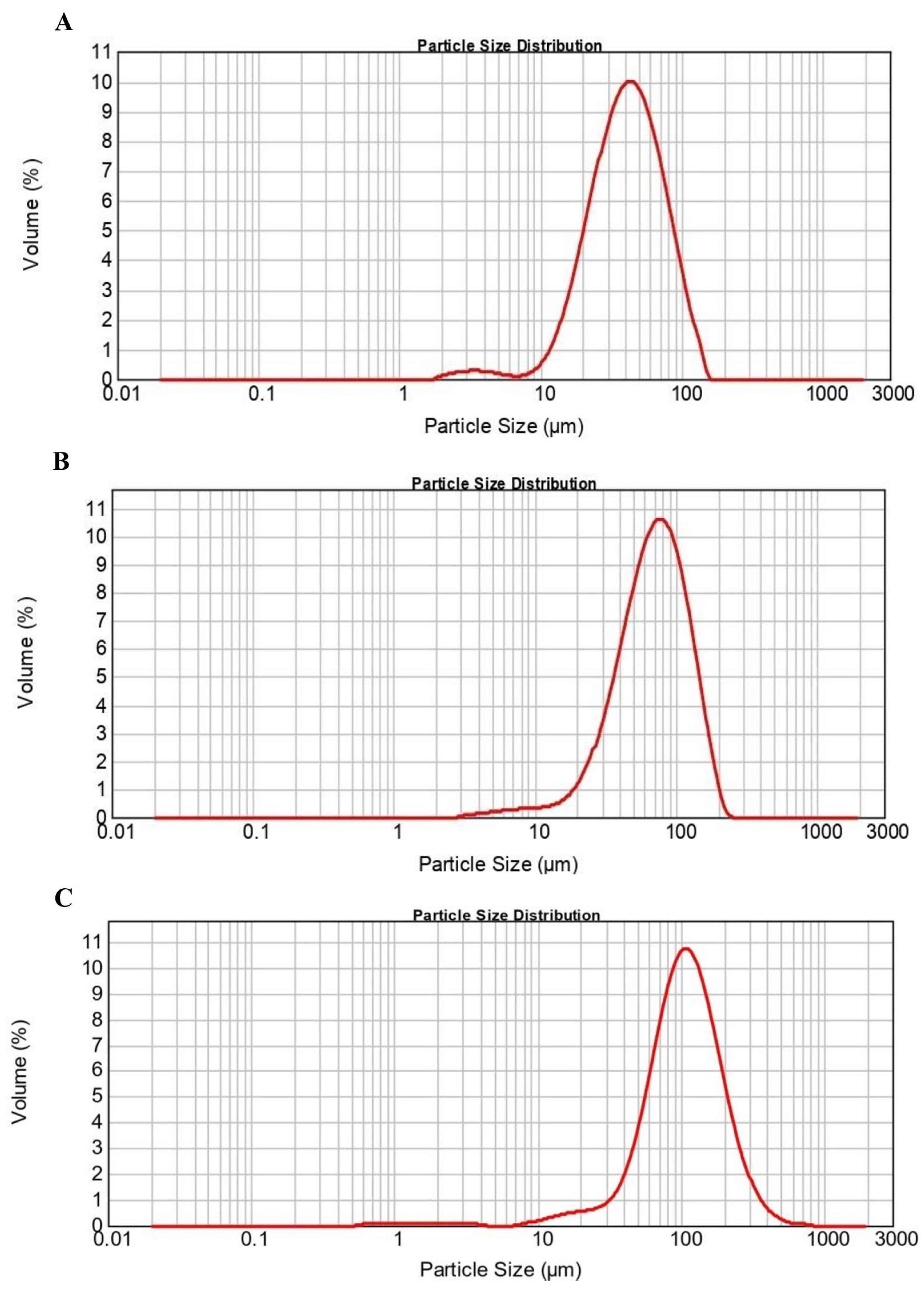
As MPs degrade, their size reduces, which in turn increases the surface area relative to their mass, thereby enhancing their reactivity and toxicity [7]. To examine the size distribution of MPs, LD was employed. The particles labeled S exhibited a broad range of sizes, with d (0.1) at 18.505 µm, d (0.5) at 41.322 µm, and d (0.9) at 85.364 µm. Median diameter of M particles was d (0.5) at 70.061 µm, with d (0.1) at 30.407 µm and d (0.9) at 134.700 µm. Finally, the third type of the particles, L, were larger than the previous two types, with d (0.1) at 46.859 µm, d (0.5) at 105.959 µm and d (0.9) at 217.150 µm (Figure 2). Although partial overlap between particle size distributions was observed, experimental groups were defined according to their median particle diameter (d (0.5)), which was used as the primary descriptor of the particle size.
Figure 2.
Size distributions of microplastic particles (MPs) generated by filing poly(ethylene terephthalate) (PET) bottles and determined by laser diffraction (LD). Median diameters were (A) 41.322 µm; (B) 70.061 µm; (C) 105.959 µm.
According to the data summarized in Table S1 (Supplementary Materials), a limited number of studies investigated changes in biochemical parameters following exposure to MPs larger than 15 µm. These studies had been conducted on mammals, reptiles such as the European pond turtle, and fish species including carp, goldfish, rainbow trout, seabream, and catfish. The experiments involved oral exposure, either through drinking water, gavage, or feed, with exposure durations ranging from 24 h to up to 90 days (Table S1). Analysis of the available data revealed a notable scarcity of studies conducted in rats; however, the findings that did exist largely mirror those reported in other species, particularly mice. These findings included elevated oxidative stress markers, disruptions in lipid metabolism, and significant alterations in hepatic enzyme activities, all indicative of oxidative imbalance and hepatocellular injury. Similar changes have also been documented in reptiles and fish, where MPs disrupted lipid profiles, impaired hepatic and renal function, and elevated oxidative stress markers, underscoring consistent physiological disturbances in different vertebrate groups.
The dataset in Table S1 was intentionally curated to include only particles larger than 15 µm. This specific focus provided two key analytical benefits: first, it allowed for a more consistent assessment of how particle size influences bioavailability, distribution, and toxicity; second, the compiled data offered clearer insight into general toxicity patterns and helped highlight critical variables that might influence the magnitude of effects.
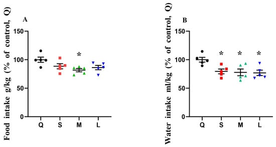
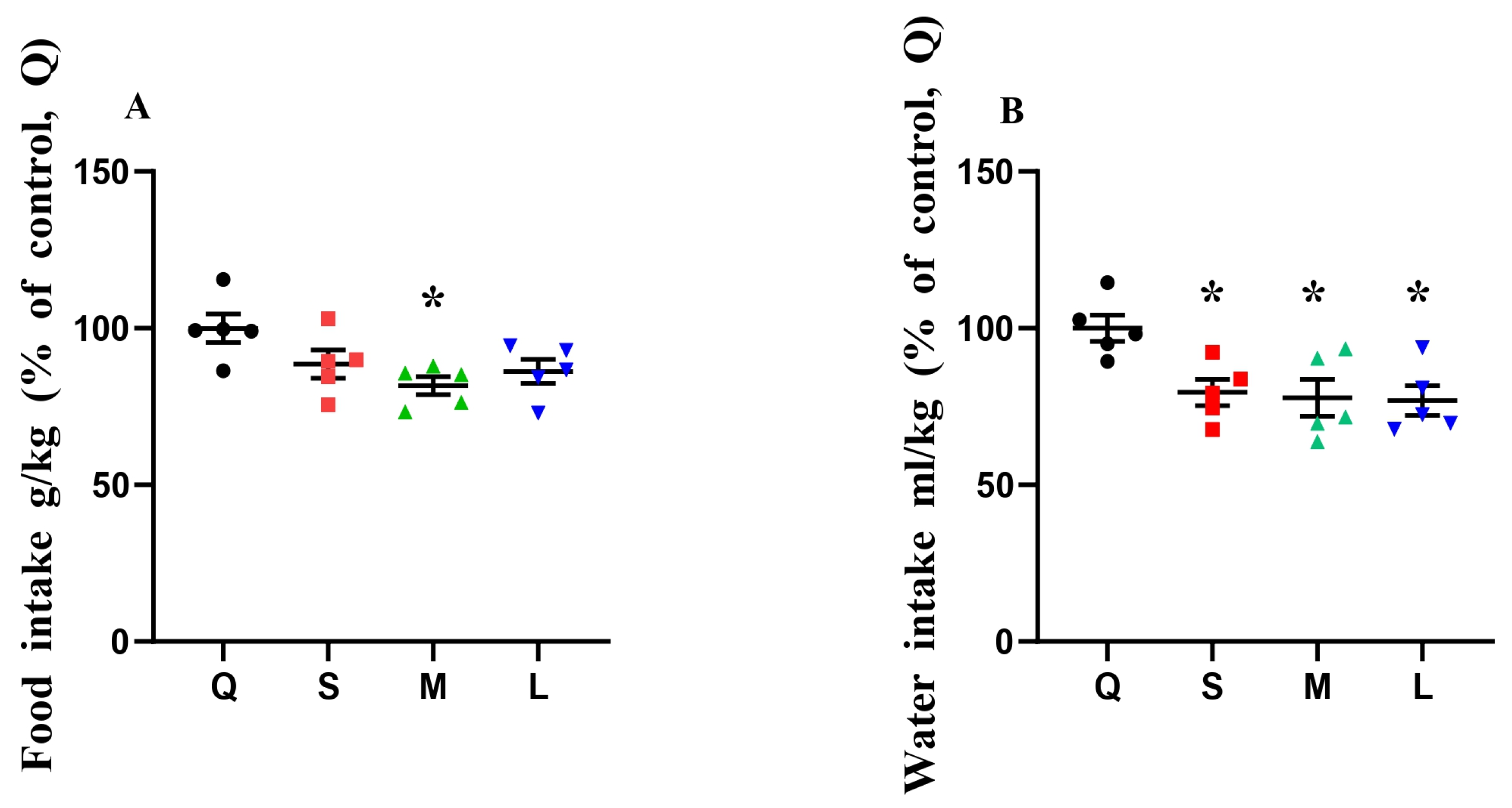
Changes in food and water intake provided valuable insights into the potential physiological effects of MPs contamination. As presented in Figure 3, exposure to MPs led to differential effects on food and water consumption, suggesting that intake of these particles might disrupt normal feeding behaviors and hydration processes.
Figure 3.
Food (A) and water (B) intake as indicators of the overall health status of adult male Wistar rats. Q—Milli-Q (2.5 mL); S—41 µm (35 mg/kg); M—70 µm (35 mg/kg); L—106 µm (35 mg/kg). The results are presented as a percentage of the mean ± SEM relative to the control (Q = 100%). Statistical significance * p < 0.05 for S, M and L vs. Q.
Compared to control, a reduction in food intake could be observed in all MPs-treated groups; however, this reached statistical significance only in the group exposed to the medium-sized particles (p < 0.05 for M vs. Q) (Figure 3A). Conversely, water intake was significantly reduced in all three MPs-treated groups (p < 0.05 for S, M and L vs. Q) (Figure 3B), which might represent a generalized response to MPs exposure, with M-sized particles exhibiting more pronounced or immediate effects on feeding behavior. Altogether, the observed reductions in food and water intake likely reflected an early, nonspecific systemic response to microplastic exposure. Although appetite and hydration are regulated through complex neuroendocrine and peripheral mechanisms, the present data do not allow conclusions regarding the direct involvement of neural pathways.
To assess whether these early changes were accompanied by functional impairment, the sensorimotor performance and clinical signs of toxicity following a single exposure to different-sized MPs were evaluated further.
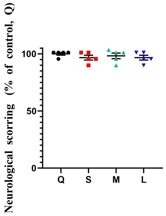
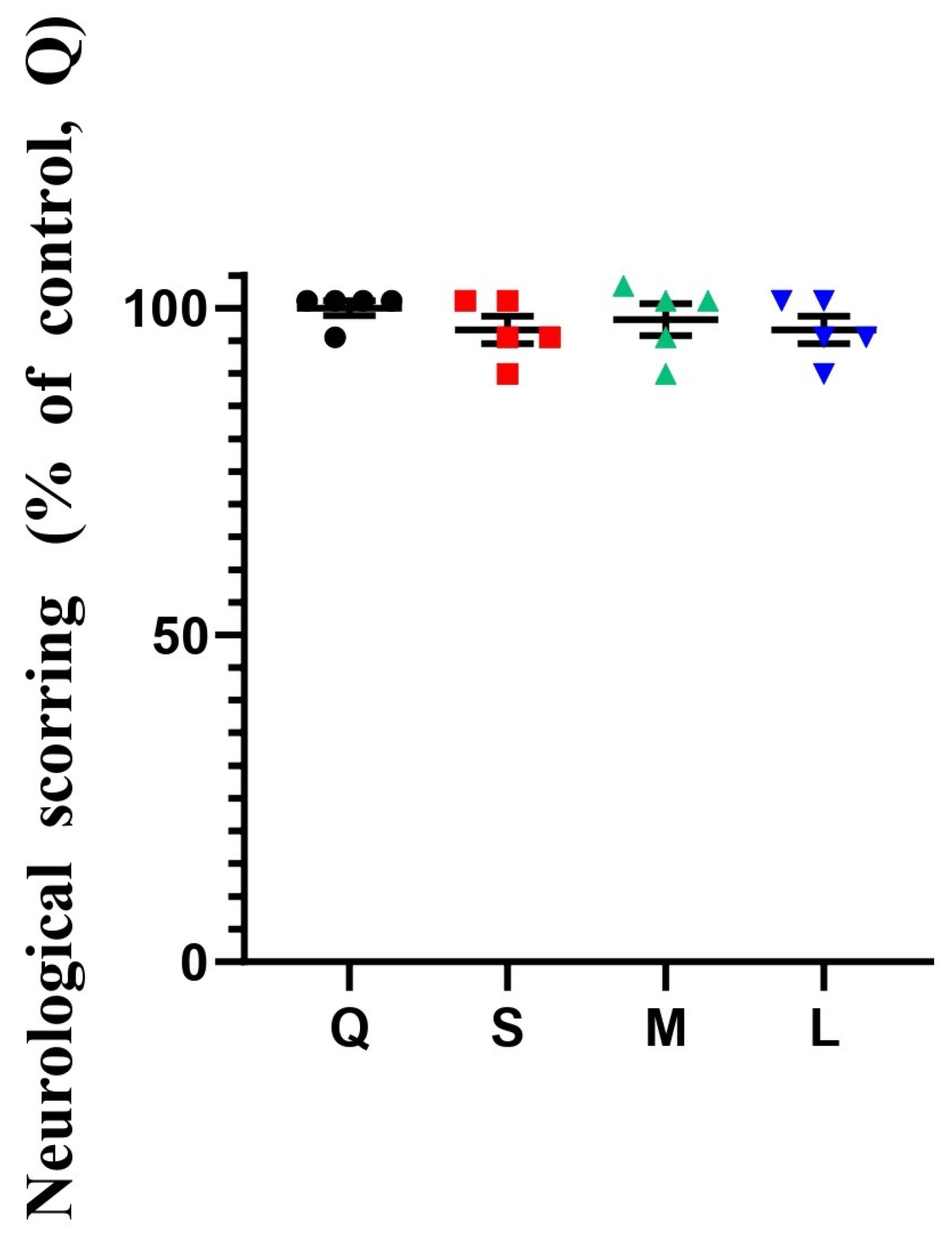
Prior to experimental exposure, all animals displayed normal sensorimotor function and exhibited no clinical signs of toxicity. In all animals treated with MPs compared to controls, neurological scoring did not reveal statistically significant changes in sensorimotor functions (Figure 4), and there were no observable clinical signs of toxicity (Table 1).
Figure 4.
Sensorimotor functions estimation as overall health indicators of adult male Wistar rats. Q—Milli-Q (2.5 mL); S—41 µm (35 mg/kg); M—70 µm (35 mg/kg); L—106 µm (35 mg/kg). The results are presented as a percentage of the mean ± SEM relative to the control (Q = 100%).
Table 1.
Clinical signs of toxicity as indicators of the overall health indicators of adult male Wistar rats Q—Milli-Q (2.5 mL); S—41 µm (35 mg/kg); M—70 µm (35 mg/kg); L—106 µm (35 mg/kg). Data are presented descriptively (- no effect).
Obtained findings might be explained by the exposure regimen used in the current study, as acute, single-dose exposure typically results in transient or subclinical changes that emerged rapidly and may not have sufficient time to develop into pronounced physiological or behavioral symptoms, whereas repeated or chronic exposure might allow cumulative effects to manifest more clearly [20].
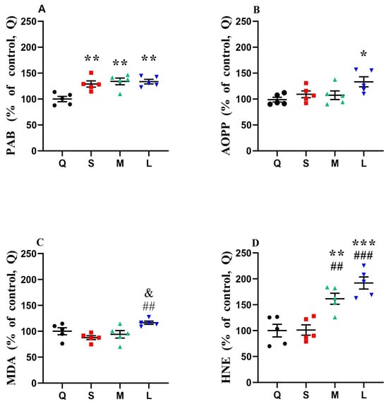
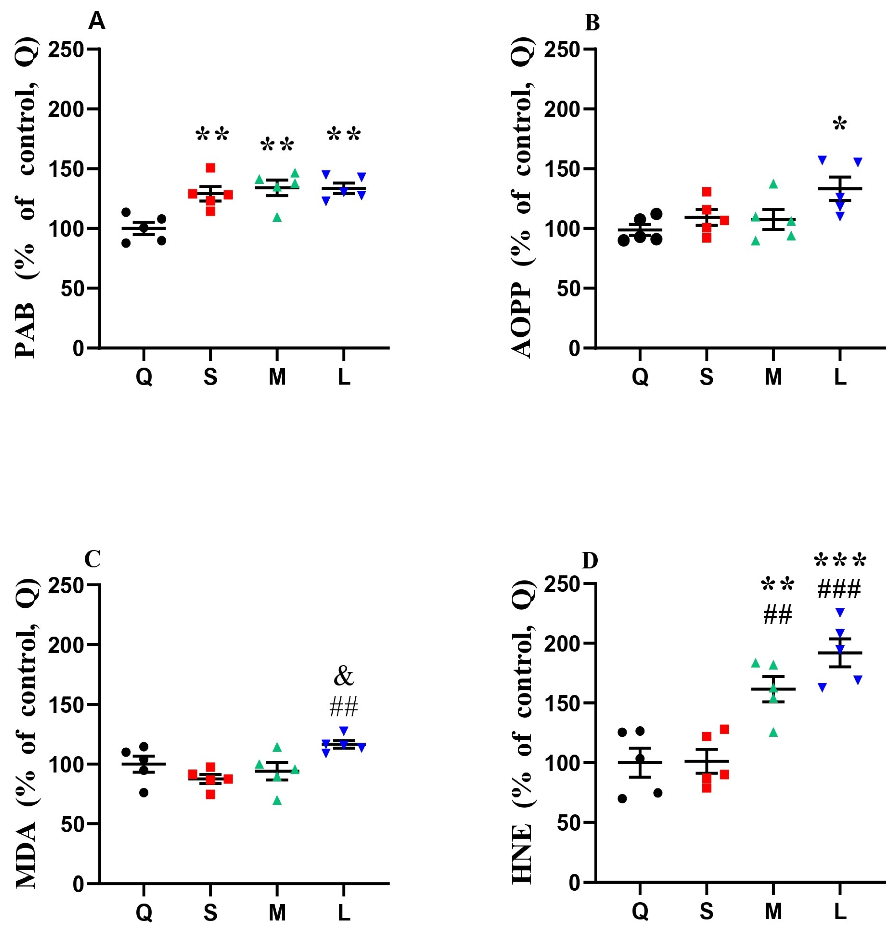
Despite the absence of statistically significant sensorimotor impairments or clinical toxicity signs following application of MPs, subclinical systemic disturbances remained possible. Therefore, to investigate more subtle, early biochemical changes, the serum markers of oxidative stress were evaluated as a sensitive and common initial response to toxic agents. The results showed that exposure to MPs induced changes in oxidative-stress parameters in serum (Figure 5). The PAB was significantly elevated in all MPs-treated groups compared to the control (p < 0.01 for S, M and L vs. Q), indicating a general shift towards a prooxidative state. Nonetheless, AOPP was significantly increased only in the group treated with the largest MPs compared to Q (p < 0.05 for L vs. Q), pointing to proteins’ oxidative damage. In parallel, MDA, a marker of lipid peroxidation, was elevated only in the L compared to S and M (p < 0.01 for L vs. S and p < 0.05 for L vs. M). The MDA augmentation had been previously observed in the brain and GIT of rats and mice exposed to various types of MPs [21,22,23], as well as in the serum and liver of fish [24,25,26]. In parallel, HNE was significantly altered in M compared to Q and S (p < 0.01 for M vs. Q and S) and in L compared to the same groups (p < 0.001 for L vs. Q and S), which was consistent with toxicological responses to MPs’ harmful insult [15]. This pattern suggests that enhanced lipid peroxidation might be a conserved response to MPs exposure, regardless of polymer type or species. Aside from our previous study [15], no other study reported measurements of PAB, AOPP or HNE as summarized in Table S1, in the context of elucidating the mechanistic response to MPs exposure, highlighting that data on these oxidative-stress parameters under given conditions remain limited to the current work only and are therefore directly incomparable.
Figure 5.
Serum levels of oxidative stress indicators in adult male Wistar rats. Q—Milli-Q (2.5 mL); S—41 µm (35 mg/kg); M—70 µm (35 mg/kg); L—106 µm (35 mg/kg) (A) Changes in the levels of prooxidant–antioxidant balance (PAB); (B) advanced oxidation protein products (AOPP); (C) malondialdehyde (MDA) and (D) 4-hydroxynonenal (HNE). The results are presented as a percentage of the mean ± SEM relative to the control (Q = 100%), whereas the values of the Q were set as 100%. Statistical significance * p < 0.05 for L vs. Q group; ** p < 0.01 for S, M, L vs. Q, *** p < 0.001 for L vs. Q; ## p < 0.01 for M and L vs. S; ### p < 0.001 for L vs. S and & p < 0.05 for L vs. M.
The observed oxidative stress profile—a systemic pro-oxidant shift accompanied by size-dependent macromolecular damage, aligns with recognized pathways of MP-induced gastrointestinal injury. The physical presence of MPs, especially larger, irregular particles, can abrade the mucosal lining, provoking localized inflammation and the generation of reactive oxygen species (ROS) that contribute to epithelial damage, a mechanism demonstrated with polystyrene MPs [27]. Alongside this physical insult, the potential leaching of chemical additives or residual monomers from plastics, including endocrine-disrupting compounds, can introduce exogenous agents that disrupt cellular redox balance and induce oxidative stress [28]. Moreover, MP exposure can also alter the gut microbial ecosystem. The resulting dysbiosis is a critical factor, as it can increase intestinal permeability and the production of pro-inflammatory microbial metabolites, thereby amplifying systemic oxidative and inflammatory responses [29]. In the current acute model, the significant rise in PAB across all MP sizes points to an immediate systemic redox challenge, potentially initiated by early dysbiosis. The more pronounced oxidative damage to proteins and lipids seen with the largest particles (~106 µm), however, suggested the greater relative contribution from direct physical abrasion and consequent localized inflammation. Thus, the acquired data on the differential oxidative outcomes were consistent with the activation of these interconnected gut-damage mechanisms, with particle size influencing their individual prominence.
Given that highly vascularized organs such as the liver, heart and kidneys [30,31,32] are often the first to respond to toxic agent exposure, monitoring the changes in serum biochemical parameters allow early insights into any underlying physiological disruptions. Among these parameters, AST and ALT are widely used as indicators of liver function. AST is distributed throughout the body, with significant amounts in the liver, heart, skeletal muscle, kidneys, brain, pancreas, lungs, leucocytes, and red cells, whereas ALT is more specifically concentrated in the liver, with lower amounts in muscles and kidneys [33,34,35]. Elevated levels of these enzymes in the bloodstream are characteristic indicators of liver injury and are frequently used to assess the extent of hepatic dysfunction in response to toxic insults [36]. Monitoring these markers thus allows for an integrated evaluation of early organ-specific and systemic effects following exposure.
As shown in Table 2, compared to the Q group, AST was significantly elevated in S (p < 0.01 for S vs. Q), while a more moderate elevation was observed in M and L (p < 0.05 for M and L vs. Q), suggesting that smaller particles may exert a stronger and/or faster effect on AST. Similar findings had been reported in previous studies, with the majority of them showing increased AST levels in all species [15,24,25,26,37,38,39]. Interestingly, despite the rise in AST in the current experimental setup, ALT levels remained similar in all investigated groups, indicating that the hepatic injury is likely mild or at an early stage, which is in accordance with the study of Yang et al., 2022 [40] who reported the absence of alterations in ALT levels in mice. In contrast, others [24,25,26,37,38,39,41] detected an increase, while our previous study noted a decrease in this serum marker [15], pointing out that these variations in ALT levels were not universal and might occur only under certain conditions.
Table 2.
Serum markers of organ/tissue function in adult male Wistar rats. Q—Milli-Q (2.5 mL); S—41 µm (35 mg/kg); M—70 µm (35 mg/kg); L—106 µm (35 mg/kg). Aspartate transaminase (AST), alanine transaminase (ALT), bilirubin, gamma-glutamyl transferase (GGT), alkaline phosphatase (ALP), cholesterol, high-density lipoprotein (HDL), low-density lipoprotein (LDL), triglycerides, glucose, urea, and creatinine. Data are presented as the mean ± SEM. Statistical significance * p < 0.05 for S, M and L groups vs. Q group and ** p < 0.01 for S group vs. Q group.
Besides AST and ALT, liver function assessment commonly includes ALP, GGT and bilirubin. ALP is mainly located in bile canaliculi but is also found in bone and intestine [34]. GGT, present on the membranes of various cells with high secretory or absorptive activity, is more specific for biliary disorders because it is absent in bone [34], while bilirubin, the main breakdown product of hemoproteins, is a useful indicator of liver function [42]. Although in some studies investigated enzymes were altered [24,26,37,38,39,43], the current research (Table 2), along with two others previously published [15,40], diverges from them, as ALP and/or GGT and bilirubin remained unchanged. A lack of change in ALP, GGT and bilirubin suggested that the primary mechanism of MPs-induced toxicity did not involve significant biliary system disruption or apparent hepatocellular necrosis, even though AST was elevated, indicating a more subtle or localized effect of MPs at the early stage, without causing widespread damage to bile ducts or hepatocytes.
Although an earlier report found that NPs were able to disturb glucose–lipid metabolism at the biochemical levels [44], in the current study, serum glucose levels remained stable, which was consistent with our previous study [15] and another by Faramazinia et al. which reported unchanged glucose levels even with longer exposure to MPs [45], suggesting that this parameter might be relatively resilient to MPs’ exposure. However, several other studies listed in Table S1 [24,25,26,39,41] demonstrated glucose dysregulation, which could reflect differences in particle size, polymer type, administered dose, or animal model used. Such heterogeneity implies that glucose response is more likely to depend on a combination of factors rather than exposure duration alone.
The literature emphasizes that exposure to polystyrene foam and polystyrene pellets can induce elevated levels of cholesterol, and LDL; however, no changes in cholesterol, LDL or HDL levels were observed (Table 2) [46]. On the other hand, serum levels of triglycerides demonstrated significant elevation in the S and M compared to control (p < 0.01 for S vs. Q and p < 0.05 for M vs. Q) (Table 2), which was in alignment with the majority of previous studies [15,22,24,25,26,39,45,47]. This finding might imply that MPs selectively interfere with triglyceride metabolism, as one of the earliest detectable metabolic alterations without altering cholesterol or glucose homeostasis, at least in this experimental setup. Moreover, the stronger response observed in the S group supports the hypothesis that smaller particles might have a greater metabolic impact compared to larger ones.
Renal biomarkers, including serum urea and creatinine, revealed additional systemic effects. As presented in Table 2, levels of urea were significantly elevated in all MPs groups compared to control (p < 0.05 for S, M and L vs. Q), suggesting that all three sizes similarly affect its clearance from the kidney or metabolic processes associated with renal function. The association between microplastic exposure and alterations in renal structure and function had been documented, with increasing attention directed toward the role of the gut–kidney axis as a mediating pathway. For example, polystyrene microplastics had been shown to impair gut barrier integrity and activate immune signaling that contributed to kidney injury via the gut–kidney axis in mice, suggesting that microplastic-induced gut dyshomeostasis might affect renal health [48].
These mechanisms typically involve intestinal dysbiosis, low-grade inflammation, and altered systemic signaling, which have been linked to changes in renal biomarkers and histopathology under repeated or chronic exposure conditions [49]. In the present acute model, elevations in serum urea and creatinine might have been partially influenced by reduced water intake and transient changes in hydration status. Nevertheless, an early contribution of microplastic-induced systemic responses could not be excluded. Therefore, these findings should be interpreted as indicators of early physiological perturbation rather than evidence of established renal dysfunction.
Comparable findings were observed in mammals [15,38,50] and turtles [36]. The changes in creatinine level, another key marker of kidney function, closely mirrored those of urea, with the S exhibiting more pronounced elevation compared to the control (p < 0.01 for S and p < 0.05 for M and L vs. Q), suggesting early glomerular stress. The parallel increase in these two markers of renal function underscored the potential nephrotoxic nature of MPs, with particle size again emerging as an important determinant of effect severity.
Taken together, these findings suggest that a single acute exposure to PET microplastics may be associated with early, size-dependent changes in selected systemic biochemical markers, in the absence of clinical toxicity or sensorimotor impairment. The observed patterns of oxidative stress, metabolic alterations, and renal biomarkers indicated that microplastics may act as physiologically active stressors rather than inert particulate matter.
4. Limitations of the Study
When interpreting the findings of the current research in the light of the literature summarized in Table S1, it is evident that the reported biological effects of MPs vary. Such inconsistency likely reflects differences in particle origin, characteristics, doses, exposure durations, and animal experimental models. Therefore, these discrepancies should be viewed as a consequence of this complexity, not as genuine contradictions.
Even though Table S1 was used solely as a reference to facilitate interpretation of the results, rather than as a comprehensive review of the field, it provides a useful overview but remains necessarily selective and cannot encompass the full variability of MPs toxicity. It is also important to note that the research used data from two major scientific databases; however, given the still limited number of available studies on the topic, these sources adequately capture the current state of knowledge.
Additionally, the experimental design was focused on evaluating early physiological and biochemical responses following a single acute oral dose of microplastics, with all endpoints assessed 24 h after administration. Regarding the dose rationale, the applied dose of 35 mg/kg used in this study might represent a high acute exposure rather than a typical daily intake. While this dose was derived from the upper-bound modeled estimates of human microplastic ingestion [51], it is acknowledged that such modeled values carry inherent uncertainties and might exceed average daily intake. Furthermore, the dose was derived using the Km scaling factor (6.2) to convert human-equivalent estimates into animal doses. It is recognized that this methodology, traditionally used for systemically absorbed substances, might have limitations when applied to non-absorbed particulate matter like MPs.
The study was conceived to identify initial response patterns rather than providing a comprehensive toxicological characterization across multiple doses or exposure durations. Within this framework, the sample size and statistical power are restricted, therefore the observed size-dependent trends should be interpreted cautiously and regarded as hypothesis-generating rather than definitive toxicological conclusions. Potential effects of chemical additives released from PET were not specifically examined in this study but could be addressed in future research to complement the understanding of microplastic-induced responses.
Further studies employing larger cohorts, chronic exposure at lower doses, are required to substantiate and expand upon the present findings.
Supplementary Materials
The following supporting information can be downloaded at: https://www.mdpi.com/article/10.3390/microplastics5010054/s1, Table S1. Results of GPT-5 assisted information extraction strategy. Table S2. Food and water intake values normalized to body weight in adult male Wistar rats. Table S3. Sensorimotor function estimation as an indicator of the overall health status of adult male Wistar rats expressed as absolute values. Table S4. Oxidative stress indicators in the serum of adult male Wistar rats expressed as absolute values. File S1. information_extraction_from_MPs_pdfs.ipynb.
Author Contributions
Z.S.: supervision, conceptualization, methodology, software, formal analysis, investigation, writing—original draft preparation, D.D.: formal analysis, investigation, writing—review and editing; J.M.: formal analysis, investigation, writing—review and editing, A.T.: formal analysis, investigation, writing—review and editing, N.F.: formal analysis. F.V.: formal analysis, I.G.S.: supervision, conceptualization, methodology, formal analysis, investigation, writing—original draft preparation. All authors have read and agreed to the published version of the manuscript.
Funding
This research was funded by the Ministry of Science, Technological Development and Innovation of the Republic of Serbia, grants 451-03-33/2026-03/200017 and 451-03-33/2026-03/200175.
Institutional Review Board Statement
Research procedures conducted on animals were approved by the Ethical Committee for the Use of Laboratory Animals of VINČA Institute of Nuclear Sciences-National Institute of the Republic of Serbia, University of Belgrade, Republic of Serbia (protocol authorization number 323-07-03460/2021-05) following the compliance with the European Communities Council Directive (2010/63/EU) for animal experiments, according to the guidelines of the EU registered Serbian Laboratory Animal Science Association, a member of the Federation of the European Laboratory Animal Science Associations.
Informed Consent Statement
Not applicable.
Data Availability Statement
Data is contained within the article.
Acknowledgments
We express our gratitude to Nevena Guševac and Dragan Guševac for their valuable technical support in obtaining microplastic particles. The authors would like to thank Željko Glumac for valuable comments and constructive feedback on the manuscript. The results presented in this manuscript are in line with Sustainable Development Goal 3 (Good Health and Well-being) and Goal 12 (Responsible Consumption and Production) of the United Nations 2030 Agenda. During the preparation of this manuscript, the authors used the GPT-5 model (version gpt-5-2025-07-15) for the purposes of information extraction from original research articles. The authors have reviewed and edited the output and take full responsibility for the content of this publication.
Conflicts of Interest
The authors declare no conflicts of interest. The funders had no role in the design of the study; in the collection, analysis, or interpretation of data; in the writing of the manuscript; or in the decision to publish the results.
References
- Ghasemlou, M.; Barrow, C.J.; Adhikari, B. The Future of Bioplastics in Food Packaging: An Industrial Perspective. Food Packag. Shelf Life 2024, 43, 101279. [Google Scholar] [CrossRef] [Scilit]
- Zhao, X.; Wang, Y.; Chen, X.; Yu, X.; Li, W.; Zhang, S.; Meng, X.; Zhao, Z.-M.; Dong, T.; Anderson, A.; et al. Sustainable Bioplastics Derived from Renewable Natural Resources for Food Packaging. Matter 2023, 6, 97–127. [Google Scholar] [CrossRef] [Scilit]
- Kannan, K.; Vimalkumar, K. A Review of Human Exposure to Microplastics and Insights Into Microplastics as Obesogens. Front. Endocrinol. 2021, 12, 724989. [Google Scholar] [CrossRef] [Scilit]
- Wythers, M.C. (Ed.) Advances in Materials Science Research; Nova Science Publishers: Hauppauge, NY, USA, 2021; Volume 48. [Google Scholar] [CrossRef] [Scilit]
- Djouina, M.; Waxin, C.; Dubuquoy, L.; Launay, D.; Vignal, C.; Body-Malapel, M. Oral Exposure to Polyethylene Microplastics Induces Inflammatory and Metabolic Changes and Promotes Fibrosis in Mouse Liver. Ecotoxicol. Environ. Saf. 2023, 264, 115417. [Google Scholar] [CrossRef] [Scilit]
- Bouwmeester, H.; Hollman, P.C.H.; Peters, R.J.B. Potential Health Impact of Environmentally Released Micro- and Nanoplastics in the Human Food Production Chain: Experiences from Nanotoxicology. Environ. Sci. Technol. 2015, 49, 8932–8947. [Google Scholar] [CrossRef] [Scilit]
- Dzierżyński, E.; Gawlik, P.J.; Puźniak, D.; Flieger, W.; Jóźwik, K.; Teresiński, G.; Forma, A.; Wdowiak, P.; Baj, J.; Flieger, J. Microplastics in the Human Body: Exposure, Detection, and Risk of Carcinogenesis: A State-of-the-Art Review. Cancers 2024, 16, 3703. [Google Scholar] [CrossRef] [Scilit] [PubMed]
- EFSA Panel on Contaminants in the Food Chain (CONTAM). Presence of Microplastics and Nanoplastics in Food, with Particular Focus on Seafood. EFSA J. 2016, 14, e04501. [CrossRef] [Scilit]
- Leslie, H.A.; Van Velzen, M.J.M.; Brandsma, S.H.; Vethaak, A.D.; Garcia-Vallejo, J.J.; Lamoree, M.H. Discovery and Quantification of Plastic Particle Pollution in Human Blood. Environ. Int. 2022, 163, 107199. [Google Scholar] [CrossRef] [Scilit] [PubMed]
- Saraluck, A.; Techarang, T.; Bunyapipat, P.; Boonchuwong, K.; Pullaput, Y.; Mordmuang, A. Detection of Microplastics in Human Breast Milk and Its Association with Changes in Human Milk Bacterial Microbiota. J. Clin. Med. 2024, 13, 4029. [Google Scholar] [CrossRef] [Scilit]
- Schwabl, P.; Köppel, S.; Königshofer, P.; Bucsics, T.; Trauner, M.; Reiberger, T.; Liebmann, B. Detection of Various Microplastics in Human Stool: A Prospective Case Series. Ann. Intern. Med. 2019, 171, 453–457. [Google Scholar] [CrossRef] [Scilit]
- Jin, M.; Wang, X.; Ren, T.; Wang, J.; Shan, J. Microplastics Contamination in Food and Beverages: Direct Exposure to Humans. J. Food Sci. 2021, 86, 2816–2837. [Google Scholar] [CrossRef] [Scilit]
- Mason, S.A.; Welch, V.G.; Neratko, J. Synthetic Polymer Contamination in Bottled Water. Front. Chem. 2018, 6, 407. [Google Scholar] [CrossRef] [Scilit]
- Novotna, K.; Cermakova, L.; Pivokonska, L.; Cajthaml, T.; Pivokonsky, M. Microplastics in Drinking Water Treatment—Current Knowledge and Research Needs. Sci. Total Environ. 2019, 667, 730–740. [Google Scholar] [CrossRef] [Scilit]
- Guševac Stojanović, I.; Drakulić, D.; Todorović, A.; Martinović, J.; Filipović, N.; Stojanović, Z. Acute Toxicity Assessment of Orally Administered Microplastic Particles in Adult Male Wistar Rats. Toxics 2024, 12, 167. [Google Scholar] [CrossRef] [Scilit]
- Garcia, J.H.; Wagner, S.; Liu, K.-F.; Hu, X. Neurological Deficit and Extent of Neuronal Necrosis Attributable to Middle Cerebral Artery Occlusion in Rats: Statistical Validation. Stroke 1995, 26, 627–635. [Google Scholar] [CrossRef] [Scilit]
- Alamdari, D.H.; Ghayour-Mobarhan, M.; Tavallaie, S.; Parizadeh, M.R.; Moohebati, M.; Ghafoori, F.; Kazemi-Bajestani, S.M.R.; Paletas, K.; Pegiou, T.; Koliakos, G. Prooxidant antioxidant balance as a new risk factor in patients with angiographically defined coronary artery disease. Clin. Biochem. 2008, 41, 375–380. [Google Scholar] [CrossRef] [Scilit]
- Witko-Sarsat, V.; Friedlander, M.; Capeillère-Blandin, C.; Nguyen-Khoa, T.; Nguyen, A.T.; Zingraff, J.; Jungers, P.; Descamps-Latscha, B. Advanced oxidation protein products as a novel marker of oxidative stress in uremia. Kidney Int. 1996, 49, 1304–1313. [Google Scholar] [CrossRef] [Scilit] [PubMed]
- Gérard-Monnier, D.; Erdelmeier, I.; Régnard, K.; Moze-Henry, N.; Yadan, J.C.; Chaudière, J. Reactions of 1-methyl-2-phenylindole with malondialdehyde and 4 hydroxyalkenals. Analytical applications to a colorimetric assay of lipid peroxidation. Chem. Res. Toxicol. 1988, 11, 1176–1183. [Google Scholar] [CrossRef] [Scilit]
- Nordberg, M.; Duffus, J.H.; Templeton, D.M. Explanatory Dictionary of Key Terms in Toxicology (IUPAC Recommendations 2007). Pure Appl. Chem. 2007, 79, 1583–1633. [Google Scholar] [CrossRef] [Scilit]
- Sincihu, Y.; Lusno, M.F.D.; Mulyasari, T.M.; Elias, S.M.; Sudiana, I.K.; Kusumastuti, K.; Sulistyorini, L.; Keman, S. Wistar Rats Hippocampal Neurons Response to Blood Low-Density Polyethylene Microplastics: A Pathway Analysis of SOD, CAT, MDA, 8-OHdG Expression in Hippocampal Neurons and Blood Serum Aβ42 Levels. Neuropsychiatr. Dis. Treat. 2023, 19, 73–83. [Google Scholar] [CrossRef] [Scilit] [PubMed]
- Xie, L.; Chen, T.; Liu, J.; Hou, Y.; Tan, Q.; Zhang, X.; Li, Z.; Farooq, T.H.; Yan, W.; Li, Y. Intestinal Flora Variation Reflects the Short-Term Damage of Microplastic to the Intestinal Tract in Mice. Ecotoxicol. Environ. Saf. 2022, 246, 114194. [Google Scholar] [CrossRef] [Scilit] [PubMed]
- Gan, H.-J.; Chen, S.; Yao, K.; Lin, X.-Y.; Juhasz, A.L.; Zhou, D.; Li, H.-B. Simulated Microplastic Release from Cutting Boards and Evaluation of Intestinal Inflammation and Gut Microbiota in Mice. Environ. Health Perspect. 2025, 133, 047004. [Google Scholar] [CrossRef] [Scilit] [PubMed]
- Banaei, M.; Forouzanfar, M.; Jafarinia, M. Toxic Effects of Polyethylene Microplastics on Transcriptional Changes, Biochemical Response, and Oxidative Stress in Common Carp (Cyprinus carpio). Comp. Biochem. Physiol. Part C Toxicol. Pharmacol. 2022, 261, 109423. [Google Scholar] [CrossRef] [Scilit]
- Iheanacho, S.C.; Odo, G.E. Dietary Exposure to Polyvinyl Chloride Microparticles Induced Oxidative Stress and Hepatic Damage in Clarias Gariepinus (Burchell, 1822). Environ. Sci. Pollut. Res. 2020, 27, 21159–21173. [Google Scholar] [CrossRef] [Scilit]
- Banihashemi, E.A.; Soltanian, S.; Gholamhosseini, A.; Banaee, M. Effect of Microplastics on Yersinia Ruckeri Infection in Rainbow Trout (Oncorhynchus Mykiss). Environ. Sci. Pollut. Res. 2022, 29, 11939–11950. [Google Scholar] [CrossRef] [Scilit]
- Liang, B.; Zhong, Y.; Huang, Y.; Lin, X.; Liu, J.; Lin, L.; Hu, M.; Jiang, J.; Dai, M.; Wang, B.; et al. Underestimated health risks: Polystyrene micro- and nanoplastics jointly induce intestinal barrier dysfunction by ROS-mediated epithelial cell apoptosis. Part. Fibre Toxicol. 2021, 18, 20. [Google Scholar] [CrossRef] [Scilit]
- Kovacs, K.; Bodis, J.; Vass, R.A. Microplastics, Endocrine Disruptors, and Oxidative Stress: Mechanisms and Health Implications. Int. J. Mol. Sci. 2025, 27, 399. [Google Scholar] [CrossRef] [Scilit] [PubMed]
- Ren, X.; Su, C.; Zhu, Y.; Fang, J.K.; Who, P.Y. Microplastic Toxicity on Gut Microbiota and Intestinal Cells: Evidence from the Simulator of the Human Intestinal Microbial Ecosystem (SHIME). Toxics 2025, 13, 1045. [Google Scholar] [CrossRef] [Scilit]
- Assarzadegan, N.; Anders, R.A.; Oshima, K. Hepatic Vascular Diseases. Diagn. Histopathol. 2017, 23, 553–558. [Google Scholar] [CrossRef] [Scilit]
- Mohamed, T.; Sequeira-Lopez, M.L.S. Development of the Renal Vasculature. Semin. Cell Dev. Biol. 2019, 91, 132–146. [Google Scholar] [CrossRef] [Scilit]
- King, O.; Cruz-Moreira, D.; Sayed, A.; Kermani, F.; Kit-Anan, W.; Sunyovszki, I.; Wang, B.X.; Downing, B.; Fourre, J.; Hachim, D.; et al. Functional Microvascularization of Human Myocardium in Vitro. Cell Rep. Methods 2022, 2, 100280. [Google Scholar] [CrossRef] [Scilit]
- Giannini, E.G. Liver Enzyme Alteration: A Guide for Clinicians. Can. Med. Assoc. J. 2005, 172, 367–379. [Google Scholar] [CrossRef] [Scilit]
- Lala, V.; Zubair, M.; Minter, D.A. Liver Function Tests. In StatPearls; StatPearls Publishing: Treasure Island, FL, USA, 2025. [Google Scholar]
- Lim, A.K. Abnormal Liver Function Tests Associated with Severe Rhabdomyolysis. World J. Gastroenterol. 2020, 26, 1020–1028. [Google Scholar] [CrossRef] [Scilit]
- Thakur, S.; Kumar, V.; Das, R.; Sharma, V.; Mehta, D.K. Biomarkers of Hepatic Toxicity: An Overview. Curr. Ther. Res. 2024, 100, 100737. [Google Scholar] [CrossRef] [Scilit]
- Razzaq, S.; Sidra, S.; Javid, A.; Mehmood, S. Impact of Dietary Exposure to Polyester Microfibers on Hematology, Serology and Histology in a Mouse Model. Environ. Res. 2024, 263, 120175. [Google Scholar] [CrossRef] [Scilit]
- Yahaya, T.O.; Ibrahim, A.B.; Kalgo, A.S.; Adewale, M.K.; Emmanuela, C.C.; Abdulkadir, B.; Fari, A.Z.; Attahiru, A.K.; Saadatu, A.; Wanda, J.D. Microplastics Exposure Altered Hematological and Lipid Profiles as Well as Liver and Kidney Function Parameters in Albino Rats (Rattus norvegicus). Environ. Anal. Health Toxicol. 2024, 39, e2024021. [Google Scholar] [CrossRef] [Scilit] [PubMed]
- Banaee, M.; Gholamhosseini, A.; Sureda, A.; Soltanian, S.; Fereidouni, M.S.; Ibrahim, A.T.A. Effects of Microplastic Exposure on the Blood Biochemical Parameters in the Pond Turtle (Emys orbicularis). Environ. Sci. Pollut. Res. 2021, 28, 9221–9234. [Google Scholar] [CrossRef] [Scilit]
- Yang, X.; Jiang, J.; Wang, Q.; Duan, J.; Chen, N.; Wu, D.; Xia, Y. Gender Difference in Hepatic AMPK Pathway Activated Lipid Metabolism Induced by Aged Polystyrene Microplastics Exposure. Ecotoxicol. Environ. Saf. 2022, 245, 114105. [Google Scholar] [CrossRef] [Scilit] [PubMed]
- Sun, X.; Zhuang, Y.; Wang, Y.; Zhang, Z.; An, L.; Xu, Q. Polyethylene Terephthalate Microplastics Affect Gut Microbiota Distribution and Intestinal Damage in Mice. Ecotoxicol. Environ. Saf. 2025, 294, 118119. [Google Scholar] [CrossRef] [Scilit]
- Ramírez-Mejía, M.M.; Castillo-Castañeda, S.M.; Pal, S.C.; Qi, X.; Méndez-Sánchez, N. The Multifaceted Role of Bilirubin in Liver Disease: A Literature Review. J. Clin. Transl. Hepatol. 2024, 12, 939–948. [Google Scholar] [CrossRef] [Scilit]
- Liu, X.; Liang, C.; Zhou, M.; Chang, Z.; Li, L. Exposure of Cyprinus Carpio Var. Larvae to PVC Microplastics Reveals Significant Immunological Alterations and Irreversible Histological Organ Damage. Ecotoxicol. Environ. Saf. 2023, 249, 114377. [Google Scholar] [CrossRef] [Scilit] [PubMed]
- Zhou, L.; Ran, L.; He, Y.; Huang, Y. Mechanisms of Microplastics on Gastrointestinal Injury and Liver Metabolism Disorder (Review). Mol. Med. Rep. 2025, 31, 98. [Google Scholar] [CrossRef] [Scilit]
- Faramazinia, M.; Sabzghabaei, G.R.; Multisanti, C.R.; Banaee, M.; Piccione, G.; Trivedi, A.; Faggio, C. Individual and Combined Effects of Microplastics and Diphenyl Phthalate as Plastic Additives on Male Goldfish: A Biochemical and Physiological Investigation. Comp. Biochem. Physiol. Part C Toxicol. Pharmacol. 2025, 290, 110144. [Google Scholar] [CrossRef] [Scilit]
- Nnoruka, U.C.; Okonkwo, C.J.; Ilechukwu, I.; Okonkwo, C.J.; Belonwu, D.C. Impact of Polystyrene Microplastic Exposure on Lipid Profile and Oxidative Stress Status of Male and Female Wistar Rats. Environ. Anal. Health Toxicol. 2022, 37, e2022024. [Google Scholar] [CrossRef] [Scilit] [PubMed]
- Del Piano, F.; Mateu, B.; Coretti, L.; Borrelli, L.; Piccolo, G.; Addeo, N.F.; Esposito, S.; Mercogliano, R.; Turco, L.; Meli, R.; et al. Polystyrene Microplastic Exposure Modulates Gut Microbiota and Gut-Liver Axis in Gilthead Seabream (Sparus aurata). Sci. Total Environ. 2024, 957, 177857. [Google Scholar] [CrossRef] [Scilit] [PubMed]
- Liang, Y.; Liu, D.; Zhan, J.; Liu, X.; Li, P.; Ma, X.; Hou, H.; Wang, P. Polystyrene microplastics induce kidney injury via gut barrier dysfunction and C5a/C5aR pathway activation. Environ. Pollut. 2024, 342, 122909. [Google Scholar] [CrossRef] [Scilit]
- Bora, S.S.; Gogoi, R.; Sharma, M.R.; Anshu; Borah, M.P.; Deka, P.; Bora, J.; Naorem, R.S.; Das, J.; Teli, A.B. Microplastics and human health: Unveiling the gut microbiome disruption and chronic disease risks. Front. Cell. Infect. Microbiol. 2024, 14, 1492759. [Google Scholar] [CrossRef] [Scilit]
- Zaheer, J.; Shanmugiah, J.; Kim, S.; Kim, H.; Ko, I.O.; Byun, B.H.; Cheong, M.A.; Lee, S.-S.; Kim, J.S. 99mTc-DMSA and 99mTc-DTPA Identified Renal Dysfunction Due to Microplastic Polyethylene in Murine Model. Chemosphere 2024, 364, 143108. [Google Scholar] [CrossRef] [Scilit]
- Senathirajah, K.; Attwood, S.; Bhagwat, G.; Carbery, M.; Wilson, S.; Palanisami, T. Estimation of the Mass of Microplastics Ingested—A Pivotal First Step towards Human Health Risk Assessment. J. Hazard. Mater. 2021, 404, 124004. [Google Scholar] [CrossRef] [Scilit]

Disclaimer/Publisher’s Note: The statements, opinions and data contained in all publications are solely those of the individual author(s) and contributor(s) and not of MDPI and/or the editor(s). MDPI and/or the editor(s) disclaim responsibility for any injury to people or property resulting from any ideas, methods, instructions or products referred to in the content. |
© 2026 by the authors. Licensee MDPI, Basel, Switzerland. This article is an open access article distributed under the terms and conditions of the Creative Commons Attribution (CC BY) license.