


Abstract
Backgrounds/Objectives: Probiotics confer health-promoting and immunomodulatory effects on the host. However, a lot remains to be understood regarding their safety profile, characteristics and functional mechanisms. This study was conducted to comprehend the functional mechanisms of Limosilactobacillus reuteri isolated from chickens and characterize some of its uncharacterized proteins (hypothetical proteins). Methods: The chicken-isolated Limosilactobacillus reuteri genome, sequenced using Illumina MiSeq sequencing technology, was explored for proteome comparison analyses, comprehensive antibiotic resistance analysis, interleukin-4 (IL-4) expression across various conditions as well as IL-4-inducing potential. Results: Five antibiotic resistance-associated genes were found. Hypothetical proteins labeled QHPv.2.1, QHPv.2.12 and QHPv.2.16 induced IL-4, but QHPv.2.12 was not immunogenic. Immunogenic IL-4-inducing mutants from QHPv.2.1 (0.7013) and QHPv.2.16 (0.7040) had the best support vector machine (SVM) values, which were significantly (p < 0.05) higher than the rest of the group, followed by those from QHPv.2.12 (0.4587). QHPv.2.5 (0.0560) had the lowest value. QHPv.2.1 (5484.71) and QHPv.2.16 (5491.05) had significantly (p < 0.05) larger molecular weights (MWs), followed by QHPv.2.12 (5302.05). The lowest value was obtained for QHPv.2.7 (3386.74). Conclusions: This explorative study revealed the IL-4 inducing capacity of the hypothetical proteins of L. reuteri and their mutants, as well as identified heat and cold shock proteins associated with L. reuteri.
1. Introduction
The benefits of probiotics, that is, live microorganisms which, when consumed adequately, provide health-promoting effects to both animals and humans, have inspired continuous research interest. The recent interest is focused on unraveling their functional mechanisms, as well as exploring their uncharacterized and hypothetical proteins [1].
Probiotics’ functional mechanisms and their hypothetical proteins remain incompletely understood despite their documented benefits and the increasing demand for their use in animal and human nutrition. This knowledge gap has necessitated continuous research interest in completely understanding their functional mechanisms, which become clearer by the day as more research outputs document their potential functional pathways and additional benefits, including their cytokine-inducing ability, such as IL-6 induction [2] and interferon-gamma in humans and mice [3].
The important roles that IL-4 plays in immunity cannot be overstated, including the key roles it plays in type 2 immunity, leading to toxin inactivation and resistance to helminth parasites. In a like manner, the importance of HSPs and CSPs to probiotics cannot be overstated. HSPs protect probiotics from harsh environments, such as the bile, heat and acid of the gastrointestinal tract. They protect the intestinal barrier by minimizing inflammation and enhancing as well as promoting HSPs in epithelial cells. CSPs are primarily essential in food processing involving refrigeration, enabling probiotics to withstand stress arising from food temperature, as well as enabling them to maintain functional metabolic activities.
The gut commensal microbiota, such as Limosilactobacillus reuteri, formerly known as Lactobacillus reuteri, offer structural and mechanical integrity and barrier functions to the intestinal mucosal surfaces, thus protecting the intestine [4] and maintaining a healthy immune system [5]. Of all the known commensal microbiota species used as probiotics for dietary supplements, therapeutics and cosmetic products, lactic acid bacteria (LAB) are the prominent probiotic bacteria used. Studies are ongoing not just to understand the functional mechanisms of probiotics but to also uncover new sources for developing new candidates. The starting point is usually the isolation of the potential probiotics from the feces, milk or gut of the respective animals [6]. In addition, potential candidates must not be pathogenic; they must be able to survive in the intestinal tract, possess the ability to adhere to the intestinal walls, have high vitality and be able to synthesize antimicrobial substances such as bacteriocins, as well as not be associated with antibiotic resistance. One major characteristic of LAB is their ability to produce bacteriocins.
A recent study focused on isolates (Lactobacillus plantarum, Bacillus subtilis, Bacillus tequilensis and Lactobacillus fermentum) from chickens tested for antagonistic ability, carbohydrate fermentation analysis, bile salt and low pH tolerance, hemolysis activity and intestinal adhesion, revealing that all the isolates showed great survival percentage at low pH, none exhibited a- and b-hemolytic activity and most of them showed optimal growth of up to 4% sodium chloride concentration [6]. The findings from such a study further confirmed the potential of LAB as probiotic bacteria. Another recent study reported the probiotic potential of chicken-isolated L. saerimneri, L. acidophilus, L. ingluviei, L. salivarius and L. reuteri [7].
Proteins that have not been experimentally assigned functions or for which translation has not been established are referred to as hypothetical proteins. It has been observed that about 2% of the genes of a genome code for protein while the remaining ones are non-coding or their functions are yet to be established [8]. Hypothetical proteins may be involved in disease development as well as help in understanding physiological and biochemical pathways such as in finding new functions, structures, pharmacological targets and markers [9,10]. Research interests are recently also focused on antibiotic-associated proteins or genes, beyond the focus on CSPs, HSPs and hypothetical proteins.
Antibiotic resistance is a global threat to human health and to the treatment of disease. In addition, antibiotic-resistant genes (ARGs) may be present in any environment including natural and engineered habitats [11], threatening public health. Probiotics are regarded as safe alternatives to antibiotics but a lot remains unexplored about their association with ARGs. Probiotics are health-promoting but their immunological mechanisms have not been fully understood, such as their relationship with IL-4. Understanding the relationships between probiotics and IL-4 is key to understanding their functional mechanisms, especially regarding hypothetical proteins. Considering the importance of LAB as probiotics and the key roles IL-4 plays in immunity, this study was conducted to understand the mechanisms underlying the functional abilities of Limosilactobacillus reuteri using the chicken-isolated Limosilactobacillus reuteri genome and to characterize some of its hypothetical proteins vis-à-vis CSPs, HSPs and IL-4-inducing potential exploration.
2. Materials and Methods
2.1. Isolation and Genome Sequencing
Fecal samples from Nigeria-reared broiler chickens served as the source of bacteria isolation; Illumina MiSeq sequencing technology was adopted for the genome sequencing. The sequencing depth was 78.0× and the assembly method was SPAdes v3.14.0. For this study, the assembled complete whole genome nucleotide sequence was retrieved from NCBI (CP059275 and version.1); annotation, proteome comparison analysis and sorting of the protein families were analyzed using PATRIC. HSPs, CSPs and hypothetical proteins were selected for antibiotic resistance analysis and IL-4-inducing potential gene expression. Table 1 shows the selected studied proteins.
Table 1.
Investigated selected proteins from L. reuteri genome.
2.2. BLAST (2.17.0) and Multiple Sequence Alignment
The HSPs and CSPs found in the L. reuteri genome were analyzed for an amino acid sequence similarity check using NCBI protein–protein blast and Blast Tree View was used to generate the trees for selected organisms: Homo sapiens (taxid:9606), Mus (taxid:10088), chicken (taxid:9031), Wistar rats (taxid:10116) and Rattus norvegicus (taxid:10116). Multiple sequence alignment of the heat and cold shock proteins was carried out with Clustal Omega (1.2.4), https://www.ebi.ac.uk/jdispatcher/msa/clustalo (accessed 17 July 2024), based on seeded guide trees and a hidden Markov model profile–profile approach for generating the presented alignments.
2.3. Antibiotic Resistance Analysis
A comprehensive antibiotic resistance analysis of the chicken-isolated L. reuteri genome was conducted using the Comprehensive Antibiotic Resistance Database (https://card.mcmaster.ca/analyze/rgi, accessed 17 July 2024). The RGI criteria used were perfect, strict and complete genes only. The detection criterion used was protein homolog model.
2.4. IL-4-Inducing Proteins and Mutants
The IL-4-inducing capacity of the selected hypothetical proteins Limosilactobacillus reuteri was investigated using IL4pred (https://webs.iiitd.edu.in/raghava/il4pred/design.php, accessed 17 July 2024). Thereafter, we designed 400 mutants from the aforementioned proteins. The mutants were designed with a single amino acid amendment for each mutant. The IL-4-inducing potential of the mutants were assessed as previously described. Support vector machine (SVM) scores were used for grouping the mutants, with the top fifty being selected and evaluated for immunogenicity using default thresholds. After this, the top fifteen (out of fifty) were assessed and immunogenic mutants with IL-4-inducing potential were statistically analyzed.
2.5. Expression of Genes
GENEVESTIGATOR was used for IL-4 gene expression and co-expression analyses (Homo sapiens and Mus musculus) considering different conditions, according to HS_AFFY_U133PLUS_2-0 (human) and MM_AFFY_430_2-1 (mouse) platforms. The expression of HSPs and CSPs across various experimental sets in Rattus norvegicus were also determined in GENEVESTIGATOR using the platform RN_AFFY_230_2-1. While RNA-seq may have offered a different result, Affymetrix microarray datasets were used for sample availability, cross-study comparability and a focus on differential expression.
2.6. Analysis of Statistics
Organization of the top fifteen mutants with IL-4-inducing potential was done according to completely randomized design (CRD). The data were analyzed according to one-way analysis of variance, using SAS (SAS 9.4) GLM. Duncan’s multiple range test was used for separating significant means. Statistical significance was set at p < 0.05.
3. Results
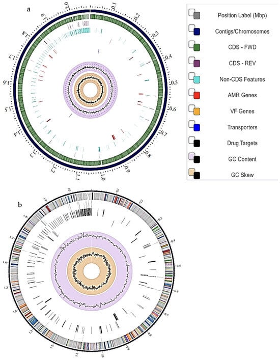
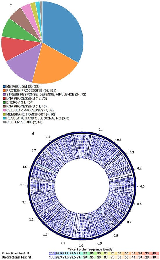
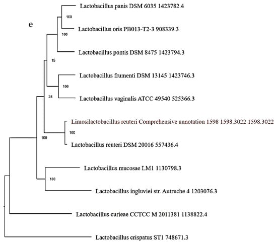
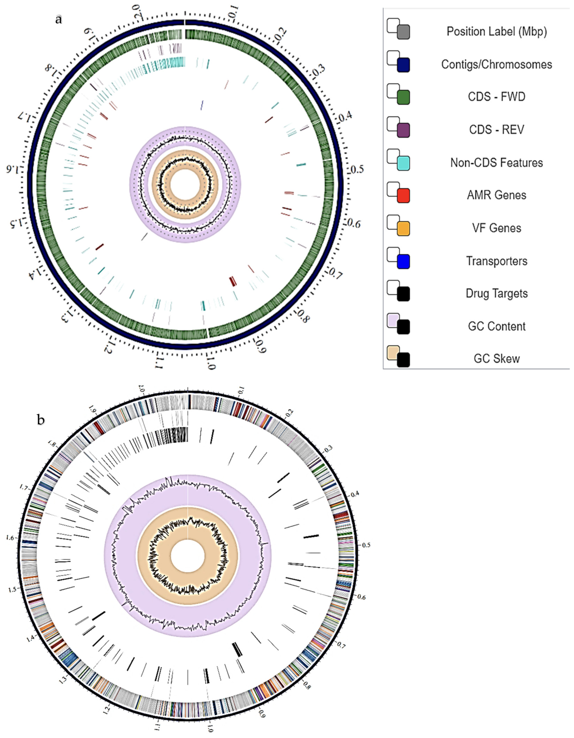
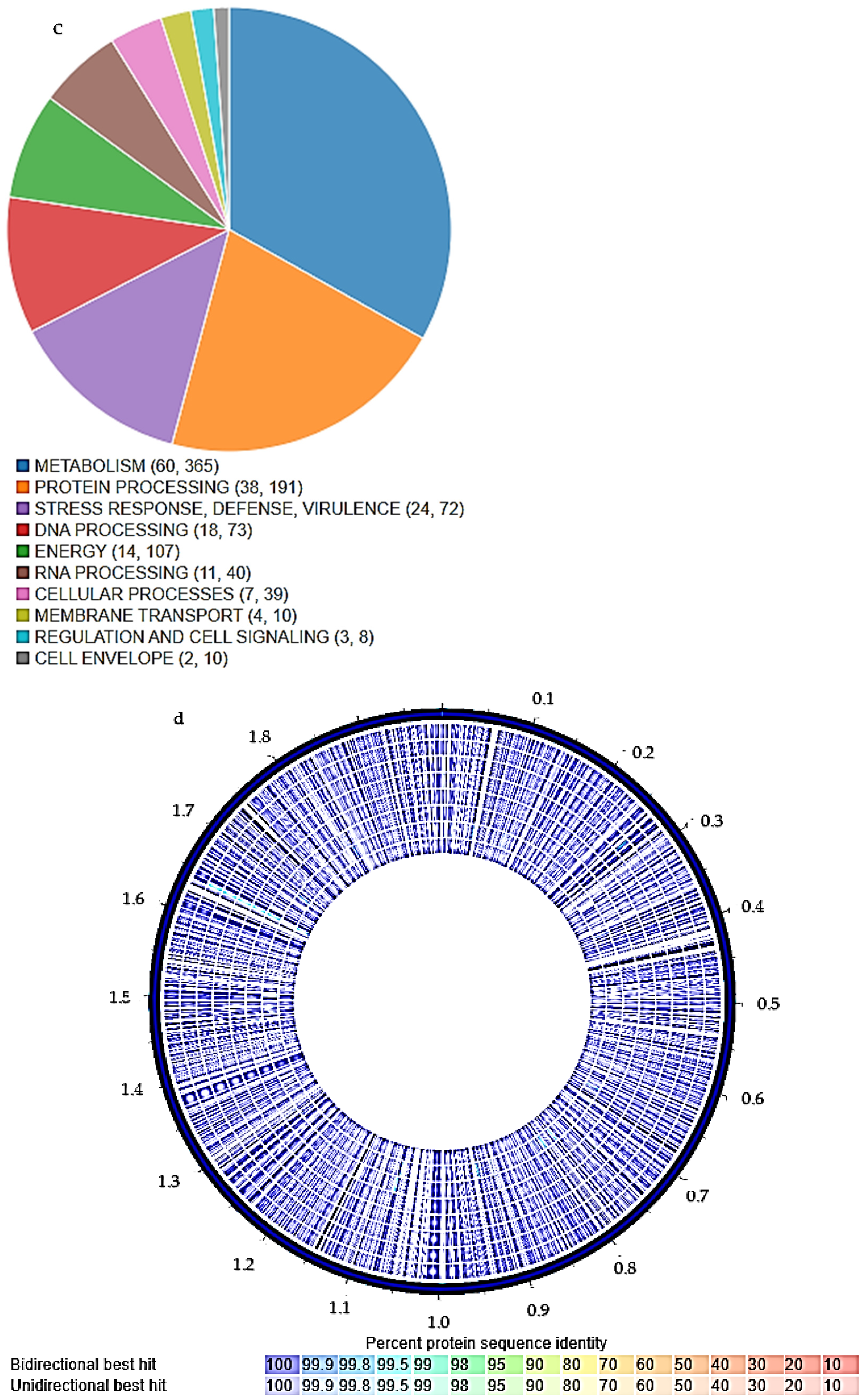
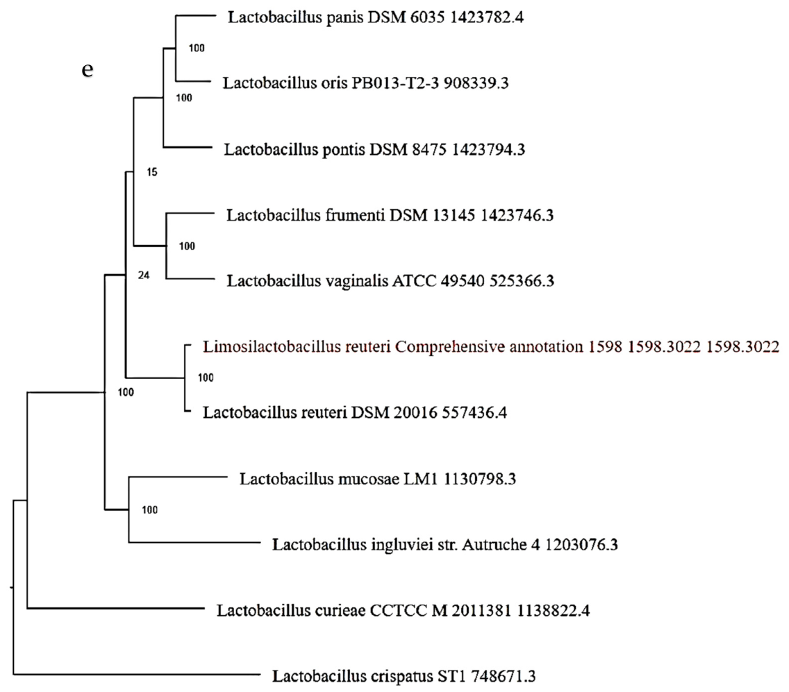
The chicken-isolated L. reuteri genome had one contig, with a length of 2,085,667 bp and an average G + C content of 39.43%. It had 2471 protein-coding sequences (CDSs), 78 transfer RNA (tRNA) genes, and 10 ribosomal RNA (rRNA) genes. A circular view of the genome, a circular graphical display of the distribution of the genome annotations, the subsystems unique to the genome, the proteome comparison analysis of similar genomes and the phylogenetic tree are shown in Figure 1a–e.
Figure 1.
Annotation results of the chicken-isolated L. reuteri genome; (a) circular view of the genome, (b) circular graphical display of the distribution of the genome annotations of the chicken-isolated L. reuteri genome, (c) a chart showing the subsystems unique to the chicken-isolated L. reuteri genome, (d) circular view of the proteome comparison of similar genomes to chicken-isolated L. reuteri genome—1. Lactobacillus acidophilus 1579cga (1579.814), 2. Lactobacillus acidophilus La-14 1314884 (1,314,884.31), 3. Lactobacillus acidophilus strain ATCC 53544 (1579.81), 4. Lactobacillus acidophilus strain FSI4 (1579.28), 5. Lactobacillus acidophilus strain LA-G80-111 (1579.364), 6. Lactobacillus acidophilus NCFM (272,621.13), 7. Lactobacillus acidophilus strain DSM 20079 (1579.223), 8. Lactobacillus acidophilus La-14 (1,314,884.3)—and (e) phylogram of the chicken-isolated L. reuteri genome.
In total, 1485 proteins with functional assignments and 986 hypothetical proteins were reported for the genome. The proteins with functional assignments included 586 proteins with Enzyme Commission (EC) numbers, 506 with Gene Ontology (GO) assignments and 422 proteins mapped to KEGG pathways. The circular graphical display of the distribution of the genome annotations included from outer to inner rings, the contigs, CDS on the forward strand, CDS on the reverse strand, RNA genes, CDS with homology to known antimicrobial resistance genes, CDS with homology to known virulence factors, GC content and GC skew. The subsystems the genes belong to are represented with the colors of the CDS on the reverse and forward strands as indicated in the subsystem chart.
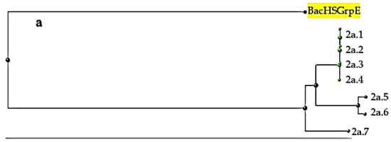
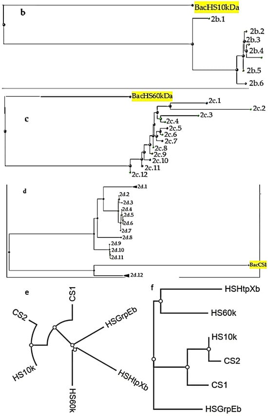
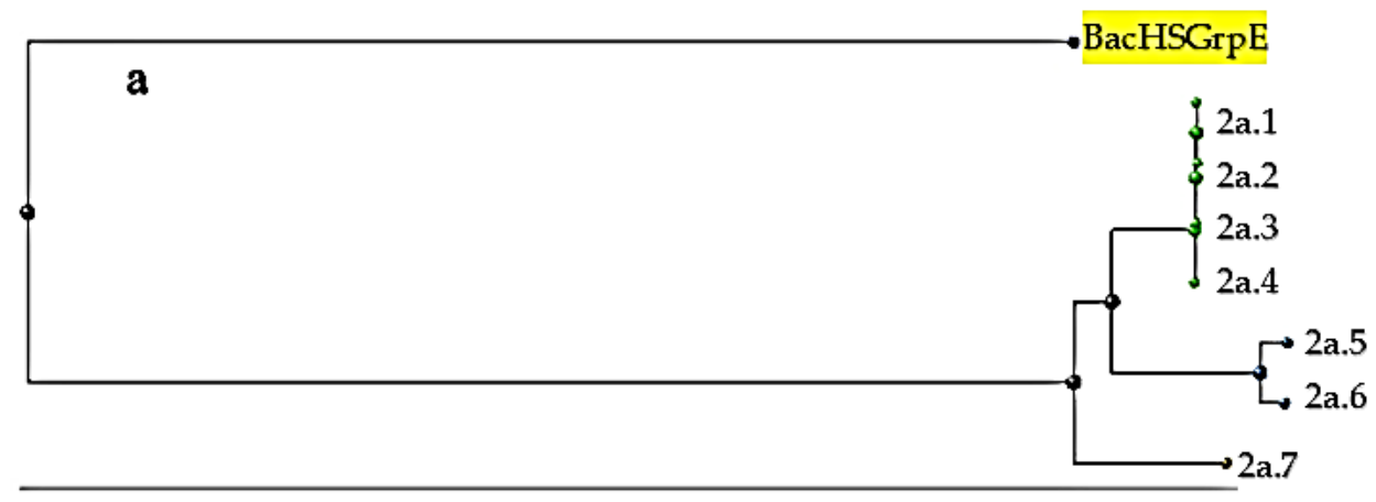
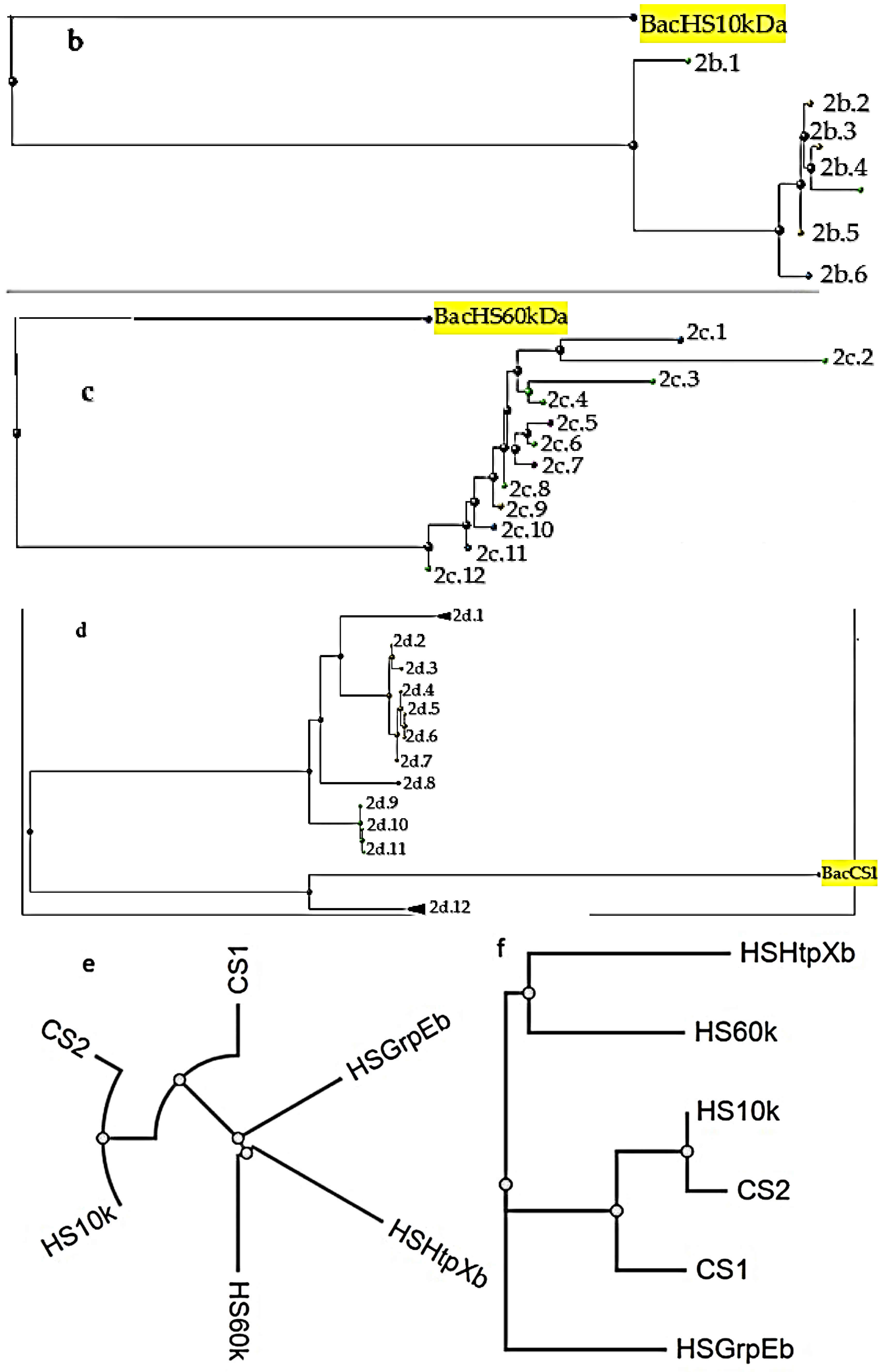
The HSP and CSP amino acid sequence similarity check of the genome was carried out in NCBI. The Blast Tree View was used to generate the trees for selected organisms, and they are presented in Figure 2a–d. There were no significant alignments for HtpX (fig|1598.3022.peg.1900) and CSP of the CSP family (fig|1598.3022.peg.703). The results of the multiple sequence alignment (MSA) carried out among HSPs and CSPs are presented in Figure 2e,f.
Figure 2.
Blast Tree View result for protein–protein blast for chicken-isolated L. reuteri genome: (a) heat shock protein GrpE (fig|1598.3022.peg.1668), (b) HSP 10 kDa family chaperone GroES (fig|1598.3022.peg.1253), (c) HSP 60 kDa family chaperone GroEL (fig|1598.3022.peg.1252), (d) CSP of CSP family (fig|1598.3022.peg.365), (e) radial view of multiple sequence alignment for the query heat and cold proteins, (f) a tree connecting the query heat and cold proteins arising from multiple sequence alignment. 2a.1 = predicted grpE protein homolog 1, mitochondrial isoform X2 (Mandri leucophaecus); 2a.2 = unnamed protein product (Homo sapiens); 2a.3 = grpE protein homolog 1, mitochondrial precursor (Pongo abelii); 2a.4 = predicted grpE protein homolog 1, mitochondrial isoform X1 (Cercocebus atys); 2a.5 = grpE protein homolog 1, mitochondrial precursor (Mus musculus); 2a.6 = predicted grpE protein homolog 1, mitochondrial isoform X1 (Rattus norvegicus); 2a.7 = grpE protein homolog 1, mitochondrial (Gallus gallus); 2b.1 = CPN 10-like protein (Mus musculus); 2b.2 = heat shock 10 kDa protein (chaperonin 10), isoform CRA_g (Homo sapiens); 2b.3 = Chain 1, kDa heat shock protein, mitochondrial (Homo sapiens); 2b.4 = 10 kDa heat shock protein, mitochondrial (Mus musculus); 2b.5 = heat shock 10 kDa protein (chaperonin 10), isoform CRA_f (Homo sapiens); 2b.6 = heat shock family E (HSP 10) member 1 (Myotis myotis); 2c.1 = low quality protein: 60 kDa heat shock protein mitochondrial-like (Mus pahari); 2c.2 = hCG33148 (Homo sapiens); 2c.3 = 60 kDa chaperonin (Homo sapiens); 2c.4 = 60 kDa chaperonin (Homo sapiens); 2c.5 = heat shock protein family D (HSP 60) member 1 (Nyotis myotis); 2c.6 = unnamed protein product (Homo sapiens); 2c.7 = heat shock protein family D (HSP 60) member 1 (Phyllostomus discolor); 2c.8 = 60 kDa heat shock protein, mitochondrial (Homo sapiens); 2c.9 = 60 kDa heat shock protein, mitochondrial (Camelus dromeda); 2c.10 = mCG116284 (Mus musculus); 2c.11 = 60 kDa heat shock protein, mitochondrial (Mus pahari); 2c.12 = unknown (Homo sapiens); 2d.1 = multiple organisms, 6 leaves; 2d.2 = rCG58531, isoform CRA_b (Rattus norvegicus); 2d.3 = protein lin-28 homology B isoform X2 (Grammomys surdaster); 2d.4 = protein lin-28 homolog B (Mus musculus); 2d.5 = lin-28 homolog B (C. elegans), isoform CRA_a (Mus musculus); 2d.6 = unnamed protein product (Mus musculus); 2d.7 = protein lin-28 homolog B isoform X1 (Mus musculus); 2d.8 = protein lin-28 homolog B isoform (Gallus gallus); 2d.9 = protein lin-28 homolog B (Homo sapiens); 2d.10 = lin-28 homolog B (Homo sapiens); 2d.11 = chain A, protein lin-28_homolog B (Homo sapiens); 2d.12 = multiple organisms, 33 leaves.
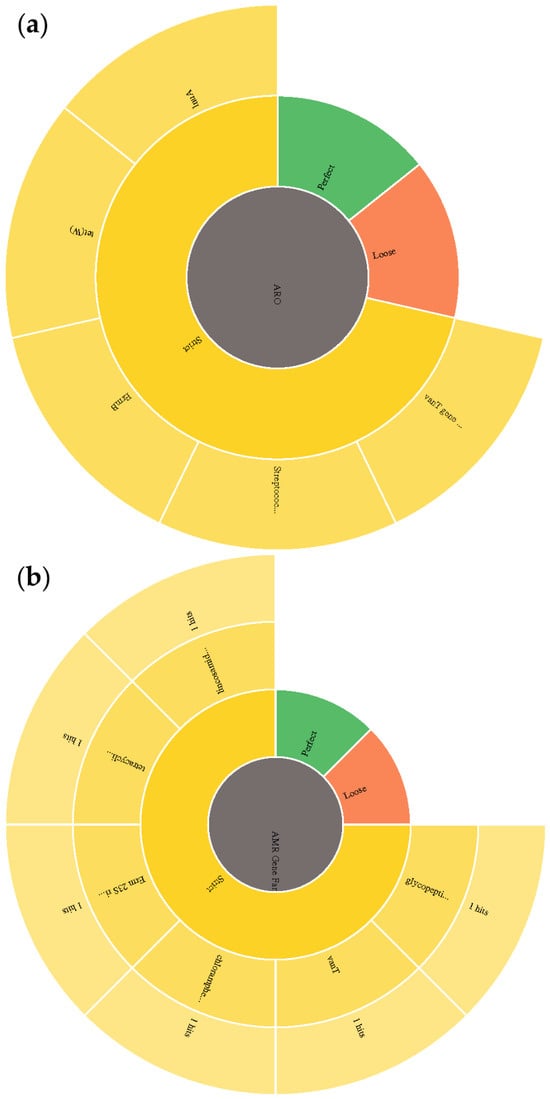
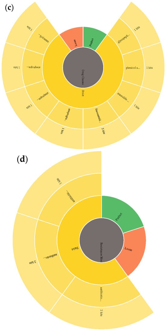
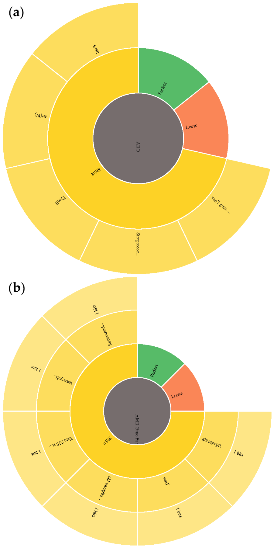
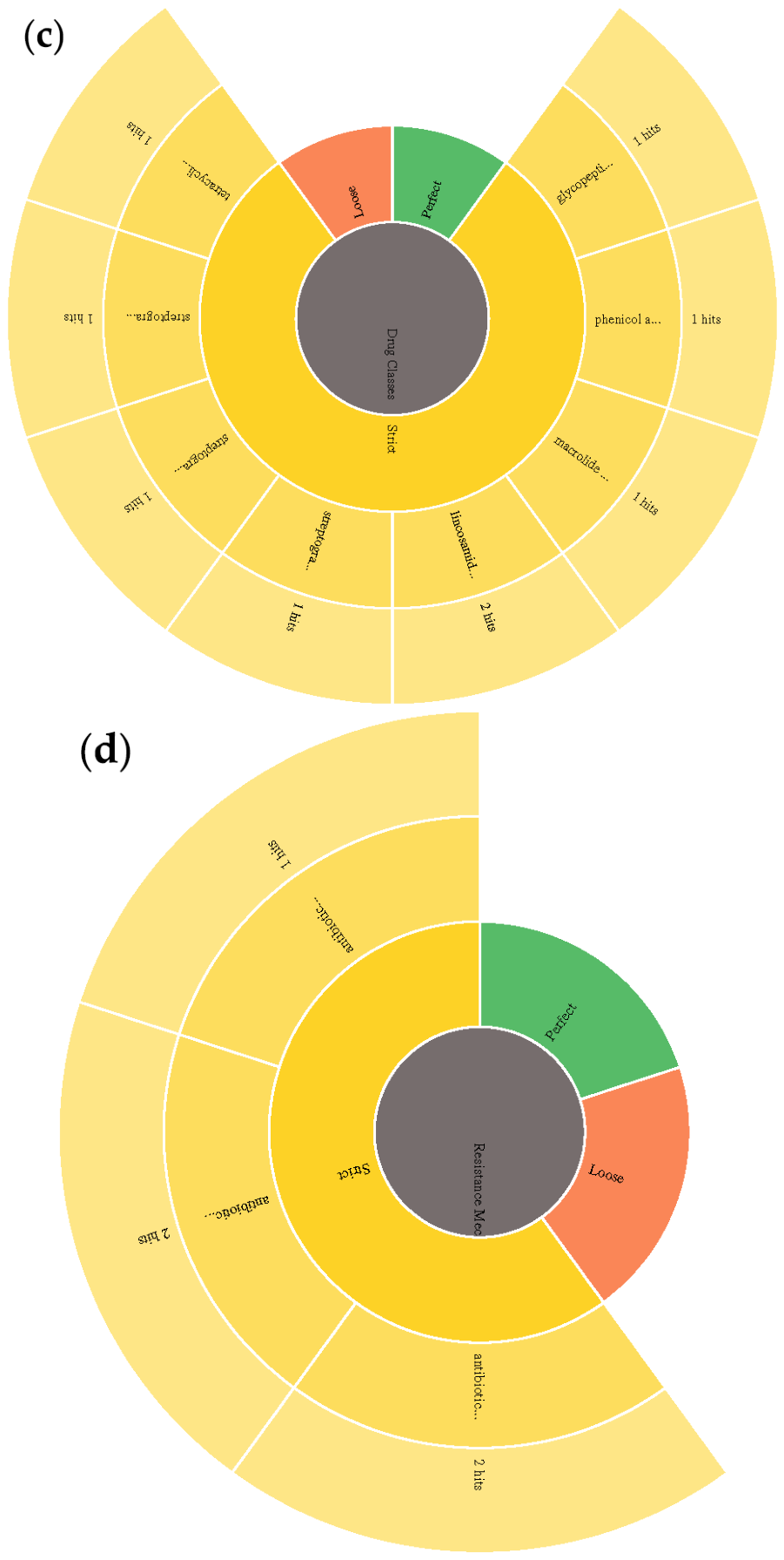
Figure 3 shows the result of the comprehensive antibiotic resistance analysis of the chicken-isolated L. reuteri genome. Five antibiotic resistance-associated gene families were found only under strict criterion: glycopeptide resistance gene cluster, vanT; chloramphenicol acetyltransferase (CAT); lincosamide nucleotidyltransferase (LNU); tetracycline-resistant ribosomal protection protein and Erm 23S ribosomal RNA methyltransferase. The glycopeptide resistance gene cluster, vanT (vanT), belonged to the glycopeptide antibiotic drug class, having a 33.87% identity-matching region. Its resistance mechanism was antibiotic target alteration with a 52.67% length of reference sequence. CAT belonged to the phenicol antibiotic drug class, having a 95.35% identity-matching region; its resistance mechanism is antibiotic inactivation, with a 100% length of reference sequence. Erm 23S ribosomal RNA methyltransferase (ErmB) belonged to macrolide, lincosamide, streptogramin, streptogramin A and streptogramin B antibiotics, with a 98.39% identity-matching region; its resistance mechanism was antibiotic target alteration, with a 100% length of reference sequence. Tetracycline-resistant ribosomal protection protein (tet(W)) belonged to the tetracycline antibiotic drug class; its resistance mechanism was antibiotic target protection, having a 96.24% identity-matching region with a 100% length of the reference sequence. Lincosamide nucleotidyltransferase (LNU) belonged to the lincosamide antibiotic drug class; its resistance mechanism was antibiotic inactivation, having a 97.52% identity-matching region with a 100% length of reference sequence.
Figure 3.
Comprehensive antibiotic resistance analysis of chicken-isolated L. reuteri analyzed with the Comprehensive Antibiotic Resistance Database, showing the (a) AMR gene, (b) AMR gene family, (c) drug class and (d) resistance mechanism. AMR Gene Fam… = AMR Gene Family; 3a: van T gene… = van T; streptoccoc… = streptococcus; 3b: Tetratcyclin… = tetracycline-resistant ribosomal 1 protection protein; Lincosamide… = lincosamide nucleotidyltransferase (LNU); Chloramphe… = chloramphenicol; Glycopepti… glycopeptide resistance gene cluster; Erm 23s ri… = Erm 23s ribosomal RNA methyltransferase; 3c: glycopepti… = glycopeptide antibiotics; phenicol a… = phenicol antibiotic; macrolide… = macrolide antibiotic; lincosamid… = lincosamide antibiotic; streptogra… = streptogramin antibiotic; tetracycli… = tetracycline antibiotic; 3d: Resistance mec… = resistance mechanism; antibiotic… = antibiotic inactivation.
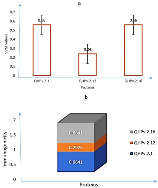
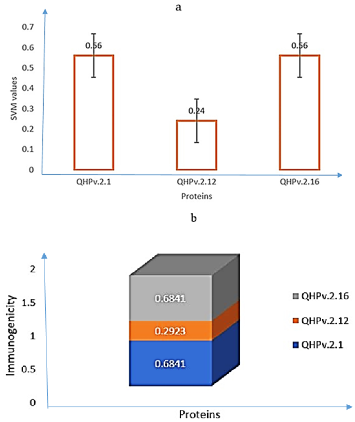
The IL-4-inducing hypothetical proteins (QHPv.2.1, QHPv.2.12 and QHPv.2.16) are shown in Figure 4, according to immunogenicity and SVM scores. Out of the three IL-4-inducing proteins, QHPv.2.12 was not immunogenic. The SVM values for QHPv.2.1 and QHPv.2.16 were 32% higher than QHPv.2.12.
Figure 4.
Hypothetical proteins with IL-4-inducing potential of the explored bacterial genome using (a) support vector machine score and (b) antigenicity value. The SVM values for QHPv.2.1 and QHPv.2.16 were 32% higher than the value obtained for QHPv.2.12 but QHPv.2.12 was not immunogenic.
The top fifteen IL-4-inducing mutants from each group, based on their immunogenicity and SVM scores were statistically analyzed. Table 2 shows the statistics of chicken-isolated Limosilactobacillus reuteri immunogenic hypothetical proteins and mutants that induced IL-4.
Table 2.
Statistical analysis result of immunogenic mutants with IL-4 inducing potential of the explored genome.
The immunogenic IL-4-inducing mutants from QHPv.2.1 (0.7013) and QHPv.2.16 (0.7040) had the best SVM values, which were significantly (p < 0.05) higher than the rest of the group, followed by those from QHPv.2.12 (0.4587). QHPv.2.5 (0.0560) had the lowest value. QHPv.2.1 (5484.71) and QHPv.2.16 (5491.05) had the largest molecular weights followed by QHPv.2.12 (5302.05). The lowest value was obtained by QHPv.2.7 (3386.74). QHPv.2.1 (7.917) and QHPv.2.16 (7.828) obtained higher theoretical isoelectric points compared with other mutants. QHPv.2.7 (4.445) and QHPv.2.12 (4.212) had the lowest values. According to immunogenicity value, QHPv.2.7 (1.3380) and QHPv.2.8 (1.2905) had the highest values, which were significantly (p < 0.05) higher than the values obtained by other mutants, followed by QHPv.2.5 (1.1673) and then QHPv.2.12 (0.4377).
Table 3 shows the corresponding position, theoretical isoelectric points and molecular weights of the selected top three immunogenic mutants with IL-4-inducing capacity from the explored bacterial genome.
Table 3.
Selected immunogenic mutants with IL-4-inducing potential.
We used cross-species IL-4 gene expression in Homo sapiens (human) and Mus musculus (mouse) using GENEVESTIGATOR because of the limitation of public chicken transcriptomic data for IL-4 under our study condition, as well as to enable a broader overview into the mammalian system, considering the broader application of probiotics in animal nutrition, food science and technology, health care and therapeutic industries. IL-4 was upregulated in the blood, peripheral, bone marrow, ovary, tumor, lung, temporal lobe and in the following cell types: CD4 helper T-cell, oogonium, CD8, cytotoxic T-cell, CD8 T-cell, myeloid cell, CD4 memory T-cell, CD8 T-cell, CD8 memory T-cell and CD4 regulatory T-cell as shown in Figure S1a,c. The IL-4 expression level was low or average in various cancer conditions (Figure S1b). Figure S1d,e show the top 50 positive genes co-expressed with IL-4 under different cells, anatomy, cancer categories and experimental sets. For instance, untreated memory CD4 T-cell samples compared with memory T-cell activation resulted in the upregulation of IL-4 (Figure S1f). In terms of the cell line, IL-4 was upregulated in 32D-BCR/ABL-ESR-Cebpa (mouse), but the expression level (Log2 ratio) was low or average across other cell lines (Figure S1g). The positive co-expression of genes with IL-4 according to cancer categories in mice is presented in Figure S1h. The 4-hydroxytamoxifen treatment for 6 h, 9 h and 12 h against control treatments resulted in the upregulated expression of IL-4 in mice (Figure S1i). Figure S1j shows the hierarchical clustering of IL-4 and IL-4ra in mice based on anatomical parts, cell types and cancer conditions. Figure S1k presents the expression level of HSPs and CSPs in Rattus norvegicus across various perturbations. For instance, Carhsp1 was upregulated in soman study 1 (48 h; untreated hippocampus tissue), sarin-induced seizures (24 h; 2-PAM, atropine sulfate, diazepam; L5; ipsilateral), and neuropathic pain study 1 (72 h; untreated hippocampus tissue).
4. Discussion
In this explorative study, we made attempts to understand the properties of the L. reuteri genome by exploring its HSPs, CSPs, hypothetical proteins and their mutants in relation to their IL-4-inducing capacity, considering the key roles IL-4 plays in immunity as well as its conserved nature. Probiotics and fermented foods have been extensively studied. The benefits of microbial and fermented food or feed in human and animal nutrition keep unfolding, and the market for microbial probiotics keeps growing. The benefits of probiotics include improved growth performance, feed conversion efficiency, immune responses, combating enteric pathogens and modulation of the intestinal microbiota [12,13]. Nonetheless, research efforts are ongoing to understand how probiotics perform its immune-boosting functions among other key roles, necessitating studies like this.
This study and its findings are of interest because it has been documented that a high population of probiotic bacteria in the intestinal tract can form colonies that protect the digestive tract by sticking to the epithelial tissue on the enterocyte wall and reducing the formation of pathogenic bacteria colonies [14,15]. Unraveling the mystery of how these functions are performed is key and requires concerted efforts to enable our understanding of probiotics in their entirety and their associated threats, if any.
Based on the documented benefits of probiotics and recent findings unfolding their ability to induce cytokines [16,17,18], which may improve immunity in human and animals as corroborated by the findings in this study, researchers continue to seek cost-effective and safe probiotic candidates for use as feed additives, food supplements and cosmetics and for therapeutic purposes. Kassa et al. [6] recently presented 12 potential probiotic bacteria from selected indigenous Ethiopian chicken gastrointestinal tracts, indicating a growing interest in microbial probiotics for human and animal consumption. The best probiotic candidates reported by the authors were the isolates GCH212 and GCM412, belonging to L. plantarum and B. subtilis. Probiotic bacteria from the native host are preferred to those isolated from other sources [19,20,21].
Efforts to identify the best probiotic candidates have suggested isolating probiotic strains from species they are fed to, such as chickens and pigs. A recent study adjudged strains of Lactobacillus obtained from contents of chicken guts to have probiotic potential and be safe [7]. The bile salt tolerance analysis of Lactobacillus strains isolated from the digestive tract of chickens in different bile salts revealed that L. reuteri had the best survival rates for both 0.30% and 1.00% bile salt, indicating its probiotic potential. However, a lot remains unclear about the functional mechanisms of these beneficial organisms.
In the current study, HSPs and CSPs were identified, five genes were associated with antibiotic resistance, and three of the hypothetical proteins including some of their mutants induced IL-4. To the best of our knowledge, this is the first study showing this kind of result thus far, thereby shedding light on the functional mechanisms of this beneficial microorganism and laying a foundation for further in vivo studies, which may benefit poultry and swine industries, food and dietary supplements as well as therapeutic industries.
Exploring HSPs and CSPs are of specific interest because extreme temperature is usually adopted to preserve food by inhibiting microbial growth, chemical reactions, autolytic enzyme activity and invasion. Such a method may also have negative impacts on beneficial microorganisms such as probiotics. In the present study, CSPs and HSPs present in L. reuteri strongly suggest that L. reuteri may be able to withstand such extreme temperatures, thereby preserving their beneficial effects for the host, although this is an explorative study that has not validated that claim with wet-lab experimental findings. The suggested benefit is based on the general knowledge that bacteria respond to the cold by preventing RNA from forming a stable secondary structure and promoting transcription and translation [22]. Two genes associated with the cold shock protein (CSP) of the CSP family were identified in the genome of chicken-isolated L. reuteri in the present study, which may need to be further explored for their specific functions.
Based on the present knowledge, CSPs play significant roles in bacterial cold adaptation and are induced under conditions of temperature drift. They are highly conserved small proteins, composed of nucleic acid-binding cold shock domains, which can bind to either a single-stranded DNA or RNA or as a chaperone [23]. The presence of one or more nucleic acid-binding cold shock domains endows them with pleiotropic functions, such as the regulation of splicing, transcription and translation [24,25]. They were previously reported in some foodborne pathogenic bacteria and food spoilage bacteria [26,27,28] but studies reporting their presence in probiotic bacteria are rare, as reported in this study.
CSPs play crucial roles in bacterial cellular functions, including differentiation and normal growth, virulence, starvation and other physiological and biochemical processes [29], making them ideal for growth, recovery and other bacterial cell functions at low temperatures, although CSPs in probiotic bacteria are rarely studied. CSPs are a ‘jack-of-all-trades’. They are conserved in both structure and function and can be found in almost all organisms from bacteria to humans (except yeast). Also, they have been detected in almost every cellular compartment. They can regulate their expression and several disease-associated genes and orchestrate multiple cellular processes [30]. As the authors [30] admitted, there is a lot to be conducted in characterizing and understanding the full cell-specific functions and mechanisms of CSPs.
Both CSPs and HSPs were identified in the studied genome in this study. Like CSPs, HSPs are ‘stress proteins’ that are highly conserved. They are a special group among chaperones and play important roles in protein folding and structuring as well as in maintaining their functional conformations, assembling multi-protein complexes and preventing non-native protein aggregation [31]. They improve membrane stability and assist during the reactive oxygen species detoxifying process by positively regulating the antioxidant enzyme system. They control cell signaling and transport to the suitable subcellular compartment and are involved in the protein translocation process and in the degradation of other proteins, avoiding the eventual formation of non-functional ones [31].
HSPs identified in this study include the 10 kDa family and HSP 60 kDa family chaperone, among others. HSPs with low a molecular weight (<40 kDa) are referred to as small HSPs and are known to perform the ‘cellular defense first line’ against stress from excessive heat and/or others, which results in protein denaturation [32]. They are present in all organisms. They form a very diverse chaperone family, which share an α-crystalline domain flanked by variable and disordered N- and C-terminal extensions, although certain structural characteristics such as an oligomeric form with a β-sandwich domain seem to remain conserved [33]. HSPs are broadly grouped according to their amino acid sequences based on the presence of certain CDs, and mainly according to their MW (kDa). They are usually grouped into six classes, which are: small HSPs, HSP100s or CLPs, HSP90s, HSP70s, HSP40s/DNAJs and chaperonins. Small HSPs protect against protein aggregation; HSP40s/DNAJs regulate the HSP70 complex; HSP70s, HSP90s and chaperonins mediate protein folding; while HSP100s/CLPs are involved in protein aggregation and solubilization [33].
Based on the documented benefits, probiotics, primarily used in food or dietary supplements, medicines and cosmetic products, are annually increasing. The global probiotic market was valued at almost USD 7 billion in 2021, with a forecast growth of 3.75% on average until 2026 having grown between 2022 and 2023 from USD 66.9 to USD 73.14 billion, with an estimated 9.3% yearly growth rate [34]. One major factor suggested for the future increasing probiotic market is the increasing consumers’ preference for healthy foods and nutritional supplements, thereby necessitating the need to ensure the all-round safety of probiotics.
It has been noted that a probiotic, as food or a supplement without a health claim or in a food or supplement with a specific health claim, must meet the criteria of being a member of a safe species in the EU [35]. It must belong to a specific probiotic strain or strains, it must be beneficial to humans or regarded as a safe microorganism based on adequate evidence and, in addition, at the end of its shelf life must deliver viable strain(s) [36]. In the present study, five genes were associated with antibiotic resistance. Probiotics are considered as safe alternatives to antibiotics and hence, should be free from antibiotic resistance, which is currently of global concern. However, being an in silico explorative study, there may be a need to extensively study this to validate the claim using wet-lab analyses, although Lactobacilli and Enterococci isolates have previously been reported to be resistant to cefoxitin, penicillin, vancomycin, aztreonam and oxacillin [37].
The IL-4-inducing capacity of the hypothetical proteins and their mutants as reported in this study is of importance, considering the important role IL-4 plays in immunity and its conserved nature across species. The immune system continuously communicates with the central nervous system (CNS) during health and disease [38]. The CNS plays a key role in controlling other systems of the body and may influence an organism’s general well-being. The CNS-resident macrophages and myeloid cells are usually the first cells to detect tissue damage or pathogens. These cells initiate an immune response in order to clear the tissue debris or fight the pathogen, but if the blood–brain barrier becomes permeable as a result of inflammation or trauma, immune cells that infiltrate the CNS from the periphery are pro-inflammatory within an acute inflammatory injury. However, they can also be regulatory or anti-inflammatory cells. The balance and timing of pro- and anti-inflammatory immune cells play key roles in tissue damage and repair [39,40]. T helper 2-like (TH2-like) cells and their cytokines including IL-4 inhibit TH1-like and TH17-mediated inflammatory processes, thereby serving an anti-inflammatory function [41].
The IL-4_inducing capacity of probiotics, as reported in this study, may be revealing the pathways toward the mode of mechanism of probiotics in cell immunity. Being a pleiotropic cytokine, IL-4 plays a crucial role in type 2 immunity, mainly secreted by not only TH2-like cells but also granulocytes and myeloid cells [42]. It can induce the differentiation of CD4+ T cells into TH2-like cells as well as the alternative activation of myeloid cells (macrophages and microglia), involving morphological and transcriptional changes such as the expression of arginase 1 (Arg1), chitinase-like protein 3 (Ym1) or CD206, resulting in wound healing and the suppression of destructive inflammation [43].
IL-4 has been shown to improve immunity; enhance the functional outcome of different types of CNS injury and disease models, exerting effects on lymphocytes, microglia and neurons; as well as be recommended for therapeutic development, although with caution for consideration of possible side effects [40]. It has been reported that IL-4-stimulated Arg+ microglia upregulated the brain-derived neurotrophic factor and enhanced hippocampal neurogenesis in mice exposed to chronic mild stress [44]. It was reported that microglia-specific IL-4Rα-deficiency suppressed neurogenesis and resulted in increased stress vulnerability. In another study involving adult zebrafish brain, IL-4 induced the phosphorylation of signal transducer and activator of transcription 6 in neural stem/progenitor cells (NSPCs) and was sufficient to restore NSPC proliferation after incubation with neurotoxic amyloid-β42 [45]. It has been documented that IL-4-induced M2-like phenotypes to help in neuroprotection and repair [46,47], as well as was an activator of M2-like markers such as Arg1, CD206 and Ym1.
5. Conclusions
The chicken-isolated L. reuteri genome has one contig, with a total length of 2,085,667 bp and an average G + C content of 39.43%. It had 2471 CDS, 78 tRNA genes and 10 rRNA genes. It was associated with five antibiotic resistance genes, which were glycopeptide resistance gene cluster, vanT; chloramphenicol acetyltransferase (CAT), Erm 23S ribosomal RNA methyltransferase, tetracycline-resistant ribosomal protection protein and lincosamide nucleotidyltransferase (LNU), although none was found under the perfect RGI criterion. Hypothetical proteins of the chicken-isolated L. reuteri genome, labeled QHPv.2.1, QHPv.2.12 and QHPv.2.16, induced IL-4. However, QHPv.2.12 was not immunogenic. The best IL-4-inducing SVM scores were obtained by mutants from QHPv.2.1 and QHPv.2.16, followed by QHPv.2.12 and QHPv.2.5. IL-4 was upregulated in the blood, peripheral, bone marrow, ovary, tumor, lung and temporal lobe. We made attempts in this explorative study to unravel the mystery surrounding the functional mechanisms of probiotic L. reuteri, suggesting its sustainable applications in feed/food, cosmetic and therapeutic industries. Being an in silico explorative study, we strongly recommend wet-lab experimental validations for the claims in this study.
Supplementary Materials
The following supporting information can be downloaded at: https://www.mdpi.com/article/10.3390/biologics6030021/s1, Figure S1. Level of expression of IL-4 and IL-4R in human and mouse at different conditions: (a) scatter plot showing IL-4 and IL-4R expression (log2 scale) in Homo sapiens across various anatomical parts; (b) IL-4 expression level (log2 scale) across various neoplasms in Homo sapiens; (c) a heatmap showing IL-4 and IL-4R expression (linear scale) across various anatomical parts, cell types and states in Homo sapiens; (d) a circular plot representing the top 50 co-expressed genes with positive correlation to IL-4 across various anatomical parts, neoplasm and cell categories in Homo sapiens; (e) a circular plot representing the top 50 co-expressed genes with positive correlation to IL-4 across various experimental sets in Homo sapiens; (f) scatter plot showing IL-4 and IL-4R expression (log2 ratio) across relevant perturbations in Homo sapiens; (g) boxplot showing IL-4 expression (log2 scale) in Mus musculus across various cell lines; (h) a circular plot showing the top 50 co-expressed genes with positive correlation to IL-4 across various cancer categories in Mus musculus; (i) a heatmap showing IL-4 and IL-4RA expression (log2 ratio) across various experimental sets in Mus musculus; (j) scatter plot showing IL-4 and IL-4RA expression (log2 scale) in Mus musculus across various perturbations in Mus musculus, (k) a heatmap showing expression of cold shock proteins across various experimental set in Rattus norvegicus and (l) a heatmap showing expression of heat shock proteins across various experimental set in Rattus norvegicus.
Author Contributions
Conceptualization: I.O.A.; methodology: I.O.A.; formal analysis: I.O.A. and O.A.A.; investigation: I.O.A.; writing—original draft: I.O.A.; writing—review and editing: I.O.A. and O.A.A. All authors have read and agreed to the published version of the manuscript.
Funding
This research received no external funding.
Institutional Review Board Statement
Not applicable.
Informed Consent Statement
Not applicable.
Data Availability Statement
The original contributions presented in this study are included in the article/Supplementary Materials. Further inquiries can be directed to the corresponding author.
Acknowledgments
The authors appreciate Olawumi Adejumo’s support during data curation and the owners/authors of some of the resources used in this study.
Conflicts of Interest
The authors declare no conflicts of interest.
Abbreviations
| CAT | Chloramphenicol acetyltransferase |
| CD | Conserved domain |
| CDS | Protein-coding sequences |
| CSP | Cold shock protein |
| ErmB | Erm 23S ribosomal RNA methyltransferase |
| GO | Gene ontology |
| HSP | Heat shock protein |
| lnu | Lincosamide nucleotidyltransferase |
| MP | Mutant position |
| MW | Molecular weight |
| NSPC | Neural stem/progenitor cell |
| pI | Isoelectric point |
| rRNA | Ribosomal RNA |
| tet(W) | Tetracycline-resistant ribosomal protection protein |
| tRNA | Transfer RNA |
References
- Adejumo, I.O. Hypothetical Proteins of Chicken-Isolated Limosilactobacillus reuteri Subjected to In Silico Analyses Induce IL-2 and IL-10. Genes Nutr. 2024, 19, 21. [Google Scholar] [CrossRef] [Scilit] [PubMed]
- Adejumo, I.O. Lactobacillus acidophilus: Boosting immunity through IL-6 induction. Biologics 2025, 5, 29. [Google Scholar] [CrossRef] [Scilit]
- Adejumo, I.O. Animal-sourced probiotic bacteria induce interferongamma in humans and mice. Silico Res. Biomed. 2026, 2, 100196. [Google Scholar] [CrossRef] [Scilit]
- Serek, P.; Oleksy-Wawrzyniak, M. The Effect of Bacterial Infections, Probiotics, and Zonulin on Intestinal Barrier Integrity. Int. J. Mol. Sci. 2021, 22, 11359. [Google Scholar] [CrossRef] [Scilit] [PubMed]
- De Vos, W.M.; Tilg, H.; Van Hul, M.; Cani, P.D. Gut Microbiome and Health: Mechanistic Insights. Gut 2022, 71, 1020–1032. [Google Scholar] [CrossRef] [Scilit]
- Kassa, G.; Alemayehu, D.; Andualem, B. Isolation, Identification, and Molecular Characterization of Probiotic Bacteria from Locally Selected Ethiopian Free-Range Chickens’ Gastrointestinal Tract. Poult. Sci. 2024, 103, 103311. [Google Scholar] [CrossRef] [Scilit] [PubMed]
- Sirisopapong, M.; Shimosato, T.; Okrathok, S.; Khempaka, S. Assessment of Lactic Acid Bacteria Isolated from the Chicken Digestive Tract for Potential Use as Poultry Probiotics. Anim. Biosci. 2023, 36, 1209–1220. [Google Scholar] [CrossRef] [Scilit] [PubMed]
- Uhlen, M.; Oksvold, P.; Fagerberg, L.; Lundberg, E.; Jonasson, K.; Forsberg, M.; Zwahlen, M.; Kampf, C.; Wester, K.; Hober, S.; et al. Towards a Knowledge-Based Human Protein Atlas. Nat. Biotechnol. 2010, 28, 1248–1250. [Google Scholar] [CrossRef] [Scilit] [PubMed]
- Nimrod, G.; Schushan, M.; Szilágyi, A.; Leslie, C.S.; Ben-Tal, N. Detection of Functionally Important Regions in “Hypothetical Proteins” of Known Structure. Structure 2008, 16, 1755–1763. [Google Scholar] [CrossRef] [Scilit] [PubMed]
- Shahbaaz, M.; Bisetty, K.; Ahmad, F. Functional Annotation of Conserved Hypothetical Proteins from Haemophilus influenzae Rd KW20. PLoS ONE 2013, 8, e84263. [Google Scholar] [CrossRef] [Scilit] [PubMed]
- Zhang, Z.; Schwarz, S.; Dean, C.J.; Li, Y.; Chandrasekhar, N.; Sen, Y.; Wang, J.; Wang, M.; Gu, X. Assessment of Global Health Risk of Antibiotic Resistance Genes. Nat. Commun. 2022, 13, 1553. [Google Scholar] [CrossRef] [Scilit] [PubMed]
- Dhama, K.; Verma, V.; Sawant, P.M.; Tiwari, R.; Vaid, R.K.; Chauhan, R.S. Applications of probiotics in poultry: Enhancing immunity and beneficial effects on production performances and health—A review. J. Immunol. Immunopathol. 2011, 13, 1–19. [Google Scholar]
- Denli, M.; Demirel, R. Replacement of antibiotics in poultry diets. CABI Rev. 2018, 13, 35. [Google Scholar]
- Shang, Y.S.; Kumar, S.; Oakley, B.; Woo, K.K. Chicken gut microbiota: Importance and detection technology. Front. Vet. Sci. 2018, 5, 254. [Google Scholar] [CrossRef] [Scilit] [PubMed]
- Sugiharto, S.; Yudiarti, T.; Isroli, I.; Widiastuti, E.; Wahyuni, H.I. Hematological parameters and selected intestinal microbiota populations in the Indonesian indigenous crossbred chickens fed basal diet supplemented with multistrain probiotic preparation in combination with vitamins and minerals. Vet. World 2018, 11, 874–882. [Google Scholar] [CrossRef] [Scilit] [PubMed]
- Adejumo, I.O.; Akintayo, A.O. Investigating the neurotoxic threat associated with Bacillus pumilus probiotic strains. Asian J. Biol. Sci. 2026, 19, 12–21. [Google Scholar] [CrossRef] [Scilit]
- Adejumo, I.O. Immunotherapy potential of animal-sourced probiotic bacteria. Biologics 2025, 5, 17. [Google Scholar] [CrossRef] [Scilit]
- Adejumo, I.O.; Adebiyi, O.A. Peptides from hypothetical proteins of Lactobacillus acidophilus induce IL-4 and IL-10. Biomed. Res. Ther. 2025, 12, 7191–7206. [Google Scholar] [CrossRef] [Scilit]
- Dowarah, R.; Verma, A.K.; Agarwal, N. Selection and characterization of probiotic lactic acid bacteria and its impact on growth, nutrient digestibility, health and antioxidant status in weaned piglets. PLoS ONE 2018, 13, 29–78. [Google Scholar] [CrossRef] [Scilit]
- Reuben, R.C.; Roy, P.C.; Sarkar, S.L.; Alam, R.A.; Jahid, I.K. Isolation, characterization, and assessment of lactic acid bacteria toward their selection as poultry probiotics. BMC Microbiol. 2019, 19, 253. [Google Scholar] [CrossRef] [Scilit] [PubMed]
- Mona, S.; Zayed, D.; Abd El-Fattah, A.; Rateb, N.A.; El-Bordeny, N.E. Isolation, and characterization, of lactic acid bacteria for potential use as feed additives for ruminant animals. Res. Sq. 2022, preprint. [Google Scholar] [CrossRef] [Scilit] [PubMed]
- Zhang, Y.; Burkhardt, D.H.; Rouskin, S.; Li, G.W.; Weissman, J.S.; Gross, C.A. A stress response that monitors and regulates mRNA structure is central to cold shock adaptation. Mol. Cell 2018, 70, 274–286.e7. [Google Scholar] [CrossRef] [Scilit] [PubMed]
- Von König, K.; Kachel, N.; Kalbitzer, H.R.; Kremer, W. RNA and DNA binding epitopes of the cold shock protein TmCsp from the hyperthermophile Thermotoga maritima. Protein J. 2020, 39, 487–500. [Google Scholar] [CrossRef] [Scilit] [PubMed]
- Brandt, S.; Raffetseder, U.; Djudjaj, S.; El Mokhtari, N.E.; En-Nia, A.; Elger, M.; Gremse, F.; Marx-Stoelting, P.; Schneider, U.; Tiefenbach, B.; et al. Cold shock Y-box protein-1 participates in signaling circuits with auto-regulatory activities. Eur. J. Cell Biol. 2012, 91, 464–471. [Google Scholar] [CrossRef] [Scilit] [PubMed]
- Lindquist, J.A.; Brandt, S.; Bernhardt, A.; Zhu, C.; Mertens, P.R. The role of cold shock domain proteins in inflammatory diseases. J. Mol. Med. 2014, 92, 207–216. [Google Scholar] [CrossRef] [Scilit] [PubMed]
- Welte, H.; Kovermann, M. Targeted expression and purification of fluorine labelled cold shock protein B by using an auxotrophic strategy. Protein Expr. Purif. 2019, 157, 86–91. [Google Scholar] [CrossRef] [Scilit] [PubMed]
- Kragh, M.L.; Muchaamba, F.; Tasara, T.; Hansen, L.T. Cold-shock proteins affect desiccation tolerance, biofilm formation and motility in Listeria monocytogenes. Int. J. Food Microbiol. 2020, 329, 108662. [Google Scholar] [CrossRef] [Scilit] [PubMed]
- Xu, J.; Li, Q.; Zhang, J.; Li, X.; Sun, T. In silico structural and functional analysis of cold shock proteins in Pseudomonas fluorescens PF08 from marine fish. J. Food Prot. 2021, 84, 1446–1454. [Google Scholar] [CrossRef] [Scilit] [PubMed]
- Behl, A.; Kumar, V.; Shevtsov, M.; Singh, S. Pleiotropic roles of cold shock proteins with special emphasis on unexplored cold shock protein member of Plasmodium falciparum. Malar. J. 2020, 19, 382. [Google Scholar] [CrossRef] [Scilit] [PubMed]
- Lindquist, J.A.; Mertens, P.R. Cold shock proteins: From cellular mechanisms to pathophysiology and disease. Cell Commun. Signal. 2018, 16, 63. [Google Scholar] [CrossRef] [Scilit] [PubMed]
- Diogo, R., Jr.; Von Pinho, E.V.R.; Pinto, R.T.; Reis, C.O.; Souza, D.C.; Silva, L.; Oliveira, T.V.; Sousa, F.J.; Ferreira, G.; Braga, L.R.; et al. Maize heat shock proteins—Prospection, validation, categorization and in silico analysis of the different ZmHSP families. Stress Biol. 2023, 3, 37. [Google Scholar] [CrossRef] [Scilit] [PubMed]
- Janowska, M.K.; Baughman, H.E.R.; Woods, C.N.; Klevit, R.E. Mechanisms of small heat shock proteins. Cold Spring Harb. Perspect. Biol. 2019, 11, a034025. [Google Scholar] [CrossRef] [Scilit] [PubMed]
- Waters, E.R.; Vierling, E. Plant small heat shock proteins—Evolutionary and functional diversity. New Phytol. 2020, 227, 24–37. [Google Scholar] [CrossRef] [Scilit] [PubMed]
- International Probiotics Association (IPA). Market Data, Probiotic Market 2021 and Trend Analysis. Available online: https://www.ipaeurope.org/legal-framework/market-data/ (accessed on 15 May 2023).
- EFSA (European Food Safety Authority). Opinion of the Scientific Committee on a request from EFSA on the introduction of a Qualified Presumption of Safety (QPS). EFSA J. 2007, 587, 1–16. [Google Scholar] [CrossRef] [Scilit]
- Zawistowska-Rojek, A.; Zaręba, T.; Mrówka, A.; Tyski, S. Assessment of microbiological status of probiotic products. Pol. J. Microbiol. 2016, 65, 97–104. [Google Scholar] [CrossRef] [Scilit] [PubMed]
- Zavišić, G.; Popović, M.; Stojkov, S.; Sladojević, Ž.; Baralić, K.; Dimitrijević, D.; Đokić, M.; Ćirić, J.; Kovačević, A.; Pantić, I.; et al. Antibiotic resistance and probiotics: Knowledge gaps, market overview and preliminary screening. Antibiotics 2023, 12, 1281. [Google Scholar] [CrossRef] [Scilit] [PubMed]
- Pape, K.; Tamouza, R.; Leboyer, M.; Zipp, F. Immunoneuropsychiatry—Novel perspectives on brain disorders. Nat. Rev. Neurol. 2019, 15, 317–328. [Google Scholar] [CrossRef] [Scilit] [PubMed]
- Irani, S.R.; Nath, A.; Zipp, F. The neuroinflammation collection: A vision for expanding neuro-immune crosstalk in brain. Brain 2021, 144, e59. [Google Scholar] [CrossRef] [Scilit] [PubMed]
- Gärtner, Y.; Bitar, L.; Zipp, F.; Vogelaar, C.F. Interleukin-4 as a therapeutic target. Pharmacol. Ther. 2023, 242, 108348. [Google Scholar] [CrossRef] [Scilit] [PubMed]
- Ellwardt, E.; Walsh, J.T.; Kipnis, J.; Zipp, F. Understanding the role of T cells in CNS homeostasis. Trends Immunol. 2016, 37, 154–165. [Google Scholar] [CrossRef] [Scilit] [PubMed]
- Egholm, C.; Heeb, L.E.M.; Impellizzieri, D.; Boyman, O. The regulatory effects of Interleukin-4 receptor signaling on neutrophils in type 2 immune responses. Front. Immunol. 2019, 10, 2507. [Google Scholar] [CrossRef] [Scilit] [PubMed]
- Chen, Z.; Trapp, B.D. Microglia and neuroprotection. J. Neurochem. 2016, 136, 10–17. [Google Scholar] [PubMed]
- Zhang, J.; Li, Y.; Duan, Z.; Zhang, X.; Wang, G.; Chen, J.; Zhao, Z. The effects of the M2a macrophage-induced axonal regeneration of neurons by arginase 1. Biosci. Rep. 2020, 40, BSR20193031. [Google Scholar] [CrossRef] [Scilit] [PubMed]
- Bhattarai, P.; Thomas, A.K.; Cosacak, M.I.; Papadimitriou, C.; Mashkaryan, V.; Zhang, Y.; Kizil, C. IL4/STAT6 signaling activates neural stem cell proliferation and neurogenesis upon amyloid-beta42 aggregation in adult zebrafish brain. Cell Rep. 2016, 17, 941–948. [Google Scholar] [CrossRef] [Scilit] [PubMed]
- Amo-Aparicio, J.; Garcia-Garcia, J.; Francos-Quijorna, I.; Ortuño-Sahagún, D.; López-Vales, R. Interleukin-4 and interleukin-13 induce different metabolic profiles in microglia and macrophages that relate with divergent outcomes after spinal cord injury. Theranostics 2021, 11, 9805–9820. [Google Scholar] [CrossRef] [Scilit] [PubMed]
- Kang, R.; Gamdzyk, M.; Luo, Y.; Chai, Y.; Tang, R.; Li, S.; Duan, L.; Hu, Q. Three days delayed recanalization improved neurological function in pMCAO rats by increasing M2 microglia—Possible involvement of the IL-4R/STAT6/PPARγ pathway. Transl. Stroke Res. 2022, 14, 250–262. [Google Scholar] [CrossRef] [Scilit] [PubMed]

Disclaimer/Publisher’s Note: The statements, opinions and data contained in all publications are solely those of the individual author(s) and contributor(s) and not of MDPI and/or the editor(s). MDPI and/or the editor(s) disclaim responsibility for any injury to people or property resulting from any ideas, methods, instructions or products referred to in the content. |
© 2026 by the authors. Licensee MDPI, Basel, Switzerland. This article is an open access article distributed under the terms and conditions of the Creative Commons Attribution (CC BY) license.