


Definition
Heart failure (HF) is a heterogeneous clinical syndrome with increasing prevalence among adults worldwide. It is characterized by complex central and peripheral alterations that contribute to exercise intolerance, fatigue, dyspnea, and reduced quality of life. Inspiratory muscle weakness (IMW) plays a key role in this vicious cycle by exacerbating symptoms and further limiting functional capacity. Inspiratory muscle training (IMT) has emerged as a potential adjuvant in comprehensive HF management and is a physiologically grounded and promising tool in the contemporary HF therapeutic toolkit. Its integration into multimodal rehabilitation programs may mitigate the cycle of dyspnea and deconditioning in patients with HF. On this basis, we provide an overview of the pathophysiological mechanisms underlying IMW and present the practical characteristics of IMT programs, synthesizing current evidence regarding its clinical efficacy and implementation challenges.
1. Introduction
Heart failure (HF) is a heterogeneous syndrome in which the heart is unable to handle a human’s metabolic needs due to its structural and/or functional abnormalities. The prevalence is 1–2% in the Western world [1], and it is accelerating due to the demographic trend of an aging population and an increase in risk factors [2]. Patients with HF are classified according to the left ventricular ejection fraction (LVEF) into HF with preserved (HFpEF: LVEF ≥ 50%), mildly reduced (HFmrEF: 41% < LVEF ≤ 49%), and reduced (HFrEF: LVEF ≤ 40%) ejection fraction [2], and they face various symptoms regardless of the disease severity. The limited exercise capacity [3], dyspnea, and poor quality of life (QoL) [4] are the most common expressions of the disease. Moreover, muscle weakness in both peripheral [5] and inspiratory muscles [6] is present in patients with HF regardless of LVEF magnitude. Inspiratory muscle weakness (IMW) is diagnosed when maximal inspiratory pressure (MIP) is reduced by less than 70% of predicted values [7]. Additionally, patients with HF demonstrated significantly reduced endurance capacity compared to healthy control subjects, indicating impaired respiratory muscle endurance in this population [8].
The skeletal muscle hypothesis, originally proposed by Coats et al. [9], describes the vicious cycle that contributes to the progression of HF and the generation of its characteristic symptoms. It emphasizes that HF is not solely a consequence of central cardiac dysfunction but also involves important peripheral abnormalities, particularly affecting skeletal and respiratory muscles. Although left ventricular dysfunction is a defining feature of the disease, it initiates a cascade of systemic effects. This dysfunction promotes a catabolic state, leading to structural and metabolic abnormalities in skeletal and respiratory muscles. Muscle alterations during physical exertion heighten ergoreflex activity, which sends signals to the central nervous system and increases sympathetic nervous system activity [9].
The resulting peripheral vasoconstriction and increased left ventricular afterload further impair cardiac performance. Through this mechanism, the skeletal muscle hypothesis explains why patients with HF experience symptoms such as exercise intolerance, early fatigue, and dyspnea, often out of the degree of left ventricular dysfunction [10]. Furthermore, it also provides a physiological basis for the beneficial effects of exercise training, which can improve muscle function and symptoms. This is why exercise training is highly recommended in clinical guidelines as a non-pharmacological therapy [11]. Aerobic exercise (AE) is the most common exercise modality in HF rehabilitation programs due to multiple benefits on patients’ exercise capacity, cardiac function [12], functional capacity, and QoL [13]. Furthermore, inspiratory muscle training (IMT) is recommended as an adjuvant exercise in the most severe patients, enriching the conventional exercise programs [14].
To date, there is a variety in training loads and modalities implemented in patients with HF. Therefore, a comprehensive patient evaluation of respiratory muscle status is essential to enable clinicians to implement appropriately tailored IMT interventions.
In light of these considerations, this entry provides a comprehensive overview of inspiratory muscle pathophysiology in patients with HF and further delineates the various modalities of IMT, outlining their theoretical rationale and mechanistic underpinnings. Additionally, it synthesizes current evidence regarding the effects of IMT and gives practical considerations supporting the structured and individualized implementation of IMT in clinical practice.
2. Rationale for Inspiratory Muscle Weakness in Heart Failure
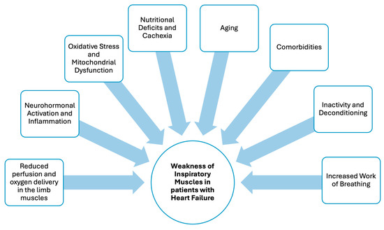
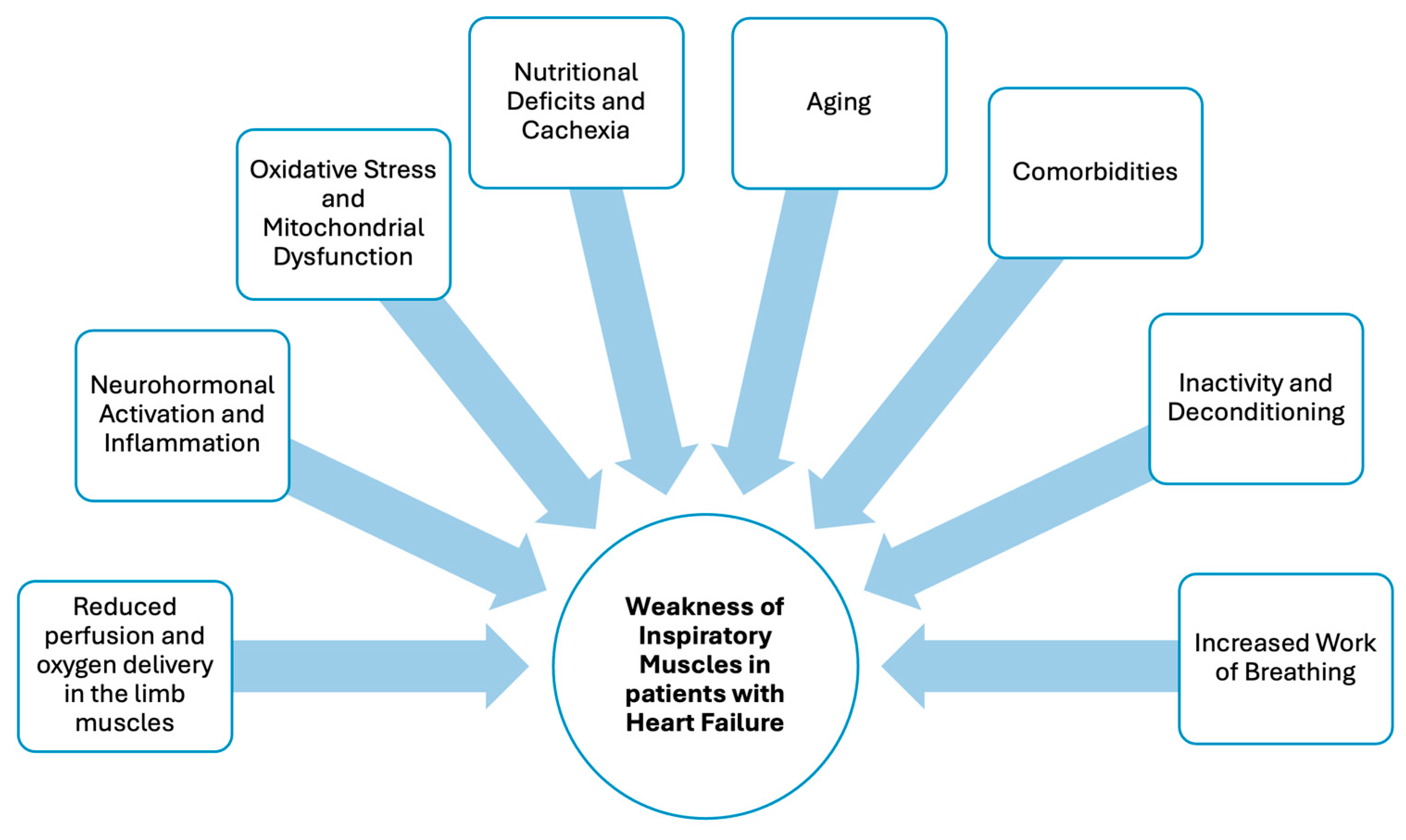
Inspiratory muscle weakness, particularly involving the diaphragm, arises from multifactorial pathophysiological mechanisms and patient-related factors. These are discussed in detail in the following subsections and are summarized in Figure 1.
Figure 1.
Pathophysiological mechanisms and patient-related factors contributing to inspiratory muscle weakness in patients with heart failure.
2.1. Impaired Oxygen Delivery and Utilization
Reduced cardiac output (CO) in patients with HF leads to chronic hypoperfusion of both respiratory and peripheral skeletal muscles [15]. This chronic reduction in blood flow restricts oxygen and nutrient delivery, resulting in a persistent state of muscular hypoperfusion and tissue hypoxia [16]. Over time, these conditions induce structural and metabolic adaptations, including muscle fiber atrophy, reduced capillary density, mitochondrial dysfunction, and alterations in fiber-type composition [15,17,18]. Diaphragm biopsies from HFrEF have shown significant alterations, such as atrophy in both type I and type II fibers, with reductions in cross-sectional area of 35% and 51%, respectively, compared with healthy controls and a significant shift towards increased fiber-type I distribution (p = 0.047) [17], reflecting chronic hypoxic stress [17,18]. Similarly, abnormalities have been reported in limb muscles, including muscle fiber atrophy, increased proportion of type II fibers, and reduced capillary density [15]. These alterations compromise muscle contractile capacity and endurance.
2.2. Neurohormonal Activation, Inflammation and Oxidative Stress
Persistent activation of neurohormonal pathways, especially the sympathetic nervous system and the renin–angiotensin–aldosterone system (RAAS), drives skeletal and respiratory muscle dysfunction in HF [19]. Although RAAS activation initially compensates for reduced CO, chronic stimulation becomes maladaptive, contributing to volume overload, increased afterload, myocardial remodeling, and fibrosis [20]. Sustained sympathetic activity and RAAS overactivation promote a catabolic state by enhancing proteolysis and suppressing anabolic signaling pathways [19].
In addition, patients with HF exhibit a chronic state of low-grade systemic inflammation, characterized by elevated circulating levels of pro-inflammatory cytokines, including tumor necrosis factor-α (TNF-α) and interleukin-6 (IL-6) [21]. These contribute to muscle wasting by increasing proteolysis, oxidative stress, and inflammatory signaling and impairing mitochondrial function.
Furthermore, the resulting oxidative stress induces damage to muscle proteins, membrane lipids, and mitochondrial DNA, thereby disrupting normal cellular structure and impairing key enzymatic processes involved in energy metabolism. Oxidative damage to mitochondria further compromises mitochondrial integrity and function, leading to reduced efficiency of oxidative phosphorylation and diminished adenosine triphosphate (ATP) production [15].
2.3. Patient-Related Factors
Multiple patient-related factors further contribute to the pathophysiology of inspiratory muscle dysfunction in patients with HF.
A subset of patients with HF exhibits deficits in their nutrition and develops cardiac cachexia as a result of a chronic imbalance between anabolic and catabolic processes [22]. Persistent neurohormonal activation and systemic inflammation lead to enhanced muscle proteolysis and inhibited protein synthesis [23]. In parallel, increased resting energy expenditure, together with impaired nutrient intake and absorption and hormonal dysregulation, contributes to a sustained negative energy balance, which accelerates muscle protein degradation [24]. Malnutrition reduces fat-free mass, particularly skeletal muscle mass [25].
Increased life expectancy over recent decades, largely attributable to advances in disease understanding, public health strategies, and therapeutic interventions, has led to a growing proportion of older adults living with chronic conditions, including HF [26]. Although fewer studies have specifically examined age-related changes in respiratory muscles, available evidence indicates a decline in inspiratory muscle strength of approximately 8–10% per decade after the age of 40 across diverse ethnic populations [27]. Mechanistically, age-related diaphragmatic myopathy, selective atrophy of type II (fast-twitch) fibers, and alterations in neuromuscular activation may underlie the observed reductions in inspiratory muscle performance [26]. In patients with HF, these physiological changes likely compound disease-related skeletal and respiratory muscle dysfunction, thereby contributing to clinically significant inspiratory muscle weakness. A recent study in patients with HFpEF demonstrated that skeletal muscle calf cross-sectional area (p = 0.014) and lean calf muscle cross-sectional area (p = 0.003), quantified using magnetic resonance imaging, were significantly lower in older compared with younger patients. Furthermore, both measures were significantly correlated with age [28]. With advancing age, patients with HF frequently present multiple comorbidities, encompassing both cardiovascular and non-cardiovascular conditions, which may either result from HF or serve as primary contributors to its pathogenesis [1]. The mean number of comorbidities differs slightly by sex, with 2.62 ± 1.55 in males and 2.8 ± 1.61 in females, and is associated with increased mortality risk; each one-point increment in the Charlson Comorbidity Index corresponds to a relative risk of 1.21 (95% CI, 1.13 to 1.29) [29]. In a European cohort, 74% of patients with HF had at least one comorbidity, with the most prevalent being chronic kidney disease (41%), anemia (29%), and diabetes mellitus (29%) [29]. Other common non-cardiovascular comorbidities include chronic obstructive pulmonary disease, malignancies, vitamin D deficiency, hyperuricemia, sleep disorders, and thyroid dysfunction, whereas cardiovascular comorbidities frequently include peripheral artery disease, coronary artery disease, atrial fibrillation, and hypertension [1]. Importantly, therapies for these comorbidities may adversely impact inspiratory muscle function [30].
Commonly observed symptoms, such as dyspnea and fatigue, lead to reduced physical activity (PA) levels and adoption of a sedentary lifestyle in the HF population [31]. A recent meta-analysis in the HF population brought to light that patients had low PA levels with a mean of 5040 steps per day [32]. This decline in habitual activity initiates a self-perpetuating cycle in which physical inactivity results in unloading of peripheral skeletal muscle, promoting progressive muscle deconditioning and subsequent worsening of symptoms, thereby further limiting PA. A prior study demonstrated a statistically significant association between both inspiratory (r2 = 0.80, p = 0.001) and expiratory (r2 = 0.45, p = 0.05) muscle strength and dyspnea during daily activities in patients with HF [33].
2.4. Increased Work of Breathing
In a given metabolic demand, patients with HF require greater ventilation due to the modified breathing pattern of rapid and shallow breathing [34]. Trying to maintain adequate ventilation under these conditions, the inspiratory muscles (predominantly the diaphragm) are required to generate higher transdiaphragmatic pressures and sustain elevated levels of neural respiratory drive. Chronic exposure to this increased respiratory workload results in persistent overuse of the inspiratory muscles, leading to metabolic stress, accelerated fatigue, and diminished mechanical efficiency. Great breathing work is demonstrated even during moderate-intensity activities of daily living [34]. A previous study comparing patients with HFrEF and healthy control subjects demonstrated that, during exertion, they frequently exhibit expiratory flow limitation and commonly breathe near residual volume during exertion [35].
Furthermore, elevated left ventricular filling pressures lead to pulmonary venous congestion and interstitial fluid accumulation within the lung parenchyma. This pulmonary congestion reduces lung compliance by increasing pulmonary stiffness and disrupting normal alveolar-capillary mechanics. Additionally, airway narrowing and lung diffusing capacity (ventilation-perfusion mismatch) may further augment resistive respiratory loads. The aforementioned pathophysiological abnormalities substantially increase the mechanical work of breathing [36].
3. Clinical Manifestations and Prognostic Prevalence
3.1. Clinical Manifestations
Patients with impaired inspiratory muscle function demonstrate respiratory and functional abnormalities, including rapid shallow breathing, reduced ventilatory efficiency, and therefore, early onset of respiratory muscle fatigue and exertional dyspnea during physical activity and exercise tolerance [37].
In patients with HF classified as New York Heart Association classification (NYHA) II–IV, inspiratory muscle strength has been shown to correlate significantly with dyspnea during activities of daily living (r2 = 0.80, p = 0.001), underscoring the critical role of the respiratory muscle pump in symptom perception [33]. Dyspnea may arise from a mismatch between limited oxygen availability to the inspiratory muscles and the increased oxygen demand of peripheral muscles during physical exertion [37].
Compared with patients without IMW, those with IMW exhibit decreased exercise capacity, vital capacity, and quadriceps femoris muscle strength [38]. Importantly, IMW has been identified as an independent determinant of impaired exercise capacity in both patients with HFrEF [38] and HFpEF [39], and for patients over 62 years old, classified as NYHA II–III, MIP is an independent biomarker for the evaluation of exercise tolerance [40]. In patients with HFrEF, reduced MIP has been identified as a contributor to impaired QoL [38] and is associated with decreased exercise capacity and increased disease severity [41,42].
3.2. Prognostic Relevance
In prognostic terms, inspiratory muscle strength is an independent predictor of one-year survival in patients with congestive HF [43] and an independent predictor of all-cause mortality in patients with HF [6]. A retrospective longitudinal observational study revealed that patients with IMW had significantly lower survival rates and higher cardiovascular mortality than those without IMW in both patients with HFpEF and HFrEF [6]. Interventional evidence further supports the clinical relevance of inspiratory muscle strength. Following cardiac rehabilitation, increases in MIP of 10 cm H2O were associated with a significant reduction in the incidence of all-cause clinical events (Z = 2.975, p = 0.003) [43].
4. Effectiveness of Inspiratory Muscle Training
Training is recognized as a targeted therapeutic strategy that elicits physiological adaptations extending beyond isolated improvements in inspiratory muscle strength. The following section summarizes the principal mechanisms through which IMT exerts its beneficial effects in patients with HF, as illustrated in Figure 2.
4.1. Inspiratory Muscle Structural and Functional Adaptations
Inspiratory muscles share morphological and physiological characteristics with other skeletal muscles and demonstrate significant plasticity in response to mechanical loading [44]. During dynamic or static loading, structural and functional adaptations result from molecular alterations in the balance between protein synthesis and degradation pathways, ultimately leading to muscle remodeling [44].
In patients with HF, particularly those with HFrEF and IMW, high-intensity inspiratory muscle training (H-IMT) has been shown to induce measurable structural adaptations in the diaphragm. Two studies implementing H-IMT at intensities exceeding 70% of MIP for 4 weeks [8] and 8 weeks [45] demonstrated significant increases in diaphragm thickness (DT) at total lung capacity. However, only the longer intervention (8 weeks) reported significant increases in DT at residual volume, suggesting that prolonged exposure to high-intensity loading may influence the magnitude and extent of diaphragmatic remodeling [45].
Studies [45,46] demonstrated significant improvements in MIP (p < 0.001) after 8–12 weeks of training, indicating that both high- and low-intensity protocols can improve inspiratory muscle strength, although the magnitude and underlying mechanisms of adaptation may differ. Inspiratory muscle endurance was significantly enhanced following 3 months of H-IMT (p < 0.05), whereas no significant changes were observed in the no-resistance group in patients with HFrEF and IMW [47]. This between-group difference supports a training-specific effect and indicates that inspiratory muscle endurance represents a clinically relevant functional adaptation to targeted inspiratory muscle loading in this population, characterized by exertional dyspnea and early respiratory muscle fatigue.
More recently, a research group [48] investigated diaphragmatic structural and functional responses during two distinct inspiratory loading modalities: the mechanical threshold loading and the electronic tapered flow-resistive loading. In 17 patients with HF across varying disease severities, DT and diaphragm thickening fraction (DTF) [(DT at the end of inspiration − DT at the end of expiration)/DT at the end of expiration] were significantly greater during both low- and high-intensity loads compared with unloaded breathing for both IMT modalities. Notably, DTF values were significantly higher during high-intensity loading than during low-intensity loading for both aforementioned modalities, suggesting an intensity-dependent diaphragmatic contractile response. Diaphragm mobility was also significantly increased compared with unloaded breathing under both low-intensity loads (Δ = 14.85 mm [18.02, 11.67] for mechanical threshold loading and Δ = 16.26 mm [19.43, 13.08] for electronic tapered flow-resistive loading) and high-intensity loads (Δ = 20.75 mm [23.92, 17.57] for mechanical threshold loading and Δ = 23.23 mm [26.40, 20.05] for electronic tapered flow-resistive loading). These findings indicate that inspiratory loading acutely enhances diaphragmatic excursion in an intensity-dependent manner under both training modalities [48].
Figure 2.
Effects of inspiratory muscle training in patients with heart failure.
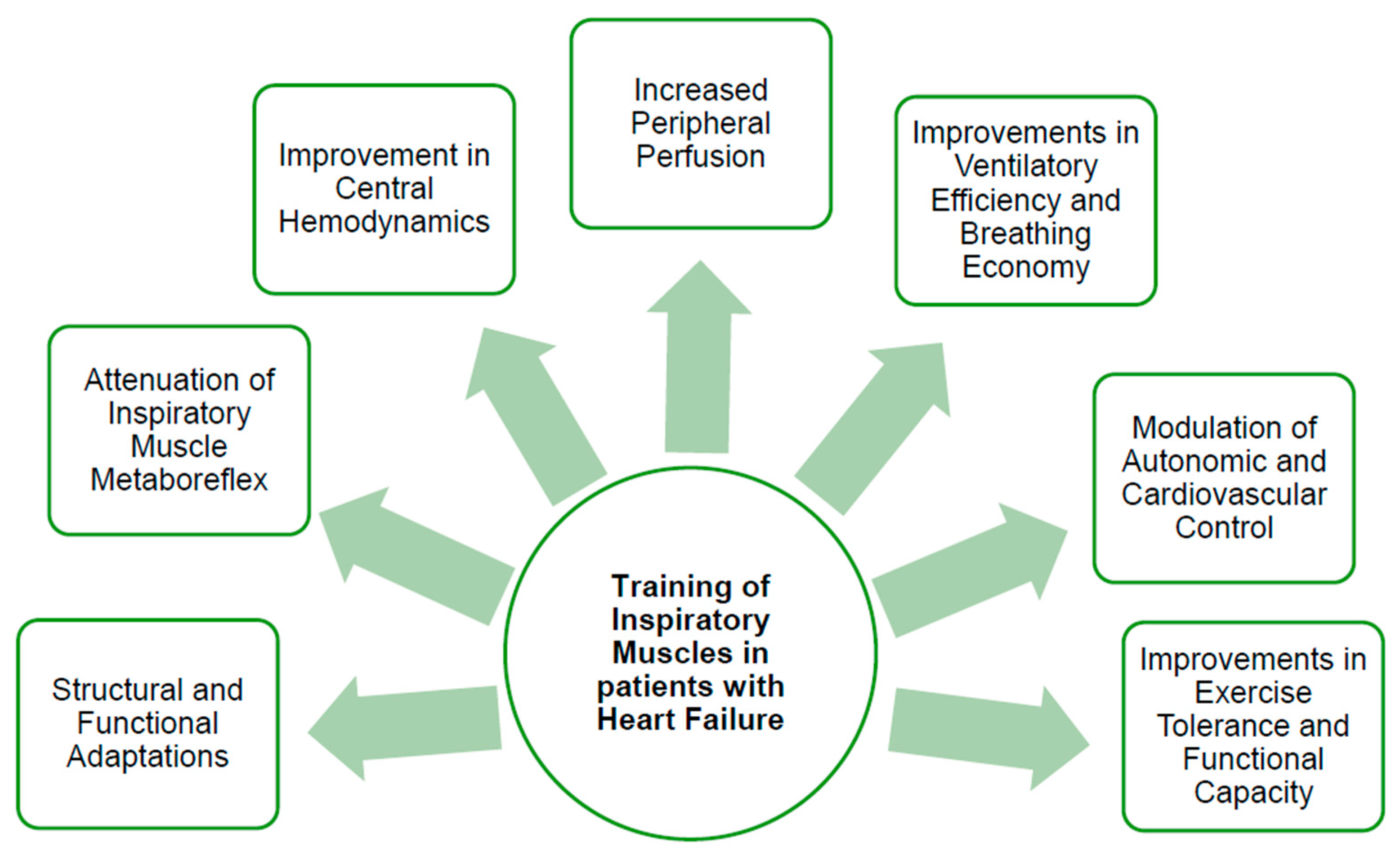
4.2. Attenuation of the Inspiratory Muscle Metaboreflex, Central Hemodynamics and Peripheral Perfusion
Structural and functional adaptations induced by IMT in patients with HFrEF, including increased DT and enhanced inspiratory muscle strength, are associated with reduced accumulation of metabolic byproducts [49] and attenuation of peripheral vasoconstriction [50]. Moreno et al. [49] demonstrated a statistically significant reduction in circulating blood lactate concentrations at fatigue in patients with HFrEF and IMW following a low-intensity IMT protocol performed at 30% of MIP, indicating improved metabolic efficiency of the respiratory muscles. Previous studies have demonstrated that both low (30% of MIP) and high-intensity (60% of MIP) IMT performed five to seven times per week for at least four weeks significantly modulate inspiratory muscle metaboreflex activity in patients with HFrEF [8,49,51]. In contrast, Laoutaris et al. [52] reported no significant effects on endothelium-dependent vasodilatation, likely attributable to the lower training frequency of three sessions per week over ten weeks.
Improvement in stroke volume has been reported after 12 weeks of H-IMT in patients with HFrEF and IMW [53]. These hemodynamic changes, along with reduced chemoreflex-mediated sympathetic activation, may explain improved peripheral oxygen saturation in the forearm and intercostal muscles, reflecting enhanced oxygen delivery and utilization [49]. Furthermore, a single H-IMT session in stable HFrEF has shown increased thenar muscle reperfusion (p = 0.007) and oxygen consumption rates (p = 0.021), suggesting acute benefits on peripheral microvascular function [50].
4.3. Improvements in Ventilatory Efficiency and Breathing Economy
Two systematic reviews and meta-analyses have shown that IMT significantly reduces the minute ventilation/carbon dioxide production (VE/VCO2) slope in HFrEF, indicating improved ventilatory efficiency [54,55]. Given that an elevated VE/VCO2 slope is a well-established marker of disease severity and poor prognosis in patients with LVEF <45% [56], these findings suggest that IMT favorably improves the ventilatory control and exercise response in this population.
In contrast, when the same outcome was examined in patients with HFpEF, only a non-significant trend toward reduction in the VE/VCO2 slope was observed in both meta-analyses [54,55]. The lack of statistical significance may be attributable to substantial heterogeneity across the included studies, particularly with respect to IMT protocol characteristics and the baseline functional status of participants [57]. Notwithstanding the overall inconclusive evidence in patients with HFpEF, Palau et al. [46] reported a significant between-group reduction in the VE/VCO2 slope following IMT, suggesting that ventilatory efficiency may improve under specific intervention conditions in this population. Furthermore, the distinct pathophysiological mechanisms underlying patients with HFpEF [58], including a greater contribution of peripheral vascular dysfunction, chronotropic incompetence, and altered ventricular compliance, may attenuate the relative contribution of inspiratory muscle dysfunction to ventilatory inefficiency, thereby limiting the magnitude of response to IMT. Weiner et al. [47] evaluated respiratory rate at rest and during peak exercise in patients with HFrEF and IMW. Following the intervention, the IMT group demonstrated reductions in respiratory rate at both rest and peak exercise, whereas no changes were observed in the sham-IMT group. These findings support the notion of reducing the oxygen cost of breathing.
4.4. Modulation of Autonomic and Cardiovascular Control
Evidence regarding the effects of IMT on autonomic function, as assessed by heart rate variability (HRV) indices, remains limited. A systematic review by de Abreu et al. [59] highlighted inconsistencies between two studies. Laoutaris et al. [52] observed no significant changes in HRV following 10 weeks of H-IMT (60% of MIP), whereas Moreno et al. [49] reported significant improvements after 12 weeks of low-intensity IMT (30% of MIP). Regarding cardiovascular control, Moreno et al. [49] demonstrated that resting mean arterial blood pressure was reduced following 8 weeks of low-intensity IMT in patients with HFrEF and IMW. However, increases in mean arterial blood pressure were comparable in both the IMT group and the non-training group during respiratory fatigue, suggesting that IMT may modulate hemodynamic responses primarily at rest rather than under maximal ventilatory stress [49].
4.5. Effects on Exercise Tolerance and Functional Capacity
IMT appears to favorably influence several contributing mechanisms, including reducing the oxygen cost of breathing and improving ventilatory efficiency (VE/VCO2 slope), which may translate into enhanced exercise performance. Two meta-analyses reported statistically significant increases in peak oxygen uptake (VO2peak) following IMT (p < 0.003 and p = 0.02, respectively) [55,60] in patients with HF across varying disease severity. In contrast, when exercise capacity was evaluated using metabolic equivalents (METs), a recent meta-analysis found no significant changes in the overall HF population [54], highlighting potential inconsistencies depending on the outcome measure used.
Regarding functional capacity assessed by the six-minute walk test (6MWT), findings remain heterogeneous. Two meta-analyses [54,60] reported a significant increase in six-minute walk distance (6MWD) in patients with HF. On the contrary, a previous meta-analysis reported no statistically significant improvements in 6MWD following IMT [55]. Nevertheless, a potentially greater responsiveness is indicated in 6MWD in patients with HFpEF [55]. The discrepancies across studies are likely attributable to substantial heterogeneity in IMT protocols, baseline muscle strength, and HF phenotype.
5. Inspiratory Muscle Training Modalities
A variety of IMT modalities are available, depending on the primary training objective, namely, the improvement of inspiratory muscle strength, endurance, or both. Mechanical threshold loading, flow-resistive loading, normocapnic hyperpnea, and electronic tapered flow-resistive loading are applied across different clinical settings. There is limited direct comparative evidence between IMT modalities in patients with HF [61]. The mechanical threshold loading and electronic tapered flow-resistive loading are most commonly used [61].
Selection of the appropriate IMT modality should be based not only on the training objective but also on several factors, including baseline MIP, device adjustability [62], training setting (home-based vs. hospital-based), level of supervision, availability of biofeedback, and the patient’s economic, functional, and clinical status [63]. Prior to IMT prescription, assessment of inspiratory muscle function is essential and should be interpreted against normative reference values. Inspiratory muscle strength is most commonly assessed by measuring MIP (cm H2O) [7,64], whereas endurance is evaluated through outcomes such as sustained MIP until task failure, time to task failure, and/or total external work performed during testing [64].
Mechanical threshold loading IMT is performed using devices that contain a spring-loaded, one-way valve. During training, the valve remains closed until the patient generates a predetermined threshold pressure. Once the generated inspiratory negative pressure exceeds the set threshold, the valve opens, and airflow is permitted. As a result, inspiration occurs when the inspiratory muscles sustain a negative pressure greater than the threshold level. Although the force generated before valve opening cannot be quantified, once the valve opens, the workload decreases markedly during a substantial portion of the inspiratory phase. The inability to determine the exact resistance generated by the inspiratory muscles constitutes a significant limitation of this technique. Calibrated spring-loaded valves are important to maintain accuracy during training sessions [57].
In flow-resistive loading IMT, inspiratory efforts are performed through devices with adjustable orifices that impose graded airflow resistance. A reduction in orifice diameter increases airflow resistance, thereby augmenting the inspiratory training load [62]. Resistance can be modified by altering the number and/or size of the openings; however, it is dependent not only on orifice characteristics but also on the inspiratory airflow rate [62].
In normocapnic hyperpnea training, the training stimulus is achieved by increasing ventilatory volume rather than inspiratory pressure. Patients are instructed to breathe at a high minute ventilation corresponding to a predetermined percentage of their maximal voluntary ventilation for a specified duration. Normocapnia is maintained through a rebreathing system or controlled CO2 supplementation. Although this modality was initially confined to laboratory settings, handheld electronic portable devices are now available for clinical and home-based use [65].
Electronic tapered flow-resistive loading training represents an alternative IMT modality in which patients perform repeated inspiratory efforts against a threshold or electronically controlled resistance that is increased in a stepwise manner. Load increments may occur after a predetermined number of repetitions or according to the patient’s tolerance. This approach offers several advantages, including the application of workload throughout the full range of inspiration isokinetic-like workload, precise control of training intensity, and the provision of biofeedback. Training intensity is typically prescribed relative to the individual’s MIP [57].
6. Clinical Guideline Recommendations for Integrating Inspiratory Muscle Training into the Multimodal Heart Failure Toolkit
European Society of Cardiology (ESC) guidelines recommend the integration of IMT as an adjunct to other exercise modalities in patients with HF [66]. Evidence from systematic reviews indicates that adding IMT to AE significantly improves MIP and QoL, although no consistent additional benefit has been demonstrated for VO2peak [67].
Current clinical guidelines provide clear direction for IMT implementation. The ESC recommends initiating IMT at low intensity (approximately 30% of MIP) with progressive increases up to 60% of MIP, alongside reassessment and adjustment of training intensity every 7–10 days [66,68]. Suggested training duration is 20–30 min per day, with 3–5 sessions per week for a minimum of 8 weeks. For deconditioned patients, similar intensity ranges (30–60% of MIP), with session durations of 15–30 min over 10–12 weeks, are recommended, supporting a gradual and individualized progression model aimed at optimizing respiratory muscle adaptation while maintaining safety [14].
Across exercise intensities, a general shift toward the application of higher training loads has been observed. Systematic reviews [69,70] have demonstrated that greater improvements in MIP were associated with higher IMT intensities (60–90% of MIP), supporting a dose–response relationship. These findings are consistent with the principle of progressive overload, whereby higher resistive loading facilitates greater inspiratory motor unit recruitment and promotes adaptive responses of the respiratory musculature [44].
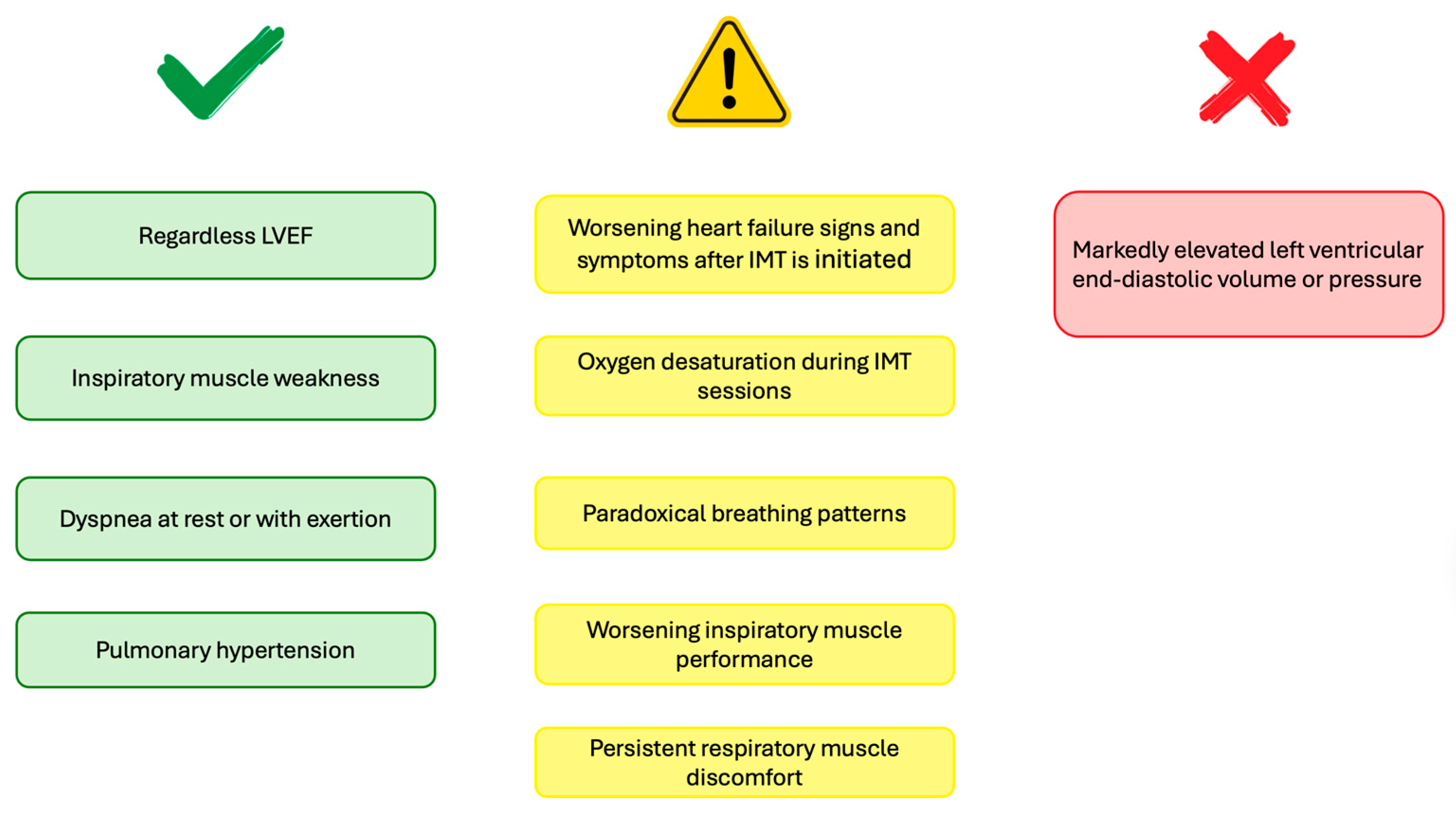
Potential indications for IMT include IMW, dyspnea at rest or during exertion, and selected cases of pulmonary hypertension. Conversely, relative contraindications include markedly elevated left ventricular end-diastolic volume or pressure, oxygen desaturation during IMT sessions, paradoxical breathing patterns, worsening inspiratory muscle performance, and persistent respiratory muscle discomfort [57]. Earlier guidelines recommended IMT primarily for patients with VO2peak < 18 mL·kg−1·min−1 or 6MWD < 450 m, or for those with higher functional capacity only in the presence of IMW [66]. More recent guidelines support IMT participation regardless of LVEF [68]. Indications, signs for readjustment, and contraindications are summarized in Figure 3.
In terms of safety, the current body of literature provides limited direct evidence, as recent systematic reviews [55,60,69] have not specifically evaluated safety outcomes in HF populations undergoing IMT. However, Gomes Neto et al. [71] reported no adverse events in a randomized trial involving clinically stable HF patients (NYHA class II–III) undergoing supervised high-intensity IMT [72]. Furthermore, no increased risk of mortality or HF decompensation has been reported with IMT implementation, supporting its safety profile as a well-tolerated adjunct therapy [73]. Accurate assessment of exercise intensity and vigilance for symptoms such as excessive dyspnea, dizziness, oxygen desaturation, or abnormal hemodynamic responses remains essential to ensure patient safety [57].
Supervision during the initial implementation of IMT may be appropriate to ensure correct technique, intensity prescription, and early detection of any adverse responses, whereas continuous or periodic monitoring is particularly important in home-based programs, as it is generally presented in clinical guidelines [68]. Piotrowicz et al. [74] described a hybrid approach in which patients initiated IMT under hospital supervision and subsequently continued the program at home. Training intensity was progressively increased from 30% to 60% of MIP, demonstrating the feasibility of combining supervised and home-based IMT within a cardiac rehabilitation framework. Although evidence regarding the integration of IMT into structured tele-rehabilitation programs remains limited, tele-rehabilitation has emerged as a safe and feasible strategy, associated with minimal adverse events, improved adherence, and favorable functional outcomes [75].
Figure 3.
Indications (green), signs for readjustment (yellow), and contraindications (red) of inspiratory muscle training in patients with heart failure.
7. Practical Considerations for Implementing IMT
When implementing IMT in patients with HF, several practical and clinical considerations must be addressed to ensure safety, effectiveness, and adherence. Initial patient assessment is essential and should include both subjective and objective measures, such as MIP, symptom severity, and clinical stability. Continuous monitoring of dyspnea, fatigue, heart rate, oxygen saturation, and arterial blood pressure is recommended, particularly during the initial sessions of training.
The selection of IMT modality and device should be individualized according to patient capability, therapeutic goals, the availability of supervision, and the exercise setting. For instance, the selection of electronic tapered flow-resistive loading may be applied during the initial training sessions as an approach that may enhance patients’ performance [57]. The aforementioned training modality provides visual feedback and allows specific quantification of the power curve (a parameter related to sustained MIP) reflecting inspiratory effort. Furthermore, this modality can be utilized at baseline, at strategic interim assessment points, and during post-intervention evaluations.
In cardiac populations, factors such as advanced age, lower socioeconomic status, female gender, and psychological characteristics, including depression, have been associated with reduced adherence to rehabilitation programs [76]. Specifically, HF patients with multiple comorbidities demonstrate a reduced likelihood of adherence to prescribed exercise interventions [76]. Given the limited evidence regarding adherence to IMT programs, a range of general strategies is proposed to optimize compliance with this modality. Clinicians may implement approaches such as fostering patient commitment, encouraging family involvement, maintaining regular contact via telephone or email, monitoring vital signs, and incorporating training diaries within cardiac rehabilitation programs [77].
Training should be modified, supplemented with oxygen, or temporarily discontinued in the presence of markedly elevated left ventricular end-diastolic volume or pressure, oxygen desaturation, paradoxical breathing patterns, or worsening symptoms such as dyspnea, dizziness, chest pain, or abnormal cardiovascular responses [57].
8. Limitations, Challenges, and Future Directions
The majority of available literature has predominantly focused on patients with HFrEF, with relative under-representation of patients with HFpEF. This imbalance limits the generalizability of findings across the broader HF spectrum. In addition, many studies are characterized by small sample sizes, thereby reducing statistical power. Another important limitation is the considerable heterogeneity across studies, particularly with respect to training intensity, duration, frequency, and supervision, which complicates comparisons and limits the ability to establish standardized recommendations. Furthermore, most investigations have not examined long-term clinical outcomes, such as mortality, hospitalization rates, or healthcare utilization, thereby restricting conclusions regarding the prognostic impact of IMT.
Future research should prioritize adequately powered randomized controlled trials that evaluate the combined effects of IMT with other exercise modalities (AE and strength exercise) within multimodal rehabilitation programs. Greater emphasis should also be placed on the integration of digital technologies and tele-rehabilitation strategies to enhance adherence, monitoring, and individualized progression. Importantly, further studies are needed to clarify the effects of IMT in patients with the HFpEF phenotype and to determine the safety, efficacy, and dose–response relationship associated with higher training loads.
9. Conclusions
IMT is a relatively low-cost, simple, and feasible therapeutic exercise modality in the management of patients with HF. The clinical benefits are supported by well-established physiological mechanisms, including structural and functional adaptations of inspiratory muscles, attenuation of the inspiratory muscle metaboreflex, improved ventilatory efficiency, and modulation of autonomic and cardiovascular control. Through these mechanisms, IMT alleviates symptoms such as dyspnea, thereby contributing to enhanced QoL. Given its effects, IMT represents a practical adjunct to standard cardiac rehabilitation programs, particularly for patients with IMW. Following a comprehensive assessment, clinicians may select the most appropriate IMT modality and prescribe tailored, individualized training programs. However, implementation should be undertaken with careful consideration of potential contraindications and may require protocol modifications in specific clinical circumstances. Current evidence suggests that higher training loads and longer intervention durations may maximize training effects. Nevertheless, adequately powered and methodologically rigorous trials are still required to determine the long-term clinical and prognostic impact of IMT and to further clarify its optimal integration into routine HF management.
Author Contributions
Conceptualization, E.A.K. and M.I.; methodology, M.I.; writing—original draft preparation, M.I.; writing—review and editing, M.I. and E.A.K.; supervision, E.A.K. All authors have read and agreed to the published version of the manuscript.
Funding
This research received no external funding.
Institutional Review Board Statement
Not applicable.
Informed Consent Statement
Not applicable.
Data Availability Statement
No new data were created or analyzed in this study. Data sharing is not applicable to this article.
Acknowledgments
The authors would like to thank Panagiotis Dalamarinis, for figure preparation.
Conflicts of Interest
The authors declare no conflicts of interest.
Abbreviations
The following abbreviations are used in this manuscript:
| 6MWD | Six-Minute Walk Distance |
| 6MWT | Six-Minute Walk Test |
| AE | Aerobic Exercise |
| ATP | Adenosine Triphosphate |
| CO | Cardiac Output |
| DT | Diaphragm Thickness |
| DTF | Diaphragm Thickening Fraction |
| ESC | European Society of Cardiology |
| HF | Heart Failure |
| HFmrEF | Heart Failure with mildly reduced Ejection Fraction |
| HFpEF | Heart Failure with preserved Ejection Fraction |
| HFrEF | Heart Failure with reduced Ejection Fraction |
| H-IMT | High-intensity Inspiratory Muscle Training |
| HRV | Heart Rate Variability |
| IL-6 | Interleukin-6 |
| IMT | Inspiratory Muscle Training |
| IMW | Inspiratory Muscle Weakness |
| LVEF | Left Ventricular Ejection Fraction |
| METs | Metabolic Equivalents |
| MIP | Maximal Inspiratory Pressure |
| NYHA | New York Heart Association classification |
| PA | Physical Activity |
| QoL | Quality of Life |
| RAAS | Renin–Angiotensin–Aldosterone System |
| TNF-α | Tumor Necrosis Factor-α |
| VE/VCO2 | Minute Ventilation/Carbon Dioxide Production |
| VO2peak | Peak Oxygen Uptake |
References
- Correale, M.; Paolillo, S.; Mercurio, V.; Limongelli, G.; Barillà, F.; Ruocco, G.; Palazzuoli, A.; Scrutinio, D.; Lagioia, R.; Lombardi, C.; et al. Comorbidities in chronic heart failure: An update from Italian Society of Cardiology (SIC) Working Group on Heart Failure. Eur. J. Intern. Med. 2020, 71, 23–31. [Google Scholar] [CrossRef] [Scilit]
- Khan, M.S.; Shahid, I.; Bennis, A.; Rakisheva, A.; Metra, M.; Butler, J. Global epidemiology of heart failure. Nat. Rev. Cardiol. 2024, 21, 717–734. [Google Scholar] [CrossRef] [Scilit]
- Miura, Y.; Fukumoto, Y.; Miura, T.; Shimada, K.; Asakura, M.; Kadokami, T.; Ando, S.; Miyata, S.; Sakata, Y.; Daida, H.; et al. Impact of physical activity on cardiovascular events in patients with chronic heart failure. A multicenter prospective cohort study. Circ. J. 2013, 77, 2963–2972. [Google Scholar] [CrossRef] [Scilit]
- Sacco, S.J.; Park, C.L.; Suresh, D.P.; Bliss, D. Living with heart failure: Psychosocial resources, meaning, gratitude and well-being. Heart Lung 2014, 43, 213–218. [Google Scholar] [CrossRef] [Scilit] [PubMed]
- von Haehling, S.; Ebner, N.; Dos Santos, M.R.; Springer, J.; Anker, S.D. Muscle wasting and cachexia in heart failure: Mechanisms and therapies. Nat. Rev. Cardiol. 2017, 14, 323–341. [Google Scholar] [CrossRef] [Scilit] [PubMed]
- Hamazaki, N.; Kamiya, K.; Matsuzawa, R.; Nozaki, K.; Ichikawa, T.; Tanaka, S.; Nakamura, T.; Yamashita, M.; Maekawa, E.; Noda, C.; et al. Prevalence and prognosis of respiratory muscle weakness in heart failure patients with preserved ejection fraction. Respir. Med. 2020, 161, 105834. [Google Scholar] [CrossRef] [Scilit]
- American Thoracic Society; European Respiratory Society. ATS/ERS Statement on respiratory muscle testing. Am. J. Respir. Crit. Care Med. 2002, 166, 518–624. [Google Scholar] [CrossRef] [Scilit] [PubMed]
- Chiappa, G.R.; Roseguini, B.T.; Vieira, P.J.; Alves, C.N.; Tavares, A.; Winkelmann, E.R.; Ferlin, E.L.; Stein, R.; Ribeiro, J.P. Inspiratory muscle training improves blood flow to resting and exercising limbs in patients with chronic heart failure. J. Am. Coll. Cardiol. 2008, 51, 1663–1671. [Google Scholar] [CrossRef] [Scilit]
- Coats, A.J.; Clark, A.L.; Piepoli, M.; Volterrani, M.; Poole-Wilson, P.A. Symptoms and quality of life in heart failure: The muscle hypothesis. Br. Heart J. 1994, 72, S36–S39. [Google Scholar] [CrossRef] [Scilit]
- Katayıfçı, N.; Boşnak Güçlü, M. Inspiratory muscle weakness further impairs exercise capacity and respiratory functions and increases dyspnea perception in patients with heart failure. Sci. Rep. 2025, 15, 32229. [Google Scholar] [CrossRef] [Scilit]
- Ponikowski, P.; Voors, A.A.; Anker, S.D.; Bueno, H.; Cleland, J.G.F.; Coats, A.J.S.; Falk, V.; Gonza’lez-Juanatey, J.R.; Harjola, V.P.; Jankowska, E.A.; et al. 2016 ESC Guidelines for the diagnosis and treatment of acute and chronic heart failure: The Task Force for the diagnosis and treatment of acute and chronic heart failure of the European Society of Cardiology (ESC). Developed with the special contribution of the Heart Failure Association (HFA) of the ESC. Eur. J. Heart Fail. 2016, 18, 891–975. [Google Scholar] [CrossRef] [Scilit] [PubMed]
- Callum, K.; Swinton, P.; Gorely, T.; Crabtree, D.; Leslie, S. Physiological and psychological outcomes of high intensity interval training in patients with heart failure compared to moderate continuous training and usual care: A systematic review with meta analysis. Heart Lung 2024, 64, 117–127. [Google Scholar] [CrossRef] [Scilit]
- Yang, C.; Zhang, L.; Cheng, Y.; Zhang, M.; Zhao, Y.; Zhang, T.; Dong, J.; Xing, J.; Zhen, Y.; Wang, C. High intensity interval training vs. moderate intensity continuous training on aerobic capacity and functional capacity in patients with heart failure: A systematic review and meta-analysis. Front. Cardiovasc. Med. 2024, 11, 1302109. [Google Scholar] [CrossRef] [Scilit]
- Pelliccia, A.; Sharma, S.; Gati, S.; Bäck, M.; Börjesson, M.; Caselli, S.; Collet, J.P.; Corrado, D.; Drezner, J.A.; Halle, M.; et al. 2020 ESC Guidelines on sports cardiology and exercise in patients with cardiovascular disease. Eur. Heart J. 2021, 42, 17–96. [Google Scholar] [CrossRef] [Scilit]
- Park, L.K.; Coggan, A.R.; Peterson, L.R. Skeletal Muscle Contractile Function in Heart Failure with Reduced Ejection Fraction—A Focus on Nitric Oxide. Front. Physiol. 2022, 3, 872719. [Google Scholar] [CrossRef] [Scilit] [PubMed]
- Magnusson, G.; Kaijser, L.; Sylvén, C.; Karlberg, K.E.; Isberg, B.; Saltin, B. Peak skeletal muscle perfusion is maintained in patients with chronic heart failure when only a small muscle mass is exercised. Cardiovasc. Res. 1997, 33, 297–306. [Google Scholar] [CrossRef] [Scilit]
- Mangner, N.; Garbade, J.; Heyne, E.; van den Berg, M.; Winzer, E.B.; Hommel, J.; Sandri, M.; Jozwiak-Nozdrzykowska, J.; Meyer, A.L.; Lehmann, S.; et al. Molecular Mechanisms of Diaphragm Myopathy in Humans with Severe Heart Failure. Circ. Res. 2021, 128, 706–719. [Google Scholar] [CrossRef] [Scilit]
- Salah, H.M.; Goldberg, L.R.; Molinger, J.; Felker, G.M.; Applefeld, W.; Rassaf, T.; Tedford, R.J.; Mirro, M.; Cleland, J.G.F.; Fudim, M. Diaphragmatic Function in Cardiovascular Disease: JACC Review Topic of the Week. J. Am. Coll. Cardiol. 2022, 80, 1647–1659. [Google Scholar] [CrossRef] [Scilit]
- Powers, S.K.; Morton, A.B.; Hyatt, H.; Hinkley, M.J. The Renin-Angiotensin System and Skeletal Muscle. Exerc. Sport Sci. Rev. 2018, 46, 205–214. [Google Scholar] [CrossRef] [Scilit]
- Maryam, P.; Treesa, P.V.; Tazneem, B. Unraveling the complex pathophysiology of heart failure: Insights into the role of renin-angiotensin-aldosterone system (RAAS) and sympathetic nervous system (SNS). Curr. Probl. Cardiol. 2024, 49, 102411. [Google Scholar] [CrossRef] [Scilit] [PubMed]
- Petretta, M.; Condorelli, G.L.; Spinelli, L.; Scopacasa, F.; de Caterina, M.; Leosco, D.; Vicario, M.L.; Bonaduce, D. Circulating levels of cytokines and their site of production in patients with mild to severe chronic heart failure. Am. Heart J. 2000, 140, E28. [Google Scholar] [CrossRef] [Scilit]
- Prokopidis, K.; Irlik, K.; Hendel, M.; Piaśnik, J.; Lip, G.Y.H.; Nabrdalik, K. Prognostic Impact and Prevalence of Cachexia in Patients with Heart Failure: A Systematic Review and Meta-Analysis. J. Cachexia Sarcopenia Muscle 2024, 15, 2536–2543. [Google Scholar] [CrossRef] [Scilit]
- Thanapholsart, J.; Khan, E.; Ismail, T.F.; Lee, G.A. The complex pathophysiology of cardiac cachexia: A review of current pathophysiology and implications for clinical practice. Am. J. Med. Sci. 2023, 365, 9–18. [Google Scholar] [CrossRef] [Scilit] [PubMed]
- Courelli, V.; Ahmad, A.; Ghassemian, M.; Pruitt, C.; Mills, P.J.; Schmid-Schönbein, G.W. Digestive Enzyme Activity and Protein Degradation in Plasma of Heart Failure Patients. Cell. Mol. Bioeng. 2021, 14, 583–596. [Google Scholar] [CrossRef] [Scilit]
- Pagnesi, M.; Serafini, L.; Chiarito, M.; Stolfo, D.; Baldetti, L.; Inciardi, R.M.; Tomasoni, D.; Adamo, M.; Lombardi, C.M.; Sammartino, A.M.; et al. Impact of malnutrition in patients with severe heart failure. Eur. J. Heart Fail. 2024, 26, 1585–1593. [Google Scholar] [CrossRef] [Scilit]
- Sharma, G.; Goodwin, J. Effect of aging on respiratory system physiology and immunology. Clin. Interv. Aging 2006, 1, 253–260. [Google Scholar] [CrossRef] [Scilit]
- Watsford, M.L.; Murphy, A.J.; Pine, M.J. The effects of ageing on respiratory muscle function and performance in older adults. J. Sci. Med. Sport 2007, 10, 36–44. [Google Scholar] [CrossRef] [Scilit] [PubMed]
- Lewsey, S.C.; Samuel, T.J.; Schär, M.; Sourdon, J.; Goldenberg, J.R.; Yanek, L.R.; Lai, S.; Steinberg, A.M.; Bottomley, P.A.; Gerstenblith, G.; et al. Skeletal Muscle Quantity Versus Quality in Heart Failure: Exercise Intolerance and Outcomes in Older Patients with HFpEF Are Related to Abnormal Skeletal Muscle Metabolism Rather Than Age-Related Skeletal Muscle Loss. Circ. Heart Fail. 2025, 18, e012512. [Google Scholar] [CrossRef] [Scilit] [PubMed]
- Sin, P.; Sanjanwala, R.; Zieroth, R. Managing Common Co-morbidities in Heart Failure. Can. J. Gen. Intern. Med. 2020, 15, 14–21. [Google Scholar] [CrossRef] [Scilit]
- Miernik, S.; Matusiewicz, A.; Olesińska, M. Drug-Induced Myopathies: A Comprehensive Review and Update. Biomedicines 2024, 12, 987. [Google Scholar] [CrossRef] [Scilit]
- Niklasson, A.; Maher, J.; Patil, R.; Sillén, H.; Chen, J.; Gwaltney, C.; Rydén, A. Living with heart failure: Patient experiences and implications for physical activity and daily living. ESC Heart Fail. 2022, 9, 1206–1215. [Google Scholar] [CrossRef] [Scilit] [PubMed]
- Jordan, C.; Charman, S.J.; Batterham, A.M.; Flynn, D.; Houghton, D.; Errington, L.; MacGowan, G.; Avery, L. Habitual physical activity levels of adults with heart failure: Systematic review and meta-analysis. Heart 2023, 109, 1357–1362. [Google Scholar] [CrossRef] [Scilit]
- McParland, C.; Krishnan, B.; Wang, Y.; Gallagher, C.G. Inspiratory muscle weakness and dyspnea in chronic heart failure. Am. Rev. Respir. Dis. 1992, 146, 467–472. [Google Scholar] [CrossRef] [Scilit]
- Olson, T.M. What makes the heart fail? New insights from defective genes. Acta. Paediatr. Suppl. 2006, 95, 17–21. [Google Scholar] [CrossRef] [Scilit]
- Johnson, B.D.; Beck, K.C.; Olson, L.J.; O’Malley, K.A.; Allison, T.G.; Squires, R.W.; Gau, G.T. Ventilatory constraints during exercise in patients with chronic heart failure. Chest 2000, 117, 321–332. [Google Scholar] [CrossRef] [Scilit]
- Poole, D.C.; Hirai, D.M.; Copp, S.W.; Musch, T.I. Muscle oxygen transport and utilization in heart failure: Implications for exercise (in)tolerance. Am. J. Physiol. Heart Circ. Physiol. 2012, 302, H1050–H1063. [Google Scholar] [CrossRef] [Scilit]
- Smith, J.R.; Taylor, B.J. Inspiratory muscle weakness in cardiovascular diseases: Implications for cardiac rehabilitation. Prog. Cardiovasc. Dis. 2022, 70, 49–57. [Google Scholar] [CrossRef] [Scilit]
- Yamada, K.; Kinugasa, Y.; Sota, T.; Miyagi, M.; Sugihara, S.; Kato, M.; Yamamoto, K. Inspiratory muscle weakness is associated with exercise intolerance in patients with heart failure with preserved ejection fraction: A preliminary study. J. Card. Fail. 2016, 22, 38–47. [Google Scholar] [CrossRef] [Scilit] [PubMed]
- Kinugasa, Y.; Yamada, K.; Sota, T.; Miyagi, M.; Sugihara, S.; Kato, M.; Yamamoto, K. Respiratory Muscle Weakness in Patients with Heart Failure with Preserved Ejection Fraction. J. Card. Fail. 2014, 20, S136. [Google Scholar] [CrossRef] [Scilit]
- Shah, R.V.; Cahalin, L.P.; Haus, J.M.; Allsup, K.; Delligatti, A.; Wolf, C.; Checko Scioli, E.R.; Aragam, J.R.; Gottlieb, D.J.; Byard, T.D.; et al. The role of maximal inspiratory pressure on functional performance in adults with heart failure. ESC Heart Fail. 2024, 11, 4231–4241. [Google Scholar] [CrossRef] [Scilit]
- Meyer, F.J.; Borst, M.M.; Zugck, C.; Kirschke, A.; Schellberg, D.; Kübler, W.; Haass, M. Respiratory muscle dysfunction in congestive heart failure: Clinical correlation and prognostic significance. Circulation 2001, 103, 2153–2158. [Google Scholar] [CrossRef] [Scilit]
- Nishimura, Y.; Maeda, H.; Tanaka, K.; Nakamura, H.; Hashimoto, Y.; Yokoyama, M. Respiratory muscle strength and hemodynamics in chronic heart failure. Chest 1994, 105, 355–359. [Google Scholar] [CrossRef] [Scilit]
- Hamazaki, N.; Kamiya, K.; Yamamoto, S.; Nozaki, K.; Ichikawa, T.; Matsuzawa, R.; Tanaka, S.; Nakamura, T.; Yamashita, M.; Maekawa, E.; et al. Changes in Respiratory Muscle Strength Following Cardiac Rehabilitation for Prognosis in Patients with Heart Failure. J. Clin. Med. 2020, 9, 952. [Google Scholar] [CrossRef] [Scilit]
- Gransee, H.M.; Mantilla, C.B.; Sieck, G.C. Respiratory muscle plasticity. Compr. Physiol. 2012, 2, 1441–1462. [Google Scholar] [CrossRef] [Scilit]
- Tanriverdi, A.; Savci, S.; Ozcan Kahraman, B.; Odaman, H.; Ozpelit, E.; Senturk, B.; Ozsoy, I.; Baran, A.; Akdeniz, B.; Acar, S.; et al. Effects of high intensity interval-based inspiratory muscle training in patients with heart failure: A single-blind randomized controlled trial. Heart Lung 2023, 62, 1–8. [Google Scholar] [CrossRef] [Scilit] [PubMed]
- Palau, P.; Domínguez, E.; Núñez, E.; Schmid, J.P.; Vergara, P.; Ramón, J.M.; Mascarell, B.; Sanchis, J.; Chorro, F.J.; Núñez, J. Effects of inspiratory muscle training in patients with heart failure with preserved ejection fraction. Eur. J. Prev. Cardiol. 2014, 21, 1465–1473. [Google Scholar] [CrossRef] [Scilit] [PubMed]
- Weiner, P.; Waizman, J.; Magadle, R.; Berar-Yanay, N.; Pelled, B. The Effect of Specific Inspiratory Muscle Training on the Sensation of Dyspnea and Exercise Tolerance in Patients with Congestive Heart Failure. Clin. Cardiol. 1999, 22, 727–732. [Google Scholar] [CrossRef] [Scilit] [PubMed]
- Zacarias Rondinel, T.; Bocchi, L.; Cipriano Júnior, G.; Chiappa, G.R.D.S.; Martins, G.S.; Mateus, S.R.M.; Cahalin, L.P.; Cipriano, G.F.B. Diaphragm thickness and mobility elicited by two different modalities of inspiratory muscle loading in heart failure participants: A randomized crossover study. PLoS ONE 2024, 19, e0302735. [Google Scholar] [CrossRef] [Scilit]
- Moreno, A.M.; Toledo-Arruda, A.C.; Lima, J.S.; Duarte, C.S.; Villacorta, H.; Nóbrega, A.C.L. Inspiratory Muscle Training Improves Intercostal and Forearm Muscle Oxygenation in Patients with Chronic Heart Failure: Evidence of the Origin of the Respiratory Metaboreflex. J. Card. Fail. 2017, 23, 672–679. [Google Scholar] [CrossRef] [Scilit] [PubMed]
- Katsoula, C.; Karatzanos, E.; Rovina, N.; Routsi, C.; Vasileiou, I.; Papadopoulou, E.; Briasoulis, A.; Nanas, S.; Vasileiadis, I. The acute effect of respiratory muscle training on microcirculation in patients with chronic heart failure. Health Res. J. 2023, 9, 96–103. [Google Scholar] [CrossRef] [Scilit]
- Antunes-Correa, L.M.; Trevizan, P.F.; Bacurau, A.V.N.; Ferreira-Santos, L.; Gomes, J.L.P.; Urias, U.; Oliveira, P.A.; Alves, M.J.N.N.; de Almeida, D.R.; Brum, P.C.; et al. Effects of aerobic and inspiratory training on skeletal muscle microRNA-1 and downstream-associated pathways in patients with heart failure. J. Cachexia Sarcopenia Muscle 2020, 11, 89–102. [Google Scholar] [CrossRef] [Scilit] [PubMed]
- Laoutaris, I.D.; Dritsas, A.; Brown, M.D.; Manginas, A.; Kallistratos, M.S.; Chaidaroglou, A.; Degiannis, D.; Alivizatos, P.A.; Cokkinos, D.V. Effects of inspiratory muscle training on autonomic activity, endothelial vasodilator function, and N-terminal pro-brain natriuretic peptide levels in chronic heart failure. J. Cardiopulm. Rehabil. Prev. 2008, 28, 99–106. [Google Scholar] [CrossRef] [Scilit]
- Sadek, Z.; Ahmaidi, S.; Youness, M.; Awada, C.; Joumaa, W.H.; Ramadan, W. Combining aerobic interval, inspiratory and resistance trainings induces better cardiac remodelling and exercise capacity in heart failure patients. Eur. J. Physiother. 2021, 25, 129–137. [Google Scholar] [CrossRef] [Scilit]
- Siddiqi, A.K.; Shahzad, M.; Kumar, A.; Ahmed, M.; Sridharan, L.; Abdou, M.H.; Naeem, M. The efficacy of inspiratory muscle training in improving clinical outcomes in heart failure patients: An updated systematic review and meta-analysis. J. Cardiol. 2025, 85, 374–385. [Google Scholar] [CrossRef] [Scilit]
- Li, H.; Tao, L.; Huang, Y.; Li, Z.; Zhao, J. Inspiratory muscle training in patients with heart failure: A systematic review and meta-analysis. Front. Cardiovasc. Med. 2022, 9, 993846. [Google Scholar] [CrossRef] [Scilit]
- Shen, Y.; Zhang, X.; Ma, W.; Song, H.; Gong, Z.; Wang, Q.; Che, L.; Xu, W.; Jiang, J.; Xu, J.; et al. VE/VCO2 slope and its prognostic value in patients with chronic heart failure. Exp. Ther. Med. 2015, 9, 1407–1412. [Google Scholar] [CrossRef] [Scilit] [PubMed]
- Cahalin, L.P.; Arena, R.; Guazzi, M.; Myers, J.; Cipriano, G.; Chiappa, G.; Lavie, C.J.; Forman, D.E. Inspiratory muscle training in heart disease and heart failure: A review of the literature with a focus on method of training and outcomes. Expert. Rev. Cardiovasc. Ther. 2013, 11, 161–177. [Google Scholar] [CrossRef] [Scilit]
- Fang, J.; Wang, Z.; Yu, J. Advances in pathophysiological mechanisms and therapeutic efficacy of exercise rehabilitation in patients with heart failure with preserved ejection fraction. Front. Cardiovasc. Med. 2025, 12, 1598878. [Google Scholar] [CrossRef] [Scilit] [PubMed]
- de Abreu, R.M.; Rehder-Santos, P.; Minatel, V.; Dos Santos, G.L.; Catai, A.M. Effects of inspiratory muscle training on cardiovascular autonomic control: A systematic review. Auton. Neurosci. 2017, 208, 29–35. [Google Scholar] [CrossRef] [Scilit]
- Yang, M.; Huang, W.; Luo, Z.; Zhang, X.; Su, J.; Yu, P. Effect of inspiratory muscle training in patients with chronic heart failure: A systematic review and meta-analysis of randomized controlled trials. J. Thorac. Dis. 2025, 17, 6242–6253. [Google Scholar] [CrossRef] [Scilit]
- Fernandez-Rubio, H.; Becerro-de-Bengoa-Vallejo, R.; Rodríguez-Sanz, D.; Calvo-Lobo, C.; Vicente-Campos, D.; Chicharro, J.L. Inspiratory Muscle Training in Patients with Heart Failure. J. Clin. Med. 2020, 9, 1710. [Google Scholar] [CrossRef] [Scilit]
- Anderson, J.; Priluck, D.; Dias, K.J. Breaths: A Knowledge Translation Framework for Optimizing Inspiratory Muscle Training in Physical Therapy Practice. Med. Res. Arch. 2025, 13, 6. [Google Scholar] [CrossRef] [Scilit]
- Menzes, K.K.P.; Nascimento, L.R.; Avelino, P.R.; Polese, J.C.; Salmela, L.F.T. A Review on Respiratory Muscle Training Devices. J. Pulm. Respir. Med. 2018, 8, 1–7. [Google Scholar] [CrossRef]
- Laveneziana, P.; Albuquerque, A.; Aliverti, A.; Babb, T.; Barreiro, E.; Dres, M.; Dubé, B.P.; Fauroux, B.; Gea, J.; Guenette, J.A.; et al. ERS statement on respiratory muscle testing at rest and during exercise. Eur. Respir. J. 2019, 53, 1801214. [Google Scholar] [CrossRef] [Scilit] [PubMed]
- Bernardi, E.; Pomidori, L.; Bassal, F.; Contoli, M.; Cogo, A. Respiratory muscle training with normocapnic hyperpnea improves ventilatory pattern and thoracoabdominal coordination, and reduces oxygen desaturation during endurance exercise testing in COPD patients. Int. J. Chron. Obstruct. Pulmon. Dis. 2015, 10, 1899–1906. [Google Scholar] [CrossRef] [Scilit]
- Piepoli, M.F.; Conraads, V.; Corrà, U.; Dickstein, K.; Francis, D.P.; Jaarsma, T.; McMurray, J.; Pieske, B.; Piotrowicz, E.; Schmid, J.P.; et al. Exercise training in heart failure: From theory to practice. A consensus document of the Heart Failure Association and the European Association for Cardiovascular Prevention and Rehabilitation. Eur. J. Heart Fail. 2011, 13, 347–357. [Google Scholar] [CrossRef] [Scilit]
- Gomes Neto, M.; Martinez, B.P.; Conceição, C.S.; Silva, P.E.; Carvalho, V.O. Combined Exercise and Inspiratory Muscle Training in Patients with Heart Failure: A SYSTEMATIC REVIEW AND META-ANALYSIS. J. Cardiopulm. Rehabil. Prev. 2016, 36, 395–401. [Google Scholar] [CrossRef] [Scilit]
- Ambrosetti, M.; Abreu, A.; Corrà, U.; Davos, C.H.; Hansen, D.; Frederix, I.; Iliou, M.C.; Pedretti, R.F.E.; Schmid, J.P.; Vigorito, C.; et al. Secondary prevention through comprehensive cardiovascular rehabilitation: From knowledge to implementation. 2020 update. A position paper from the Secondary Prevention and Rehabilitation Section of the European Association of Preventive Cardiology. Eur. J. Prev. Cardiol. 2021, 28, 460–495. [Google Scholar] [CrossRef] [Scilit]
- Azambuja, A.C.M.; de Oliveira, L.Z.; Sbruzzi, G. Inspiratory Muscle Training in Patients with Heart Failure: What Is New? Systematic Review and Meta-Analysis. Phys. Ther. 2020, 100, 2099–2109. [Google Scholar] [CrossRef] [Scilit] [PubMed]
- Sadek, Z.; Salami, A.; Joumaa, W.H.; Awada, C.; Ahmaidi, S.; Ramadan, W. Best mode of inspiratory muscle training in heart failure patients: A systematic review and meta-analysis. Eur. J. Prev. Cardiol. 2018, 25, 1691–1701. [Google Scholar] [CrossRef] [Scilit] [PubMed]
- Gomes Neto, M.; Ferrari, F.; Helal, L.; Lopes, A.A.; Carvalho, V.O.; Stein, R. The impact of high-intensity inspiratory muscle training on exercise capacity and inspiratory muscle strength in heart failure with reduced ejection fraction: A systematic review and meta-analysis. Clin. Rehabil. 2018, 32, 1482–1492. [Google Scholar] [CrossRef] [Scilit]
- Marco, E.; Ramírez-Sarmiento, A.L.; Coloma, A.; Sartor, M.; Comin-Colet, J.; Vila, J.; Enjuanes, C.; Bruguera, J.; Escalada, F.; Gea, J.; et al. High-intensity vs. sham inspiratory muscle training in patients with chronic heart failure: A prospective randomized trial. Eur. J. Heart Fail. 2013, 15, 892–901. [Google Scholar] [CrossRef] [Scilit]
- Palau, P.; Domínguez, E.; López, L.; Ramón, J.M.; Heredia, R.; González, J.; Santas, E.; Bodí, V.; Miñana, G.; Valero, E.; et al. Inspiratory Muscle Training and Functional Electrical Stimulation for Treatment of Heart Failure with Preserved Ejection Fraction: The TRAINING-HF Trial. Rev. Esp. Cardiol. 2019, 72, 288–297. [Google Scholar] [CrossRef] [Scilit]
- Piotrowicz, E.; Pencina, M.J.; Opolski, G.; Zareba, W.; Banach, M.; Kowalik, I.; Orzechowski, P.; Szalewska, D.; Pluta, S.; Glówczynska, R.; et al. Effects of a 9-Week Hybrid Comprehensive Telerehabilitation Program on Long-term Outcomes in Patients with Heart Failure: The Telerehabilitation in Heart Failure Patients (TELEREH-HF) Randomized Clinical Trial. JAMA Cardiol. 2020, 5, 300–308. [Google Scholar] [CrossRef] [Scilit] [PubMed]
- Isernia, S.; Pagliari, C.; Morici, N.; Toccafondi, A.; Banfi, P.I.; Rossetto, F.; Borgnis, F.; Tavanelli, M.; Brambilla, L.; Baglio, F.; et al. Telerehabilitation Approaches for People with Chronic Heart Failure: A Systematic Review and Meta-Analysis. J. Clin. Med. 2022, 12, 64. [Google Scholar] [CrossRef] [Scilit]
- Resurrección, D.M.; Moreno-Peral, P.; Gómez-Herranz, M.; Rubio-Valera, M.; Pastor, L.; Caldas de Almeida, J.M.; Motrico, E. Factors associated with non-participation in and dropout from cardiac rehabilitation programmes: A systematic review of prospective cohort studies. Eur. J. Cardiovasc. Nurs. 2019, 18, 38–47. [Google Scholar] [CrossRef] [Scilit] [PubMed]
- Barbour, K.A.; Miller, N.H. Adherence to exercise training in heart failure: A review. Heart Fail. Rev. 2008, 13, 81–89. [Google Scholar] [CrossRef] [Scilit] [PubMed]
Disclaimer/Publisher’s Note: The statements, opinions and data contained in all publications are solely those of the individual author(s) and contributor(s) and not of MDPI and/or the editor(s). MDPI and/or the editor(s) disclaim responsibility for any injury to people or property resulting from any ideas, methods, instructions or products referred to in the content. |
© 2026 by the authors. Licensee MDPI, Basel, Switzerland. This article is an open access article distributed under the terms and conditions of the Creative Commons Attribution (CC BY) license.