


Abstract
Bacterial infections, predominantly affecting the gastrointestinal tract and oral cavity, represent a considerable health burden and remain among the leading causes of morbidity and mortality globally. Exploration of therapeutic mediators from medicinal plants continues to be a foundational strategy in modern drug development. The objective of the current study was to synthesize a Mentha arvensis-mediated biogenic iron oxide heterophase nanocomposite as a green therapeutic approach to controlling oral and gastrointestinal (GI) bacteria. Green synthesis is sustainable and an economic alternative for producing nanoparticles, because it is ecologically benign. For the first time native Al-Baha M. arvensis biomass was used for green preparation of mixed-phase iron oxide nanoparticles, and these were characterized by visual observation, X-ray diffraction, scanning electron microscopy, and Energy-Dispersive X-ray and FT-IR spectroscopy. The materialization of a dark brown color preliminarily points toward the establishment of iron oxide heterophase NPs due to reduction by phytochemicals. SEM demonstrated particle sizes approximately ranging between 30 and 80 nm, whereas EDX evidenced the predominance of Fe and O in the nanocomposite. Iron oxide heterophase NPs were also tested against human pathogens. The results displayed effective antibacterial activity, which seems concentration-dependent. The largest zone was recorded for S. aureus, with a size of 25 mm, and the smallest zone for L. garvieae at 100 µg/mL, with a size of 11 mm. The findings of this study indicate that iron oxide heterophase composites possess potential as biogenic antibacterial agents with environmentally friendly properties. Further development and optimization of this nanocomposite could contribute meaningfully to advancements in pharmaceutical and medical areas.
1. Introduction
The advent of communicable diseases, in general, poses a serious threat to community health worldwide, especially with the development of antibiotic-resistant microorganisms. Particularly, both Gram-positive and Gram-negative bacteria are believed to pose devastating public health issues [1]. The oral cavity and lower gastrointestinal region are densely populated with bacterial strains surpassing counts of approximately 109 CFU/mL, while the concentration in stomach and bowel is generally lower than 105 CFU/mL. Nevertheless, an overgrowth of bacteria indicates anomalous bacterial establishment of the upper gut zone. Normally, there is also a segmented distribution of bacteria. For instance, obligate anaerobes are typically restricted to oral cavities and the colon. Once the upper gut defense mechanism collapses owing to disease or dysfunction, bacterial overgrowth occurs, and consequences range from no symptoms to life-threatening conditions, such as sepsis and severe fluid and electrolyte loss [2].
Pathogens resistant to existing antimicrobial compounds continue to emerge and therapeutic efficacy is drastically impaired by antimicrobial resistance; therefore, threat of cross-infections in hospitals is continuing to increase. Antibiotic resistance has amplified internationally, which awfully hinders the eradication of recurrent chronic infestations. At present, nanoparticles are an appropriate substitute for existing antibiotics, holding massive potential to resolve problematic resistance [3]. The fabrication of metal oxide nanoparticles using plant-based extracts is one of the most facile, expedient, inexpensive, and ecologically welcoming approaches influencing the engrossment of lethal substances [4,5]. Henceforth, currently, various eco-friendly procedures have been described utilizing extracts of diverse plant parts for facile preparation of nanoparticles [6,7,8].
M. arvensis is a petite biennial herb that may reach a height of 75 cm. It has branching stems with tiny sapphire flower blossoms on a spike of aromatic green verdures. It is a member of the Lamiaceae family, which has more than 4000 species from over 200 genera [9]. It is thought to have originated in Eurasia and is currently found in a variety of settings worldwide [10]. It is also referred to as field mint, corn mint, and podina, among other names. It has many health benefits; wild mint is frequently utilized as a household herbal treatment. Several compounds, including menthol, menthofuran, isomenthone, pinene, carvone, piperitenone oxide, linalyl acetate, tannin, phenols, flavonoids, terpenes, alkaloids, eugenol, steroids, tocopherol, glycosides, sugars, and others, have been found in this plant. Additionally, it contains flavonoids such menthoside, quercetin, and isorhoifolin [11,12]. Recent studies have shown that M. arvensis has antiviral, antibacterial, antifungal, and anti-helminthic activities [13]. The leaves in particular have strong pharmacological effects [12]. The antibacterial excellence of the plant can be attributed to the presence of essential oils [10] as well. Additionally, natural substances found in M. arvensis have the potential to speed up the green production of NPs and their antibacterial effects [3].
Nevertheless, the environmentally friendly amalgamation of iron NPs has attracted more attention in recent years owing to characteristic chattels, pertinent to diverse arenas of technology. In a published research study, aqueous extracts of Kappaphycus alvarezii have been used to synthesize Fe3O4 NPs, demonstrating effective catalytic degradation of textile waste and antibacterial activity [14]. In another research investigation, iron oxide NPs were produced via Centella asiatica extract and histopathological analysis in mice displayed no tissue toxicity, signifying biomedical safety [15]. Likewise, iron oxide NPs have been prepared using Juglans regia husk, which depicted no cytotoxicity in normal or cancerous cell lines [16]. Furthermore, in recent work, iron oxide NPs have been established and exploited as a competent nanocatalyst for heavy metal adsorption [17]. In addition, bactericidal effect of the iron oxide NPs has been assessed against Gram +ve bacteria S. aureus, Gram −ve bacteria Shigella dysenteriae and E. coli [18]. Similarly, in another study the destruction of the mitochondrial membrane of S. aureus led to effective bactericidal propensity which suggests promise for iron oxide NPs to become a clinically approved antibacterial agent [19]. The increasing incidence of infectious diseases caused by antimicrobial resistance organisms urged the necessity to develop more potent, selective, and safe antimicrobial agents.
Al-Baha Province inhabits a geographically diverse region, encompassing coastal plains and high mountainous areas categorized by noteworthy floristic assortment. However, this botanical richness associated with local plant species remains insufficiently documented [20]. In this study, for the first time the synthesis of biogenic iron oxide heterophase (Fe2O3/Fe3O4) nanocomposite through reduction of Fe salts employing Al-Baha M. arvensis leaf biomass have been reported and their antimicrobial efficacy against pathogenic GI such as Klebsiella pneumoniae and Escherichia coli and oral bacteria, isolated from oral cavity such as Staphylococcus aureus and Lactococcus garvieae, was assessed. The composite NPs are composed of iron oxide phases, specifically hematite (Fe2O3) and magnetite (Fe3O4) which highlight nanoscale integration and interface between Fe2O3 and Fe3O4 spheres, emphasizing their synergistic interaction within the composite system. The exceptional magnetic and flexible assets of iron oxide NPs brand them as auspicious material for diverse theragnostic uses, such as antimicrobials [21]. Furthermore, the greenly synthesized heterophase iron oxide NPs were investigated for physiochemical parameters, as well as the influence of their antibacterial action. Conclusively, these newly synthesized NPs hold promise for therapeutic applications and will be utilized in future.
2. Results and Discussion
M. arvensis is a small perennial herb that naturally grows in the Al-Baha region of Saudi Arabia [22,23]. There has been a lot of interest in nanotechnology and the study of nanoparticles [24,25,26]. Additionally, efforts are being made to identify more efficient methods for synthesizing nanoparticles; these efforts have recently gained momentum [4,5]. Principally, recycling agricultural biomass has progressively become imperative for both environmental sustainability and the recovery of valuable bioactive compounds [5]; for instance, mint leaves, which are a rich source of such compounds, have significant potential applications in the pharmaceutical industry. These compounds inherit noteworthy biological activities [12].
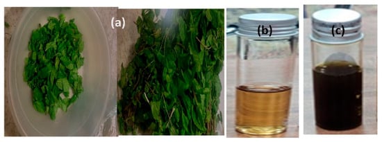
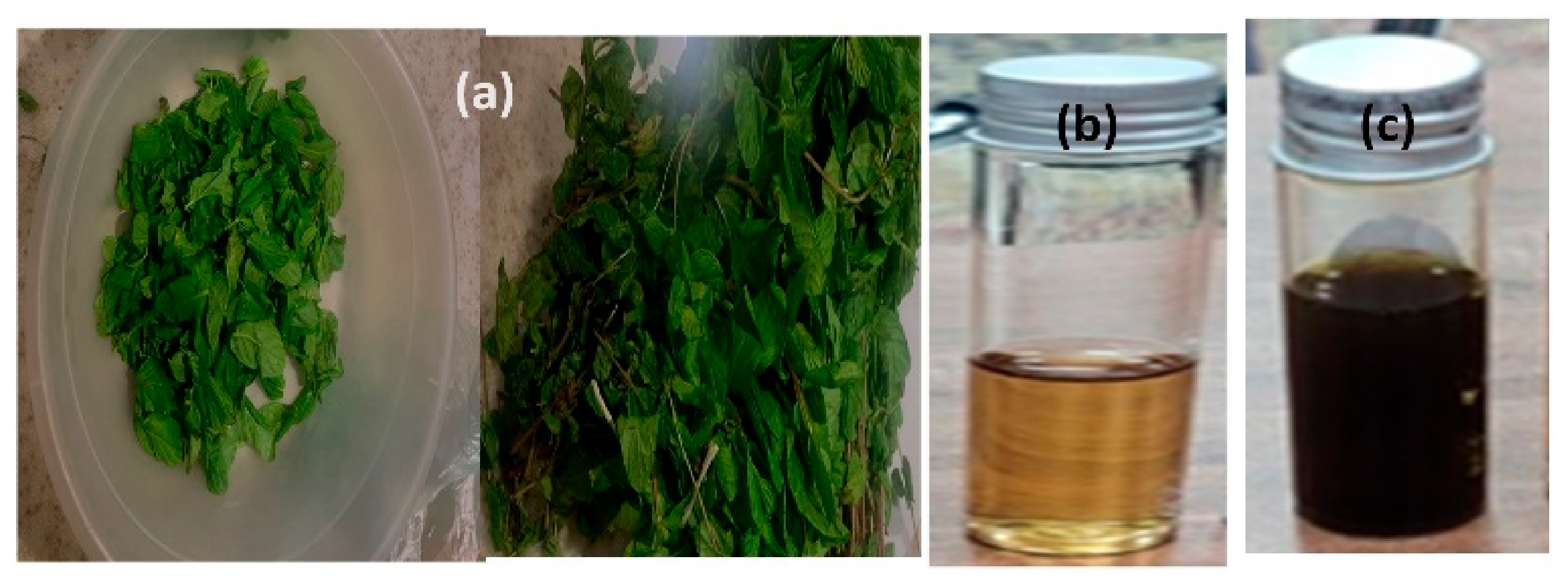
In the current study iron oxide heterophase composite NPs have been prepared via extract of M. arvensis leaves. M. arvensis had been cleaned and then allowed air dry (Figure 1a). Dehydrated leaf biomass was refluxed to get a concoction (Figure 1b) which was used for green synthesis and antibacterial tests. It is clear from this study that agricultural material such as leaf biomass can be recycled to synthesize environmentally friendly iron oxide heterophase NPs, and these NPs could have biological uses. The supplementation of the plant concoction induced reduction of metal precursors to yield NPs. The reduction capability of the mint concoction is possibly due to the occurrence of polyphenols and flavonoids; nonetheless, the precise mechanism remains unidentified. The initial color of the FeCl3 solution was golden yellow; it changed to a deep blackish brown (Figure 1c) after reacting with the aqueous extract of leaf biomass.
Figure 1.
(a) M. arvensis leaf biomass; (b) aqueous extract of biomass; (c) FeCl3 precursor solution which resulted in iron oxide heterophase NPs via green synthesis.
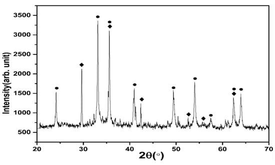
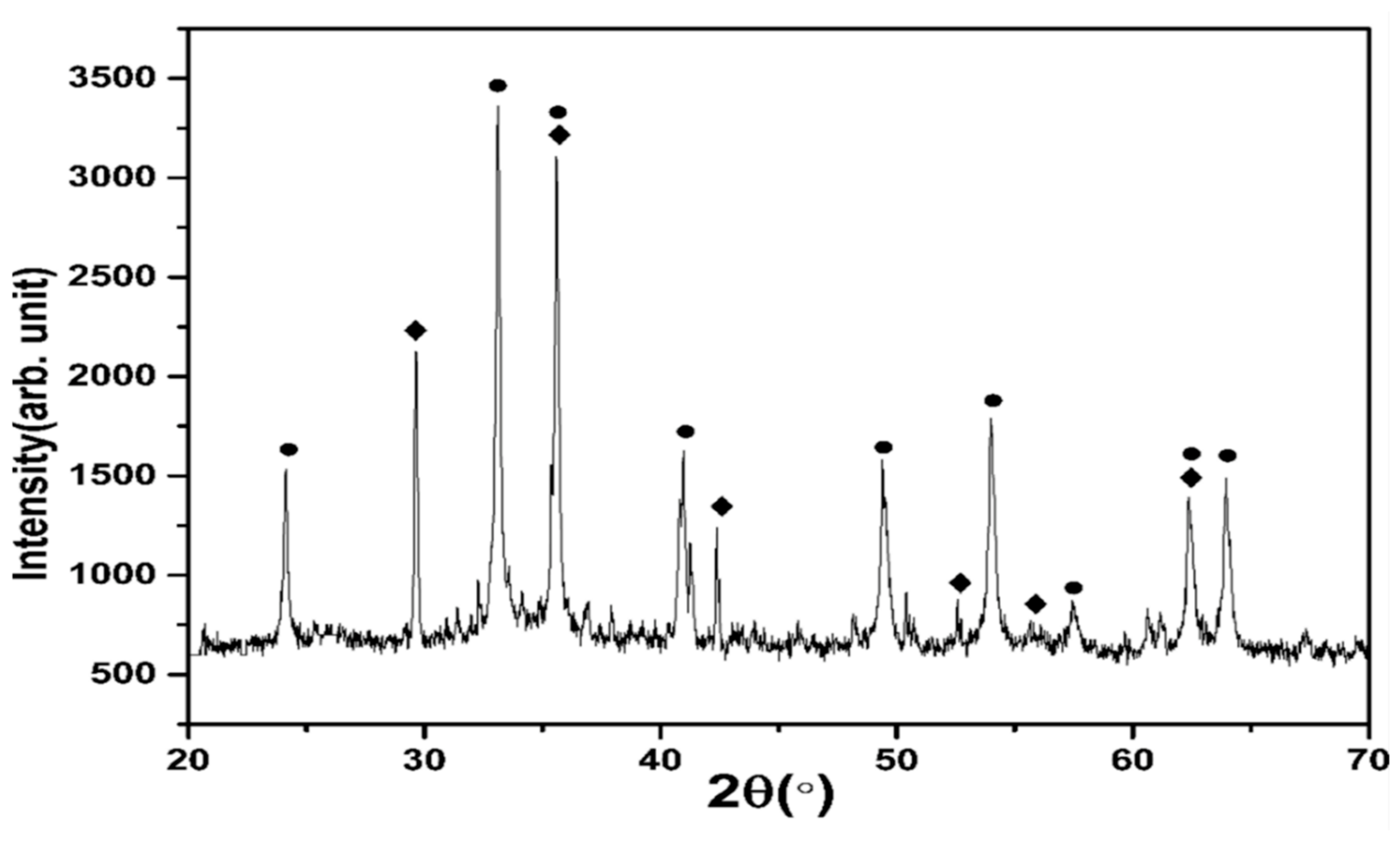
The appearance of a dark brown color indicated the formation of iron oxide heterophase composite NPs due to the reduction [27] mechanism. To authenticate the formation of Fe2O3/Fe3O4 composite NPs, the greenly synthesized NPs were subjected to XRD analysis. The XRD pattern shows a mixture of diffraction patterns corresponding to different phases of iron oxide NPs (Figure 2). The diffraction peaks at 24.13, 33.13, 35.61, 40.9, 49.45, 54.05, 57.46, 62.43, and 63.9° for the Fe2O3, correspond to the (012), (104), (110), (113), (024), (116), (018), (214), and (300) planes of α-Fe2O3 (JCPDS No.33-0664), respectively. Whereas the additional peaks at 30.02, 35.61, 42.38, 52.66, 55.75, and 62.43° correspond to the (220), (311), (400), (422), (511) and (440) planes of Fe3O4 (JCPDS number 99-0073), respectively, confirming coexistence of Fe2O3 and Fe3O4 phases [27].
Figure 2.
X-ray diffraction of iron oxide heterophase composite NPs after calcination.
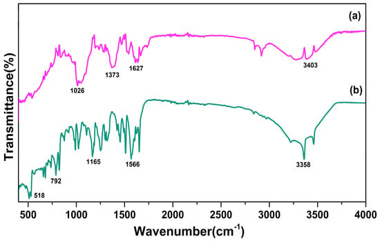
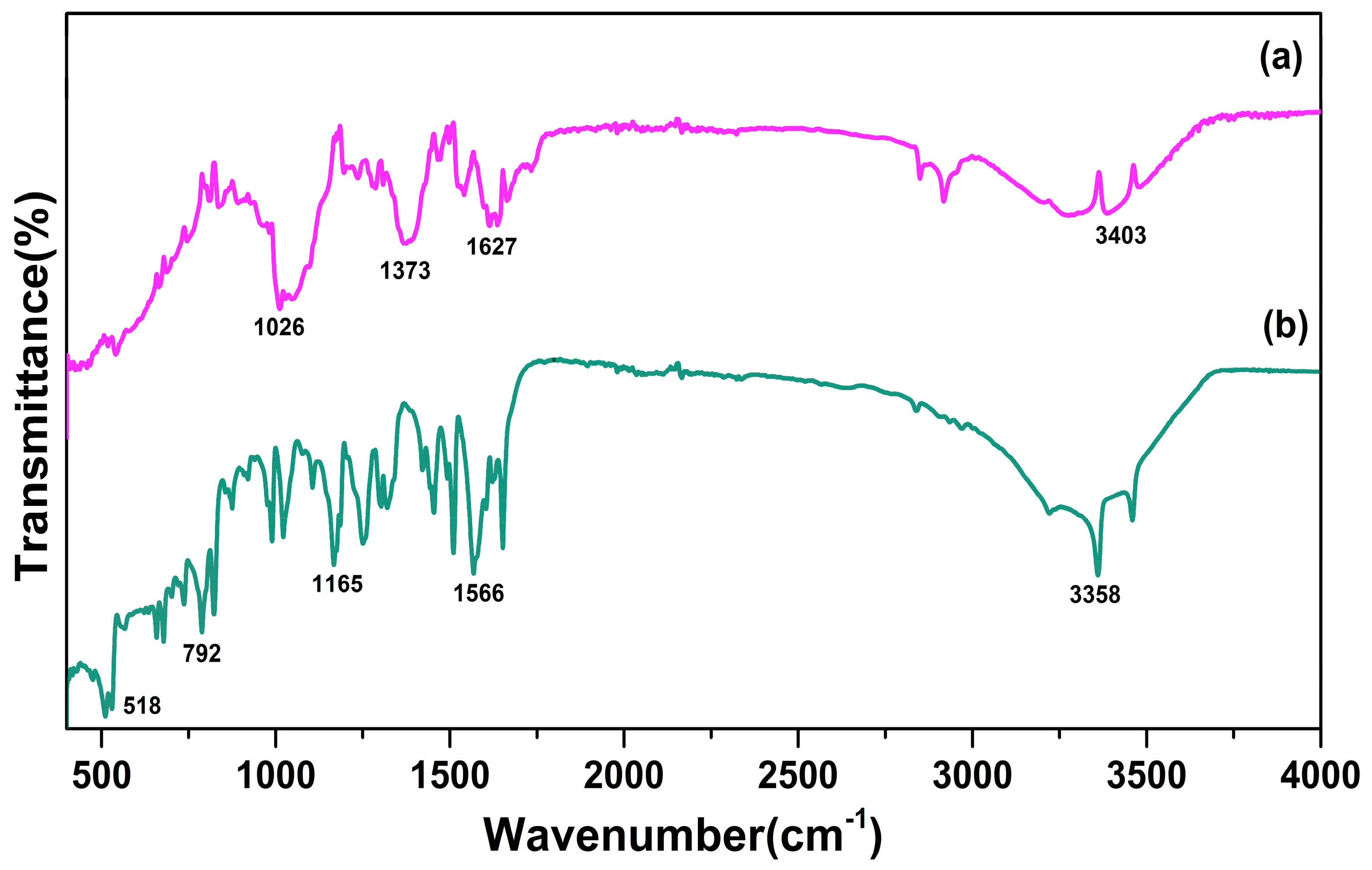
Figure 3 demonstrates FT–IR spectra of M. arvensis and Fe2O3/Fe3O4 composite NPs. The FT–IR spectrum of M. arvensis extract displayed characteristic bands at 3403, 1627, 1373, and 1026 cm−1. The broad band at 3403 cm−1 was allocated to O–H stretching vibrations of hydroxyl-encompassing phytochemicals, whereas the band at 1627 cm−1 was associated with aromatic C=C and/or carbonyl-related vibrations. The band at 1373 cm−1 was ascribed to C–H bending and oxygen comprising plant bioactive chemicals, whereas the band at 1026 cm−1 matched C–O stretching vibrations mainly [27]. On the other hand, in the case of the iron oxide heterophase nanocomposite, substantial alterations in the mentioned bands have been noted. For instance, the O–H band shifted from 3403 to 3358 cm−1, whereas the band at 1627 cm−1 shuffled to 1566 cm−1, demonstrating transformations in chemical milieu of corresponding functional groups, an outcome of their interaction with iron precursors. Alterations/or desertion of 1373 cm−1 band and alterations in C–O-linked fingerprint further confirms contribution of oxygen encompassing phytochemicals in the formation nanocomposite. Prominently, the emergence of a pronounced band at 518 cm−1 in iron oxide heterophase nanocomposite is attributed to Fe–O stretching vibrations, verifying development of an iron oxide structure [28]. Nonetheless, it can be expected that the Fe–O vibrational area may comprehend contributions from both magnetite and hematite, due to their distinct Fe–O coordination backgrounds. Additionally, celebrated low-wavenumber absorption, concurrent with variation of organic functional group bands after synthesis, further proposes development of a mixed iron oxide system. The existence of dual oxidation states of iron could possibly be coupled with the reducing and coordinating influence of plant constituents upfolded in the aqueous extract. Hence, the FTIR spectrum produces encouraging evidence about the organization of iron oxide heterophase nanocomposite, and phase-specific validation has been achieved from XRD as well.
Figure 3.
FT–IR spectra of (a) M. arvensis and (b) iron oxide heterophase nanocomposite.
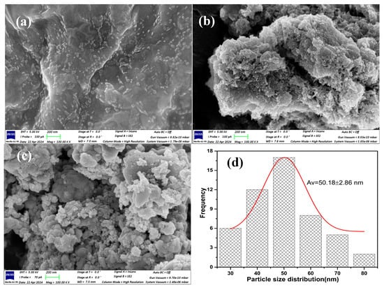
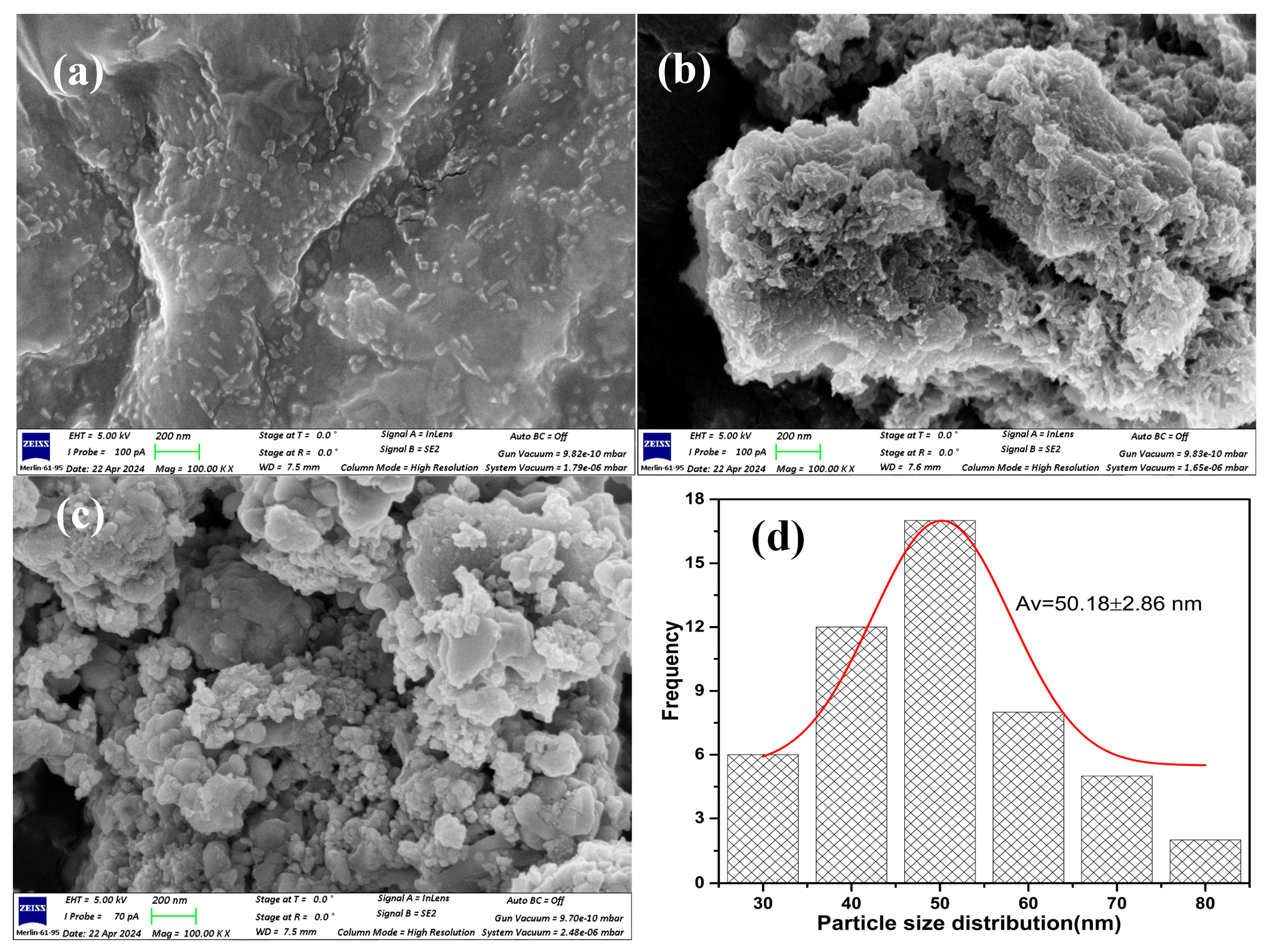
The smattering and overall microstructure of unified samples were categorized by SEM at lower and higher resolution (Figure 4a–c). The Fe2O3/Fe3O4 composite NPs, which are distributed homogeneously throughout the surface of the sample, were smooth in appearance. However, at high magnification, some of the aggregated particles look like small rod-shaped structures; owing to their small size and high surface energy, some tiny particles were seen aggregating into secondary particles. The PSA diagram (Figure 4d) illustrates the diameter distribution of iron oxide NPs and the distribution curve denotes variation in particle diameters across the analyzed sample. The observed profile offers insight into size, uniformity and variability of NPs with a diameter approximately ranging between 30 and 80 nm, with typical average size of 50 nm.
Figure 4.
SEM images of iron oxide heterophase nanocomposite at different magnifications (a–c) and particle size distribution histogram (d) illustrating frequency distribution of NPs diameter.
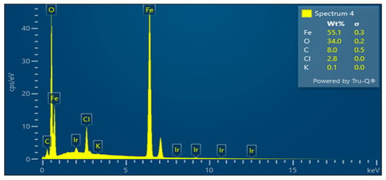
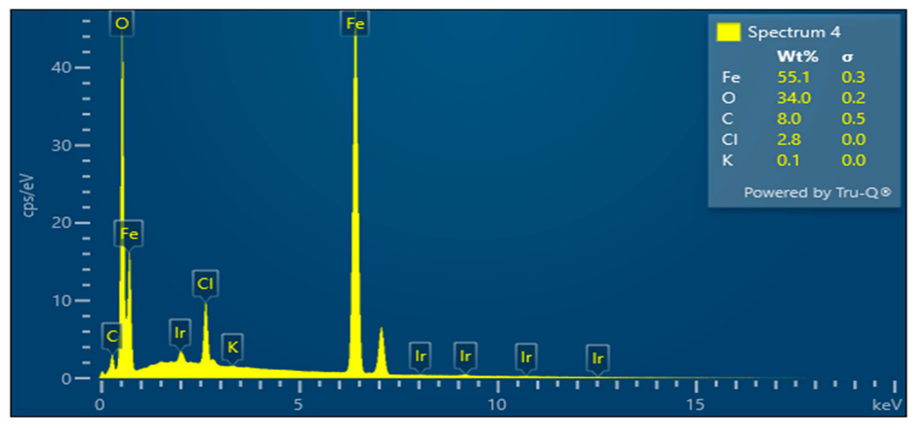
To explain the anticipated mechanism of iron oxide heterophase nanocomposite generation, it can be suggested that possibly phytochemically induced reduction reactions resulted in clusters, which sequentially initiated NP development, exhausting leaf biomass [5]. Additionally, the EDX exploration of iron oxide heterophase composite NPs has been determined to accomplish the reaction and its chemical configuration. Figure 5 demonstrates the EDX spectrum of iron oxide heterophase composite NPs which confirmed predominant presence of Fe and O elements in addition to C in the composite sample. The elemental peak of iridium (Ir) in the spectrum was due to the coating of Ir while taking the SEM images. C, Cl and K have also been detected, but at insignificant levels. These minor signals are unlikely to denote residual extract components, especially after calcination steps. These may be associated with trace contamination during the sample mounting or instrumental background. The percent elemental composition of elements in heterophase composite has been given in the table (Inset of Figure 5).
Figure 5.
EDX spectrum of iron oxide heterophase composite NPs which confirmed presence of Fe and O elements in sample. The elemental peak Ir is due to coating for taking SEM.
Oral microbial illnesses can develop intermittently and spread, leading to oral infections like toothaches, pimples and gum ulcers, etc. Likewise, due to GI ailments, a person may have nausea, discomfort in their abdomen, or stomach ulcers as well. Food poisoning can also be caused by microbial illnesses [29]. It is already known that Mentha biomass inherits antibacterial activity due to the presence of a plethora of novel compounds [30,31].
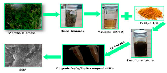
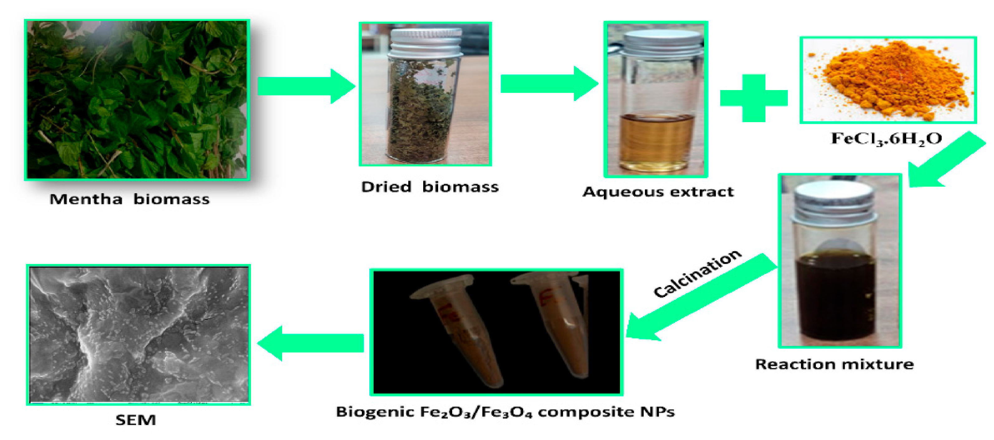
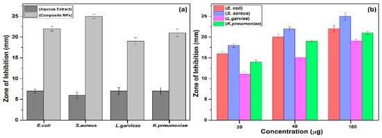
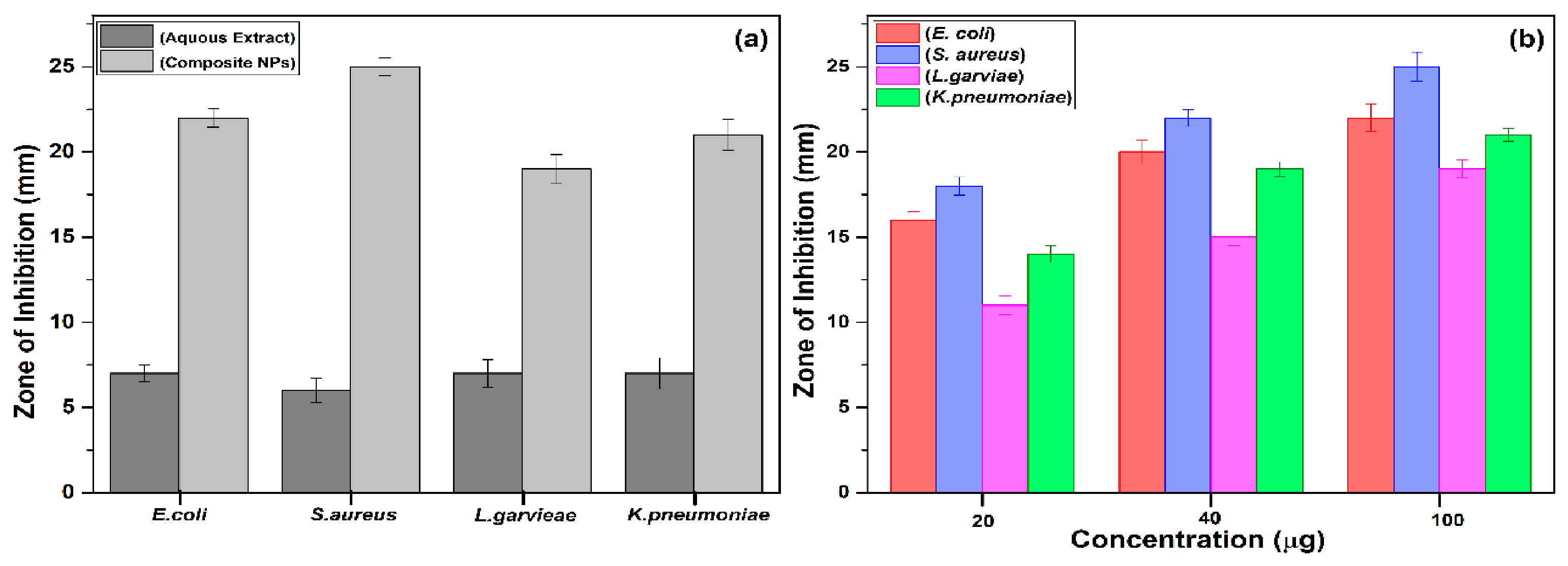
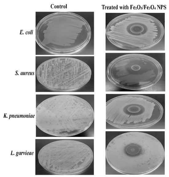
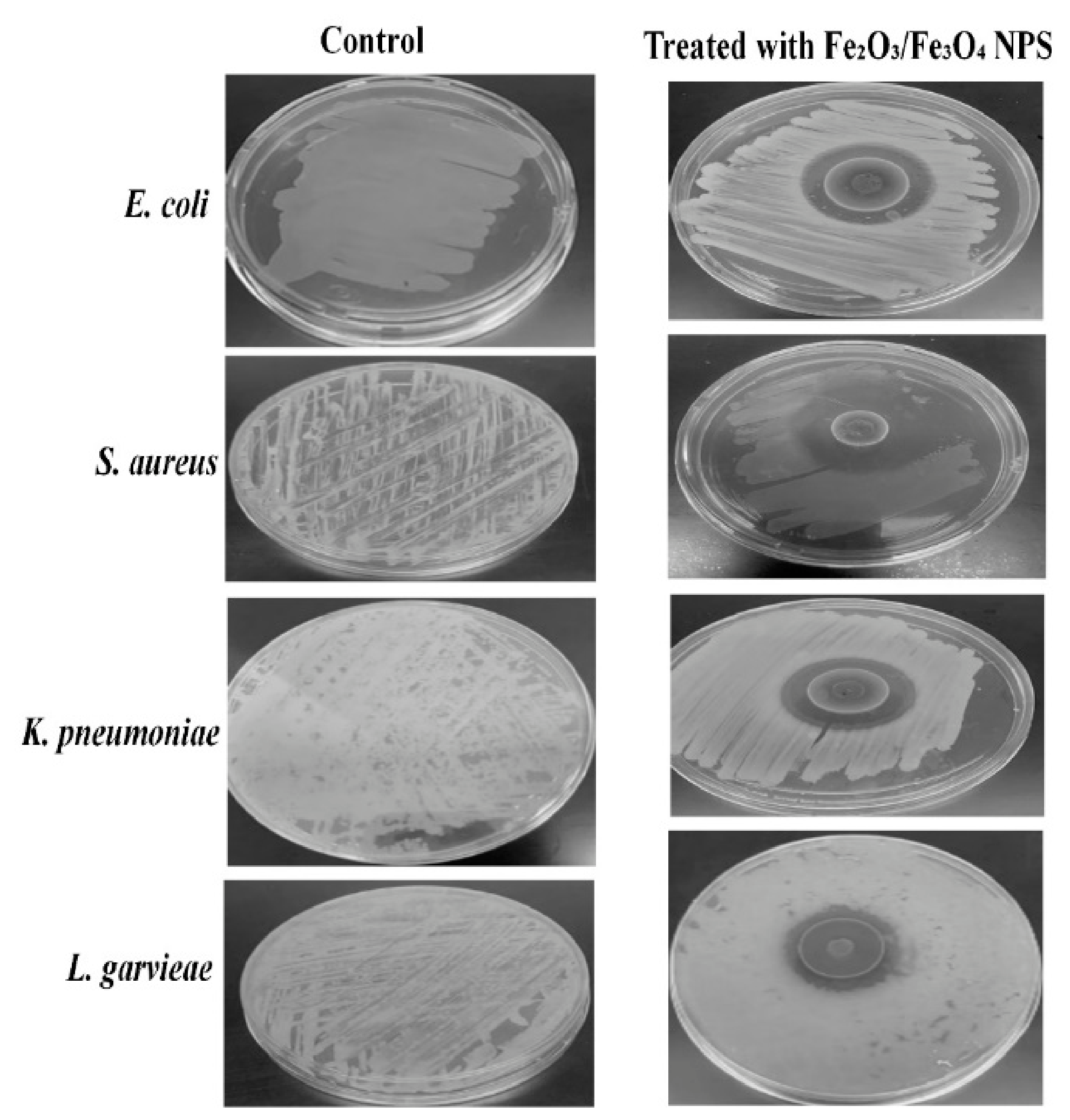
Hence, a green synthetic approach was operated in the present study, for the fabrication of iron oxide heterophase nanocomposite and these composite NPs of hematite and magnetite exhibit nanoscale integration and synergistic interaction between their domains (Figure 6). The antibacterial activity of aqueous extract and iron oxide heterophase nanocomposite was confirmed by a zone of inhibition assay against oral and GI bacteria and the results are shown in Figure 7. In an in vitro antimicrobial bioassay, the bactericidal effect was assessed by agar diffusion assay using the highest concentration. It was observed that the aqueous extract from leaf biomass displaced negligible activity against the selected strains (Figure 7a). However, the iron oxide heterophase composite NPs depicted promising activity (Figure 7a). The as-synthesized composite NPs were further tested at various concentrations, 20, 40 and 100 µg/mL, to determine the lowest possible concentration for bacteriostatic impact. DW was used as a control and for the mint extract, only the highest concentration (100 µg/mL) was used as the other tried concentrations did not produce any observable antibacterial impact. It is evident that pristine aqueous extract does not possess prominent antibacterial activity against any selected bacterial strain while the Fe2O3/Fe3O4 composite NPs show antibacterial activity (Figure 7b) against all the tested strains. Figure 8 represents the average diameter distribution of the inhibition zones.
Figure 6.
A detailed schematic representation depicting preparation of iron oxide heterophase composite NPs and morphological characteristics.
Figure 7.
Antimicrobial vulnerability outline of selected oral and GI bacteria (a) with aqueous extracts and composite NPs (100 µg/mL), (b) with different concentrations of iron oxide heterophase composite NPs (20–100 µg/mL). Data signifies average values and standard deviation.
Figure 8.
Representative plate morphology of natural isolates before and after treatment with iron oxide heterophase nanocomposite (100 µg/mL).
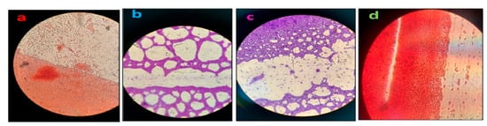
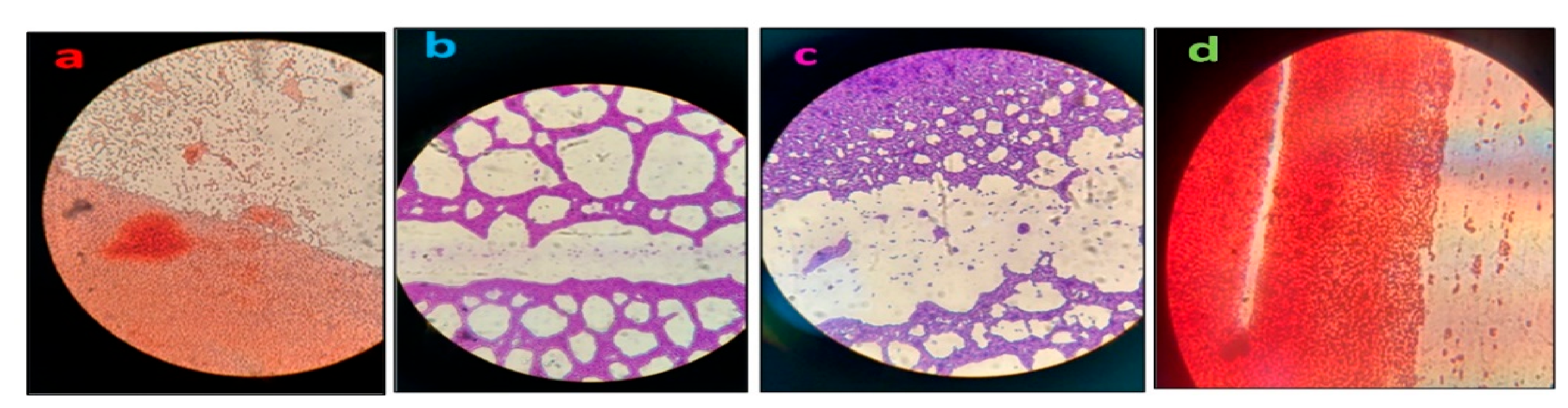
The determined lowest values were 20 µg/ mL for both oral and GI bacteria (E. coli, S. aureus, K. pneumoniae and L. garvieae). The iron oxide heterophase composite NPs showed activity against E. coli and the maximum zone size observed was 21 mm and minimum zone size recorded was 16 mm. Similarly, for S. aureus the maximum zone size is 22 mm and minimum zone size recorded is 18 mm. The heterophase NPs also showed activity against L. garvieae, and maximum zone size is 19 mm and minimum zone size recorded is 11mm. Additionally, iron oxide heterophase NPs showed activity against K. pneumoniae as well with maximum zone size 20 mm, and minimum zone size 14 mm respectively. The obtained results clearly depicted that iron oxide heterophase nanocomposite inherit superlative activity at maximum (100 µg) concentration against S. aureus among the tested oral and GI bacteria and the maximum zone of inhibition recorded was 25 mm. The variance in zones of inhibition amongst strains might be accredited to alterations in membrane configurations [8]. Even so, in the case of both oral and GI bacteria, eclectic zones have been perceived with iron oxide NPs, providing evidence of their future role as potent antibacterial material against all strains. Additionally, smaller dimension of iron oxide heterophase nanocomposite are also credited with improved penetrating competence through bacterial cells, consequently, killing them. Figure 9 signifies representative microscopic images of bacterial cultures treated with green synthesized iron oxide heterophase nanocomposite, following staining. Distinct areas devoid of bacterial growth are evident across all the samples, indicating inhibitory and bactericidal effects exerted by the composite iron oxide NPs.
Figure 9.
Representative microscopic images of bacterial cultures treated with green synthesized iron oxide heterophase nanocomposite following staining. Distinct areas devoid of bacterial growth signify inhibitory effects exerted by NPs, (a) E. coli, (b) S. aureus, (c) L. garvieae and (d) K. pneumoniae.
Furthermore, the nanoscale integration and synergy between Fe2O3 and Fe3O4 in the composite system could probably be the driver for enhanced broad-spectrum bactericidal action. Antibiotics frequently avert progress of numerous bacterial genera or species but are not species-specific, for instance, Polymyxin B, amphotericin B, nalidixic acid, trimethoprim, and azlocillin [32,33]. Furthermore, it is recognized that microorganisms are frequently found in dangerous environmental settings. Their survival under such harsh circumstances is exclusively dependent on intrinsic mechanisms that confer resistance to external stress. In other terms, microbes possess innate defense systems that enable them to withstand various forms of stress, including toxic compounds [34]. Therefore, to deal with these resistant bacteria, in recent times NPs have been found to be very effective.
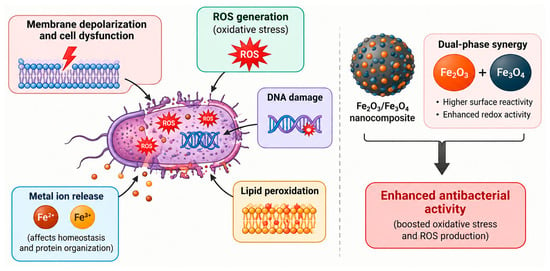
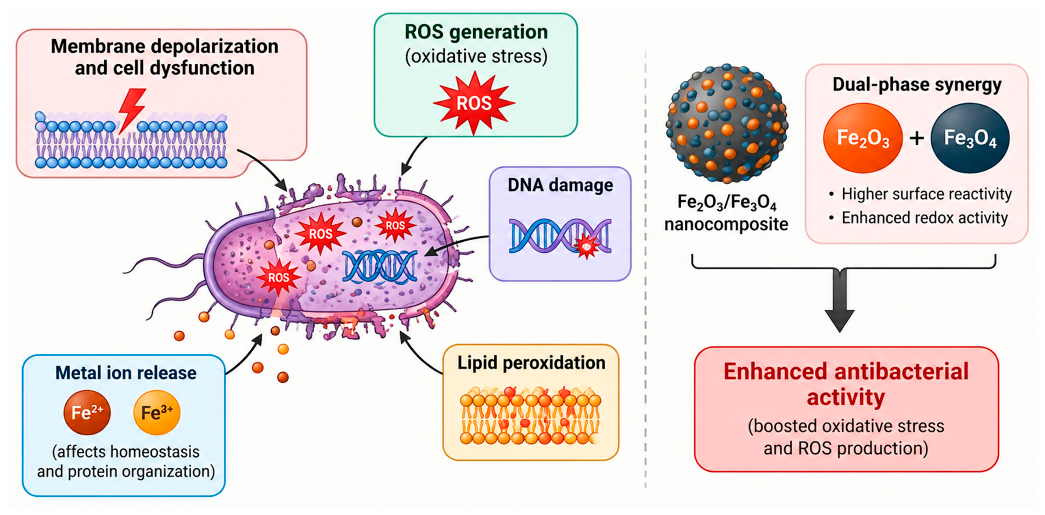
Keeping in mind the unique properties of these nanomaterials, in the present study the iron oxide heterophase composite NPs were developed, characterized and were tested against various bacteria and were found to be effective against the tested strains, showing broad-spectrum antimicrobial potential. According to previous studies, there are a number of interactions that might lead to magnetic nanoparticle-induced microbial toxicity, such as membrane depolarization and ensuing cell dysfunction [35], generation of reactive oxygen species (ROS) coupled with DNA damage and lipid peroxidation, and liberation of metal ions that influence the cellular homeostasis and protein organization. Moreover, it could also be said that the coexistence of Fe2O3 and Fe3O4 phases might have provided harmonizing physicochemical features, such as increased surface reactivity and redox reaction, which in turn could advance the performance of nanocomposites. This mixed-phase nanotextured structure possibly will boost antibacterial activity via boosted oxidative stress and ROS production, pointing towards significant damage of cellular components in bacteria (Figure 10). Earlier researchers have also reported the multifunctionality of iron oxide NPs which have been briefly tabulated (Table 1). Nevertheless, there are no reports about the biogenic synthesis of Fe2O3/Fe3O4 composite from the native Al-Baha M. arvensis and their antimicrobial activity against oral and GI bacteria; thus these NPs could be used as disinfectants, which relish them as an innovative future biomaterial after detailed in vivo screening.
Figure 10.
Schematic sketch depicting proposed antibacterial mechanism of iron oxide heterophase nanocomposite.
Table 1.
Multifunctional applications of green synthesized iron oxide NPs.
3. Materials and Methods
3.1. Chemicals and Cultures
The iron oxide precursor FeCl3 ·6H2O (≥99.9%) was purchased from Sigma-Aldrich Inc. St. Louis, Missouri, United States. The bacterial cultures of E. coli, S. aureus, K. pneumoniae and L. garvieae were obtained from the blood bank of Al-Baha University. All materials employed for present study were of analytical grade.
3.2. Biomass Collection and Preparation of Mentha Aqueous Extract
In the present study, Mentha leaf biomass (Figure 1a) was gathered from a home garden in Al-Baha, Saudi Arabia, and identified and authenticated by Prof. Dr. Brajeshwar Singh. The leaf biomass was splashed with running water and distilled water (DW), subsequently several times to eliminate impurities, then dehydrated in air. The aqueous extract was prepared by refluxing dried leaf biomass in DW at 100 °C for 24 h. The extract was cooled to ambient temperature. Whatman No.1 paper was utilized to drain the concoction. The filtrate was concentrated on a rotary evaporator and finally kept in a refrigerator, away from light at 4 °C until used for further experiments [5].
3.3. Fabrication of Biogenic Iron Oxide Heterophase Nanocomposite via Mentha Biomass
Green preparation of iron oxide heterophase nanocomposite has been carried out using previously reported procedures [5] with suitable modifications. To start with the synthesis, 5 g of FeCl3.6H2O was taken into an Erlenmeyer flask comprising 100 mL of DW. Aqueous solution was stimulated on magnetic stirrer at 70 °C for about 2 h for uniform mixing. When a homogeneous mixture was obtained, it was added to the leaf biomass aqueous extract and further stirred for another 5 h until color change occurred. The mixture was cooled prior to centrifugation at 3000 rpm for 15–25 min. The residue was desiccated at 80 °C for 12 h, washed with DW, lastly calcined at 300 °C in an oven (BI/Barnstead Thermolyne Inc., Dubuque, IA, USA) for 3h.
3.4. Physicochemical Characterization of Iron Oxide Heterophase Nanocomposite
The primary validation of iron oxide heterophase composite NP formation was carried out by visual detection indicated by color change after treatment with precursor salt. To further affirm the formation of nanocomposite, XRD analysis was performed. XRD of iron oxide heterophase composite NPs was carried out on Bruker D8 Advance powder/polycrystalline X-ray (PXRD) diffractometer equipped with Cu X-ray source (Wavelength = 1.5406 Å) operated in Bragg–Brentano geometry at 40 mA and 40 kV. Powder pattern acquisition was carried out from 10 to 70 degrees to confirm the crystalline structure of the iron oxide heterophase composite NPs. The Fourier transform infrared (FTIR, Thermo Scientific, Waltham, MA, USA) analysis was conducted in range of 400–4000 cm−1 to explore functional groups of M. arvensis aqueous extract involved in green synthesis of iron oxide heterophase nanocomposite.
Scanning electron microscopy (SEM) images were acquired on a Zeiss Merlin SEM operated at 10 kV acceleration voltage, 70 mA current and 10 mm working distance. Samples were mounted on aluminum SEM stubs using adhesive carbon tape and were sputter-coated with 5 nm iridium to avoid charging during analysis. To analyze NPs size in iron oxide heterophase nanocomposite, ImageJ 1.54g software was used. The SEM images were introduced to fix the average size and size distribution of NPs in iron oxide nanocomposite. At random, some sections were selected with diverse NPs size and thoroughly explored, and the particle size analysis (PSA) histogram was assembled. Energy-Dispersive X-ray analysis (EDX) spectra were acquired from two different areas for each sample. On several spots marked on the SEM image to reveal the presence of elements present in iron oxide heterophase nanocomposite specimens. EDX spectra were acquired with an X-Max 80 EDX detector (Oxford Instruments, Ultim® Max Infinity, High Wycombe, UK) detector using 20 kV acceleration voltage and 1 mA beam current at 10 mm working distance.
3.5. Estimation of Bactericide Impact of Iron Oxide Heterophase Nanocomposite on Oral and GI Bacteria
The microorganisms under examination were S. aureus, K. pneumoniae, E. coli and L. garvieae. The strains were a kind gift from the blood bank, Al-Baha hospital. The agar well diffusion technique was utilized to ascertain antibacterial activity [39]. The bactericide activity of iron oxide heterophase nanocomposite was estimated against aforementioned oral and GI bacteria as described elsewhere [40] with suitable modifications. Practically, 100 µg of colloidal suspension of composite NPs was poured in the agar plates inoculated with different bacteria, to screen the bactericidal impact.
To determine the lowest tested concentration which could determine the observable inhibition zone, three serial dilutions (20, 40, 100 µg/mL) were prepared. Subsequently, culture tubes were kept for 48 h at 37 °C [41]. Also, for mint aqueous extract the same procedure was followed. Mueller–Hinton agar and MacConkey agar were used for bacterial culture. Each isolate was then cultivated on to solid medium and plates having wells (6 mm diameter), made with the help of cork-borer were supplemented with different concentrations of Fe2O3/Fe3O4 composite NPs. The inoculated Petri dishes were kept at 37 °C for 24 h. The inhibition zone diameter was used to gauge antimicrobial activity of iron oxide heterophase composite NPs. To gain further insight, E. coli and K. pneumoniae were stained using safranin stain, while L. garvieae and S. aureus were stained with crystal violet. The stained bacterial smears were then examined under a light microscope for morphological and staining characteristics [26].
4. Conclusions
In conclusion, it is given to understand, that mint plants naturally growing in city of Al-Baha, can be exploited for synthesis of novel metal oxide nano-compounds. Herein, the mint leaves biomass extract was used for synthesis of iron oxide heterophase nanocomposite by green synthesis method and these as-synthesized NPs were tested against oral and GI bacteria which led to positive results. SEM, EDX, FT-IR and XRD were used to characterize the iron oxide heterophase nanocomposite. The results displayed encouraging antibacterial activity of the samples, which are concentration-dependent. The current study concludes that green production of mixed-phase iron oxide NPs is a promising, cost-effective and environmentally safe technique. In the preliminary investigation it was detected that S. aureus possesses utmost sensitivity to iron oxide heterophase composite NPs. The superior sensitivity of S. aureus might be ascribed to differences in the cell organization or classical configuration of bacteria. Henceforth, antimicrobial NPs could be further developed into novel antibiotics which may be employed against oral and intestinal pathogenic microbes in Al-Baha region. However, for wide-ranging utilization and commercialization of iron oxide heterophase NPs, detailed investigation is mandatory on bio-fabrication techniques possessing distinct dimensions and silhouettes. In addition, biocompatibility/biosafety investigations are extremely important prior to real-world usage. Upcoming research should focus on purifying bioactive entities, which might be employed for synthesis of nanomedicine considering apt biosafety procedures.
Author Contributions
Conceptualization T.A.; methodology, T.A. and A.A.; software, validation and formal analysis, T.A.; investigation, resources, A.A.; data curation, T.A. and A.A.; writing—original draft preparation, T.A.; writing—review and editing, T.A.; visualization, T.A. and A.A.; supervision, T.A.; project administration, T.A. All authors have read and agreed to the published version of the manuscript.
Funding
This research received no external funding.
Institutional Review Board Statement
Not applicable.
Informed Consent Statement
Not applicable.
Data Availability Statement
The datasets used and analyzed during the current study are available from the corresponding author upon reasonable request.
Acknowledgments
The authors sincerely thank the Dean, HOD, and Department of Biology, Al-Baha University for providing the necessary institutional facilities. The authors are very grateful to the Blood Bank for providing oral and GI bacteria and KAUST Centre for Physicochemical characterization. The authors have reviewed and edited the output and take full responsibility for the content of this publication.
Conflicts of Interest
The authors declare no conflicts of interest.
References
- Azam, A.; Ahmed, A.S.; Oves, M.; Khan, M.S.; Habib, S.S.; Memic, A. Antimicrobial activity of metal oxide nanoparticles against Gram-positive and Gram-negative bacteria: A comparative study. Int. J. Nanomed. 2012, 7, 6003–6009. [Google Scholar] [CrossRef] [Scilit]
- Husebye, E. The pathogenesis of gastrointestinal bacterial overgrowth. Chemotherapy 2005, 51, 1–22. [Google Scholar] [CrossRef] [Scilit]
- Thiyagarajan, S.; Kanchana, S. Green synthesis of silver nanoparticles using leaf extracts of Mentha arvensis Linn. and demonstration of their in vitro antibacterial activities. Braz. J. Pharm. Sci. 2022, 58, e19898. [Google Scholar] [CrossRef] [Scilit]
- Alghamdi, S.Q.; Alotaibi, N.; Al-Ghamdi, S.N.; Alqarni, L.S.; Amna, T.; Moustafa, S.M.; Alsohaimi, I.H.; Alruwaili, I.; Nassar, A. High antiparasitic and antimicrobial performance of biosynthesized nio nanoparticles via wasted olive leaf extract. Int. J. Nanomed. 2024, 19, 1469–1485. [Google Scholar] [CrossRef] [Scilit]
- Alotaibi, N.F.; ALqarni, L.S.; Alghamdi, S.Q.; Al-Ghamdi, S.N.; Amna, T.; Alzahrani, S.S.; Moustafa, S.M.; Hasanin, T.H.; Nassar, A.M. Green synthesis of uncoated and olive leaf extract-coated silver nanoparticles: Sunlight photocatalytic, antiparasitic, and antifungal activities. Int. J. Mol. Sci. 2024, 25, 3082. [Google Scholar] [CrossRef] [Scilit]
- Vanlalveni, C.; Lallianrawna, S.; Biswas, A.; Selvaraj, M.; Changmai, B.; Rokhum, S.L. Green synthesis of silver nanoparticles using plant extracts and their antimicrobial activities: A review of recent literature. RSC Adv. 2021, 11, 2804–2837. [Google Scholar] [CrossRef] [Scilit]
- Beheshtkhoo, N.; Kouhbanani, M.A.J.; Savardashtaki, A.; Amani, A.M.; Taghizadeh, S. Green synthesis of iron oxide nanoparticles by aqueous leaf extract of Daphne mezereum as a novel dye removing material. Appl. Phys. A 2018, 124, 363. [Google Scholar] [CrossRef] [Scilit]
- Alghamdi, M.D.; Nazreen, S.; Ali, N.M.; Amna, T. ZnO nanocomposites of Juniperus procera and Dodonaea viscosa extracts as antiproliferative and antimicrobial agents. Nanomaterials 2022, 12, 664. [Google Scholar] [CrossRef] [Scilit]
- Shelepova, O.; Voronkova, T.; Kondrat’eva, V.; Semenova, M.; Bidyukova, G.; Olehnovich, L. Phenotypic and phytochemical differences between Mentha arvensis L. and Mentha canadiensis L. Biol. Bull. 2014, 41, 19–23. [Google Scholar] [CrossRef] [Scilit]
- Snoussi, M.; Noumi, E.; Trabelsi, N.; Flamini, G.; Papetti, A.; De Feo, V. Mentha spicata essential oil: Chemical composition, antioxidant and antibacterial activities against planktonic and biofilm cultures of Vibrio spp. strains. Molecules 2015, 20, 14402–14424. [Google Scholar] [CrossRef] [Scilit]
- Thuyen, N.; Phong, H.; Hanh, C.; Huong, H.; Men, T. Study on morphology, distillation process, chemical compositions, and biological activities of Mentha arvensis L. var. javanica (Blume) Hook. f essential oil. J. Taiwan Inst. Chem. Eng. 2023, 153, 105209. [Google Scholar] [CrossRef] [Scilit]
- Thawkar, B.S. Phytochemical and pharmacological review of Mentha arvensis. Int. J. Green Pharm. (IJGP) 2016, 10, 71–76. [Google Scholar]
- Biswas, N.N.; Saha, S.; Ali, M.K. Antioxidant, antimicrobial, cytotoxic and analgesic activities of ethanolic extract of Mentha arvensis L. Asian Pac. J. Trop. Biomed. 2014, 4, 792–797. [Google Scholar] [CrossRef] [Scilit]
- Arularasu, M.; Devakumar, J.; Rajendran, T. An innovative approach for green synthesis of iron oxide nanoparticles: Characterization and its photocatalytic activity. Polyhedron 2018, 156, 279–290. [Google Scholar] [CrossRef] [Scilit]
- Pravallika, P.L.; Mohan, G.K.; Rao, K.V.; Shanker, K. Biosynthesis, characterization and acute oral toxicity studies of synthesized iron oxide nanoparticles using ethanolic extract of Centella asiatica plant. Mater. Lett. 2019, 236, 256–259. [Google Scholar] [CrossRef] [Scilit]
- Izadiyan, Z.; Shameli, K.; Miyake, M.; Hara, H.; Mohamad, S.E.B.; Kalantari, K.; Taib, S.H.M.; Rasouli, E. Cytotoxicity assay of plant-mediated synthesized iron oxide nanoparticles using Juglans regia green husk extract. Arab. J. Chem. 2020, 13, 2011–2023. [Google Scholar] [CrossRef] [Scilit]
- Rajendran, S.; Wanale, S.G.; Gacem, A.; Yadav, V.K.; Ahmed, I.A.; Algethami, J.S.; Kakodiya, S.D.; Modi, T.; Alsuhaibani, A.M.; Yadav, K.K. Nanostructured iron oxides: Structural, optical, magnetic, and adsorption characteristics for cleaning industrial effluents. Crystals 2023, 13, 472. [Google Scholar] [CrossRef] [Scilit]
- Saqib, S.; Munis, M.F.H.; Zaman, W.; Ullah, F.; Shah, S.N.; Ayaz, A.; Farooq, M.; Bahadur, S. Synthesis, characterization and use of iron oxide nano particles for antibacterial activity. Microsc. Res. Tech. 2019, 82, 415–420. [Google Scholar] [CrossRef] [Scilit]
- Das, S.; Diyali, S.; Vinothini, G.; Perumalsamy, B.; Balakrishnan, G.; Ramasamy, T.; Dharumadurai, D.; Biswas, B. Synthesis, morphological analysis, antibacterial activity of iron oxide nanoparticles and the cytotoxic effect on lung cancer cell line. Heliyon 2020, 6, e04953. [Google Scholar] [CrossRef] [Scilit]
- Al-Robai, S.A.; Ahmed, A.A.; Mohamed, H.A.; Ahmed, A.A.; Zabin, S.A.; Alghamdi, A.A. Qualitative and quantitative ethnobotanical survey in Al Baha province, southwestern Saudi Arabia. Diversity 2022, 14, 867. [Google Scholar] [CrossRef] [Scilit]
- Shoudho, K.N.; Uddin, S.; Rumon, M.M.H.; Shakil, M.S. Influence of physicochemical properties of iron oxide nanoparticles on their antibacterial activity. ACS Omega 2024, 9, 33303–33334. [Google Scholar] [CrossRef] [Scilit]
- Abdelhady, M. A comparative study of the Saudi Arabia flora, medicinal plants of al-Mandaq province, Al-Baha region. Al-Azhar J. Pharm. Sci. 2021, 63, 57–71. [Google Scholar] [CrossRef] [Scilit]
- Al-robai, S. Patterns of spatial distribution and diversity of invasive plants and their correlation with environmental elements in six dam areas, Al-Baha, Saudi Arabia. Appl. Ecol. Environ. Res. 2024, 22, 1983. [Google Scholar] [CrossRef] [Scilit]
- Hassan, M.S.; Khan, R.; Amna, T.; Yang, J.; Lee, I.-H.; Sun, M.-Y.; EL-Newehy, M.H.; Al-Deyab, S.S.; Khil, M.-S. The influence of synthesis method on size and toxicity of CeO2 quantum dots: Potential in the environmental remediation. Ceram. Int. 2016, 42, 576–582. [Google Scholar] [CrossRef] [Scilit]
- Hassan, M.S.; Amna, T.; Yang, O.-B.; El-Newehy, M.H.; Al-Deyab, S.S.; Khil, M.-S. Smart copper oxide nanocrystals: Synthesis, characterization, electrochemical and potent antibacterial activity. Colloids Surf. B Biointerfaces 2012, 97, 201–206. [Google Scholar] [CrossRef] [Scilit]
- Amna, T.; Alghamdi, A.A.; Shang, K.; Hassan, M.S. Nigella Sativa-Coated Hydroxyapatite Scaffolds: Synergetic Cues to Stimulate Myoblasts Differentiation and Offset Infections. Tissue Eng. Regen. Med. 2021, 18, 787–795. [Google Scholar] [CrossRef] [Scilit]
- Elkhateeb, O.; Atta, M.B.; Mahmoud, E. Biosynthesis of iron oxide nanoparticles using plant extracts and evaluation of their antibacterial activity. AMB Express 2024, 14, 92. [Google Scholar] [CrossRef] [Scilit]
- Begum, S.K.; Shabnam, D.; Haque, N.; Alam, M.J.; Ferdous, J.; Nur, U.J.B.; Fatema, K.; Shabiha, R.J.; Clarke, R.J.; Chowdhury, P. Green synthesis of magnetite (Fe3O4) and hematite (Fe2O3) nanoparticles using Moringa oleifera and Psidium guajava leaf extracts for sustainable applications. Sci. Rep. 2025, 15, 36465. [Google Scholar] [CrossRef] [Scilit]
- Muronetz, V.I.; Kurochkina, L.P.; Leisi, E.V.; Kudryavtseva, S.S. Are gastrointestinal microorganisms involved in the onset and development of amyloid neurodegenerative diseases? Microbiol. Res. 2023, 14, 1942–1955. [Google Scholar] [CrossRef] [Scilit]
- Gabriela, Á.-M.; Gabriela, M.D.O.-V.; Luis, A.-M.; Reinaldo, P.-R.; Michael, H.-M.; Rodolfo, G.-P.; Roberto, V.-B.J. Biosynthesis of silver nanoparticles using mint leaf extract (Mentha piperita) and their antibacterial activity. Adv. Sci. Eng. Med. 2017, 9, 914–923. [Google Scholar] [CrossRef] [Scilit]
- Malankina, E.; Kuzmenko, A.; Evgrafov, A.; Evgrafova, S.; Zaychik, B.; Ruzhitskiy, A. Chemical Features of Long-Leaved Mint (Mentha longifolia L.) Varieties. Mosc. Univ. Chem. Bull. 2025, 80, 33–38. [Google Scholar] [CrossRef] [Scilit]
- Subramanyam, B.; Siyaramakrishnan, G.; Dusthackeer, A.; Kumar, V. Phage lysin as a substitute for antibiotics to detect Mycobacterium tuberculosis from sputum samples with the BACTEC MGIT 960 system. Clin. Microbiol. Infect. 2012, 18, 497–501. [Google Scholar] [CrossRef] [Scilit]
- Chevallier, L.; Cameron, M.; Mcfeters, G. New medium for improved recovery of coliform bacteria from drinking water. Appl. Environ. Microbiol. 1983, 45, 484–492. [Google Scholar] [CrossRef] [Scilit]
- Aziz, W.J.; Abid, M.A.; Hussein, E.H. Biosynthesis of CuO nanoparticles and synergistic antibacteria activity using mint leaft extract. Mater. Technol. 2020, 35, 447–451. [Google Scholar] [CrossRef] [Scilit]
- Pelgrift, R.Y.; Friedman, A.J. Nanotechnology as a therapeutic tool to combat microbial resistance. Adv. Drug Deliv. Rev. 2013, 65, 1803–1815. [Google Scholar] [CrossRef] [Scilit]
- Saod, W.M.; Al-Janaby, M.S.; Gayadh, E.W.; Ramizy, A.; Hamid, L.L. Biogenic synthesis of iron oxide nanoparticles using Hibiscus sabdariffa extract: Potential for antibiotic development and antibacterial activity against multidrug-resistant bacteria. Curr. Res. Green Sustain. Chem. 2024, 8, 100397. [Google Scholar] [CrossRef] [Scilit]
- Duan, Y.-T.; Soni, K.; Patel, D.; Choksi, H.; Sangani, C.B.; Saeed, W.S.; Ameta, K.L.; Ameta, R.K. Green synthesis of iron oxide nanoparticles using Nicotiana plumbaginifolia and their biological evaluation. J. Mol. Liq. 2024, 396, 123985. [Google Scholar] [CrossRef] [Scilit]
- Parmanik, A.; Kothari, P.P.; Bose, A.; Biswas, S. Evaluation of antibacterial and antibiofilm activities of green-synthesized iron oxide nanoparticles using Cyperus rotundus extract as a reducing and stabilizing agent. J. Mater. Sci. Mater. Med. 2025, 36, 60. [Google Scholar] [CrossRef] [Scilit]
- Hussein, S.; Mahmoud, A.M.; Elgebaly, H.A.; Hendawy, O.M.; Hassanein, E.H.; Moustafa, S.M.; Alotaibi, N.F.; Nassar, A.M. Green synthesis of trimetallic nanocomposite (Ru/Ag/Pd)-Np and Its In Vitro antimicrobial and anticancer activities. J. Chem. 2022, 2022, 4593086. [Google Scholar] [CrossRef] [Scilit]
- Amna, T.; Yang, J.; Ryu, K.-S.; Hwang, I. Electrospun antimicrobial hybrid mats: Innovative packaging material for meat and meat-products. J. Food Sci. Technol. 2015, 52, 4600–4606. [Google Scholar] [CrossRef] [Scilit]
- Meskin, M.; Esmaeili, D.; Ghorbani, M.; Ghorban Alizadehgan, M. Comparison of micro-dilution and macro-dilution methods for mic determination of plant extracts. In Proceedings of the 5th National & the 1th International Congress on Novel & Innovative Laboratory Technologies, Tehran, Iran, 25–27 October 2017. [Google Scholar]

Disclaimer/Publisher’s Note: The statements, opinions and data contained in all publications are solely those of the individual author(s) and contributor(s) and not of MDPI and/or the editor(s). MDPI and/or the editor(s) disclaim responsibility for any injury to people or property resulting from any ideas, methods, instructions or products referred to in the content. |
© 2026 by the authors. Licensee MDPI, Basel, Switzerland. This article is an open access article distributed under the terms and conditions of the Creative Commons Attribution (CC BY) license.