


Abstract
This study evaluated the microwave-assisted green synthesis of zinc oxide nanoparticles (ZnO NPs) utilizing Melaleuca quinquenervia leaf extract (MQLE) as a sustainable reducing and capping agent. The synthesized bio-capped ZnO NPs exhibited a pure crystalline structure with an average hydrodynamic size of 82.6 nm. The biological profiles of the nanoparticles were systematically evaluated. Results demonstrated that bio-capped ZnO NPs possessed superior antioxidant, antityrosinase, anti-aging, and anti-inflammatory activities compared to chemically synthesized ZnO NPs (Che-ZnO NPs) and raw MQLE. Specifically, bio-capped ZnO NPs effectively suppressed melanin synthesis in human epidermal melanocytes (HEMn) and down-regulated key inflammatory mediators. Biocompatibility assessments based on ISO 10993-5 criteria revealed that bio-capped ZnO NPs maintained high cell viability at their effective working doses, with safe dosage thresholds (viability > 70%) determined at 175 mg/L for CCD966SK, 225 mg/L for HEMn, and 175 mg/L for Raw264.7 cells. The safe doses significantly exceeded the concentrations required for their physiological functions, whereas Che-ZnO NPs displayed prominent cytotoxicity. In conclusion, MQLE-mediated green synthesis provided a safe, biocompatible, and high-performance platform for ZnO NPs, offering great potential for advanced cosmetic and transdermal biomedical applications.
1. Introduction
Over recent decades, the emergence of nanomaterial-based technology has transformed biomedical and microbiological research. Nanomaterials possess a high surface-area-to-volume ratio along with distinctive physical, chemical, and biological characteristics. Specifically, research on nanoparticles (NPs) has attracted significant attention; their superior electrochemical reactivity, thermal stability, and surface-reactive properties have paved the way for diverse innovative applications [1]. Zinc oxide (ZnO) nanostructures have emerged at the forefront of material science research, driven by their distinctive properties and versatile applications. Particularly among various metal oxides, the synthesis of ZnO via “green synthesis” methods has garnered significant interest. This approach offered a clean, secure, sustainable, and ecologically non-toxic alternative for fabricating nanoparticles [2]. Moreover, zinc oxide nanoparticles (ZnO NPs) exhibit advantageous optical and physicochemical properties, notably a high photoluminescence quantum yield and tunable emission spectra, making them promising tools for antimicrobial therapy and bio-imaging. Zinc oxide nanoparticles (ZnO NPs) have drawn substantial interest owing to their multifunctional properties in biomedical, environmental, industrial and agricultural fields [3]. Previous research demonstrated that ZnO NPs effectively promoted wound healing and inhibited tyrosinase activity to achieve skin-whitening effects [4]. Recent studies indicated that topical application of ZnO nanoparticles (NPs) could effectively suppress allergen-induced cutaneous inflammation [5]. Furthermore, combining ZnO nanorods with diallyl sulfide exerts a significant synergistic effect, accelerating the healing of acute inflammation in dermatitis [6].
Various conventional methods are utilized to synthesize zinc oxide nanoparticles ZnO NPs, including spray pyrolysis, sol–gel processing, hydrothermal synthesis, and precipitation techniques. However, these traditional approaches typically demand substantial energy inputs and involve hazardous chemicals, thereby posing significant biological and environmental risks [7]. Previous research indicated that bio-synthesized zinc oxide nanoparticles (ZnO NPs) feature an in situ capping layer composed of plant-derived phytochemicals, predominantly polyphenols, which imparts distinct photochemical advantages, such an approach corresponded to the previously mentioned green synthesis method [8].
Melaleuca quinquenervia is widely cultivated as a roadside tree in Taiwan. Its leaf extract (MQLE) exhibits substantial therapeutic potential, particularly owing to its anti-melanogenic and antioxidant activities. Utilizing MQLE as a dual reducing and capping agent in microwave-assisted green synthesis can effectively maximize the functional advantages of the synthesized metallic nanoparticles [9].
Herein, we report the microwave-assisted green synthesis of zinc oxide nanoparticles (ZnO NPs) utilizing Melaleuca quinquenervia leaf extract (MQLE) as a sustainable reducing and capping agent. The successful formation of ZnO NPs was confirmed via ultraviolet-visible (UV-Vis) spectroscopy, while dynamic light scattering (DLS), energy-dispersive X-ray spectroscopy (EDS) and X-ray diffraction (XRD) elucidated their chemical composition and crystalline structure, respectively. Furthermore, the antioxidative activities, tyrosinase inhibitory activities, anti-aging activities and anti-inflammatory activities of the biosynthesized ZnO NPs were systematically evaluated. To the best of our knowledge, this study is the first to investigate the inhibition of melanin synthesis in human epidermal melanocytes (HEMn) using MQLE-mediated ZnO NPs. These findings indicated that our green synthesis approach provides a feasible, sustainable, and cost-effective platform with great potential for cosmetic and biomedical applications.
2. Materials and Methods
2.1. Preparation of Raw Materials
The experimental biomass consisted of freshly fallen M. quinquenervia leaves collected in Taipei, Taiwan, and identified by Professor Bau-Yuan Hu following the protocol of Lin et al. (2024) [9].
2.2. Preparation of Melaleuca quinquenervia Leaf Extract
Freshly harvested leaves of Melaleuca quinquenervia were rinsed with distilled water and oven-dried at 60 °C for 1.5 h, modifying the method described by Lin et al. (2024) [9]. A 0.5 mm screen was used to sieve the pulverized dry leaf powder. For the extraction process, 10 g of this powder was mixed with 200 mL of distilled water, maintaining a 1:20 (w/v) ratio. The extraction was carried out using a microwave-assisted method (SINEO system, Shanghai, China) set to 80 °C and 700 W (2.45 GHz) for 180 s. To remove insoluble residues, the solution was subjected to centrifugation at 4800 rpm for 20 min and subsequently passed through Whatman No. 1 filter paper. The final filtrate, which functioned as both a bio-reducing and stabilizing agent for nanoparticle synthesis, was lyophilized and preserved.
2.3. Synthesis of Green ZnO NPs
The green fabrication of ZnO NPs involved mixing 5 mL of M. quinquenervia leaf filtrate into 95 mL of 2 mM zinc acetate Zn(CH3CO2)2 dissolved in water (final pH 7.1) while maintaining constant stirring. Microwave-assisted synthesis was performed using a SINEO system at a stable temperature of 80 °C with an output power of 700 W (2.45 GHz) for durations spanning 0 to 60 s. For nanoparticle recovery, the solution underwent a centrifugation process at 6000 rpm (15 min). The supernatant was decanted, and the resulting precipitate was rinsed three times using deionized water for purification. Oven-drying the product at 150 °C for 1 h successfully produced the ZnO NP powder. As a control benchmark, commercial-grade ZnO nanoparticles (size < 100 nm) were obtained from Sigma-Aldrich. The optimized biosynthetic protocol was completed within 75.5 min, including 30 s of microwave heating, 15 min of centrifugation, and 1 h of drying. The M. quinquenervia leaf extract (MQLE)-mediated ZnO NPs yielded within 30 s via first-order reduction kinetics (k = 0.3305 s−1), accompanied by a reduction potential surge from 14.8 to 62.3 mV (pH/ORP Meter, TES-1381K, TES Electrical Electronic Corp., Taiwan). The synthesized particles exhibited a characteristic UV absorption peak at 365 nm, and the optimized reaction yielded a final production efficiency of approximately 100%. Long-term evaluation confirmed remarkable colloidal stability for up to 11 months.
2.4. Physicochemical Characterization of ZnO Nanoparticles
The optical characteristics and successful formation of the biogenic ZnO NPs were initially confirmed via ultraviolet–visible (UV–Vis) spectrophotometry (Thermo Fisher Scientific, Waltham, MA, USA) [10]. The hydrodynamic size and relative distribution profile of the synthesized particles were assessed by dynamic light scattering (DLS) on a Malvern NanoSizer-ZS instrument (Worcestershire, UK). Additionally, the crystallographic phase purity, crystal lattice structure, and crystallite size were evaluated using an X-ray powder diffractometer (XRD; Bruker, Billerica, MA, USA) equipped with Cu Kα radiation (λ = 0.154 nm). Finally, energy-dispersive X-ray spectroscopy (EDS; JEOL system) was performed at an accelerating voltage of 20 kV to verify the elemental composition of the fabricated ZnO nano-entities. The structural, hydrodynamic, and elemental properties of Bio-synthesized ZnO NPs were systematically characterized. XRD analysis confirmed a highly pure, polycrystalline wurtzite phase matching JCPDS card no. 36-1451, with an average crystallite size of 26.7 nm derived via Scherrer’s formula. DLS analysis indicated a monodisperse and uniform particle distribution, yielding an average hydrodynamic diameter of 82.6 nm with a polydispersity index (PDI) of 0.242 ± 0.01. Furthermore, energy-dispersive X-ray spectroscopy (EDS) confirmed the exclusive presence of Zn and O, revealing weight percentages of 78.16% Zn and 21.84% O (45.68% and 54.32% atomic percentages, respectively). Furthermore, these experimental values closely aligned with theoretical weight distributions (80.3% Zn, 19.7% O), corroborating the excellent crystalline purity of the fabricated nanoparticles. In contrast, the commercial-grade ZnO NPs (with a specified primary size of <100 nm) exhibited a significantly larger average hydrodynamic diameter of 113.6 ± 6.2 nm with a polydispersity index (PDI) of 0.243 ± 0.02.
2.5. Antioxidant Activity of Bio-Synthesized ZnO NPs
The 1,1-diphenyl-2-picrylhydrazyl (DPPH) free radical scavenging capacity of the synthesized zinc oxide nanoparticles (ZnO NPs) was evaluated according to the methods reported by Prachi (2018) [11], with minor modifications. Briefly, various concentrations of ZnO NP suspensions were adjusted to a final volume of 100 µL using methanol, followed by the addition of a 0.1 mM DPPH methanolic solution. The reaction mixtures were incubated in the dark at room temperature for 20 min to allow complete radical quenching. Subsequently, the absorbance was recorded at 517 nm using a UV–Vis spectrophotometer. Butylated hydroxyanisole (BHA) was used as the reference standard (positive control), while a mixture of methanol and DPPH served as the negative control. All assays were conducted in triplicate.
The DPPH radical scavenging percentage was calculated using Equation (1):
where Control OD is the absorbance of the negative control and Sample OD is the absorbance of the synthesized ZnO NPs, leaf extract or Vitamin C reference standard. The half-maximal inhibitory concentration IC50 was determined by plotting the percentage inhibition against the corresponding concentrations using linear or non-linear regression analysis.
2.6. Extracellular Tyrosinase Inhibition Assay
To evaluate the extracellular antityrosinase potential, a mushroom tyrosinase inhibition assay was conducted as a screening platform. Briefly, lyophilized bio-capped ZnO NPs were initially dissolved in DMSO (1 mg/L) and subsequently diluted into a series of test concentrations. A 30 μL aliquot of each sample solution was combined with 970 μL of phosphate buffer (0.05 mM, pH 6.8), followed by the addition of 1 mL tyrosine substrate (100 mg/L) and 1 mL tyrosinase solution (350 U/mL). Following a 20-min incubation period, the optical density was recorded at a wavelength of 490 nm using a UV–Vis spectrophotometer (Thermo Fisher Scientific, Waltham, MA, USA) [12]. For comparison, kojic acid and α-arbutin were processed under identical experimental conditions to serve as positive controls. The IC50 value was calculated as the sample concentration required to suppress 50% of the baseline enzymatic activity using the following equation:
where A denotes the control absorbance in the absence of the extract; B corresponds to the blank absorbance without both extract and enzyme; C represents the test absorbance with both extract and enzyme present; and D indicates the sample background absorbance containing the extract without tyrosinase.
2.7. Assays for Tyrosinase Activity and Melanin Content in Human Epidermal Melanocytes (HEMn)
To assess the skin-whitening efficacy of bio-capped ZnO nanoparticles (NPs) on human cells, cellular tyrosinase activity and melanin content were evaluated using human epidermal melanocytes (HEMn; C-102-5C, Cascade Biologics, Portland, OR, USA). Cells were maintained in Medium 254 supplemented with Human Melanocyte Growth Supplement (S-002-5, Cascade Biologics) at 37 °C in a humidified atmosphere containing 5% CO2. After 24 h of incubation, cells were harvested and seeded into 6-well plates at a density of 3 × 10⁵ cells/well (identical to cytotoxicity assay conditions). Cells were then treated with various concentrations of the sample for 24 h under identical culture conditions.
Tyrosinase Activity Assay: Following treatment, cells were washed with PBS, detached using trypsin, and lysed via sonication. The lysate was centrifuged to collect the supernatant containing active enzymes. The supernatant was incubated with 2.5 mM L-DOPA in a 96-well plate for 60 min. Tyrosinase activity was quantified by monitoring dopachrome formation at an absorbance of 475 nm using an Epoch ELISA microplate reader (BioTek Instruments, Santa Clara, CA, USA), following the method described by Wu et al. (2018) [13]. In this study, α-arbutin and kojic acid were used as positive controls.
Melanin Content Measurement: Treated cells were washed with PBS, trypsinized, and lysed in 1 N NaOH containing 10% DMSO at 80 °C for 1 h. The absorbance of the resulting solution was measured at 450 nm using the Epoch microplate reader. Melanin concentration was interpolated using a standard curve generated from synthetic melanin [13].
2.8. Assessment of ANTI-AGING ACTIVITY
To evaluate the anti-aging potential, the intracellular activity of matrix metalloproteinase-1 (MMP-1) alongside the extracellular activities of collagenase, elastase, and hyaluronidase were determined. The MMP-1 activity assay was performed as follows. Briefly, CCD966SK cells, obtained from the Bioresource Collection and Research Center (BCRC, Hsinchu City, Taiwan), were seeded into 24-well plates at a density of 3 × 10⁵ cells/well in Minimum Essential Medium (MEM) supplemented with 10% fetal bovine serum (FBS), and incubated at 37 °C in a 5% CO2 atmosphere. After 24 h of incubation, the culture medium was discarded, and the cells were treated with 200 μL of bio-capped ZnO NPs for an additional 48 h [14,15]. Subsequently, the intracellular MMP-1 activity was quantified using a commercial human MMP-1 ELISA kit (RayBiotech, Norcross, GA, USA) according to the manufacturer’s instructions.
Collagenase activity was evaluated using the dye-quenched (DQ) gelatin fluorogenic assay [15,16]. Briefly, 20 μL of bio-capped ZnO NPs at various concentrations, 100 μL of collagenase (1 U/mL), and 15 μg/mL DQ-gelatin were mixed in a 96-well plate and incubated for 15 min. Fluorescence intensity was measured at an excitation wavelength of 485 nm and an emission wavelength of 528 nm using a Synergy 2 microplate reader (BioTek Instruments, Winooski, VT, USA).
Elastase activity was determined according to the protocol described by Wu et al. (2018) [13]. In brief, 20 μL of bio-capped ZnO NPs, 50 μL of PBS, and 50 μL of neutrophil elastase (0.3 U/mL) were sequentially introduced into a 48-well plate and incubated at 37 °C for 10 min. Subsequently, 5 μL of the substrate, methoxysuccinyl-Ala-Ala-Pro-Val-p-nitroanilide, was added, followed by an additional 10-min incubation. The absorbance was then recorded at 405 nm using an ELISA microplate reader.
Hyaluronidase activity was assessed based on the method outlined by Thring et al. [17]. Briefly, a mixture containing 100 μL of bio-capped ZnO NPs, 100 μL of hyaluronidase (3000 U/mL), and 500 μL of hyaluronic acid solution (5000 mg/L) was incubated at 37 °C for 40 min, after which 2 mL of p-dimethylaminobenzaldehyde was added. The absorbance was subsequently quantified at 570 nm using a UV–Vis spectrophotometer. Epigallocatechin gallate (EGCG) served as the positive control for all four activity assays. Additionally, gallic acid was used for the collagenase assay, while oleanolic acid was employed for the elastase and hyaluronidase assays.
2.9. Assessment of Anti-Inflammatory Activity
The intracellular levels of nitric oxide (NO), interleukin (IL)-6, and tumor necrosis factor (TNF)-α were quantified following the method described by Huang et al. [18] and Smeriglio et al. (2020) [19], with minor modifications. Murine macrophage RAW 264.7 cells (6 × 10⁵ cells/mL; obtained from the Bioresource Collection and Research Center, BCRC) were seeded and cultured in Dulbecco’s Modified Eagle Medium (DMEM). To induce an inflammatory response, cells were co-incubated with lipopolysaccharide (LPS; 1 mg/L) and varying concentrations of bio-capped ZnO NPs for 24 h. Two control groups were established parallelly: a negative control consisting of RAW 264.7 cells cultured in DMEM alone, and a positive control consisting of cells stimulated with LPS in the absence of bio-capped ZnO NPs.
To determine NO production, equal volumes of the cultured cell suspension and Griess reagent were mixed and incubated at room temperature for 10 min. The absorbance was subsequently measured at 550 nm using an enzyme-linked immunosorbent assay (ELISA) microplate reader, and NO concentration was calculated against a standard curve generated with sodium nitrite (NaNO2). Concurrently, secretion levels of IL-6 and TNF-α in the culture supernatants were quantified using the IL-6 and TNF-α Quantikine ELISA Kits (R&D Systems, Minneapolis, MN, USA), respectively, according to the manufacturer’s instructions.
CLPS: The concentration of the inflammatory mediators (NO, IL-6, TNF-α) in the LPS-only stimulated group; Csample: The concentration of the inflammatory mediator in the cells treated with both LPS and Bio-capped ZnO NPs.
2.10. Nanoliposome Preparation by Thin-Layer Hydration-Sonication Method
Liposome-coating ZnO NPs (nanoliposomes) were prepared using a modified thin-film hydration-sonication method based on the protocol described by Cao et al. [20] Briefly, zinc oxide nanoparticles (nano-ZnO) were dispersed in an ethanolic lecithin solution (3% w/v) to achieve mass ratios of lecithin to nano-ZnO at 15:1 (w/w). Each mixture was subsequently introduced into a phosphate-buffered solution (pH 7.2–7.4) containing Tween 80, followed by vigorous and continuous stirring for 2 h at 60 °C. Ethanol was removed from the suspension using a rotary evaporator (LabTech EV311H, Beijing, China) operated at 110 rpm and 60 °C. The resulting dried thin lipid film was hydrated with 10 mL of sterile deionized water to facilitate the formation of liposomal structures. To ensure initial homogenization, the suspension was processed using a high-pressure homogenizer (DLAB D500 Homogenizer, Beijing, China) at 5000 rpm for 5 min. Finally, the liposomal dispersions were subjected to probe sonication (Ultrasonic Homogenizer UH-600) to obtain small nanoliposomes with a uniform particle size distribution.
2.11. Cell Viability Assay
The viability of HaCaT, HEMn, CCD966SK, and Raw264.7 cells was evaluated using the MTT assay, as previously described [21]. Briefly, cells were seeded in 48-well plates at a density of 3 × 10⁵ cells/well and incubated at 37 °C in a humidified atmosphere with 5% CO2. After 24 h, the cells were treated with various concentrations of MQLE or fresh medium (control) for 72 h. Culture media differed by cell line: HaCaT cells were maintained in DMEM supplemented with 10% FBS and 1% penicillin–streptomycin; HEMns in Medium 254 supplemented with HMGS; CCD966SK cells in MEM supplemented with 10% FBS; and Raw264.7 cells in DMEM alone. Following treatment, cell viability was quantified using the MTT Cell Growth Assay Kit (Sigma-Aldrich, St. Louis, MO, USA) according to the manufacturer’s protocol.
2.12. In Vitro Transdermal Absorption and Permeation Analyses of ZnO NPs and Liposome-Coating ZnO NPs
In vitro diffusion studies were conducted using a Phoenix DB-6 Diffusion Testing System (Teledyne Technologies, Chatsworth, CA, USA). Bio-capped ZnO NPs and liposome-coating ZnO NPs were introduced into the donor chamber, while the receptor chamber was filled with physiological saline. A Strat-M® membrane (Merck Millipore, Billerica, MA, USA), featuring an effective diffusion area of 1.77 cm2, was clamped between the donor and receptor compartments to mimic skin permeability. Furthermore, the Strat-M® membrane was utilized for the 24-h diffusion study, with sampling performed every 3 h. The concentration of Bio-capped ZnO NPs and liposome-coating ZnO NPs in the collected receptor solutions was determined by measuring the absorbance intensity at its maximum absorption wavelength (365 nm) using UV-vis spectroscopy [22].
2.13. Statistical Analysis
Statistical analyses were performed using IBM SPSS Statistics (version 20.0; IBM Corp., Armonk, NY, USA). Data from at least three independent replicates were expressed as means ± standard deviations. Intergroup differences were assessed using one-way analysis of variance (ANOVA) followed by Duncan’s multiple range test, with statistical significance set at p < 0.05.
3. Results and Discussion
3.1. Antioxidant Activity of Bio-Capped ZnO NPs
The antioxidant potential of Melaleuca quinquenervia leaf extract (MQLE), chemically synthesized zinc oxide nanoparticles (Che-ZnO NPs), biosynthesized nanoparticles (Bio-capped ZnO NPs), and vitamin C was evaluated using DPPH, ABTS, and hydroxyl radical (·OH) scavenging assays. The results were expressed as IC50 values (mg/L), where a lower value denoted higher free radical scavenging efficiency. As summarized in Table 1, Che-ZnO NPs exhibited the highest IC50 values across all assays (DPPH: 287.2 ± 31.4 mg/L, ABTS: 199.8 ± 20.1 mg/L, ·OH: 273.8 ± 12.5 mg/L), indicating the weakest antioxidant capacity among the tested groups. In contrast, the biosynthesized Bio-capped ZnO NPs demonstrated significantly enhanced antioxidant activity, with IC50 values dropping sharply compared to both Che-ZnO NPs and the raw leaf extract. Although vitamin C maintained the highest efficiency in scavenging DPPH and ABTS radicals, the Bio-capped ZnO NPs outperformed vitamin C in quenching hydroxyl radicals, yielding a lower IC50 value of 61.3 ± 8.5 mg/L compared to 90.4 ± 5.6 mg/L for the control.
Table 1.
Antioxidant activity of Melaleuca quinquenervia leaf extract (MQLE), chemically synthesized nanoparticles (Che-ZnO NPs), MQLE biosynthesized nanoparticles (Bio-capped ZnO NPs), and vitamin C (control).
The superior antioxidant performance of Bio-capped ZnO NPs compared to their chemically synthesized counterparts might be driven by the surface “capping” phenomenon. To support this hypothesis, we provided two lines of indirect evidence. First, according to Section 2.4, the average crystallite size calculated from the Scherrer equation was 26.7 nm, whereas the DLS spectrum showed a uniform and monodisperse particle distribution with an average hydrodynamic diameter of 82.6 nm. While the discrepancy between these two values was consistent with the presence of a surrounding organic layer, it served as an indirect indicator rather than definitive proof of a stable capping shell. Second, preliminary Fourier-transform infrared spectroscopy (FTIR) screening revealed a characteristic Zn-O vibration peak at approximately 440 cm−1, along with functional groups associated with polyphenols. Although the FTIR signals suggested the adsorption of plant-derived biomolecules on the nanoparticle surface, further quantitative and high-resolution surface characterization is required to fully confirm the exact nature and thickness of these capping effects. During the green synthesis process, biogenic components from MQLE, such as polyphenols and flavonoids, act as reducing and stabilizing agents that coat the nanoparticle surface [23]. These residual bioactive functional groups were speculated to potentially act synergistically with the inorganic ZnO core, thereby boosting the radical-neutralizing capacity compared to bare particles [24]; however, further validation is required to confirm the mechanism.
3.2. Antityrosinase Activity and Melanin Content Regulation
The comparative efficacy of the leaf extract, chemically synthesized zinc oxide nanoparticles (Che-ZnO NPs), and biosynthesized zinc oxide nanoparticles (Bio-capped ZnO NPs) was evaluated across in vitro, in vivo, and cellular models. The results suggested that the Bio-capped ZnO NPs possessed significantly higher anti-melanogenic and antityrosinase potency compared to both the crude leaf extract and Che-ZnO NPs (Table 2). In the antityrosinase assays, the Bio-capped ZnO NPs exhibited the highest in vitro activity (35.5 ± 7.8), compared to the leaf extract (120.2 ± 18.5) and Che-ZnO NPs (152 ± 22.7) (Table 2). This superior performance was consistent in the in vivo model, where Bio-capped ZnO NPs maintained the lowest value (32.7 ± 3.6), indicating synergistic effects from the biological capping agents. The present study showed superior data compared to previous findings, where the IC50 of zinc oxide nanoparticles synthesized using Alchornea laxiflora leaf extract against tyrosinase was reported to be approximately 66.28 mg/L [25]. In human epidermal melanocytes (HEMn), Bio-capped ZnO NPs further proved the strongest reduction in melanin content (21.3 ± 2.4) compared to the leaf extract (68.9 ± 5.1) and Che-ZnO NPs (116 ± 19.3), which highlighted their superior potential for hyperpigmentation treatment. Actually, ZnO NPs can bind to tyrosinase, inducing conformational changes that hinder the interaction between the substrate and the enzyme, thereby achieving an inhibitory effect [26]. For Che-ZnO NPs, the consistent efficacy between enzyme inhibition and cellular melanin reduction indicated a single-target mode of action, primarily driven by the direct inactivation of tyrosinase catalytic function. Conversely, the inconsistent trends exhibited by the leaf extract and bio-capped ZnO NPs—where melanin suppression significantly outperformed tyrosinase inhibition—suggested a potential multi-target synergistic mechanism [27]. However, cellular tyrosinase activity and melanin content were measured in this work, and the multi-target mechanism represented a hypothesis requiring further molecular-level validation.
Table 2.
Effect of Melaleuca quinquenervia leaf extract (MQLE), chemically synthesized nanoparticles (Che-ZnO NPs), MQLE biosynthesized nanoparticles (Bio-capped ZnO NPs). IC50 represents the concentration of the extracts giving 50% inhibition of the enzyme activity.
3.3. Anti-Aging Activity of Bio-Capped ZnO NPs
The intracellular or extracellular anti-aging efficacy of the leaf extract, chemically synthesized zinc oxide nanoparticles (Che-ZnO NPs), and biosynthesized zinc oxide nanoparticles (Bio-capped ZnO NPs) was evaluated by determining the IC50 against four key extracellular matrix (ECM)-degrading enzymes: MMP-1, collagenase, elastase, and hyaluronidase. Bio-capped ZnO NPs demonstrated the highest inhibitory potential across all tested enzymes (Table 3). The bio-capped ZnO NPs demonstrated highly potent inhibitory activity against elastase and hyaluronidase, followed by collagenase and MMP-1, with overall IC50 values ranging from 36.2 to 76.8 mg/L. Conversely, the Che-ZnO NPs exhibited poor efficacy against MMP-1. Furthermore, the control group showed that EGCG inhibited four types of enzymes with IC50 values ranged from 45.6 to 392.6 mg/L, the IC50 of gallic acid against collagenase was 125.7 mg/L, and the IC50 values of oleanolic acid against elastase and hyaluronidase were 77.5 and 96.3 mg/L, respectively. These findings indicated that bio-capped ZnO NPs might possess comparable activity to these commercial components.
Table 3.
Anti-aging activity of Melaleuca quinquenervia leaf extract (MQLE), chemically synthesized nanoparticles (Che-ZnO NPs), MQLE biosynthesized nanoparticles (Bio-capped ZnO NPs). About positive controls, EGCG inhibited four types of enzymes with IC50 values ranged from 45.6 to 392.6 mg/L, the IC50 of gallic acid against collagenase was 125.7 mg/L, and the IC50 values of oleanolic acid against elastase and hyaluronidase were 77.5 and 96.3 mg/L, respectively.
The significantly enhanced enzyme inhibitory activity of Bio-capped ZnO NPs compared to Che-ZnO NPs and the raw leaf extract could be attributed to a hypothesized synergistic effect between the inorganic ZnO core and the organic bioactive capping agents from the plant extract. The green synthesis of zinc oxide nanoparticles (ZnO NPs) using Punica granatum extract showed significant anti-aging activities by synergistic effect between ZnO and the phytochemicals of Punica granatum [26]. Che-ZnO NPs exhibited poor enzyme inhibition. Without the functional organic layer, bare zinc oxide particles were less effective at binding to and blocking complex enzyme structures like MMP-1. Furthermore, the coating of phytochemicals on the surface of the synthesized green NPs made them biologically neutral and compatible [27].
3.4. Anti-Inflammatory Activity of Bio-Capped ZnO NPs
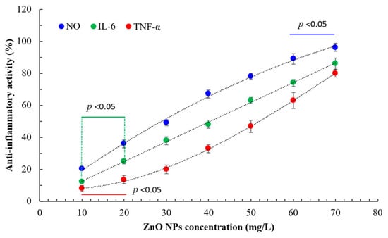
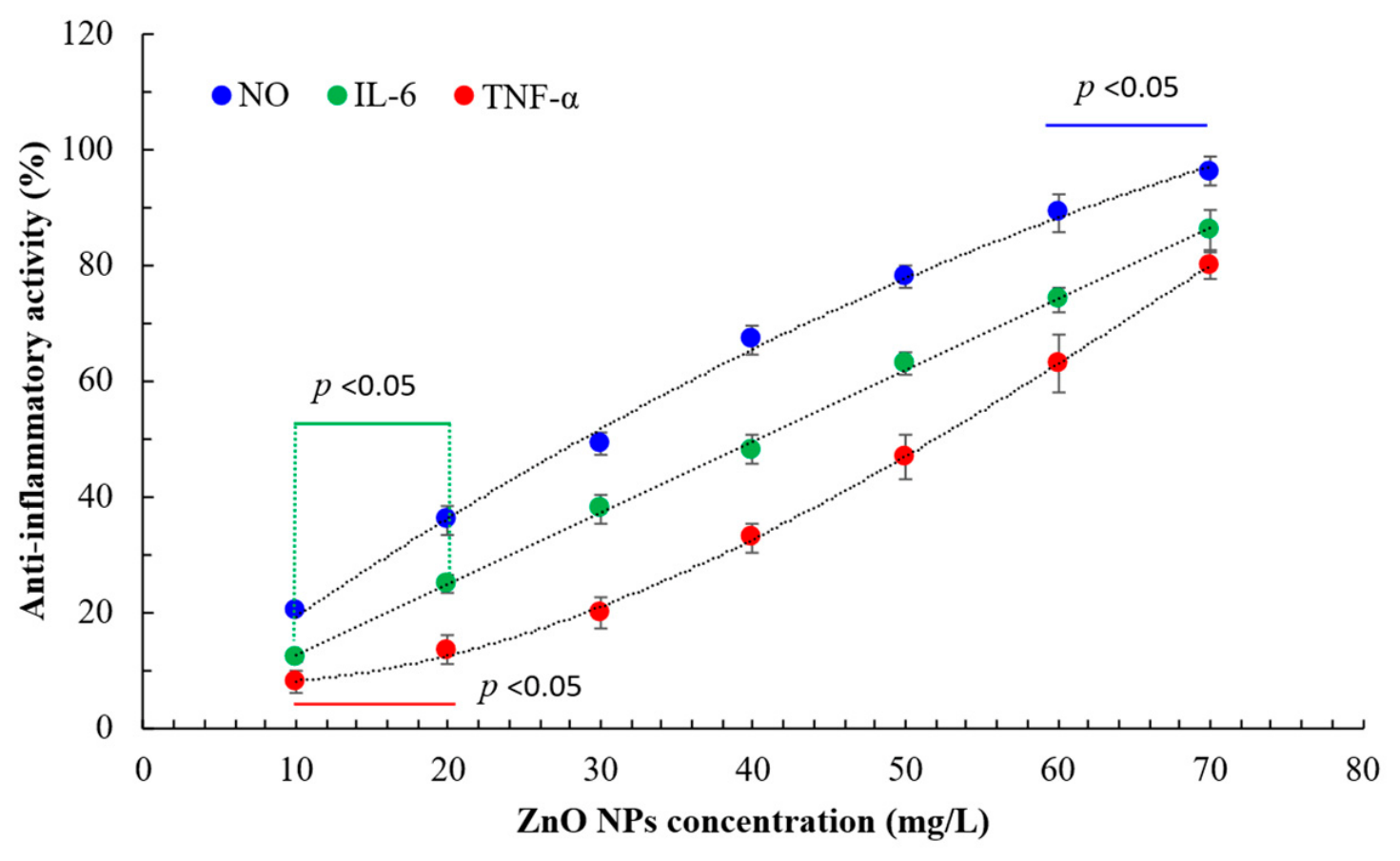
This study evaluated the anti-inflammatory effects using RAW 264.7 cells as the test model. The results showed that bio-capped zinc oxide nanoparticles (ZnO NPs) exhibited dose-dependent anti-inflammatory activity, with IC50 values of 30.6 ± 0.8, 42.5 ± 1.5, and 51.3 ± 2.0 mg/L for nitric oxide (NO), interleukin-6 (IL-6), and tumor necrosis factor-alpha (TNF-α), respectively (Figure 1). In comparison, the control leaf extract showed IC50 values of 40.6 ± 1.3, 58.7 ± 3.5, and 45.2 ± 2.5 mg/L for the same markers. These findings suggested that the anti-inflammatory efficacy of bio-capped ZnO NPs did not solely rely on the leaf extract.
Figure 1.
Anti-inflammatory activity of bio-capped ZnO NPs under optimal conditions. Data are presented as the means and standard deviations of three independent experiments. Statistical significance was determined using one-way ANOVA followed by Duncan’s multiple range test. (p < 0.05 indicates significant).
Nitric oxide played a critical role in mitigating inflammation and inhibiting cancer cell proliferation. Furthermore, the activation of macrophages significantly increased the secretion of nitric oxide. The bio-capped ZnO NPs synthesized by nutmeg oil have been reported to significantly inhibited the production of nitric oxide and exhibited an excellent reduction in secretions of inflammatory cytokines (IL-6, IL-10, and TNF-α) [28]. The results of Elhabal et al. (2024) [29] demonstrated improved anti-inflammatory effects with reduced downregulation of IL-6, IL-1β, and TNF-α in the Althaea officinalis flower extract coated ZnO NPs.
3.5. Assessment of Cell Viability
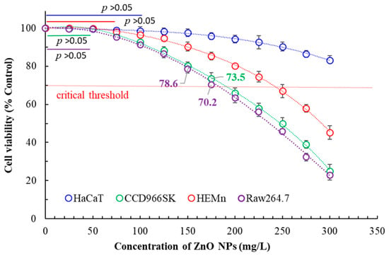
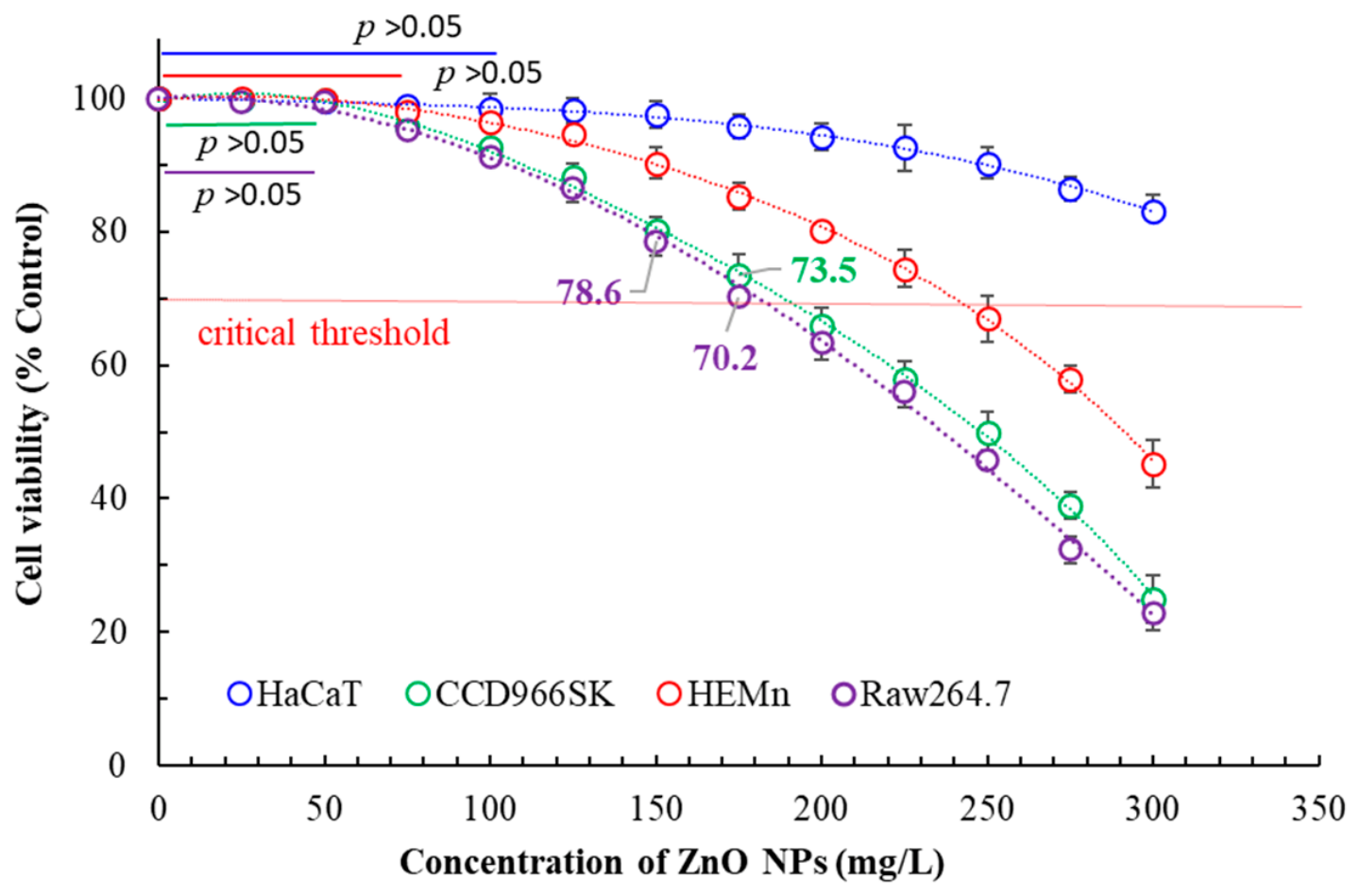
Various physiological activities of bio-capped ZnO NPs have been evaluated previously to determine their effective working doses; however, all doses must demonstrate non-toxic toward cells to possess practical application value. Based on the ISO 10993-5 criteria [30] for the biocompatibility evaluation of medical devices, a cell viability of greater than 70% was the critical threshold to determine the absence of in vitro cytotoxicity. Based on this criterion, the safe dosage thresholds of bio-capped ZnO NPs were at least 300 mg/L for HaCaT cells, and ≤175, ≤225, and ≤175 mg/L for CCD966SK, HEMn, and Raw264.7 cells, respectively (Figure 2). Notably, these values were significantly higher than the concentrations required for bio-capped ZnO NPs to exert their respective physiological activities, thereby confirming the safety of bio-capped ZnO NPs for further applications. Furthermore, the leaf extract exhibited no cytotoxicity within the range of 0–300 mg/L (cell viability > 80%). In contrast, the safe dosages of chemically synthesized ZnO NPs (Che-ZnO NPs) for HaCaT, CCD966SK, and HEMn cells were 175, 100, and 175 mg/L, respectively, indicating relatively higher toxicity compared to the bio-capped ZnO NPs. Additionally, the calculated IC50 values of bio-capped ZnO NPs against CCD966SK, HEMn, and Raw264.7 cells were 250, 286.4, and 238.2 mg/L, respectively.
Figure 2.
Cytotoxic effects of bio-capped ZnO NPs under optimal conditions. Data are presented as the means and standard deviations of three independent experiments (p > 0.05 indicates nonsignificant).
Compared with previous studies, ZnO NPs biosynthesized by B. subtilis were highly effective against MDA-MB-231 (human breast cancer cells) with an IC50 of approximately 60 mg/L [31]. The results of Tabrez et al. (2022) [32] demonstrated the significant anticancer potential of biosynthesized ZnO NPs from pumpkin seeds’ extract against the breast cancer cell line (IC50: 10 mg/L). ZnO NPs exhibited selective cytotoxicity, demonstrating significantly higher lethality toward cancer cells compared to normal cells. It was hypothesized that the selectivity was driven by the accelerated metabolism and inherently acidic microenvironment (lower pH) characteristic of cancer cells. These conditions were presumed to expedite the dissolution of ZnO NPs, resulting in a rapid influx of intracellular Zn2+ ions. The surge subsequently triggered severe oxidative stress within the malignant cells, ultimately inducing apoptosis [33].
3.6. Analyses of Transdermal Absorption and Permeation of Bio-Capped ZnO NPs
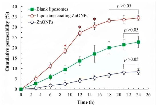
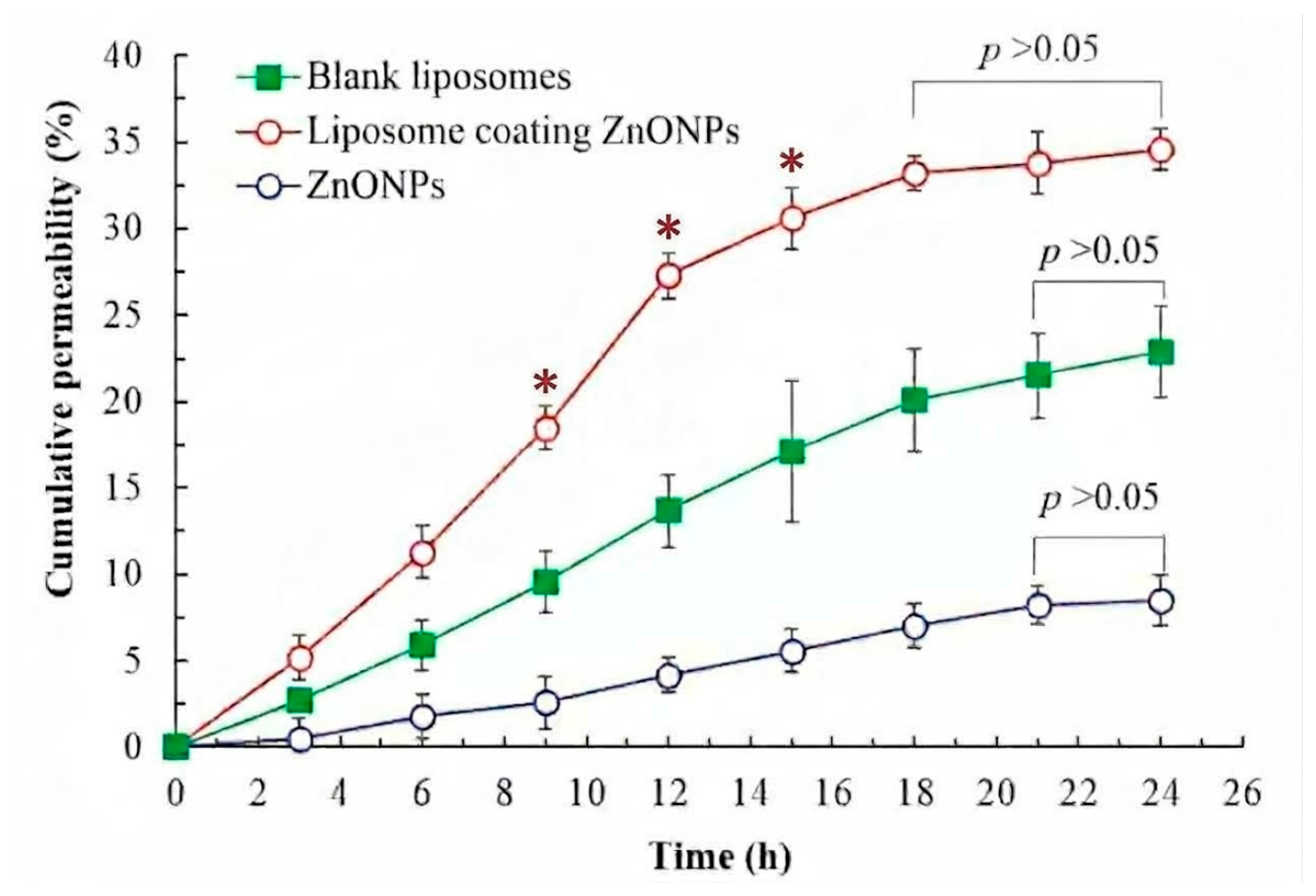
The average hydrodynamic diameter of the bio-capped ZnO NPs was 82.6 nm, which significantly increased to 103.4 nm after being wrapped in the liposome membrane. The formulation exhibited a polydispersity index (PDI) value of 0.24, and the zeta-potential value was recorded at −25.6 mV. As shown in Figure 3, the cumulative permeation rate increased gradually over time. The permeation of ZnO NPs leveled off between 21 and 24 h, reaching 8.5 ± 1.5% at 24 h, which was less than ideal for topical or dermal applications. To improve transdermal delivery, liposome-containing ZnO NPs were prepared using the thin-layer hydration-sonication method. Furthermore, the cumulative permeation rate of the liposomal formulation increased rapidly over time (Figure 3). The permeation rate stabilized within 18–24 h, reaching 34.6 ± 1.2% at 24 h for the liposome-coating ZnO NPs, which was 4.07-fold higher than that of the unencapsulated ZnO NPs. Based on the cytotoxicity profiles evaluated in Section 3.6, a concentration of 150 mg/L was selected as the optimal dosage for the ZnO NPs, balancing maximum efficacy with high cell viability. Furthermore, the cumulative permeability of the blank liposomes fell between the two curves, reaching 22.8%. When ethanolic lecithin solution was directly mixed with ZnO, the resulting system was often classified as a form of “ethosomes” and could potentially exhibit the following ranking in terms of transdermal absorption efficiency: liposome-ZnO > ethanolic lecithin liposome > ZnO [34]. Two possible mechanisms were speculated as follows: 1. Potential Synergistic Effect of liposome-ZnO (Drug-loaded Ethosomes): Ethanol was hypothesized to disrupt the highly organized lipid bilayer arrangement of the stratum corneum (outermost skin layer), which might enhance the overall fluidity. 2. High Deformability of the Vesicles: The combination of lecithin and ethanol was suggested to form highly elastic and flexible vesicles (ethosomes). These adaptable structures might be capable of carrying the encapsulated ZnO through minute gaps in the stratum corneum, allowing for deeper skin penetration and resulting in optimized transdermal delivery and controlled release outcomes. However, Strat-M® membranes could not fully recapitulate native human skin; in vitro permeation results were not equal to in vivo skin performance.
Figure 3.
Transdermal absorption analysis of the bio-capped ZnO NPs and liposome-coating ZnO NPs. * p < 0.05 indicates a statistically significant difference compared with both the blank liposomes and ZnO NPs groups at the same time point; p > 0.05 indicates no significant difference.
Commonly used zinc oxide nanoparticles (ZnO NPs) range in size from approximately 20 to 150 nm [35], whereas molecules capable of naturally penetrating the skin typically require a molecular weight below 500 Daltons [36]. For nanoparticles, traversing the epidermal layer into the dermis is highly challenging. Consequently, to enhance transdermal absorption, this study indicated that encapsulation within liposomes can effectively improve transdermal delivery performance. Nanotechnology has significantly advanced transdermal drug delivery by providing precise control over nanoparticle physicochemical properties, thereby enhancing skin permeation and achieving targeted delivery. Liposomes serve as a prime exemplar of this technological paradigm [37].
4. Conclusions
This study successfully established an eco-friendly and high-efficiency nanoplatform by merging microwave-assisted green synthesis with liposomal engineering. Beyond validating the multi-functional biological benefits of Melaleuca quinquenervia leaf extract (MQLE)-mediated ZnO NPs, this work addressed a critical bottleneck in inorganic nanomaterial application: poor transdermal permeability. Specifically, the results demonstrated that liposome-coating ZnO NPs effectively enhanced its transdermal delivery performance. This lipid-bilayer coating successfully resolved skin-permeation constraints, yielding a remarkable 4.07-fold increase in cumulative transdermal permeability compared to bare nanoparticles. Future investigations will focus on in vivo anti-aging efficacy and long-term dermatological safety assessments to accelerate its translation into industrial and clinical biomedical products.
Author Contributions
Y.-C.C.: Conceptualization, Supervision, Investigation, Validation, Data curation, Resources, Funding acquisition. C.-Y.C.: Supervision, Conceptualization, Writing—original draft, Writing—review and editing, Funding acquisition. All authors have read and agreed to the published version of the manuscript.
Funding
This research was funded by the National Science and Technology Council, grant numbers NSTC 112-2313-B-157-001-MY3.
Institutional Review Board Statement
Not applicable.
Informed Consent Statement
Not applicable.
Data Availability Statement
All data generated or analyzed during this study are included in this published article.
Acknowledgments
The authors would like to thank Pin-Kuan Chiang, Kuan-Fu Yang, and Yun-Erh Kuan for their assistance with sampling and partial analytical measurements.
Conflicts of Interest
The authors declare no conflicts of interest.
References
- Mandal, A.K.; Katuwal, S.; Tettey, F.; Gupta, A.; Bhattarai, S.; Jaisi, S.; Bhandari, D.P.; Shah, A.K.; Bhattarai, N.; Parajuli, N. Current research on zinc oxide nanoparticles: Synthesis, characterization, and biomedical applications. Nanomaterials 2022, 12, 3066. [Google Scholar] [CrossRef] [Scilit] [PubMed]
- Rouhi, J.; Mahmud, S.; Naderi, N.; Ooi, C.R.; Mahmood, M.R. Physical properties of fish gelatin-based bio-nanocomposite films incorporated with ZnO nanorods. Nanoscale Res. Lett. 2013, 8, 364. [Google Scholar] [CrossRef] [Scilit] [PubMed]
- Mishra, P.K.; Mishra, H.; Ekielski, A.; Talegaonkar, S.; Vaidya, B. Zinc oxide nanoparticles: A promising nanomaterial for biomedical applications. Drug Discov. Today 2017, 22, 1825–1834. [Google Scholar] [CrossRef] [Scilit] [PubMed]
- Sarkhail, P.; Foroughian, S.; Gholami, M.; Navidpour, L. Tyrosinase inhibition and angiogenesis studies of zinc oxide nanoparticles green synthesized using aqueous extract of Peucedanum knappii. Nanomed. Res. J. 2025, 10, 295–305. [Google Scholar]
- Ilves, M.; Palomäki, J.; Vippola, M.; Lehto, M.; Savolainen, K.; Savinko, T.; Alenius, H. Topically applied ZnO nanoparticles suppress allergen induced skin inflammation but induce vigorous IgE production in the atopic dermatitis mouse model. Part. Fibre Toxicol. 2014, 11, 38. [Google Scholar] [CrossRef] [Scilit] [PubMed]
- Rauf, M.A.; Zubair, S.; Ateeq, H.; Dabeer, K.; Pachauri, S.; Ajmal, M.; Owais, M. Synergistic effect of diallyl sulfide with zinc oxide nanorods: A novel and effective approach for treatment of acute dermatitis in model animals. Front. Microbiol. 2018, 9, 586. [Google Scholar] [CrossRef] [Scilit] [PubMed]
- Rehman, H.; Ali, W.; Khan, N.Z.; Aasim, M.; Khan, T.; Khan, A.A. Delphinium uncinatum mediated biosynthesis of zinc oxide nanoparticles and in-vitro evaluation of their antioxidant, cytotoxic, antimicrobial, anti-diabetic, anti-inflammatory, and anti-aging activities. Saudi J. Biol. Sci. 2023, 30, 103485. [Google Scholar] [CrossRef] [Scilit] [PubMed]
- Yassin, M.T.; Al-Otibi, F.O.; Al-Askar, A.A.; Elmaghrabi, M.M. Synergistic anticandidal effectiveness of greenly synthesized zinc oxide nanoparticles with antifungal agents against nosocomial candidal pathogens. Microorganisms 2023, 11, 1957. [Google Scholar] [CrossRef] [Scilit] [PubMed]
- Lin, T.K.; Leu, J.Y.; Lai, Y.L.; Chang, Y.C.; Chung, Y.C.; Liu, H.W. Application of microwave-assisted water extraction (MAWE) to fully realize various physiological activities of Melaleuca quinquenervia leaf extract. Plants 2024, 13, 3362. [Google Scholar] [CrossRef] [Scilit] [PubMed]
- Hajos, M.; Starowicz, M.; Brzychczyk, B.; Basista, G.; Francik, S. Size distribution of zinc oxide nanoparticles depending on the temperature of electrochemical synthesis. Materials 2025, 18, 458. [Google Scholar] [CrossRef] [Scilit] [PubMed]
- Prachi; Negi, D.S. Antioxidant and photocatalytic activity of green synthesized zinc oxide nanoparticles using stem extract of Rubia cordifolia. Int. J. Eng. Sci. Math. 2018, 7, 402–412. [Google Scholar]
- Wang, G.H.; Chen, C.Y.; Tsai, T.H.; Chen, C.K.; Cheng, C.Y.; Huang, Y.H.; Hsieh, M.C.; Chung, Y.C. Evaluation of tyrosinase inhibitory and antioxidant activities of Angelica dahurica root extracts for four different probiotic bacteria fermentations. J. Biosci. Bioeng. 2017, 123, 679–684. [Google Scholar] [CrossRef] [Scilit] [PubMed]
- Wu, L.; Chen, C.Y.; Cheng, C.Y.; Dai, H.; Ai, Y.; Lin, C.; Chung, Y.C. Evaluation of tyrosinase inhibitory, antioxidant, antimicrobial, and antiaging activities of Magnolia officinalis extracts after Aspergillus niger fermentation. BioMed. Res. Int. 2018, 5201786. [Google Scholar] [CrossRef] [Scilit] [PubMed]
- Yoshikawa, H.; Oyamada, I.; Usuku, G.; Kim, N.Y.; Kim, J.H.; Choi, G.P. An assay of collagenase activity using enzyme-linked immunosorbent assay for mammalian collagenase. Anal. Biochem. 1987, 166, 172–177. [Google Scholar] [CrossRef] [Scilit] [PubMed]
- Chen, C.Y.; Hu, C.Y.; Chen, Y.H.; Li, Y.T.; Chung, Y.C. Submerged fermentation with Lactobacillus brevis significantly improved the physiological activities of Citrus aurantium flower extract. Heliyon 2022, 8, e10498. [Google Scholar] [CrossRef] [Scilit] [PubMed]
- Vandooren, J.; Geurts, N.; Martens, E.; Van den Steen, P.E.; Jonghe, S.D.; Herdewijn, P. Gelatin degradation assay reveals MMP-9 inhibitors and function of O-glycosylated domain. World J. Biol. Chem. 2011, 2, 14–24. [Google Scholar] [CrossRef] [Scilit] [PubMed]
- Thring, T.S.; Hili, P.; Naughton, D.P. Anti-collagenase, anti-elastase and anti-oxidant activities of extracts from 21 plants. BMC Complement. Altern. Med. 2009, 9, 27. [Google Scholar] [CrossRef] [Scilit] [PubMed]
- Huang, H.W.; Cheng, M.C.; Chen, B.Y.; Wang, C.Y. Effects of high pressure extraction on the extraction yield, phenolic compounds, antioxidant and antityrosinase activity of Djulis hull. J. Food Sci. Technol. 2019, 56, 4016–4024. [Google Scholar] [CrossRef] [Scilit] [PubMed]
- Smeriglio, A.; Denaro, M.; D’Angelo, V.; Germano, M.P.; Trombetta, D. Antioxidant, anti-inflammatory and anti-angiogenic properties of Citrus lumia Juice. Front. Pharmacol. 2020, 11, 593506. [Google Scholar] [CrossRef] [Scilit] [PubMed]
- Cao, D.; Shu, X.; Zhu, D.; Liang, S.; Hasan, M.; Gong, S. Lipid-coated ZnO nanoparticles synthesis, characterization and cytotoxicity studies in cancer cell. Nano Converg. 2020, 7, 14. [Google Scholar] [CrossRef] [Scilit] [PubMed]
- Wang, G.H.; Lin, Y.M.; Kuo, J.T.; Lin, C.P.; Chang, C.F.; Hsieh, M.C. Comparison of biofunctional activity of Asparagus cochinchinensis (Lour.) Merr. extract before and after fermentation with Aspergillus oryzae. J. Biosci. Bioeng. 2019, 127, 59–65. [Google Scholar] [CrossRef] [Scilit] [PubMed]
- Chen, C.Y.; Wang, G.H.; Kuo, J.T.; Lin, Y.T.; Huang, H.J.; Chang, Y.C.; Chung, Y.C. Ultrasound-assisted extraction of Peucedanum ostruthium leaves: A feasible alternative to rhizomes for industrial applications. Front. Pharmacol. 2025, 16, 1636312. [Google Scholar] [CrossRef] [Scilit] [PubMed]
- Thanjavur, N.; Lakshmi, B.A.; Bugude, L.; Theyagarajan, K.; Kim, Y.J. Bio-capped ZnO nanoparticles formulated from Prosopis juliflora gum: Phytochemical characterization and therapeutic potential. Int. J. Biol. Macromol. 2025, 334, 149092. [Google Scholar] [CrossRef] [Scilit] [PubMed]
- Vieira, I.R.S.; Mirres, A.C.D.M.; Conte-Junior, C.A. Zinc Oxide Nanomaterials: Green Synthesis, Properties, and Potentials in Active Food Packaging, Edible Coatings, and Nanomedicine Applications. ChemistrySelect 2026, 11, e04228. [Google Scholar] [CrossRef] [Scilit]
- Ekennia, A.C.; Uduagwu, D.N.; Nwaji, N.N.; Oje, O.O.; Emma-Uba, C.O.; Mgbii, S.I.; Olawale, J.O.; Nwanji, O.L. Green synthesis of biogenic zinc oxide nanoflower as dual agent for photodegradation of an organic dye and tyrosinase inhibitor. J. Inorg. Organomet. Polym. Mater. 2021, 31, 886–897. [Google Scholar] [CrossRef] [Scilit]
- Kapare, H.S.; Bhosale, M.; Karwa, P.; Kulkarni, D.; Bhole, R.; Labhade, S. Phyto-assisted synthesis and investigation of zinc oxide nanoparticles for their anti-aging, sun protection and antibacterial activity. Cosmetics 2025, 12, 238. [Google Scholar] [CrossRef] [Scilit]
- Radulescu, D.M.; Surdu, V.A.; Ficai, A.; Ficai, D.; Grumezescu, A.M.; Andronescu, E. Green synthesis of metal and metal oxide nanoparticles: A review of the principles and biomedical applications. Int. J. Mol. Sci. 2023, 24, 15397. [Google Scholar] [CrossRef] [Scilit] [PubMed]
- Amina, M.; Al Musayeib, N.M.; Alarfaj, N.A.; El-Tohamy, M.F.; Al-Hamoud, G.A.; Al-Yousef, H.M. Immunomodulatory and antioxidant potential of biogenic functionalized polymeric nutmeg oil/polyurethane/ZnO bionanocomposite. Pharmacetics 2021, 13, 2197. [Google Scholar] [CrossRef] [Scilit] [PubMed]
- Elhabal, S.F.; Abdelaal, N.; Saeed Al-Zuhairy, S.A.K.; Elrefai, M.F.M.; Elsaid Hamdan, A.M.; Khalifa, M.M.; Hababeh, S.; Khasawneh, M.A.; Khamis, G.M.; Nelson, J.; et al. Green synthesis of zinc oxide nanoparticles from Althaea officinalis flower extract coated with chitosan for potential healing effects on diabetic wounds by inhibiting TNF-α and IL-6/IL-1β signaling pathways. Int. J. Nanomed. 2024, 3045–3070. [Google Scholar] [CrossRef] [Scilit] [PubMed]
- ISO 10993-5; Biological Evaluation of Medical Devices—Part 5: Tests for in Vitro Cytotoxicity. ISO: Geneva, Switzerland, 2009.
- Hanumitha, S.; Mahalakshmi, V.; Abirami, S. Biosynthesis of zinc oxide nanoparticles using Bacillus species potentiates anticancer and antimicrobial activity. Int. J. Trend Sci. Res. Dev. 2018, 2, 1796–1802. [Google Scholar] [CrossRef] [Scilit]
- Tabrez, S.; Khan, A.U.; Hoque, M.; Suhail, M.; Khan, M.I.; Zughaibi, T.A. Biosynthesis of ZnO NPs from pumpkin seeds’ extract and elucidation of its anticancer potential against breast cancer. Nanotechnol. Rev. 2022, 11, 2714–2725. [Google Scholar] [CrossRef] [Scilit]
- Nsairat, H.; Abdulsahib, W.K.; Jyothi, S.R.; Nayak, P.P.; Chauhan, A.S.; Singla, S.; Sead, F.F.; Polatova, D. Evaluating the mechanisms and therapeutic potential of ZnO nanoparticles as selective anticancer agents for lung malignancies. Discov. Nano. 2026, 21, 215. [Google Scholar] [CrossRef] [Scilit] [PubMed]
- Jaiswal, P.K.; Kesharwani, S.; Kesharwani, R.; Patel, D.K. Ethosome: A new technology used as topical & transdermal delivery system. J. Drug Deliv. Ther. 2016, 6, 7–17. [Google Scholar] [CrossRef] [Scilit]
- Verma, L.M.; Kumar, A.; Kumar, A.; Singh, G.; Singh, U.; Chaudhary, S.; Kumar, S.; Sanwaria, A.R.; Ingole, P.P.; Sharma, S. Green chemistry routed sugar press mud for (2D) ZnO nanostructure fabrication, mineral fortification, and climate-resilient wheat crop productivity. Sci. Rep. 2024, 14, 4074. [Google Scholar] [CrossRef] [Scilit] [PubMed]
- Bos, J.D.; Meinardi, M.M. The 500 Dalton rule for the skin penetration of chemical compounds and drugs. Exp. Dermatol. 2000, 9, 165–169. [Google Scholar] [CrossRef] [Scilit] [PubMed]
- Dudhat, K. Emerging trends in transdermal drug delivery: Nanoparticle formulations and technologies for enhanced skin penetration and drug efficiency. Pharm. Nanotechnol. 2026, 14, 209–229. [Google Scholar] [CrossRef] [Scilit] [PubMed]

Disclaimer/Publisher’s Note: The statements, opinions and data contained in all publications are solely those of the individual author(s) and contributor(s) and not of MDPI and/or the editor(s). MDPI and/or the editor(s) disclaim responsibility for any injury to people or property resulting from any ideas, methods, instructions or products referred to in the content. |
© 2026 by the authors. Licensee MDPI, Basel, Switzerland. This article is an open access article distributed under the terms and conditions of the Creative Commons Attribution (CC BY) license.