


Abstract
Healing wounds and skin lesions has been practiced since ancient times, and various mechanisms have been used to treat them, ranging from bandages and herbs to ointments and modern alternatives such as dressings. This work aimed to develop bioactive dressings for wound healing. Dressings were prepared using the solution-drained plate technique and comprised 2% (w/v) sodium alginate with varying concentrations of Melipona beecheii honey, up to 5% (w/v). The dressings obtained showed suitable flexibility and elongation as the honey content increased, and had a suitable thickness for skin application, ranging from 0.03 to 0.21 mm. They were also evaluated for barrier, optical, structural, thermal, morphological, bioactive, and cytotoxic properties. The dressings demonstrated good moisture absorption and maintained antimicrobial activity for up to 12 months. Moreover, they retained bioactive compounds, demonstrated strong antioxidant activity, and showed biocompatibility in biological assays. The dressings did not induce cytotoxic effects in human keratinocytes. Dressing with a 2.5% honey concentration exhibited physicochemical and bioactive properties that could improve skin wound healing.
1. Introduction
Wound trauma, a significant public health issue, can potentially affect a diverse global population of all ages due to the long-term morbidity associated with tissue restoration and regeneration, as well as the risk of infection, hypertrophy, and scar formation [1]. The wound-healing process involves stages (hemostasis, inflammation, cell proliferation, and tissue regeneration) to repair damaged tissues and restore the injured area [2]. Various dressings on the market promote the healing of both acute and chronic wounds. Among the characteristics to consider for their use are wettability, antibacterial activity, and anti-adherent properties, primarily to combat bacterial infection by sealing the lesion microenvironment against peripheral contaminants. At the same time, dressings should remove excess exudate, promote autolysis-driven debridement, be flexible and biodegradable, and maintain barrier properties to accelerate healing and reduce the risk of infection [3].
Dressings developed from bioactive polysaccharides provide a therapeutic effect on wound healing, an ideal characteristic required for their use. These can include sodium alginate, a biocompatible, biodegradable, and hydrophilic polysaccharide composed of varying amounts of the negatively charged β-D-mannuronic acid and α-L-guluronic acid, linked via glycosidic bonds [4]. It is used in the food and pharmaceutical industries as a drug and dressing. Several types of alginate-based dressings (hydrogels, nanofibers, foams, sponges, and films) can maintain a moist environment and absorb excess exudate in the wound. Although alginate has promising therapeutic properties for wounds, it lacks the antibacterial properties required to prevent wound infections. Therefore, adding honey to different materials has been shown to avoid infection and accelerate wound healing [4]. Honey is a high-nutritional-value food product that has been used in traditional medicine since ancient times [5]. It has been used as a natural dressing due to its healing properties, including antioxidant and free-radical-scavenging activities, as well as antimicrobial and anti-inflammatory effects. These effects are due to bioactive factors, including the phenolic and flavonoid compounds present in honey, which can promote tissue growth, prevent infections, and promote healing [6]. Melipona honey is produced by stingless bees of the Melipona beecheii species and has very particular organoleptic characteristics that consist of a high degree of moisture and acidity, which make it almost liquid; its consumption has been shown to strengthen the immune system for eye ailments and to heal wounds [7]. Therefore, this research aimed to develop and characterize bioactive dressings from Melipona honey in an alginate matrix, focusing on evaluating their physical, mechanical, and bioactive properties to promote an environment that could allow the healing of cutaneous wounds, with the potential to support the wound healing process and bring optimism about the effectiveness of wound dressing applications.
2. Materials and Methods
2.1. Materials
Sodium alginate (A), a medium-molecular-weight, high-solubility food-grade additive from Deiman S.A. de C.V. brand (Mexico City, Mexico), was used to prepare the dressings. Melipona honey (M) produced by the Melipona beecheii bee was collected from a rural meliponary in the town of Mani, Yucatan, Mexico (20.385444114994154 N, −89.39841799586078 W) in April 2022.
2.2. Dressing Preparation
Dressings were prepared using the solution-drained plate technique (casting). Dressing formulations were prepared at different honey concentrations (1, 2.5, and 5% w/v) with a constant A concentration of 2% w/v (chosen based on its optimal performance in preliminary tests), as shown in Table 1. Alginate was solubilized in distilled water at 60 °C with agitation for 4 h. Subsequently, M was added to the mixture at different concentrations, and the mixture was agitated for 2 h at the same temperature. The solution was poured into Petri dishes to avoid bubble formation and dried at 60 °C for 24 h in a convection oven (Binder model ED115, Tuttlingen, Germany). The formed dressings were removed from the Petri dishes, protected from light by dark plastic bags, and stored in a desiccator to prevent moisture gain or loss and to prevent natural light from affecting the bioactive compounds.
Table 1.
Formulations of the dressings.
2.3. Dressing Characterization
2.3.1. Thickness and Bulk Density
The thickness of each dressing formulation was measured using an analog micrometer (Mitutoyo model N7301; Kawasaki-Shi, Kanagawa, Japan). The average thickness was calculated from 6 measurements taken from different regions of each sample.
Rectangular specimens of the plastic film were cut (2 × 2 cm) with precision to obtain uniform dimensions. Thickness was measured at five random points using a digital micrometer (±0.001 mm), Mitutoyo model N7301 (Kawasaki-Shi, Kanagawa, Japan), and the measurements were averaged. Length and width were determined with a calibrated caliper, Mitutoyo model 500-197-32 (Kawasaki-Shi, Kanagawa, Japan). The mass of each specimen was recorded using an analytical balance, Ohaus Corp. model Adventure™ Semi-Micro (±0.1 mg) (Parsippany, NJ, USA). Density was calculated as the ratio of mass to volume (g/cm3), where volume was obtained from the product of average thickness, length, and width. All measurements were performed on ten samples of each formulation at ambient laboratory conditions to minimize variability.
2.3.2. Moisture and Water Vapor Transmission Rate (WVTR)
The moisture of the dressings was determined by gravimetry, and the weight of 2 × 2 cm dressing samples was taken on an Ohaus Corp. (Parsippany, NJ, USA) model Adventure analytical balance. Subsequently, these samples were placed in a convection oven at 80 °C for 24 h, and their weight was recorded. The moisture percentage was calculated based on the weight difference using Equation (1).
where W0 is the initial weight, and Wf is the dry weight of the films after drying.
The WVTR tests were performed using the ASTM E96/E96M method, with modifications made by Chan et al. [8]. A container holding 30 mL of distilled water was used, with the dressing placed over the lid, leaving a small space between the sample and the water to achieve a relative humidity of 100%. The container was closed and stored at 36 °C in a desiccator containing silica gel to maintain a relative humidity of 25% inside, and the weight was recorded every 2 h for 12 h. The WVTR (g/h·m2) was calculated using the following Equation (2).
where b is the slope of the straight line (g/h) of the graph obtained by the difference in weight versus time, and A is the transfer area of the dressing (m2).
2.3.3. Opacity
The light transmission values of the dressings were obtained by measuring the transmittance of selected wavelengths between 200 and 800 nm using a Varian, Inc. (Walnut Creek, CA, USA) model Cary 50 ultraviolet–visible (UV–Vis) spectrophotometer. Dressings were cut into 40 × 10 mm rectangles and placed in the spectrophotometer cell, using the cell free of any substance as the calibration blank. The opacity was determined by measuring the absorbance at 600 nm and calculating it using Equation (3).
where the opacity (mm−1), Abs600 is the absorbance value at 600 nm, and δ is the thickness of the dressing (mm).
2.3.4. Color
The color was determined using an X-Rite colorimeter, model SP62 (X-Rite, Inc., Grandville, MI, USA), calibrated with a standard mosaic (Y = 94.05333, x = 1.1366667, y = −4.36) and an opacity reference (5.32%). The samples were evaluated using the CIELab scale, with the following parameters: lightness (L*), the a* value (red-green), and the b* value (yellow-blue); these were used to calculate chromaticity (C*) and Hue angle (°Hue). Measurements were performed at five points on each sample. The total color difference (ΔE) was determined using the control film (alginate only, A) as the reference. The parameters ΔE, °Hue, and C* were calculated from Equations (4)–(6).
where L* is the brightness value, a* red-green values, b* yellow-blue values, and L0, a0, b0 are control film values.
2.3.5. Mechanical Properties
Mechanical properties were measured in accordance with ASTM D882. Samples were cut into 60 × 10 mm rectangular strips. A universal mechanical testing machine, Instron model 4442 (Canton, MA, USA), was used for tensile tests. Previously, the dressing thickness was measured with a Mitutoyo digital micrometer (model H2780, Aurora, IL, USA) at the center of the rectangle, with an accuracy of ±0.001 mm, to calculate its cross-sectional area and ensure precise measurements. Adhesive tape was applied to the ends to prevent the dressing from tearing between the jaws. A 50 kg load cell with a 30 mm jaw spacing and a stretching speed of 5 mm/min was used for the tensile test. Six specimens of each formulation were tested, and the mechanical parameters recorded were tensile strength (TS), percentage elongation at break (%Eb), and modulus of elasticity (ME) (N/mm2 ≡ MPa), which were calculated using Equations (7)–(9).
where Fmax is the maximum force at break (N), At is the cross-sectional area of the specimen (mm2), Lf is the final length (mm), Li is the initial length (mm), Fmax is the force in (N) corresponding to the maximum stress, and ϵ is the strain (mm/mm). TS or σ is tensile strength or stress (N/mm2 ≡ MPa), and ϵ is the strain (mm/mm) associated with the relative elongation with respect to the initial length.
2.3.6. Morphological Analysis
The dressing’s surface morphology was characterized using a field-emission scanning electron microscope (FESEM) model JEOL JSM-7600F (Peabody, MA, USA). Samples were placed on a 10 mm cylindrical specimen holder using double-sided carbon-conductive adhesive tape. Subsequently, before analysis, these were coated with a thin layer of Au-Pd using a Quorum model QI5OR-EN plasma coater (Sussex, UK). Surface morphology was observed on 5 × 5 mm samples by visual inspection of the surface at 200× magnification.
2.3.7. Infrared Spectroscopy
FTIR analysis was performed on the dressings using a Fourier transform infrared (FTIR) spectrometer (Thermo Scientific™ model Nicolet™ iS50™) (Waltham, MA, USA) with an attenuated total reflection (ATR) accessory. Spectra were recorded in the 4000 to 600 cm−1 range at a resolution of 4 cm−1, a scan rate of 0.675 cm−1/s, and after 100 sweeps. The spectrum normalization process accounted for the band at 808 cm−1, using the spectrophotometer software (OMNIC Paradigm).
2.3.8. Thermogravimetric Analysis (TGA)
Thermal properties were analyzed using a thermogravimetric analyzer (TGA-8000, Perkin-Elmer Inc., Waltham, MA, USA) to determine the degradation temperatures of the dressings. The thermograms of weight loss (%) were obtained over a temperature range of 24 °C to 700 °C, with a heating rate of 10 °C/min under an inert atmosphere (N2).
2.4. Bioactive Properties
2.4.1. Total Phenol Content
The Folin–Ciocalteu technique was used to quantify the total phenol content (TPC). One gram of each dressing was used to prepare the extract; the dressings were placed in test tubes, and 13 mL of distilled water was added to each. The samples were placed in a Hielscher USA, Inc. model UP200st sonicator (Wanaque, NJ, USA) at 31 °C for 30 min. The samples were then centrifuged at 4500 rpm for 10 min, and 5 mL of the extract (supernatant) was collected. To each test tube, 20 μL of the extract obtained from each dressing was added, followed by 1.5 mL of distilled water, 100 μL of Folin–Ciocalteu’s reagent, and 300 μL of 7.5% Na2CO3 solution. The samples were read at a wavelength of 765 nm. Previously, a calibration curve was generated over the concentration range of 0–0.5 mg/mL using gallic acid as the standard. The results are expressed in mg gallic acid equivalents in 100 g of sample (mg GAE/100 g).
2.4.2. Antioxidant Capacity
For the dressings, the absorbance was measured using the 2,2′-azino-bis(3-ethylbenzothiazoline)-6-sulfonate (ABTS) method described by Re et al. [9]. We prepared the solution using 0.7 mM ABTS radical and 4.9 mM potassium persulfate, mixed in a 1:1 ratio, and then diluted with 20% ethanol until an absorbance of 0.7 ± 0.2 was reached. A total of 3.0 mL of this solution was taken, and 0.1 mL of the aforementioned extract (2.4.1) was added; the absorbance was measured at 740 nm during the first seven minutes. This method is commonly applied to measure the antioxidant capacity of a substance. The results were expressed as a % inhibition of free radicals using Equation (10).
where Abs0 is the absorbance at 740 nm at the beginning of the reaction, and Absf is the absorbance at 740 nm after 7 min.
2.4.3. Antimicrobial Analysis
Microbiological tests corroborated the dressing’s ability to provide antimicrobial protection. These were performed by contaminating the dressings with microorganisms under standard conditions and verifying their growth after 24 h of contact. Subsequently, the disc diffusion method was used to determine the bacteria’s sensitivity to the dressing [10]. The microorganisms were inoculated onto Müller–Hinton (MH) agar and allowed to grow by spreading the strain with a saline-soaked swab. This procedure was performed using an inoculum at a concentration of 1 × 10⁶ colony-forming units (CFU). This concentration was determined using a 0.85% saline solution as a blank, to which the inoculum was gradually added while measuring absorbance at 600 nm until a value of 0.10–0.11 was reached, corresponding to 0.5 units on the McFarland scale and equivalent to 1 × 10⁸ CFU/mL. From this inoculum, the appropriate dilution was performed to obtain a final concentration of 1 × 10⁶ CFU/mL. The various dressings, cut into 4 cm2 pieces, were strategically placed in designated areas and sterilized with UV light (at a wavelength of 254 nm and a distance of 52 cm) for 15 min on each side. Ten microliters of amikacin (antibiotic) at 1 mg/mL were deposited in the center to compare the diameters of the zones of inhibition. Two strains were used for the antimicrobial activity assay: Staphylococcus aureus ATCC 25923 (Gram-positive) and Pseudomonas aeruginosa ATCC 27853 (Gram-negative) (American Type Culture Collection. Manassas, Virginia. Distributor in Mexico: Cientifica Senna S.A. de C.V., Mexico City, Mexico). All samples were incubated for 36 h at 37 °C. The inhibition zone was observed every 12 h until 36 h had elapsed, and the zone was recorded. The dressings were stored for 12 months at 25 °C with relative humidity below 30% in a desiccator and in the absence of light. Antimicrobial assays were performed on dressings stored for 3, 6, 9, and 12 months. All experiments were conducted in triplicate.
2.4.4. MTT Toxicity Assay
To determine the dressing’s effect on the viability of human cells, a 3-(4,5-dimethylthiazol-2-yl)-2,5-diphenyltetrazole bromide (MTT) colorimetric test described by Mosmann [11], with some modifications, was performed. This assay evaluated samples of three dressings at different honey concentrations (A, AM2.5, and AM5), selected based on the best results from the physical, colorimetric, mechanical, structural, thermal, morphological, antimicrobial, and antioxidant tests.
To prepare the treatments, pre-weighed membrane samples from each formulation were completely dissolved in culture medium. Because the dressings were soluble, each formulation was evaluated at concentrations of 0 (Control), 3, 8, 15, 30, and 60 mg/mL (calculated as the mass of dissolved membrane per volume of medium). Then, the membrane solutions were filtered through a 0.22 µm membrane filter to ensure sterility.
The cell model used was immortalized human keratinocytes (HaCaT, obtained from Sigma-Aldrich (St Louis, MO, USA) (Cat. 300493, CLS/Cytion) by Dr. Leidy Carrillo-Cocom, Universidad Autónoma de Yucatán, Facultad de Ingeniería Química. Mérida, Yucatán. México), a type of essential skin cell. The cells were routinely cultured in DMEM-F12 medium without phenol red and supplemented with 10% fetal bovine serum (complete medium). The culture was incubated at 37 °C in a 5% CO2 humidified atmosphere using Thermo Scientific™ equipment. For assays, cells were washed with phosphate-buffered saline (PBS) and incubated with 0.25% trypsin-EDTA for 5 min. Upon completion, the enzyme was inactivated with complete culture medium, and the cells were recovered by centrifugation for 5 min at 1000 rpm. The cell pellet was resuspended in culture medium and counted using a Neubauer chamber and the trypan blue technique. Cells were seeded at 2 × 10⁴ cells/well in 96-well microtiter plates in 100 µL of complete DMEM-F12 medium. At 24 h, the medium was removed, and 100 μL of each sample was added to the wells, resulting in final concentrations of 0 (Control), 3, 8, 15, 30, and 60 mg/mL. The cells were incubated for 48 h, and at the end, 20 μL of MTT solution (5 mg/mL in PBS) was added to each well. The plate was incubated at 37 °C for 4 h. Afterward, the medium was removed, and 50 μL of dimethyl sulfoxide (DMSO) was added to the wells to solubilize the formazan crystals. The resulting absorbance was measured with a Multiskan FC microplate reader (Thermo Scientific™) at 570 nm. The percentage of cell viability (proliferation) relative to the no-sample control (cells with medium only) was determined using Equation (11). All experiments were carried out in two independent assays performed in triplicate.
where AbsS is the absorbance of the sample at 570 nm, and AbsC is the absorbance at 570 nm of the control (untreated cells).
2.5. Statistical Analysis
The experiments on the formulation and manufacture of the dressings were conducted in duplicate. The physical, optical, barrier, and bioactive analyses were performed in triplicate, and the mechanical analysis in sextuple, and all were subjected to a one-way analysis of variance (ANOVA). The means were compared using the Tukey–Kramer test at a significance level (p) of 5% in the statistical package STATGRAPHIC Centurion® (The Plains, VA, USA). The results are presented as the mean value ± standard deviation.
2.6. Generative AI
Declaration of generative AI in scientific writing: During the preparation of this manuscript, the authors used Grammarly for Windows Education version and licensed it to CIAD, A.C. (24 July 2026) for the sole purpose of improving language readability, checking spelling, and refining the manuscript’s grammar and syntax. This tool was utilized to enhance the clarity and flow of the text, ensuring it meets the linguistic standards expected for scientific communication. After using this tool, the authors reviewed and edited the content as needed and took full responsibility for the manuscript. No generative AI tools were used for data analysis, interpretation, or the generation of scientific content.
3. Results
3.1. Thickness and Bulk Density
The thickness of a dressing can vary significantly, influenced by both processing conditions and composition [12]. Table 2 shows the thickness and bulk density of the formulated dressings. The dressing thickness increased significantly across the different honey concentrations, ranging from 0.03 to 0.21 mm. The control dressing (A) exhibited the lowest thickness, whereas the dressing with the highest honey concentration (AM5) showed the greatest thickness.
Table 2.
Results of thickness, density, humidity, opacity, and WVTR.
Similarly, the bulk density showed a significant increase (p < 0.05) with increasing solids concentrations (honey), and the density of the dressings ranged from 1.43 to 2.78 g/cm3. The control dressing (A) had the lowest density, while the AM5 dressing had the highest, as shown in Table 2. In general, both thickness and bulk density show the same relationship: they increase significantly (p < 0.05) as a function of the content of honey in the alginate matrix.
3.2. Humidity
A low moisture percentage allows dressings to remain dry and stable. The moisture content of the alginate-based dressings is shown in Table 2. The moisture values ranged from 40.98% to 7.59%. Dressing A showed the highest moisture content, while AM5 showed the lowest. The moisture content of the formulated dressings decreased significantly (p < 0.05) as the honey content increased.
3.3. Water Vapor Transmission Rate (WVTR)
The WVTR values for the alginate-based dressings are shown in Table 2. The formulations’ values range from 154.1 to 204.2 g/h·m2. Dressing A had the highest WVTR, whereas the AM2.5 formulation had the lowest. The dressings showed significant differences (p < 0.05). The lowest WVTR values are due to the honey concentration used in the dressing formulations.
3.4. Opacity and Color
The opacity and color parameters of the alginate-based dressings are shown in Table 2 and Table 3, respectively. The opacity of the analyzed dressings ranged from 0.63 to 1.48 mm−1. Dressing A exhibited the highest opacity, whereas it decreased significantly (p < 0.05) as the concentration of Melipona honey increased.
Table 3.
Color parameters of luminosity (L*), angle (°Hue), color difference (ΔE), and chromaticity (C*) measured in alginate–Melipona honey dressings.
The color parameters of the dressings were determined in terms of lightness (L*), color difference (ΔE), hue angle (°Hue), and chromaticity (C*), as presented in Table 3. The dressings maintained their structural integrity while exhibiting a progressive yellowish coloration as the honey concentration increased. All dressings showed significant differences (p < 0.05); however, the AM5 sample presented the lowest L* value (47.88), while the angle (°Hue), which represents the tonality within the color notation system diagram (CIELab), was 119.6 due to the change in pH corresponding to the number of phenolic compounds present in the honey, causing a color change in the L*, a*, and b* parameters. Dressing AM5 showed the largest color difference relative to A, the control.
3.5. Mechanical Properties
The mechanical parameter values for dressings are presented in Table 4. The mechanical properties of a material describe how it behaves under the action of external forces, which depend on the structure and composition of the components. The incorporation of honey resulted in a significant change in mechanical behavior and increased flexibility (p < 0.05), improving the mechanical properties of dressings with a sodium alginate matrix. The AM1 sample showed a lower percentage of elongation at break (3.08%) than the AM5 dressing (43.86%). Tensile strength and modulus of elasticity decreased progressively as honey concentration increased. The AM5 dressing exhibited the lowest tensile strength (2.87 MPa) and modulus of elasticity (5.88 MPa), indicating that incorporating honey altered the dressing’s mechanical behavior.
Table 4.
Results of the percentage of elongation at break, tensile strength, and elastic modulus for alginate–Melipona honey dressings.
3.6. Morphological Analysis
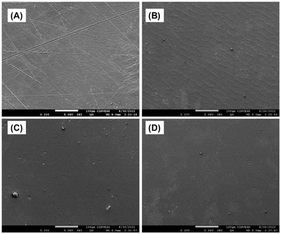
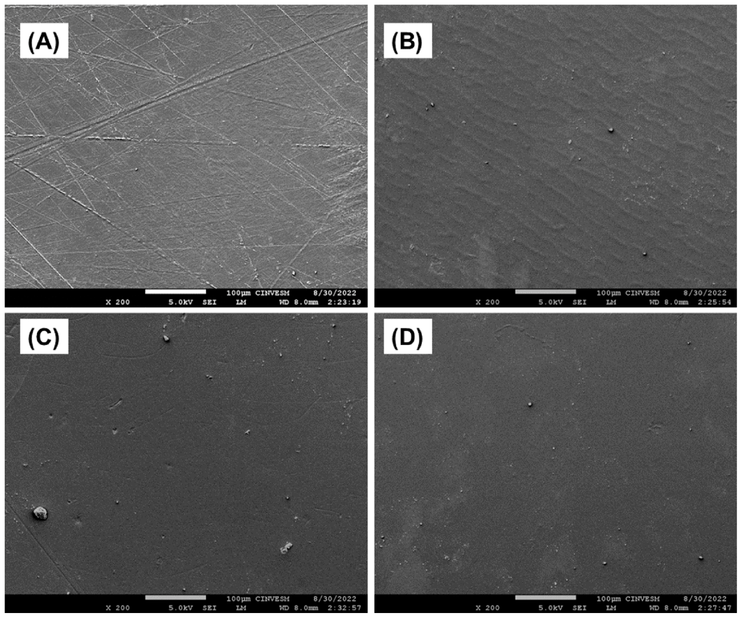
SEM micrographs of the alginate-based dressings at 200× magnification are shown in Figure 1. The formulations exhibited continuous structures with no apparent phase separation at the microdomain level on the dressing surface, indicating good compatibility between sodium alginate and Melipona honey. The control dressing (A) presented roughness, indentations, and large pores on its surface. As the honey content increased, the surface morphology of the dressings became progressively more homogeneous. Dressings AM5 showed the most uniform microstructure, reduced pores and agglomerates, and a smoother surface compared with the other dressings.
Figure 1.
Micrographs of the surface morphology of alginate-honey dressings A (A), AM1 (B), AM2.5 (C), and AM5 (D) by SEM at 200× magnification.
3.7. Infrared Spectroscopy
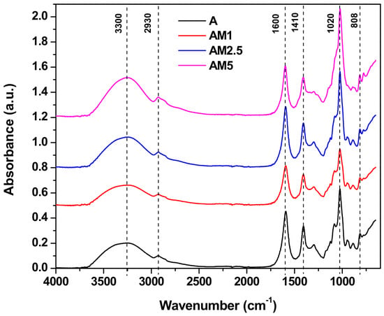
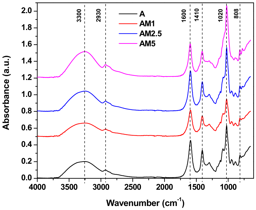
Infrared (IR) spectroscopy, a technique used to identify and study the chemical composition of materials, was performed to determine changes in the functional groups of dressings after incorporating honey into the alginate matrix. The FTIR spectra of alginate-based dressing formulations of Melipona honey are shown in Figure 2. The spectrum of dressing A showed the characteristic absorption band of sodium alginate. A broad absorption band of the hydroxyl group (-OH) stretching at 3300 cm−1 was observed. Likewise, the stretching at 2930 cm−1 (C-H) corresponds to carbon-hydrogen bonds in sp3-hybridized carbon, and finally, the tension at 1600 cm−1 (-COO-) is attributed to the symmetrical stretching of carboxylic groups [13]. The stretches between 1300 and 1000 Cm−1 correspond to C-C-H, O-C-H, and C-O-C bonds associated with pyranose [14]. FTIR spectra confirmed the integration of honey into the alginate matrix without altering its structural chemistry, indicating successful loading. Spectra of the AM2.5 and AM5 dressings showed increased absorption signals for the predominant alginate bands at 1600 cm−1 and 1410 cm−1 (-COO), attributed to symmetric and asymmetric vibrations of the carboxyl group [15]. However, in AM5 dressing, an increase in signal absorption intensity at 3300 cm−1 is observed due to the abundant presence of hydroxyl (-OH) groups associated with glucose, sucrose, and fructose [16], as well as polyphenol and flavonoid content [17] present in honey.
Figure 2.
IR spectra of the formulated dressings: A, AM1, AM2.5, and AM5.
3.8. Thermogravimetric Analysis (TGA)
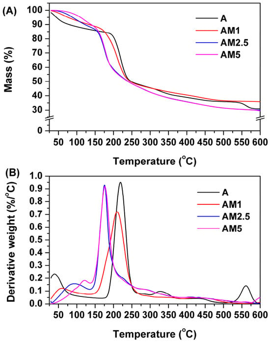
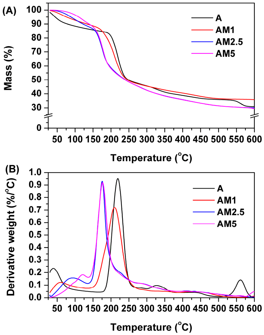
The thermal stability and decomposition of alginate-based dressings were studied by thermogravimetric analysis (TGA), and the corresponding derivative thermogravimetric (DTG) thermograms are shown in Figure 3. The formulations analyzed exhibited a three-stage thermal degradation process characterized by mass loss, as observed in the DTG (Figure 3B). The first degradation stage occurred between 24 °C and 100 °C and was associated with moisture release. The dressings showed mass loss values of 12% for A, 8% for AM1, 7% for AM2.5, and 4% for AM5. The second degradation stage occurred between 101 °C and 299 °C, representing the greatest mass loss in the analysis. The values were as follows: A at 43%, AM1 at 47%, AM2.5 at 51%, and AM5 at 54%. The third degradation step occurred from 300 °C to 500 °C, with mass losses of 14% for A and AM1, and 11% for AM2.5 and AM5. The DTG thermograms revealed that the maximum decomposition temperatures were approximately 220 °C for A, 212 °C for AM1, 175 °C for AM2.5, and 174.5 °C for AM5.
Figure 3.
Thermograms of alginate–Melipona honey dressings, TGA (A) and DTG (B).
3.9. Total Phenol Content
The total phenol content (TPC) of the alginate-based dressings is presented in Table 5. As can be observed, the TPC of the dressings differed significantly (p < 0.05). Dressing A has the lowest TPC value (55.06 mg GAE/100 g of sample), whereas the incorporation of Melipona honey resulted in a progressive and significant increase (p < 0.05) in TPC; AM2.5 and AM5 dressings showed the highest values, reaching 199.99 and 255.48 mg GAE/100 g, respectively.
Table 5.
Values of total phenolic content (TPC) and antioxidant activity (ABTS) of the formulated wound dressings.
3.10. Antioxidant Capacity
The values of the antioxidant capacity of the alginate-based dressings, determined by the ABTS radical scavenging assay, are shown in Table 5. The developed dressings showed significant differences (p < 0.05) in total phenolic content (TPC) and a proportional relationship with honey content. Dressing A showed the lowest antioxidant activity (4.10% ABTS radical inhibition), while the dressing with a lower honey concentration, AM1, showed 13.9%. In contrast, dressings with higher honey concentrations (AM2.5 and AM5) showed higher inhibition of the free radical ABTS, with 36.09% and 42.48% inhibition, respectively.
3.11. Antimicrobial Analysis
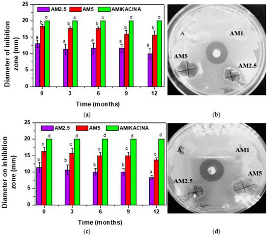
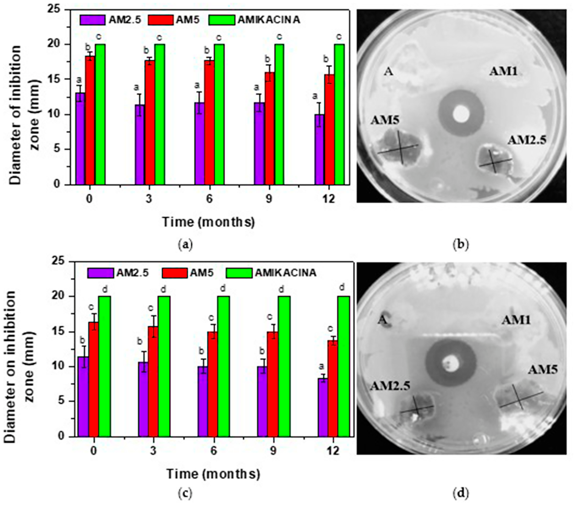
The antimicrobial activity of the formulated dressings was evaluated against Pseudomonas aeruginosa ATCC 27853 (Gram-negative) and Staphylococcus aureus ATCC 25923 (Gram-positive) over a period of 12 months. The antimicrobial efficacy of each formulation was assessed every three months and compared with the antibiotic Amikacin® at 1 mg/mL (positive control). The control dressing (A) and the AM1 did not show antimicrobial activity under experimental conditions, as shown in Figure 4b,d. In contrast, dressings AM2.5 and AM5 showed clear inhibition zones against both bacterial strains throughout the 12-month evaluation period (Figure 4). The antimicrobial activity of these dressings was stable, with inhibition persisting for 36 h after application. These results show that Melipona honey preserves its antibacterial properties for up to 12 months.
Figure 4.
Antimicrobial kinetics of AM2.5 and AM5 dressings every three months for 12 months. (a,b) against Pseudomonas aeruginosa ATCC 27853. (c,d) against Staphylococcus aureus ATCC 25923. Amikacin® 1 mg/mL was used as the antibiotic. The error bars represent the standard deviation. Different letters in each column indicate significant differences between the given formulations (p < 0.05).
3.12. MTT Cytotoxicity Assay
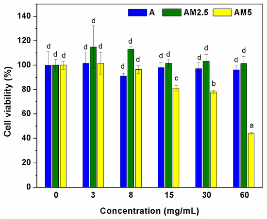
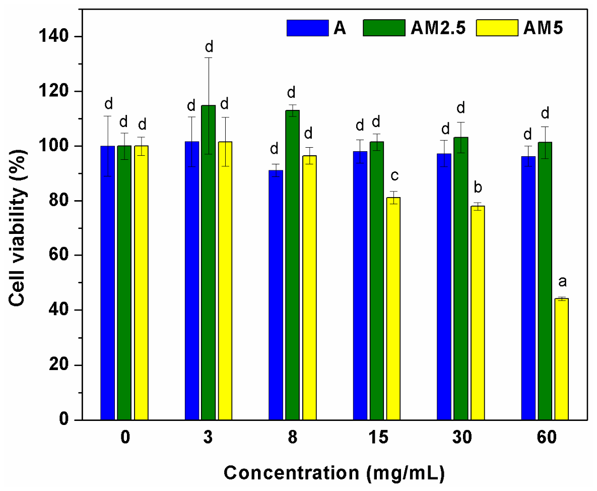
The effect of the dressing on the HaCaT cell line was assessed by measuring cell viability indirectly using the MTT assay; the results are shown in Figure 5. The results show that dressing A did not affect cell viability compared to the control (untreated cells). Similarly, the AM2.5 dressing showed no effect on cells at the concentrations tested, further highlighting the potential of these dressings to promote healing. In contrast, dressings containing 5% honey (AM5) significantly reduced cell viability (p < 0.05) at concentrations of 15 mg/mL and above.
Figure 5.
Relative cell viability compared to the control HaCaT cell line produced by contact with different concentrations of Melipona honey in the dressings (A, AM2.5, and AM5). Error bars represent standard deviation. Different letters in each column indicate significant differences between formulations (p < 0.05).
4. Discussion
An increase in the thickness and bulk density of the dressing was observed, attributed to the gradual increase in the concentration of Melipona honey in the alginate matrix. The solids content in the honey likely promoted the formation of a thicker and more compact structure, resulting in a higher mass per unit volume in the AM5 dressing. Furthermore, honey acts as a natural plasticizer, reducing intermolecular interactions within the alginate network, thereby increasing flexibility and reducing the inherent brittleness typically observed in sodium alginate films.
The thickness values obtained in this study are consistent with those reported by Hernandez et al. [18]. They reported dressings with a thickness range of 0.03–0.07 mm, containing up to 1.6% Melipona honey in an alginate matrix at 2% (w/v). Sodium alginate stands out for its fragility; the use of a plasticizer, such as honey, decreased intermolecular forces, thereby increasing flexibility and reducing fragility. Likewise, Feketshane et al. [19] described thicknesses of alginate-based dressings ranging from 0.09 to 0.31 mm loaded with cephalexin monohydrate, while Zhang et al. [20] described another kind of dressing based on sodium alginate and carboxymethylcellulose with a thickness between 0.072 and 0.078 mm, reporting that the concentration of this polymer in an alginate matrix influenced dressing thickness. Another very important physical property of dressings is bulk density, which helps keep the structure of wound dressings intact while they are handled and applied. Lozano-Navarro et al. [21] note that adequate density is essential for maintaining the mechanical stability of the dressing while it covers the wound. Consequently, the increase in density at higher honey concentrations indicates the formation of a more compact matrix, which can improve structural integrity. However, this characteristic must be considered alongside other mechanical and swelling properties when evaluating the dressing’s overall performance.
The low moisture content prevents bioactive dressings from becoming brittle during storage, preserving their integrity and stability [22]. Similar results were reported by Feketshane et al. [19] for sodium alginate-based dressings with a range of 16.7–3.4% and by Ullah et al. [23] for gatifloxacin carboxymethylcellulose-based dressings with values between 12.39% and 8.71%. The moisture content of the prepared dressings decreased as the amount of honey increased; such a decrease is desirable, as honey, being hygroscopic, enhances the dressing’s ability to absorb wound exudate through its hydrophilic groups, which allow it to interact with water molecules [24].
Humidity (moisture) content in wound dressings is an important parameter because it affects their storage stability and physical integrity. Low moisture levels reduce the risk of premature degradation and brittleness during storage, thereby preserving the dressing’s structural stability [22]. The decrease in moisture content observed as honey concentration increases is consistent with previous studies on polysaccharide-based wound dressings. Feketshane et al. [19] reported moisture contents ranging from 3.4 to 16.7% in sodium alginate dressings. Furthermore, Ullah et al. [23] reported values ranging from 8.71 to 12.39% for carboxymethylcellulose dressings loaded with gatifloxacin. Although absolute values vary with formulation and processing conditions, the observed trend is consistent with the behavior of hydrocolloid-based dressings.
The reduction in moisture content as honey concentration increases may be related to its physicochemical characteristics. Although honey is highly hygroscopic, it contains numerous hydrophilic functional groups that strongly bind to water molecules. This enhances the dressing’s ability to absorb wound exudate during application [24]. Consequently, a lower initial moisture content is advantageous, as it can increase the dressing’s ability to absorb fluids upon application to the wound and maintain structural integrity during storage.
WVTR is a key parameter for wound dressings because it regulates moisture exchange between the wound and the external environment, influences exudate management, and maintains optimal moisture levels for tissue repair. An appropriate WVTR prevents excessive wound dehydration while also preventing the accumulation of exudate that could lead to tissue maceration. WVTR decreases with the incorporation of Melipona honey; the decrease may be due to structural changes in the alginate matrix. The honey interacts with the polymer network via hydrogen bonds, which may increase matrix compaction and reduce water vapor diffusion through the dressing. Likewise, the increase in dressing thickness with increasing honey concentration contributes to greater mass-transfer resistance, thereby reducing water vapor permeability. During hydration, the swelling behavior of the alginate matrix can further alter the diffusion path of water molecules, reinforcing this effect. The WVTR values obtained in this study fall within the range described for alginate-based wound dressings. Niranjan et al. [25] reported WVTR values ranging from 279 to 5138 g/h m2 for sodium alginate dressings containing turmeric. Although the formulations and additives differ from those employed in the present study, the reported values remain below the upper limit of 104.16 g/h m2, which is considered suitable for maintaining an optimal moist environment at the wound site [25]. Accordingly, the WVTR values of the developed dressings demonstrate their capacity to regulate moisture exchange while effectively managing wound exudate.
Opacity and color are important characteristics of wound dressings, as they influence their aesthetic appearance, their ability to protect light-sensitive bioactive compounds, and their acceptability for clinical use. The progressive decrease in opacity upon the incorporation of honey suggests that the optical properties of the alginate matrix were altered, possibly due to changes in its internal structure and light-scattering behavior. These changes may alter the matrix’s refractive index, enabling greater light transmission despite the dressing’s increased thickness. The dressings showed a gradual decrease in lightness (L*), an increase in color difference (ΔE), and an increase in chromaticity (C*), indicating that they darkened and became more intensely yellow-orange as the honey concentration increased. These changes are related to the natural color of Melipona honey, which contains phenolic compounds, flavonoids, pigments, and Maillard reaction products that contribute to its characteristic color. Consequently, the increase in honey content altered the visual appearance of the dressings, increasing color saturation and shifting the °Hue toward yellow-orange. The optical properties observed in this study are consistent with those described for other alginate-based dressings containing natural bioactive compounds. Santos et al. [26] reported sodium alginate dressings loaded with anthocyanins from Clitoria ternatea, with values ranging from 1.51 to 43.11. This same effect is reflected in the chromaticity value (C*), which represents the degree of saturation; AM1 dressing had the lowest value at 2.53, and AM5 the highest at 8.82. Irmak et al. [27] reported similar results for dressings based on sodium alginate, honey, and phycocyanin, ranging from 25.32 to 27.48. The honey concentration directly influenced the color change in the dressings, as the large number of phenolic compounds present at low levels can range from transparent white to dark red.
The mechanical properties of wound dressings are key factors that determine their use and application characteristics, as they must adapt to irregular wound surfaces without mechanical failure. The ideal dressing must combine sufficient mechanical strength for handling with adequate flexibility that reduces the likelihood of cracking during handling, allowing it to adapt to body movements (patient comfort) during application and use. Elongation at break shows a progressive increase, accompanied by a decrease in tensile strength and modulus of elasticity, indicating that Melipona honey acted as a natural plasticizer within the sodium alginate matrix. Honey contains low-molecular-weight sugars and hydrophilic components that can interfere with intermolecular interactions between alginate chains, thereby reducing hydrogen bonding and increasing polymer chain mobility. This plasticizing effect of Melipona honey results in a dressing that is more flexible and more extensible, but with lower stiffness and tensile strength. Similar results have been reported by Rocha-Lemus et al. [28] for dressings in a matrix based on reduced graphene oxide, agar, and Melipone honey, with % elongation at break ranging between 6.87 and 21.49; this is because the incorporation of the natural polymer in the alginate matrix improved the structural support of dressings [29]. The addition of honey resulted in a gradual decrease in the tensile strength and elastic modulus of the dressings; AM5 showed the lowest values for these parameters, 2.87 and 5.88, respectively. This indicates the plasticizing characteristics of alginate, which decrease intermolecular forces and increase mobility, thereby facilitating dressing application to the wound without breaking [30]. Moreover, a similar trend has been reported by Cazón et al. [31] for dressings based on cellulose, glycerol, and polyvinyl alcohol, with elongation at break ranging from 5.88% to 19.85%, tensile strength from 17.57 to 92.65 MPa, and elastic modulus from 227.89 to 3639.09 MPa. Therefore, dressings with higher honey content exhibit greater flexibility and lower tensile strength, thereby decreasing rigidity and increasing mobility.
SEM observations indicate that Melipona honey was uniformly incorporated into the sodium alginate matrix without causing phase separation, suggesting good compatibility between the two components. The dressings exhibited a smoother and more homogeneous surface as the honey concentration increased, which is attributed to honey’s plasticizing effect and its ability to promote a more uniform polymer network during film formation. The rough and porous morphology of the control dressing is consistent with previously published data on alginate-based materials. Azam et al. [29] reported that alginate dressings exhibited a compact surface with high roughness and brittleness. The micrographs confirm that incorporating honey into the alginate matrix enables the formation of soft, elastic dressings. AM5 showed the highest homogeneity at the microdomain level, with reduced agglomeration, fewer pores, and a uniform surface with a smoother, softer texture. This is attributed to the adequate honey concentration and more uniform distribution within the alginate matrix. Dai et al. [2] reported that honey is an additive with eutectic solvent characteristics. It is used to improve the dissolution properties of its components in matrices, providing smoother surfaces and thus improving dressing efficacy. The characteristics exhibited by the dressings are attributed to molecular interactions that improve the dispersion of the components within the polymer matrices, thereby promoting the formation of more continuous and homogeneous films. Morphological uniformity in wound dressings is desirable because it ensures consistent mechanical properties, provides a more uniform contact surface between the dressing and the wound, and promotes a more uniform release of bioactive compounds.
The FTIR spectra of the dressings containing Melipona honey showed the characteristic absorption bands of sodium alginate, indicating that the preparation of the dressing does not alter the chemical structure of this biopolymer. Furthermore, the spectra show no new absorption bands or significant shifts in existing bands, suggesting that Melipona honey was physically incorporated into the alginate matrix via noncovalent interactions, without forming new chemical bonds. The bands at 1600 and 1410 cm−1, as the honey concentration increases, are attributed to a greater contribution from carboxyl groups and hydrogen-bond interactions between the alginate matrix and honey. In this context, the broad and intense absorption band at 3300 cm−1 in the AM5 dressing indicates a higher number of hydroxyl groups, which are contributed by sugars (glucose, fructose, and sucrose), as well as by phenolic compounds and flavonoids naturally present in Melipona honey. Consequently, these hydroxyl-group-rich components form hydrogen-bond interactions with the alginate chains, thereby improving compatibility between the two components.
The FTIR results are consistent with previous studies on alginate systems containing honey. Mukhopadhyay et al. [24] reported similar findings for 10% w/v alginate and 10% w/v honey-based dressings, where they present the most prominent spectral variations observed in the fingerprint region (between 1500 cm−1 and 750 cm−1), where absorption bands associated with sugars become more evident. The absorption bands at 778 cm−1 are essentially attributed to the presence of glucose, sucrose, and fructose in honey; in addition, absorption bands at 1076 and 1261 cm−1 are due to C-O stretching and single-bond C-C stretching, respectively. Spectra of honey dressings revealed absorption bands between 950 and 850 cm−1 corresponding to skeletal vibrations due to C-C stretching, and this has been reported in the vibration of glucose and fructose occurring at 922 cm−1 and 864 cm−1, as well as at 817 cm−1 due to C-C-H deformation [32].
All dressings exhibited a three-stage degradation pattern characteristic of alginate-based biomaterials. The first stage involved mass loss below 100 °C, attributed to the release of physically adsorbed and bound water; this is consistent with the moisture contents of the dressings reported in Table 2. Furthermore, the gradual reduction in mass loss during this stage coincided with an increase in honey content, consistent with the formulation containing the highest amount of Melipona honey. The second stage of degradation (101 and 299 °C) is attributed specifically to the thermal decomposition of polysaccharides, the breakdown of glycosidic bonds, and the degradation of the alginate backbone. It is during this stage that the greatest mass loss is observed in dressings with and without honey, indicating the thermal decomposition of honey-derived carbohydrates, such as glucose, fructose, and sucrose. The final stage of degradation (300–500 °C) is characterized by the decomposition of the remaining carbonaceous residue and the progressive disintegration of the polymeric structure. This degradation behavior is similar to that of alginate-based dressings containing soy protein reported by Hannah et al. [33], confirming the characteristic thermal decomposition profile of these biopolymer systems. As the honey concentration in the dressings increases, the maximum decomposition temperature decreases, suggesting that honey slightly reduces the thermal stability of the alginate matrix. This behavior is due to the low-molecular-weight sugars present in honey, which decompose at lower temperatures than sodium alginate. On the other hand, the dressings exhibited thermal stability above 100 °C, indicating adequate stability for processing, storage, and handling under normal conditions. Although TGA analysis does not directly evaluate sterilization performance, the observed thermal stability suggests that the dressings could withstand moderate heat treatments—commonly used during manufacturing—without significant structural degradation.
The increase in the concentration of Melipona honey in the dressings progressively increased the total phenolic content (TPC), confirming the successful incorporation of the honey’s phenolic compounds into the sodium alginate matrix. Alginate has been reported as an effective carrier of bioactive molecules, retaining and preserving their functional properties [34]. The TPC values obtained for the honey-containing dressings in this study are comparable to those reported for other alginate-based bioactive films enriched with natural antioxidants [35], demonstrating that Melipona honey is an excellent source of phenolic compounds for wound care applications. The high TPC values are attributed to flavonoids, phenolic acids, and other antioxidant components naturally present in honey, whose composition depends on its botanical and geographical origin [36]. These compounds can neutralize reactive oxygen species, reduce oxidative stress, and modulate inflammatory responses, all of which are essential for effective wound healing. The increase in phenolic content seen in this study not only demonstrates the successful incorporation of honey into the alginate matrix. It also supports that these dressings could be bioactive materials that promote tissue healing and protect wounds from oxidative damage.
The gradual increase in antioxidant activity was associated with the aforementioned increase in total phenolic content, indicating that Melipona honey is the primary contributor to the dressing’s free-radical- scavenging capacity. Honey contains flavonoids, sugars, phenolic acids, and other compounds with antioxidant properties [37,38]. Consequently, a higher amount of honey results in greater antioxidant activity. So, increasing the amount of honey in the alginate matrix increased the dressing’s antioxidant activity. In line with the findings of Xindi et al. [39], they act as a free-radical scavenger and hydrogen donor, creating a unique antioxidant system. This is further supported by the work of Ghorbani et al. [40] on ethyl cellulose and tragacanth gum-based dressings loaded with honey, which enhanced the antioxidant and antibacterial activity of the dressings, allowing cell growth and proliferation in fibroblast cells. These findings imply that honey-loaded dressings, with their unique antioxidant system, can significantly promote wound healing, offering a promising solution for wound care.
The antimicrobial activity observed in dressings AM2.5 and AM5 demonstrates that Melipona honey contains sufficient antibacterial compounds to inhibit both Gram-positive (Staphylococcus aureus) and Gram-negative (Pseudomonas aeruginosa) bacteria. Meanwhile, the absence of inhibition in the control (A) and AM1 dressings indicates that low honey concentrations lack antimicrobial activity, suggesting a concentration-dependent response. The AM2.5 and AM5 dressings exhibited antimicrobial activity even after 12 months of storage, indicating that the bioactive components of honey remain stable within the alginate matrix. Long-term stability is a marketing advantage for wound dressings, as it ensures a long shelf life and retention of biological functionality. Similar antibacterial results have been reported in the literature for biomaterials containing honey in polysaccharide dressings, such as chitosan-based materials against P. aeruginosa [41] and dressings containing 50% honey against S. aureus [42]. Antimicrobial activity confirms the dressings’ stability, as the honey concentration used does not affect it. Phytocompounds, especially flavonoids, aromatic acids, and phenolic antioxidants, are recognized as inhibiting a wide range of Gram-positive and Gram-negative bacteria and are produced by plants throughout the year, thereby influencing the variation in secondary metabolites collected by bees for honey production [43]. These complementary mechanisms can inhibit bacterial growth while maintaining a moist wound environment, making honey-loaded alginate dressings promising candidates for preventing wound infections and supporting tissue repair.
The MTT assay showed that the alginate and AM2.5 dressings are cytocompatible with HaCaT keratinocytes across most of the evaluated conditions, supporting their suitability for wound-dressing applications. Such et al. [44] reported that alginate does not affect cell proliferation, and dressings made from this biopolymer show null cytotoxicity, high biocompatibility, reduced odor, and reduced wound pain. Through new reactions, these dressings can accelerate healing by absorbing excess exudate, preserving a pleasant, moist environment, and significantly promoting healing. Morais et al. [45] point out that incorporating a material into an alginate matrix can maintain nutrient and oxygen flow. It allows for the maintenance of a more viable system, as honey can nourish cells until then. Candra et al. [46] noted that honey possesses unique properties (pH, osmotic effect, high osmolarity, and low hydrogen peroxide content), as well as polyphenols, flavonoids, proteins, vitamins, and minerals, which have a positive impact on cell proliferation, anticancer, and antidiabetic activity. Gope et al. [22] reported similar results for an alginate hydrogel treated with cow ghee and jamun honey, showing adequate fibroblast viability in cell–matrix interactions compared with the control. On the other hand, at a concentration of 15 mg/mL, the AM5 dressing showed reduced cell viability, probably due to the high concentration of soluble components in honey released as the dressing dissolves. Honey contains simple sugars, low levels of hydrogen peroxide, and other bioactive compounds that can increase the osmotic pressure of the culture medium, potentially affecting cellular metabolism. This interpretation should be viewed with caution, as the cytotoxicity assay was performed using dissolved dressings rather than intact materials. In practice, wound dressings are applied as solid matrices; therefore, the gradual release of honey is expected to reduce cells’ direct exposure to these high concentrations. The cytocompatibility of the dressings is similar to that reported by other authors for dressings containing natural products such as honey [22,46].
5. Conclusions
In this study, wound dressings based on sodium alginate and different concentrations of Melipona honey were developed. The dressings retained their physical and biological properties during 12 months of storage. Furthermore, the incorporation of honey modified the physicochemical, structural, mechanical, and biological properties of the alginate matrix, resulting in homogeneous, flexible dressings with adequate moisture management and satisfactory thermal stability. The spectroscopic and morphological analyses corroborated a successful incorporation of the honey into the biopolymeric network without altering the structural integrity of the alginate matrix.
Increasing the honey concentration improved the total phenolic content, antioxidant capacity, and antimicrobial activity of the dressings, indicating that Melipona honey acts not only as a natural plasticizer but also as a source of bioactive compounds. Among the dressings evaluated, AM2.5 exhibited a favorable balance between biological activity and cytocompatibility. This dressing maintained high HaCaT cell viability and provided sustained antibacterial activity against Staphylococcus aureus and Pseudomonas aeruginosa. These findings highlight the potential of alginate dressings with Melipona honey as bioactive materials for treating skin wounds. However, further in vivo studies are planned to confirm their therapeutic efficacy, safety, and clinical performance under physiological conditions.
Author Contributions
E.A.H.-G.: Investigation, Methodology, Formal analysis, Writing—original draft. M.V.-y.V.: Investigation, Supervision, Data curation, Writing—review and editing. P.Q.-O.: Investigation, Methodology, Validation, Resources. L.C.-C.: Methodology, Validation, Resources. E.O.-V.: Methodology, Validation, Resources. J.R.-S.: Methodology, Validation, Visualization. T.J.M.-S.: Conceptualization, Supervision, Project administration, Writing—review, Editing and Revision. All authors have read and agreed to the published version of the manuscript.
Funding
E.A. Hernández Gómez would like to thank SECIHTI for the resources granted through the MSc scholarship CVU: 1155044.
Institutional Review Board Statement
Not applicable.
Informed Consent Statement
Not applicable.
Data Availability Statement
The data that support the findings of this study are available from the corresponding authors upon reasonable request.
Acknowledgments
The authors acknowledge the technical assistance of William Santiago González and Víctor Rejón-Moo at CINVESTAV-IPN Unidad Mérida in performing the SEM-EDS, TGA, and DSC measurements. These analyses were performed at Laboratorio Nacional CONAHCYT LANNBIO-CINVESTAV-Unidad Mérida (PROY No. 321119).
Conflicts of Interest
The authors declare no conflicts of interest.
References
- Sharma, S.; Bhende, M.; Goel, A. A review: Polysaccharide-based hydrogels and their biomedical applications. Polym. Bull. 2024, 81, 8573–8594. [Google Scholar] [CrossRef] [Scilit]
- Dai, Y.; Jin, R.; Verpoorte, R.; Lam, W.; Cheng, Y.; Xiao, Y.; Xu, J.; Zhang, L.; Qin, X.; Chen, S. Natural deep eutectic characteristics of honey improve the bioactivity and safety of traditional medicines. J. Ethnopharmacol. 2020, 250, 112460. [Google Scholar] [CrossRef] [Scilit] [PubMed]
- Alberts, A.; Tudorache, D.-I.; Niculescu, A.-G.; Grumezescu, A.M. Advancements in wound dressing materials: Highlighting recent progress in hydrogels, foams, and antimicrobial dressings. Gels 2025, 11, 123. [Google Scholar] [CrossRef] [Scilit]
- Sadeghi, M.; Mostafa, R.; Hassan, A.; Farideh, F. Catecholamines polymerization crosslinking for alginate-based burn wound dressings developed with ciprofloxacin and zinc oxide interactions. Int. J. Biol. Macromol. 2024, 260, 129400. [Google Scholar] [CrossRef] [Scilit] [PubMed]
- Ávila, S.; Beux, M.R.; Ribani, R.H.; Zambiazi, R.C. Stingless bee honey: Quality parameters, bioactive compounds, health-promotion properties and modification detection strategies. Trends Food Sci. Technol. 2018, 81, 37–50. [Google Scholar] [CrossRef] [Scilit]
- Faraji, N.; Parizad, N.; Goli, R.; Nikkhah, F.; Golhkar, M. Fighting diabetic foot ulcer by combination therapy, including larva therapy, medi honey ointment, and silver alginate dressings: A case report. Int. J. Surg. 2023, 113, 109055. [Google Scholar] [CrossRef] [Scilit] [PubMed]
- Pimentel, T.; Rosset, M.; Sousa, J.; Oliveira, L.; Mafaldo, I.; Pintado, M.; Souza, E.; Magnani, M. Stingless bee honey: An overview of health benefits and main market challenges. J. Food Biochem. 2022, 46, e13883. [Google Scholar] [CrossRef] [Scilit] [PubMed]
- Chan, D.; Toledo, V.; Vargas, M.; Rincón, S.; Rodríguez, A.; Madera, T. Preparation and characterization of chitosan-based bioactive films incorporating Moringa oleifera leaves extract. J. Food Meas. Charact. 2021, 15, 4813–4824. [Google Scholar] [CrossRef] [Scilit]
- Re, R.; Pellegrini, N.; Proteggente, A.; Pannala, A.; Yang, M.; Rice, C. Antioxidant activity applying an improved ABTS radical cation decolorization assay. Free Radic. Biol. Med. 1999, 26, 1231–1237. [Google Scholar] [CrossRef] [Scilit] [PubMed]
- Barth, R.L.; Weinstein, M.; Jorgensen, J.H.; Ferraro, M.J. Antimicrobial susceptibility testing: A review of general principles and contemporary practices. Clin. Infect. Dis. 2009, 49, 1749–1755. [Google Scholar] [CrossRef] [Scilit] [PubMed]
- Mosmann, T. Rapid colorimetric assay for cellular growth and survival: Application to proliferation and cytotoxicity assays. J. Immunol. Methods 1983, 65, 55–63. [Google Scholar] [CrossRef] [Scilit] [PubMed]
- Mora, D.S.; Madera, T.J.; López, C.J.; Sánchez, D.I.; Ayala, J.F.; Soto, V.H. Development and characterization of chitosan-collagen films loaded with honey. Polimeros 2023, 33, e20230028. [Google Scholar] [CrossRef] [Scilit]
- Seyedeh, F.; Arash, K.; Behrouz, G.; Mohebbat, M. Formation of alginate/ Alyssum homolocarpum seed gum (AHSG) microgels through electrospraying technique to encapsulate and release curcumin. Food Hydrocol. Health 2024, 5, 100177. [Google Scholar] [CrossRef] [Scilit]
- Hayley, A.; Smith, P.B.; Cabling, L.P.B.; Nathaniel, A.; Leonard, N.A.; Kristian, L.; Dubrawski, K.L.; Heather, L.; Buckley, H.L. Green alginate extraction from Macrocystis pyrifera for bioplastic applications: Physicochemical, environmental impact, and chemical hazard analyses. ACS Sustain. Resour. Manag. 2024, 1, 958–969. [Google Scholar] [CrossRef] [Scilit]
- Sun, X.; Zhang, H.; Wang, J.; Dong, M.; Jia, P.; Bu, T.; Wang, L. Sodium alginate-based nanocomposite films with strong antioxidant and antibacterial properties enhanced by polyphenol-rich kiwi peel extracts bio-reduced silver nanoparticles. Food Packag. Shelf Life 2021, 29, 100741. [Google Scholar] [CrossRef] [Scilit]
- Gok, S.; Severcan, M.; Goormaghtigh, E.; Kandemir, I.; Severcan, F. Differentiation of Anatolian honey samples from different botanical origins by ATR-FTIR spectroscopy using multivariate analysis. Food Chem. 2015, 170, 234–240. [Google Scholar] [CrossRef] [Scilit] [PubMed]
- Damto, T.; Zewdu, A.; Birhanu, T. Impact of different adulterants on honey quality properties and evaluating different analytical approaches for adulteration detection. J. Food Prot. 2024, 87, 100241. [Google Scholar] [CrossRef] [Scilit] [PubMed]
- Hernández, E.A.; Madera, T.J.; Quintana, P.; Ortiz, E.; Ramón, J.M.; Vargas, M.L. Physicochemical and bioactive properties of edible films based on alginate and melipona honey. Biotecnia 2024, 26, 16–25. [Google Scholar] [CrossRef] [Scilit]
- Feketshane, Z.; Adeyemi, S.; Ubanako, P.; Ndinteh, D.; Ray, S.; Choonara, Y.; Aderibigbe, B. Dissolvable sodium alginate-based antibacterial wound dressing patches: Design, characterization, and in vitro biological studies. Int. J. Biol. Macromol. 2023, 232, 123460. [Google Scholar] [CrossRef] [Scilit] [PubMed]
- Zhang, K.; Wang, Y.; Wei, Q.; Li, X.; Guo, Y.; Zhang, S. Design and fabrication of sodium alginate/carboxymethyl cellulose sodium blend hydrogel for artificial skin. Gels 2021, 7, 115. [Google Scholar] [CrossRef] [Scilit] [PubMed]
- Lozano-Navarro, J.; Díaz, N.; Velasco, C.; Melo, J.; Páramo, U.; Paraguay, F.; García, R.; Martínez, A.; Zapién, S. Chitosan-Starch films with natural extracts: Physical, chemical, morphological and thermal properties. Materials 2018, 11, 120. [Google Scholar] [CrossRef] [Scilit] [PubMed]
- Gope, A.; Mukhopadhyay, A.; Mukhopadhyay, O.; Chatterjee, J. Regenerative repair of full thickness skin wound assisted by dual crosslinking percolative gel casting maneuvered alginate hydrogel embedded with honey ghee blend resembles standard cutaneous properties. J. Tissue Viability 2022, 31, 657–672. [Google Scholar] [CrossRef] [Scilit] [PubMed]
- Ullah, W.; Nawaz, A.; Akhlaq, M.; Shah, K.; Latif, M.; Doolaanea, A.; Alfatama, M. Transdermal delivery of gatifloxacin carboxymethyl cellulose-based patches: Preparation and characterization. J. Drug Deliv. Sci. Technol. 2021, 66, 102783. [Google Scholar] [CrossRef] [Scilit]
- Mukhopadhyay, A.; Rajput, M.; Barui, A.; Chatterjee, S.; Kumar, N.; Chatterjee, J.; Mukherjee, R. Dual cross-linked honey coupled 3D antimicrobial alginate hydrogels for cutaneous wound healing. Mater. Sci. Eng. 2020, 116, 111218. [Google Scholar] [CrossRef] [Scilit] [PubMed]
- Niranjan, R.; Kaushik, M.; Selvi, R.; Prakash, J.; Venkataprasanna, K.; Prema, D.; Pannerselvam, B.; Venkatasubbu, G. PVA/SA/TiO2-CUR patch for enhanced wound healing application: In vitro and in vivo analysis. Int. J. Biol. Macromol. 2019, 138, 704–717. [Google Scholar] [CrossRef] [Scilit] [PubMed]
- Santos, J.; Alves, G.; Guimarães, V. Active-intelligent and biodegradable sodium alginate films loaded with Clitoria ternatea anthocyanin-rich extract to preserve and monitor food freshness. Int. J. Biol. Macromol. 2022, 220, 866–877. [Google Scholar] [CrossRef] [Scilit] [PubMed]
- Irmak, O.; Kubra, U.; Neslihan, A.; Ekrem, M.; Betul, L.; Fatih, A.; Turker, F.; Simsek, S. Functional properties and bioaccessibility of alginate based phycocyanin-honey hydrogels. LWT Food Sci. Technol. 2023, 184, 115099. [Google Scholar] [CrossRef] [Scilit]
- Rocha, L.; Azamar, J.; Ortiz, E.; Quintana, P.; Freile, Y.; Gamboa, F.; Madera, T. Development and physical characterization of novel bio-nanocomposite films based on reduced graphene oxide, agar and melipona honey. Carbohydr. Polym. Technol. Appl. 2021, 2, 100133. [Google Scholar] [CrossRef] [Scilit]
- Azam, F.; Ahmad, F.; Ahmad, S.; Zafar, M.; Ulker, Z. Synthesis and characterization of natural fibers reinforced alginate hydrogel fibers loaded with diclofenac sodium for wound dressings. Int. J. Biol. Macromol. 2023, 241, 124623. [Google Scholar] [CrossRef] [Scilit] [PubMed]
- Bal, A.; Torkay, G.; İdil, N.; Özkahraman, B.; Özbaş, Z. Gellan gum/guar gum films incorporated with honey as potential wound dressings. Polym. Bull. 2024, 81, 1211–1228. [Google Scholar] [CrossRef] [Scilit]
- Cazón, P.; Velázquez, G.; Vazquez, M. Regenerated cellulose films combined with glycerol and polyvinyl alcohol: Effect of moisture content on the physical properties. Food Hydrocol. 2020, 103, 105657. [Google Scholar] [CrossRef] [Scilit]
- Svečnjak, L.; Prđun, S.; Rogina, J.; Bubalo, D.; Jerković, I. Characterization of Satsuma mandarin (Citrus unshiu) nectar-to-honey transformation pathway using FTIR-ATR spectroscopy. Food Chem. 2017, 232, 286–294. [Google Scholar] [CrossRef] [Scilit] [PubMed]
- Hannah, S.; Mustapa, A.; Muhammad, A.; Nik, R.; Suhaiza, H.; Martín, A.; Cocero, M. Characteristics of hybrid alginate/soy protein isolate wound dressing aerogels dried by supercritical carbon dioxide. Mater. Today Proc. 2023, in press. [Google Scholar] [CrossRef] [Scilit]
- Soylu, Z.; Oktay, B.; Erarslan, A.; Özerol, E.A. Multifunctional polymeric wound dressings. Polym. Bull. 2025, 82, 5325–5383. [Google Scholar] [CrossRef] [Scilit]
- Tao, W.; Zhikun, Y.; Changcai, Z.; Xiaodong, Z.; Xinyu, Z.; Xiaowei, H.; Zhihua, L.; Xinai, Z.; Xiaobo, Z.; Jiyong, S. Chitosan-cinnamon essential oil/sodium alginate-TiO2 bilayer films with enhanced bioactive retention property: Application for mango preservation. Int. J. Biol. Macromol. 2022, 222, 2843–2854. [Google Scholar] [CrossRef] [Scilit] [PubMed]
- Alves, V.F.; Chaul, L.T.; Bueno, C.A.; Reinecke, I.; Silva, C.G.; Brito, P.V.; Martinis, E.P. Associated bacterial microbiota of honey and related products from stingless bees as novel sources of bioactive compounds for biotechnological applications. Curr. Opin. Food Sci. 2024, 55, 101122. [Google Scholar] [CrossRef] [Scilit]
- Jiang, W.; Paolini, J.; Bereau, D.; Battesti, M.; Yang, Y.; Costa, J.; Robinson, J. French Guiana honeys from the Amazon biome: First description of volatile fraction and antioxidant capacity. Heliyon 2023, 9, e18526. [Google Scholar] [CrossRef] [Scilit] [PubMed]
- Olate, V.; Guzmán, L.; López, X.; Cornejo, R.; Nachtigall, F.; Doorn, M.; Bejarano, A. Comparison of Chilean honeys through MALDI-TOF-MS profiling and evaluation of their antioxidant and antibacterial potential. An. Agric. Sci. 2021, 66, 152–161. [Google Scholar] [CrossRef] [Scilit]
- Xindi, Z.; Jia, K.; Hao, H.; Hongyue, W.; Zuncao, L.; Wei, Z.; Xiangrong, Z. Impacts of tannin inclusion on fabrication, characterization and antioxidant activity of sodium alginate-silk fibroin-tannin films and their application on fresh-cut apple packaging. Food Biosci. 2023, 54, 102897. [Google Scholar] [CrossRef] [Scilit]
- Ghorbani, M.; Ramezani, S.; Mohammad, R. Fabrication of honey-loaded ethylcellulose/gum tragacanth nanofibers as an effective antibacterial wound dressing. Colloid. Surf. A 2021, 621, 126615. [Google Scholar] [CrossRef] [Scilit]
- Escárcega, A.A.; Sánchez, D.I.; López, J.; Sánchez, A.; Madera, T.J.; Paseiro, P. Characterization data of chitosan-based films: Antimicrobial activity, thermal analysis, elementary composition, tensile strength and degree crystallinity. Data Brief 2018, 21, 473–479. [Google Scholar] [CrossRef] [Scilit] [PubMed]
- Velásquez, P.; Montenegro, G.; Leyton, F.; Ascar, L.; Ramirez, O.; Giordano, A. Bioactive compounds and antibacterial properties of monofloral Ulmo honey. CyTA–J. Food 2019, 18, 11–19. [Google Scholar] [CrossRef] [Scilit]
- Rikohe, I.; Mlozi, S.; Ngondya, I. Seasons and bee foraging plant species strongly influence honey antimicrobial activity. J. Agric. Food Res. 2023, 12, 100622. [Google Scholar] [CrossRef] [Scilit]
- Such, A.; Wisła, A.; Węsierska, E.; Nowak, E.; Szatkowski, P.; Kopcińska, J.; Koronowicz, A. Edible chitosan-alginate based coatings enriched with turmeric and oregano additives: Formulation, antimicrobial and non-cytotoxic properties. Food Chem. 2023, 426, 136662. [Google Scholar] [CrossRef] [Scilit] [PubMed]
- Morais, E.; Schroeder, H.; Muller, C.; Rodrigues, S.; Souza, C.; Dos Santos, J. Evaluation of the effect of alginate matrices combination on insulin-secreting MIN-6 cell viability. J. Drug Deliv. Sci. Technol. 2021, 64, 64. [Google Scholar] [CrossRef] [Scilit]
- Candra, A.; Eko, B.; Fentahun, H. Honey utilization in soursop leaves (Annona muricata) kombucha: Physicochemical, cytotoxicity, and antimicrobial activity. Biocatal. Agric. Biotechnol. 2023, 52, 102815. [Google Scholar] [CrossRef] [Scilit]

Disclaimer/Publisher’s Note: The statements, opinions and data contained in all publications are solely those of the individual author(s) and contributor(s) and not of MDPI and/or the editor(s). MDPI and/or the editor(s) disclaim responsibility for any injury to people or property resulting from any ideas, methods, instructions or products referred to in the content. |
© 2026 by the authors. Licensee MDPI, Basel, Switzerland. This article is an open access article distributed under the terms and conditions of the Creative Commons Attribution (CC BY) license.