


Abstract
Protoporphyrin IX (PpIX) occupies a unique nexus in photodynamic therapy (PDT) by bridging the endogenous metabolism of heme biosynthesis with highly efficient, light-induced cytotoxicity. Administered via its precursor 5-aminolevulinic acid (5-ALA) or its derivatives, intracellularly generated PpIX undergoes intersystem crossing upon irradiation to predominantly initiate Type II photochemistry. This process yields singlet oxygen and ancillary reactive oxygen species (ROS) characterized by a strict, nanometer-scale radius of molecular attack. Consequently, the spatiotemporal kinetics of PDT are dictated by the precise subcellular compartmentalization of PpIX within the mitochondria, mitochondrial-associated membranes (MAMs), endoplasmic reticulum, lysosomes, and plasma membrane. Beyond inducing direct biomolecular damage, PpIX-PDT operates as a localized selector of complex signaling networks, orchestrating organelle communication, redox-mediated pathways, and cell fate adaptations. This comprehensive review systematically delineates the metabolic parameters governing intracellular PpIX accumulation, the biophysical mechanisms driving its excitation, and the subsequent cascade from primary oxidative stress to downstream signaling responses. Crucially, we present a novel, integrated framework that seamlessly connects upstream transporter kinetics and metabolic flux directly to downstream immunogenic and therapeutic outcomes. Ultimately, these multifaceted properties establish 5-ALA-mediated PpIX-PDT as a powerful theranostic platform, advancing pathway-oriented and personalized strategies to optimize modern anticancer interventions.
1. Introduction
Photodynamic therapy (PDT) relies on a simple yet elegant triad: a photosensitizer, light of an appropriate wavelength, and molecular oxygen. This foundational simplicity, however, gives rise to remarkable complexity at the systemic level [1,2]. In PDT, the final biological outcome depends not only on the absolute quantum yield of singlet oxygen, but crucially on its spatial and temporal generation profiles [3,4,5]. Consequently, the tumor selectivity of 5-ALA-mediated PDT does not depend solely on passive drug delivery; instead, it is governed by metabolic pathway flux, differential enzyme activities, intracellular iron availability, transporter dynamics, and complex pharmacokinetics. Thus, the distribution of PpIX mirrors that of a native metabolite rather than a conventional drug, varying directly with the metabolic phenotype of the target tissue [4,5,6,7,8]. This intrinsic metabolic linkage offers distinct translational advantages for 5-ALA-inducible PpIX. This platform is highly effective for fluorescence-guided surgery (FGS), photodynamic diagnosis (PDD), tumor-localized PDT, and the evaluation of premalignant lesions. Currently, 5-ALA-based formulations are clinically approved for managing conditions such as actinic keratosis, basal cell carcinoma, bladder cancer, and high-grade gliomas. Parallel to these clinical successes, there is growing interest in mapping the complex metabolic and signaling networks altered by PpIX-PDT. Unraveling these responses requires a deeply integrated framework spanning photophysics, cell biology, mitochondrial metabolism, redox chemistry, and oncogenic signaling. While existing literature predominantly reviews the macro-clinical outcomes of 5-ALA therapies, this review uniquely bridges the gap between endogenous metabolism and nanoscale organelle signaling to establish PpIX as a precision theragnostic tool. This review is organized around this interdisciplinary approach. The following sections summarize the biophysical foundations of PpIX in PDT, focusing on its spectral properties, the diffusion radius of singlet oxygen, subcellular localization, and endogenous metabolic synthesis. Subsequent chapters will evaluate heme pathway alterations in tumors, mechanisms of PpIX-mediated reactive oxygen species (ROS) generation, metabolic reprogramming under photodynamic stress, and the activation of apoptotic versus non-apoptotic death pathways. Finally, we explore the emergence of immunogenic cell signaling and the development of therapy resistance linked to hypoxia and antioxidant adaptive responses. Despite these clinical successes, a fundamental knowledge gap remains unresolved in the current literature: the lack of a unified, mechanistic framework that explains how sub-lethal photodynamic stress dictates long-term tumor adaptation and survival. Consequently, it leaves a critical question unanswered: how do the subtle, localized variations in PpIX spatial distribution and temporal activation trigger systemic cellular reprogramming? This omission is particularly problematic given that incomplete photodynamic clearance often leads to the emergence of highly aggressive, therapy-resistant tumor phenotypes.
This review directly addresses this critical gap by systematizing recent data through a nanoscale, organelle-specific lens. The pressing clinical problem is that conventional PDT protocols frequently overlook the heterogenous metabolic rewiring of the tumor microenvironment. By failing to account for how low-dose or peripheral photodynamic stress modulates endogenous signaling cascades, current literature cannot explain why certain tumors undergo rapid recurrence while others exhibit complete regression. Here, we address this challenge by shifting the paradigm from global cytotoxicity to precise, pathway-oriented regulation. We systematically map how the intra-organelle localization of PpIX specifically within the mitochondria-associated membranes (MAMs) and endoplasmic reticulum acts as a selective master-switch. This positioning allows photodynamic stress to control inter-organelle communication, retrograde signaling, and adaptive cell-fate decisions long before the execution of apical apoptotic pathways. Furthermore, our data systematization resolves a long-standing contradiction in PDT research regarding the dual role of ROS as both destructive agents and signaling molecules. By critically evaluating the cross-talk between PpIX photodynamics and metabolic pathways, this work highlights specific molecular nodes such as the Nrf2 antioxidant response, HIF-1alpha hypoxia adaptation, and autophagy-mediated survival networks that are inadvertently activated during sub-optimal treatment. Bridging this gap between nanoscale organelle signaling and macro-environmental adaptation allows us to transform a traditionally compilatory narrative into a predictive conceptual model. Ultimately, this comprehensive systematization provides the field with a clearer understanding of how to pharmacologically exploit heme-pathway alterations, overcome adaptive resistance, and establish PpIX as a truly precision theranostic tool. The main novelty of this narrative review lies in shifting the focus of protoporphyrin IX (PpIX)-PDT from a conventional cytotoxic treatment to a highly precise, pathway-oriented, organelle-specific signaling regulator. Unlike existing literature that treats PpIX merely as a bulk photosensitizer, this work reconceptualizes its role at the nanoscale level (Figure 1).
Figure 1.
The dual-fate paradigm of 5-ALA-mediated PpIX photodynamic therapy. Endogenous biosynthesis of protoporphyrin IX (PpIX) from exogenous 5-aminolevulinic acid (5-ALA) sets the foundation for photodynamic intervention upon light irradiation and molecular oxygen engagement. Rather than acting merely as a non-specific, bulk cytotoxic agent, the spatial and temporal generation profiles of singlet oxygen 1O2 at the nanoscale level operate as a precise organelle-specific master-switch. High photodynamic stress drives overwhelming reactive oxygen species (ROS) production, apical apoptotic pathways, and successful tumor clearance (Path A). Conversely, sub-lethal or peripheral photodynamic stress activates complex inter-organelle communication and survival networks (Path B). This regulatory arm triggers adaptive tumor responses via the Nrf2 antioxidant cascade, HIF-1 α hypoxia signaling, and autophagy-mediated pathways, establishing a cellular blueprint for therapy resistance and tumor recurrence.
2. Literature Search Strategy and Selection Criteria
To ensure a comprehensive and objective synthesis of the literature, a structured search strategy was employed across major biomedical databases, including PubMed/MEDLINE, Scopus, and Web of Science. The search queries combined keywords and MeSH terms relevant to the review’s core scope: “photodynamic therapy”, “protoporphyrin IX”, “5-aminolevulinic acid”, “heme biosynthesis”, “reactive oxygen species”, “ABCG2”, and “organelle signaling”. Articles were evaluated for inclusion based on a targeted approach to isolate mechanistic, nanoscale data.
Inclusion Criteria: Primary research and critical reviews focusing on the subcellular, microscale, or nanoscale localization of PpIX; studies evaluating redox signaling pathways, organelle cross-talk (mitochondria, MAMs, ER, lysosomes), and cellular adaptation or cell-fate regulation post-PDT; molecular studies addressing ABCG2 transport dynamics and ferrochelatase regulation.
Exclusion Criteria: Papers focusing strictly on clinical surgical protocols or macro-level tumor destruction without evaluating molecular mechanisms; studies evaluating synthetic, non-endogenous photosensitizers unrelated to the 5-ALA pathway; conference abstracts, non-peer-reviewed preprints, and non-English publications. A comprehensive literature search was conducted across the PubMed, Scopus, and Web of Sciences databases to identify relevant studies. The search strategy utilized combinations of the following medical subject headings (MeSH) and keywords: photodynamic therapy; protoporphyrin IX; 5-aminolevulinic acid; heme biosynthesis; reactive oxygen species; singlet oxygen; ABCG2; ferrochelatase; mitochondria; signal transduction. Only peer-reviewed articles published in English were included. Document types were limited to original research articles, systematic reviews, and meta-analyses to ensure a rigorous synthesis of the current literature.
3. View on PpIX as Endogenous Photosensitizer
PpIX is directly a precursor of heme in the eight-enzyme-driven heme biosynthetic pathway of the heme biosynthesis. The first substrates are glycine and succinyl-CoA. Under normal physiological conditions, an enzyme called ferrochelatase (FECH) inserts Fe2+ into PpIX. This process suppresses the accumulation of PpIX in tissues, because all excess of this compound is used to synthesize the heme [8,9].
Incubation of the cancer cells with 5-ALA allows for bypassing of the first stage of this biosynthetic pathway. Such a condition stimulates porphyrin flux. In many cancer cells, the balance between biosynthesis of heme and its degradation is changed so that it becomes a way of accumulating intracellular PpIX [4,5]. This malformed balance is a reflection of reduced FECH activity and changed iron homeostasis, increased pathway time of throughput, altered cancer signaling, and higher sensitivity of the mitochondria to oxidative stress [4,6,8].
This endogenous route is a dominant difference between ALA-PpIX and traditional exogenous photosensitizers. Because of the intracellular origin of PpIX, the spatial location of its synthesis and accumulation became highly important. Beginning in the mitochondria from glycine and succinyl-CoA, biosynthesis moves to the cytoplasm as a metabolite of 5-ALA, and then comes back to the mitochondria at the stage of coproporphyrin III, in close contact with membranes [2,3,10].
Similar to other porphyrins, PpIX, has a typical optical spectrum, with an intense Soret band. It is usually located in the blue part of the absorption spectrum, in the visible region, with multiple Q-bands. These parameters have their roots in a conjugated tetrapyrrolic macrocycle, which is a determinant of both fluorescence and PDT activation efficiency. Excitation to the singlet (S1) state is followed by decomposition pathways; among them are fluorescence, internal conversion, and intersystem crossing to the triplet state. This is caused by the transfer of electrons from the triplet state of the photosensitizers to the molecules of the substrate. Such perturbation can initiate Type II and Type I photochemistry [1,2,11].
If there are oxygen-rich conditions, photochemistry Type II predominates. It causes the production of singlet oxygen (1O2), which is more chemically reactive in comparison with unexcited normal oxygen. In contrast, Type I systems generate ROS through electron or hydrogenase transfer reactions. Inside the biological system, each pathway does not exclude the others. They can coexist, depending on local oxygen concentration and substrate availability, microenvironmental polarity, and the physicochemical state of the sensitizer [1,2,11]. Light does not provide oxidative output alone, as we can understand from PpIX-mediated PDT. The oxidative chemistry of PpIX is dependent on excitation physics and the biochemical context of protoporphyrin usage.
Singlet oxygen has a very short lifetime, and this limits its diffusion. It is critical for PDT effectiveness, the radius of which is measured in 10–20 nm [12], depending on the microenvironment and media [1,3,13]. Because of this, PPT has influence and damages cell molecules near the site of propagation. It does not irradiate long distances or whole cells. In the case of ROS produced at comparable levels, localization of the origin of ROS in the mitochondrial inner membrane, outer membrane, endoplasmic reticulum, lysosomes, or plasma membrane causes different downstream signaling reactions [1,2,3].
From a biophysical standpoint, cells synthesize the photosensitizer directly within their most oxidatively vulnerable nanodomains. Furthermore, this metabolic bottleneck is tightly regulated by the tumor’s genetic landscape. The malformed balance leading to PpIX accumulation is not merely passive; it is actively driven by oncogenic signaling pathways (such as MYC and HIF-1α) that transcriptionally suppress FECH expression and disrupt iron homeostasis. During subsequent irradiation, the intracellular microenvironment undergoes a dynamic evolution.
Why Subcellular Localization Determines Signaling Outcome
As we understand from the previous subchapter, PDT acts on very short distances. So, the cellular localization of photosensitizers is very important for PDT effectiveness and determines cell signaling that appears as a response, and metabolic pathways that are under influence [2]. Mitochondrial localization is most beneficial for PDT, where it can damage cardiolipin-rich membranes, respiratory complexes, or transport (permeability)-regulated proteins. It can also have consequences like rapidly amplifying stress signaling, collapsing membrane potential, impairing oxidative phosphorylation, and promoting the release of pro-apoptotic factors [2]. From the points of view of Zhang and colleagues [3], ROS generated in mitochondria can have a biological effect on cancer stem cells and the whole tumor population. Damage of this organelles can be too effective to convert local oxidative consequences to fate-determining decisions.
Another prospective target is the endoplasmic reticulum. In this compartment, protein folding and calcium storage are connected with the IRN1(IRE1) signaling pathway [14,15]. Photo-oxidation in the ER can provoke signaling in the unfolded protein response pathway, calcium mobilization, and, possibly, cross-talk with mitochondria [16,17].
The critical point is the lysosome. Lysosomes are historically referred as “a suicidal bag of the cell”, because damage to a lot of lysosomes can lead to resolvins in their enzymes, which work at a pH of 4.0–5.0 but still have activity near a pH of 7.2 [18]. The acid content of the lysosomes can make cellular cytoplasm pH lower and, released from lysosomes, cathepsins and other proteases can damage cell skeletons, and even mitochondria. The last organelle can release cytochrome C and start apoptosis [19,20].
In the case of PDT damaging a plasma membrane, a cell can lose its Nernst potential and experience a collapse of transport processes. Also, processes such as apoptosis and ferroptosis can be easily started from a cell’s surface membrane. More information about affected organelles can be found in Table 1 below. PpIX serves as the immediate precursor to heme in the eight-enzyme-driven biosynthetic pathway, where 5-ALA incubation allows cancer cells to bypass the initial rate-limiting step, leading to PpIX accumulation, primarily in the mitochondria due to reduced ferrochelatase (FECH) activity [4,5,6,8,9]. The 5-ALA-PpIX platform relies on this endogenous, tumor-specific pathway rather than external photosensitizer delivery, enabling selective treatment via the localized production of reactive oxygen species (ROS) that affect organelles like the ER and lysosomes, in addition to the mitochondria [1,2,11]. The primary mechanism involves Type II photochemistry in oxygen-rich zones, producing singlet oxygen, while local oxygen consumption during treatment causes a shift to Type I ROS generation [1,2,11,12,13]. The efficacy of this therapy, and the resulting cellular death pathways, is directly dictated by the specific subcellular localization of the within a tumor cell [2,3,14,15,16,17,18,19,20].
Table 1.
Cell organelles affected by PpIX-PDT.
So, the effectiveness of PDT can depend on localization, dose, oxygenation, and the adaptive state of the cancer cell [1,2,3,11]. This situation can explain why different photosensitizers can induce apoptosis, autophagy-associated survival, inflammation, or immunogenic cell death in the last case. The primary effect is that PDT injury is local, and the signaling consequences of it are secondary, and they are emergent. With this focus, the rest of this review will consist of an analysis of PpIX PDT as a localized–constrained signal in the cellular pathway generator and stimulator. This approach is different from the mechanistic point of view, where PpIX is only an ROS generator. Then, we will focus on metabolic determinants of PpIX accumulation in tumors.
Given that the diffusion radius of singlet oxygen is strictly limited to 10–20 nm, PDT-induced damage is highly compartmentalized. This physical constraint means that the therapeutic outcome is not generalized cellular stress, but a precise, localized destruction of molecular structures immediately adjacent to the accumulated PpIX. Because the local microenvironment, oxygen tension, and structural composition vary drastically between cellular compartments, each organelle dictates a completely distinct molecular trajectory upon irradiation. To systematically clarify how these sub-cellular coordinates govern the therapeutic mechanism, Table 2 maps the primary nanometer-scale targets of PpIX-PDT to their immediate molecular damage and subsequent downstream cell death pathways.
Table 2.
Nanometer-scale targets and signaling outcomes of PpIX-mediated PDT.
4. Metabolic Determinants of the Accumulation of PpIX in Terms of Tumor Selectivity
To achieve clinical success, usually 5-ALA is added to cancer. This causes intracellular accumulation of PpIX and leads to the ability to provide photodiagnosis and PDT. This accumulation provides the selectivity of influence of PpIX-mediated PDT, which arises from several mechanisms. First, exogenous 5-ALA can increase pathway flux. It helps to explore bottlenecks in the cancer cells’ metabolism. Second, reduced activity of FECH or low ferrum availability decreases heme formation and increases PpIX accumulation. Third, there are transporter systems like ABCG2 that can increase the efflux of intracellular PpIX. Fourth, the oncogenic stage can activate reprogramming of cell signaling, causing the accumulation of PpIX in cancer cells, leading to a malformed balance between synthesis and efflux of this compound [4,5,6,7,9].
FECH activity is clinically important, and a decrease in FECH activity has repeatedly been associated with enhanced PpIX accumulation in tumors in experimental cancer models [4,8,9]. In a lot of research, ABCG2 has been marked as the most important negative regulator of PpIX level. Activity of ABCG2 modulates tissue distribution of PpIX. Also, it has an influence on PpIX-mediated PDT toxicity, decreasing PDT by exporting PpIX from cells [6]. Due to this specific influence, in terms of the inhibition of ABCG2, a suitable strategy was proposed to increase the intracellular concentration of this photosensitizer, which causes the 5-ALA-induced fluorescence and PDT process to be induced in tumor cells [7]. This finding has made therapies with respect to multidrug resistance of cancer using membrane transport pharmacology and PDT more effective and promising.
Oncogenic signaling has an influence on PpIX metabolism. Yoshioka et al. demonstrated Ras/MEK signaling in the case of PpIX accumulation in cancer. If the pathway is inhibited, it can enhance PpIX levels in vivo and in vitro [5]. This is a very important observation. It turns the situation from simply mitochondrial dysfunction in cancer to the specific regulated phenotype of oncogenic pathways. From another point of view, ALA-PpIX is a readout of a tumor’s metabolic profile [5]. This finding opens the possibility of using pathway inhibitors, such as ferrum modulators or transporter inhibitors with PDT to improve selectivity and potency. Unlike earlier studies that treat 5-ALA metabolic components as isolated mechanisms, this review synthesizes FECH activity, ABCG2 transport, iron levels, and oncogenic signaling into a unified regulatory network [4,5,6,7,8,9]. It shifts the paradigm from individual observations to a multi-pronged theranostic strategy, utilizing PpIX accumulation as a diagnostic readout of the tumor’s metabolic profile to guide combined therapeutic interventions.
To achieve clinical success, photodiagnosis and photodynamic therapy (PDT) rely on the selective intracellular accumulation of protoporphyrin IX (PpIX) in cancer cells following exogenous 5-aminolevulinic acid (5-ALA) administration. This selectivity is mediated by a multi-pronged regulatory network consisting of four core mechanisms: enhanced metabolic pathway flux that exposes tumor-specific bottlenecks, reduced ferrochelatase (FECH) activity or limited iron availability that prevents the conversion of PpIX into heme, transport proteins like ABCG2 that drive intracellular PpIX efflux, and oncogenic signaling pathways—such as Ras/MEK—that reprogram cellular machinery to distort the balance between synthesis and excretion [4,5,6,7,9]. Recent evidence shifts the understanding of this phenomenon from a generic mitochondrial dysfunction to a strictly regulated oncogenic phenotype; for instance, inhibiting the Ras/MEK pathway has been shown to significantly enhance PpIX levels both in vitro and in vivo. Consequently, the ALA-PpIX axis serves as a direct readout of the tumor’s metabolic profile, opening promising theranostic avenues that combine PDT with ABCG2 inhibitors, iron modulators, or signaling pathway blockers to reverse multidrug resistance and boost phototoxicity [5,6,7]. Such visual aids are crucial to summarize, categorize, and cross-reference contradictory data from various experimental sources [4,5,6,7,8,9].
5. Clinical and Conceptual Importance of PpIX in Theranostics
Clinical conception of ALA-induced PpIX demonstrates the power and flexibility of the platform. In medical practice, PpIX is used not only for PDT, but for fluorescence-guided detection during cancer resection. According to recent reviews, ALA-based agents are very successful in translational medicines. They unify diagnostics and PDT with a single metabolic pathway [4,5,6,7,8,9]. Such theranostic logic has had success in oncology, where fluorescence can mark cancer tissue for detection, and where, for PDT, the same chromophore can be used [4,26].
From a mechanistic point of view, the connection between the metabolic state of the cell and PpIX-mediated cytotoxicity in PDT is very valuable. This is the main fundamental difference between 5-ALA-mediated therapy and cytostatics and other PDT agents, which are only secondarily modified by metabolism [27]. So, PpIX-PDT is a pathway-oriented approach. It can have an influence on mitochondrial activity, heme-pathway flux, transporter status, oncogenic signaling, and hypoxia-linked redox adaptation. At the same time, PpIX PDT can provide localized oxidative damage to cell components [28].
Taken together, in terms of PDT and diagnostics, the theranostic approach is focused on a specific biosynthetic pathway of heme biosynthesis. The following chapters of this review will describe and provide a detailed analysis of pathway-specific responses, signaling in the case of mitochondrial damage, endoplasmic reticulum stress, calcium transfer, ferroptosis and lipid peroxidation, immunogenic signaling, and adaptive resistance under hypoxia. While traditional exogenous photosensitizers rely primarily on passive cellular uptake, the 5-ALA-PpIX platform represents a unique, pathway-oriented theranostic paradigm that is intrinsically hardwired into the cancer cell’s metabolic architecture. Rather than treating 5-ALA metabolic flux, FECH downregulation, ABCG2 transport, and oncogenic signaling (such as Ras/MEK, MYC, and HIF-1α) as isolated phenomena, this review provides a novel, comprehensive synthesis of these components as a unified regulatory network. By evaluating PpIX accumulation as a direct readout of the tumor’s distinct metabolic profile, we bridge the critical gap between basic photophysical principles—such as the strict 10–20 nm diffusion limit of singlet oxygen—and organelle-specific stress responses. The following chapters provide a systematic, multi-organelle roadmap detailing how localized photo-oxidative damage to the mitochondria, endoplasmic reticulum, and lysosomes directly cross-talks to dictate complex signaling cascades, including calcium mobilization, ferroptosis, immunogenic cell death (ICD), and adaptive resistance under hypoxia. Ultimately, this integrated perspective moves beyond descriptive biochemistry to outline how shifting tumor phenotypes can be strategically exploited using targeted pathway inhibitors and transport pharmacology to maximize the selectivity and potency of next-generation photodynamic regimes.
The clinical conception of ALA-induced PpIX demonstrates the power and flexibility of the platform. In medical practice, PpIX is used not only for PDT, but for fluorescence-guided detection during cancer resection. According to recent reviews, ALA-based agents are highly successful in translational medicine because they unify diagnostics and PDT within a single metabolic pathway. Such theranostic logic has achieved significant success in oncology, where fluorescence can mark cancer tissue, allowing the same chromophore to be used for both detection and targeted destruction. From a mechanistic point of view, the connection between the cellular metabolic state and PpIX-mediated cytotoxicity in PDT is uniquely valuable. This represents the primary, fundamental difference between 5-ALA-mediated therapy and traditional cytostatics or other PDT agents, which are only secondarily modified by metabolism. Consequently, PpIX-PDT is a pathway-oriented approach capable of influencing mitochondrial activity, heme-pathway flux, transporter status, oncogenic signaling, and hypoxia-linked redox adaptation, while simultaneously providing localized oxidative damage to cell components.
Despite this paradigm-shifting metabolic precision, the translational trajectory of the 5-ALA-PpIX platform is tightly bottlenecked by critical pharmacological, physiological, and physical limitations that challenge its routine clinical efficacy. Systemically, the administration of 5-ALA can induce transient toxicities, most notably acute systemic skin photosensitivity requiring strict post-treatment patient isolation from ambient light—as well as transient hepatotoxicity characterized by elevated liver transaminases. These safety concerns are further compounded by unfavorable pharmacokinetics; 5-ALA suffers from rapid clearance and a narrow therapeutic window, which often risks systemic saturation before adequate, tumor-selective accumulation can be achieved.
Furthermore, the fundamental operational threshold of the platform is strictly bounded by the optical properties of biological tissue. Because the absorption maxima required for both blue-light PpIX fluorescence excitation (~405 nm) and red-light photodynamic activation (~635 nm) sit outside or at the absolute edge of the near-infrared biological window, photon scattering and endogenous tissue absorption heavily restrict light penetration depth to just a few millimeters. This physical constraint renders deep-seated tumors, large bulky masses, or infiltrative margins largely inaccessible to standard surface illumination, necessitating invasive interstitial fiber-optic setups or complex multi-organelle strategies to salvage therapeutic efficacy.
Therefore, taking this theranostic approach as a whole focused entirely on the biosynthetic pathway of heme biosynthesis requires a comprehensive framework that addresses both its biochemical brilliance and these translational hurdles (Table 3).
Table 3.
Comprehensive synthesis of the 5-ALA-PpIX platform: regulatory mechanisms, multi-organelle targets, and theranostic outcomes.
5-ALA-induced PpIX acts as a pathway-oriented theranostic platform that connects tumor-specific metabolic states with localized, organelle-specific photo-oxidative damage, rather than relying on passive cellular uptake [4,27]. This review synthesizes heme biosynthesis, FECH down-regulation, and ABCG2 transport into a unified regulatory network, providing a comprehensive roadmap for exploiting adaptive resistance to maximize the potency of next-generation photodynamic regimes [4,28].
6. The Metabolic “Factory”: Heme Biosynthesis and PpIX Accumulation
Protoporphyrin IX can selectively accumulate after exposure of cancer cells to 5-aminolevulinic acid. In real cells, PpIX accumulates during the long metabolic pathway, which includes cytosol, mitochondria, membranes, iron-trafficking machinery, and efflux transporters [9,26]. This long pathway works like a “biochemistry factory”, which reacts with respect to the present substrate and iron availability, enzyme kinetics, organelle trafficking, and amount of photosensitizer available for fluorescent diagnostics and PDT [9,29]. This network works for PpIX accumulation in heterogeneous tumors and its distribution between normal and malignant tissues, and it can behave differently even between subclones of the same cancer.
In the case of cancer, this factory rarely stays neutral [5]. Tumor cells often demonstrate altered mitochondrial function, an abnormal Krebs cycle, dysregulated iron homeostasis, and oncogenic signaling programs that modify the flux from 5-ALA to heme [28]. Some influences increase the accumulation of PpIX, while others decrease it; for example, ferrochelatase activity reduction and porphyrin efflux inhibition cause the accumulation of PpIX in cells. Thus, the amount of intracellular PpIX depends on the balance between production, conversion, sequestration, and export rather than on simple synthesis and 5-ALA uptake [30].
6.1. Pathway Flow: Through 5-ALA to Mitochondrial PpIX
The addition of 5-ALA helps to bypass the first step of the synthesis of PpIX. It is converted to PpIX by ALA dehydratase to porphobilinogen. Then, four molecules of porphobilinogen condense to the linear tetrapyrrole hydroxymethylbilane via the hydroxymethylbilane synthase enzyme [31]. After this stage, tetrapyrrole hydroxymethylbilane cycles from the uroporphyrinogen III synthase to uroporphyrinogen III [31,32].
The subsequent decarboxylation of uroporphyrinogen III by uroporphyrinogen decarboxylase leads to the origin of coproporphyrinogen III, which comes back to the mitochondria. In the mitochondria, two enzymes, coproporphyrinogen oxidase and protoporphyrinogen oxidase, start the synthesis of protoporphyrin IX. PpIX becomes heme under the influence of ferrochelatase (FECH), the terminal stage of the heme pathway, in which Fe2+ is inserted into PpIX [28]. This sequence has sensitive nodes that are important for PpIX accumulation during theranostic or therapeutic 5-ALA loading. The first is overloading of the pathway, which is caused by the addition of 5-ALA. The second is a downstream process that leads to overloading of the cell by PpIX. In cancer, both of these effects exist simultaneously [33,34]. It explains how malignant cells accumulate more PpIX than normal cells after 5-ALA exposition following injection (Figure 2) [35].
Figure 2.
PPIX/heme biosynthesis and disposition in mammalian cells. Multiple enzymes, cofactors, and transporters are involved in the pathway of PPIX/heme biosynthesis and in PPIX disposition. Abbreviations: ALAS, 5-aminolevulinic acid synthase; ALAD, aminolevulinic acid dehydratase; HMBS, hydroxymethylbilane synthase; UROS, uroporphyrinogen III synthase; UROD, uroporphyrinogen decarboxylase; CPOX, coproporphyrinogen oxidase; PPOX, protoporphyrinogen oxidase; FECH, ferrochelatase; PLP, pyridoxal phosphate; SLC25A38, solute carrier family 25 member 38; MFRN1, mitoferrin-1; FLVCR1, feline leukemia virus subgroup C receptor 1; PBzR, peripheral benzodiazepine receptor; ABCB6/ABCB10/ABCG2, ATP-binding cassette transporters.
6.2. The Central Role of Ferrochelatase and Enzymatic Bottlenecks in the PpIX Pathway
In terms of pathway flow, the FECH stage is the strongest bottleneck, controlling PpIX accumulation. Localization of FECH—on the inner side of the inner mitochondrial membrane, where it provides catalytic activity—inserts Fe2+ in PpIX [36]. When FECH is blocked, PpIX arises because it is the substrate of FECH. So, silencing this enzyme in the experiment causes PpIX accumulation and an increase in photosensitivity of such cancer models. Activation of FECH expression has been associated with decreasing PpIX fluorescence, induced by 5-ALA in tumors in a lot of cancers. Among them, bladder cancer, colon cancer, and glioma provide evidence that decreased FECH correlates positively with PpIX accumulation in these types of cancers [37,38,39].
The role of FECH is not limited by the expression level; its function also depends on mitochondrial function, the integrity of iron–sulfur clusters, and the availability of ferrum in the cell. FECH function can decrease in the presence of oxidative stress, changed mitochondrial membrane potential and malformation of Fe-S clusters; disturbances in Fe and sulfur homeostasis can dramatically change enzyme activity, even when expressed [40,41,42]. This situation is typical for cancer cells, which are under metabolic stress. In such cells, FECH activity may decrease under conditions of cell stress, hypoxia, nutrient stress, or oncogene signaling and mitochondrial dysfunction [43]. As a result of blocking the terminal pathway reaction, the entire chain of transformation causes the accumulation of photosensitizers [44,45].
Other enzymes make their contributions to phenotypes, but not in a dominant manner. Specific tumor settings can occur during porphobilinogen deaminase changes, malformed expression of coproporphyrinogen oxidase, and enhanced protoporphyrinogen oxidase. However, according to the literature, upstream points of the pathway do not cause higher PpIX accumulation if FECH is not blocked or decreased. During this process, FECH occupies a central place in the mechanistic model of ALA-PDT selectivity [46,47].
In summary, ferrochelatase (FECH) serves as the primary metabolic bottleneck and central determinant of ALA-PDT tumor selectivity. Rather than being governed strictly by upstream enzymatic kinetics, the accumulation of PpIX is overwhelmingly dictated by the downregulation or functional impairment of FECH at the inner mitochondrial membrane. Crucially, this review highlights that FECH activity is not merely a reflection of protein expression levels, but a highly sensitive readout of the tumor microenvironment. Metabolic stress, altered mitochondrial membrane potential, hypoxia, and disruptions in iron–sulfur (Fe-S) cluster biogenesis all act as environmental brakes that suppress FECH activity in malignant cells, forcing the accumulation of the photosensitizer even when the enzyme is physically present. While other heme pathway enzymes contribute to the overall tumor phenotype, they play a subordinate role; without a functional or expression-based block at the FECH stage, upstream pathway flux fails to yield significant PpIX retention. Recognizing FECH as a multi-layered regulatory hub explains the heterogeneous success of 5-ALA across different cancer types and transitions the understanding of PDT selectivity from a simple gene-expression model to a dynamic, stress-responsive network, laying the groundwork for the detailed discussion on iron trafficking that follows.
6.3. Ion Metabolism, Ferroportin, and Metabolic Control of Terminal Heme Synthesis
To complete biosynthesis of heme, FECH needs ferrous ions. Because of this, iron metabolism is closely connected to PpIX accumulation. It is commonly understood that heme synthesis is determined not only by FECH activity but also by the delivery process of ferrous ions to the cell through the cell membrane and then to the right compartment of mitochondria [47]. The delivery process can have its own timeline. Cancer cells often demonstrate malformed and deregulated iron metabolism in terms of import, storage and export. The deregulation may affect transferrin receptor 1 and ferritin dynamics, and iron utilization may be redirected because of rapid proliferation [48]. These changes mostly do not support an effective iron supply to FECH [40]. So, bioactive mitochondrial iron can be limited with respect to the entire pathway flux, causing PpIX retention.
The only known mammalian cellular iron transporter is ferroportin [38,49,50]. It governs cell iron balance, and its influence is greater than that of porphyrin transporters. Reduced ferroprotein expression in different tumors can cause iron sequestration and deregulation [51,52], while hepcidin proinflammatory signaling can malformed incoming iron much more. The relationship between ferroprotein and ALA-induced PpIX accumulation is not linear [52,53]. The main variable is not just total iron, but the relationship between cytosolic iron, labile pools, and mitochondrial delivery. Because of this, a strategy such as iron chelation can increase intracellular PpIX by blocking its access to FECH [49]. But total cell iron is still able to be measured. Classic works with deferoxamine and similar chelators have demonstrated increased PpIX concentration and greater phototoxicity [54]. This principle is most important with respect to demonstrating terminal metalation as a checkpoint of the whole pathway.
Two mitochondrial iron transporters are mitoferrin-based, and proteins coordinating iron–sulfur clusters are responsible for influencing this checkpoint of the pathway [55]. They give evidence of the variability of the model. Together, such observations give birth to the concept: the heme pathway is not an isolated biosynthetic chain, but a sequence of transformation included in metabolism [40]. Any tumor with a phenotype allowing disruption between iron metabolism and PpIX accumulation can create a therapeutic window for PpIX PDT that can be exploited. Malignant cells frequently rewire their iron trafficking networks—upregulating transferrin receptor 1 while dysregulating ferritin dynamics and suppressing the sole iron exporter, ferroportin, to sustain rapid proliferation. Crucially, this systemic iron deregulation often starves the inner mitochondrial membrane of bioavailable ferrous Fe2+ ions, creating a localized iron deficiency that forces PpIX retention even in the presence of functional FECH enzymes. Rather than being determined by total cellular iron concentrations, the kinetic throughput of the pathway relies on the continuous delivery of iron through mitochondrial transporters like mitoferrin and iron–sulfur cluster assembly machinery. This intricate linkage is validated by the clinical success of iron chelation strategies (e.g., using deferoxamine), which artificially sever the iron-FECH axis to drastically enhance PpIX fluorescence and phototoxicity. Ultimately, this interplay demonstrates that the heme pathway is not an isolated biosynthetic chain, but a deeply integrated metabolic circuit.
6.4. Transport Processes from the Influx to Mitochondrial Trafficking and ABCG2-Dependent Efflux
Transport systems form PpIX accumulation on different levels. At the first checkpoint, exogenous 5-ALA must cross the plasmatic membrane. This process is driven by multiple transporters, including proton-coupled amino acid and peptide transport systems (PEPT1, PEPT2) [56]. There is a difference between free 5-ALA and its esters, like methyl-aminolevulinate [57]. At the second checkpoint, porphyrin intermediates must be transferred from the cytosol to the mitochondria. Also, we have omitted multiple enzyme steps, which appear in the cytosol during the process of the synthesis of PpIX.
The most important export component is ATP-binding cassette transporter ABCG2. This is a breast cancer-resistant protein. ABCG2 excretes porphyrins and PpIX, which decreases the concentration of this porphyrin in the cell. High ABCG2 activity decreases PDT efficacy and fluorescence. Pharmacology or genetic inhibition of ABCG2 increases intracellular PpIX concentration and PDT efficacy [58]. This principle has broad pharmacological significance because ABCG2 is also known as a multidrug-resistant transporter. Resistant to a broader line of pharmacology compounds, typical of some tumors, it can also be resistant to 5-ALA-based theranostics and PDT. They can simply pump out PpIX from the cell to less effective levels [57].
Some studies have expanded this model. It has been shown that ABCG2 does not work alone in the process of PpIX excretion. In the process of porphyrin evacuation, other processes that participate are endocytic trafficking, vesicular transport, membrane cholesterol composition, and dynamin-dependent processes [59]. Nevertheless, ABCG2 remains the best validated PpIX efflux node. Some inhibitors, like Ko143, were shown to be able to increase ALA-induced PpIX concentration in the cytosol. The importance of ABCG2 is in demonstrating that a tumor is not only a hub with synthetic capacity. What matters is how effectively a cell can move photosensitizers from the cytoplasm to the outside. If we provide an analogy with “the metabolic factory”, ABCG2 is the export gate, which can leak all the products synthesized in the “factory” before they can be used [60].
Transmembrane transport systems, particularly proton-coupled peptide transporters (PEPT1/2) and ATP-binding cassette transporter ABCG2, act as key checkpoints for the uptake and export of 5-ALA and PpIX, directly impacting photodynamic therapy (PDT) efficacy [56,58]. ABCG2 serves as the primary export mechanism, with its inhibition or modulation of cellular processes like endocytic trafficking, and is capable of increasing intracellular PpIX concentration to overcome tumor resistance [58,59].
6.5. Oncogenic Regulation: Influence of Ras/MAPK/ERK Pathways on PpIX Accumulation
The finding that oncogenic signaling regulates the accumulation of PpIX changes the field of research from metabolism to signaling and cell function regulation. The best-studied examples are Ras/MAPK/ERK pathways. Yoshioka et al. demonstrated, in the case of inhibited Ras/MEK, the accumulation of PpIX after 5-ALA addition to the cells [5]. The situation occurred as a reflection of two effects that are coordinated. Suppression of ABCG2-mediated PpIX excretion is the first; the second is the decreasing conversion of PpIX to heme. Signaling through RSKs and hypoxia-inducible factor-related modules is a great illustration of how classic pathways adapted to growth can influence PpIX metabolism remodeling [5,61].
ABCG2/BCRP is another important PpIX efflux transporter. It participates in growing 5-ALA-dependent PpIX accumulation. Do not confuse it with RSK–ABCB1, downstream of Ras/MEK [61].
This observation has a few implications. Oncogenic pathways not only facilitate proliferation, they actively influence the metabolic parameters of the cell, which are important for PpIX PDT. Second, the last step of pathway inhibition can be used for increased photosensitizer concentration and efficacy of PDT. It provides selectivity rather than just the simple accumulation of PpIX in the nearest tissues [5,61]. Third, the presence of intertumoral heterogeneity, found in PDT and fluorescence: It can express not only histology, but also the expression level of enzymes of heme metabolism. Tumors with active Ras/ERK may have lower PpIX retention. It can have a place despite mitochondrial properties. In this case, target inhibition can increase success of PDT [62,63].
Other signaling pathways are also connected with signaling, like PI3K-AKT, HIF-1, MYC, and mitochondrial programs connected to cancer. Stress-adaptive transcription factors like Nrf2 can influence transporter expression, antioxidant capacity, mitochondrial function, and iron metabolism [26,46]. Each of these subsystems can have an influence on the accumulation of PpIX. This oncogenic control is well known as a control layer of the core enzymatic pathway [46]. That explains why the same pathway with its biosynthesis demonstrates such different behavior in different PDT or theranostic conditions [61]. In the chapter below we will focus on accumulation of photosensitizers in real tumors with their features. Oncogenic signaling regulates 5-ALA-induced protoporphyrin IX (PpIX) accumulation by controlling metabolic pathways through modules like Ras/MAPK/ERK and HIF, influencing both exporter activity and heme conversion [5,61]. This mechanism, often involving ABCG2/BCRP, suggests that inhibiting pathways like MEK/ERK can increase photosensitization efficacy, while tumor heterogeneity and stress-responsive transcription factors like Nrf2 further modulate PpIX retention and PDT outcomes [26,61,62,63].
6.6. Reasons for PpIX Accumulation in the Real Tumors
Clinical and preclinical trials demonstrate that fluorescence of the cancer tissue, induced by 5-ALA, is spatial and heterogeneous. Even in the case of a single cancer tumor, some regions demonstrate strong fluorescence, while others are dim. This chapter explains only the mechanistic point of view on heterogeneity. Heterogeneity can be caused by nonuniform drug delivery. It can also depend on regional differences in oxygenation, mitochondrial mass, proliferation state, iron availability, FECH activity, and ABCG2 expression. Hypoxic zones in the tumor can cause changes in fluorescence and PDT because of a lack of oxygen, and hypoxia can dramatically change both heme metabolism and transporter programs [62,64].
Cells that are in a dormant or more differentiated state can accumulate more PpIX than aggressive malignancies or migrated cells. This suggests that malignancy does not correlate with fluorescence. This is a very relevant finding for clinical fluorescence and relevant surgery. Non-fluorescent cells that infiltrate lesions can contain malignant cells with malformed PpIX metabolism [65,66].
Multiple layers of biology explain heterogeneity rather than technical noise. For the PDT optimization, metabolic priming may have the same importance as light delivery. Different strategies are important with respect to increasing PpIX accumulation before light activation: iron chelators, ABCG2 inhibitors, Ras/MEK blockade, and other formulation strategies [26,66].
6.7. Common Concluding Remarks with Respect to the Biochemical PpIX Synthetic Pathway and Its Regulation
This long, multicompartment metabolic pathway acts as a “biochemical factory”, producing PpIx from priming ALA. In this “factory assembly line”, FECH and iron availability form the end bottleneck, and ABCG2 is a checkpoint whereby accumulated PpIX is excreted from the cell [66]. ABCG2 in the next turn is under the control of an oncogenic signaling pathway, such as Ras/MAPK/ERK. They modulate not only retention but conversation [67]. PpIX accumulation is the product of heme pathway flux, metalation efficiency, transporter balance, and signaling state. Understanding the relationship between signaling and PpIX accumulation is important with respect to explaining tumor selectivity, growing resistance heterogeneity, and therapy strategies.
Heterogeneity in 5-ALA-induced fluorescence arises from biological factors including, but not limited to, inconsistent drug delivery, varying iron levels, mitochondrial mass, and ABGC2 expression, which create distinct “bottleneck” points for PpIX accumulation [62,64].
7. Signaling Pathways and Second Messengers, Activated by ROS
The introduction of PDT is typical as an approach that allows for killing cells by oxidative damage. This is the correct point of view, but incomplete. When PpIX generates ROS in biological systems, it does not behave as an indiscriminate toxin. They also have a signaling function or participate in cell signaling cascades [68].
The critical point of PpIX-PDT is that it begins from a localized photochemical event. And only after the consequences have cumulated does the cell stress response expand to the system level. Very short lifetime and short, diffuse distance are typical of singlet oxygen. The first oxidized compounds are proteins, lipids, and coenzymes located near the pool of PpIX. Once the local oxidation appears, the weaves of intracellular events appear [69]. Among them are: mitochondrial dysfunction, calcium redistribution, kinase activation, endoplasmic reticulum stress, and proteostasis collapse. From this point of view, the cascade of signaling, initiated by ROS, leads to the connection of global photophysics and cell-fate-determined events [70].
In the signaling point of view, endogenously produced PpIX from 5-ALA priming can accumulate in membrane organelles or the plasma membrane: mitochondria and mitochondria-associated membranes, ER, and lysosomes. This compound has an amphiphilic nature, so it can coexist in the cytoplasm, depending on cell type and physiological state. Accumulation of PpIX in the mitochondria gives cells the ability to oxidize damage to respiratory complexes, cardiolipin-rich membranes, permeability transition mechanisms and proteins, and ER–mitochondria contact sites. Even moderate photodamage, if it takes place in the mitochondria, can initiate apoptosis [1]. Moderate photodamage may be the cause of structured apoptosis or stress adaptation, while hard photodamage may collapse chemiosmotic coupling and force necrotic or apoptotic death [16]. Signaling during light irradiation in PDT depends on light dose, fluence rate, oxygenation, organelle localization, and pre-existing metabolic state. This alters both treatment efficacy and the type of cell death. In the chapter below we will discuss spatial problems that appear in the ROS generation process.
In the signaling point of view, endogenously produced PpIX from 5-ALA priming can accumulate in membrane organelles or the plasma membrane: mitochondria and mitochondria-associated membranes, ER, and lysosomes. This compound has an amphiphilic nature, so it can coexist in the cytoplasm, depending on cell type and physiological state. Accumulation of PpIX in the mitochondria gives cells the ability to oxidize damage to respiratory complexes, cardiolipin-rich membranes, permeability transition mechanisms and proteins, and ER–mitochondria contact sites. Even moderate photodamage, if it takes place in the mitochondria, can initiate apoptosis [1]. Moderate photodamage may be the cause of structured apoptosis or stress adaptation, while hard photodamage may collapse chemiosmotic coupling and force necrotic or apoptotic death [16].
7.1. Spatial Restriction of ROS Generation and Redox Logic
After being illuminated, the PpIX electron comes to an excited state and can be transferred to the surrounding molecules. In this process, singlet oxygen and ROS can be generated. Singlet oxygen has a very low diffusion distance before quenching [12]. So the first radius of oxidative injury is topology-restricted. Nearest-neighbor proteins can be quickly oxidized, but the rest of the cells are not affected. In the case of mitochondria, the targets can be cardiolipin, respiratory chain components, and transporters [1]. For PpIX, associated with ER, potential targets can be different. They can be luminal folding machinery, redox enzymes, calcium-handling proteins, and membrane lipids. This locality explains, from a mechanistic point of view, the original dependent signaling as a response to PDT [16].
However, the localized injury does not stay local all of the time. The electron transport chain can leak superoxide. The membrane, after damage, can release calcium, and the ATP diffusion can have an influence on enzyme kinetics and protein quantity control in the cell. So, in this way, the secondary consequence can amplify PDT damage. PDT-mediated signaling can work as a redox relay. If the first singlet oxygen is generated at the PDT site, the second ROS and damaging response is created by the damaged organelles by themselves [71].
7.2. Mitochondrial Signaling During PDT, Oxidation of Cardiolipin, mPTP Generation, and Release of Death-Associated Factors
In case of PpIX-mediated PDT, mitochondria are a central point of attachment of the “PDT force”. This is the central hub converting photodamage to PDT signals and proapoptotic signals. Early events during PDT are oxidation of cardiolipin, an tetra-acyl phospholipid of the inner mitochondrial membrane [72]. The latter stabilizes respiratory supercomplexes and anchors cytochrome c. Oxidation of cardiolipins alters interaction with cytochrome c, and it modifies the inner membrane structure. A parallel process is the damage to the membrane respiratory complex [73]. This damage has the following consequences: it lowers membrane potential, limits oxidative phosphorylation, and increases electron leakage. As a result of this chain of transformation caused by PDT, mitochondria are damaged and become a source of ROS for the whole cell.
In the case of growth of oxidative damage intensity, the mitochondrial membrane begins to leak because of permeabilization. There are two major models of permeabilization that can take place with the outer membrane. The first case is governed by the BCL-2 family of proteins, and the second case is the working process of the mitochondrial transition pore. mPTP is regulated by calcium overload, oxidation, adenine nucleotide depletion, and matrix conditions [74]. It is not a simple on–off switch model. As soon as membrane changes appear and a solution enters, mitochondria begin swelling, there is collapsed membrane potential, and decomposition of outer membrane integrity takes place. These transformations identify mitochondria as the origin of death signals.
Cytochrome c and Smac/DIABLO are two molecules that are released from the mitochondria and have proapoptotic properties. Cytochrome c is an apoptosome activator through caspase-9 [75]. This means that there exists a linkage between the organelle and the executable protein of the caspase cascade. Smac/DIABLO facilitates this transition by inactivating AIP, an apoptosis inhibitor protein. This process makes caspase activation more probable. In some cells, AIP and endonuclease G also require caspase-independent nuclear damage [76]. Looking for these consequences, the final death situation may look like apoptosis, necrosis, or necroptosis, depending strongly on ATP availability. If ATP is present enough, the cell will organize caspase-dependent disassembly. If ATP reserves are lost rapidly, cells demonstrate a necrotic phenotype.
This dosage-dependent behavior has practical consequences. It can explain the ability to induce different cell death programs by the same photosensitizer in the same cell and irradiation conditions. It also gives us information as to why PpIX localization is more important than total fluorescence alone [77]. In the case of intramitochondrial localization of PpIX, even a small amount of photosensitizer can cause mitochondrial signaling and cell death [2]. From a cell signaling point of view, the main concept is that mitochondria do not undergo simple oxidative injury. An active cell-wide decision process is started, leading to cell death. Mitochondrial membrane permeabilization, triggered by increased oxidative stress, leads to either apoptosis or necrosis depending on ATP availability, highlighting that the precise localization of PpIX is more critical than total fluorescence intensity [74,76].
7.3. ER Stress and Unfolded Protein Response
The endoplasmic reticulum is a large membrane organelle, like mitochondria. If PpIX is mitochondrion-only localized, the PDT consequences can quickly cover the ER, because of ATP depletion and calcium exchange at membrane contact sites. ER is sensitive to redox imbalance, which is caused by protein folding and disulfide bond formation [16]. These parameters depend on a deeply regulated luminal environment. In the case of PDT, if photosensitizers are present in the ER or topically placed on its membrane or dissolved in it, they can have a direct influence by singlet oxygen on chaperones, membrane proteins, and redox enzymes. It can trigger the accumulation of unfolded protein in the lumen. These events activate proteins PERK, IRE1(ERN1), and ATF6, which support the unfolded protein response [78].
The most relevant to PDT biology signaling is PERK-eIF2alpha signaling. Rapid phosphorylation of eIF2alpha suppresses the translation process globally. This helps reduce the amount of incoming protein, avoiding the growth of stress [16]. At the same time, the growth of selective synthesis of transcription factors like ATF4 activates the expression of genes related to amino acid metabolism, antioxidant defense, autophagy, and adaptation. In the early phase after photodamage, this activation of the genome can have cytoprotective consequences [79]. It stimulates chaperone induction, degradation of oxidized proteins, and restoration of organellar homeostasis. If PDT pressure is prolonged, this pathway induces CHOP, mitochondrial dysfunction, and death commitment.
Such dual properties of PDT response do not predict biological effect. In the case of equal oxidative damage, the response of the two cells may be different because of a more effective proteostasis response from one cell. Some cells can operate near the proteostasis limit even without treatment; for example, secretory tumors, aneuploid tumors, and rapidly proliferating cells [16]. So, PpIX-mediated PDT may exploit a specific vulnerability, which appears under ER stress. If cells have better chaperones, proteasomal reserve, or higher basal antioxidant defenses, they may survive, and the tumor will repopulate. Because of these facts, we can conclude that ER stress, mediated by PpIX PDT, is better viewed as not simply peripheral damage, but as a decision node. It governs the balance between survival and resistance or delayed death [80].
Photosensitizer-mediated ER stress, particularly through PpIX-induced damage, triggers the unfolded protein response (UPR) via PERK, IRE1, and ATF6 pathways, leading to either cell death or adaptation. The PERK-eIF2alpha pathway acts as a critical decision node, providing initial cytoprotection via translation halt and chaperone induction before committing to destructive apoptosis if stress persists.
7.4. Calcium Messenger Governs Coupling Signaling Between Organelles
Calcium is the most effective signal transmitter in a cell. In the case of cell signaling, there can be a burst of calcium influx locally, transformed into whole-cell signaling. The ER is a storage of the majority of calcium ions. Because the mitochondria membrane is connected to the ER, the first organelle provides the rapid exchange of calcium in the second—between the ER and cytoplasm [81]. Cytoplasmic calcium appears from the ER, under PpIX-mediated PDT. The PDT has an influence on this process by oxidation of ER calcium channels or membrane lipids, along with ATP depletion that disables Ca2+-ATPases. Depolarization of mitochondria changes the organelle’s capacity to buffer calcium. In this way, a redistribution of calcium appears, which is not a passive leak. It is a signal that has a strong influence on metabolism, the cytoskeleton, proteases, and permeability transitions [81].
One of the PpIX-mediated PDT consequences is calcium-mediated damage of mitochondria. If in damaged mitochondria calcium uptake appears, it causes permeability transition, matrix swelling, and further ROS production. The present feedback joins the coupling between ER injury and mitochondrial death signaling [82].
A consequence of such activation is calpains, which are calcium-dependent cysteine proteases. They cleave the cytoskeleton and regulatory proteins. The BCL-2 family can be modified by calpains, which can influence lysosomal stability. It causes membrane blebbing or necrotic processes. In some models of PDT, described above, processes intersect with caspase activation, leading to canonical apoptosis or bypassing of it [2].
Typically, it is in a dose-dependent manner that calcium influences bioenergetics. In normal conditions, the physiological concentration of calcium is supportive with respect to dehydrogenase activity and ATP production. Post-PDT, it is very different: inlet calcium entry into oxidatively damaged mitochondria causes failure instead of adaptation and survival. This case helps us to explain how identical changes in calcium concentration may have opposite influences on cell survival [83,84,85]. This result depends on the cell’s redox state. In PpIX-mediated PDT, calcium is often a complex, not an independent, event. It functions like a connective bridge between ROS generation, mitochondrial permeability, ER stress, protease activation, and structural collapse.
7.5. Redox-Sensetive Kinases and Transcriptional Response
Downstream signaling in the place of a PpIX-mediated PDT lesion touches downstream kinases, organelle signaling, cytosolic kinases, and the transcriptional system. Stress can activate kinases like JNK, p38 MAPK, and ERK. It is a response to oxidation. Also, the reaction is to changes in the level of calcium and mitochondrial stress. Their effect depends on the context in which they appear. It can promote apoptosis, inflammatory signaling, temporary survival, or, later, adaptive rewiring. PDT-induced oxidation may also affect the NF-kappaB and AP-1 signaling pathway. This pathway does not respond directly to organellar damage. It responds to the intensity and duration of organelle-derived stress [72,85]. More information about affected PpIX-mediated PDT kinases can be found in Table 4, and information about transcription factors is summarized in Table 5.
Table 4.
Kinases, directly or indirectly affected by PpIX-PDT.
Table 5.
Transcription factors which have influence on PpIX-PDT results or are affected by PDT.
This situation with pathways causes threshold behavior. If the level of injury becomes lower, signaling remains plastic. It slows translation, chaperones increase, calcium rises, and kinases are activated. All this supports recovery. In the case of an intermediate level of damage, mitochondria start apoptotic commitment; they are damaged but still energized. They can start to execute the dismantling of the cell structure via caspases. In case of a high level of injury, ATP depletion and membrane leakage appear. Oxidation in such cases can produce a necrotic or mixed phenotype. So, signaling depends on the dose of PpIX and the time of exposition, and the signaling landscape can change dramatically according to these variables.
7.6. Translation Optimization of PpIX-PDT
To understand signaling changes after PpIX-mediated PDT, we can use PpIX-mediated PDT in the design of treatment and therapy. The first strategy is proper localization. It is better to pay attention to subcellular localization rather than the total amount of PpIX in the cell. The second strategy is pressure on weakening organelles and interference with signaling cascades like anti-apoptotic BCL-2 proteins, proteostasis networks, or calcium homeostasis. The third strategy responds to strong illumination consuming oxygen too quickly, causing local oxygen depletion, photochemical changes, and ROS amplification. Lower illumination may preserve oxygen, stabilizing the PDT process [77,103].
Most important in the translation of PpIX-mediated PDT are conceptions of scheduling and fractionation. In case of interrupted illumination, there appears to be partial reoxidation. This can cause organellar recovery and influence the balance between adaptive signaling and death-led damage. This finding means that dosimetry is not a pure physics question. It has a biological component with respect to timing [104]. In PpiX-mediated PDT, the time and duration of photosensitizer accumulation and irradiation, and exposure of the photosensitizer, are important in terms of balancing the fate of mitochondria in terms of survival and recovery or the start of apoptotic death [105].
Signal analysis helps to predict resistance. Cells that survive may have similar signaling patterns, like attenuation of mitochondrial amplification, suppression of calcium overload, or resolving ER stress more effectively. These are not abstract possibilities. They are experimentally governed by gaps in cell death and resistance to signaling systems. Rational PDT optimization requires integrating photophysics with mitochondrial biology, ER quality control, and inter-organelle communication [71,85]. In PpIX-mediated PDT the best therapeutic window is achieved not only by generating ROS, but also by doing it in the right place, along with preventing cellular buffering of residual signals.
Optimizing PpIX-mediated PDT involves targeting specific subcellular locations rather than relying solely on total photosensitizer amounts, while using fractionated, lower-intensity light to manage oxygen levels and prevent organelle recovery [77,103,104]. Successful treatment strategies rely on modulating signaling pathways like BCL-2 proteins, calcium homeostasis, and proteostasis to prevent resistance and induce apoptosis [71,85,105].
7.7. Summary of Direct Signaling ROS and Cell Second Messengers
After the accumulation of PpIX, the generation of ROS under light illumination begins. These oxidants act not only directly on biomolecules, but also as messengers. This signaling involves mitochondria, the ER, and calcium-coupled communication between organelles, cardiolipin oxidation, permeability transition, release of cytochrome c and Smac/DIABLO activation of the PERK-eIF2alpha arm of the UPR, and calcium-dependent amplification [16,71]. It can cause the cell to adapt, undergo apoptosis, or collapse into necrosis. This organelle engagement provides a connection between signaling and metabolic sensitization and the result of death phenotypes.
8. Post-PDT Metabolic Reprogramming
During PDT, the cell synthesizes endogenously PpIX from 5-ALA, but the influence of this PpIX and oxidative damage of the cell do not simply recover via acute oxidation. The cell is conducted through a metabolic state transition. After PpIX undergoes photoinactivation, the first wave of singlet oxygen overrides mitochondria and the ER. The cell must rearrange the internal flux of oxidation, reducing power, ATP, amino acid flux, and lipid repair capacity. The cell has to make a decision with respect to death and survival. This decision is rarely neutral. If the oxidative damage exceeds the buffering reserve of the system, dramatic changes in reprogramming of carbon metabolism, iron handling, proteostasis, and membrane homeostasis appear. The reprogramming turns the cell toward collapse or toward a quickly adaptive strategy. In this chapter, we examine PpIX-PDT as a metabolic reprogramming event rather than a trigger of death [2,106].
The conception of metabolic reprogramming of the cell during PDT is grounded in approaches with respect to priming 5-ALA tissue and PpIX-based PDT as a result. This treatment is based on mitochondrial bioenergetics, and it intersects with redox chemistry and iron-dependent enzymology. PDT causes perturbation of the respiratory chain function and ATP production. Singlet oxygen oxidizes cysteines in the active center of enzymes, which are sensitive to oxidation. It causes non-physiological consumption of oxygen. This process involves glutathione in terms of signaling to redox homeostasis and detoxification [106] (Table 6).
Table 6.
Metabolic pathways or regulatory processes affected by PpIX-PDT.
The resulting phenotype looks like a decompensated cancer phenotype. The glycolytic pathway becomes inefficient. Mitochondrial compensation falls, and membrane lipids lead to peroxidation. The chaperone system is overloaded. It works beyond its capacity. These changes are part of the same stage, not independent side effects of PpIX-mediated PDT.
8.1. The Consequences of PDT Mitochondrial Damage for Bioenergetics
Mitochondria play a pivotal role with respect to the consequences of PpIX-mediated PDT: firstly, because they consist of a lot endogenously synthesized PpIX; secondly, because they are the main place of oxidative phosphorylation, acid cycle coupling, and several biosynthetic reactions. When oxidation touches the respiratory complexes, cardiolipin-rich membranes, and transport systems of the mitochondria, the first fails the membrane potential, and the second fails ATP generation. Even before cell death becomes morphologically distinguished, the cell moves forward to an energy-constrained state in which repair programs, ion transport, and proteostasis become less energized [72].
So, a bioenergetic crisis appears. This has two important consequences. First, the cell is dependent on substrate-level production of ATP, mostly in terms of glycolysis. The second consequence is the fall of ATP-dependent systems of the cell. Among them are: calcium pumps, ubiquitin-proteasome flux, chaperone cycling, and membrane trafficking. They all need energy. When the ATP level falls below the minimum threshold, adaptation and recovery become more difficult, even if oxidation damage is not at its maximum. From a mechanistic point of view, mitochondrial-associated PDT is hitting two birds with one stone: because this organelle couples oxidative stress with energy production. Cells can survive even in moderate ROS, but the ATP level for this must be acceptable, because cells need ATP for repair processes [2]. But cells will not survive if they lose respiratory competence and repair capacity.
Another important checkpoint is that mitochondrial dysfunction has an influence on metabolite signaling. Redox equilibrium is usually lost, and an accumulation of NADH, an incomplete TCA cycle, and altered metabolite exchange appear. This changes the ratio of reduced to oxidized glutathione, causing a shift in detoxification pathways that are activated in the cell. But prolonged mitochondrial lesions in PDT leak antioxidant reserves. As a result of the influence of PDT on such cells, metabolism does not simply become slower; it becomes a little altered. Oxidative damage and malformation appear. Oxidative damage feeds back into carbon flux, amino acid demand, and lipid remodeling [107]. Mitochondrial damage during PpIX-mediated PDT triggers a severe bioenergetic crisis by destroying respiratory complexes and dropping ATP levels. This failure forces the cell to rely on glycolysis, disables energy-dependent repair systems (like calcium pumps and proteasome flux), and alters metabolite signaling (such as NADH accumulation and glutathione depletion). Ultimately, the loss of respiratory competence and repair capacity prevents adaptation, shifting cell metabolism toward structural collapse.
8.2. Shift of Glycolysis, Enzyme Oxidation and the Limits of Compensatory Mechanisms in Metabolism
The main concept is that tumor cells compensate for the dysfunction of mitochondria, caused by PDT, with the activation of glycolysis. Such compensation takes place after PDT. But we have to take into account that, normally, activation of aerobic glycolysis, known as the Warburg phenomenon, is typically found in cancer cells. Mostly, this phenomenon has its roots in cancer dysfunction of mitochondria. Part of the glycolytic enzymes are redox sensitive. Glyceraldehyde-3-phosphate dehydrogenase is particularly sensitive to oxidation because of active site cysteine. Oxidation of GAPDH interrupts the central chain in glycolytic flux, which decreases the ATP level generated from the substrate. This blockade causes upstream metabolite accumulation [106]. So, another pathway, supplying ATP, can be oxidatively inhibited by the same oxidative burst that appears in the mitochondria.
This is why a simple “glycolytic escape” is often not effective for cell survival after PDT. In some cases, glucose uptake increases after such influence because of the pentose phosphate pathway and signaling in stress-responsive cascades. The longer the oxidation, the less effective the glycolysis. The pentose phosphate pathway can save its unique active position because it provides NADPH+H for the glutathione subsystem and thioredoxin. This is metabolically not a very profitable redirection. The cell has to excrete carbon dioxide via the antioxidant-producing pathway but take it from the ATP-producing pathway. This takes place when the cell is energy limited [116]. This transient redistribution may postpone cell death, but a stable proliferation state can rarely be achieved.
Some authors describe this effect of PpIX-mediated PDT as “Warburg reversal”. From some points of view, this phrase must be used very cautiously, because PDT does not restore normal oxidative metabolism of the cell in the case of cancer. It disrupts both oxidative and glycolytic metabolism, exposing how flexible cancer redox metabolism is.
The cells move to deadlock, where oxidative metabolism is not enough to support cell needs, but glycolytic enzymes are also too damaged to provide intact glycolysis [108]. So, cells cannot rely on only mitochondria, and also, redox-sensitive glycolytic enzymes are damaged, along with their cofactors.
8.3. Lipid Peroxidation and Ferroptosis Process
Lipid peroxidation is not only a simple consequence of PDT, but also a signaling event, which can start the process of ferroptotic initiation. In organelles’ membranes, generated by PpIX, oxygen reacts with lipids, especially unsaturated ones. The results are a loss of membrane integrity, altered permeability, and dysfunction of membrane proteins. In this process, the consequences are an accumulation of lipid hydroperoxides and quickly decomposing products, which can damage proteins and membranes. The damage does not stop even after the light pulse has ended [117].
Typical ferroptosis is organized by iron-dependent lethal lipid peroxidation. This process has the following characteristics: antioxidant failure, suppression of ferroptosis inhibitors in many systems, and iron-dependent lethal lipid peroxidation. PpIX-PDT interacts with this logic because it is involved in iron-linked metabolism, generates extensive lipid peroxidation, and can intersect with GPX4- and glutathione-dependent defense pathways. In cancer cells, PDT often activates apoptotic, necrotic, and autophagic programs simultaneously [117]. So the after-PDT state may be described more as ferroptosis-like than purely ferroptotic.
Most important is the GPX4 node. GPX4 detoxifies phospholipid peroxides and hydroperoxides; in this way, it protects membranes from oxidative decomposition. In the case of exogenous glutathione reserve and a decrease in NADPH + H regeneration or when GPX4 is deregulated or overloaded, cells lose their most effective protection from membrane destruction, even in the dark, after PDT. The availability of iron ions facilitates this exploitation of the cell membrane defense system. Iron links between metabolism and lipid damage demonstrate an autocatalytic Fenton reaction. So, PpIX-mediated PDT joins together the photochemistry of photosynthesizers and iron-dependent membrane chemistry [117]. It transforms local injury into self-propagating chemical autocatalytic oxidized decomposition. Lipid peroxidation in PpIX-mediated PDT damages organelle membranes and initiates a self-propagating, autocatalytic destructive cycle that continues even after the light is turned off. This process triggers a ferroptosis-like death pathway by exhausting glutathione and NADPH reserves, which overloads the GPX4 protective node. Concurrently, available iron ions drive the Fenton reaction, transforming local photochemical injury into widespread membrane decomposition.
8.4. Head Shock Proteins, Protein Folding Stress, and Proteostasis Failure
Due to enzymes, transporters, and signaling proteins being oxidized protein targets for PDT, PpIX-mediated PDT can be connected to proteostasis. Kinases, receptors, and folding intermediates are stabilized by chaperones like HSP90 to allow cancer to survive under ER and PDT stress [117].
Looking at these facts, PpIX is interesting and provides a perspective not only as a photosensitizer, but as a modulator of protein-stress signaling networks. Some studies have demonstrated that PpIX-mediated PDT can interfere with HSP90 and its function, and can interact with protein maturation pathways, weakening a major adaptive scaffold in malignant cells [118].
Oxidized protein loses its conformation, unfolds, aggregates, and loses its catalytic activity. If the heat shock response was successful, cells may survive and preserve viability. They prevent the aggregate mechanism by refolding damaged proteins. In this way, the cell avoids aggregate toxicity. If this refolding response is overwhelming, misfolded proteins can create a wave of metabolic stress after accumulation. A lot of enzymes are lost not only with direct oxidation in PDT, but also during downstream instability [85]. Among them are enzymes involved in glycolysis, mitochondrial respiration, cytoskeletal remodeling, and signal transduction. For this reason, the combination of PDT and chaperone inhibitors provides an interesting perspective. They convert the recoverable stress effect of folding and protein-producing systems of the cell to a lethal death lesion [119].
The proteasome is responsible for degrading such old or misfolded proteins. The assembly of the proteasome is ATP-dependent. It is sensitive to the stress state of the cell. Tudno et al. highlight the maturation of the proteasome and proteostatic exploits as therapeutically favorable nodes in oncology. Oxidative protein lesions and loss of energy and folding capacity are simultaneously associated with these states. Such a situation generates a mismatch in folding capacity and amount of misfolded protein, causing proteostatic insolvency [120].
8.5. Connection Between Redox Metabolism, Consumption of Aminocids, and Survival Signaling Cascades
Pathways related to glutathione synthesis and carbox/redox balance are under the influence of PDT and undergo post-PDT reprogramming. Cysteine availability is limited when the amount of glutathione decreases. Serine and glycine are important to nucleotide synthesis and DNA repair, one-carbon metabolism, and antioxidant defense, which is important to the malignant cell state. The signaling pathways, responsible for stress, reduce general biosynthetic activities, for example, attenuating translation. They switch resources for survival. This is a defensive move, so the proliferation is under suppression, but translation is turned to emergency metabolism maintenance [120].
PpIX-mediated PDT disrupts glutathione synthesis and limits key amino acids (cysteine, serine, glycine), forcing the cell to suppress global proliferation and switch translation to emergency survival metabolism [121]. While stress-responsive transcription factors like Nrf2 and ATF4 activate antioxidant defenses and reprogram amino acid transport to temporarily postpone cell death, optimized PDT ultimately overwhelms this metabolic buffering to achieve lethality [85].
8.6. Translation Perspectives: Exploiting Vulnerabilities of Metabolism After PpIX-PDT
Viewing PpIX-PDT through a metabolic lens can provide an answer to the question of why certain combinations are successful and others are not. We should not use considered add-ons which inhibit glutathione synthesis, GPX4, disrupt iron sequestration, impair HSP90, or suppress proteasome function, because they target pathways necessary for recovery of cells after PDT. But the best way is to press on glucose availability, mitochondrial respiration, or NADPH generation. This can change the balance from cell survival to irreversible collapse [85]. Explanation of timing is the most advanced strategy. After illumination, the therapeutic window is characterized by high oxidative loading and a larger reducing reserve, making the cell more sensitive to secondary metabolic hits.
These consequences clarify the fact that not all PpIX-positive tumors respond equally. The parameters in different cancers vary: baseline iron metabolism, antioxidant capacity, membrane lipid composition, mitochondrial density, and chaperone expression. Which tumors are more sensitive? More sensitive tumors are enriched in polyunsaturated lipids and with developed HSP90-supported signaling. Tumors with upregulated ABC transporters can require combined treatment with medicines and PpIX-mediated PDT [121]. Therefore, PDT must be oriented toward biomarkers; it cannot be assigned as therapy in the blind. So we need biomarker selection for personalization of 5-ALA/PpIX therapy.
PpIX-mediated photodynamic therapy (PDT) efficacy depends on targeting survival mechanisms during a post-irradiation window, when cells are vulnerable to secondary metabolic hits against glucose, respiration, or NADPH [85]. Because tumors vary in antioxidant capacity and chaperone expression, successful treatment requires biomarker-driven personalization to target sensitive, polyunsaturated lipid-rich, or HSP90-dependent tumors [121].
8.7. Summary with Respect to Metabolic and Signaling Pathways Affected by PpIX-Mediated PDT
After photoactivation of PpIX appears, the cell demonstrates metabolic failure, which is not a passive push of the cell into death. It is a part of the mechanism that governs cell death, based on signaling. The cell undergoes mitochondrial dysfunction, caused by ATP depletion, GAPDH and other glycolytic enzyme oxidation, lipid peroxidation, and ferroptosis, overwhelming proteostasis systems. These nodes of the overall process can explain how PDT can convert localized photochemical energy to cell death as a consequence of metabolic collapse [106].
9. Non-Apoptotic Cell Death and Immunogenic Signaling
If apoptosis is one of the canonical outcomes of PDT, it is not sufficient to explain cell death in the caspase-activated suicidal cascade. The oxidative injury after PpIX -mediated PDT can overwhelm the formal boundaries of apoptosis and activate distinct morphologically and immunologically programmed cell death. This statement is true when PpIX accumulates in mitochondria, endoplasmic reticulum, lysosomal membranes, and plasma-membrane-adjacent compartments. In such places, when oxidation occurs, it causes organellar rupture, defective proteostasis, lipid peroxidation, and release of intracellular alarm molecules [106]. The resulting phenotype depends on the type of cell death. It can be autophagy-associated death, necrotic breakdown, pyroptotic execution, or mixed forms. Not all cell death phenotypes can be classified in accordance with a single book category.
What is the matter with non-apoptotic programs in the translational perspective of PDT? They normally determine if the PDT remains a local cytotoxic procedure or if it can be a basis for immunological therapy. All cells that die after the mitochondria are damaged move to death with a small level of inflammation. Some cells can activate adaptive anticancer immunity, stimulate dendritic cells’ maturation, or recruit outer immune cells. Such cells usually express calreticulin, HMGB1, release ATP, or undergo membrane pore formation [122]. In this chapter, we will examine PpIX-PDT in terms of movement from a stress to immune response. It starts from autophagy and an immune response, then pyroptotic and necrotic signaling, and, finally, with damage to molecular patterns and immunogenic cell death. This works like a bridge between photochemistry and immune response on the tissue level. PpIX-mediated PDT goes beyond canonical apoptosis to trigger diverse, mixed cell death phenotypes—such as necrotic, pyroptotic, or autophagy-associated death—driven by organelle rupture and lipid peroxidation. From a translational perspective, these non-apoptotic programs are crucial because they release intracellular alarm molecules (like calreticulin, HMGB1, and ATP) that stimulate dendritic cell maturation and recruit immune cells. Consequently, this process transforms PDT from a strictly local cytotoxic procedure into a trigger for systemic immunogenic cell death, bridging photochemistry and tissue-level immune response.
9.1. Autophagy Is Cytotoxic and an Adaptive Response
Autophagy is most probably the first response to PDT. In the canonical form, autophagy is maintained by lysosomes. This is a lysosome-dependent recycling pathway that can digest damaged organelles and aggregated proteins. This pathway is triggered after PpIX-mediated PDT because of injured mitochondria and endoplasmic reticulum, and accumulation of oxidized, aggregated proteins. If mitochondria are damaged, they are utilized by mitophagy. Stress adaptation is stimulated by aggregated protein in the ER. The cell is trying to decrease the overwhelming amount of denatured material and activate autophagy facilities. The early phase of autophagy is usually cytoprotective [123]. It saves time and limits secondary ROS by removing damaged mitochondria. It caused the relocalization of metabolic routes during post-PDT recovery.
However, the protective role of autophagy is dose- and context-dependent. In the case of moderate oxidative injury, autophagy does not lead to rapid apoptosis or necrosis. If the damage is extensive, the autophagy mechanism is overwhelmed, and its incorrect function pushes cells into death pathway activation [124]. In such a condition, autophagy becomes a crisis, with catastrophic self-degeneration instead of a normal recirculation process. This is accompanied by energy exhaustion. Because of this, autophagy after PDT is always a double-ended process. So, the same cell machinery that supports autophagy in small illumination participates in cell death after PDT with therapeutic doses. This balance is sensitive to subcellular targeting, ATP availability, and lysosomal activity [124]. It is important because autophagosomes cannot charge their cargo if the lysosomes are deregulated by PDT.
Autophagy has an intersection with redox signaling, calcium signaling, and unfolded protein response pathways. They are discussed earlier. PERK-ATF4 signaling leads to activation of stress-adaptive genes. At the same time, AMPK activation in the condition of energetic stress promotes autophagy. Suppression of mTOR in such a state demonstrates a transition from growth signaling to emergency survival. This molecular pathway can explain why it is better not to view autophagy as an isolated process. After PpIX-mediated PDT, there is a broad effort at restoration of homeostasis after organelle damage by singlet oxygen [124]. After membrane integrity is lost, compounds like ATP drop below the normal threshold, which is acceptable for cell machinery to work. Lysosomal trafficking and acidification stop, and autophagy turns to one of the components of a broad terminal phenotype. Autophagy operates as a double-edged sword following PpIX-mediated PDT, functioning as a cytoprotective mechanism under low-dose illumination [123]. However, excessive damage from therapeutic doses overwhelms the system, causing lysosomal dysfunction and converting of the process into catastrophic, energy-depleting self-degeneration [124].
9.2. After Apoptosis: Necrotic and Necroinflammatory Processes
In the case of higher PDT doses, the oxidative damage of the cells progresses to necrotic or necroinflammatory processes. This is not simply death with decomposition of the cell. PpIX-mediated PDT causes oxidation of the membranes. Apoptosis of ion pumps occurs, mitochondrial bioenergetics fall in dysregulation, ATP depletion is present, and membrane gaps appear. For classical apoptosis, cells need energy. Cells must have membrane ion asymmetry, energy for caspase cascades, chromatin fragmentation, and the ability to package the residues into apoptotic bodies [125]. When ATP supply is lost too quickly, the counted steps of organized death become impossible. And so necrotic phenotypes form.
Necrosis as a terminal phenotype is most typical when lysosomes and plasma membrane are damaged. This causes a lack of enzymes and potassium and a rupture of the membrane, which amplifies inflammatory consequences. The difference between primary necrosis and regulated necrosis is still hardly defined in the case of PDT. From a practical point of view, the general cell death process can cause many more inflammatory consequences than silent apoptosis. This matters because inflammation can cause local vascular effects, phagocytosis attraction, and secondary cytokine production. It can change the shape and condition of the tumor microenvironment, exposing stromal and immune cells to components secreted from damaged cells.
PDT induces oxidative stress, which can intersect with necrosis-related processes, determined by canonical signals RIPK1-RIPK3-MLKL [126]. Realization of this signaling can vary between models of photosensitizers.
In the case of high PDT damage, where membrane damage is extensive, caspase activation is not full, and inflammatory output is high, regulated lytic pathways may contribute to phenotype formation. So, non-apoptotic death after PpIX-PDT can vary from non-regulated necrotic death with strong inflammatory consequences to well-regulated apoptosis [127]. At higher photodynamic therapy (PDT) doses, extensive membrane oxidation and rapid ATP depletion make energy-dependent, silent apoptosis impossible, forcing the cell into necrotic or necroinflammatory pathways. The resulting rupture of the plasma membrane releases intracellular contents like potassium and enzymes, changing the tumor microenvironment by attracting phagocytes and stimulating cytokine production. This lytic death can range from unregulated necrosis to programmed necroptosis governed by the RIPK1-RIPK3-MLKL cascade, shifting the treatment from a localized effect to a strong inflammatory tissue response.
9.3. Pyroptosis and Gasdermin-Dependent Membrane Permiabilization
Pyroptosis appears after PDT as an interesting type of lytic cell death after ROS injury. To start canonical pyroptosis, cells need an inflammasome assembly and activation. Pyroptosis is different from apoptosis in that it prefers caspase 1 activation and membrane damage, which results in excretion of cytoplasmic components and activation of inflammation with temperature and IL-1β i IL-18 secretion. The process receives its name from this phenomenon. Membrane damage is associated with gasdermin, which is cleaved by caspase and forms pores [128]. The noncanonical variant of this protein is important for cancer cases: gasdermin E, which is cleaved by caspase 3. So, expression of GASDME converts apoptotic commitment to pyroptotic. This mechanism has a high level of activation in the PDT case. Because ROS can cause both mitochondrial and plasma membrane damage and create aggregated protein stress, it favors pore-mediated lysis in the end [129].
In PpIX-mediated PDT, the ROS-caspase-3-GSDME axis is more and more broadly described in the literature related to PDT-induced pyroptosis. First, the mitochondrial burst of caspases is activated by PDT-mediated oxidation. Second, caspase-3 cleaves the N-terminal fragment of the presented GSDME. And third, it translocates to the plasma membrane and assembles pores. The result is membrane self-perforation, swelling, membrane decomposition, and release of inflammatory mediators [129]. It is another pathway rather than the classical apoptotic program. Such pyroptotic cells release a lot of DAMPs and cytokine-like signals.
In accordance with the outlined observation, pyroptosis should be interpreted carefully. Not every lytic event after PDT can be evaluated as pyroptosis. Not every tumor has enough GSDME expression levels to dominate the pyroptosis route. Some cancers have silenced gasdermin genes. These consequences limit the activity of the described pathway. But the main idea of this concept is still useful. This concept explains how ROS-based therapy gives rise to a phenotype that begins from apoptosis signals and terminates with inflammation and membrane failure. In medical terms, expression of GSDME is an acceptable biomarker of PpIX -mediated PDT immunogenic lytic inflammation, rather than apoptotic clearance [128]. Pyroptosis is a lytic, highly inflammatory form of cell death induced by PpIX-mediated PDT, driven primarily by the ROS–caspase-3–GSDME axis. Following photodynamic irradiation, the mitochondrial burst activates caspase-3, which cleaves gasdermin E (GSDME) instead of completing the canonical apoptotic cascade. The N-terminal fragment of GSDME translocates to the plasma membrane to form pores, causing self-perforation, cell swelling, and massive release of inflammatory mediators (IL-1β, IL-18) and DAMPs. Because GSDME expression is frequently silenced in various cancers, its baseline presence serves as a valuable biomarker in the prediction of whether PDT will trigger immunogenic lytic inflammation or silent apoptotic clearance.
9.4. Associated with Damage to Biomolecules and Immunogenic Cell Death
Non-apoptotic death has clinically significant consequences. It realizes and exposes molecules associated with damage or modified by damage to molecules. Immunogenic cell death is not simply cell destruction. It is also a spectrum and quality of signal emitted during destruction [130]. In terms of PDT consequences, there are three important Damage-Associated Molecular Patterns (DAMP): surface exposure of calreticulin, active or passive release of ATP, and extracellular release of HMGB1. Taken together, these signals stimulate phagocytosis, chemotaxis of the immune cells, dendritic cell activation, and focus on adaptive immunity [122].
In the case of PpIX-PDT, all these hallmarks are generated simultaneously because PpIX-PDT perturbs mitochondria, the ER, and plasma membrane. As ER stress appears, it causes the translocation of calreticulin to the cell surface. Here, it works as an “Eat me” signal for phagocytes [131,132]. ATP is released from pannexin channels or through damaged membranes. It acts as a chemoattractive signal, providing activation of purinergic signaling. Another biomolecule, HMBG1, is released from the damaged nucleus after membrane permeabilization. It is recognized by receptors like TLR4 and supports antigen-presenting cell maturation. This process forms from the local injury and immunological signal [133].
It is important to note that cell death is not a binary process. In the timeline, DAMP generation is dependent on the PDT dose, access to oxygen, tumor type, and the progression of fate decision, which starts in the cell signaling pathways, including apoptosis and lytic death. If sublethal oxidative stress can induce stress markers at a level not high enough to activate the antigen, then stronger PDT can generate acceptable amounts of tumor antigens. Between them are adjuvant-like danger signals that cause local inflammation. The balance occurs when vascular effects, stromal responses, and immune cell infiltration can be brought to attention when engineering PpIX-PDT systems and evaluating their efficacy [122].
9.5. Innate and Adaptive Immunity and PpIX-PDT
When DAMP appears, the effect of PpIX-PDT overwhelms one separate cell. Local response includes neutrophils, macrophages, dendritic cells, and complement-associated mechanisms. They can be converted into a local response. First, the neutrophils arrive. They participate in tumor destruction through oxidative and proteolytic mechanisms. Inflammation that appears can excessively damage the nearest tissues. Macrophages clean debris and produce cytokines and shaping. When dendritic cells are central components for cross-presentation of realized tumor antigens, the quality of the early stages of innate response depends on the type and time of PDT. This determines if treatment gives us durable adaptive immunity or remains just a local response [130].
Adaptive consequences are interesting in oncology cases, because locally adequate PDT can function as in situ vaccination. Oxidatively damaged cells release tumor-associated antigens, which are accepted antigens presented by cells in inflammatory conditions that favor T-cell priming more than tolerance [134]. A big layer of PDT research has shown that immune competence needs the long-term life of the tumor under immune control. Loss of immune effectors can make therapeutic durability weaker. Even in the case of tumors, killing is especially effective in the initial stages [135].
In PpIX-mediated PDT, the non-apoptotic death of peripheral cancer cells is not believed to be interesting or a new phenomenon. But it helps to evaluate if PDT gains system relevance. This approach allows us to explain why combined strategies with immune modulators and immune checkpoint blockade adjuvants are very attractive [136]. In cases where PpIX-mediated PDT releases antigens and endogenous adjuvant signals, the inhibitors of the checkpoints may facilitate and amplify T-cells, while agents that stimulate dendritic cells rescale local immune response to a whole-organism response to the cancer antigens. So, the main question is not if PDT can stimulate immunity, but under which biological and physical conditions it is more effective [137,138,139,140]. PpIX-mediated non-apoptotic death drives immunogenic cell death (ICD) by triggering three critical Damage-Associated Molecular Patterns (DAMPs) caused by simultaneous mitochondria, ER, and plasma membrane stress. A stressed ER translocates calreticulin to the surface as an “eat me” signal, while ATP leaks out to attract immune cells, and nuclear HMGB1 escapes to mature antigen-presenting cells. This coordinated release transforms local photodynamic injury into a systemic adaptive immune response, heavily dependent on precise PDT dosing and tumor characteristics.
9.6. Fate Determinants That Shift PpIX-Mediated PDT Among Immunogenic Versus Silent Death
What determinants influence the PpIX-PDT outcome as highly immunogenic? They are: dose fractionation, fluence rate, oxygen dynamics, and shape ROS topology, balanced between recoverable stress state and critical unrecoverable injury. Subcellular localization of PpIX is important because ER stress causes calreticulin exposure. Damage to the plasma membrane and lysosomes can cause the lytic release of ATP and HMGB1. The second important factor is tumor genotype: expression of gasdermins, basal antioxidant capacity, and autophagic competence [122]. What also matters is the antigen-presenting quality of the tumor microenvironment. All these features affect immunity.
The third factor is the host context. The immunosuppressive microenvironment may terminate an earlier good response to DAMPs from productive T-cell priming. Tumors that retain some degree of immune accessibility may be more responsive to PpIX-mediated PDT in an immunogenic context [126].
The highly immunogenic outcome of PpIX-PDT is determined by a system-level interplay of photophysical parameters, tumor genotype, and host context. Specifically, factors like dose fractionation, fluence rate, and subcellular PpIX localization shape the ROS topology needed to cause unrecoverable organelle injury, driving the release of critical DAMPs (calreticulin, ATP, HMGB1). Furthermore, the tumor’s genotype (such as gasdermin expression and autophagic competence) and the host’s immune microenvironment dictate whether these danger signals successfully prime T-cells or are suppressed by tissue hypoxia.
9.7. Conclusions on Metabolic and Signaling Pathways Affected by PDT with Protoporphyrin IX
Protoporphyrin IX-mediated PDT generates a continuum of death fate selection—from apoptotic to nonapoptotic responses. The progression is from adaptive autophagy to inflammatory and lethal death with immunogenic signaling. Autophagy can turn to self-digestion; strong PDT damage can turn to necroinflammatory disintegration; activation of gasdermin-dependent pathways can be a switcher from apoptosis to pyroptosis; accumulation of DAMPs can trigger a local response to systemic immunity. These mechanisms are not exchangeable labels; they are independent pathways, which determine whether PDT stays as local therapy or has exposure on a whole-organism level [122,126].
After we have finished with metabolic and signaling pathways, we will focus on the hypoxic state and its influence on PpIX-PDT results. Protoporphyrin IX-mediated PDT triggers diverse cell fates, spanning a broad continuum from adaptive autophagy to immunogenic and necrotic inflammatory death. These independent signaling pathways—including gasdermin activation and DAMP accumulation—critically determine whether the therapy remains localized or elicits a systemic whole-organism response. The upcoming section explores key barriers to this therapeutic burst, focusing on resistance mechanisms, hypoxic modulation, and cellular redox adaptation.
10. Resistance Mechanisms and Influence from the Hypoxic State
Photodynamic therapy, which is based on PpIX, is a local oxidative cell-damaging event. More often, it is presented in such a way in the literature. The efficacy of the therapy depends on light dose, oxygen availability, and photosensitizer accumulation. These variables are fundamental and shape the design of PDT [140]. The solution of these shaped and designed programs can have quenching or stimulating influence on phototoxicity. The phototoxicity is not just passive injury of tumor cells and oxidative tissues. It has an influence on coordinated changes in transcription, metabolism, membrane transport, iron handling, proteostasis, and stromal communication. The changes that can appear may reduce PpIX synthesis, accelerate its export, detoxify reactive oxygen species, stabilize mitochondria, and favor survival rather than terminal damage [86]. According to this multiple-point phenotype, resistance to PpIX-mediated PDT should be understandable as a system-level phenotype change, not as a single-point mutation result.
There are two main factors in terms of this adaptive landscape. The first is hypoxia. It has an influence on every stage of PDT, from the formation of PpIX in cells to singlet oxygen generation under light illumination and reparation processes in cells. The second factor is stress-redox adaptation, which is hardly controlled by HIF-1α and Nrf2. The function of HIF-1α is oxygen sensing; it integrates oxygen sensing with angiogenesis, glycolysis, and immune suppression. On the other hand, under the control of Nrf2 is a large number of cytoprotective genes: glutathione metabolism, NADPH regeneration, heme oxygenase-1, ferritin control, and xenobiotic transporters (Figure 3). PpIX-mediated photodynamic therapy induces system-level, adaptive changes in tumor cells—including altered metabolism, iron handling, and proteostasis—rather than just passive injury, leading to resistance [86]. This resistance is driven by an adaptive landscape characterized by hypoxia and stress-redox mechanisms mediated by HIF-1α and Nrf2 [140].
Figure 3.
System-level adaptive landscape of PpIX-PDT resistance. The baseline efficacy of photodynamic therapy is determined by the triad of light, oxygen, and protoporphyrin IX (PpIX) accumulation. Sub-optimal treatment parameters trigger an adaptive landscape governed by two molecular pillars: (1) hypoxia via HIF-1α, which impairs PDT execution and drives metabolic shifts, and (2) redox adaptation via Nrf2, which activates cytoprotective and antioxidant pathways. Together, these mechanisms prompt an orchestrated, system-level phenotypic shift toward cell survival and therapy resistance rather than a single-point mutation.
10.1. Defining Resistance in PpIX-PDT: Primary, Adaptive, Influence of Microenvironment
Resistance to protoporphyrin IX (PpIX)-mediated photodynamic therapy (PDT) develops across three distinct chronological stages: prior to PDT (during PpIX formation), during irradiation, and during the recovery phase post-irradiation. The initial stage is governed by factors that limit intracellular photosensitizer availability. These include deficient exogenous 5-ALA uptake, insufficient metabolic flux through the heme biosynthetic pathway, inadequate mitochondrial targeting, and accelerated active excretion mediated by ABCG2 and ABCB1 efflux transporters. Consequently, some tumors fail to reach the threshold concentration of PpIX required to trigger a lethal photodynamic response. This stage is predominantly dictated by photophysics and tissue architecture. Even when photosensitizer levels are sufficient, the immediate efficacy of PDT is strictly limited by local oxygen availability. Rapid oxygen consumption during irradiation, coupled with baseline vascular dysfunction and localized hypoxia, forces a kinetic shift that blunts singlet oxygen production. Furthermore, dense extracellular matrix barriers restrict both drug diffusion and light penetration, shielding tumor cells from direct phototoxicity. Cells that survive the initial oxidative burst initiate robust survival mechanisms. This recovery phase is driven by the rapid upregulation of antioxidant enzymes, heat-shock proteins, pro-survival kinases, and cytoprotective autophagy, orchestrated by a specialized transcription factor network. Ultimately, these temporal resistance axes share hallmarks with both chemotherapeutic and radiotherapeutic tolerance. The final biological endpoint of this multi-stage adaptation is clonal regrowth, characterized by modified ROS scavenging capacities, decreased damage-associated molecular pattern (DAMP) signaling, and a fundamental failure to convert localized phototoxicity into a systemic, immunogenic anti-tumor immune response.
10.2. Hypoxia as a PpIX-PDT Restraint, Decreasing Its Efficacy
PDT is often limited by hypoxia. Most clinically important reactions in PDT depend on oxygen, because they need molecular oxygen to generate singlet oxygen or another type of ROS, depending on the type of reaction.
When partial tissue oxygen is low, the formation of the triplet-state of photosensitizers is less efficient. This means that the ability of energy transfer is inhibited and the efficacy of PDT is low. But the influence of the hypoxia is wider than the direct influence on the restriction of the photochemical reaction. Chronic hypoxia, continuous or cyclic, has an influence on different organs or tissue parameters. Among them are transporter expression, heme-pathway flux, mitochondrial metabolism, angiogenic signaling, and immune composition of the treated tissue [138,139,140,141,142].
Oxygen is strongly consumed in PDT during the irradiation phase. It causes the production of local hypoxic conditions even if the tissue or organ is not deficient in oxygen. This often happens when rapid photon delivery consumes more and more oxygen from the local oxygen reservoir in the tissue, exhausting its reserve. This is typical photodynamics, which explains that moderate illumination can produce more singlet oxygen than under high illumination conditions. Cells preserve oxygen long enough for singlet oxygen generation over a longer time progression. So, for the PpIX-mediated PDT, hypoxia is both a naturally occurring biological state, but also a therapeutically achieved condition [142].
Synthesis of endogenous PpIX from 5-ALA is under the influence of hypoxia. Heme metabolism is highly integrated with mitochondria. So, hypoxia can influence iron availability and intermediate utilization. Some models show lower ALA-priming PpIX accumulation under hypoxic conditions. Another model shows the outcome dependent on genotype, nutrient availability, and transporter activity. The node of this stage is that oxygen tension is an important parameter of PDT during illumination [142]. It also helps to check if there is enough intracellular PpIX accumulated before the act of illumination is started. Hypoxia limits PpIX-mediated PDT both as a naturally occurring tissue state and as a treatment-induced condition caused by rapid oxygen consumption during high-intensity irradiation. This oxygen deficiency directly inhibits singlet oxygen generation while driving chronic cellular adaptations in metabolism and vascular signaling. Furthermore, hypoxia disrupts the pre-treatment synthesis of PpIX from 5-ALA by altering mitochondrial iron availability, making moderate illumination strategies more effective at preserving oxygen for sustained ROS production.
10.3. The HIF-1α Node: Survival, Angiogenesis, and Metabolic Escape
HIF-1α is an oxygen-sensitive transcription factor. It links hypoxia and resistance to PDT. Under normoxic conditions, it is hydroxylated by proline hydroxylase and degraded in the proteasome. Under hypoxia conditions, it is stabilized due to a lack of oxygen. After that, it translocates into the nucleus. There, it binds to DNA and activates gene expression. The genes responsive to angiogenesis, glucose transport, glycolysis, pH regulation, invasion, and cell survival are activated. After the PDT round, HIF-1α can be induced by hypoxia and inflammatory signaling with mitochondrial dysfunction. This creates a recovery pathway for injured tissue and facilitates regeneration [142]. It has a very strong impact on recovery after PDT, because HIF-1α reduces the efficacy of damage by ROS, promotes vascular remodeling, and facilitates metabolic compensation.
The most important transcription factor regulated by HIF-1α is VEGF. It participates in vascular endothelial growth signaling. It can positively influence revascularization and regrowth of the tumor after particular tumor destruction after PDT. On the other hand, VEGF enhances the expression of the genes of glycolytic enzymes and glucose transporters. This fact allows damaged cells to rely less on mitochondria and more on glycolysis as a source of energy. This is very relevant, because the core mechanism of PpIX-mediated PDT is mitochondrial damage. So, if a threatened cell survives due to glycolysis, it escapes PDT, and clonal recovery and repopulation of cancer will be more probable [142]. Another important consequence of HIF-1α activation is extracellular acidosis through carbonic anhydrase and lactate-associated pathways, reshaping and changing the microenvironment by decreasing the efficacy of immune function and drug penetration.
In the case of analyzing PpIX-mediated PDT consequences, the heme pathway and porphyrin accumulation are regulated by HIF-1α. There are malformations of iron metabolism, mitochondrial biogenesis, and transporter expression under hypoxic signaling. All these facts affect PpIX accumulation and heme biosynthesis. HIF-1α starts stromal remodeling, attracts suppressive myeloid populations, and facilitates local tissue state, when the local tumor can avoid control from the immune system side [61]. As a consequence, HIF-1α is not simply a marker of hypoxia. It organizes an active network of resistance to therapy. HIF-1α is a critical transcription factor that coordinates an active network of resistance to PDT, moving far beyond its role as a simple hypoxia marker. Stabilized by oxygen deficiency and post-irradiation inflammatory signaling, it protects damaged tumor cells by promoting VEGF-driven angiogenesis and stromal remodeling. Furthermore, HIF-1α shifts cellular metabolism toward glycolysis, allowing cancer cells to bypass the mitochondrial damage caused by PpIX-PDT, while simultaneously disrupting pre-treatment PpIX synthesis and inducing extracellular acidosis that suppresses the local immune response.
10.4. Nrf2 and Antioxidant Buffer as a Barrier to Oxidative-Associated Death
HIF-1α is a controller of the major oxygen-sensing branch of the signaling network, and Nrf2 controls the main redox-buffering signaling network. Normally, KEAP1 provides the degradation of Nrf2 with ubiquitin and prohibits its accumulation in the nucleus. Transcription factor Nrf2 regulates a lot of genes involved in glutathione synthesis, thioredoxin metabolism, NADPH production, ferritin and iron sequestration, heme oxygenase-1, quinone detoxification, and multidrug transport [143]. This therapy relies on ROS, such as a wide set of pathways, which always have protective and resistance-favoring value.
After PpIX-mediated PDT is finished, Nrf2 is activated. It can reduce injury in a few ways. The first is detoxification of ROS by glutathione and thioredoxin. They normally reduce oxidized proteins. Second, proteins such as NAD(P)H quinone oxidoreductase 1 and glucose-6-phosphate dehydrogenase give additional reduced equivalents for cell recovery [143]. Third, sequestration of the iron via ferritin can decrease lipid peroxidation and Fenton-like reaction. It can also decrease ferroptosis.
Fourth, ABC-transporters and xenobiotic defense systems can pump out photosynthesizers or their intermediates from the cell to reduce stress and damage. Nrf2’s influence on cell genes is particularly important for PpIX-PDT. It happens because protoporphyrin and heme metabolism are connected to redox homeostasis and mitochondrial physiology state [143].
Heme is degraded by heme oxygenase-1 and generates molecules such as biliverdin, bilirubin, and carbon monoxide. They are signal molecules with cytoprotective functions. Upregulation of ferritin and a changed labile iron pool can be associated with a decrease in the amplification of iron oxidative processes. From a biochemical point of view, this transcription factor does not neutralize ROS directly or prohibit their formation. It reorganizes the expression of genes, changing the biochemical environment in which ROS spreads [144]. So, downstream pathways like ferroptosis or the collapse of mitochondria do not reach their final stage—cell death. Nrf2 acts as the primary redox-buffering system that protects tumor cells from PDT-induced oxidative stress, preventing downstream processes like ferroptosis or mitochondrial collapse. Activated post-therapy, Nrf2 does not neutralize ROS directly; instead, it reorganizes the biochemical environment by upregulating genes for glutathione synthesis, protein reduction, ABC-transporter export, and iron sequestration via ferritin. A critical part of this defense is the induction of heme oxygenase-1 (HO-1), which degrades heme into cytoprotective signaling molecules, shifting the cellular landscape toward survival.
10.5. Transporters, Iron Metabolic Pathways, and Decreasing PpIX Availability
The main marker of PDT resistance is a decrease in intracellular PpIX concentration. If the light level and oxygen amount are adequate, photochemical production of the singlet oxygen can still be weak because the concentration of PpIX is under the working threshold. The most important mechanism of PpIX efflux from the cell is the activity of ABCG2. It can also efflux porphyrin-related substances, for example, porphyrin precursors. This export of substances can be linked with poor ALA-PDT sensitivity. If the level of expression of ABCG2 is high, the cytosolic and mitochondrial levels of PpIX decrease [7]. It reduces theranostics and fluorescence used for diagnostics. Because of this, the fluorescence outcome after light exposure is also lower. Inhibitors of ABCG2 are also possible solutions for tumors of this type [6]. Using PpIX-mediated PDT with tumors with low ABCG2 expression levels is also a potentially effective strategy.
Iron metabolism has a few layers of control. The second is ferrochelatase, which inserts a ferrous ion into PpIX, so it produces heme in this way. The enzyme consumes PpIX in this way, reducing the efficacy of PDT. In cases when ferrochelatase demonstrates high activity, or when mitochondria receive a lot of iron, accumulation of PpIX is decreased. On the other hand, all pathways are under the influence of proteins such as mitoferrins, frataxin, ferroportin, and ferritin. They balance the intracellular distribution of ferrum and the redox state [49]. So, lipid peroxidation and ferroptotic activity are connected with the signaling-controlled PpIX accumulation pathway and downstream signaling of cell death.
We observe these facts as a valuable reason why resistance cannot be defeated only by increasing the light dose. If ABCG2 actively exhausts cells’ reserves of PpIX or synthesis of heme consumes additional PpIX, the extra light will not increase efficacy of PDT. So, to increase efficacy of PDT we need to generate a strategy with rational sensitization [35]. This can consist of blocking the efflux of PpIX and inhibiting iron chelation.
10.6. Survival Signaling, Different from HIF-1α and Nrf2
In cell survival signaling, the most dominant players are HIF-1α and Nrf2. But there are more signaling pathways involved in post-PpIX-mediated PDT survival. The MAPK/ERK pathway supports survival and proliferation, and the oncogenic Ras pathway also makes a contribution to survival in the case of cancer cells. Another biosynthetic pathway is PI3K-AKT-mTOR, which gives the cells metabolic plasticity. At the same time, NF-kappaB can maintain cytokine-dependent cell protection [143]. A component of the head-shock response can interact with damaged proteins and stabilize them, while BCL-2 proteins are released in cases where mitochondrial membranes are damaged. Also, an important survival process is autophagy, which is described in terms of its signaling level in the previous chapter. Autophagy allows the remove of oxidized proteins and organelles in time to avoid metabolic collapse and protein aggregation [143].
The contribution of these pathways is not equal; it depends on dose, tissue, and localization. If mitochondria are damaged, the cells’ signaling system induces autophagy and ERK signaling, but without lethal consequences. On the other hand, membrane oxidation can push cells to a lethal program of ferroptosis (no need for caspase activity) and pyroptosis (caspase-1 activity). The resistance is partial, not absolute.
10.7. Therapeutic Strategies to Defeat Growth Resistance and Hypoxia
There are a few ways to overcome resistance, and they are described above. Manipulation with light delivery is a fundamental basis of PDT. It includes lower fluence rates, fractionated irradiation, and oxygen-preserving schedules, which can reduce the level of hypoxia caused by consumption of oxygen in PDT [142].
To improve the efficacy of PDT, scientists have proposed using oxygen-carrying and oxygen-generating nanoplatforms. Among them are perfluorocarbon systems, catalase-containing carriers, and metal–organic frameworks. Such strategies allow for the generation of ROS in poorly perfused tumors [145,146]. The approach is particularly suitable for PpIX-mediated PDT, because of the combination of target delivery systems and oxygen dynamics and realizing ROS generation [146].
At the level of transcription factors, HIF-1α and signaling protein VEGF can provide an escape from PDT consequences. At the same time, inhibiting the Nrf2 pathway can lower antioxidant reserves and increase the probability of oxidative-dependent death. Metabolic modifications of the heme biosynthetic pathway can lead to increased PpiX accumulation in cancer cells. The most important are ABCG2 inhibitors and iron chelators, which can be combined with ferroptosis sensitizers, proteostasis inhibitors, or an immune checkpoint blockade [35]. They can improve the response on PDT, mediated by PpIX, making it an irreversible and immunogenic endpoint.
This is very important: the resistance and defeat strategy needs to overcome the defense barrier of the present tumor. A case with excellent PpIX accumulation and a low level of hypoxia does not depend highly on transporter blockade, but more on oxygen supply conditions: according to this thought, a normoxic tumor with powerful porphyrin efflux, rather than 5-ALA-priming and irradiation and spatial or geometry conditions [121,141].
10.8. Summary and Remarks with Respect to Therapeutic Strategies to Defeat Growth Resistance and Hypoxia
Resistance to PpIX-PDT is complex, and not a mutation phenomenon. It depends on metabolic adaptation and microenvironment status. Hypoxia decreases ROS generation, and at the same time activates HIF-1α and, depending on it, programs of angiogenesis, glycolysis, and tissue recovery in terms of Nrf2 signaling, activation of antioxidant defense, and influence on iron buffering [35]. This pathway stops oxidative propagation and decreases ferroptosis probability. The amount of intracellular PpIX is dependent on ABCG2, iron-trafficking networks, and ferrochelatase activity. At the same time, activation of ERK, AKT, autophagy processes, and response to proteostasis activates survival mechanisms to allow the cell to escape death from oxidation [141].
11. Future Perspectives of the Target Nanoplatforms and Precision PpIX-PDT
The therapeutic efficacy of PpIX-mediated PDT is strictly governed by a complex, interdependent matrix of intracellular variables. These include the kinetic rates of endogenous PpIX biosynthesis, its nanoscale compartmentalization within specific organelle membranes, local ground-state oxygen 3O2 tension, the baseline cellular redox buffer capacity, and the rapid transcription of adaptive stress-response pathways. This multifactorial dependency underpins both the unique clinical utility and the primary therapeutic limitations of the 5-ALA platform. Paradoxically, the endogenous metabolic route that imparts tumor selectivity also introduces profound therapeutic heterogeneity. Because intracellular PpIX accumulation is a dynamic function of mitochondrial electron transport chain (ETC) status, ferrochelatase (FECH) expression, intracellular labile iron pools, and ABCG2-mediated active efflux, the inherent spatial and temporal heterogeneity of solid tumors leads to highly inconsistent photosensitizer payloads. Furthermore, during initial light irradiation, the rapid, localized consumption of oxygen often outpaces microvascular diffusion, triggering a local kinetic shift from Type II singlet oxygen 1O2 production to Type I radical-driven photochemistry. Simultaneously, this localized oxidative stress activates secondary survival mechanisms—such as the HIF-1 alpha pathway, Nrf2-mediated antioxidant synthesis, and upregulation of ABCG2 efflux pumps—which collectively blunt the therapeutic outcome. Consequently, achieving a uniform cytotoxic effect requires interventions that extend far beyond the precise dosimetry of light alone. To circumvent these biological bottlenecks, multifunctional nanoplatforms are engineered to transform the unpredictable endogenous photosensitizer pathway into a programmable, highly controlled therapeutic system. Rather than acting as passive delivery vehicles, these advanced architectures are designed to intercept and modulate specific metabolic and signaling nodes. Mechanistically, these nanoplatforms maximize therapeutic yield through several coordinated pathways: Organelle-Targeted Metabolon Modulation: Nanocarriers functionalized with mitochondrial-homing moieties (e.g., triphenylphosphonium) bypass cytosolic diffusion barriers, delivering 5-ALA directly to the mitochondrial intermembrane space. This hyper-localizes PpIX synthesis to the inner mitochondrial membrane, ensuring that subsequent 1O2 generation immediately disrupts cardiolipin-rich respiratory complexes and triggers intrinsic apoptosis via cytochrome c release. Efflux Pump Defeat and Transporter Inhibition: By co-encapsulating 5-ALA alongside small-molecule ABCG2 inhibitors (e.g., Ko143) or tyrosine kinase inhibitors, these platforms intercept the active export of PpIX, maintaining a critical cytotoxic concentration threshold within the tumor parenchyma. Alleviation of Hypoxia and Oxygen Management: To sustain Type II photochemistry, modern nanoplatforms integrate oxygen-generating systems (such as manganese dioxide MnO2 nanoparticles that catalyze endogenous hydrogen peroxide H2O2 decomposition into O2 or oxygen-carrying perfluorocarbons). This artificial oxygen supply systematically represses the stabilization of HIF-1alpha, preventing the upregulation of downstream hypoxia-induced survival genes.
Synergistic Signaling Cascade Interception: Nanoparticles can co-deliver specific inhibitors of the unfolded protein response (UPR) or antioxidant pathways (such as glutathione-depleting agents). By blocking the cell’s secondary-level vulnerability responses, the platform ensures that PDT-induced ER stress and lipid peroxidation culminate unhindered into ferroptosis or immunogenic cell death. We evaluate the precise molecular mechanisms of these nanotechnology-driven interventions, focusing on the biophysical design of organelle-targeted systems, the chemical kinetics of oxygen-evolving architectures, the emergence of autonomous self-illuminating platforms, and the translational barriers encountered when moving these sophisticated multi-component systems into clinical oncology [147,148].
11.1. The Attractive Role of Nanoplatforms in PpIX-Mediated PDT
PpIX has a unique position between photosensitizers, because it is synthesized in cells from 5-ALA, delivered from outside. It makes a synthesized photosensitizer and universal compound, which means that a higher intracellular dose not only participates in PDT as an effective dose, but also interacts in the metabolism. Nano-designed compounds or nanoplatforms compensate for this dual nature, which is caused by distribution and time variability [147]. We need intracellular PpIX to improve the delivery of 5-ALA, combining metabolic modulators with the precursor, and to provide stabilization of derivative molecules. They have a basis in the heme pathway, providing higher PpIX accumulation [149].
Another opportunity is to synchronize precursor delivery with inhibitors of ABCG2 and ferrochelatase-dependent PpIX utilization, and inhibitors of antioxidant defense.
A second major benefit is spatial and temporal resolution. 5-ALA is a small molecule that is distributed between cancer cells and tissue, depending on the metabolic properties of the tissue and selectivity. Nanocarriers add an additional point of control in terms of retention and permeability. This effect should be more broadly studied in human tumors because it varies in different pathogenic tissues. Variants of targeting can be organized by ligands, pH selectivity and sensitivity, charge engineering, and enzymes, depending on response, or with respect to organelle targeting motives, such as triphenylphosphonium [150]. Such carriers can pump or enrich PpIX to the organelles or compartments. So, singlet oxygen can be produced in connection with or inside mitochondria, lysosomes, the endoplasmic reticulum, or vasculature. This approach exploits the fact that singlet oxygen diffuses only over a small distance; the effectiveness of PDT depends not only on the concentration of PpIX in the local compartments, but also on the time of illumination [151]. Nanotechnology enhances 5-ALA/PpIX-mediated photodynamic therapy by addressing metabolic challenges and improving targeted delivery to specific intracellular compartments, such as mitochondria. To maximize efficacy, the approach utilizes metabolic modulators and stimuli-responsive nanocarriers to improve the spatial and temporal precision of singlet oxygen production.
11.2. Organelle-Targeting Systems with Respect to Lysosomes, Mitochondria, and the Endoplasmic Reticulum
Among organelles, mitochondria are the most beneficial and interesting as a target. This is grounded both on PpIX biology and PDT logic. Endogenous PpiX is associated with mitochondria, which is caused by the fact that the last step of the synthesis of porphyrin appears here. Mitochondrial photodamage is also very beneficial. Photodamage triggers permeability of the mitochondrial membrane, release of cytochrome c, and apoptosis processes. The consequences are also bioenergetic collapse, amplification, and lipid peroxidation [152]. Carriers, targeting the mitochondria, can be conjugated with lipophilic cations, peptides with transport motifs, and mitochondrial membrane-sensitive groups. Such a nano-based system can facilitate PDT even if intracellular PpIX is low and not high enough, because it can activate death-associated signaling machinery. In the resistant tumor, it can be the best variant in terms of exploiting such vulnerabilities, because damaging mitochondria is the fastest way to induce adaptation or irreversible cell collapse [153].
The lysosome targeting strategy is similar. Lysosomes are organelles that are critical to the regulation and maintenance of autophagy flux, iron turnover, and membrane permeabilization. When lysosomes are damaged, a lot of consequences appear: the releasing of redox-associated iron, destabilizing of cathepsins, and disrupting of autophagic repair programs. The last allows for autorecovery of the cell after PpIX-PDT [154].
First, lysosome damage can proceed synergistically with the mitochondria damage and cause downstream activation of ferroptosis and apoptosis. Second, the photodamage of the ER can influence protein-folding machinery and unfolded protein signaling, danger-associated exposition of molecular patterns, and turn on immunogenic cell death. The best new-generation PpIX nanoplatforms do not target just one organelle, but are capable of inducing multi-organellar complex damage. Especially, a favorite target is ER–mitochondria contact sites, where calcium exchange is present [155]. Mitochondria are the primary targets in 5-ALA/PpIX-PDT, as their damage triggers bioenergetic collapse and apoptosis, while lysosomal damage enhances this effect by disrupting autophagic recovery [152,154]. Advanced nanoplatforms target multi-organellar complexes, specifically ER–mitochondria contact sites, to induce complex damage and promote immunogenic cell death [155].
11.3. Oxygen Management: The Critical Node of PDT
Along with these challenges, which appeared at the start of PDT technology in the timeline of its development, is the problem of oxygen management. Photochemistry type II strongly depends on the presence of oxygen; some photosensitizers switch to Type I photochemistry if oxygen is absent, and some just stop the photochemistry reaction. PpIX is predominantly a Type II photochemical photosensitizer that needs oxygen to provide PDT. For PpIX, poorly reperfused tumors cannot rapidly replace the consumed oxygen via Type II photochemistry due to the low volume of perfusion. Paradoxically, an increasing level of irradiation can decrease the efficiency of PDT because of rapid oxygen consumption without replacement [142,156].
The problem seems more complex because of local hypoxia, local vascular compression, and high interstitial pressure. The solution to these three problems has its roots in the development of nanoparticle delivery technology and carriers. Hemoglobin-like carriers and perfluorocarbons can carry oxygen physically, and others generate oxygen using catalase and platinum nanozymes, manganese dioxide, or similar catalytic systems that decompose hydrogen peroxide, produced by mitochondria. The last strategy is based on reducing oxygen demand and enabling the use of a lower dose and self-sustained activation approaches [145,146].
In the case of PpIX systems, oxygen management must be integrated into metabolic timeline processes. When the carrier can enrich 5-ALA in the cancer tissue but fails to preserve overwhelming consumption of oxygen during illumination, such PDT also fails, and the level of photocytotoxicity cannot be reached correctly [142]. Due to these consequences, oxygen generation by the carrier cannot be a successful solution to increasing photodamage, if oxygen is generated in a region far from the photosensitizer.
The most proficient nanoplatforms couple oxygen generation and 5-ALA or PpIX delivery in one nanocarrier system. Metal–organic compositions are very attractive in terms of this approach, due to their high loading capacity, catalytic functionality, tunable porosity, and modular surface chemistry. So, a single metal–organic construct can participate in drug loading, transport, oxygen management, imaging, and controlled release, making it a scaffold for controlled PpIX-PDT [157].
11.4. Metal–Organic Composition of Nanoplatform and Multifunctional Carrier Design
Metal–organic nanoparticles have had a long journey from conception to a major platform for PDT engineering. The main benefit of this approach is structural programmability. The variables of such constructions are pore size, metal node identity, linker chemistry, and surface functionalization. They influence drug loading, catalytic activity, degradation, and tissue interaction. In the case of PpIX-containing PDT systems, metal–organic particles can encapsulate 5-ALA, porphyrin derivatives, iron modulators and chelators, immunoreagents, and oxygen-generation components [158]. Carriers can consist of photodynamic linkers, where the carrier itself participates directly in photodynamics. This multifunctional and highly relevant approach to PDT can benefit from control of hypoxia, sensitizer production, and stress survival mechanisms [157,159].
It is important to know that the future of metal–organic nanoparticles depends not only on sophisticated design and constructive approaches. Suitable-for-clinic systems must have reproducible particle sizes, scalable manufacturing, low off-target toxicity, acceptable biodegradation, and predictable pharmacokinetics. Sometimes, highly effective complexes in cell or animal models can be acceptable for preclinical study, but difficult to translate [160], because their composition is too robust or too composite for regulatory development. A realistic future approach favors modularity and simplicity. Carriers must have a limited number of functions with a clear number of modular components. To rephrase: not every PpIX-mediated PDT can be solved with a single particle solution. The most successful are platforms that solve two—or a few—main limitations of nanoparticle-based PDT, rather than changing ten variables superficially [161].
11.5. Combination of Nanoplatforms with Metabolic and Signaling Inhibitors
The previous chapters of this review provide an understanding that, in the future, the efficiency of PDT will increase due to the rational combination of structural nanoparticle materials. Nanoparticles can co-deliver agents that provide different pharmacokinetics and tissue distribution of PpIX. Partners in encapsulation of PpIX in nanoparticles can behave differently, with ABCG2 inhibitors to decrease porphyrin efflux, iron chelators or iron modulators to increase PpIX accumulation, HIF-1α or VEGF-dependent gene inhibitors to regulate hypoxia-associated escape, and Nrf2-pathway antagonists to decrease antioxidant defense [162]. Another perspective with respect to components for loading includes sensitizers of ferroptosis, inhibitors of proteostasis, and potentiating immune molecules such as STING agonists or checkpoint blockade adjuvants [35].
An important property of releasing compounds is not only nanoscale design, but time alignment. An ABCG2 inhibitor is most useful before PpIX accumulation; and immune stimulation is most beneficial during the period after PDT stimulation and after photodamage begins to form DAMPs—if carriers can respond to stimuli, such as pH, specific enzymatic activity, ROS level, or light. Such programming is temporal and can be important for PDT, because of its solid, noninterrupted release profile, which is typical in terms of coinciding with phases of accumulation, oxidative damage, irradiation, oxidative amplification, and immune conversion. We can draw a conclusion: that the future of PDT depends on kinetics as closed from composition [163,164].
11.6. Chemiluminescence-Dependent and Self-Illuminating PpIX-PDT
One of the most important approaches to designing effective PpIX-PDT systems is chemiluminescence-dependent and bioluminescence-dependent PDT systems, which do not require an external light source for light penetration. This approach is very attractive to the deep lesions and wounds embedded in dense matrix cancers, where illumination from the surface is not optimal. In such systems, the origin of photons can processes such as radionuclide emission, Cerenkov radiation, chemical reactions, or enzyme-triggered luminescence [165]. They can generate photons locally and transfer energy to the PpIX. In the case of PpIX, this method can preserve the benefits of PDT, reducing the influence of geometric constraints and problems in penetrating light to the tissue. While technically challenging, self-illuminated systems are important as a concept to PDT development, because they transmit PDT from device-dependent systems (lamps and lasers) and intervene as more autonomous, intratumoral self-illuminated systems [148].
This strategy has non-standard physics constraints. Emission spectra of fluorophores and absorption spectra of photosensitizers must overlap and be adequate. The biological environment must support adequate energy transfer. Photon generation must be enough for ROS maintenance without nonspecific biotoxicity. Most of all, the oxygen consumption problem does not disappear. Oxygen deficiency can switch ROS generation from Type II photochemistry to Type I. For PpIX it is less effective, but possible if there is an ability to transmit electrons to substrates like lipids. Because of this, chemiluminescence-dependent PDT should be reviewed not as a replacement for oxygen delivery, but as an extension to address the problem of light delivery. The most promising design for such systems is to use chemiluminescence or bioluminescence in combination with oxygen management systems in the same nanocomposites [148,166,167,168].
11.7. Translational Problems in Designed Clinically Realistic and Acceptable Systems
Clinical translation is reached only for a few suitable nanoparticle medications. The barriers between development and implementation are well known: synthetic complexity, unclear biodegradation products, batch-to-batch variability, limited reproducibility across tumor models, insufficient large-animal validation, and regulatory uncertainty due to complex and synthetic materials [160]. In the case of PpIX-mediated PDT, clinicians have access to clinical documentation about the soluble PpIX therapy or 5-ALA-based therapy. Because of this, the new systems must not only be new and modern, but also improve selectivity, performance, and resistance formation ability, as well as clinical workflow. Evidence: where the decrease in a rodent tumor is not enough to prove or predict clinical efficacy [150].
From the perspective of translation, a few design principles stand out. The first parameter, the mechanisms, should be explicit and minimal. Each carrier should solve problems such as hypoxia, subcellular localization, or efflux. Second, pharmacological effects and the medicine concentration should stay measurable. If is possible to prepare a platform that is not too complex, each component must respond to the accumulation of one compound or feature. The third is that there must be compatibility with the present clinical infrastructure. The system has to retain the advantages of soluble 5-ALA and integrate standard endoscopic, surgical, or dermatologic illumination workflows [168]. It may have a shorter path to adoption.
11.8. Conclusions About Metabolic and Signaling Pathways Affected by PDT with Protoporphyrin IX
Future prognosis of usage of PpIX-PDT will move from the dogma that all cancers should be treated with the same dose of PpIX and light illumination. The approach will include oxygen supplementation, control of metabolism, localization, and immune conversion. Nanoplatforms form the central paradigm of this approach in PDT, mediated by 5-ALA or PpIX. It can transform PpIX from an endogenous photosynthesizer to a morphogenetic PDT system. Mitochondrial and ER-directed transport can accept special carriers, oxygen release and metal–organic nanostructures and multifunctional platforms, with connection to self-illuminated systems, showing the real translation problems in physical organization that have been demonstrated by this review [35].
12. Discussion
PpIX-mediated PDT maintains a separate position among all oncogenic photomedicines, because it is located on the cross-section of endogenous metabolism and external origin of photons. This dual nature determines a conceptual detail, while PpIX-PDT demonstrates both selectivity and heterogeneity. The previous chapters showed that the therapeutic effect depends on three levels of control. The first level, metabolic control, demonstrates how cells synthesize, retain, and distribute PpIX after it is synthesized from 5-aminolevulonic acid [4]. The second level lies in photochemistry; it determines the efficiency of conversion of light to amounts of singlet oxygen and related ROS. The third level is signaling control. It transmits and interprets the result of oxidative damage in mitochondria, the ER, plasmatic membrane, proteostasis system, and calcium exchange and deposition systems. It also influences the extracellular microenvironment. A full discussion of PpIX-PDT can be shortened to the list of affected pathways [35]. According to this strategy of observation, we will examine how these levels interact to form a durable therapeutic effect, expressed in cytotoxicity, systemic or local immune response, or stress adaptation.
One of the most important points is that the PpIX-PDT is not simply a uniform ROS input in a tumor. Different tumors or different subpopulations of the cells of the same tumor can undergo different 5-ALA uptake and then demonstrate different cytotoxic effects after PDT. Access to oxygen, mitochondrial state, and unequal antioxidant reserve have also influenced PDT outcomes. Because of this, identical drugs and light conditions can have different outcomes, which include those associated with mitochondrial apoptosis, necrotic process, and autophagy-linked survival, immune signaling, and cell-fate determination. This situation can explain why strong preclinical phototoxicity does not always guarantee the clinical benefits of PDT. The ability to produce ROS in specific tissues, cells, and compartments is being studied now. Also, the influence of its cellular background is being clinically discussed [36,126].
12.1. Metabolism Is the First Player in the Play of the Final Therapeutic Outcome
A therapy that uses PDT should be the first to regulate PpIX-PDT. A tumor may have downregulated ferrochelatase, while the iron metabolism is deregulated. It works in this way, rather than as a simple source of photosensitization. The condition of PpIX accumulation is created by tumor-associated downregulation of ferrochelatase activity, malformed iron trafficking, increased precursor uptake, and decreased porphyrin efflux from the cell [49]. Generation of PpIX is not facilitated by the addition of precursor dose alone. The generation of this photosensitizer is metabolically regulated by the state of the cell, by the activity of enzymes ALAS1 and FECH, by mitochondrial membrane integrity, and by transporters such as ABCG2. They directly limit total intracellular retention. The discussion is formed around a central conclusion: any attempt to regulate PpIX level without connection to the heme pathway and iron handling will be unsuccessful and incomplete [49].
Such metabolic dependence has two issues. First, we must provide rational biomarkers. If the tumor is abundant in ABCG2, it supports the insertion of iron into PpIX; if it has hypoxia-related regulation of the metabolism, perspectives in terms of using PpIX-mediated PDT are very limited, even in cases where fluorescence is reached and well detectable [121]. On the other hand, this creates practical, valuable points of intervention via inhibitors of ABCG2, chelation of iron, inhibition of iron inclusion into PpIX, and modulation of oncogenic pathways Ras/MEK. A strategy to accumulate porphyrin in the cell and prolong its retention in the cytoplasm or organelles can be developed. From the translation point of view, the most promising strategy is to use biomarker analysis and priming with 5-ALA tissue, where the accumulation of PpIX increases especially before light irradiation, and then stays at the same level by default [35].
12.2. Organelle Localization Can Be a Reason Why the Same Photosensitizer Triggers Different Signaling Pathways
A second critical point is subcellular localization. Singlet oxygen only reaches targets within a distance of 10–20 nanometers. So, the targets must have the same localization as the photosensitizer. After mitochondria enrichment of PpIX, the next consequences appear: the first is membrane depolarization, then permeability transition, release of cytochrome c, ATP depletion, and apoptosis acceleration after illumination. The mitochondria are not the endpoint of the processes and are not the only organelles touched by PDT [169]. PDT has consequences like endoplasmic reticulum stress, destruction of mitochondria and associated membranes, redox-dependent calcium flux, destabilization of lysosomes, and oxidation of membrane lipids, which can, all together, dramatically change the signaling process in the cells. What can be reviewed as a single method of treatment, in reality, is a group of organ-dependent perturbations and responses [151].
These facts have methodological consequences for the field of PDT. In many studies, the outcome is interpreted as the total PpIX level or in terms of global ROS detection method. These measurements are important, but insufficient. Similar cells with the same level of PpIX can demonstrate activation of different pathways, because PpIX can concentrate in different compartments [151]. In the future, research work will benefit from pairing measurement of porphyrins by optical assays with use of sensitive microscopy. The work can also involve organelle-specific redox assays and measurement of mitochondria, ER, and plasma membrane damage. This design of experiments helps to determine if activation of pathways responds to PDT and reflects the oxidative damage, or if it appears as a secondary stress burst in a later response [169].
12.3. Mixed Type, Plastic, and Dose-Dependent Cell Death After PpIX-Mediated PDT
There is a diversity of pathways activated or inhibited after PpIX-mediated PDT. Their activation can better interpret regulatory plasticity than an inconsistent approach. Apoptosis is one of the best studied outcomes of such PDT. It occurs when damage to the mitochondria is enough to activate caspases. The cell must have enough energy to maintain apoptosis. However, the PDT quickly exceeds the boundaries of the traditional apoptotic model. In contrast, the necrotic phenotype can form membrane damage, avalanche-like ATP loss, or extensive lysosomal disruption [126]. Autophagy can in particular buffer or compensate for the damage by removing oxidized proteins and organelles. It activates self-digestion and switches on compensatory programs. Decreases in GPX4 function, rapid lipid peroxidation, and lower levels of glutathione buffering lead to the increased possibility of a ferroptotic end of the cell. The selected models can vary, from caspase-dependent cleavage of gasdermins to pyroptosis and inflammatory signaling [170].
The discussion point is not in the relevance of each pathway and its correspondence to each model. In reality, PDT creates media, where the death pathway is chosen by the context. These factors have an influence on the final phenotype: light rate, photosensitizer concentration, oxygen availability, intracellular PpIX distribution, antioxidant buffering capacity, and redox enzyme activity. This avoids labeling pathways according to one or two markers [126]. For more precise analysis, the authors recommend assays for membrane integrity status, mitochondrial readouts, lipid-peroxidation indices, caspase activation, autophagic flux measurements, and immunogenic damage-associated patterns. Only such a wide analysis can resolve the issue, if this protocol gives simple toxicity as a readout or unique activation of a therapeutically beneficial form of cell death [122,133].
12.4. Adaptive Stress Response—The Main Force in the Balance Between Lethal Photodamage and Successful Survival
Through an analysis of the literature, we can reach a conclusion that photodamaging stress is only the beginning of the story of cell response. The reaction of cells to photodamage is not passive. They try to repair, buffer, and reprogram damage. HIF-1α and Nrf2 are central to the recovery process. Hypoxia changes photodynamic therapy dramatically because of a lack of oxygen [35,171], but it has an influence on metabolism, signaling networks, and the antioxidant defense system. Hypoxia also stimulates heme biosynthesis, causing the PpIX involved in this process and decreasing PpIX concentration. Another pathway activated by transcription factor Nrf2 facilitates the glutathione system, recovery from redox imbalance, and peroxide detoxification [143]. A second layer of adaptation above these two is determined by heat-shock responses, proteasomal buffering, and the unfolded protein response.
These observations explain why resistance to PDT cannot develop in the same manner as classical chemotherapeutic drug mechanisms. Resistance can grow through different mechanisms, such as lower PpIX synthesis, altered organelle targeting, higher efflux by ABCG2, improved oxygen adaptation, or faster stress adaptation after illumination [35,141]. A practical approach to successful PDT is in particular the inhibition of stress adaptation pathways, enhancing the initial photochemical injury. This generates a strong recommendation in terms of combining PDT with inhibitors of antioxidant adaptation, chaperone systems, ER-stress buffering, or metabolic rescue. We note that such combinations should be used carefully, because they touch normal tissue. This makes the therapeutic window narrow. The main challenge in this case is to destabilize tumor resistance, not to decrease the therapeutic dose of PDT [143].
12.5. The Immune Results of PpIX-Mediated PDT Depend on Transferring Local Damage to the Full Inflammatory Process
An interesting point of view on PDT relates to observing it not as local ablation, but as a process generating normally initiating immune signaling, calreticulin exposition, cleavage, and release of HMGB1, producing cytokines (IL-1ϐ, IL-18, ATP secretion, and vascular and stromal remodeling in the tumor neighborhood of the cancer can form together a response to PDT, finished by immunogenic cell death) [122,130]. But there are no strong guarantees of such conversion. A similar treatment can produce a high level of cytotoxicity, but a low level of immune priming. So, it can fail to generate a correct inflammatory response. Conversely, sublethal treatment can activate inflammatory programs without enough clarity in terms of the tumor. The consequences of this are that immunogenicity is not an automatic result of ROS production. It depends on the dose and context of PDT [126].
This issue is relevant in the case of the usage of photosensitizers in combination with a checkpoint blockade or usage of an innate immune adjuvant. PpIX-mediated PDT fits well with such combinations due to the intracellular nature of the photosensitizer and intracellular danger signals, tumor vascularization, and stress phenotype [172]. In the literature, we need to obtain a stronger connection between immunogenic cell death and the formation of antitumor immunity in vivo. In future studies, it would be better to take into account not only single damage-associated markers alone, but also the evaluation of dendritic cell activation, T-cell recruitment, antigen presentation, and, as a control, untreated lesions [173]. Only by such a complex approach can we distinguish protocols that simplify inflammation tissues from protocols that can reprogram antitumor immunity [174].
12.6. Future Translation Progress Will Depend on Precision Design and Nanotechnology Rather than a Wide Escalation of Phototoxicity
The translation perspectives of PpIX-PDT are determined by precision design across the entire treatment chain, and then increasing the photosensitizer or light dose. This approach includes the selection of patients, metabolic priming, and dosimetry. Here, we can also add real-time fluorescence guidance, oxygen-dependent calculation of illumination, and rational combinations with nanocarriers or signal-sensitizing modulators [4]. Nanoplatforms and organelle-targeted systems look very attractive. They can hit “two birds with one stone”: prolong tumor retention, improve precursor delivery, favor intracellular distribution, and help resolve hypoxia. But complexity alone does not guarantee a solution. High-technology nanoplatforms can be clinically relevant only in cases where they maintain simpler 5-ALA-based workflows in reproducible, scalable, and safe ways [35,150].
There is therefore a need in terms of the translational discipline. Mechanistic sophistication must be matched by different criteria, such as pharmacokinetic interpretability, regulatory plausibility, manufacturability, and procedural simplicity. The best new-generation approaches can be turned into multifunctional constructs [160]. They must solve issues: poor retention in ABCG2-high tumors or oxygen limitation in deeply hypoxic environments. In this case, movement should be from precision PDT logic to precision and personalized oncology logic [121,167].
12.7. Methodological Limitation of 5-ALA (PpIX)-Mediated PDT
In this discussion chapter, we will focus on methodological limitations that constrain the field of study. Cross-study comparison provides more questions, because protocols are different and hardly comparable. Protocols in the literature vary widely in precursor formulation, light wavelength, total fluence, incubation time, fluence rate, endpoint timing, oxygen context, and the markers used to define cell death [35]. In many responses, global methods of ROS generation and evaluation of the outcome have interpretation as a pathway proof. But valuable data, direct assessments of FECH and ABCG2, and iron-state measurements are absent. Similar issues apply to the pyroptotic, ferroptotic, or immunogenic mechanisms described in the literature. Sometimes, these are inferred from incomplete marker panels. This inconsistency does not decrease the value of the published articles, but limits how all cellular mechanisms can be generalized [126].
The field needs improved standardization. At a minimum, studies should report the quantitative conditions of PpIX accumulation, oxygen status, and optical exposure. This should be grouped together with localization analyses and multiparametric death-pathway readouts [175]. Using different, but clinically relevant biological models, such as organoids, orthotopic tumors, and immune-competent systems, would help bridge the gap between cell-culture observations and organismal observed reality. Such precision is very important in PpIX-mediated PDT, because differences in the level of oxygenation can lead to larger differences in biological outcome [176].
12.8. Overall Interpretation of PpIX-PDT Research
If all these facts are taken together, they make PpIX-PDT a rich platform. The technology grows from the intersection of metabolic selectivity and controlled oxidative stress. It is not a therapy whose bulls-eye is a single dominant pathway. It is built on a cascade of regulation of the heme pathway, organelle topology, adaptive stress response, immune signaling, and redox metabolism [4]. This complexity is not a weakness of the system. On the contrary, it has a modular structure and universality. The problem is that universality can exceed programmable control. The next phase of the study must focus on mechanistic reach into operational rules for patient stratification, rational combinations of approaches and compounds, and design [35].
In connection to the practical terms, the most promising future model of PpIX-PDT is applicable to tumors, characterized not only by anatomical accessibility to loaded 5-ALA or PpIX, but also in terms of expected organelle targeting, stress response systems, and oxygen context. Under such conditions, PpIX-mediated PDT can evolve from being generally useful as a treatment based on light to a complex, intelligence-guided intervention [35].
13. Clinical Limitations of 5-ALA-PDT
Despite its established clinical utility as a precision theragnostic platform, 5-ALA-PDT is not universally effective. Translating preclinical successes into predictable macro-clinical outcomes is frequently challenged by distinct biophysical, anatomical, and physiological barriers that lead to low therapeutic efficacy or adverse clinical drawbacks.
The fundamental thermodynamic constraint of PDT is determined by the optical window of biological tissue. PpIX is traditionally excited using blue light (~405 nm) for diagnostics due to its high quantum yield, or red light (~635 nm) for therapy to achieve deeper penetration. However, even red light rapidly attenuates in tissue, with effective penetration typically limited to 5–10 mm. Consequently, 5-ALA-PDT demonstrates remarkably low efficacy against deeply seated, nodular, or bulky tumors, frequently leaving peripheral or deep margins under-treated, which directly drives rapid tumor recurrence.
The presence of endogenous chromophores, such as melanin in melanoma or hemoglobin in highly vascularized structures, introduces a severe competitive absorption barrier. These chromophores intercept incident photons, shielding PpIX from direct excitation, drastically decreasing singlet oxygen generation rates, and rendering conventional protocol definitions ineffective.
The enzymatic synthesis of PpIX from exogenous 5-ALA is an oxygen- and nutrient-dependent metabolic process. Advanced solid tumors routinely develop large, poorly vascularized necrotic cores characterized by chronic hypoxia and extracellular acidosis. In these compromised microenvironments, the delivery of systemically administered 5-ALA is highly restricted. Furthermore, cells residing within these zones exhibit suppressed metabolic flux through the heme biosynthetic pathway, resulting in sub-optimal, heterogeneous PpIX accumulation that fails to cross the threshold for lethal cytotoxicity upon irradiation.
As a localized interventional modality, conventional 5-ALA-PDT is inherently unsuited for managing widespread systemic or metastatic diseases. While it can successfully clear a primary focal lesion, it exerts negligible direct therapeutic effects on distant, occult micrometastases, unless paired with emerging strategies designed to trigger a systemic abscopal immune response.
A major clinical drawback during dermatological and endoscopic 5-ALA-PDT applications is the induction of severe, localized, burning pain during light irradiation. This pain stems from immediate ROS-mediated damage to peripheral nerve endings and acute local inflammatory cascades. In real-world scenarios, intense pain frequently necessitates treatment interruption, lowering the total delivered light dose and directly compromising therapeutic efficacy.
Although 5-ALA clears from the body relatively quickly compared to first-generation photosensitizers, patients still experience transient systemic skin and ocular hypersensitivity to sunlight and bright indoor lighting for 24 to 48 h post-administration. This window requires strict behavioral compliance, and failure to avoid ambient light can result in painful cutaneous erythema and phototoxic burns.
14. Conclusions
Protoporphyrin IX-mediated photodynamic therapy (PpIX-PDT) represents a sophisticated form of oncology that integrates endogenous metabolism with localized light illumination to induce targeted metabolic reprogramming, rather than relying on non-specific oxidative toxicity. The ultimate therapeutic outcome hinges entirely on the nanoscale, organelle-specific localization of PpIX. Within this framework, mitochondrial accumulation induces lipid peroxidation and respiratory chain failure, endoplasmic reticulum stress triggers protein response failure, and plasma membrane damage leads to regulated necrosis, pyroptosis, or ferroptosis. This review emphasizes that PpIX-PDT disrupts glycolytic enzymes and mitochondrial respiration, positioning it as an active, pathway-oriented therapy. Transitioning this modality into a personalized theranostic paradigm requires shifting away from universal clinical protocols toward biologically customized strategies.
However, moving from generalized clinical application to a personalized framework requires researchers to overcome four critical scientific and technological hurdles in the coming years:
Pharmacological Modulation of the ABCG2/FECH Axis: Designing potent, non-toxic metabolic priming agents to inhibit ABCG2 efflux pumps and modulate ferrochelatase kinetics, thereby eliminating the natural heterogeneity of PpIX accumulation across diverse tumor tissues.
Engineering Bio-Responsive, Oxygen-Releasing Nanoplatforms: Fabricating smart nanocarriers capable of precise organelle targeting that actively generate or release molecular oxygen to sustain photodynamic toxicity within deeply hypoxic tumor cores.
Standardization of Multiplexed Biomarker Panels: Developing reproducible, real-time clinical screening methods to track patient-specific PpIX fluorescence kinetics, mitochondrial membrane potential, and iron-handling status for personalized patient selection.
Optimization of Immunogenic Cell Death Protocols: Decoupling the balance between local phototoxicity and systemic immunity by establishing fractionated illumination strategies that reliably expose calreticulin and release high-mobility group box 1 (HMGB1).
Ultimately, overcoming these system-level resistance mechanisms prevents tumors from repopulating from resistant clones. By combining fractionated illumination protocols, metabolic priming, and direct organelle targeting, clinicians can successfully exploit specific cancer vulnerabilities—such as ferroptosis-like lipid oxidation, proteasome overload, and depletion of reducing capacity. Future progress relies on demonstrating reproducible, clinically realistic, and mechanistically transparent studies. When properly engineered, PpIX-PDT will successfully transition into a personalized, pathway-oriented intervention where endogenous photosensitizer metabolism, organelle stress, and immune activation dictate a predictable, systemic antitumor response.
Funding
This research received no external funding.
Data Availability Statement
No new data were created or analyzed in this study. Data sharing is not applicable to this article.
Conflicts of Interest
The authors declare no conflicts of interest.
References
- Castano, A.P.; Demidova, T.N.; Hamblin, M.R. Mechanisms in Photodynamic Therapy: Part One-Photosensitizers, Photochemistry and Cellular Localization. Photodiagn. Photodyn. Ther. 2004, 1, 279–293. [Google Scholar] [CrossRef] [Scilit] [PubMed]
- Mroz, P.; Yaroslavsky, A.; Kharkwal, G.B.; Hamblin, M.R. Cell Death Pathways in Photodynamic Therapy of Cancer. Cancers 2011, 3, 2516–2539. [Google Scholar] [CrossRef] [Scilit] [PubMed]
- Zhang, Z.-J.; Wang, K.-P.; Mo, J.-G.; Xiong, L.; Wen, Y. Photodynamic Therapy Regulates Fate of Cancer Stem Cells through Reactive Oxygen Species. World J. Stem Cells 2020, 12, 562–584. [Google Scholar] [CrossRef] [Scilit] [PubMed]
- Howley, R.; Chandratre, S.; Chen, B. 5-Aminolevulinic Acid as a Theranostic Agent for Tumor Fluorescence Imaging and Photodynamic Therapy. Bioengineering 2023, 10, 496. [Google Scholar] [CrossRef] [Scilit] [PubMed]
- Yoshioka, E.; Chelakkot, V.S.; Licursi, M.; Rutihinda, S.G.; Som, J.; Derwish, L.; King, J.J.; Pongnopparat, T.; Mearow, K.; Larijani, M.; et al. Enhancement of Cancer-Specific Protoporphyrin IX Fluorescence by Targeting Oncogenic Ras/MEK Pathway. Theranostics 2018, 8, 2134–2146. [Google Scholar] [CrossRef] [Scilit] [PubMed]
- Qi, Q.; Gu, R.; Zhu, J.; Anderson, K.E.; Ma, X. Roles of the ABCG2 Transporter in Protoporphyrin IX Distribution and Toxicity. Drug Metab. Dispos. 2024, 52, 1201–1207. [Google Scholar] [CrossRef] [Scilit] [PubMed]
- Chandratre, S.; Olsen, J.; Howley, R.; Chen, B. Targeting ABCG2 Transporter to Enhance 5-Aminolevulinic Acid for Tumor Visualization and Photodynamic Therapy. Biochem. Pharmacol. 2023, 217, 115851. [Google Scholar] [CrossRef] [Scilit] [PubMed]
- Hussain, Z.; Qi, Q.; Zhu, J.; Anderson, K.E.; Ma, X. Protoporphyrin IX-Induced Phototoxicity: Mechanisms and Therapeutics. Pharmacol. Ther. 2023, 248, 108487. [Google Scholar] [CrossRef] [Scilit] [PubMed]
- Sachar, M.; Anderson, K.E.; Ma, X. Protoporphyrin IX: The Good, the Bad, and the Ugly. J. Pharmacol. Exp. Ther. 2016, 356, 267–275. [Google Scholar] [CrossRef] [Scilit] [PubMed]
- Mik, E.G. Special Article: Measuring Mitochondrial Oxygen Tension: From Basic Principles to Application in Humans. Anesth. Analg. 2013, 117, 834–846. [Google Scholar] [CrossRef] [Scilit] [PubMed]
- Pignatelli, P.; Umme, S.; D’Antonio, D.L.; Piattelli, A.; Curia, M.C. Reactive Oxygen Species Produced by 5-Aminolevulinic Acid Photodynamic Therapy in the Treatment of Cancer. Int. J. Mol. Sci. 2023, 24, 8964. [Google Scholar] [CrossRef] [Scilit] [PubMed]
- Ożog, Ł.; Aebisher, D. Singlet Oxygen Lifetime and Diffusion Measurements. Eur. J. Clin. Exp. Med. 2018, 16, 123–126. [Google Scholar] [CrossRef] [Scilit]
- Matsui, H.; Ito, H. Heme-Porphyrin Metabolism in Photodynamic Therapy Associated with Reactive Oxygen Species. J. Clin. Biochem. Nutr. 2026, 78, 1–5. [Google Scholar] [CrossRef] [Scilit] [PubMed]
- Garg, A.D.; Krysko, D.V.; Verfaillie, T.; Kaczmarek, A.; Ferreira, G.B.; Marysael, T.; Rubio, N.; Firczuk, M.; Mathieu, C.; Roebroek, A.J.M.; et al. A Novel Pathway Combining Calreticulin Exposure and ATP Secretion in Immunogenic Cancer Cell Death. EMBO J. 2012, 31, 1062–1079. [Google Scholar] [CrossRef] [Scilit] [PubMed]
- Zheng, Z.-Y.; Lin, W.; Su, J.-W.; Huang, Q.-F.; Zhang, C.; Pan, W.-X.; Li, E.-M.; Zhang, H.-F.; Xu, L.-Y. NIR-715 Photodynamic Therapy Induces Immunogenic Cancer Cell Death by Enhancing the Endoplasmic Reticulum Stress Response. Cell Death Dis. 2024, 15, 890. [Google Scholar] [CrossRef] [Scilit] [PubMed]
- Moserova, I.; Kralova, J. Role of ER Stress Response in Photodynamic Therapy: ROS Generated in Different Subcellular Compartments Trigger Diverse Cell Death Pathways. PLoS ONE 2012, 7, e32972. [Google Scholar] [CrossRef] [Scilit] [PubMed]
- Garg, A.D.; Agostinis, P. ER Stress, Autophagy and Immunogenic Cell Death in Photodynamic Therapy-Induced Anti-Cancer Immune Responses. Photochem. Photobiol. Sci. 2014, 13, 474–487. [Google Scholar] [CrossRef] [Scilit] [PubMed]
- Turk, B.; Turk, V. Lysosomes as “Suicide Bags” in Cell Death: Myth or Reality? J. Biol. Chem. 2009, 284, 21783–21787. [Google Scholar] [CrossRef] [Scilit] [PubMed]
- Domagala, A.; Fidyt, K.; Bobrowicz, M.; Stachura, J.; Szczygiel, K.; Firczuk, M. Typical and Atypical Inducers of Lysosomal Cell Death: A Promising Anticancer Strategy. Int. J. Mol. Sci. 2018, 19, 2256. [Google Scholar] [CrossRef] [Scilit] [PubMed]
- Johansson, A.-C.; Appelqvist, H.; Nilsson, C.; Kågedal, K.; Roberg, K.; Öllinger, K. Regulation of Apoptosis-Associated Lysosomal Membrane Permeabilization. Apoptosis 2010, 15, 527–540. [Google Scholar] [CrossRef] [Scilit] [PubMed]
- Grebeňová, D.; Kuželová, K.; Smetana, K.; Pluskalová, M.; Cajthamlová, H.; Marinov, I.; Fuchs, O.; Souček, J.; Jarolím, P.; Hrkal, Z. Mitochondrial and Endoplasmic Reticulum Stress-Induced Apoptotic Pathways Are Activated by 5-Aminolevulinic Acid-Based Photodynamic Therapy in HL60 Leukemia Cells. J. Photochem. Photobiol. B Biol. 2003, 69, 71–85. [Google Scholar] [CrossRef] [Scilit] [PubMed]
- Shahzidi, S.; Cunderlíková, B.; Więdłocha, A.; Zhen, Y.; Vasovič, V.; Nesland, J.M.; Peng, Q. Simultaneously Targeting Mitochondria and Endoplasmic Reticulum by Photodynamic Therapy Induces Apoptosis in Human Lymphoma Cells. Photochem. Photobiol. Sci. 2011, 10, 1773–1782. [Google Scholar] [CrossRef] [Scilit] [PubMed]
- Zeng, Q.; Liu, J.; Yan, Y.; Zhang, G.; Wang, P.; Zhang, H.; Liu, X.; Zhang, L.; Wang, X. Modified 5-Aminolevulinic Acid Photodynamic Therapy Suppresses Cutaneous Squamous Cell Carcinoma through Blocking Akt/mTOR-Mediated Autophagic Flux. Front. Pharmacol. 2023, 14, 1114678. [Google Scholar] [CrossRef] [Scilit] [PubMed]
- Wang, L.; Chelakkot, V.S.; Newhook, N.; Tucker, S.; Hirasawa, K. Inflammatory Cell Death Induced by 5-Aminolevulinic Acid-Photodynamic Therapy Initiates Anticancer Immunity. Front. Oncol. 2023, 13, 1156763. [Google Scholar] [CrossRef] [Scilit] [PubMed]
- Wei, J.; Peng, X.; Wang, S.; Xu, M.; Liu, H.; Zhong, Y.; Chen, X.; Wang, Q.; Huang, X.; Zeng, K. The Effect of 5-Aminolevulinic Acid Photodynamic Therapy in Promoting Pyroptosis of HPV-Infected Cells. Photonics 2022, 9, 408. [Google Scholar] [CrossRef] [Scilit]
- Harada, Y.; Murayama, Y.; Takamatsu, T.; Otsuji, E.; Tanaka, H. 5-Aminolevulinic Acid-Induced Protoporphyrin IX Fluorescence Imaging for Tumor Detection: Recent Advances and Challenges. Int. J. Mol. Sci. 2022, 23, 6478. [Google Scholar] [CrossRef] [Scilit] [PubMed]
- Gunaydin, G.; Gedik, M.E.; Ayan, S. Photodynamic Therapy—Current Limitations and Novel Approaches. Front. Chem. 2021, 9, 691697. [Google Scholar] [CrossRef] [Scilit] [PubMed]
- Kiening, M.; Lange, N. A Recap of Heme Metabolism towards Understanding Protoporphyrin IX Selectivity in Cancer Cells. Int. J. Mol. Sci. 2022, 23, 7974. [Google Scholar] [CrossRef] [Scilit] [PubMed]
- Yang, X.; Palasuberniam, P.; Myers, K.A.; Wang, C.; Chen, B. Her2 Oncogene Transformation Enhances 5aminolevulinic Acidmediated Protoporphyrin IX Production and Photodynamic Therapy Response. Oncotarget 2016, 7, 57798–57810. [Google Scholar] [CrossRef] [Scilit] [PubMed]
- Chelakkot, V.S.; Som, J.; Yoshioka, E.; Rice, C.P.; Rutihinda, S.G.; Hirasawa, K. Systemic MEK Inhibition Enhances the Efficacy of 5-Aminolevulinic Acid-Photodynamic Therapy. Br. J. Cancer 2019, 121, 758–767. [Google Scholar] [CrossRef] [Scilit] [PubMed]
- Wu, A.; Niu, J.; Hong, Z.; Gu, L.; Huang, Y.; Qiu, L. The Effects of 5-Aminolevulinic Acid Photodynamic Therapy on the Local Immune Response of Women with Cervical Intraepithelial Neoplasia Grade 2. Front. Immunol. 2023, 14, 1211114. [Google Scholar] [CrossRef] [Scilit] [PubMed]
- Mathews, M.A.A.; Schubert, H.L.; Whitby, F.G.; Alexander, K.J.; Schadick, K.; Bergonia, H.A.; Phillips, J.D.; Hill, C.P. Crystal Structure of Human Uroporphyrinogen III Synthase. EMBO J. 2001, 20, 5832–5839. [Google Scholar] [CrossRef] [Scilit] [PubMed]
- Kitajima, Y.; Ishii, T.; Kohda, T.; Ishizuka, M.; Yamazaki, K.; Nishimura, Y.; Tanaka, T.; Dan, S.; Nakajima, M. Mechanistic Study of PpIX Accumulation Using the JFCR39 Cell Panel Revealed a Role for Dynamin 2-Mediated Exocytosis. Sci. Rep. 2019, 9, 8666. [Google Scholar] [CrossRef] [Scilit] [PubMed]
- Nakai, Y.; Tatsumi, Y.; Miyake, M.; Anai, S.; Kuwada, M.; Onishi, S.; Chihara, Y.; Tanaka, N.; Hirao, Y.; Fujimoto, K. Expression of Ferrochelatase Has a Strong Correlation in Protoporphyrin IX Accumulation with Photodynamic Detection of Bladder Cancer. Photodiagn. Photodyn. Ther. 2016, 13, 225–232. [Google Scholar] [CrossRef] [Scilit] [PubMed]
- Ebrahimi, S.; Khaleghi Ghadiri, M.; Stummer, W.; Gorji, A. Enhancing 5-ALA-PDT Efficacy against Resistant Tumor Cells: Strategies and Advances. Life Sci. 2024, 351, 122808. [Google Scholar] [CrossRef] [Scilit] [PubMed]
- Zhong, G.; Li, Q.; Luo, Y.; Liu, Y.; Liu, D.; Li, B.; Wang, T. FECH Expression Correlates with the Prognosis and Tumor Immune Microenvironment in Clear Cell Renal Cell Carcinoma. J. Oncol. 2022, 2022, 8943643. [Google Scholar] [CrossRef] [Scilit] [PubMed]
- Ohgari, Y.; Miyata, Y.; Miyagi, T.; Gotoh, S.; Ohta, T.; Kataoka, T.; Furuyama, K.; Taketani, S. Roles of Porphyrin and Iron Metabolisms in the δ-Aminolevulinic Acid (ALA)-Induced Accumulation of Protoporphyrin and Photodamage of Tumor Cells. Photochem. Photobiol. 2011, 87, 1138–1145. [Google Scholar] [CrossRef] [Scilit] [PubMed]
- Berg, K.; Anholt, H.; Bech, O.; Moan, J. The Influence of Iron Chelators on the Accumulation of Protoporphyrin IX in 5-Aminolaevulinic Acid-Treated Cells. Br. J. Cancer 1996, 74, 688–697. [Google Scholar] [CrossRef] [Scilit] [PubMed]
- Rahman, K.M.M.; Foster, B.A.; You, Y. Preclinical Evaluation of Singlet Oxygen-Cleavable Prodrugs in Combination with Protoporphyrin IX-Photodynamic Therapy in an Orthotopic Rat Model of Non-Muscle-Invasive Bladder Cancer. Photochem. Photobiol. 2024, 100, 1590–1602. [Google Scholar] [CrossRef] [Scilit] [PubMed]
- Obi, C.D.; Bhuiyan, T.; Dailey, H.A.; Medlock, A.E. Ferrochelatase: Mapping the Intersection of Iron and Porphyrin Metabolism in the Mitochondria. Front. Cell Dev. Biol. 2022, 10, 894591. [Google Scholar] [CrossRef] [Scilit] [PubMed]
- Crooks, D.R.; Ghosh, M.C.; Haller, R.G.; Tong, W.-H.; Rouault, T.A. Posttranslational Stability of the Heme Biosynthetic Enzyme Ferrochelatase Is Dependent on Iron Availability and Intact Iron-Sulfur Cluster Assembly Machinery. Blood 2010, 115, 860–869. [Google Scholar] [CrossRef] [Scilit] [PubMed]
- Sellers, V.M.; Johnson, M.K.; Dailey, H.A. Function of the [2Fe−2S] Cluster in Mammalian Ferrochelatase: A Possible Role as a Nitric Oxide Sensor. Biochemistry 1996, 35, 2699–2704. [Google Scholar] [CrossRef] [Scilit] [PubMed]
- Kemmner, W.; Wan, K.; Rüttinger, S.; Ebert, B.; Macdonald, R.; Klamm, U.; Moesta, K.T. Silencing of Human Ferrochelatase Causes Abundant Protoporphyrin-IX Accumulation in Colon Cancer. FASEB J. 2008, 22, 500–509. [Google Scholar] [CrossRef] [Scilit] [PubMed]
- Phillips, J.D. Heme Biosynthesis and the Porphyrias. Mol. Genet. Metab. 2019, 128, 164–177. [Google Scholar] [CrossRef] [Scilit] [PubMed]
- Otake, M.; Nishiwaki, M.; Kobayashi, Y.; Baba, S.; Kohno, E.; Kawasaki, T.; Fujise, Y.; Nakamura, H. Selective Accumulation of ALA-Induced PpIX and Photodynamic Effect in Chemically Induced Hepatocellular Carcinoma. Br. J. Cancer 2003, 89, 730–736. [Google Scholar] [CrossRef] [Scilit] [PubMed][Green Version]
- Yang, X.; Palasuberniam, P.; Kraus, D.; Chen, B. Aminolevulinic Acid-Based Tumor Detection and Therapy: Molecular Mechanisms and Strategies for Enhancement. Int. J. Mol. Sci. 2015, 16, 25865–25880. [Google Scholar] [CrossRef] [Scilit] [PubMed]
- Ruhi, M.K. Protoporphyrin IX Beyond Conventional Applications: A Review of Emerging Research Directions. Life 2025, 15, 1516. [Google Scholar] [CrossRef] [Scilit] [PubMed]
- Bu, X.; Wang, L. Iron Metabolism and the Tumor Microenvironment: A New Perspective on Cancer Intervention and Therapy (Review). Int. J. Mol. Med. 2024, 55, 39. [Google Scholar] [CrossRef] [Scilit] [PubMed]
- Gawecki, R.; Rawicka, P.; Rogalska, M.; Serda, M.; Mrozek-Wilczkiewicz, A. Iron Metabolism in Aminolevulinic Acid-Photodynamic Therapy with Iron Chelators from the Thiosemicarbazone Group. Int. J. Mol. Sci. 2024, 25, 10468. [Google Scholar] [CrossRef] [Scilit] [PubMed]
- Ganz, T. Cellular Iron: Ferroportin Is the Only Way Out. Cell Metab. 2005, 1, 155–157. [Google Scholar] [CrossRef] [Scilit] [PubMed]
- Ramey, G.; Deschemin, J.-C.; Durel, B.; Canonne-Hergaux, F.; Nicolas, G.; Vaulont, S. Hepcidin Targets Ferroportin for Degradation in Hepatocytes. Haematologica 2010, 95, 501–504. [Google Scholar] [CrossRef] [Scilit] [PubMed]
- Vela, D.; Vela-Gaxha, Z. Differential Regulation of Hepcidin in Cancer and Non-Cancer Tissues and Its Clinical Implications. Exp. Mol. Med. 2018, 50, e436. [Google Scholar] [CrossRef] [Scilit] [PubMed]
- Schwartz, A.J.; Goyert, J.W.; Solanki, S.; Kerk, S.A.; Chen, B.; Castillo, C.; Hsu, P.P.; Do, B.T.; Singhal, R.; Dame, M.K.; et al. Hepcidin Sequesters Iron to Sustain Nucleotide Metabolism and Mitochondrial Function in Colorectal Cancer Epithelial Cells. Nat. Metab. 2021, 3, 969–982. [Google Scholar] [CrossRef] [Scilit] [PubMed]
- Choudry, K.; Brooke, R.C.C.; Farrar, W.; Rhodes, L.E. The Effect of an Iron Chelating Agent on Protoporphyrin IX Levels and Phototoxicity in Topical 5-Aminolaevulinic Acid Photodynamic Therapy. Br. J. Dermatol. 2003, 149, 124–130. [Google Scholar] [CrossRef] [Scilit] [PubMed]
- Paradkar, P.N.; Zumbrennen, K.B.; Paw, B.H.; Ward, D.M.; Kaplan, J. Regulation of Mitochondrial Iron Import through Differential Turnover of Mitoferrin 1 and Mitoferrin 2. Mol. Cell Biol. 2009, 29, 1007–1016. [Google Scholar] [CrossRef] [Scilit] [PubMed]
- Anderson, C.M.H.; Jevons, M.; Thangaraju, M.; Edwards, N.; Conlon, N.J.; Woods, S.; Ganapathy, V.; Thwaites, D.T. Transport of the Photodynamic Therapy Agent 5-Aminolevulinic Acid by Distinct H+-Coupled Nutrient Carriers Coexpressed in the Small Intestine. J. Pharmacol. Exp. Ther. 2010, 332, 220–228. [Google Scholar] [CrossRef] [Scilit] [PubMed]
- Rud, E.; Gederaas, O.; Høgset, A.; Berg, K. 5-Aminolevulinic Acid, but Not 5-Aminolevulinic Acid Esters, Is Transported into Adenocarcinoma Cells by System BETA Transporters. Photochem. Photobiol. 2000, 71, 640–647. [Google Scholar] [CrossRef] [Scilit]
- Hagiya, Y.; Endo, Y.; Yonemura, Y.; Takahashi, K.; Ishizuka, M.; Abe, F.; Tanaka, T.; Okura, I.; Nakajima, M.; Ishikawa, T.; et al. Pivotal Roles of Peptide Transporter PEPT1 and ATP-Binding Cassette (ABC) Transporter ABCG2 in 5-Aminolevulinic Acid (ALA)-Based Photocytotoxicity of Gastric Cancer Cells in Vitro. Photodiagn. Photodyn. Ther. 2012, 9, 204–214. [Google Scholar] [CrossRef] [Scilit] [PubMed]
- Hamza, I.; Dailey, H.A. One Ring to Rule Them All: Trafficking of Heme and Heme Synthesis Intermediates in the Metazoans. Biochim. Biophys. Acta 2012, 1823, 1617–1632. [Google Scholar] [CrossRef] [Scilit] [PubMed]
- Kobuchi, H.; Moriya, K.; Ogino, T.; Fujita, H.; Inoue, K.; Shuin, T.; Yasuda, T.; Utsumi, K.; Utsumi, T. Mitochondrial Localization of ABC Transporter ABCG2 and Its Function in 5-Aminolevulinic Acid-Mediated Protoporphyrin IX Accumulation. PLoS ONE 2012, 7, e50082. [Google Scholar] [CrossRef] [Scilit] [PubMed]
- Chelakkot, V.S.; Liu, K.; Yoshioka, E.; Saha, S.; Xu, D.; Licursi, M.; Dorward, A.; Hirasawa, K. MEK Reduces Cancer-Specific PpIX Accumulation through the RSK-ABCB1 and HIF-1α-FECH Axes. Sci. Rep. 2020, 10, 22124. [Google Scholar] [CrossRef] [Scilit] [PubMed]
- Mischkulnig, M.; Roetzer-Pejrimovsky, T.; Lötsch-Gojo, D.; Kastner, N.; Bruckner, K.; Prihoda, R.; Lang, A.; Martinez-Moreno, M.; Furtner, J.; Berghoff, A.; et al. Heme Biosynthesis Factors and 5-ALA Induced Fluorescence: Analysis of mRNA and Protein Expression in Fluorescing and Non-Fluorescing Gliomas. Front. Med. 2022, 9, 907442. [Google Scholar] [CrossRef] [Scilit] [PubMed]
- Traylor, J.I.; Pernik, M.N.; Sternisha, A.C.; McBrayer, S.K.; Abdullah, K.G. Molecular and Metabolic Mechanisms Underlying Selective 5-Aminolevulinic Acid-Induced Fluorescence in Gliomas. Cancers 2021, 13, 580. [Google Scholar] [CrossRef] [Scilit] [PubMed]
- McNicholas, K.; MacGregor, M.N.; Gleadle, J.M. In order for the light to shine so brightly, the darkness must be present—Why do cancers fluoresce with 5-aminolaevulinic acid? Br. J. Cancer 2019, 121, 631–639. [Google Scholar] [CrossRef] [Scilit] [PubMed]
- Nakayama, T.; Otsuka, S.; Kobayashi, T.; Okajima, H.; Matsumoto, K.; Hagiya, Y.; Inoue, K.; Shuin, T.; Nakajima, M.; Tanaka, T.; et al. Dormant Cancer Cells Accumulate High Protoporphyrin IX Levels and Are Sensitive to 5-Aminolevulinic Acid-Based Photodynamic Therapy. Sci. Rep. 2016, 6, 36478. [Google Scholar] [CrossRef] [Scilit] [PubMed]
- Kasai, S.; Pustimbara, A.; Daneshwaran, G.; Takahashi, K.; Nakajima, M.; Fukuhara, H.; Yamamoto, S.; Inoue, K.; Ogura, S. Highly Malignant Tumor Cells Accumulate Less PpIX and Enhanced Cell Dormancy Increases PpIX Accumulation. Photodiagn. Photodyn. Ther. 2025, 53, 104551. [Google Scholar] [CrossRef] [Scilit] [PubMed]
- Palasuberniam, P.; Kraus, D.; Mansi, M.; Braun, A.; Howley, R.; Myers, K.A.; Chen, B. Ferrochelatase Deficiency Abrogated the Enhancement of Aminolevulinic Acid-Mediated Protoporphyrin IX by Iron Chelator Deferoxamine. Photochem. Photobiol. 2019, 95, 1052–1059. [Google Scholar] [CrossRef] [Scilit] [PubMed]
- Skovsen, E.; Snyder, J.W.; Lambert, J.D.C.; Ogilby, P.R. Lifetime and Diffusion of Singlet Oxygen in a Cell. J. Phys. Chem. B 2005, 109, 8570–8573. [Google Scholar] [CrossRef] [Scilit] [PubMed]
- Bacellar, I.O.L.; Tsubone, T.M.; Pavani, C.; Baptista, M.S. Photodynamic Efficiency: From Molecular Photochemistry to Cell Death. Int. J. Mol. Sci. 2015, 16, 20523–20559. [Google Scholar] [CrossRef] [Scilit] [PubMed]
- Maharjan, P.S.; Bhattarai, H.K. Singlet Oxygen, Photodynamic Therapy, and Mechanisms of Cancer Cell Death. J. Oncol. 2022, 2022, 7211485. [Google Scholar] [CrossRef] [Scilit] [PubMed]
- Castano, A.P.; Demidova, T.N.; Hamblin, M.R. Mechanisms in Photodynamic Therapy: Part Two—Cellular Signaling, Cell Metabolism and Modes of Cell Death. Photodiagn. Photodyn. Ther. 2005, 2, 1–23. [Google Scholar] [CrossRef] [Scilit] [PubMed]
- Kriska, T.; Korytowski, W.; Girotti, A.W. Role of Mitochondrial Cardiolipin Peroxidation in Apoptotic Photokilling of 5-Aminolevulinate-Treated Tumor Cells. Arch. Biochem. Biophys. 2005, 433, 435–446. [Google Scholar] [CrossRef] [Scilit] [PubMed]
- Morgan, J.; Oseroff, A.R. Mitochondria-Based Photodynamic Anti-Cancer Therapy. Adv. Drug Deliv. Rev. 2001, 49, 71–86. [Google Scholar] [CrossRef] [Scilit] [PubMed]
- Kale, J.; Osterlund, E.J.; Andrews, D.W. BCL-2 Family Proteins: Changing Partners in the Dance towards Death. Cell Death Differ. 2018, 25, 65–80. [Google Scholar] [CrossRef] [Scilit] [PubMed]
- Du, C.; Fang, M.; Li, Y.; Li, L.; Wang, X. Smac, a Mitochondrial Protein That Promotes Cytochrome c-Dependent Caspase Activation by Eliminating IAP Inhibition. Cell 2000, 102, 33–42. [Google Scholar] [CrossRef] [Scilit] [PubMed]
- van Loo, G.; Schotte, P.; van Gurp, M.; Demol, H.; Hoorelbeke, B.; Gevaert, K.; Rodriguez, I.; Ruiz-Carrillo, A.; Vandekerckhove, J.; Declercq, W.; et al. Endonuclease G: A mitochondrial protein released in apoptosis and involved in caspase-independent DNA degradation. Cell Death Differ. 2001, 8, 1136–1142. [Google Scholar] [CrossRef] [Scilit] [PubMed]
- Ji, Z.; Yang, G.; Vasovic, V.; Cunderlikova, B.; Suo, Z.; Nesland, J.M.; Peng, Q. Subcellular Localization Pattern of Protoporphyrin IX Is an Important Determinant for Its Photodynamic Efficiency of Human Carcinoma and Normal Cell Lines. J. Photochem. Photobiol. B 2006, 84, 213–220. [Google Scholar] [CrossRef] [Scilit] [PubMed]
- Zeeshan, H.M.A.; Lee, G.H.; Kim, H.-R.; Chae, H.-J. Endoplasmic Reticulum Stress and Associated ROS. Int. J. Mol. Sci. 2016, 17, 327. [Google Scholar] [CrossRef] [Scilit] [PubMed]
- Rozpędek, W.; Pytel, D.; Mucha, B.; Leszczyńska, H.; Diehl, J.A.; Majsterek, I. The Role of the PERK/eIF2α/ATF4/CHOP Signaling Pathway in Tumor Progression During Endoplasmic Reticulum Stress. Curr. Mol. Med. 2016, 16, 533–544. [Google Scholar] [CrossRef] [Scilit] [PubMed]
- Oakes, S.A. Endoplasmic Reticulum Stress Signaling in Cancer Cells. Am. J. Pathol. 2020, 190, 934–946. [Google Scholar] [CrossRef] [Scilit] [PubMed]
- Hoorelbeke, D.; Decrock, E.; Van Haver, V.; De Bock, M.; Leybaert, L. Calcium, a Pivotal Player in Photodynamic Therapy? Biochim. Biophys. Acta (BBA) —Mol. Cell Res. 2018, 1865, 1805–1814. [Google Scholar] [CrossRef] [Scilit] [PubMed]
- Rizzuto, R.; Marchi, S.; Bonora, M.; Aguiari, P.; Bononi, A.; De Stefani, D.; Giorgi, C.; Leo, S.; Rimessi, A.; Siviero, R.; et al. Ca2+ Transfer from the ER to Mitochondria: When, How and Why. Biochim. Biophys. Acta (BBA) —Bioenerg. 2009, 1787, 1342–1351. [Google Scholar] [CrossRef] [Scilit] [PubMed]
- Brookes, P.S.; Yoon, Y.; Robotham, J.L.; Anders, M.W.; Sheu, S.-S. Calcium, ATP, and ROS: A Mitochondrial Love-Hate Triangle. Am. J. Physiol. Cell Physiol. 2004, 287, C817–C833. [Google Scholar] [CrossRef] [Scilit] [PubMed]
- Bernardi, P.; Gerle, C.; Halestrap, A.P.; Jonas, E.A.; Karch, J.; Mnatsakanyan, N.; Pavlov, E.; Sheu, S.-S.; Soukas, A.A. Identity, Structure, and Function of the Mitochondrial Permeability Transition Pore: Controversies, Consensus, Recent Advances, and Future Directions. Cell Death Differ. 2023, 30, 1869–1885. [Google Scholar] [CrossRef] [Scilit] [PubMed]
- Broekgaarden, M.; Weijer, R.; van Gulik, T.M.; Hamblin, M.R.; Heger, M. Tumor Cell Survival Pathways Activated by Photodynamic Therapy: A Molecular Basis for Pharmacological Inhibition Strategies. Cancer Metastasis Rev. 2015, 34, 643–690. [Google Scholar] [CrossRef] [Scilit] [PubMed]
- Chen, J.; Huang, J.-H.; Wang, Z.; Song, X.; Chen, Z.; Zeng, Q.; Zhou, X.; Zuo, Z.; Zhao, S.; Chen, X.; et al. Endoplasmic Reticulum Stress-Mediated Autophagy Contributes to 5-Ethylamino-9-Diethylaminobenzo[a]Phenoselenazinium-Mediated Photodynamic Therapy via the PERK–eIF2α Pathway. OncoTargets Ther. 2018, 11, 4315–4325. [Google Scholar] [CrossRef] [Scilit] [PubMed]
- Xie, J.; Wang, S.; Li, Z.; Ao, C.; Wang, J.; Wang, L.; Peng, X.; Zeng, K. 5-Aminolevulinic Acid Photodynamic Therapy Reduces HPV Viral Load via Autophagy and Apoptosis by Modulating Ras/Raf/MEK/ERK and PI3K/AKT Pathways in HeLa Cells. J. Photochem. Photobiol. B 2019, 194, 46–55. [Google Scholar] [CrossRef] [Scilit] [PubMed]
- Li, Z.; Teng, M.; Wang, Y.; Wang, Q.; Feng, Y.; Xiao, Z.; Li, C.; Zeng, K. The Mechanism of 5-Aminolevulinic Acid Photodynamic Therapy in Promoting Endoplasmic Reticulum Stress in the Treatment of HR-HPV-Infected HeLa Cells. Photodermatol. Photoimmunol. Photomed. 2021, 37, 348–359. [Google Scholar] [CrossRef] [Scilit] [PubMed]
- Bhowmick, R.; Girotti, A.W. Signaling Events in Apoptotic Photokilling of 5-Aminolevulinic Acid-Treated Tumor Cells: Inhibitory Effects of Nitric Oxide. Free Radic. Biol. Med. 2009, 47, 731–740. [Google Scholar] [CrossRef] [Scilit] [PubMed]
- Cuadrado, A.; Nebreda, A.R. Mechanisms and Functions of P38 MAPK Signalling. Biochem. J. 2010, 429, 403–417. [Google Scholar] [CrossRef] [Scilit] [PubMed]
- Wu, R.W.K.; Yow, C.M.N.; Wong, C.K.; Lam, Y.H. Photodynamic Therapy (PDT)—Initiation of Apoptosis via Activation of Stress-Activated P38 MAPK and JNK Signal Pathway in H460 Cell Lines. Photodiagn. Photodyn. Ther. 2011, 8, 254–263. [Google Scholar] [CrossRef] [Scilit] [PubMed]
- Klotz, L.O.; Fritsch, C.; Briviba, K.; Tsacmacidis, N.; Schliess, F.; Sies, H. Activation of JNK and P38 but Not ERK MAP Kinases in Human Skin Cells by 5-Aminolevulinate-Photodynamic Therapy. Cancer Res. 1998, 58, 4297–4300. [Google Scholar] [PubMed]
- Wang, Q.; Suo, Y.; Shi, R.; Wang, Y. Studies Related to the Enhanced the Effect of 5-Aminolevulinic Acid-Based Photodynamic Therapy Combined with Tirapazamine. Photodiagn. Photodyn. Ther. 2024, 49, 104287. [Google Scholar] [CrossRef] [Scilit] [PubMed]
- Pluskalová, M.; Pešlová, G.; Grebeňová, D.; Halada, P.; Hrkal, Z. Photodynamic Treatment (ALA-PDT) Suppresses the Expression of the Oncogenic Bcr-Abl Kinase and Affects the Cytoskeleton Organization in K562 Cells. J. Photochem. Photobiol. B Biol. 2006, 83, 205–212. [Google Scholar] [CrossRef] [Scilit] [PubMed]
- Krieg, R.C.; Herr, A.; Raupach, K.; Ren, Q.; Schwamborn, K.; Knuechel, R. Analyzing Effects of Photodynamic Therapy with 5-Aminolevulinic Acid (ALA) Induced Protoporphyrin IX (PPIX) in Urothelial Cells Using Reverse Phase Protein Arrays. Photochem. Photobiol. Sci. 2007, 6, 1296–1305. [Google Scholar] [CrossRef] [Scilit] [PubMed]
- Zhang, D.-Y.; Gu, Z.; Shi, L.; Li, Z.; Du, X.; Li, Y. 5-ALA Mediated PDT Induces Pyroptosis in Colorectal Cancer Cells by Targeting the Caspase-1/GSDMD Pathway. Lasers Med. Sci. 2025, 40, 344. [Google Scholar] [CrossRef] [Scilit] [PubMed]
- de Keijzer, M.J.; de Klerk, D.J.; de Haan, L.R.; van Kooten, R.T.; Franchi, L.P.; Dias, L.M.; Kleijn, T.G.; van Doorn, D.J.; Heger, M. Photodynamic Therapy Study Group Inhibition of the HIF-1 Survival Pathway as a Strategy to Augment Photodynamic Therapy Efficacy. Methods Mol. Biol. 2022, 2451, 285–403. [Google Scholar] [CrossRef] [Scilit] [PubMed]
- Lamberti, M.J.; Pansa, M.F.; Vera, R.E.; Fernández-Zapico, M.E.; Vittar, N.B.R.; Rivarola, V.A. Transcriptional Activation of HIF-1 by a ROS-ERK Axis Underlies the Resistance to Photodynamic Therapy. PLoS ONE 2017, 12, e0177801. [Google Scholar] [CrossRef] [Scilit] [PubMed]
- Coupienne, I.; Bontems, S.; Dewaele, M.; Rubio, N.; Habraken, Y.; Fulda, S.; Agostinis, P.; Piette, J. NF-kappaB Inhibition Improves the Sensitivity of Human Glioblastoma Cells to 5-Aminolevulinic Acid-Based Photodynamic Therapy. Biochem. Pharmacol. 2011, 81, 606–616. [Google Scholar] [CrossRef] [Scilit] [PubMed]
- Kushibiki, T.; Tu, Y.; Abu-Yousif, A.O.; Hasan, T. Photodynamic Activation as a Molecular Switch to Promote Osteoblast Cell Differentiation via AP-1 Activation. Sci. Rep. 2015, 5, 13114. [Google Scholar] [CrossRef] [Scilit] [PubMed]
- Ruhdorfer, S.; Sanovic, R.; Sander, V.; Krammer, B.; Verwanger, T. Gene Expression Profiling of the Human Carcinoma Cell Line A-431 after 5-Aminolevulinic Acid-Based Photodynamic Treatment. Int. J. Oncol. 2007, 30, 1253–1262. [Google Scholar] [CrossRef] [Scilit]
- Lv, T.; Huang, J.; Wu, M.; Wang, H.; Zeng, Q.; Wang, X. Halofuginone Enhances the Anti-Tumor Effect of ALA-PDT by Suppressing NRF2 Signaling in cSCC. Photodiagn. Photodyn. Ther. 2022, 37, 102572. [Google Scholar] [CrossRef] [Scilit] [PubMed]
- Aniogo, E.C.; George, B.P.A.; Abrahamse, H. Role of Bcl-2 Family Proteins in Photodynamic Therapy Mediated Cell Survival and Regulation. Molecules 2020, 25, 5308. [Google Scholar] [CrossRef] [Scilit] [PubMed]
- Iinuma, S.; Schomacker, K.T.; Wagnieres, G.; Rajadhyaksha, M.; Bamberg, M.; Momma, T.; Hasan, T. In Vivo Fluence Rate and Fractionation Effects on Tumor Response and Photobleaching: Photodynamic Therapy with Two Photosensitizers in an Orthotopic Rat Tumor Model. Cancer Res. 1999, 59, 6164–6170. [Google Scholar] [PubMed]
- de Souza, A.L.R.; Marra, K.; Gunn, J.; Samkoe, K.S.; Kanick, S.C.; Davis, S.C.; Chapman, M.S.; Maytin, E.V.; Hasan, T.; Pogue, B.W. Comparing Desferrioxamine and Light Fractionation Enhancement of ALA-PpIX Photodynamic Therapy in Skin Cancer. Br. J. Cancer 2016, 115, 805–813. [Google Scholar] [CrossRef] [Scilit] [PubMed]
- Weijer, R.; Clavier, S.; Zaal, E.A.; Pijls, M.M.E.; van Kooten, R.T.; Vermaas, K.; Leen, R.; Jongejan, A.; Moerland, P.D.; van Kampen, A.H.C.; et al. Multi-OMIC Profiling of Survival and Metabolic Signaling Networks in Cells Subjected to Photodynamic Therapy. Cell. Mol. Life Sci. 2016, 74, 1133–1151. [Google Scholar] [CrossRef] [Scilit] [PubMed]
- Tsai, J.-C.; Wu, C.-L.; Chien, H.-F.; Chen, C.-T. Reorganization of Cytoskeleton Induced by 5-Aminolevulinic Acid-Mediated Photodynamic Therapy and Its Correlation with Mitochondrial Dysfunction. Lasers Surg. Med. 2005, 36, 398–408. [Google Scholar] [CrossRef] [Scilit] [PubMed]
- Gan, J.; Li, S.; Meng, Y.; Liao, Y.; Jiang, M.; Qi, L.; Li, Y.; Bai, Y. The Influence of Photodynamic Therapy on the Warburg Effect in Esophageal Cancer Cells. Lasers Med. Sci. 2020, 35, 1741–1750. [Google Scholar] [CrossRef] [Scilit] [PubMed]
- Zhang, J.; Liu, L.; Li, X.; Shen, X.; Yang, G.; Deng, Y.; Hu, Z.; Zhang, J.; Lu, Y. 5-ALA-PDT Induced Ferroptosis in Keloid Fibroblasts via ROS, Accompanied by Downregulation of xCT, GPX4. Photodiagn. Photodyn. Ther. 2023, 42, 103612. [Google Scholar] [CrossRef] [Scilit] [PubMed]
- Yan, X.; Li, Z.; Chen, H.; Yang, F.; Tian, Q.; Zhang, Y. Photodynamic Therapy Inhibits Cancer Progression and Induces Ferroptosis and Apoptosis by Targeting P53/GPX4/SLC7A11 Signaling Pathways in Cholangiocarcinoma. Photodiagn. Photodyn. Ther. 2024, 47, 104104. [Google Scholar] [CrossRef] [Scilit] [PubMed]
- Nakayama, T.; Sano, T.; Oshimo, Y.; Kawada, C.; Kasai, M.; Yamamoto, S.; Fukuhara, H.; Inoue, K.; Ogura, S. Enhanced Lipid Metabolism Induces the Sensitivity of Dormant Cancer Cells to 5-Aminolevulinic Acid-Based Photodynamic Therapy. Sci. Rep. 2021, 11, 7290. [Google Scholar] [CrossRef] [Scilit] [PubMed]
- Jiang, S.; Dong, B.; Peng, X.; Chen, Y.; Mao, H. 5-Aminolaevulinic Acid-Based Photodynamic Therapy Suppresses Lipid Secretion by Inducing Mitochondrial Stress and Oxidative Damage in Sebocytes and Ameliorates Ear Acne in Mice. Int. Immunopharmacol. 2024, 140, 112795. [Google Scholar] [CrossRef] [Scilit] [PubMed]
- Postiglione, I.; Chiaviello, A.; Aloj, S.M.; Palumbo, G. 5-Aminolaevulinic Acid/Photo-Dynamic Therapy and Gefitinib in Non-Small Cell Lung Cancer Cell Lines: A Potential Strategy to Improve Gefitinib Therapeutic Efficacy. Cell Prolif. 2013, 46, 382–395. [Google Scholar] [CrossRef] [Scilit] [PubMed]
- Ji, H.-T.; Chien, L.-T.; Lin, Y.-H.; Chien, H.-F.; Chen, C.-T. 5-ALA Mediated Photodynamic Therapy Induces Autophagic Cell Death via AMP-Activated Protein Kinase. Mol. Cancer 2010, 9, 91. [Google Scholar] [CrossRef] [Scilit] [PubMed]
- Qu, Z.; Chen, Y.; Du, K.; Qiao, J.; Chen, L.; Chen, J.; Wei, L. ALA-PDT Promotes the Death and Contractile Capacity of Hypertrophic Scar Fibroblasts through Inhibiting the TGF-B1/Smad2/3/4 Signaling Pathway. Photodiagn. Photodyn. Ther. 2024, 45, 103915. [Google Scholar] [CrossRef] [Scilit] [PubMed]
- TeSlaa, T.; Ralser, M.; Fan, J.; Rabinowitz, J.D. The Pentose Phosphate Pathway in Health and Disease. Nat. Metab. 2023, 5, 1275–1289. [Google Scholar] [CrossRef] [Scilit] [PubMed]
- Shui, S.; Zhao, Z.; Wang, H.; Conrad, M.; Liu, G. Non-Enzymatic Lipid Peroxidation Initiated by Photodynamic Therapy Drives a Distinct Ferroptosis-like Cell Death Pathway. Redox Biol. 2021, 45, 102056. [Google Scholar] [CrossRef] [Scilit] [PubMed]
- Yang, W.S.; Stockwell, B.R. Ferroptosis: Death by Lipid Peroxidation. Trends Cell Biol. 2016, 26, 165–176. [Google Scholar] [CrossRef] [Scilit] [PubMed]
- Rodríguez, M.E.; Cogno, I.S.; Sanabria, L.S.M.; Morán, Y.S.; Rivarola, V.A. Heat Shock Proteins in the Context of Photodynamic Therapy: Autophagy, Apoptosis and Immunogenic Cell Death. Photochem. Photobiol. Sci. 2016, 15, 1090–1102. [Google Scholar] [CrossRef] [Scilit] [PubMed]
- Tundo, G.R.; Sbardella, D.; Santoro, A.M.; Coletta, A.; Oddone, F.; Grasso, G.; Milardi, D.; Lacal, P.M.; Marini, S.; Purrello, R.; et al. The Proteasome as a Druggable Target with Multiple Therapeutic Potentialities: Cutting and Non-Cutting Edges. Pharmacol. Ther. 2020, 213, 107579. [Google Scholar] [CrossRef] [Scilit] [PubMed]
- Mastrangelopoulou, M.; Grigalavicius, M.; Raabe, T.H.; Skarpen, E.; Juzenas, P.; Peng, Q.; Berg, K.; Theodossiou, T.A. Predictive Biomarkers for 5-ALA-PDT Can Lead to Personalized Treatments and Overcome Tumor-specific Resistances. Cancer Rep. 2020, 5, e1278. [Google Scholar] [CrossRef] [Scilit] [PubMed]
- Alzeibak, R.; Mishchenko, T.A.; Shilyagina, N.Y.; Balalaeva, I.V.; Vedunova, M.V.; Krysko, D.V. Targeting Immunogenic Cancer Cell Death by Photodynamic Therapy: Past, Present and Future. J. Immunother. Cancer 2021, 9, e001926. [Google Scholar] [CrossRef] [Scilit] [PubMed]
- Kessel, D.; Reiners, J.J. Photodynamic Therapy: Autophagy and Mitophagy, Apoptosis and Paraptosis. Autophagy 2020, 16, 2098–2101. [Google Scholar] [CrossRef] [Scilit] [PubMed]
- Martins, W.K.; Belotto, R.; Silva, M.N.; Grasso, D.; Suriani, M.D.; Lavor, T.S.; Itri, R.; Baptista, M.S.; Tsubone, T.M. Autophagy Regulation and Photodynamic Therapy: Insights to Improve Outcomes of Cancer Treatment. Front. Oncol. 2021, 10, 610472. [Google Scholar] [CrossRef] [Scilit] [PubMed]
- Noodt, B.B.; Berg, K.; Stokke, T.; Peng, Q.; Nesland, J.M. Apoptosis and Necrosis Induced with Light and 5-Aminolaevulinic Acid-Derived Protoporphyrin IX. Br. J. Cancer 1996, 74, 22–29. [Google Scholar] [CrossRef] [Scilit] [PubMed]
- Mishchenko, T.; Balalaeva, I.; Gorokhova, A.; Vedunova, M.; Krysko, D.V. Which Cell Death Modality Wins the Contest for Photodynamic Therapy of Cancer? Cell Death Dis. 2022, 13, 455. [Google Scholar] [CrossRef] [Scilit] [PubMed]
- de Souza, Á.C.; Mencalha, A.L.; de Souza da Fonseca, A.; de Paoli, F. Necroptosis as a Consequence of Photodynamic Therapy in Tumor Cells. Lasers Med. Sci. 2024, 39, 267. [Google Scholar] [CrossRef] [Scilit] [PubMed]
- Li, L.; Song, D.; Qi, L.; Jiang, M.; Wu, Y.; Gan, J.; Cao, K.; Li, Y.; Bai, Y.; Zheng, T. Photodynamic Therapy Induces Human Esophageal Carcinoma Cell Pyroptosis by Targeting the PKM2/Caspase-8/Caspase-3/GSDME Axis. Cancer Lett. 2021, 520, 143–159. [Google Scholar] [CrossRef] [Scilit] [PubMed]
- Wang, Y.; Gao, W.; Shi, X.; Ding, J.; Liu, W.; He, H.; Wang, K.; Shao, F. Chemotherapy Drugs Induce Pyroptosis through Caspase-3 Cleavage of a Gasdermin. Nature 2017, 547, 99–103. [Google Scholar] [CrossRef] [Scilit] [PubMed]
- Turubanova, V.D.; Balalaeva, I.V.; Mishchenko, T.A.; Catanzaro, E.; Alzeibak, R.; Peskova, N.N.; Efimova, I.; Bachert, C.; Mitroshina, E.V.; Krysko, O.; et al. Immunogenic Cell Death Induced by a New Photodynamic Therapy Based on Photosens and Photodithazine. J. Immunother. Cancer 2019, 7, 350. [Google Scholar] [CrossRef] [Scilit] [PubMed]
- Gardai, S.J.; McPhillips, K.A.; Frasch, S.C.; Janssen, W.J.; Starefeldt, A.; Murphy-Ullrich, J.E.; Bratton, D.L.; Oldenborg, P.-A.; Michalak, M.; Henson, P.M. Cell-Surface Calreticulin Initiates Clearance of Viable or Apoptotic Cells through Trans-Activation of LRP on the Phagocyte. Cell 2005, 123, 321–334. [Google Scholar] [CrossRef] [Scilit] [PubMed]
- Obeid, M.; Tesniere, A.; Ghiringhelli, F.; Fimia, G.M.; Apetoh, L.; Perfettini, J.-L.; Castedo, M.; Mignot, G.; Panaretakis, T.; Casares, N.; et al. Calreticulin Exposure Dictates the Immunogenicity of Cancer Cell Death. Nat. Med. 2007, 13, 54–61. [Google Scholar] [CrossRef] [Scilit] [PubMed]
- Fucikova, J.; Kepp, O.; Kasikova, L.; Petroni, G.; Yamazaki, T.; Liu, P.; Zhao, L.; Spisek, R.; Kroemer, G.; Galluzzi, L. Detection of Immunogenic Cell Death and Its Relevance for Cancer Therapy. Cell Death Dis. 2020, 11, 1013. [Google Scholar] [CrossRef] [Scilit] [PubMed]
- Gollnick, S.O. Photodynamic Therapy and Antitumor Immunity. J. Natl. Compr. Cancer Netw. 2012, 10, S40–S43. [Google Scholar] [CrossRef] [Scilit] [PubMed]
- Korbelik, M. Cancer Vaccines Generated by Photodynamic Therapy. Photochem. Photobiol. Sci. 2011, 10, 664–669. [Google Scholar] [CrossRef] [Scilit] [PubMed]
- Chou, W.; Sun, T.; Peng, N.; Wang, Z.; Chen, D.; Qiu, H.; Zhao, H. Photodynamic Therapy-Induced Anti-Tumor Immunity: Influence Factors and Synergistic Enhancement Strategies. Pharmaceutics 2023, 15, 2617. [Google Scholar] [CrossRef] [Scilit] [PubMed]
- Sun, Z.; Zhao, M.; Wang, W.; Hong, L.; Wu, Z.; Luo, G.; Lu, S.; Tang, Y.; Li, J.; Wang, J.; et al. 5-ALA Mediated Photodynamic Therapy with Combined Treatment Improves Anti-Tumor Efficacy of Immunotherapy through Boosting Immunogenic Cell Death. Cancer Lett. 2023, 554, 216032. [Google Scholar] [CrossRef] [Scilit] [PubMed]
- Shen, Z.; Ma, Q.; Zhou, X.; Zhang, G.; Hao, G.; Sun, Y.; Cao, J. Strategies to Improve Photodynamic Therapy Efficacy by Relieving the Tumor Hypoxia Environment. NPG Asia Mater. 2021, 13, 39. [Google Scholar] [CrossRef] [Scilit]
- Fu, Z.; Mowday, A.M.; Smaill, J.B.; Hermans, I.F.; Patterson, A.V. Tumour Hypoxia-Mediated Immunosuppression: Mechanisms and Therapeutic Approaches to Improve Cancer Immunotherapy. Cells 2021, 10, 1006. [Google Scholar] [CrossRef] [Scilit] [PubMed]
- Casas, A.; Di Venosa, G.; Hasan, T.; Batlle, A. Mechanisms of Resistance to Photodynamic Therapy. Curr. Med. Chem. 2011, 18, 2486–2515. [Google Scholar] [CrossRef] [Scilit] [PubMed]
- Dąbrowska, A.; Mastalerz, J.; Wilczyński, B.; Osiecka, B.; Choromańska, A. Determinants of Photodynamic Therapy Resistance in Cancer Cells. Int. J. Mol. Sci. 2024, 25, 12069. [Google Scholar] [CrossRef] [Scilit] [PubMed]
- Scholz, M.; Petusseau, A.F.; Gunn, J.R.; Chapman, M.S.; Pogue, B.W. Imaging of Hypoxia, Oxygen Consumption and Recovery in Vivo during ALA-Photodynamic Therapy Using Delayed Fluorescence of Protoporphyrin IX. Photodiagn. Photodyn. Ther. 2020, 30, 101790. [Google Scholar] [CrossRef] [Scilit] [PubMed]
- Ajuwon, O.R.; Nsole-Biteghe, F.A.; Ndong, J.D.; Davids, L.M.; Ajiboye, B.O.; Brai, B.; Bamisaye, F.A.; Falode, J.A.; Odoh, I.M.; Adegbite, K.I.; et al. Nrf2-Mediated Antioxidant Response and Drug Efflux Transporters Upregulation as Possible Mechanisms of Resistance in Photodynamic Therapy of Cancers. OncoTargets Ther. 2024, 17, 605–627. [Google Scholar] [CrossRef] [Scilit] [PubMed]
- Yan, R.; Lin, B.; Jin, W.; Tang, L.; Hu, S.; Cai, R. NRF2, a Superstar of Ferroptosis. Antioxidants 2023, 12, 1739. [Google Scholar] [CrossRef] [Scilit] [PubMed]
- Zuo, T.; Li, X.; Ma, X.; Zhang, Y.; Li, X.; Fan, X.; Gao, M.; Xia, D.; Cheng, H. Engineering Tumor-Oxygenated Nanomaterials: Advancing Photodynamic Therapy for Cancer Treatment. Front. Bioeng. Biotechnol. 2024, 12, 1383930. [Google Scholar] [CrossRef] [Scilit] [PubMed]
- Nkune, N.W.; Abrahamse, H. Anti-Hypoxia Nanoplatforms for Enhanced Photosensitizer Uptake and Photodynamic Therapy Effects in Cancer Cells. Int. J. Mol. Sci. 2023, 24, 2656. [Google Scholar] [CrossRef] [Scilit] [PubMed]
- Zhang, C.; Zhao, X.; Li, D.; Ji, F.; Dong, A.; Chen, X.; Zhang, J.; Wang, X.; Zhao, Y.; Chen, X. Advances in 5-Aminoketovaleric Acid(5-ALA) Nanoparticle Delivery System Based on Cancer Photodynamic Therapy. J. Drug Deliv. Sci. Technol. 2022, 78, 103933. [Google Scholar] [CrossRef] [Scilit]
- An, Y.; Xu, D.; Wen, X.; Chen, C.; Liu, G.; Lu, Z. Internal Light Sources-Mediated Photodynamic Therapy Nanoplatforms: Hope for the Resolution of the Traditional Penetration Problem. Adv. Healthc. Mater. 2024, 13, e2301326. [Google Scholar] [CrossRef] [Scilit] [PubMed]
- Lou, L.; Zhou, S.; Tan, S.; Xiang, M.; Wang, W.; Yuan, C.; Gao, L.; Xiao, Q. Amplifying the Efficacy of ALA-Based Prodrugs for Photodynamic Therapy Using Nanotechnology. Front. Pharmacol. 2023, 14, 1137707. [Google Scholar] [CrossRef] [Scilit] [PubMed]
- Bhattacharya, S.; Prajapati, B.G.; Singh, S.; Anjum, M.M. Nanoparticles Drug Delivery for 5-Aminolevulinic Acid (5-ALA) in Photodynamic Therapy (PDT) for Multiple Cancer Treatment: A Critical Review on Biosynthesis, Detection, and Therapeutic Applications. J. Cancer Res. Clin. Oncol. 2023, 149, 17607–17634. [Google Scholar] [CrossRef] [Scilit] [PubMed]
- Tao, J.; Yuan, Z.; Zhou, M. Targeted Photodynamic Therapy: Enhancing Efficacy through Specific Organelle Engagement. Front. Pharmacol. 2025, 16, 1667812. [Google Scholar] [CrossRef] [Scilit] [PubMed]
- Pandya, S.R.; Singh, H.; Desimone, M.F.; Singh, J.; George, N.; Jasani, S. Circumventing Challenges in Mitochondrial Targeting for Cancer Treatment: Leveraging Nanoplatforms for Effective Solutions. Mater. Adv. 2024, 5, 409–431. [Google Scholar] [CrossRef] [Scilit]
- Owari, T.; Tanaka, N.; Nakai, Y.; Miyake, M.; Anai, S.; Kishi, S.; Mori, S.; Fujiwara-Tani, R.; Hojo, Y.; Mori, T.; et al. 5-Aminolevulinic Acid Overcomes Hypoxia-Induced Radiation Resistance by Enhancing Mitochondrial Reactive Oxygen Species Production in Prostate Cancer Cells. Br. J. Cancer 2022, 127, 350–363. [Google Scholar] [CrossRef] [Scilit] [PubMed]
- Yaya-Candela, A.P.; Ravagnani, F.G.; Dietrich, N.; Sousa, R.; Baptista, M.S. Specific Photodamage on HT-29 Cancer Cells Leads to Endolysosomal Failure and Autophagy Blockage by Cathepsin Depletion. J. Photochem. Photobiol. B Biol. 2024, 255, 112919. [Google Scholar] [CrossRef] [Scilit] [PubMed]
- Feng, J.; Wang, Z.-X.; Bin, J.-L.; Chen, Y.-X.; Ma, J.; Deng, J.-H.; Huang, X.-W.; Zhou, J.; Lu, G.-D. Pharmacological Approaches for Targeting Lysosomes to Induce Ferroptotic Cell Death in Cancer. Cancer Lett. 2024, 587, 216728. [Google Scholar] [CrossRef] [Scilit] [PubMed]
- Correia, J.H.; Rodrigues, J.A.; Pimenta, S.; Dong, T.; Yang, Z. Photodynamic Therapy Review: Principles, Photosensitizers, Applications, and Future Directions. Pharmaceutics 2021, 13, 1332. [Google Scholar] [CrossRef] [Scilit] [PubMed]
- Luo, T.; Fan, Y.; Mao, J.; Jiang, X.; Albano, L.; Yuan, E.; Germanas, T.; Lin, W. Metal-Organic Layer Delivers 5-Aminolevulinic Acid and Porphyrin for Dual-Organelle-Targeted Photodynamic Therapy. Angew. Chem. Int. Ed. Engl. 2023, 62, e202301910. [Google Scholar] [CrossRef] [PubMed]
- Ye, Y.; Zhao, Y.; Sun, Y.; Cao, J. Recent Progress of Metal-Organic Framework-Based Photodynamic Therapy for Cancer Treatment. Int. J. Nanomed. 2022, 17, 2367–2395. [Google Scholar] [CrossRef] [Scilit] [PubMed]
- Xu, D.; Duan, Q.; Yu, H.; Dong, W. Photodynamic Therapy Based on Porphyrin-Based Metal–Organic Frameworks. J. Mater. Chem. B 2023, 11, 5976–5989. [Google Scholar] [CrossRef] [Scilit] [PubMed]
- Gawne, P.J.; Ferreira, M.; Papaluca, M.; Grimm, J.; Decuzzi, P. New Opportunities and Old Challenges in the Clinical Translation of Nanotheranostics. Nat. Rev. Mater. 2023, 8, 783–798. [Google Scholar] [CrossRef] [Scilit] [PubMed]
- Hua, S.; de Matos, M.B.C.; Metselaar, J.M.; Storm, G. Current Trends and Challenges in the Clinical Translation of Nanoparticulate Nanomedicines: Pathways for Translational Development and Commercialization. Front. Pharmacol. 2018, 9, 790. [Google Scholar] [CrossRef] [Scilit] [PubMed]
- Rodrigues, J.A.; Correia, J.H. Enhanced Photodynamic Therapy: A Review of Combined Energy Sources. Cells 2022, 11, 3995. [Google Scholar] [CrossRef] [Scilit] [PubMed]
- Hu, D.; Li, Y.; Li, R.; Wang, M.; Zhou, K.; He, C.; Wei, Q.; Qian, Z. Recent Advances in Reactive Oxygen Species (ROS)-Responsive Drug Delivery Systems for Photodynamic Therapy of Cancer. Acta Pharm. Sin. B 2024, 14, 5106–5131. [Google Scholar] [CrossRef] [Scilit] [PubMed]
- Kang, W.; Liu, Y.; Wang, W. Light-Responsive Nanomedicine for Cancer Immunotherapy. Acta Pharm. Sin. B 2023, 13, 2346–2368. [Google Scholar] [CrossRef] [Scilit] [PubMed]
- Zhang, Y.; Hao, Y.; Chen, S.; Xu, M. Photodynamic Therapy of Cancers with Internal Light Sources: Chemiluminescence, Bioluminescence, and Cerenkov Radiation. Front. Chem. 2020, 8, 770. [Google Scholar] [CrossRef] [Scilit] [PubMed]
- Yu, L.; Liu, Z.; Xu, W.; Jin, K.; Liu, J.; Zhu, X.; Zhang, Y.; Wu, Y. Towards Overcoming Obstacles of Type II Photodynamic Therapy: Endogenous Production of Light, Photosensitizer, and Oxygen. Acta Pharm. Sin. B 2024, 14, 1111–1131. [Google Scholar] [CrossRef] [Scilit] [PubMed]
- Joyce, P.; Allen, C.J.; Alonso, M.J.; Ashford, M.; Bradbury, M.S.; Germain, M.; Kavallaris, M.; Langer, R.; Lammers, T.; Peracchia, M.T.; et al. A Translational Framework to DELIVER Nanomedicines to the Clinic. Nat. Nanotechnol. 2024, 19, 1597–1611. [Google Scholar] [CrossRef] [Scilit] [PubMed]
- Przygoda, M.; Bartusik-Aebisher, D.; Dynarowicz, K.; Cieślar, G.; Kawczyk-Krupka, A.; Aebisher, D. Cellular Mechanisms of Singlet Oxygen in Photodynamic Therapy. Int. J. Mol. Sci. 2023, 24, 16890. [Google Scholar] [CrossRef] [Scilit] [PubMed]
- Zhang, X.; Wang, J.; Wang, S.; Fu, L.; Du, X.; Rong, Q.; Yao, C.; Xin, J. Subcellular Organelle-Targeted Drug Delivery of Photodynamic Therapy and Gas-Photodynamic Combination Therapy. Int. J. Nanomed. 2025, 20, 13661–13687. [Google Scholar] [CrossRef] [Scilit] [PubMed]
- Chang, Q.; Wang, P.; Zeng, Q.; Wang, X. A Review on Ferroptosis and Photodynamic Therapy Synergism: Enhancing Anticancer Treatment. Heliyon 2024, 10, e28942. [Google Scholar] [CrossRef] [Scilit] [PubMed]
- Bae, T.; Hallis, S.P.; Kwak, M.-K. Hypoxia, Oxidative Stress, and the Interplay of HIFs and NRF2 Signaling in Cancer. Exp. Mol. Med. 2024, 56, 501–514. [Google Scholar] [CrossRef] [Scilit] [PubMed]
- Zhao, Y.; Liu, X.; Liu, X.; Yu, J.; Bai, X.; Wu, X.; Guo, X.; Liu, Z.; Liu, X. Combination of Phototherapy with Immune Checkpoint Blockade: Theory and Practice in Cancer. Front. Immunol. 2022, 13, 955920. [Google Scholar] [CrossRef] [Scilit] [PubMed]
- Thiruppathi, J.; Vijayan, V.; Park, I.-K.; Lee, S.E.; Rhee, J.H. Enhancing Cancer Immunotherapy with Photodynamic Therapy and Nanoparticle: Making Tumor Microenvironment Hotter to Make Immunotherapeutic Work Better. Front. Immunol. 2024, 15, 1375767. [Google Scholar] [CrossRef] [Scilit] [PubMed]
- Mishchenko, T.A.; Balalaeva, I.V.; Turubanova, V.D.; Saviuk, M.O.; Shilyagina, N.Y.; Krysko, O.; Vedunova, M.V.; Krysko, D.V. Gold Standard Assessment of Immunogenic Cell Death Induced by Photodynamic Therapy: From in Vitro to Tumor Mouse Models and Anti-Cancer Vaccination Strategies. Methods Cell Biol. 2024, 183, 203–264. [Google Scholar] [CrossRef] [Scilit] [PubMed]
- Dick, M.; Jamal, H.; Liu, Y.R.; Celli, J.P.; Lilge, L. On the Need for Standardized Reporting of Photophysical Parameters of in Vitro Photodynamic Therapy Studies. Photodiagn. Photodyn. Ther. 2023, 41, 103263. [Google Scholar] [CrossRef] [Scilit] [PubMed]
- Pedrosa, L.; Bedia, C.; Diao, D.; Mosteiro, A.; Ferrés, A.; Stanzani, E.; Martínez-Soler, F.; Tortosa, A.; Pineda, E.; Aldecoa, I.; et al. Preclinical Studies with Glioblastoma Brain Organoid Co-Cultures Show Efficient 5-ALA Photodynamic Therapy. Cells 2023, 12, 1125. [Google Scholar] [CrossRef] [Scilit] [PubMed]
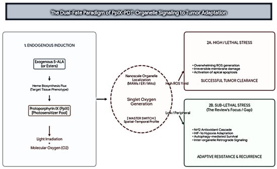
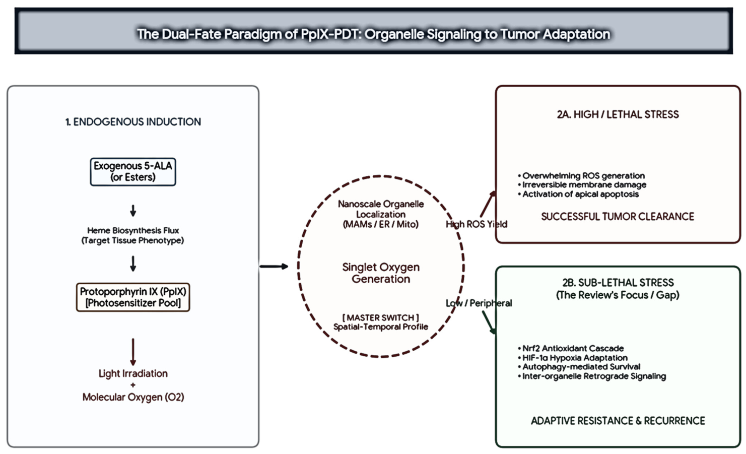
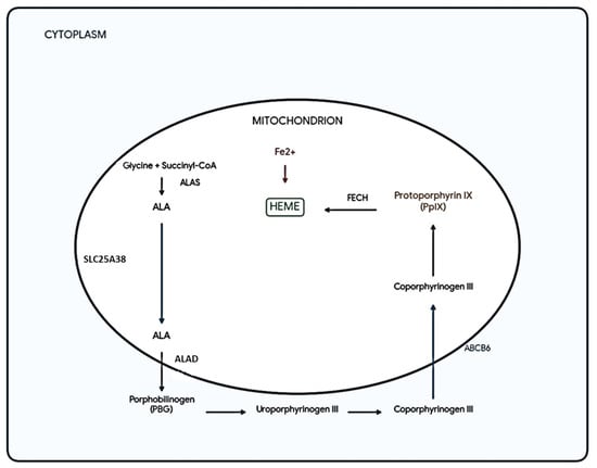
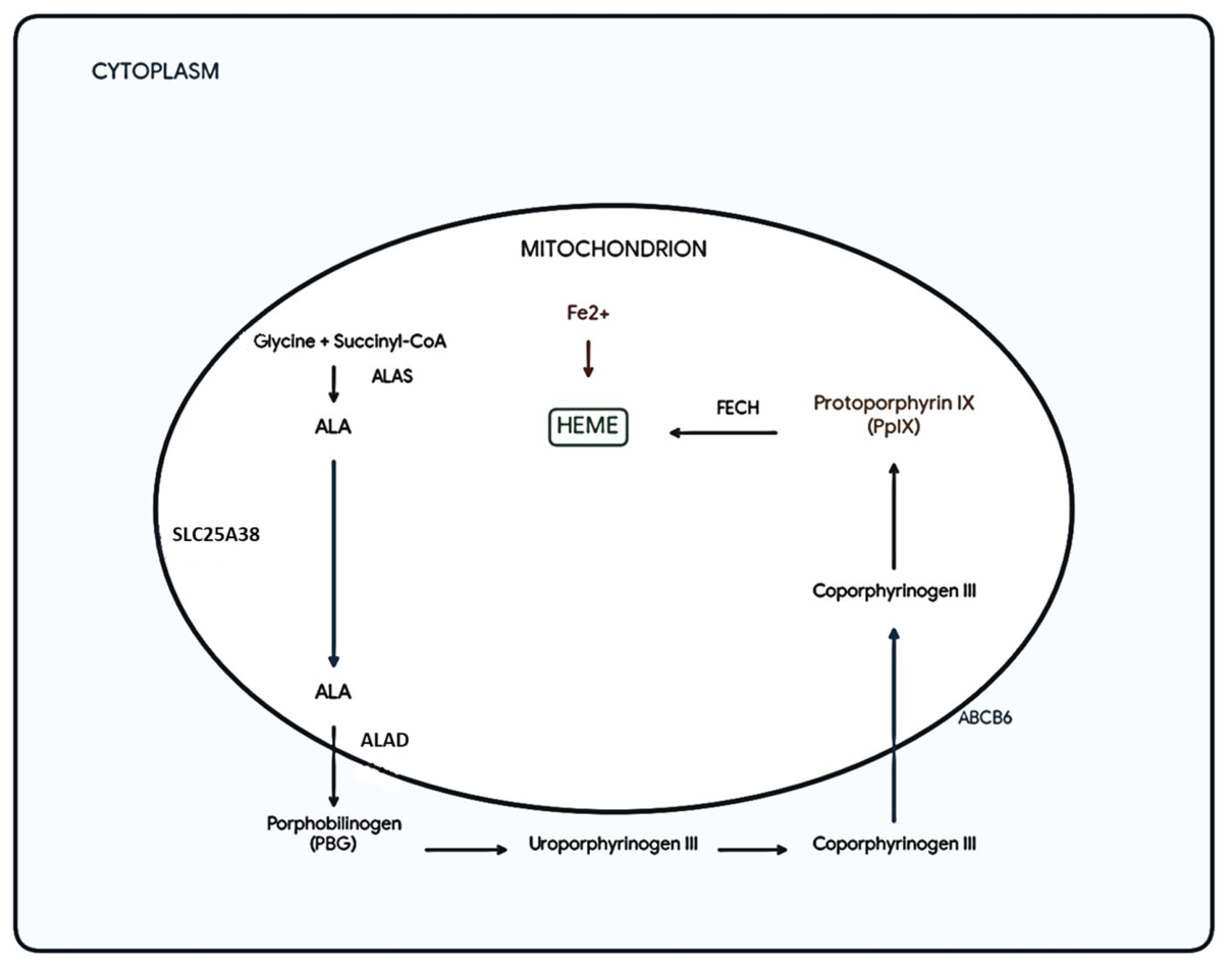
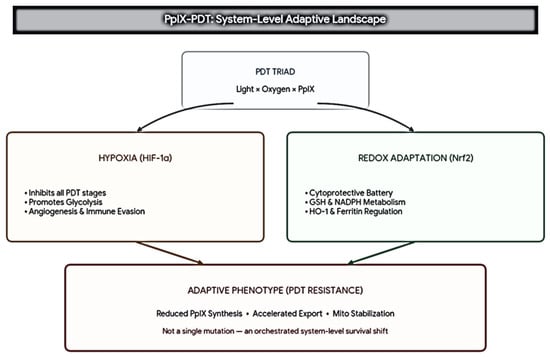
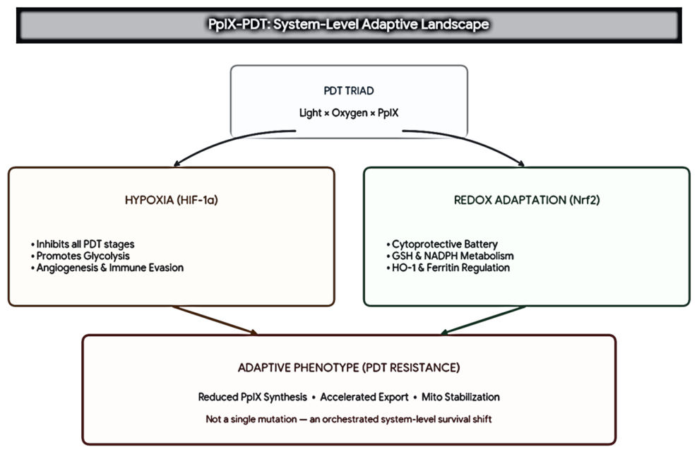

Disclaimer/Publisher’s Note: The statements, opinions and data contained in all publications are solely those of the individual author(s) and contributor(s) and not of MDPI and/or the editor(s). MDPI and/or the editor(s) disclaim responsibility for any injury to people or property resulting from any ideas, methods, instructions or products referred to in the content. |
© 2026 by the authors. Licensee MDPI, Basel, Switzerland. This article is an open access article distributed under the terms and conditions of the Creative Commons Attribution (CC BY) license.