
Article

Formulation and Physicochemical Characterization of Gelatin–Curcuma longa L. Extract Nanoparticles Prepared by Coaxial Electrospraying

by
Sebastián Guzmán-León
1,
Carlos Gregorio Barreras-Urbina
2,*,
Tomás Jesús Madera-Santana
3,*,
Lorena Armenta-Villegas
1,
Maritza Lizeth Álvarez-Ainza
1,
José Agustín Tapia-Hernández
2,
Itzel Yanira López-Peña
4 and
Francisco Rodríguez-Félix
2
1
Departamento de Ciencias Químico Biológicas, Universidad de Sonora, Blvd. Luis Encinas y Rosales, S/N, Colonia Centro, Hermosillo 83000, Sonora, Mexico
2
Departamento de Investigación y Posgrado en Alimentos (DIPA), Universidad de Sonora, Blvd 2. Luis Encinas y Rosales, S/N, Colonia Centro, Hermosillo 83000, Sonora, Mexico
3
Centro de Investigación en Alimentación y Desarrollo A.C. (CIAD, A.C.) Subsede Hidalgo, San Agustín Tlaxiaca 42162, Hidalgo, Mexico
4
Coordinación de Tecnología de Alimentos de Origen Vegetal (CTAOV), Centro de Investigación en Alimentación y Desarrollo A.C. (CIAD, A.C.), Hermosillo 83304, Sonora, Mexico
*
Authors to whom correspondence should be addressed.
Micro 2026, 6(3), 74; https://doi.org/10.3390/micro6030074
Submission received: 3 August 2026 / Revised: 31 August 2026 / Accepted: 11 September 2026 / Published: 15 September 2026
(This article belongs to the Special Issue Fabrication and Applications of Micro/Nano Colloidal Materials)

Abstract

Curcuma longa L. rhizomes contain curcuminoids and other compounds of interest, but their incorporation into aqueous systems is limited by poor dispersibility and chemical instability. This study formulated gelatin–Curcuma longa L. extract nanoparticles by coaxial electrospraying. A dried hydroethanolic extract was obtained from commercial Curcuma longa L. powder by ultrasound-microwave-assisted extraction (UMAE), with a mean gravimetric yield of 12.46 ± 1.47%. Because the recovered material was not purified or quantified by a compound-specific method, it is described as Curcuma longa L. extract rather than curcumin. Gelatin solutions at 8 and 10% (w/v) were used as the outer feed, whereas extract solutions at 0.1, 0.5, and 1.0% (w/v) were used as the inner feed. The precursor solutions showed near-Newtonian behavior. The 8% gelatin–0.5% extract formulation exhibited predominantly spherical morphology, a hydrodynamic diameter of 266.27 ± 0.57 nm, and the lowest PDI (0.24 ± 0.01). FTIR-ATR results were consistent with non-covalent interactions, mainly hydrogen bonding. These findings support coaxial electrospraying as a suitable method for preparing gelatin–Curcuma longa L. extract nanoparticles.

1. Introduction

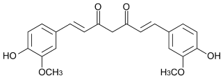
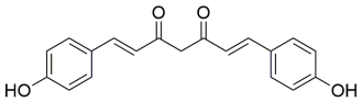
Bioactive compounds from natural sources include polyphenols, carotenoids, flavonoids, alkaloids, terpenoids, and phenolic acids [1]. Curcuma longa L. rhizomes contain a mixture of curcuminoids, principally curcumin, demethoxycurcumin, and bisdemethoxycurcumin (Table 1) [2,3]. Curcumin is a specific hydrophobic diarylheptanoid and is not synonymous with a non-purified botanical extract. Its phenolic hydroxyl, methoxy, aromatic, and β-diketone groups contribute to molecular interactions but also to poor aqueous solubility and chemical instability [2,3,4]. Accordingly, the material recovered and used in this study is identified throughout as Curcuma longa L. extract.
Encapsulation consists of entrapping or associating an active substance within a carrier to improve its handling, dispersibility, protection, or delivery. Nanoencapsulation approaches for hydrophobic bioactives include lipid-based emulsions, complex coacervation, nanoprecipitation or self-assembly, spray drying, and electrohydrodynamic techniques, such as electrospraying and electrospinning [4,5,6,7,8,9]. Lipid carriers promote dispersion of hydrophobic compounds but commonly require surfactants; coacervation relies on phase separation between interacting polymers; nanoprecipitation uses solvent displacement; spray drying is readily scalable but involves thermal and dehydration stresses; and electrohydrodynamic methods form particles or fibers from electrically charged solutions at comparatively low bulk temperatures. Method selection therefore depends on the solubility of the active and carrier, solvent compatibility, target size, and sensitivity to processing conditions.
Type B bovine gelatin, obtained by alkaline processing of collagen, is a biocompatible and biodegradable protein with film-forming, gelling, and particle-forming properties [10,11]. Its amino, carboxyl, hydroxyl, carbonyl, and amide groups can participate in hydrogen bonding, electrostatic association, hydrophobic interactions, and van der Waals forces. Gelatin has therefore been investigated as a carrier for phenolic and other hydrophobic compounds, including curcumin, quercetin, and resveratrol [12,13,14].
Electrospraying is based on electrohydrodynamic atomization: when electrical stress overcomes surface tension at a liquid meniscus, a Taylor cone and charged jet form, the jet breaks into droplets, and solvent evaporates as the droplets travel toward the collector [8,9,15]. In coaxial electrospraying, inner and outer precursor solutions are delivered independently through concentric capillaries, allowing two formulations to be processed simultaneously. Stable atomization depends on voltage, flow rates, needle geometry, tip-to-collector distance, solution viscosity and conductivity, solvent evaporation, temperature, and relative humidity [9,15]. This mild, tunable process has been used to incorporate hydrophobic and phenolic bioactives into biopolymeric particles [14,16,17].
Although gelatin-based electrosprayed systems and purified-curcumin carriers have been reported, information remains limited regarding nanoparticles prepared with a non-purified hydroethanolic Curcuma longa L. extract. Therefore, this study aimed to formulate gelatin–Curcuma longa L. extract nanoparticles by coaxial electrospraying and to evaluate the effects of gelatin and extract concentrations on precursor rheology, dry morphology, hydrodynamic diameter, polydispersity, and FTIR-ATR spectral features.

2. Materials and Methods

2.1. Materials

Type B bovine-skin gelatin (G9382; 225 g Bloom; CAS 9000-70-8; Sigma-Aldrich Co., St. Louis, MO, USA) was used as the polymer. Commercial food-grade Curcuma longa L. rhizome powder was obtained from Zazueta (Hermosillo, Sonora, Mexico). Ethanol (97%, v/v; CAS 64-17-5; Fagalab, Mexico City, Mexico) and glacial acetic acid (≥99.7%; CAS 64-19-7; J.T. Baker, Sanford, MA, USA) were used to prepare the extraction and precursor solutions. Ultrapure water produced using a Milli-Q system (CAS 7732-18-5; resistivity 18.2 MΩ·cm at 25 °C, Molsheim, France) was used for all aqueous dilutions.

2.2. Ultrasound-Microwave-Assisted Extraction (UMAE)

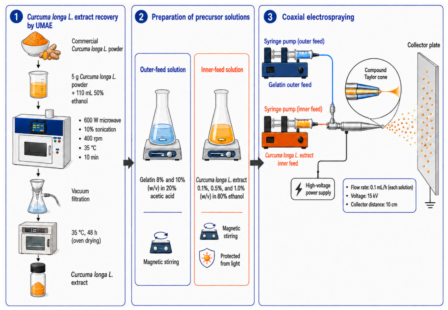
A modified ultrasound-microwave-assisted extraction (UMAE) procedure was used, taking as a methodological reference the conditions reported by Madera-Santana et al. for obtaining an aqueous Moringa oleifera leaf extract in a 250 mL UMAE reactor [18]. Each extraction unit contained 5.00 g of commercial Curcuma longa L. powder and 110 mL of 50% (v/v) aqueous ethanol, corresponding to a solid-to-solvent ratio of 1:22 (w/v; 45.45 mg mL−1). The selected ratio maintained sufficient liquid volume for complete wetting, stable suspension, and efficient solid–liquid mass transfer while remaining close to the 1:20 ratios used in the cited UMAE reactor study [18] and in the optimized microwave-assisted extraction of Curcuma longa L. with ethanol reported by Fernández-Marín et al. [19]. Extraction was performed in an XO-SM50 ultrasonic microwave reaction system (Nanjing Xianou Instruments Manufacture Co., Ltd., Nanjing, China) at 600 W microwave power, a 10% sonication-power setting, a nominal ultrasound frequency of 25 kHz, 35 °C, and for 10 min. The instrument’s integrated magnetic stirrer was operated at 400 rpm to maintain powder suspension and uniform solid-solvent contact. The suspension was vacuum-filtered, and the filtrate was oven-dried at 35 °C for 48 h. For each independent pooled replicate, four 5.00 g extraction units were processed under identical conditions, and their filtrates were combined before drying; the total starting mass was therefore approximately 20 g, while the 1:22 ratio remained unchanged in every unit. The recovered dried extract was weighed. Extraction yield was calculated using Equation (1):
E x t r a c t i o n   y i e l d   ( % ) = m e x t m p o w d e r × 100
where mext is the mass of the dried Curcuma longa L. extract and mpowder is the initial mass of the Curcuma longa L. powder.

2.3. Preparation of Precursor Solutions

Outer-feed solutions were prepared by dissolving gelatin at 8 or 10% (w/v) in 20% (v/v) aqueous acetic acid. These concentrations were selected to span the particle-forming interval reported for gelatin–acetic acid electrospraying: particles were obtained at 8 and 10% gelatin, whereas 12% favored sheets and fibrils because of greater chain interaction and entanglement [20]. The two concentrations therefore permitted evaluation of the effect of polymer content on rheology and particle morphology without entering the higher-concentration fiber-forming regime. Inner-feed solutions contained dried Curcuma longa L. extract at 0.1, 0.5, or 1.0% (w/v) in 80% (v/v) aqueous ethanol. This graded range was used to evaluate the influence of increasing extract content while retaining a low-viscosity inner phase, which is an important requirement for stable droplet breakup [13,14,20]. A 2.0% (w/v) extract solution was included only as an upper-concentration rheological control and was not electrosprayed. All solutions were stirred until homogeneous and protected from light before use.

2.4. Preparation of Gelatin–Curcuma longa L. Extract Nanoparticles by Coaxial Electrospraying

Gelatin and Curcuma longa L. extract solutions were placed in separate 10 mL syringes and delivered through plastic tubing to the outer and inner channels, respectively, of a coaxial metallic needle comprising a 20 G inner capillary and a 14 G outer capillary. A dual syringe pump (Tongli Tech, model TL-F6, Shenzhen, China) supplied each solution at 0.1 mL h−1. A high-voltage power supply (Spellman, model CZE 1000R, New York, NY, USA) applied 15 kV. Nanoparticles were collected on a 10 × 10 cm aluminum plate positioned 10 cm from the needle tip (Figure 1). The room temperature was 25.0 °C and the relative humidity was 20%.

2.5. Physicochemical Characterization

2.5.1. Rheological Analysis

The rheological behavior of each precursor solution was measured in triplicate using an Anton Paar MCR102 (Ostfildern, Germany) rheometer equipped with concentric cylinders (1 mm gap). Measurements were performed at 25 °C over a shear-rate range of 0.1–1000 s−1. Shear stress was fitted to the power–law model shown in Equation (2):
τ = K γ ˙ n
where τ is shear stress, K is the consistency index, γ ˙ is shear rate, and n is the flow-behavior index [21].

2.5.2. Scanning Electron Microscopy (SEM)

Nanoparticle morphology was examined using a JEOL JSM-7600F field-emission scanning electron microscope (JEOL Ltd., Tokyo, Japan) equipped with a secondary electron imaging (SEI) detector, following sample-preparation conditions reported for gelatin-based coaxially electrosprayed nanoparticles [20]. Samples were mounted on metallic stubs with conductive carbon tape, sputter-coated with Au/Pd, and imaged at 10 kV. Micrographs were acquired at 10,000×, 20,000×, or 30,000×, as indicated in each panel. Projected particle diameters were measured using ImageJ (Software 1.38e). One hundred particles were analyzed per formulation, except for the 8% gelatin–0.5% extract and 10% gelatin–1.0% extract formulations, for which 41 and 56 individually resolved particles, respectively, were measured.

2.5.3. Fourier Transform Infrared Spectroscopy-Attenuated Total Reflectance (FTIR-ATR)

FTIR-ATR spectra were acquired in triplicate using an Agilent Cary 630 spectrometer (Santa Clara, CA, USA) equipped with a diamond ATR accessory. Spectra were collected from 4000 to 500 cm−1 at a resolution of 8 cm−1 using 32 scans after background collection. Representative spectra are shown.

2.5.4. Dynamic Light Scattering (DLS)

Hydrodynamic diameter and cumulant polydispersity index (PDI) were determined using a Malvern Zetasizer Nano-ZS90 (Malvern, UK). Three measurements were obtained for each formulation and are reported as mean ± standard deviation.

2.5.5. Gravimetric Production Yield and Theoretical Extract Loading

Gravimetric production yield was determined using a dry-mass balance approach previously applied to electrosprayed pharmaceutical particles [22,23,24,25]. The mass of collected particles (mc) was determined from the difference between the final and initial masses of the aluminum-foil collector after solvent evaporation. The theoretical mass of non-volatile solids delivered during each experiment (mi) was calculated from the net volumes of the inner and outer solutions supplied through the coaxial system and their respective solids concentrations, as shown in Equation (3):
m i = C E V E + C G V G
where CE and CG are the extract and gelatin concentrations, respectively, and VE and VG are the corresponding net-delivered volumes. Gravimetric production yield was calculated using Equation (4):
P r o d u c t i o n   y i e l d   ( % ) = m c m i × 100
The theoretical extract loading was calculated from the relative mass-flow contribution of the extract to the total non-volatile solids supplied through the coaxial feeds using Equation (5):
T h e o r e t i c a l   e x t r a c t   l o a d i n g   ( % ) = C E Q E C E Q E + C G Q G × 100
where QE and QG are the inner and outer flow rates, respectively. This feed-based calculation is consistent with theoretical-loading approaches reported for coaxial electrohydrodynamic processing [26,27]. Because both solutions were supplied at 0.1 mL h−1, Equation (5) simplifies to Equation (6):
T h e o r e t i c a l   e x t r a c t   l o a d i n g   ( % ) = C E C E + C G × 100
The resulting value represents the nominal extract mass fraction expected in the dry particles under the assumption of complete and non-selective co-deposition and should not be interpreted as chemically measured curcumin loading or encapsulation efficiency.

2.5.6. Statistical Analysis

Results are reported as mean ± standard deviation. The extraction experiment comprised three independent pooled replicates, each formed from four 5.00 g extraction units. Rheological, FTIR-ATR, DLS, and gravimetric particle-production-yield measurements were performed in triplicate. Nanoparticle formulations were evaluated in a completely randomized 2 × 4 factorial design, with gelatin concentration (8 or 10%, w/v) and extract concentration (0, 0.1, 0.5, or 1.0%, w/v) as fixed factors. Hydrodynamic diameter and PDI were analyzed across all eight formulations by two-way analysis of variance (ANOVA) followed by Tukey’s honestly significant difference test at α = 0.05. SEM-derived particle diameters were analyzed across the eight formulations by one-way ANOVA followed by Tukey’s test at α = 0.05. Statistical analyses were performed using InfoStat 2021.

3. Results and Discussion

3.1. Gravimetric Yield of the Curcuma longa L. Extract

The three independent pooled UMAE replicates produced 2.166, 2.579, and 2.737 g of dried Curcuma longa L. extract from 20.00, 20.01, and 20.02 g of powder, respectively. The corresponding gravimetric yields were 10.83, 12.89, and 13.67%, giving an overall mean of 12.46 ± 1.47% (Table 2). These values represent total dried hydroethanolic extract rather than purified curcumin. For quantitative context, Fernández-Marín et al. [19] reported yields of 10.32 ± 0.69% for optimized microwave-assisted extraction and 8.44 ± 0.17% for conventional Soxhlet extraction of Curcuma longa L. oil. Thus, the present UMAE yield was numerically higher under the conditions evaluated. This behavior is consistent with the complementary effects of ultrasonic cavitation and microwave dielectric heating: collapse of cavitation bubbles promotes disruption of plant tissue and renews the solid–liquid interface, whereas rapid volumetric heating improves solvent penetration and diffusion [28,29,30,31]. Their simultaneous action can intensify mass transfer and shorten extraction time, providing a mechanistic basis for the recovery obtained in this study.

3.2. Rheological Properties of Precursor Solutions

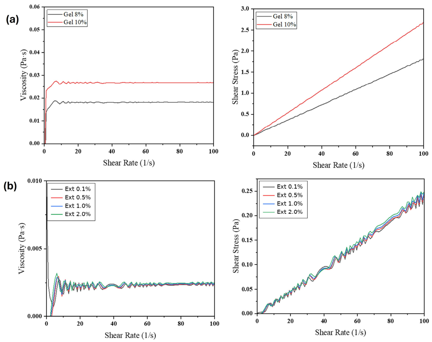
Figure 2 shows the rheological profiles of the precursor solutions over the displayed range of 0–100 s−1; measurements were acquired over 0.1–1000 s−1 at 25 °C. The 8 and 10% gelatin solutions showed nearly constant apparent viscosities of approximately 0.018 and 0.026 Pa·s, respectively. The 0.1–2.0% Curcuma longa L. extract solutions showed lower apparent viscosities of approximately 0.002–0.003 Pa·s. The nearly constant viscosity and linear shear-stress response indicate near-Newtonian behavior under the tested conditions, which supports predictable delivery through the concentric capillaries and stable droplet atomization [17,21].
The power–law parameters are summarized in Table 3. Flow-behavior indices were close to 1 (1.0014–1.1200), and R2 values ranged from 0.96 to 0.99, confirming the near-Newtonian response. The higher viscosity of the 10% gelatin solution reflects stronger intermolecular interactions and can reduce droplet breakup, increasing the tendency toward fused particles or fiber formation [13,20,21].

3.3. Morphology, Hydrodynamic Size Distribution, and Formulation Selection

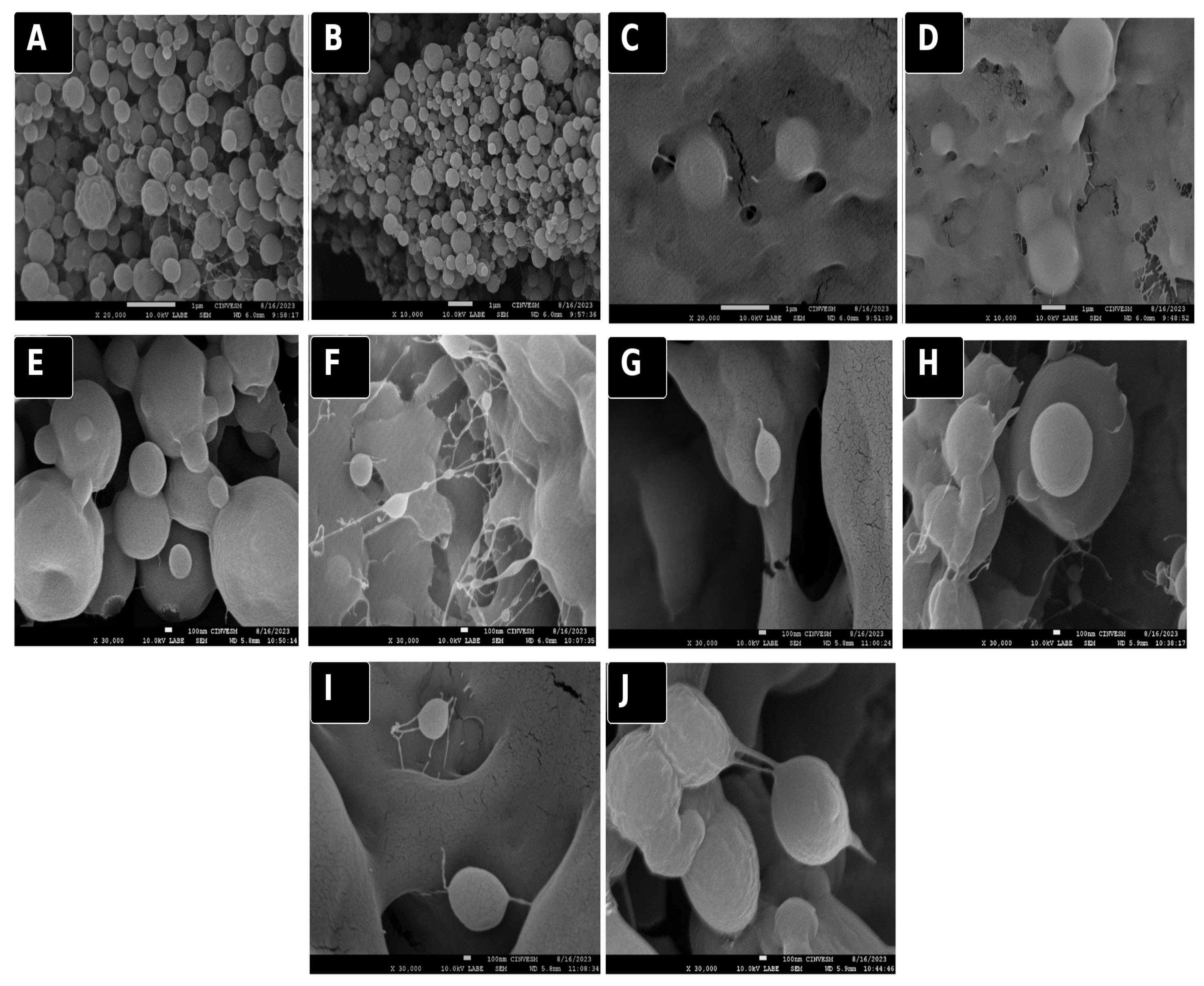
Blank gelatin nanoparticles were examined before incorporation of the extract. The 8% gelatin formulation produced predominantly spherical nanoparticles with extensive particle-to-particle contact (Figure 3A,B), whereas the 10% gelatin formulation showed more fused structures (Figure 3C,D). The higher polymer concentration and viscosity may increase chain interactions and reduce droplet breakup, favoring partial coalescence or a mixed particle–fiber morphology [13,20,21,32].
Agglomeration may occur when droplets reach the collector before complete solvent evaporation, allowing partially wet nanoparticles to fuse [33,34]. The effect was more evident in several 10% gelatin fields. Relative humidity was 20%, with a small number of final runs near 30%; increased humidity can slow solvent removal during flight [35]. Residual agglomeration may reduce redispersibility, promote sedimentation or irreversible fusion, broaden the effective size distribution, and lead to less uniform performance in downstream applications. These implications support the use of both morphology and PDI, rather than diameter alone, when selecting a formulation.
The extract-containing formulations showed concentration-dependent morphology (Figure 3E–J). Several fields contained spherical nanoparticles, whereas the 10% gelatin systems showed a greater proportion of fused or fiber-like material, particularly at 0.1% extract (Figure 3F). The 8% gelatin–0.5% extract formulation contained well-defined spherical nanoparticles together with some agglomerated regions (Figure 3G).
DLS results are summarized in Table 4. The cumulant PDI describes the breadth of the intensity-weighted hydrodynamic size distribution; values below approximately 0.3 are commonly associated with relatively narrow distributions, whereas higher values indicate increasing heterogeneity and may reflect aggregation [36]. Blank 8% gelatin nanoparticles showed the largest hydrodynamic diameter (1253.00 ± 178.12 nm) and highest PDI (0.84 ± 0.02). Among extract-containing formulations, 8% gelatin–0.5% extract showed a hydrodynamic diameter of 266.27 ± 0.57 nm and the lowest PDI (0.24 ± 0.01), indicating the narrowest hydrodynamic distribution in the tested set. The global factorial comparison confirmed that its PDI differed from all other formulations. The PDI values of 0.45–0.84 observed for most other formulations are consistent with the heterogeneity and agglomeration observed by SEM.
The 8% gelatin–0.5% extract formulation provided the most favorable combination of hydrodynamic diameter, PDI, and predominantly spherical dry morphology. Although the 10% gelatin–1.0% extract formulation had a smaller hydrodynamic diameter (150.43 ± 15.06 nm), its broader distribution (PDI 0.56 ± 0.06) and residual agglomeration made it less uniform. The 8% gelatin–0.5% extract formulation was therefore selected as the best-performing formulation among those evaluated.
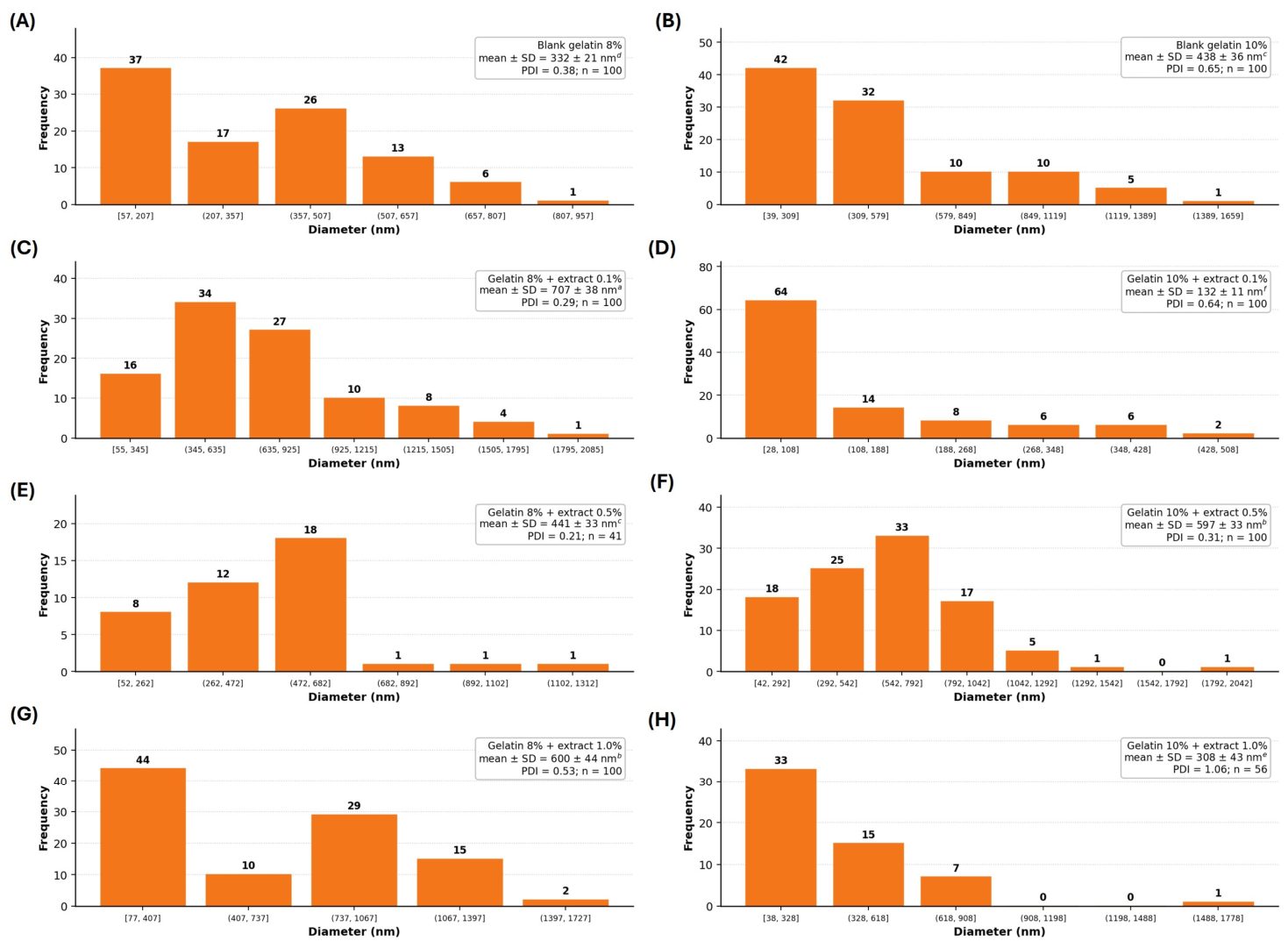
ImageJ analysis provided complementary dry-state particle-size distributions (Figure 4). The mean diameter ± standard deviation and the number of measured particles are reported for every formulation. The distributions ranged from relatively concentrated to broad or multimodal, consistent with the differences observed among the SEM fields. ANOVA/Tukey lettering showed six statistical groups: the 8% gelatin–0.1% extract formulation had the largest mean dry diameter, the 8% gelatin–1.0% and 10% gelatin–0.5% formulations formed the next group, and the 10% gelatin–0.1% formulation had the smallest mean dry diameter.
DLS and SEM yielded different numerical diameters because the methods measure different particle states and physical properties. DLS estimates the hydrodynamic diameter of particles dispersed in liquid and is strongly weighted toward large scatterers or aggregates. SEM measures the projected diameter of dried nanoparticles in selected fields; dehydration, residual solvent, deposition, particle fusion, conductive coating, field selection, and agglomerate counting can alter the apparent dry-state diameter [33,34]. Thus, the 266.27 ± 0.57 nm hydrodynamic diameter of the 8% gelatin–0.5% extract formulation is not expected to match its 441 ± 33 nm dry diameter exactly. The two methods are complementary and together indicate a comparatively narrow dispersion with predominantly spherical particles and residual dry-state agglomeration.
Overall, gelatin concentration influenced both atomization and particle organization. The 10% gelatin formulations showed a greater tendency toward fused or fiber-like regions, whereas the 8% gelatin–0.5% extract formulation produced the narrowest DLS distribution and the most favorable morphology among the tested formulations.

3.4. Gravimetric Production Yield and Theoretical Extract Loading

Gravimetric production yield was consistently high across the six extract-containing formulations, ranging from 86.9 ± 5.4% to 89.1 ± 3.8% (Table 5). Theoretical extract loading increased with inner-feed extract concentration and, at the same extract concentration, was higher for the 8% gelatin formulations because gelatin contributed a smaller fraction of the total solid mass flow. The nominal values ranged from 0.99 to 11.11 wt%. These feed-based values describe the intended dry-particle composition and should not be interpreted as chemically measured extract retention or encapsulation efficiency.

3.5. FTIR-ATR Analysis of Gelatin–Curcuma longa L. Extract Nanoparticles

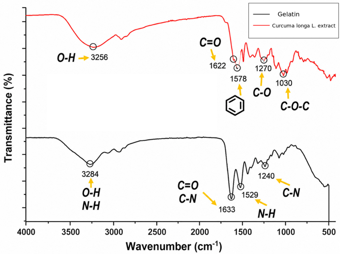
FTIR-ATR was used to compare the functional-group regions of gelatin and the dried hydroethanolic Curcuma longa L. extract (Figure 5). The extract spectrum showed a broad O-H-associated band near 3256 cm−1, a band near 1622 cm−1, aromatic-region absorption near 1578 cm−1, and C-O/C-O-C-region features between approximately 1270 and 1030 cm−1. These bands are compatible with curcuminoid-containing material [12,37] but are not specific to purified curcumin. Gelatin showed Amide A near 3284 cm−1, Amide I near 1633 cm−1, Amide II near 1529 cm−1, and Amide III near 1240 cm−1 [38,39].
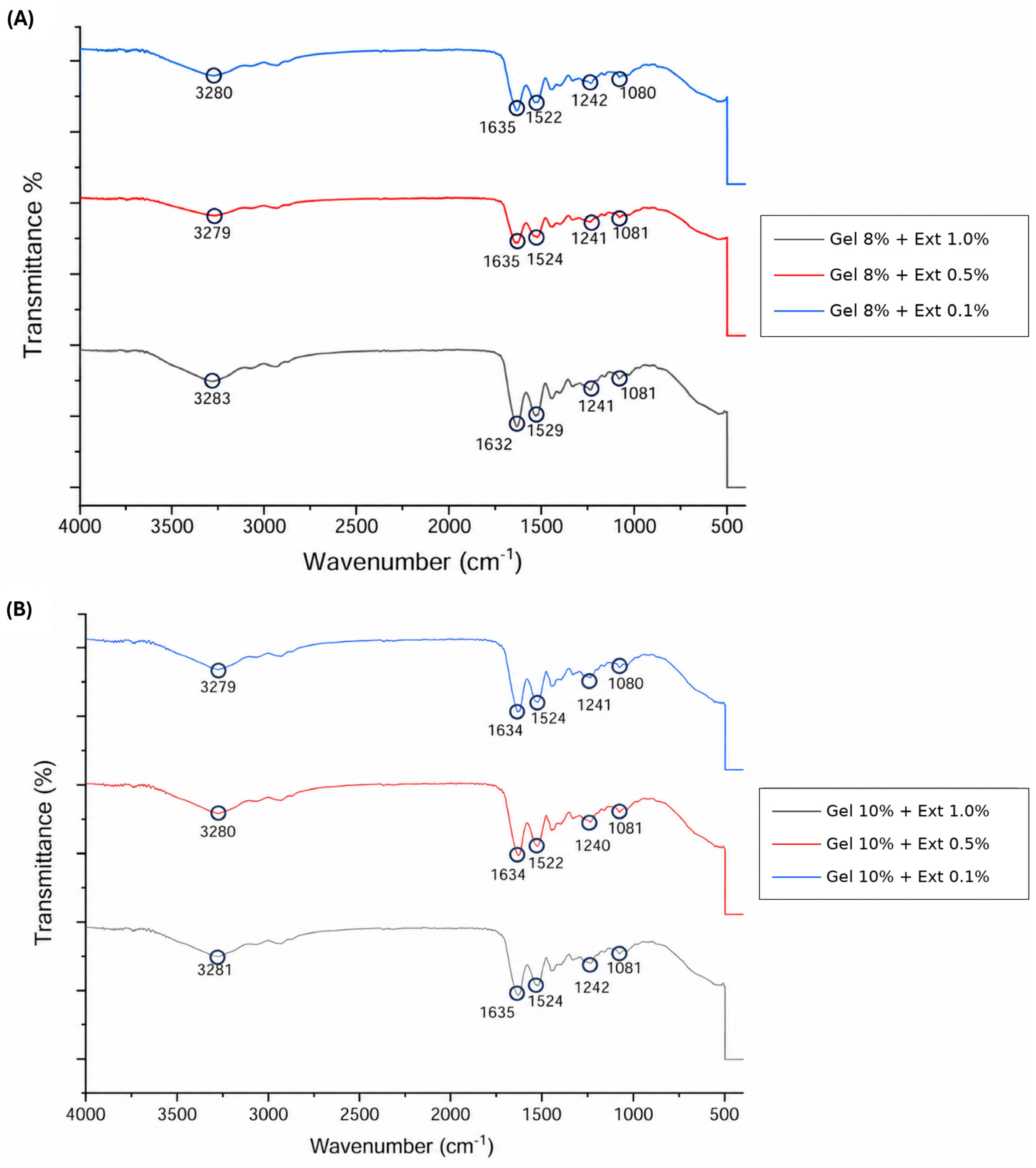
Figure 6 presents representative spectra of gelatin–Curcuma longa L. extract nanoparticles. All formulations were dominated by gelatin amide bands, with features near 3279–3283, 1632–1635, 1522–1529, 1240–1242, and 1080–1081 cm−1. The predominance of gelatin bands is consistent with the higher gelatin concentration and the surface-sensitive nature of ATR; it does not establish the internal distribution of the extract.
Small formulation-dependent shifts in the amide regions, together with the absence of new absorption bands, are consistent with non-covalent association rather than formation of new covalent bonds. Hydrogen bonding between hydroxyl- or carbonyl-containing extract constituents and gelatin amide or hydroxyl groups is a plausible principal contribution [12,37]. Hydrophobic interactions and van der Waals forces may provide additional stabilization because the extract contains aromatic constituents and gelatin contains nonpolar amino acid residues.
The 50% aqueous ethanol used in UMAE has intermediate polarity and therefore can recover a multicomponent fraction rather than a single purified compound. In addition to curcumin, Curcuma longa L. rhizomes contain demethoxycurcumin and bisdemethoxycurcumin [2,3], and ethanolic extraction can recover a broader phenolic fraction [40]. Consequently, the observed FTIR bands are assigned to functional groups within the Curcuma longa L. extract and cannot be attributed exclusively to curcumin.
The FTIR-ATR results therefore support physical association between gelatin and constituents of the extract. Table 5 presents gravimetric production yield and nominal theoretical extract loading as reproducible mass-balance descriptors derived from the collector and feed-composition data. Together, these measurements complement the morphological, DLS, rheological, and spectroscopic characterization and provide a quantitative basis for comparing the formulations.

4. Conclusions

Coaxial electrospraying produced gelatin–Curcuma longa L. extract nanoparticles from near-Newtonian precursor solutions. UMAE provided a mean gravimetric extract yield of 12.46 ± 1.47% across three independent pooled replicates. Among the formulations evaluated, 8% gelatin–0.5% extract showed the most favorable combination of predominantly spherical morphology, hydrodynamic diameter (266.27 ± 0.57 nm), and PDI (0.24 ± 0.01). Particle production yields of 86.9–89.1% and theoretical feed-based extract loadings of 0.99–11.11 wt% provide additional mass-balance descriptors of the process. The quantitative comparison with published microwave and Soxhlet extraction yields, together with the established cavitation and dielectric-heating mechanisms, supports the effectiveness of the UMAE conditions used in this study.
FTIR-ATR indicated a non-covalent association between gelatin and extract constituents, without evidence of new covalent bonds. The integrated morphological, DLS, rheological, spectroscopic, and mass-balance results identify 8% gelatin–0.5% extract as the most favorable formulation under the evaluated conditions and provide a reproducible basis for subsequent performance studies.

Author Contributions

S.G.-L. performed the experimental work, data analysis, and original draft preparation. C.G.B.-U. contributed to conceptualization, supervision, resources and data analysis. L.A.-V., T.J.M.-S. and M.L.Á.-A. contributed to conceptualization, supervision and resources. J.A.T.-H. and F.R.-F. contributed to manuscript review. I.Y.L.-P. contributed to review and editing. All authors have read and agreed to the published version of the manuscript.

Funding

This research received no external funding.

Institutional Review Board Statement

Not applicable.

Informed Consent Statement

Not applicable.

Data Availability Statement

The data are contained within the article.

Acknowledgments

The authors acknowledge the technical assistance of William Santiago González and Víctor Rejón-Moo at CINVESTAV-IPN Unidad Mérida in performing SEM and FTIR measurements. These analyses were performed at Laboratorio Nacional CONAHCYT LANNBIO-Cinvestav-Mérida (PROY No. 321119). During the preparation of this manuscript, the authors used ChatGPT[M14.1][CB14.2] (GPT-5.6 Sol; OpenAI, San Francisco, CA, USA) for the purposes to generate graphical elements. The authors have reviewed and edited the output and take full responsibility for the content of this publication.

Conflicts of Interest

The authors declare no conflicts of interest.

References

  1. Sundhari, D.; Dhineshbabu, N.R.; Sutha, S.; Raja Saravanan, M.E. Encapsulation of Bioactive Agent (Curcumin, Moringa) in Electrospun Nanofibers-Some Insights into Recent Research Trends. Mater. Today Proc. 2021, 46, 2682–2685. [Google Scholar] [CrossRef] [Scilit]
  2. Nelson, K.M.; Dahlin, J.L.; Bisson, J.; Graham, J.; Pauli, G.F.; Walters, M.A. The Essential Medicinal Chemistry of Curcumin. J. Med. Chem. 2017, 60, 1620–1637. [Google Scholar] [CrossRef] [Scilit]
  3. Priyadarsini, K.I. The Chemistry of Curcumin: From Extraction to Therapeutic Agent. Molecules 2014, 19, 20091–20112. [Google Scholar] [CrossRef] [Scilit]
  4. Rafiee, Z.; Nejatian, M.; Daeihamed, M.; Jafari, S.M. Application of Curcumin-Loaded Nanocarriers for Food, Drug and Cosmetic Purposes. Trends Food Sci. Technol. 2019, 88, 445–458. [Google Scholar] [CrossRef] [Scilit]
  5. Jain, A.; Dasgupta, N.; Ranjan, S.; Singh, V.; Singh, H.; Purohit, S.D.; Mishra, N.C.; Yadav, N.P.; Haque, S.; Mishra, B.N.; et al. Whey Protein Based Electrosprayed Nanospheres for Encapsulation and Controlled Release of Bioactive Compounds from Tinospora cordifolia Extract. Innov. Food Sci. Emerg. Technol. 2021, 69, 102671. [Google Scholar] [CrossRef] [Scilit]
  6. Panagiotopoulou, M.; Papadaki, S.; Krokida, M. Formation and Characterization of Zein Electrosprayed Nanoparticles Containing Bioactive Compounds. S. Afr. J. Chem. Eng. 2022, 40, 32–47. [Google Scholar] [CrossRef] [Scilit]
  7. Wen, P.; Zong, M.H.; Linhardt, R.J.; Feng, K.; Wu, H. Electrospinning: A Novel Nano-Encapsulation Approach for Bioactive Compounds. Trends Food Sci. Technol. 2017, 70, 56–68. [Google Scholar] [CrossRef] [Scilit]
  8. Tapia-Hernández, J.A.; Torres-Chávez, P.I.; Ramírez-Wong, B.; Rascón-Chu, A.; Plascencia-Jatomea, M.; Barreras-Urbina, C.G.; Rangel-Vázquez, N.A.; Rodríguez-Félix, F. Micro- and Nanoparticles by Electrospray: Advances and Applications in Foods. J. Agric. Food Chem. 2015, 63, 4699–4707. [Google Scholar] [CrossRef] [Scilit]
  9. Wang, P.; Ding, M.; Zhang, T.; Wu, T.; Qiao, R.; Zhang, F.; Wang, X.; Zhong, J. Electrospraying Technique and Its Recent Application Advances for Biological Macromolecule Encapsulation of Food Bioactive Substances. Food Rev. Int. 2022, 38, 566–588. [Google Scholar] [CrossRef] [Scilit]
  10. Khan, M.R.; Sadiq, M.B. Importance of Gelatin, Nanoparticles and Their Interactions in the Formulation of Biodegradable Composite Films: A Review. Polym. Bull. 2021, 78, 4047–4073. [Google Scholar] [CrossRef] [Scilit]
  11. Sultana, S.; Ali, M.E.; Ahamad, M.N.U. Gelatine, Collagen, and Single Cell Proteins as Natural and Newly Emerging Food Ingredients. In Preparation and Processing of Religious and Cultural Foods; Ali, M.E., Nizar, N.N.A., Eds.; Woodhead Publishing: Duxford, UK, 2018; pp. 215–239. [Google Scholar] [CrossRef] [Scilit]
  12. Roy, S.; Rhim, J.W. Preparation of Antimicrobial and Antioxidant Gelatin/Curcumin Composite Films for Active Food Packaging Application. Colloids Surf. B Biointerfaces 2020, 188, 110761. [Google Scholar] [CrossRef] [Scilit]
  13. Rodríguez-Félix, F.; Del-Toro-Sánchez, C.L.; Cinco-Moroyoqui, F.J.; Juárez, J.; Ruiz-Cruz, S.; López-Ahumada, G.A.; Carvajal-Millan, E.; Castro-Enríquez, D.D.; Barreras-Urbina, C.G.; Tapia-Hernández, J.A. Preparation and Characterization of Quercetin-Loaded Zein Nanoparticles by Electrospraying and Study of In Vitro Bioavailability. J. Food Sci. 2019, 84, 2883–2897. [Google Scholar] [CrossRef] [Scilit]
  14. Leena, M.M.; Anukiruthika, T.; Moses, J.A.; Anandharamakrishnan, C. Co-Delivery of Curcumin and Resveratrol through Electrosprayed Core-Shell Nanoparticles in 3D Printed Hydrogel. Food Hydrocoll. 2022, 124, 107200. [Google Scholar] [CrossRef] [Scilit]
  15. Gañán-Calvo, A.M.; López-Herrera, J.M.; Herrada, M.A.; Ramos, A.; Montanero, J.M. Review on the Physics of Electrospray: From Electrokinetics to the Operating Conditions of Single and Coaxial Taylor Cone-Jets, and AC Electrospray. J. Aerosol Sci. 2018, 125, 32–56. [Google Scholar] [CrossRef] [Scilit]
  16. Gómez-Mascaraque, L.G.; Tordera, F.; Fabra, M.J.; Martínez-Sanz, M.; Lopez-Rubio, A. Coaxial Electrospraying of Biopolymers as a Strategy to Improve Protection of Bioactive Food Ingredients. Innov. Food Sci. Emerg. Technol. 2019, 51, 2–11. [Google Scholar] [CrossRef] [Scilit]
  17. Fallahasghari, E.Z.; Chronakis, I.S.; Mendes, A.C. Lipophilic Bioactive Compounds Nano-Micro Encapsulation via Coaxial Electrohydrodynamic Atomization: A Review. Food Hydrocoll. Health 2025, 7, 100202. [Google Scholar] [CrossRef] [Scilit]
  18. Madera-Santana, J.T.; De Dios-Aguilar, M.A.; Colín-Chávez, C.; Mariscal-Amaro, L.A.; Núñez-Colín, C.A.; Veloz-García, R.; Guzmán-Maldonado, S.H.; Peña-Caballero, V.; Grijalva-Verdugo, C.P.; Rodríguez-Núñez, J.R. Recubrimiento a base de quitosano y extracto acuoso de hoja de Moringa oleifera obtenido por UMAE y su efecto en las propiedades fisicoquímicas de fresa (Fragaria × ananassa). Biotecnia 2019, 21, 155–163. [Google Scholar] [CrossRef] [Scilit]
  19. Fernández-Marín, R.; Fernandes, S.C.M.; Andrés, M.A.; Labidi, J. Microwave-Assisted Extraction of Curcuma longa L. Oil: Optimization, Chemical Structure and Composition, Antioxidant Activity and Comparison with Conventional Soxhlet Extraction. Molecules 2021, 26, 1516. [Google Scholar] [CrossRef] [Scilit]
  20. Figueroa-Enriquez, C.E.; Rodríguez-Félix, F.; Plascencia-Jatomea, M.; Sánchez-Escalante, A.; Vargas-López, J.M.; Tapia-Hernández, J.A.; Canizales-Rodríguez, D.F.; Castro-Enriquez, D.D.; Ruiz-Cruz, S.; Santos-Sauceda, I.; et al. Nanoparticles of Betalain-Gelatin with Antioxidant Properties by Coaxial Electrospraying: Preparation and Characterization. ACS Omega 2023, 8, 41156–41168. [Google Scholar] [CrossRef] [Scilit]
  21. Castro-Enríquez, D.D.; Castillo-Ortega, M.M.; Romero-García, J.; Rodríguez-Félix, D.E.; Dórame-Miranda, R.F.; Torres-Arreola, W.; Vargas-López, J.M.; Burruel-Ibarra, S.E.; Rodríguez-Félix, F. Development of Microparticles from Wheat Glutenins by Electrospray and Potential Application as Controlled-Release Fertilizers. Bull. Mater. Sci. 2019, 42, 41. [Google Scholar] [CrossRef] [Scilit]
  22. Bohr, A.; Kristensen, J.; Dyas, M.; Edirisinghe, M.; Stride, E. Release Profile and Characteristics of Electrosprayed Particles for Oral Delivery of a Practically Insoluble Drug. J. R. Soc. Interface 2012, 9, 2437–2449. [Google Scholar] [CrossRef] [Scilit]
  23. Zhang, Y.; Shams, T.; Harker, A.H.; Parhizkar, M.; Edirisinghe, M. Effect of Copolymer Composition on Particle Morphology and Release Behavior In Vitro Using Progesterone. Mater. Des. 2018, 159, 57–67. [Google Scholar] [CrossRef] [Scilit]
  24. Zhang, Y.; Zhang, R.; Illangakoon, U.E.; Harker, A.H.; Thrasivoulou, C.; Parhizkar, M.; Edirisinghe, M.; Luo, C.J. Copolymer Composition and Nanoparticle Configuration Enhance In Vitro Drug Release Behavior of Poorly Water-Soluble Progesterone for Oral Formulations. Int. J. Nanomed. 2020, 15, 5389–5403. [Google Scholar] [CrossRef] [Scilit]
  25. Zhang, Y.; Harker, A.H.; Luo, C.J.; Parhizkar, M.; Edirisinghe, M. Co-Delivery of Saxagliptin and Dapagliflozin by Electrosprayed Trilayer Poly(D,L-lactide-co-glycolide) Nanoparticles for Controlled Drug Delivery. Int. J. Pharm. 2022, 628, 122279. [Google Scholar] [CrossRef] [Scilit]
  26. Wang, Y.; Yang, X.; Liu, W.; Zhang, F.; Cai, Q.; Deng, X. Controlled Release Behaviour of Protein-Loaded Microparticles Prepared via Coaxial or Emulsion Electrospray. J. Microencapsul. 2013, 30, 490–497. [Google Scholar] [CrossRef] [Scilit]
  27. Abdelhakim, H.E.; Coupe, A.; Tuleu, C.; Edirisinghe, M.; Craig, D.Q.M. Utilising Co-Axial Electrospinning as a Taste-Masking Technology for Paediatric Drug Delivery. Pharmaceutics 2021, 13, 1665. [Google Scholar] [CrossRef] [Scilit]
  28. Ciuca, M.D.; Racovita, R.C. Curcumin: Overview of Extraction Methods, Health Benefits, and Encapsulation and Delivery Using Microemulsions and Nanoemulsions. Int. J. Mol. Sci. 2023, 24, 8874. [Google Scholar] [CrossRef] [Scilit]
  29. Kumar, M.; Dahuja, A.; Tiwari, S.; Punia, S.; Tak, Y.; Amarowicz, R.; Bhoite, A.G.; Singh, S.; Joshi, S.; Panesar, P.S.; et al. Recent Trends in Extraction of Plant Bioactives Using Green Technologies: A Review. Food Chem. 2021, 353, 129431. [Google Scholar] [CrossRef] [Scilit]
  30. Manasa, P.S.L.; Kamble, A.D.; Chilakamarthi, U. Various Extraction Techniques of Curcumin-A Comprehensive Review. ACS Omega 2023, 8, 34868–34878. [Google Scholar] [CrossRef] [Scilit]
  31. López-Salazar, H.; Camacho-Díaz, B.H.; Arenas Ocampo, M.L.; Jiménez-Aparicio, A.R. Microwave-Assisted Extraction of Functional Compounds from Plants: A Review. BioResources 2023, 18, 6614–6638. [Google Scholar] [CrossRef] [Scilit]
  32. Zhang, L.; Huang, J.; Si, T.; Xu, R.X. Coaxial Electrospray of Microparticles and Nanoparticles for Biomedical Applications. Expert Rev. Med. Devices 2012, 9, 595–612. [Google Scholar] [CrossRef] [Scilit]
  33. Faramarzi, A.R.; Barzin, J.; Mobedi, H. Effect of Solution and Apparatus Parameters on the Morphology and Size of Electrosprayed PLGA Microparticles. Fibers Polym. 2016, 17, 1806–1819. [Google Scholar] [CrossRef] [Scilit]
  34. Alfatama, M.; Shahzad, Y.; Choukaife, H. Recent Advances of Electrospray Technique for Multiparticulate Preparation: Drug Delivery Applications. Adv. Colloid Interface Sci. 2024, 325, 103098. [Google Scholar] [CrossRef] [Scilit]
  35. Kavadiya, S.; Niedzwiedzki, D.M.; Huang, S.; Biswas, P. Electrospray-Assisted Fabrication of Moisture-Resistant and Highly Stable Perovskite Solar Cells at Ambient Conditions. Adv. Energy Mater. 2017, 7, 1700210. [Google Scholar] [CrossRef] [Scilit]
  36. Danaei, M.; Dehghankhold, M.; Ataei, S.; Hasanzadeh Davarani, F.; Javanmard, R.; Dokhani, A.; Khorasani, S.; Mozafari, M.R. Impact of Particle Size and Polydispersity Index on the Clinical Applications of Lipidic Nanocarrier Systems. Pharmaceutics 2018, 10, 57. [Google Scholar] [CrossRef] [Scilit]
  37. Chen, X.; Zou, L.Q.; Niu, J.; Liu, W.; Peng, S.F.; Liu, C.M. The Stability, Sustained Release and Cellular Antioxidant Activity of Curcumin Nanoliposomes. Molecules 2015, 20, 14293–14311. [Google Scholar] [CrossRef] [Scilit]
  38. Feng, X.; Dai, H.; Ma, L.; Yu, Y.; Tang, M.; Li, Y.; Hu, W.; Liu, T.; Zhang, Y. Food-Grade Gelatin Nanoparticles: Preparation, Characterization, and Preliminary Application for Stabilizing Pickering Emulsions. Foods 2019, 8, 479. [Google Scholar] [CrossRef] [Scilit]
  39. Shi, R.; Geng, H.; Gong, M.; Ye, J.; Wu, C.; Hu, X.; Zhang, L. Long-Acting and Broad-Spectrum Antimicrobial Electrospun Poly(ε-caprolactone)/Gelatin Micro/Nanofibers for Wound Dressing. J. Colloid Interface Sci. 2018, 509, 275–284. [Google Scholar] [CrossRef] [Scilit]
  40. Insuan, W.; Hansupalak, N.; Chahomchuen, T. Extraction of Curcumin from Turmeric by Ultrasonic-Assisted Extraction, Identification, and Evaluation of the Biological Activity. J. HerbMed Pharmacol. 2022, 11, 188–196. [Google Scholar] [CrossRef] [Scilit]
Figure 1. Schematic workflow for obtaining gelatin–Curcuma longa L. extract nanoparticles: (1) UMAE recovery of the extract, (2) preparation of precursor solutions, and (3) coaxial electrospraying. The schematic was prepared by the authors using graphical elements generated with ChatGPT (GPT-5.6 Sol; OpenAI, San Francisco, CA, USA).
Micro 06 00074 g001
Figure 2. Rheograms of (a) gelatin outer-feed solutions at 8 and 10% (w/v) and (b) Curcuma longa L. extract inner-feed solutions at 0.1, 0.5, 1.0, and 2.0% (w/v). The displayed shear-rate range is 0–100 s−1; measurements were acquired at 25 °C over 0.1–1000 s−1. Ext., Curcuma longa L. extract.
Micro 06 00074 g002
Figure 3. Combined FE-SEM micrographs of blank gelatin nanoparticles and gelatin–Curcuma longa L. extract nanoparticles. Blank nanoparticles: (A) 8% gelatin, 20,000×; (B) 8% gelatin, 10,000×; (C) 10% gelatin, 20,000×; and (D) 10% gelatin, 10,000×. Extract-containing nanoparticles, all at 30,000×: (E) 8% gelatin–0.1% extract; (F) 10% gelatin–0.1% extract; (G) 8% gelatin–0.5% extract; (H) 10% gelatin–0.5% extract; (I) 8% gelatin–1.0% extract; and (J) 10% gelatin–1.0% extract.
Micro 06 00074 g003
Figure 4. Dry-particle diameter frequency distributions obtained from FE-SEM micrographs using ImageJ. (A) Blank 8% gelatin (n = 100); (B) blank 10% gelatin (n = 100); (C) 8% gelatin–0.1% extract (n = 100); (D) 10% gelatin–0.1% extract (n = 100); (E) 8% gelatin–0.5% extract (n = 41); (F) 10% gelatin–0.5% extract (n = 100); (G) 8% gelatin–1.0% extract (n = 100); and (H) 10% gelatin–1.0% extract (n = 56). Insets report mean ± SD and image-derived PDI. Different superscript letters indicate significant differences in mean dry diameter by one-way ANOVA followed by Tukey’s test (α = 0.05).
Micro 06 00074 g004
Figure 5. Representative FTIR-ATR spectra of gelatin and dried hydroethanolic Curcuma longa L. extracts.
Micro 06 00074 g005
Figure 6. Representative FTIR-ATR spectra of gelatin–Curcuma longa L. extract nanoparticles prepared with (A) 8% and (B) 10% gelatin and inner extract feeds of 0.1, 0.5, or 1.0% (w/v). Ext., Curcuma longa L. extract.
Micro 06 00074 g006
Table 1. Main curcuminoids reported in Curcuma longa L.
CompoundMain Structural Feature
Curcumin
Micro 06 00074 i001
Two methoxy groups, phenolic OH, β-diketone chain
Demethoxycurcumin
Micro 06 00074 i002
One methoxy group
Bisdemethoxycurcumin
Micro 06 00074 i003
No methoxy groups
Table 2. Gravimetric yield of dried Curcuma longa L. extract from three independently pooled UMAE replicates.
Extraction ReplicateCurcuma longa L. Powder (g)Dried Curcuma longa L. Extract (g)Extraction Yield (%)
120.002.16610.83
220.012.57912.89
320.022.73713.67
Mean ± SD (n = 3)20.01 ± 0.012.494 ± 0.29512.46 ± 1.47
Each pooled replicate comprised four 5.00 g extraction units (110 mL solvent per unit; 1:22, w/v). The four filtrates were combined before drying. Values in the final row are mean ± standard deviation across the three independent pooled replicates (n = 3).
Table 3. Power–law fitting parameters for gelatin and Curcuma longa L. extract precursor solutions.
Precursor SolutionConcentration (% w/v)Flow-Behavior Index, nR2
Gelatin (outer feed)81.0225 ± 0.00320.99
Gelatin (outer feed)101.0014 ± 0.00090.99
Curcuma longa L. extract (inner feed)0.11.0076 ± 0.00320.96
Curcuma longa L. extract (inner feed)0.51.0700 ± 0.00410.98
Curcuma longa L. extract (inner feed)1.01.0501 ± 0.00380.99
Curcuma longa L. extract (rheology only)2.01.1200 ± 0.00400.99
Values are expressed as mean ± standard deviation from triplicate measurements. n, flow-behavior index; R2, coefficient of determination. Extract concentrations of 0.1–1.0% constituted the nanoparticle formulation screen; 2.0% was evaluated only in the rheological analysis.
Table 4. Hydrodynamic diameter, PDI, and dry morphology of gelatin–Curcuma longa L. extract nanoparticles.
Gelatin (%)Curcuma longa L. Extract (%)Hydrodynamic Diameter (nm) ± SDPDI ± SDDry Morphology by SEM
801253.00 ± 178.12 a0.84 ± 0.02 aSpherical; extensive contacts
80.1473.50 ± 26.31 b0.49 ± 0.01 c,dHeterogeneous spherical
80.5266.27 ± 0.57 c,d0.24 ± 0.01 ePredominantly spherical; some agglomeration
81.0315.37 ± 10.52 b,c,d0.68 ± 0.02 bAgglomerated spherical
100381.37 ± 15.54 b,c0.45 ± 0.05 dFused/agglomerated
100.1373.17 ± 38.66 b,c0.49 ± 0.03 c,dSpherical, oval, and fibrous
100.5348.80 ± 17.76 b,c0.47 ± 0.02 c,dAgglomerated spherical
101.0150.43 ± 15.06 d0.56 ± 0.06 cSpherical; agglomerated regions
Values are mean ± standard deviation from three DLS measurements (n = 3). For each response column, formulation means that share at least one superscript letter are not significantly different. Letters compare all eight formulations using two-way ANOVA for the 2 × 4 factorial design followed by Tukey’s honestly significant difference test (α = 0.05).
Table 5. Gravimetric production yield and theoretical extract loading of gelatin–Curcuma longa L. extract nanoparticles.
FormulationGravimetric Production Yield (%)Theoretical Extract Loading (wt%)
G10-E0.188.5 ± 4.20.99
G10-E0.587.2 ± 5.14.76
G10-E1.089.1 ± 3.89.09
G8-E0.186.9 ± 5.41.23
G8-E0.588.0 ± 4.55.88
G8-E1.087.5 ± 4.911.11
Values of production yield are presented as mean ± standard deviation (n = 3). G, gelatin concentration (% w/v); E, Curcuma longa L. extract concentration (% w/v). Theoretical extract loading is the nominal extract fraction of the non-volatile solids delivered through the two feeds.
Disclaimer/Publisher’s Note: The statements, opinions and data contained in all publications are solely those of the individual author(s) and contributor(s) and not of MDPI and/or the editor(s). MDPI and/or the editor(s) disclaim responsibility for any injury to people or property resulting from any ideas, methods, instructions or products referred to in the content.