


Abstract
Background: Taurine is a bioactive amino acid that has great potential for wound healing, but it struggles with poor skin penetration and quick clearance. This study aimed to develop taurine-loaded nanoliposomes (Nlp-Tau) to enable sustained local delivery and improve effectiveness. Methods: Nlp-Tau were prepared using thin-film hydration. We characterized them for size, charge, shape, encapsulation efficiency (EE%), loading efficiency (LE%), and in vitro release. Also, its biocompatibility on human foreskin fibroblasts (HFF) with a 3-(4,5-dimethylthiazol-2-yl)-2,5-diphenyltetrazolium bromide (MTT assay) has been assessed. In vitro wound healing potential was further evaluated using a scratch assay. Results: Optimized Nlp-Tau showed favorable properties, including an average hydrodynamic diameter of about 282 nm, a polydispersity index of 0.2, a strong negative zeta potential of −31.3 mV, and a spherical shape. However, transmission electron microscopy (TEM) images revealed diameters of about 142 nm and 194 nm for the drug-free and taurine-loaded particles, respectively. Additionally, EE% and LE% were 20% and 2.5%, respectively. In vitro release in PBS (pH 7.4) followed Higuchi kinetics, showing sustained release over 72 h. Nlp-Tau displayed excellent biocompatibility, with HFF viability significantly higher than other groups at concentrations up to 7 mg/mL. Importantly, in the scratch assay, Nlp-Tau treatment resulted in just 6.8% of the wound area remaining after 48 h, which outperformed free taurine at 10.7%. Conclusions: The Nlp-Tau system we developed offers a stable, biocompatible, and effective delivery method for sustained taurine release. It demonstrates greatly improved in vitro wound closure and shows strong potential for future wound care applications.
1. Introduction
Chronic and acute wounds represent a significant global healthcare challenge, imposing a heavy economic burden and severely impacting patients’ quality of life [1]. The complex wound healing process, encompassing hemostasis, inflammation, proliferation, and remodeling, can be disrupted by factors such as diabetes, persistent infection, and excessive oxidative stress, leading to impaired tissue regeneration and fibrosis [2]. Consequently, there is a pressing need for advanced therapeutic strategies that can modulate the wound microenvironment and deliver bioactive agents in a sustained, targeted manner to accelerate healing [3]. Recently, bioactive compounds have shown great potential in overcoming wound healing barriers [4]. Among these, Taurine (2-aminoethanesulfonic acid), a conditionally essential β-amino acid, has emerged as a promising candidate for wound repair due to its pleiotropic biological activities [5]. It is one of the most abundant free amino acids in mammalian tissues and exhibits potent antioxidant, anti-inflammatory, and membrane-stabilizing properties [6]. Studies have demonstrated that taurine can enhance wound healing by mitigating oxidative damage, stimulating the proliferation and migration of fibroblasts and keratinocytes, promoting angiogenesis, and improving collagen synthesis and organization [7]. Furthermore, endogenous taurine derivatives, such as N-acyl taurines (NATs), have been identified as crucial signaling lipids that regulate skin homeostasis and wound closure through cannabinoid receptor pathways [8,9]. Despite its therapeutic potential, the clinical translation of taurine is hampered by its high hydrophilicity, which results in rapid clearance from the wound site, poor penetration across skin barriers, and the need for frequent administration to maintain effective concentrations [10,11]. Hence, designing a carrier for its release within the wound microenvironment is crucial.
Nanotechnology-based drug delivery systems represent a significant option for addressing these challenges. Among these systems, nanoliposomes are particularly promising. These spherical vesicles consist of one or more phospholipid bilayers that enclose an aqueous core [12,13,14,15].Their biocompatible and biodegradable nature, along with their ability to encapsulate both hydrophilic drugs in the core and hydrophobic drugs in the bilayer, make them an excellent choice as a carrier for wound healing applications [16]. Nanoliposomes can shield encapsulated drugs from degradation, enhance their bioavailability through improve their localization at the wound site, and allow for controlled, sustained release. This leads to better therapeutic effectiveness and fewer doses required [17].
Recent advances in liposomal formulations for wound care are significant. Researchers have successfully included various agents, such as growth factors like epidermal growth factor (EGF) [18], natural compounds like curcumin [19,20], and plant extracts [21,22], into liposomal systems. These formulations have shown improved wound contraction, better collagen deposition, and less inflammation in both in vitro and in vivo models [23,24]. Also, the combination of drug-loaded liposomes with biocompatible hydrogels has led to the creation of multifunctional dressings. These dressings provide a moist environment for wounds, mechanical support, and controlled drug release [25,26]. For instance, liposomes loaded with therapeutic agents like exosomes have been incorporated into hydrogels to enable sequential or cooperative release. This approach greatly enhances scarless healing and tissue regeneration [27].
Despite the therapeutic potential of taurine, the development of a specially optimized nanoliposomal system for its sustained delivery in wound healing remains underexplored [28,29]. Therefore, the aim of this study was to develop and characterize a novel taurine-loaded nanoliposome (Nlp-Tau) formulation for enhanced wound healing applications. Specifically, we sought to: (i) prepare Nlp-Tau using the thin-film hydration method; (ii) comprehensively evaluate its physicochemical properties, including particle size, polydispersity index, zeta potential, morphology, encapsulation efficiency, loading capacity, and chemical interactions; (iii) assess its in vitro taurine release kinetics under simulated physiological conditions; and (iv) determine its biocompatibility and wound healing efficacy using HFF cells via MTT and scratch assays. We hypothesized that a well-engineered Nlp-Tau system would overcome the delivery limitations of free taurine—specifically rapid clearance and poor skin penetration—by providing a controlled-release depot that maintains therapeutic levels at the wound site, ultimately improving the healing process (Scheme 1).
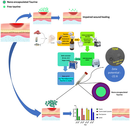
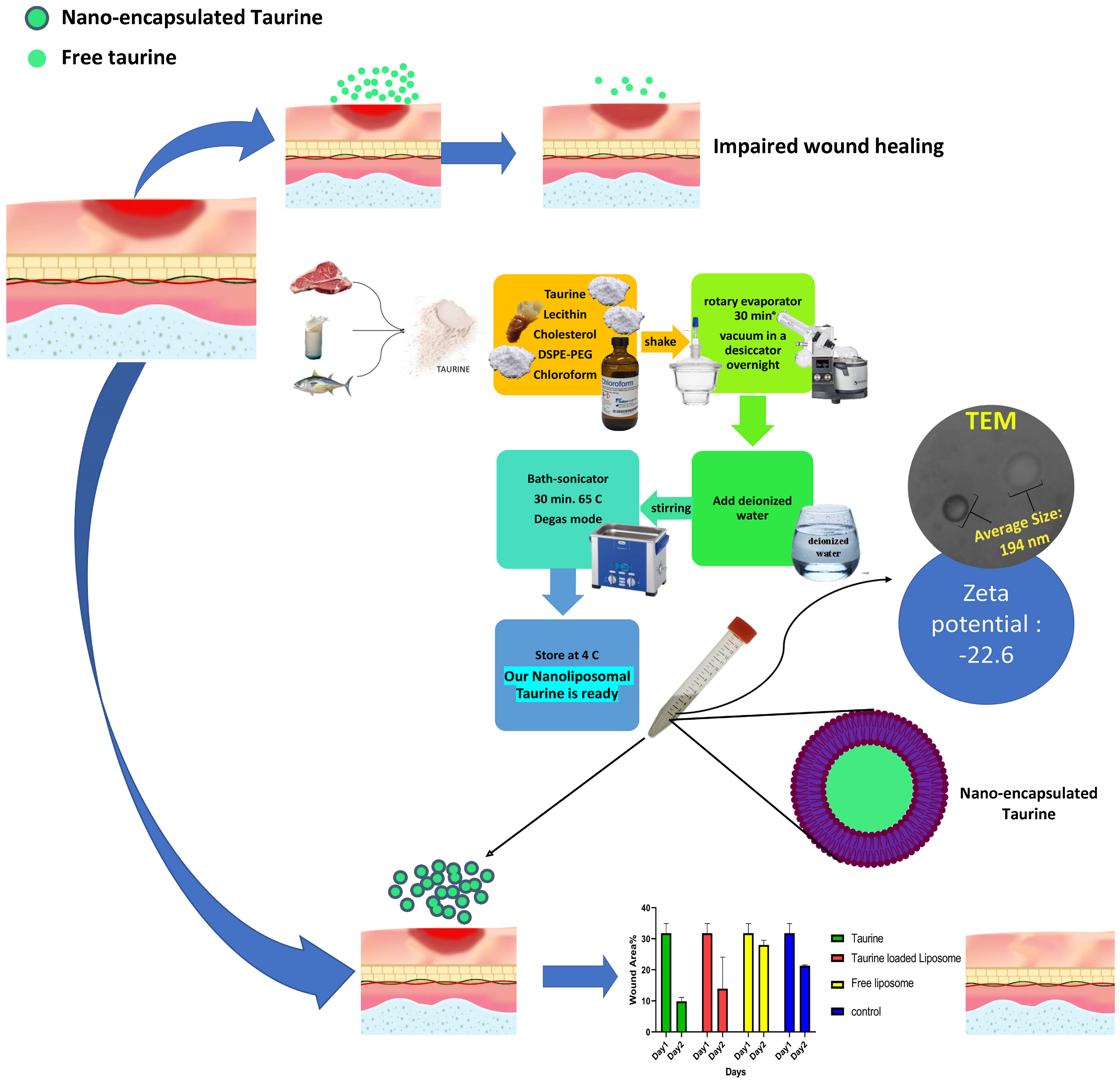
Scheme 1.
Schematic illustration of the preparation, physicochemical characterization, and experimental design of taurine-loaded liposomal nanoparticles (Nano-encapsulated Taurine). Taurine, lecithin, cholesterol, and DSPE-PEG were dissolved in chloroform and processed via rotary evaporation (30 min), followed by overnight vacuum desiccation to form a thin lipid film. The film was rehydrated with deionized water, shaken, and subjected to bath sonication (30 min, 65 °C, degas mode) to yield taurine-encapsulated liposomes. The resulting nanocarriers exhibited a uniform average size of approximately 194 nm and a zeta potential of −22.6 mV, with spherical morphology confirmed by transmission electron microscopy (TEM). The therapeutic efficacy of the taurine-loaded liposomes was evaluated in an in vitro wound healing model, with parallel comparisons against free taurine, empty liposomes (free liposome), and untreated control groups.
2. Materials and Methods
2.1. Materials
The components for nanoliposome formation were prepared as follows: HSPC (L-α-phosphatidylcholine, hydrogenated), cholesterol, and polyethylene glycol (DSPE-PEG-Amine, molecular weight 2000 Da) were obtained from Avanti Polar Lipids, Inc. (Alabaster, AL, USA). Also, the solvents chloroform and ethanol were purchased from Sigma-Aldrich (St. Louis, MO, USA). Moreover, the drug taurine was supplied by Shanghai D&B Biological Science and Technology Co., Ltd. (Shanghai, China). Cell culture media and reagents, including Dulbecco’s Modified Eagle’s Medium (DMEM), RPMI-1640 medium, fetal bovine serum (FBS), phosphate-buffered saline (PBS), and trypsin, were acquired from Gibco Life Technologies Corporation (Carlsbad, CA, USA).
2.2. Preparation of Taurine-Loaded Nanoliposomes (Nlp-Tau)
Taurine-loaded nanoliposomes were prepared based on methods from earlier research, with some minor changes [30]. The nanoliposomes containing taurine were made using the thin-film hydration method. For the control group, which consisted of empty nanoliposomes, we dissolved 10 mg of lecithin, 2 mg of cholesterol, and 1.5 mg of DSPE-PEG in 5 mL of chloroform in a round-bottom flask, and then shook for 1 min. We evaporated the organic solvent using a rotary evaporator (Heidolph, Schwabach, Germany) at 65 °C and 200 rpm by a vacuum pump for 30 min. This process formed a thin lipid film on the flask’s inner wall. We then kept the lipid film under vacuum in a desiccator overnight to ensure that any leftover solvent was completely removed. To make the drug-loaded nanoliposome group (Nlp-Tau), we first ensured the solvent was completely evaporated and the dry lipid film was formed, as described above. We then dissolved 0.896 mg of taurine in 5 mL of water and added to the dried lipid film in the flask. Next, we initiated the hydration process by adding 20 mL of deionized water, which had been pre-warmed to 65 °C. We performed the hydration under continuous stirring using a temperature-controlled magnetic stirrer (Heidolph, Germany) set at 65 °C and 960 rpm. This involved a specific stirring method: first stirring in a water bath for 5 min, then manually swirling outside the bath for 2 min. We repeated this cycle twice. After that, we stirred the mixture in the water bath for 15 min, followed by another 2 min of manual swirling, and then an additional 15 min of stirring in the bath. To ensure even dispersion and decrease the particle size and distribution, we sonicated the resulting suspension for 30 min at 65 °C using a bath-sonicator (Elmasonic, Singen, Germany) operating in pulse mode was used (20 s on/10 s off). We stored the final nanoliposome suspensions at 4 °C, ready for characterization and further use [31].
2.3. Characterization
2.3.1. Dynamic Light Scattering (DLS) and Zeta Potential
A 1:10 (v/v) dilution (100 µL nanoliposome suspension + 900 µL deionized water) of each nanoliposome suspension was prepared to reduce particle concentration sufficiently and avoid multiple scattering effects. each nanoliposome suspension was prepared. The particle size distribution range and the peak particle size, reported as the polydispersity index (PDI), were determined using the Dynamic Light Scattering (DLS) technique. To find the surface charge, or zeta potential, of the prepared nanoliposomes (both empty and taurine-loaded), measurements were taken with a Dynamic Light Scattering instrument (Malvern Zetasizer Nano ZS, Malvern Instruments, Worcestershire, UK) at a 90-degree angle and room temperature. The samples were diluted with deionized water to avoid multiple scattering, and the analysis was performed in triplicate at 25 °C [32].
2.3.2. Fourier-Transform Infrared (FT-IR) Spectroscopy
FT-IR spectroscopy (Spectrum Two, PerkinElmer, Waltham, USA) was used to study possible chemical interactions between taurine and the lipid components, including lecithin, cholesterol, and DSPE-PEG, within the nanoliposomes. The spectra of pure taurine, empty nanoliposomes, and taurine-loaded nanoliposomes were recorded in the wavenumber range of 4000–400 cm−1 [33].
2.3.3. Encapsulation Efficiency (EE%) and Loading Efficiency (LE%)
The EE% and LE%, also known as drug loading (DL%), of taurine in nanoliposomes was determined by measuring the amount of unencapsulated, or free, drug. We centrifuged the nanoliposome suspension in an ultracentrifuge (Amicon Ultra-15, MWCO: 30 kDa, Merck Millipore, Darmstadt, Germany) at 14,000 rpm for 30 min at 7 °C. This process separated the free taurine from the encapsulated drug. We measured the concentration of free taurine in the filtrate using a UV-Vis spectrophotometer (JASCO V-530, Tokyo, Japan) at the specific wavelength for taurine (570 nm) and based on the standard calibration curve (Y = aX + b, R2 = 0.999). All measurements were performed in triplicate [34].
2.3.4. Transmission Electron Microscopy (TEM)
The morphology and structural integrity of the nanoliposomes were examined using a Transmission Electron Microscope (TEM, Zeiss EM900, Jena, Germany). A drop of the diluted nanoliposome suspension was placed on a carbon-coated copper grid and left to adsorb for 2 min. We removed the excess liquid with filter paper. Next, we stained the sample with a 2% (w/v) uranyl acetate solution as a negative stain. The grid was dried in air before imaging at an accelerating voltage of 80 kV.
2.3.5. In Vitro Taurine Release
The study examined the release of taurine from nanoliposomes (Nlp-Tau) under conditions that mimic the body (in vitro) using the dialysis method. For this, 60 mg of freeze-dried taurine-loaded nanoliposomes were dissolved in 10 mL of phosphate-buffered saline (PBS) at pH 7.4. The resulting mixture was placed in a dialysis bag (cut-off: 12 kDa) and submerged in 50 mL of the same buffer containing 0.5% (w/v) sodium dodecyl sulfate (SDS) as the release medium. The setup was put in an incubator shaker (Heidolph, Germany) at 37 °C and 100 rpm to mimic physiological conditions. At specific time points (2, 4, 8, and 12 h, and days 1, 2, and 3), 1 mL of the release medium was taken out and replaced with an equal volume of fresh, pre-warmed release medium to keep the conditions consistent. The concentration of released taurine was measured using a UV-Vis spectrophotometer (JASCO V-530 model, Champaign, IL, USA) at taurine’s maximum absorption wavelength (570 nm). The cumulative percentage of taurine released at each time point was calculated, and the release profile curve was plotted. Finally, the release profile data were fitted to several mathematical models, including the zero-order, first-order, Higuchi, and Korsmeyer-Peppas models, to examine the release behavior and find the main release mechanism. The model that best described the release kinetics was chosen based on the highest correlation coefficient (R2). Given the steady and slow release observed over the 3-day study period, it is expected that taurine release mainly happens through diffusion across the lipid membrane of the nanoliposomes, consistent with the Higuchi or Korsmeyer-Peppas models [35,36].
2.4. Biological Analysis
2.4.1. In Vitro Biocompatibility (MTT Assay)
The researchers assessed the biocompatibility of the samples using the MTT assay on the human fibroblast cell line (HFF). The human fibroblast (HFF) line was obtained from the National Cell Bank of Iran (NCBI), Pasteur Institute of Iran (No. 69, Pasteur Avenue, Tehran 1316943551, Iran). They seeded the cells at a density of 1 × 10⁴ cells per well in a 96-well plate and incubated them for 24 h in DMEM culture medium with 10% FBS and 1% penicillin-streptomycin. The incubator was set to 37 °C and 5% CO2. After this initial incubation, they replaced the culture medium with medium containing different concentrations of the samples: taurine-loaded nanoliposomes (Nlp-Tau) at 3.5, 7, and 14 mg/mL. After 24 and 48 h of incubation, they added MTT to a final concentration of 0.5 mg/mL and incubated for 4 h. They then aspirated the culture medium and dissolved the formed formazan crystals in 100 µL of DMSO. They measured the optical density at a wavelength of 570 nm using an ELISA reader. All experiments were done in three independent replicates. The researchers analyzed the data using GraphPad Prism software version 9.0. They used one-way analysis of variance (One-way ANOVA) followed by Tukey’s test to compare the means. p-values less than 0.05 were considered statistically significant.
2.4.2. In Vitro Wound Healing (Scratch Assay)
The migratory potential of HFF cells, which indicates in vitro wound closure, was evaluated using a scratch assay. HFF cells were seeded into 24-well plates at a density of 2 × 10⁵ cells per well and incubated until a confluent monolayer formed. A sterile 200 µL pipette tip was used to make a uniform scratch across the center of each well. The detached cells and debris were removed by gently washing the monolayers with PBS. The scratched monolayers were treated with DMEM media containing the optimized concentration of taurine-loaded nanoliposomes (Nlp-Tau), the same concentration of free taurine, empty nanoliposomes, or just the medium as a negative control. The plates were incubated under standard conditions at 37 °C with 5% CO2. The scratch area was photographed on Day 1 and Day 2 after scratching using an inverted microscope with a digital camera. The wound area at each time point was measured with ImageJ software (version 1.41, NIH, USA), and the percentage of wound closure was calculated relative to the initial wound area at 0 h. All experiments were conducted in triplicate, and the data were analyzed using GraphPad Prism version 9.0 [37].
2.5. Statistical Analysis
Statistical analysis, which included curve fitting and one-way ANOVA, was performed using GraphPad Prism version 8 from GraphPad Software, Inc., based in San Diego, CA, USA. Data from three independent experiments are shown as the mean ± standard deviation. We set statistical significance at α = 0.05. In the charts, data represent the mean ± SD (n = 3), with significance indicated as * p < 0.05, ** p < 0.01, and *** p < 0.001.
2.6. Generative AI
Declaration of generative AI in scientific writing: During the preparation of this work, the authors used [DeepSeek, and Grammarly DeepSeek-V4 (the latest major model series from DeepSeek) and Grammarly for Windows v1.2.261.1889 (20 May 2026)] for the sole purpose of improving language readability and refining the manuscript’s grammar and syntax. This tool was utilized to enhance the clarity and flow of the text, ensuring it meets the linguistic standards expected for scientific communication. After using these tools, the authors reviewed and edited the content as needed and take full responsibility for the content of the published article.
3. Results
3.1. Physicochemical Characterization of Taurine-Loaded Nanoliposomes
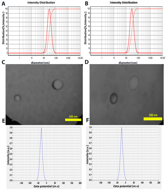
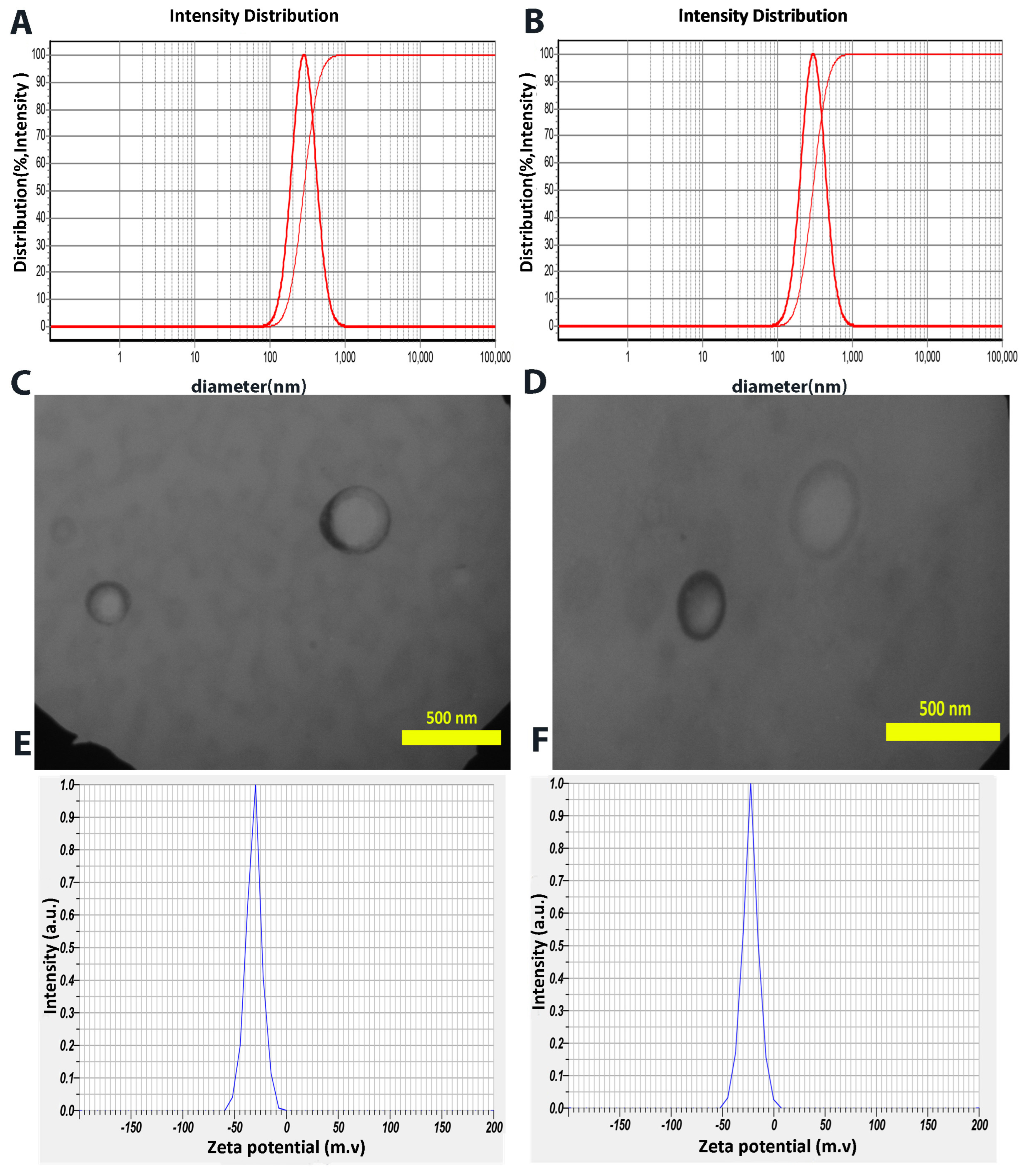
The results from dynamic light scattering (DLS) analysis of the drug-loaded nanoliposomes provided key information regarding particle size and distribution (Figure 1A,B). This method usually estimates nanoparticle size based on hydrodynamic volume; the actual particle size is often determined through TEM. All measurements were performed in triplicate using three independently prepared batches (n = 3). According to the DLS data, the mean hydrodynamic diameter (±SD) of drug-free nanoliposomes was 282.4 ± 8.7 nm (range: 273.1–291.6 nm), while taurine-loaded nanoliposomes (Nlp-Tau) showed a mean diameter of 302.1 ± 10.3 nm (range: 290.5–313.8 nm). The mean polydispersity index (PDI) for drug-free and drug-loaded formulations was 0.20 ± 0.02 (range: 0.18–0.22) and 0.25 ± 0.03 (range: 0.22–0.28), respectively, indicating acceptable size uniformity.
Figure 1.
Physicochemical characterization of the engineered nanoliposomes. (A,B) Dynamic light scattering (DLS) intensity-based size distribution profiles and corresponding mean hydrodynamic diameters for (A) empty liposomes and (B) taurine-loaded liposomes (Nlp-Tau). (C,D) Representative transmission electron microscopy (TEM) images confirming the morphology and size of (C) empty and (D) drug-loaded liposomes. (E,F) Zeta potential distribution curves indicating the surface charge for (E) empty and (F) loaded formulations.
TEM images (Figure 1C,D) confirmed the particle sizes. The diameters were about 142 nm for the drug-free particles and 194 nm for the taurine-loaded ones. We assessed surface charge, an important factor for particle stability and cellular interactions, using zeta potential measurement. According to the data (Figure 1E,F), the mean zeta potential (±SD) for drug-free nanoliposomes was −31.3 ± 2.1 mV (range: −33.8 to −28.9 mV), and for taurine-loaded nanoliposomes was −22.6 ± 1.8 mV (range: −24.9 to −20.7 mV), based on triplicate measurements (n = 3). The negative zeta potential values indicate that the particles possess a net negative surface charge. Regarding colloidal stability, absolute zeta potential values around 30 mV usually mean a suspension with excellent stability. This stability comes from the strong electrostatic repulsion between particles, which effectively stops them from clumping together or settling. Therefore, these results suggest that the formulated nanoliposomes have very good physical stability in suspension. This is critical for their storage, transport, and effectiveness as a drug delivery system. Quantitative analyses were performed to determine the encapsulation and loading percentages in the nanoliposomal carrier. The EE% and LE% of taurine in the nanoliposomes were 20% and 2.5%, respectively. These values are within the desirable range for drug delivery applications. Additionally,
Table 1
shows the DLS and TEM data.
Table 1.
Physicochemical characterization data of blank and taurine-loaded nanoliposomes.
3.2. FTIR Characterization of Taurine-Loaded Nanoliposomes
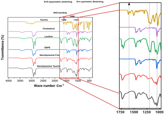
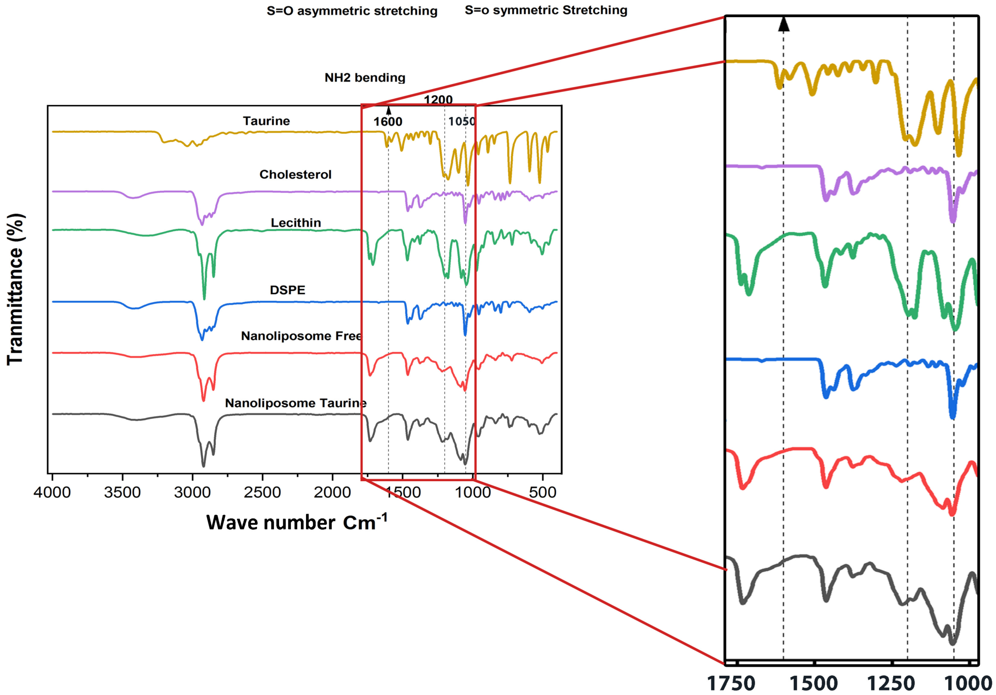
The chemical structures of the nanocarrier, drug, and taurine-loaded nano liposomes are shown in Figure 2. The FTIR spectral analysis confirms that taurine was successfully encapsulated in the nanoliposome formulation. It also clarifies the nature of the molecular interactions involved. The spectrum of pure taurine displays its characteristic fingerprint. There is a distinct peak at about 1050 cm−1 for the symmetric S=O stretching vibration, a band near 1200 cm−1 for asymmetric S=O stretching, and a peak around 1600 cm−1 for NH2 bending. These key diagnostic peaks for taurine are clearly present in the spectrum of the taurine-loaded nanoliposome, confirming that the compound is included in the final formulation. However, their appearance shows notable shifts and changes in intensity compared to the pure taurine spectrum. This provides evidence of interaction rather than just physical mixing. The peaks, especially the S=O stretches, are broadened and weakened. This suggests that taurine molecules are engaged in hydrogen-bonding interactions with the polar headgroups of the phospholipid bilayer, specifically with phosphate and carbonyl groups.
Figure 2.
FTIR spectra of taurine, lipid components (cholesterol, DSPE, lecithin), free nanoliposome, and taurine-loaded nanoliposome, demonstrating the successful encapsulation and molecular interaction of taurine within the liposomal structure.
The spectra of the lipid components—cholesterol, DSPE, and lecithin—show their own characteristic bands. These include C-H stretches between 2800 and 3000 cm−1 and carbonyl (C=O) stretches around 1730–1740 cm−1. The spectrum of the free, or empty, nanoliposome is a mix of these lipid signatures. When comparing the free nanoliposome to the taurine-loaded version, the most significant observation is the change in the region of taurine’s functional groups and subtle changes in the lipid headgroup regions. The absence of entirely new peaks in the loaded liposome spectrum indicates that the encapsulation process did not cause covalent chemical bonding, which helps preserve taurine’s molecular integrity. Although the broad O–H/N–H stretching region (3200–3500 cm−1) appeared similar between empty and taurine-loaded liposomes—likely due to dominant signals from residual water and cholesterol hydroxyl groups—the key evidence for hydrogen bonding came from lower-frequency regions. Specifically, the S=O stretching bands of taurine (1050–1200 cm−1) exhibited broadening and reduced transmittance, while the P=O (~1240 cm−1) and C=O (~1735 cm−1) stretches of phospholipids showed slight shifts. These changes indicate non-covalent interactions, including hydrogen bonds between the sulfonic acid group of taurine and the polar headgroups of the lipid bilayer. This successful integration without chemical modification is key to keeping the bioactive compound in its therapeutically active form. The spectral data strongly supports the conclusion that taurine is effectively encapsulated within the nanoliposome’s aqueous core and/or interfacial region. This establishes a stable host-guest association, which is critical for its potential controlled release and improved delivery efficacy.
3.3. In Vitro Release Profile of Taurine from Nanoliposomes
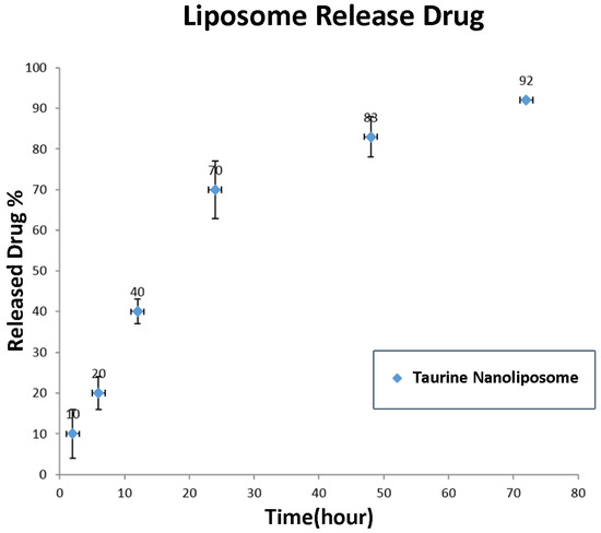
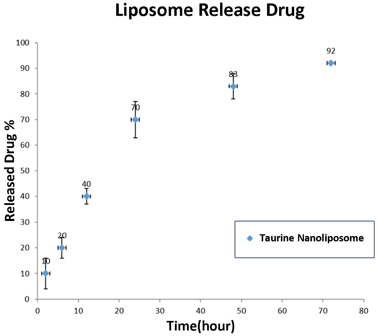
The loading and release profile of taurine from the nanoliposomal carrier in PBS was plotted at a wavelength of 570 nm (Figure 3). This study looked closely at the release profile and kinetics of taurine from the nanoliposomes (Nlp-Tau). First, a standard calibration curve was created using taurine standard solutions in phosphate-buffered saline (PBS) at 570 nm. This curve, which was linear within the tested concentration range (R2 > 0.999), provided a reliable basis for measuring the amount of released taurine in all later stages.
Figure 3.
In vitro release profile and kinetics of taurine from nanoliposomes (Nlp-Tau). The cumulative percentage of taurine released in PBS over 72 h demonstrates a biphasic, controlled release pattern. An initial phase of relatively faster release within the first 24 h is followed by a sustained, slow release phase. The release kinetics best fit the Higuchi model (R2 > 0.999), indicating a diffusion-driven (Fickian) release mechanism from the lipid matrix.
The taurine release profile from the nanoliposomes was monitored over 3 days. The observed pattern showed a slow, continuous, and controlled release from the nanocarrier. This profile clearly follows two phases: an initial phase with a faster release during the first 24 h, likely due to the release of drug adsorbed on the liposome surface. This is followed by a sustained phase with a gentle slope that continued until the end of the third day. This second phase, in which most of the drug was released, mainly shows the mechanism of drug diffusion through the lipid bilayer of the nanoliposomes. This controlled release pattern is a key advantage of the drug delivery system, as it can maintain a stable therapeutic concentration of taurine at the wound site for a long time without needing frequent dressing changes or drug re-administration.
The cumulative release data were fitted to several kinetic models, including zero-order, first-order, Higuchi, and Korsmeyer-Peppas models. The highest correlation coefficient (R2) was found for the Higuchi model. This strong fit confirms that Fickian diffusion mainly controls taurine release from the nanoliposomes, meaning the drug release rate is proportional to the square root of time, indicating drug diffusion from a lipid matrix system. In summary, the findings from this section show that the nanoliposomal formulation has successfully encapsulated taurine and demonstrates a good ability for slow and controlled drug release over a long period. This feature makes the Nlp-Tau system a promising option for controlled-release wound dressings, which could effectively improve the wound-healing effects of taurine.
3.4. Taurine-Loaded Nanoliposomes Effects on Cell Viability
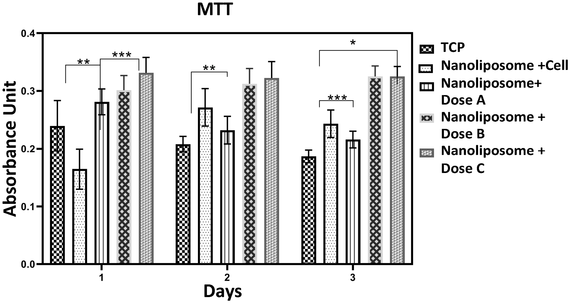
The effect of taurine-loaded nanoliposomes on cell viability has been studied in relation to different doses and is shown in Figure 4. The MTT assay results revealed a dose-dependent effect on cell viability after 48 h of incubation, as measured by optical density (OD). The OD data from the MTT assay conducted over three days indicate a treatment-dependent effect on cell metabolic activity. The control group in the tissue culture plate (TCP) had OD values between 0.20 and 0.24. The group treated with empty nanoliposomes (Nanoliposome Cell) had similar values, ranging from 0.24 to 0.26. In contrast, cells treated with taurine-loaded nanoliposomes had higher OD values, showing a clear positive trend as the dose increased. Dose A had values from 0.17 to 0.22, Dose B ranged from 0.28 to 0.32, and Dose C, which was the highest concentration, showed the greatest metabolic activity with OD values from 0.33 to 0.36. This pattern, consistent across all time points, confirms that taurine-loaded nanoliposomes enhance cell viability in a dose-dependent manner, with Dose C having the most significant effect. Statistical analysis confirmed the significance of these increases, especially for the higher doses compared to the control and vehicle groups.
Figure 4.
MTT assay results demonstrating the dose-dependent effect of taurine-loaded nanoliposomes on cell viability. Over three days, the control (TCP) OD ranged from 0.20 to 0.24, while empty nanoliposomes showed a similar range of 0.24–0.26. Taurine-loaded nanoliposomes increased metabolic activity dose-dependently: Dose A (0.17–0.22), Dose B (0.28–0.32), and Dose C (0.33–0.36). Statistical analysis confirmed that higher doses, particularly Dose C, significantly enhanced viability compared to control and vehicle groups (* p < 0.05, ** p < 0.01, and *** p < 0.001).
3.5. In Vitro Wound Healing
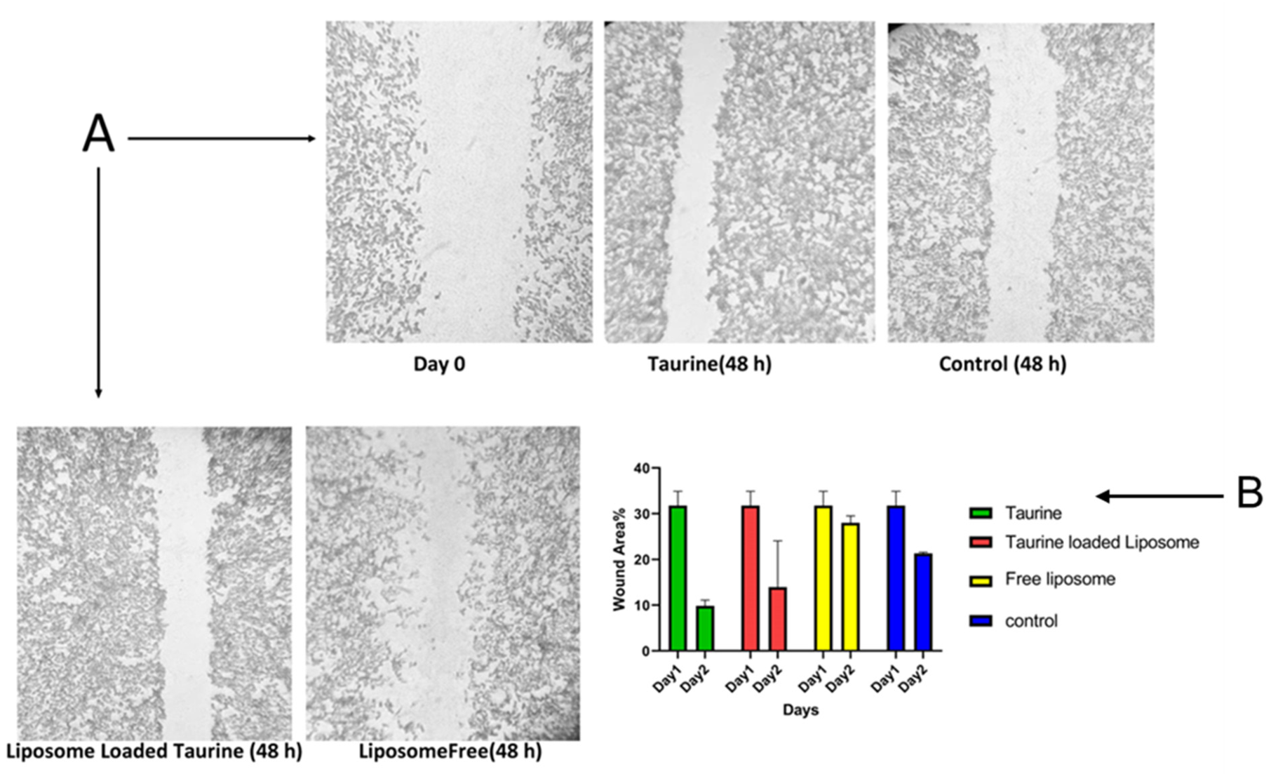
The results from the scratch assay, which is an important measure for evaluating wound healing in vitro, are shown in Figure 5. The best dose of taurine, based on MTT results, was used for the scratch analysis. The scratch assay results showed noticeable differences in cell migration and wound closure among the treatment groups after 48 h. Visually, Figure 5A indicates that the wound gap in the taurine-treated sample was significantly smaller compared to the control, suggesting improved migration. The quantitative data in Figure 5B provided a clear measurement of this effect. It showed that free taurine reduced the remaining wound width to 10.734% of the initial scratch after 2 days. The most significant effect came from taurine-loaded liposomes, which achieved the highest degree of closure, leaving only 6.797% of the wound area open after 2 days. In contrast, both empty liposomes (26.954%) and the untreated control (21.11%) had much less wound closure. To exclude the possibility that reduced closure in the empty liposome group resulted from cytotoxicity, we directly correlated these data with MTT viability results (Figure 4). At the same concentration used in the scratch assay, empty liposomes exhibited OD values of 0.24–0.26 over 72 h, which were statistically indistinguishable from the untreated control (0.20–0.24) and well above cytotoxic thresholds. Microscopic examination during the scratch assay showed no cell rounding or detachment in empty liposome-treated wells (Figure 5A). This confirms that the liposome vehicle alone did not enhance migration and that the therapeutic effect was due to taurine, especially when delivered through the liposomal formulation. Thus, the minimal closure observed with empty liposomes reflects a lack of pro-migratory bioactivity rather than any toxic effect on fibroblast viability or function.
Figure 5.
Scratch assay analysis of cell migration after 48 h, comparing free taurine, taurine-loaded liposomes, empty liposomes, and an untreated control. (A) Representative microscope images showing wound closure. (B) Quantitative analysis of the remaining wound area. Taurine-loaded liposomes achieved the most potent closure (6.8% remaining wound), outperforming free taurine (10.7%). Empty liposomes (27.0%) and the control (21.1%) showed significantly less closure, confirming the enhanced therapeutic effect of taurine via liposomal delivery.
4. Discussion
Chronic wounds, such as diabetic ulcers, pressure sores, and venous leg ulcers, present a serious and increasing global healthcare issue. It is estimated that these wounds affect about 2% of the general population. The prevalence sharply rises with age and conditions like diabetes. Individuals with diabetes have a lifetime risk of developing a foot ulcer that can reach 25% [38,39]. The economic impact is immense, with annual wound care costs in developed countries exceeding billions of dollars, primarily due to long hospital stays, frequent dressing changes, and lost productivity [40]. This situation highlights the urgent need for improved therapeutic strategies that can modulate the wound microenvironment and accelerate healing. Recent studies showed that herbal remedies hold promise for wound healing [33,35]. Taurine, a strong natural antioxidant and anti-inflammatory amino acid, has shown significant potential in promoting various stages of wound repair, including resolving inflammation, boosting fibroblast proliferation and collagen maturation [41]. However, its use in clinical settings is limited by certain challenges: its high-water solubility, poor ability to cross cell membranes, and quick removal from the wound site. These factors require frequent reapplication to keep therapeutic levels effective [42]. To overcome these barriers, we developed a nanoliposomal delivery system.
The successful development of an effective nanocarrier relies on careful optimization of its physicochemical properties. These properties directly affect its stability, biodistribution, cellular interaction, and therapeutic efficacy [43]. Hence, it seems that the formulated taurine-loaded nanoliposomes (Nlp-Tau) appear to be a promising option for achieving the key objectives.
Results of this study showed that the particle size analysis using Dynamic Light Scattering (DLS) reported a median diameter (d50) of 282 nm for neat liposomes and 302 nm for loaded liposomes (Figure 1A,B). Although DLS measures the hydrodynamic diameter and usually overestimates the core size, this value is still within a suitable range for nanocarriers meant for topical delivery [44]. Sizes in this range are considered ideal because they help with effective skin penetration while keeping a high drug-loading capacity [45]. The narrow size distribution (0.2 for neat liposomes and 0.25 for loaded liposomes) indicates a consistent and reliable formulation process, which is important for predictable drug release kinetics [46].
Complementary TEM analysis provided direct visual confirmation of the nanoliposomes’ shape (Figure 1C,D). The images showed clearly defined, spherical vesicles for both empty and drug-loaded versions, with an estimated diameter of about 142 and 194 nm, matching the expected core size. Importantly, the encapsulation process did not affect the vesicle structure, which is vital for stability. The high negative zeta potential of −31.3 mV and −22.6 (Figure 1E,F) is a key finding. A zeta potential around |±30| mV creates strong electrostatic repulsion, ensuring long-term colloidal stability in suspension. This stability is essential for shelf-life and consistent dosage [47].
FT-IR spectroscopy provided important insights into the interactions between the drug and the carrier (Figure 2). The distinctive peaks of pure taurine were clearly visible in the spectrum of Nlp-Tau, proving that loading was successful. The lack of new covalent bond peaks shows that taurine is physically trapped. The slight shifts in the peaks probably result from physical interactions like hydrogen bonding. This preservation of the drug’s chemical integrity is crucial for keeping its pharmacological activity [48].
A practical consideration in designing nanocarriers is finding the right balance between desirable physical and chemical properties and high drug loading. Our formulation achieved an EE% of 20% and an LE% of 2.5% (see Results, EE% and LE%). It is important to contextualize these encapsulation values within the intrinsic physicochemical properties of taurine. Taurine is a small (125 Da), highly hydrophilic, zwitterionic amino acid with a log P of approximately −4.2. Its high water solubility leads to a strong thermodynamic preference for the aqueous phase over the lipid bilayers during thin-film hydration, which inherently limits passive encapsulation. Furthermore, its low molecular weight facilitates rapid diffusion across the nascent liposomal membrane during and after formation, allowing significant drug leakage during sonication and purification steps. These same properties—high hydrophilicity and small size—underlie the very clinical limitations that nanoliposomes aim to overcome (rapid clearance and poor skin penetration). Consequently, achieving high EE% for such molecules without active or remote loading techniques is inherently challenging. Our obtained EE% of 20% and LE% of 2.5%, therefore, reflect a rational optimization trade-off. Rather than pursuing higher drug loading at the expense of colloidal stability, we prioritized a small and uniform particle size (≈282 nm, PDI 0.2) and a high negative zeta potential (−31.3 mV) to ensure long-term stability and enhanced cellular interaction. This approach successfully produced a functionally effective system, as evidenced by sustained Higuchi-type release and superior in vitro wound closure [49].
Although these values may seem modest, they reflect a careful optimization choice. It is often crucial to prioritize a small, uniform particle size and a high zeta potential to ensure stability and improve tissue penetration, even if it reduces the total drug load [50,51]. Our drug release data strongly back this approach. The in vitro release profile of taurine from Nlp-Tau showed a favorable biphasic, sustained release pattern over 72 h (Figure 3). The best fit to the Higuchi model confirms that the release mechanism mainly involves Fickian diffusion [52]. This controlled, sustained release is exactly what we want in a wound dressing, as it maintains a therapeutic concentration at the wound site for a long time [53].
The most compelling evidence of any biomaterial’s potential is its biocompatibility. The MTT assay results on HFF cells were clearly positive (Figure 4). The Nlp-Tau system showed no significant cytotoxicity at concentrations up to 7 mg/mL over 72 h. It maintained high cell viability with OD values between 0.33 and 0.36. This confirms the safety of the lipid components and the harmless nature of the nanostructure [54]. The ability to support fibroblast viability and growth over several days is especially important for wound healing. Fibroblasts are the main cells involved in tissue remodeling [55]. Additionally, this trend is consistent with previous research and relates to inflammation inhibition, reduced oxidative stress, promoted macrophage polarization, and stimulated proliferation [5,8,42].
The scratch assay results (Figure 5) demonstrate that taurine significantly enhances in vitro wound closure, an effect that is markedly potentiated by liposomal encapsulation. The strong performance of taurine-loaded liposomes, which achieve nearly complete wound resolution, highlights the importance of how the treatment is delivered. This increased activity is hypothesized to result from the liposomes’ ability to protect taurine, promote its steady release, and enhance cellular interaction and internalization at the wound site. Although direct uptake mechanism studies (e.g., confocal microscopy, endocytosis inhibition assays) were not performed in this work, it is well-established in the literature that nanoliposomes of similar size (150–300 nm) and negative surface charge (zeta potential ≈ −30 mV) are typically internalized by mammalian cells via endocytic pathways, including clathrin-mediated endocytosis, caveolae-mediated endocytosis, and micropinocytosis, or may fuse directly with the cell membrane [56,57,58,59]. This enhanced cellular delivery is likely a key factor in the superior wound closure observed with Nlp-Tau compared to free taurine. This process helps sustain the pro-migratory signal. The minimal closure seen with empty liposomes shows that the vehicle is inactive and that the biological effect is due only to taurine [60]. The results of this study reveal better cell migration and enhance wound healing, which align with previous research. Also, critically, the MTT assay confirmed that empty liposomes did not reduce cell viability (OD 0.24–0.26 vs. control 0.20–0.24, p > 0.05), ruling out cytotoxicity as a confounder. This is consistent with previous reports that liposomes composed of HSPC, cholesterol, and DSPE-PEG are intrinsically biocompatible and lack intrinsic wound-healing bioactivity [34,55]. Therefore, the enhanced closure in the Nlp-Tau group is specifically attributable to sustained taurine delivery and its pro-migratory effects on fibroblasts. For instance, the significant study by Sasso examined the role of natural taurine metabolites known as N-acyl taurines (NATs) in the wound healing process. This research found that NATs serve as signaling molecules, much like endocannabinoids. By activating CB2 receptors, they help control keratinocyte movement and wound closure. When NAT levels drop in the skin, wound healing gets worse. However, applying these compounds directly to the skin can greatly speed up skin regeneration. These results underline the crucial role of taurine and its derivatives in skin health [9]. A study looked into how taurine affects the production of ceramides and hyaluronic acid in cultured dermal fibroblasts and the epidermis. It found that taurine boosts ceramide production by activating the enzyme ceramide synthase and increasing the expression of genes linked to sphingolipid metabolism. Taurine also improves hyaluronic acid production by raising the expression of the enzyme hyaluronan synthase 2. These two effects improve skin barrier function and raise stratum corneum hydration, which ultimately leads to better wound healing [61].
5. Conclusions
This study successfully developed and characterized taurine-loaded nanoliposomes (Nlp-Tau) as a delivery system designed to overcome the limitations of free taurine in wound healing applications. The formulation showed optimal properties, including a nano-sized spherical shape (about 282 nm), a high negative zeta potential (around −31.3 mV) that ensures stability, and good encapsulation efficiency. FTIR analysis confirmed that the taurine was physically trapped without any chemical interactions. Importantly, Nlp-Tau demonstrated a sustained, diffusion-controlled release profile over 72 h, following Higuchi kinetics. In vitro tests on human fibroblasts showed no signs of cell damage and high cell viability. The scratch assay also demonstrated that the liposomal formulation significantly improved taurine’s therapeutic effect, leading to better wound closure compared to free taurine. These findings demonstrate that the Nlp-Tau system effectively addresses the challenges of rapid clearance and poor retention, offering a stable, biocompatible method for the local delivery of taurine. This research lays a solid foundation for future in vivo studies to confirm the clinical potential of this nanocarrier in improving tissue repair and treating chronic wounds.
Author Contributions
M.J.: Investigation, Methodology, Formal analysis, Writing—original draft. F.N.: Investigation, Methodology, Validation, Resources. H.G.J.: Investigation, Data curation, Visualization. A.R.F.: Conceptualization, Supervision, Project administration, Writing—review and editing, Funding acquisition. A.G.: Conceptualization, Supervision, Writing—review and editing, Funding acquisition. Z.A.: Formal analysis, Writing—review and editing, Revision. M.S.: Visualization, Writing—review and editing Revision. All authors have read and agreed to the published version of the manuscript.
Funding
This work was supported by School of Advanced Technologies in Medicine, Fasa University of Medical Sciences (Grant Number 402148).
Institutional Review Board Statement
This article does not contain any studies with human participants or animals performed by any of the authors. The study utilized a commercially available human foreskin fibroblast (HFF) cell line; followed by ethical approval code: IR.FUMS.AEC.1403.004.
Informed Consent Statement
Statement is not applicable.
Data Availability Statement
The data that support the findings of this study are available from the corresponding author upon reasonable request.
Acknowledgments
Authors want to acknowledge Fasa University of Medical Sciences for providing research facilities.
Conflicts of Interest
The authors declare no conflict of interest.
References
- Chitca, D.-D.; Popescu, V.; Mastalier, B.; Busu, C. The economic burden of chronic wounds on global healthcare systems. In Proceedings of the International Conference on Business Excellence, Bucharest, Romania, 20–22 March 2025; pp. 1995–2002. [Google Scholar]
- Jing, S.L.; Suh, E.J.; Huang, K.X.; Griffin, M.F.; Wan, D.C.; Longaker, M.T. Understanding and Advancing Wound Healing in the Era of Multi-Omic Technology. Bioengineering 2025, 13, 51. [Google Scholar] [CrossRef] [Scilit] [PubMed]
- Deng, X.; Gould, M.; Ali, M.A. A review of current advancements for wound healing: Biomaterial applications and medical devices. J. Biomed. Mater. Res. Part B Appl. Biomater. 2022, 110, 2542–2573. [Google Scholar] [CrossRef] [Scilit] [PubMed]
- Najafi, F.; Farrokhzad, N.; Ghaemi, A.; Khezri, D.A.; Hajiabbas, M.; Khanizadeh, A.; Farsani, N.K.; Khoramipour, M.; Fatemipayam, N.; Zadeh, E.S.; et al. Advancement in nanocarrier-mediated delivery of herbal bioactives: From bench to beside. Nat. Prod. Bioprospecting 2025, 15, 69. [Google Scholar] [CrossRef] [Scilit] [PubMed]
- Ashkani-Esfahani, S.; Zarifi, F.; Asgari, Q.; Samadnejad, A.Z.; Rafiee, S.; Noorafshan, A. Taurine improves the wound healing process in cutaneous leishmaniasis in mice model, based on stereological parameters. Adv. Biomed. Res. 2014, 3, 204. [Google Scholar] [CrossRef] [Scilit] [PubMed]
- Zhao, P.; Qi, Y. Taurine: A Comprehensive Review of Its Origin, Pharmacological Properties, Potential Health Benefits, Therapeutic Applications, and Safety Profile. Food Sci. Hum. Wellness 2025, 15, 925043. [Google Scholar] [CrossRef] [Scilit]
- Yuan, J.; Hou, Q.; He, X.; Zhong, L.; Li, M.; Fu, X.; Liu, H. Chitosan-taurine nanoparticles cross-linked carboxymethyl chitosan hydrogels facilitate both acute and chronic diabetic wound healing. Int. J. Biol. Macromol. 2024, 273, 132762. [Google Scholar] [CrossRef] [Scilit] [PubMed]
- Wu, L.; Zhang, Q.; Li, Y.; Song, W.; Chen, A.; Liu, J.; Xuan, X. Collagen sponge prolongs taurine release for improved wound healing through inflammation inhibition and proliferation stimulation. Ann. Transl. Med. 2021, 9, 1010. [Google Scholar] [CrossRef] [Scilit] [PubMed]
- Sasso, O.; Pontis, S.; Armirotti, A.; Cardinali, G.; Kovacs, D.; Migliore, M.; Summa, M.; Moreno-Sanz, G.; Picardo, M.; Piomelli, D. Endogenous N-acyl taurines regulate skin wound healing. Proc. Natl. Acad. Sci. USA 2016, 113, E4397–E4406. [Google Scholar] [CrossRef] [Scilit] [PubMed]
- Munusamy, T.; Shanmugam, R. Enhancing wound healing with nanoparticle-infused hydrogels: A review of current applications and future prospects. Discov. Biotechnol. 2025, 2, 34. [Google Scholar] [CrossRef] [Scilit]
- Abd El-Hack, M.E.; Alawam, A.S.; Aldhalmi, A.K.; Elsherbeni, A.I.; Rudayni, H.A.; Allam, A.A.; Khafaga, A.F. Taurine-functionalized carbohydrate polymers as emerging multifunctional biotherapeutics for human and veterinary medicine: A comprehensive review. Int. J. Biol. Macromol. 2026, 340, 149789. [Google Scholar] [CrossRef] [Scilit] [PubMed]
- Alvandi, H.; Shafie, A.; Najafi, F.; Sabzini, M.; Mashayekhi, M.; Omami, S.H.; Eskandarisani, M.M.; Dashti, S.; Javanmard, A.; Tajik, M.; et al. Carbon-based nanostructure drug delivery systems and their biologic applications—A review. Carbon Lett. 2025, 35, 1529–1587. [Google Scholar] [CrossRef] [Scilit]
- Ghodsi, R.; Sadr Tahouri, S.K.; Amjad, F.; Nasiri, M.R.; Toutounchi, A.; Salehi, H.; Moeinzadeh, A.; Sadeghi, S.; Soleimani, N.; Onagh, B.; et al. Chitosan/alginate@niosome-curcumin: High-performance wound dressing with enhanced antibacterial activity. J. Drug Deliv. Sci. Technol. 2025, 114, 107438. [Google Scholar] [CrossRef] [Scilit]
- Gowda, B.H.J.; Mohanto, S.; Singh, A.; Bhunia, A.; Abdelgawad, M.A.; Ghosh, S.; Ansari, M.J.; Pramanik, S. Nanoparticle-based therapeutic approaches for wound healing: A review of the state-of-the-art. Mater. Today Chem. 2023, 27, 101319. [Google Scholar] [CrossRef] [Scilit]
- Choudhury, A.; Kirti, A.; Lenka, S.S.; Naser, S.S.; Sinha, A.; Kumari, S.; Kaushik, N.K.; Ghosh, A.; Verma, S.K. Strategic advances in liposomes technology: Translational paradigm in transdermal delivery for skin dermatosis. J. Nanobiotechnol. 2025, 23, 576. [Google Scholar] [CrossRef] [Scilit] [PubMed]
- de Assis, S.A.; Alves, S.L. Chapter 10—Liposomes and niosomes approaches for wound healing. In Recent Advances in Nanomedicines Mediated Wound Healing; Kesharwani, P., Ed.; Academic Press: Cambridge, MA, USA, 2025; pp. 161–185. [Google Scholar]
- Moradi, R.; Taghizadeh, B.; Ghaffari, R.; Mirzavi, F.; Sobhani, B.; Movaffagh, J.; Afshari, J.T.; Kamali, H.; Chamani, J.; Alavizadeh, S.H.; et al. Development of nano-liposomal platelet-poor plasma (PPP) for topical application in wound healing and burn treatment. J. Pharm. Investig. 2025, 55, 919–933. [Google Scholar] [CrossRef] [Scilit]
- Parchen, G.P.; Jacumazo, J.; Koop, H.S.; Biscaia, S.M.P.; Trindade, E.S.; Silveira, J.L.M.; de Freitas, R.A. Modulation of Epidermal Growth Factor Release by Biopolymer-Coated Liposomes. J. Pharm. Sci. 2020, 109, 2294–2301. [Google Scholar] [CrossRef] [Scilit] [PubMed]
- Cardoso-Daodu, I.M.; Ilomuanya, M.O.; Azubuike, C.P. Development of curcumin-loaded liposomes in lysine–collagen hydrogel for surgical wound healing. Beni-Suef Univ. J. Basic Appl. Sci. 2022, 11, 100. [Google Scholar]
- Choudhary, V.; Shivakumar, H.; Ojha, H. Curcumin-loaded liposomes for wound healing: Preparation, optimization, in-vivo skin permeation and bioevaluation. J. Drug Deliv. Sci. Technol. 2019, 49, 683–691. [Google Scholar] [CrossRef] [Scilit]
- Risaliti, L.; Kehagia, A.; Daoultzi, E.; Lazari, D.; Bergonzi, M.C.; Vergkizi-Nikolakaki, S.; Hadjipavlou-Litina, D.; Bilia, A.R. Liposomes loaded with Salvia triloba and Rosmarinus officinalis essential oils: In vitro assessment of antioxidant, antiinflammatory and antibacterial activities. J. Drug Deliv. Sci. Technol. 2019, 51, 493–498. [Google Scholar] [CrossRef] [Scilit]
- Demirel, F.; Sağıroğlu, A.A.; Toraman GÖ, A.; Gunaydin-Akyildiz, A.; Keskin, Z.; Aydın, B.S.; Topçu, G. Development and Evaluation of Platanus orientalis L. Extract-Loaded Liposomes for Enhanced Wound Healing. Pharmaceuticals 2025, 19, 32. [Google Scholar] [CrossRef] [Scilit] [PubMed]
- Mathure, D.; Deshmukh, P.; Salunke, M.; Ranpise, H. Liposome integrated hydrogel for diabetic wound healing: Molecular mechanisms in inflammation and oxidative stress mitigation. Inflammopharmacology 2025, 33, 7479–7498. [Google Scholar] [CrossRef] [Scilit] [PubMed]
- Guidoni, M.; de Sousa Júnior, A.D.; Aragão, V.P.M.; de Melo Costa Pereira, T.; dos Santos, W.C.; Monteiro, F.C.; Guimarães, M.C.C.; Fronza, M. Liposomal stem cell extract formulation from Coffea canephora shows outstanding anti-inflammatory activity, increased tissue repair, neocollagenesis and neoangiogenesis. Arch. Dermatol. Res. 2023, 315, 491–503. [Google Scholar] [CrossRef] [Scilit] [PubMed]
- Erdal Altıntaş, Ö.; Aytar Çelik, P. Levan-based bioactive hydrogels containing herbal liposome in treating acute wounds. J. Bioact. Compat. Polym. 2023, 38, 437–457. [Google Scholar] [CrossRef] [Scilit]
- Braido, B.; Rukavina, Z.; Grimstad, Ø.; Franzè, S.; Cilurzo, F.; Vanić, Ž.; Škalko-Basnet, N.; Hemmingsen, L.M. Liposomes-in-hydrogel for topical drug delivery: Mechanical, kinetic, and biological insights. J. Drug Deliv. Sci. Technol. 2025, 113, 107380. [Google Scholar] [CrossRef] [Scilit]
- Pourtalebi Jahromi, L.; Rothammer, M.; Fuhrmann, G. Polysaccharide hydrogel platforms as suitable carriers of liposomes and extracellular vesicles for dermal applications. Adv. Drug Deliv. Rev. 2023, 200, 115028. [Google Scholar] [CrossRef] [Scilit] [PubMed]
- Ashtikar, M.; Nagarsekar, K.; Fahr, A. Transdermal delivery from liposomal formulations—Evolution of the technology over the last three decades. J. Control. Release 2016, 242, 126–140. [Google Scholar] [CrossRef] [Scilit] [PubMed]
- El-Zaafarany, G.M.; Nasr, M. Insightful exploring of advanced nanocarriers for the topical/transdermal treatment of skin diseases. Pharm. Dev. Technol. 2021, 26, 1136–1157. [Google Scholar] [CrossRef] [Scilit] [PubMed]
- Sinaei, A.; Osanloo, M.; Zarenezhad, E.; Alipanah, H.; Ghanbariasad, A. Preparation of liposomal nanocarriers containing all-trans retinoic acid and docetaxel and their evaluation in a lung cancer cell line. Sci. Rep. 2025, 15, 39066. [Google Scholar] [CrossRef] [Scilit] [PubMed]
- Gaballu, F.A.; Abbaspour-Ravasjani, S.; Mansoori, B.; Yekta, R.; Hamishehkar, H.; Mohammadi, A.; Dehghan, G.; Shokouhi, B.; Dehbokri, S.G.; Baradaran, B. Comparative of in-vitro evaluation between erlotinib loaded nanostructured lipid carriers and liposomes against A549 lung cancer cell line. Iran. J. Pharm. Res. 2019, 18, 1168. [Google Scholar]
- Pavlović, N.; Mijalković, J.; Đorđević, V.; Pecarski, D.; Bugarski, B.; Knežević-Jugović, Z. Ultrasonication for production of nanoliposomes with encapsulated soy protein concentrate hydrolysate: Process optimization, vesicle characteristics and in vitro digestion. Food Chem. X 2022, 15, 100370. [Google Scholar] [CrossRef] [Scilit] [PubMed]
- Noori, F.; Bozorgi, A.; Farmani, A.R.; Abbasi, A.; Ai, J.; Tavassoli, A.; Ghasemian, A.; Morovvati, H.; Alipanah, H.; Ataollahi, M.R.; et al. Fabrication and in Vivo Evaluation of Hybrid Squalene-Loaded Nanofiber Scaffolds Based on Poly(ε-Caprolactone)/Polyvinyl Alcohol/Chitosan for Wound Healing Applications. J. Polym. Environ. 2025, 33, 2515–2536. [Google Scholar] [CrossRef] [Scilit]
- Mirshekari, M.; Ghomi, A.B.; Hamishehkar, H.; Farahpour, M.R. In Vivo, Evaluation of Wound Healing Activity of Nanoliposomes Loaded Withania somnifera Extract. Adv. Pharm. Bull. 2024, 14, 846. [Google Scholar] [CrossRef] [Scilit] [PubMed]
- Bidaki, A.; Rezaei, N.; Kazemi, S.; Ali, S.N.; Ziaei, S.; Moeinzadeh, A.; Hosseini, F.; Noorbazargan, H.; Farmani, A.R.; Ren, Q. 3D printed bioengineered scaffold containing chitosan, alginate, and Barijeh-loaded niosomes enabled efficient antibiofilm activity and wound healing. Int. J. Biol. Macromol. 2025, 311, 143743. [Google Scholar] [CrossRef] [Scilit] [PubMed]
- Gonzalez Gomez, A.; Syed, S.; Marshall, K.; Hosseinidoust, Z. Liposomal nanovesicles for efficient encapsulation of staphylococcal antibiotics. ACS Omega 2019, 4, 10866–10876. [Google Scholar] [CrossRef] [Scilit] [PubMed]
- Allaw, M.; Manconi, M.; Caboni, P.; Bacchetta, G.; Escribano-Ferrer, E.; Peris, J.E.; Nacher, A.; Diez-Sales, O.; Manca, M.L. Formulation of liposomes loading lentisk oil to ameliorate topical delivery, attenuate oxidative stress damage and improve cell migration in scratch assay. Biomed. Pharmacother. 2021, 144, 112351. [Google Scholar] [CrossRef] [Scilit] [PubMed]
- Sen, C.K. Human Wound and Its Burden: Updated 2025 Compendium of Estimates. Adv. Wound Care 2025, 14, 429–438. [Google Scholar] [CrossRef] [Scilit] [PubMed]
- Burhan, A.; Syafiqah, N.; Ruangdet, K.; Oanh, L.T.K.; MacLeod, E.R.; Roy, A.D.; Norrström, E.M.; Susanti, I. Hidden Wounds: Prevalence of Chronic Wounds in Asia, A Systematic Review and Meta-Analysis. Java Nurs. J. 2025, 3, 230–245. [Google Scholar] [CrossRef] [Scilit]
- Queen, D.; Harding, K. Estimating the cost of wounds both nationally and regionally within the top 10 highest spenders. Int. Wound J. 2024, 21, e14709. [Google Scholar] [CrossRef] [Scilit] [PubMed]
- Lak, S.; Ostadrahimi, A.; Nagili, B.; Asghari-Jafarabadi, M.; Beigzali, S.; Salehi, F.; Djafarzadeh, R. Anti-Inflammatory Effect of Taurine in Burned Patients. Adv. Pharm. Bull. 2015, 5, 531–536. [Google Scholar] [CrossRef] [Scilit] [PubMed]
- Luo, Z.; Zheng, S.; Hu, Z.; Li, P.; Zeng, J.; Lu, Y.; Ali, M.; Chen, Z.; Wang, Q.; Qi, F. Ultrasound-responsive taurine lipid nanoparticles attenuate oxidative stress and promote macrophage polarization for diabetic wound healing. Free Radic. Biol. Med. 2025, 233, 302–316. [Google Scholar] [CrossRef] [Scilit] [PubMed]
- Tripathi, D.; Pandey, P.; Sharma, S.; Rai, A.K.; Prabhu, B.H.M. Advances in nanomaterials for precision drug delivery: Insights into pharmacokinetics and toxicity. BioImpacts BI 2025, 15, 30573. [Google Scholar] [CrossRef] [Scilit] [PubMed]
- Danaei, M.; Dehghankhold, M.; Ataei, S.; Hasanzadeh Davarani, F.; Javanmard, R.; Dokhani, A.; Khorasani, S.; Mozafari, M.R. Impact of Particle Size and Polydispersity Index on the Clinical Applications of Lipidic Nanocarrier Systems. Pharmaceutics 2018, 10, 57. [Google Scholar] [CrossRef] [Scilit] [PubMed]
- Liu, X.; Shen, B.; Shen, C.; Zhong, R.; Wang, X.; Yuan, H. Nanoparticle-loaded gels for topical delivery of nitrofurazone: Effect of particle size on skin permeation and retention. J. Drug Deliv. Sci. Technol. 2018, 45, 367–372. [Google Scholar] [CrossRef] [Scilit]
- Rodriguez-Loya, J.; Lerma, M.; Gardea-Torresdey, J.L. Dynamic light scattering and its application to control nanoparticle aggregation in colloidal systems: A review. Micromachines 2023, 15, 24. [Google Scholar] [CrossRef] [Scilit] [PubMed]
- Motsoene, F.; Abrahamse, H.; Dhilip Kumar, S.S. Multifunctional lipid-based nanoparticles for wound healing and antibacterial applications: A review. Adv. Colloid Interface Sci. 2023, 321, 103002. [Google Scholar] [CrossRef] [Scilit] [PubMed]
- Ahmed, O.; Abdel-Halim, M.; Farid, A.; Elamir, A. Taurine loaded chitosan-pectin nanoparticle shows curative effect against acetic acid-induced colitis in rats. Chem.-Biol. Interact. 2022, 351, 109715. [Google Scholar] [CrossRef] [Scilit] [PubMed]
- Eloy, J.O.; de Souza, M.C.; Petrilli, R.; Barcellos, J.P.A.; Lee, R.J.; Marchetti, J.M. Liposomes as carriers of hydrophilic small molecule drugs: Strategies to enhance encapsulation and delivery. Colloids Surf. B Biointerfaces 2014, 123, 345–363. [Google Scholar] [CrossRef] [Scilit] [PubMed]
- Shah, R.; Eldridge, D.; Palombo, E.; Harding, I. Optimisation and stability assessment of solid lipid nanoparticles using particle size and zeta potential. J. Phys. Sci. 2014, 25, 59–75. [Google Scholar]
- Németh, Z.; Csóka, I.; Semnani Jazani, R.; Sipos, B.; Haspel, H.; Kozma, G.; Kónya, Z.; Dobó, D.G. Quality by Design-Driven Zeta Potential Optimisation Study of Liposomes with Charge Imparting Membrane Additives. Pharmaceutics 2022, 14, 1798. [Google Scholar] [CrossRef] [Scilit] [PubMed]
- Jain, A.; Jain, S.K. In vitro release kinetics model fitting of liposomes: An insight. Chem. Phys. Lipids 2016, 201, 28–40. [Google Scholar] [CrossRef] [Scilit]
- Tran, H.Q.; Shahriar, S.M.S.; Yan, Z.; Xie, J. Recent Advances in Functional Wound Dressings. Adv. Wound Care 2023, 12, 399–427. [Google Scholar] [CrossRef] [Scilit] [PubMed]
- Lu, T.; Zhou, X.; Jiang, S.Y.; Zhao, Q.A.; Liu, Z.Y.; Ding, D.F. Natural Bioactive Compound-Integrated Nanomaterials for Diabetic Wound Healing: Synergistic Effects, Multifunctional Designs, and Challenges. Molecules 2025, 30, 2562. [Google Scholar] [CrossRef] [Scilit] [PubMed]
- Roggia, I.; Gomes, P.; Dalcin, A.J.F.; Ourique, A.F.; Mânica da Cruz, I.B.; Ribeiro, E.E.; Mitjans, M.; Vinardell, M.P. Profiling and evaluation of the effect of guarana-loaded liposomes on different skin cell lines: An in vitro study. Cosmetics 2023, 10, 79. [Google Scholar] [CrossRef] [Scilit]
- Szymanowski, F.; Hugo, A.A.; Alves, P.; Simões, P.; Gómez-Zavaglia, A.; Pérez, P.F. Endocytosis and intracellular traffic of cholesterol-PDMAEMA liposome complexes in human epithelial-like cells. Colloids Surf. B Biointerfaces 2017, 156, 38–43. [Google Scholar] [PubMed]
- Caviglia, C.; Garbarino, F.; Canali, C.; Melander, F.; Raiteri, R.; Ferrari, G.; Sampietro, M.; Heiskanen, A.; Andresen, T.L.; Zór, K.; et al. Monitoring cell endocytosis of liposomes by real-time electrical impedance spectroscopy. Anal. Bioanal. Chem. 2020, 412, 6371–6380. [Google Scholar] [CrossRef] [Scilit] [PubMed]
- Peruš, M.; Marevci, M.K.; Kotnik, P. Liposomes: Recent progress on nanoparticles production and their usage in medicine. Biomater. Adv. 2025, 180, 214585. [Google Scholar] [CrossRef] [Scilit] [PubMed]
- Yang, M.; Lu, X.; Xu, J.; Liu, X.; Zhang, W.; Guan, R.; Zhong, H. Cellular uptake, transport mechanism and anti-inflammatory effect of cyanidin-3-glucoside nanoliposomes in Caco-2/RAW 264.7 co-culture model. Front. Nutr. 2022, 9, 995391. [Google Scholar] [CrossRef] [Scilit] [PubMed]
- Farzamfar, S.; Naseri-Nosar, M.; Samadian, H.; Mahakizadeh, S.; Tajerian, R.; Rahmati, M.; Vaez, A.; Salehi, M. Taurine-loaded poly (ε-caprolactone)/gelatin electrospun mat as a potential wound dressing material: In vitro and in vivo evaluation. J. Bioact. Compat. Polym. 2018, 33, 282–294. [Google Scholar]
- Yoshimura, T.; Manabe, C.; Nagumo, J.-I.; Nagahama, T.; Sato, T.; Murakami, S. Taurine accelerates the synthesis of ceramides and hyaluronic acid in cultured epidermis and dermal fibroblasts. Exp. Ther. Med. 2023, 26, 512. [Google Scholar] [CrossRef] [Scilit] [PubMed]

Disclaimer/Publisher’s Note: The statements, opinions and data contained in all publications are solely those of the individual author(s) and contributor(s) and not of MDPI and/or the editor(s). MDPI and/or the editor(s) disclaim responsibility for any injury to people or property resulting from any ideas, methods, instructions or products referred to in the content. |
© 2026 by the authors. Licensee MDPI, Basel, Switzerland. This article is an open access article distributed under the terms and conditions of the Creative Commons Attribution (CC BY) license.