


Abstract
Phytochemical-assisted green synthesis of silver nanoparticles offers a sustainable alternative to conventional fabrication routes by utilising plant-derived metabolites as multifunctional reducing, capping, and stabilising agents. Polyphenols, flavonoids, tannins, alkaloids, and related biomolecules mediate the reduction of Ag+ to Ag0 under mild conditions while controlling nucleation, growth, and surface stabilisation, thereby dictating nanoparticle size, morphology, and colloidal stability. This review establishes clear links between phytochemical composition and the mechanistic pathways governing nanoparticle formation and biofunctional performance. Variations in extract chemistry influence electron transfer dynamics, surface functionalisation, and physicochemical properties, ultimately modulating biological activity. Enhanced antimicrobial and antioxidant effects arise from synergistic interactions between the silver core and phytochemical capping layers, promoting membrane disruption, reactive oxygen species generation, and biomolecular interference. Despite promising applications in antimicrobial coatings, food preservation, agriculture, and anticancer systems, key challenges remain, including compositional variability, limited mechanistic standardisation, and insufficient toxicological evaluation. Nonetheless, phytochemical-assisted synthesis provides a tunable and sustainable platform for AgNP production, aligning nanomaterial design with green chemistry principles while enabling multifunctional bioactivity. By integrating phytochemical composition, mechanistic synthesis pathways, and structure–activity relationships across diverse applications, this review provides a critical framework for the rational design, standardisation, and scalable development of next-generation phytochemical-mediated AgNP systems.
1. Introduction
The rapid advancement of nanotechnology has led to the development of nanomaterials with unique physicochemical and biological properties that differ significantly from their bulk counterparts [1]. Among these, silver nanoparticles (AgNPs) have gained considerable attention due to their exceptional antimicrobial, catalytic, optical, and biomedical properties [2,3]. However, although conventional physical and chemical synthesis methods are effective, they often require high energy inputs and toxic reagents, such as sodium borohydride, hydrazine hydrate, and ethylene glycol. In addition, stabilising agents, including cetyltrimethylammonium bromide and polyvinylpyrrolidone, may generate hazardous residues, such as borate by-products, hydrazine derivatives, glycol oxidation products, quaternary ammonium compounds, and persistent polymeric residues, thereby raising concerns regarding environmental sustainability and biocompatibility [4]. In response to these limitations, green synthesis approaches have emerged as a sustainable alternative, with plant-mediated synthesis gaining prominence [5]. This method utilises plant extracts from various plant parts (as illustrated in Figure 1) as natural reducing and stabilising agents, enabling the eco-friendly production of nanoparticles (NPs) under mild conditions.
Figure 1.
Plant-derived extracts as sources of phytochemicals for green nanoparticle synthesis.
Plant extracts contain phytochemicals, including flavonoids, phenolic acids, tannins, alkaloids, terpenoids, proteins, and polysaccharides, which play a central role in the bioreduction of silver ions (Ag+) to metallic silver (Ag0) [6]. These compounds not only drive NP formation but also influence nucleation, growth kinetics, morphology, and long-term stability.
The use of plant extracts introduces a multifunctional advantage, as the phytochemical capping layer enhances NP stability and often contributes intrinsic biological activity, such as antioxidant and antimicrobial effects [7]. Consequently, phytochemically synthesised AgNPs exhibit a wide spectrum of biological and industrial applications, encompassing antibacterial, antifungal, antiviral, anticancer, anti-inflammatory, and insecticidal activities, as well as roles in food preservation, water treatment, and agriculture [8]. These functionalities are strongly influenced by synthesis parameters, including pH, temperature, reaction time, precursor concentration, and plant extract composition, which collectively govern NP physicochemical properties. However, the inherent complexity and variability of plant metabolite profiles, arising from differences in plant species, sources, and extraction methods, can lead to inconsistencies in NP characteristics, thereby posing significant challenges for reproducibility and standardisation [9,10]. Furthermore, progress in this field is constrained by inadequate toxicological evaluation and difficulties associated with large-scale production. Addressing these interconnected challenges is critical for the rational design of reproducible, scalable, and application-specific NP systems.
Although several reviews have discussed the plant-mediated synthesis and applications of AgNPs [7,11,12,13], the present review introduces a mechanistic phytochemical-centred framework linking metabolite chemistry with nanoparticle nucleation kinetics, morphology regulation, colloidal stability, and biofunctional performance. Emphasis is placed on structure–property–bioactivity relationships, including the influence of hydroxyl-rich phytochemicals on reduction potential and the role of phytochemical coronas in antimicrobial and antioxidant mechanisms. The review critically examines the mechanistic contributions of polyphenols, flavonoids, tannins, alkaloids, terpenoids, saponins, proteins, and polysaccharides in nanoparticle reduction, capping, stabilisation, and morphology control. It further integrates advanced phytochemical characterisation methods, including HPLC, LC–MS/MS, and FTIR, to identify metabolites directly involved in AgNP synthesis and stabilisation. Additionally, antioxidant activity is evaluated as a predictive synthesis parameter, alongside emerging strategies for controlled, tunable, and application-specific nanoparticle design.
2. Methodology of the Review
Relevant literature was systematically retrieved from major scientific databases, including PubMed, Scopus, Web of Science, ScienceDirect, and Google Scholar. The search strategy used combinations of keywords, including green synthesis, silver NPs, plant-mediated synthesis, phytochemicals, nanobiotechnology, antimicrobial activity, biological activity, NP characterisation, and physicochemical properties. Only peer-reviewed articles, reviews, and relevant reports published mainly between 2021 and 2026 were included, although earlier foundational studies were considered where mechanistically relevant. Inclusion criteria focused on studies reporting phytochemical-mediated synthesis of silver NPs, synthesis conditions, phytochemical profiling, NP characterisation, biological activities, physicochemical properties, and application-specific performance. High-quality studies with detailed experimental and mechanistic information were prioritised. Duplicate, non-English, and methodologically weak studies were excluded. The selected studies were critically analysed and synthesised to identify mechanisms of synthesis, influencing factors, biological activities, emerging applications, and research gaps. The extracted data were organised into a structured review ensuring clarity, coherence, and comprehensive coverage of high-quality evidence.
3. Phytochemicals Used in Green Synthesis
Plant-derived phytochemicals are central to the green synthesis of AgNPs, functioning as reducing, capping, and stabilising agents under mild, environmentally benign conditions. Their redox-active groups drive metal ion reduction and nucleation, while surface adsorption enhances colloidal stability and prevents aggregation. Phytochemical capping can also impart intrinsic antimicrobial and antioxidant properties [7,12,14].
3.1. Major Classes of Phytochemicals
Phytochemicals involved in green NP synthesis can be broadly categorised into polyphenols (including flavonoids and tannins), alkaloids, terpenoids, saponins, and biomacromolecules such as proteins and polysaccharides [9]. These classes contribute differently to NP formation depending on their chemical structure and functional groups. Figure 2 summarises the functions of selected phytochemical classes involved in AgNP synthesis, with quercetin, ellagic acid, and gallic acid presented as representative examples of flavonoids, tannins, and phenolics, respectively.
Figure 2.
Phytochemicals and their roles in the green synthesis and stabilisation of AgNPs.
Polyphenols, a diverse class of phytochemicals that includes simple phenols, flavonoids, and tannins [15], are widely regarded as central mediators in green NP synthesis due to their pronounced redox activity and strong metal-chelating capacity. These compounds possess multiple hydroxyl (-OH) groups and conjugated aromatic systems that facilitate efficient electron donation, thereby enabling the reduction of metal ions (Ag+ → Ag0) [16]. This redox-driven transformation is a critical initiation step in the nucleation and subsequent growth of AgNPs. Beyond reduction, polyphenols also stabilise NPs by adsorbing to the particle surface, forming a protective capping layer that prevents aggregation through steric and electrostatic effects [17]. These compounds are ubiquitous in the plant kingdom, often accumulating to high concentrations in leaves, seeds, and bark to serve as natural antioxidants; thus, plant extracts rich in these metabolites are ideal precursors for NP synthesis [18]. The practical efficacy of these compounds is evidenced by the correlation between total phenolic concentration and the success of the synthesis process. For example, in the aqueous leaf extract of Barleria gibsonii, the high Total Phenolic Content (TPC) of 70.5 mg GAE/g indicates a substantial abundance of redox-active constituents capable of driving NP formation. This elevated phenolic concentration strongly suggests that polyphenols play a significant role in the bioreduction process during AgNP synthesis [19]. However, while TPC provides a useful quantitative estimate of the overall phenolic pool, it does not resolve the contributions of individual compounds or their specific reducing efficiencies. Therefore, attributing dominance to polyphenols as a class should be interpreted with caution and, ideally, substantiated by compound-specific analytical techniques. Comparable high phenolic levels have also been reported in other plant extracts used in NP synthesis. For instance, Azadirachta indica (91.40 mg/mL) [20] and Plathymenia reticulata (34.36 mg GAE/g) [21] have also demonstrated that high phenolic content is correlated with efficient NP formation, supporting the broader applicability of polyphenol-mediated reduction mechanisms across plant species.
Flavonoids, a major subclass of polyphenols, play a central role in green NP synthesis due to their hydroxyl-rich structures, which facilitate electron transfer and metal ion chelation. In B. gibsonii, flavonoids were present at moderate levels (TFC: 38.2 mg QE/g) [19], indicating a supportive, rather than dominant, contribution to the overall reducing and stabilising capacity of the extract (relative to polyphenols). These compounds are well known for their strong redox activity, enabling efficient NP formation while simultaneously enhancing biological properties such as antimicrobial activity. The effectiveness of flavonoids in NP synthesis is largely attributed to their structural diversity and multiple hydroxyl groups, which enhance both the reduction of metal ions and the stabilisation of formed NPs [22,23].
Tannins, characterised by multiple hydroxyl groups and relatively high molecular weight, exhibit a strong metal-binding capacity, making them effective reducing and stabilising agents in green NP synthesis. In B. gibsonii, their moderate presence [19], together with measurable levels reported in P. reticulata (9.58 mg CE/g) [21], supports their dual functionality in both metal-ion reduction and NP capping. Similar roles have been documented in plant systems such as Stevia rebaudiana and A. indica, where tannins significantly enhance NP stability [20,24]. Beyond their individual activities, tannins often act synergistically with phenols and flavonoids, accelerating the reduction of metal ions while simultaneously regulating nucleation and growth processes. This cooperative interaction improves control over NP size and morphology, ultimately enhancing the stability and uniformity of the synthesised nanomaterials [6].
Alkaloids and terpenoids generally play secondary but complementary roles. Alkaloids, through nitrogen-containing functional groups, interact with NP surfaces via coordination and electrostatic interactions, primarily stabilising the NPs [25]. Terpenoids, which contain functional groups such as alcohols, aldehydes, and ketones, can act as mild reducing agents but are more commonly used as capping agents, where their hydrophobic nature promotes steric stabilisation and influences particle morphology [26,27]. For example, their presence has been reported in A. indica and S. rebaudiana [20,24], where they contribute to NP formation and stabilisation processes.
Saponins, owing to their amphiphilic structure, act as natural surfactants that reduce surface tension, enhance NP dispersion, and prevent aggregation via steric stabilisation [28,29]. Their presence significantly contributes to colloidal stability and the uniformity of the synthesised NPs.
Proteins and polysaccharides further enhance NP formation and stabilisation. Proteins provide amino, carboxyl, and thiol groups that facilitate metal ion binding, reduction, and strong coordination interactions with NP surfaces [30]. Polysaccharides primarily function as biocompatible capping agents, forming a hydrophilic protective matrix that prevents aggregation and improves dispersion [31]. Collectively, these biomacromolecules enhance NP stability and biocompatibility, making them particularly relevant for biomedical applications.
Advanced analytical techniques provide further mechanistic insight. HPLC and LC–MS/MS analyses have identified key compounds, including quercetin, luteolin, naringenin, kaempferol, gallic acid, ellagic acid, cinnamic acid, caffeic acid, and shikimic acid, as active participants in NP synthesis [21,22,32,33]. For example, ellagic acid (146.49 µg/g) and cinnamic acid (125.32 µg/g) dominate bitter apple seed extract [32], while S. viscosa contains high levels of shikimic acid (39.71 µg/g) [33]. Similarly, HPLC analysis of P. reticulata identified trigonelline, theobromine, gallic acid, and caffeic acid [21]. Complementary FTIR analyses consistently reveal functional groups such as hydroxyl (–OH), carbonyl (C=O), and amine (N–H), whose spectral shifts after NP synthesis confirm their involvement in reduction and capping processes [19,20,21,22,24]. These findings indicate that phytochemicals remain adsorbed on NP surfaces, forming a stabilising organic corona.
To further illustrate the mechanistic and functional contributions of different phytochemical groups during AgNP synthesis, Table 1 summarises the major phytochemical classes, their key functional groups, specific roles in NP formation, effects on NP physicochemical properties, and associated biological activities.
Table 1.
Major phytochemical classes involved in the green synthesis of AgNPs and their functional roles, effects on NP properties, and contributions to bioactivity.
3.2. Plant-Mediated Green Synthesis of Silver Nanoparticles
Plant-derived phytochemicals provide a sustainable route for AgNP synthesis under mild conditions while minimising the use of toxic reagents, energy consumption, and waste generation [7,9]. Consequently, plant-mediated synthesis aligns closely with green chemistry principles and is increasingly applied across the biomedical, environmental, food, and agricultural sectors [48].
The efficiency of this process depends on factors such as plant species, phytochemical composition, extraction method, precursor concentration, pH, temperature, and reaction time [49]. These parameters govern nucleation, growth, morphology, and stability, ultimately determining NP performance [50]. Table 2 summarises recent studies on plant-mediated AgNP synthesis, highlighting sources, synthesis conditions, physicochemical characteristics, and reported biological activities.
Table 2.
Synthesis of AgNPs using plant-derived extracts: synthesis conditions, morphology, particle size, and reported biological activities.
Table 2 highlights the green synthesis of AgNPs using biologically derived materials, with a strong emphasis on medicinal plants as renewable and eco-friendly biogenic agents for NP synthesis. In most reported studies, plant extracts act simultaneously as reducing, capping, and stabilising agents, converting Ag+ ions from silver nitrate into metallic AgNPs under relatively mild reaction conditions [14]. The phytochemicals in the extracts adsorb onto the NP surface (capping), thereby preventing aggregation, while also providing steric and/or electrostatic stabilisation, thereby enhancing colloidal stability and controlling particle growth, size, and morphology [6,75]. This is a major advantage over conventional chemical methods, which often rely on hazardous reducing agents and synthetic surfactants that can leave toxic residues and limit biological applications. The plant material used included leaves, seeds, bark, bulbs, roots, fruits, stems, and whole plants, demonstrating that NP biosynthesis is not limited to a specific plant group or tissue type. Leaf extracts were the most commonly used plant parts in the reported studies [51,52,53,54,55], likely because they are abundant, renewable, and easy to harvest. They are also rich in secondary metabolites, including flavonoids, tannins, terpenoids, phenolic acids, alkaloids, proteins, and reducing sugars, which act as effective reducing and capping agents [76]. As a result, leaf extracts often enable faster and more stable NP synthesis than those from roots, stems, or fruits [77]. Although leaves are widely used due to their accessibility and ease of processing, the strong research focus on leaf extracts has constrained the exploration of phytochemical diversity. In contrast, comparatively fewer studies have investigated roots, bark, flowers, peels, or reproductive tissues, despite their potential to yield distinct phytochemical profiles and, consequently, NPs with unique physicochemical and functional properties.
The synthesis conditions varied widely among studies, indicating that green NP production remains highly system-dependent and strongly influenced by reaction parameters. Most methods involved simple batch mixing of an aqueous extract with AgNO3, followed by room-temperature stirring or moderate heating. For example, A. ilicifolius leaf extract reacted with 5 mM AgNO3 at 60 °C for 6 h, producing spherical NPs of 86.9 nm with antibacterial activity [51], whereas D. lablab seed extract yielded smaller spherical particles of 15.7 nm after incubation at 70 °C for 45 min under alkaline conditions [61]. These examples support the established principle that temperature accelerates reduction kinetics, while pH influences the ionisation state of phytochemicals and therefore their reducing capacity. Several studies optimised synthesis under alkaline conditions, including pH 11 for A. rosea and B. lacera, and pH 10 for P. dactylifera and C. ternatea [52,56,59,69]. Under such conditions, deprotonation of hydroxyl and carboxyl groups enhances electron donation and often promotes the formation of smaller, more uniform particles. However, most reports lack systematic optimisation or kinetic studies, limiting reproducibility. More advanced synthesis strategies were also reported, including microwave-assisted synthesis of A. indica and T. peruviana NPs [55] and the electrochemical synthesis of C. asiatica extract NPs using silver electrodes under direct current [58]. These approaches demonstrate that green synthesis is compatible with process intensification technologies, although further studies should assess scale-up feasibility, energy efficiency, purification requirements, and manufacturing economics.
NP morphology was another critical variable identified in Table 2. Most biosynthesised AgNPs were spherical, consistent with the thermodynamic preference for isotropic growth and the ability of nonspecific phytochemical adsorption to stabilise curved surfaces [78]. Spherical NPs were reported for A. ilicifolius, F. indica, L. angustifolia, Ocimum sanctum, Rumex crispus, and numerous other species [24,33,51,64,65,68,74]. However, several extracts produced anisotropic or mixed morphologies, suggesting selective interactions between phytochemicals and particular crystal facets. For example, C. Chrysantha generated spherical, triangular, and trigonal bipyramidal particles [57], whereas C. pepo produced spherical, triangular, and elliptical forms [22]. S. rebaudiana generated rod-shaped NPs in addition to spheres [24]. The nanocubes were synthesised from Myrmecodia sp. bulb extract [67] indicated that certain extracts may contain strong shape-directing ligands. In contrast, irregular, flaky, or agglomerated particles reported in some A. indica preparations and Echinops species are likely due to poor stabilisation or inadequate control of nucleation and growth [55,62]. This is not merely a structural concern, since morphology directly affects exposed crystal facets, plasmonic behaviour, biological activity, dissolution kinetics, and cellular uptake. Thus, for practical applications, achieving uniform shape and size is very important.
Table 2 also shows that NP size varies widely across plant-mediated synthesis systems, ranging from very small particles (≈2–10 nm) to much larger structures exceeding 100 nm, and in one case reaching 805 nm [54,59,67]. This variation is largely influenced by synthesis conditions, plant phytochemical composition, and the characterisation technique used. Plant extracts rich in flavonoids, phenolics, terpenoids, and proteins not only reduce metal ions but also act as capping agents, thereby influencing nucleation and growth kinetics, which ultimately dictate NP size and morphology [6]. Most bioactive NPs fall within the small-to-medium size range (≈5–50 nm), which is widely reported as optimal for biological interactions. For example, A. lerchiana (4–19 nm) [53], C. ternatea (5.9 nm) [59], J. sambac (10.5 nm) [59], and S. viscosa (12.1 nm) [33] produced very small NPs, which are typically associated with enhanced biological activity due to their high surface area-to-volume ratio, which increases reactivity, facilitates ion release, and promotes interaction with microbial cell membranes and intracellular targets [79]. Similarly, particles in the 15–40 nm range, such as those from B. lacera [56], E. angustifolia [63], and C. asiatica [58], also demonstrate strong antibacterial, antioxidant, or anti-inflammatory activities, suggesting this size range is particularly effective for bioactivity applications. However, it is important to critically note that size alone does not determine activity; surface chemistry, charge, and capping biomolecules also play decisive roles in mediating biological responses [79]. In contrast, larger NPs (>50 nm), such as those from A. ilicifolius (86.9 nm) [51] and C. colocynthis (85 nm) [32], retain biological activity but may exhibit reduced efficiency compared to smaller counterparts due to lower surface reactivity and limited cellular uptake. Extremely large or aggregated particles, such as Myrmecodia sp. (805 nm) [67], deviate significantly from the typical nanoscale range. Their reported bioactivity should therefore be interpreted cautiously, as such sizes may at times indicate aggregation rather than true NP formation. In some cases, these larger structures may still be relevant for applications such as wound healing or localised drug delivery, where slower release and reduced penetration are advantageous [80].
It is important to note that reported NP sizes vary significantly across characterisation techniques, which represents a key limitation for cross-study comparisons. Techniques such as Transmission Electron Microscopy (TEM) and X-ray Diffraction (XRD) typically measure the primary particle or crystallite size and therefore report smaller values. In contrast, Dynamic Light Scattering (DLS) determines the hydrodynamic diameter, which includes the solvation layer, surface-bound phytochemicals, and any degree of aggregation, often resulting in substantially larger sizes [81]. For example, B. gibsonii, NPs exhibit sizes of ~40 nm by TEM/XRD but 139 nm by DLS [19], clearly illustrating this discrepancy. Consequently, failure to account for methodological differences can lead to misleading interpretations, and NP size data should always be evaluated within the context of the specific measurement technique used.
Additionally, several studies report broad size ranges, indicating polydispersity (e.g., P. maderaspatensis [70] and A. indica [55]). This suggests limited control over nucleation and growth processes during synthesis, which can compromise reproducibility, colloidal stability, and consistency in biological performance. High polydispersity may lead to variable interactions with biological systems, affecting efficacy and reliability [82]. From an application perspective, monodisperse NPs are generally preferred, as they ensure more uniform behaviour and predictable pharmacokinetics [83]. Therefore, although smaller NPs are often associated with enhanced bioactivity, meaningful comparisons must consider size distribution and characterisation methods, rather than relying solely on average particle size.
Antibacterial activity was the most extensively investigated biological endpoint and remains one of the most compelling applications of green AgNPs. The NPs were tested against a broad range of Gram-positive and Gram-negative bacteria, including Staphylococcus aureus, Bacillus subtilis, Escherichia coli, Pseudomonas aeruginosa, Klebsiella pneumoniae, Salmonella spp., methicillin-resistant S. aureus (MRSA), and extended-spectrum β-lactamase-producing E. coli [54,60,62,63,73]. Several systems demonstrated notable potency. B. gibsonii, AgNPs showed minimum inhibitory concentrations (MICs) of 15.6 µg/mL against E. coli and Pseudomonas stutzeri, 31.2 µg/mL against S. aureus, and inhibition zones of 19–22 mm [19]. A. rosea NPs exhibited MIC and minimum bactericidal concentration (MBC) values of 6.25 and 12.5 µg/mL, respectively, against S. aureus and Shigella sonnei [52]. S. rebaudiana NPs inhibited S. aureus and B. subtilis at only 5 µg/mL [24], while C. arvensis NPs were effective against multidrug-resistant clinical isolates [60]. These results strongly support the broad-spectrum antimicrobial capacity of plant-mediated AgNPs. Nevertheless, methodological inconsistency remains a major limitation. Studies variably employed agar diffusion, broth microdilution, growth curves, or turbidity assays, and positive controls were not always standardised. Inhibition zone diameters depend not only on antimicrobial potency but also on NP diffusion through agar matrices. Accordingly, reported values should not be interpreted as directly equivalent measures of efficacy. Mechanistically, antibacterial effects are commonly attributed to membrane disruption, reactive oxygen species generation, Ag+ release, interference with thiol-containing enzymes, DNA damage, and inhibition of cell wall biosynthesis pathways [55,60,62]. However, few studies quantitatively dissected the relative contribution of each mechanism under biologically relevant conditions.
Antifungal activity has also been reported across several systems and is of increasing importance given the global rise in fungal resistance. F. indica AgNPs inhibited Candida albicans and Aspergillus flavus with zones of inhibition of 27–29 mm, comparable to fluconazole [64]. M. alternifolia NPs displayed stronger activity against A. fumigatus than Candida tropicalis [66], while P. dactylifera NPs inhibited both C. albicans and A. niger [69]. These observations broaden the relevance of AgNPs beyond clinical therapeutics to agriculture, crop protection, and food preservation.
Antioxidant activity was another frequently reported property, largely attributable to phytochemicals retained on NP surfaces rather than to metallic silver itself. In several cases, the NPs outperformed the parent extract, suggesting that immobilisation of bioactive molecules on a nanoscale substrate may enhance electron transfer or increase accessibility of redox-active groups. F. indica AgNPs exhibited 55.8% DPPH radical scavenging activity with a lower IC50 than the crude extract [64]. M. alternifolia NPs reached 69.29% DPPH scavenging [66]. S. rebaudiana NPs also demonstrated greater ferric-reducing antioxidant power than the extract [24]. While these results are promising, common antioxidant assays such as DPPH, ABTS, and FRAP are simple chemical tests performed in solution and may not fully reflect redox processes in living cells. This is important because many anticancer studies also link NP toxicity to increased ROS production [84]. As a result, green AgNPs may show antioxidant activity in test tubes but still trigger oxidative stress in cells. Future studies should therefore clearly separate extracellular radical scavenging from intracellular redox effects.
Anticancer activity was evaluated against several human cancer cell lines, including MCF-7, A549, HCT116, HeLa, and PC-3. Among the most potent systems, M. alternifolia NPs displayed an IC50 of 8.16 µg/mL against MCF-7 cells [66], while B. gibsonii, NPs exhibited an IC50 of 28.93 µg/mL, markedly stronger than the corresponding crude extract [19]. A. rosea NPs inhibited A549 and HeLa cells with IC50 values of 54.46 and 92.93 µg/mL, respectively [52], and E. angustifolia NPs showed dose-dependent toxicity toward PC-3 prostate cancer cells [63]. Although these findings suggest therapeutic promise, many studies assessed only malignant cell lines without parallel evaluation of normal cells. In the absence of a selectivity index, it remains unclear whether the NPs preferentially target cancer cells or simply exhibit generalised cytotoxicity. Furthermore, in vitro potency does not necessarily translate into in vivo efficacy because protein corona formation, immune clearance, biodistribution, and off-target accumulation can substantially alter NP behaviour after systemic administration [85]. Thus, more rigorous preclinical models are required.
Anti-inflammatory activity has been reported for several NP systems, particularly through inhibition of protein denaturation assays. B. lacera AgNPs showed approximately 92.44% inhibition, with activity comparable to diclofenac sodium, particularly for the smallest particles synthesised at pH 11 [56]. C. arvensis NPs suppressed protein denaturation by 69% at 500 µg/mL [60]. C. asiatica NPs were likewise described as anti-inflammatory [58]. This is encouraging, given the central role of inflammation in the pathogenesis of chronic diseases. However, protein denaturation assays are only preliminary screening methods and do not reflect the complexity of inflammatory signalling pathways, including cytokine regulation, cyclooxygenase activity, macrophage polarisation, NF-κB signalling, and the involvement of oxidative mediators. Therefore, validation using relevant cell-based inflammatory models and in vivo systems is essential before any firm therapeutic conclusions can be made.
Several specialised applications further illustrate the multifunctionality of green AgNPs. S. rebaudiana NPs inhibited α-amylase by 58.6%, suggesting potential antidiabetic activity through delayed carbohydrate hydrolysis [24]. Myrmecodia sp. nanocubes promoted in vivo wound healing [67], likely through a combination of antimicrobial activity and support of tissue repair processes. P. alba NPs exhibited ovicidal, larvicidal, pupicidal, and antifeedant activity against Spodoptera litura [72], highlighting the potential utility of nano-enabled pest management. P. reticulata NPs promoted root growth [21], while P. maderaspatensis NPs inhibited microbial biofilm formation [70].
The table also suggests emerging antiviral opportunities. NPs synthesised from C. Chrysantha were evaluated computationally against SARS-CoV-2 using molecular docking, molecular dynamics simulations, and density functional theory. Surface-associated phytochemicals such as rutin and epigallocatechin gallate showed favourable interactions with the viral spike receptor-binding domain and the ACE2 binding interface, suggesting potential interference with viral attachment and entry [57]. While such in silico findings are useful for hypothesis generation, they should not be considered evidence of antiviral efficacy without validation in cell culture and viral replication models.
As shown, plant-mediated AgNPs are versatile nanomaterials with antibacterial, antifungal, antioxidant, anticancer, anti-inflammatory, agricultural, and potential antiviral applications. Their activity depends on plant source, phytochemical profile, synthesis conditions, size, and morphology. Smaller, uniform particles generally show enhanced antimicrobial and cytotoxic effects, while phytochemical capping improves antioxidant and anti-inflammatory properties [19,24,52,56,60,66]. However, challenges persist, including non-standardised methods, inconsistent characterisation, limited in vivo and toxicological studies, and poor scalability, all of which require standardisation and safety validation.
4. Mechanisms and Controlling Factors in Phytochemical-Assisted Green Synthesis of AgNPs
An understanding of phytochemical-assisted green synthesis of AgNPs requires integrating the mechanistic basis of NP formation with the external factors that govern and refine these processes.
4.1. Mechanisms of Phytochemical-Assisted Green Synthesis of AgNPs
The green synthesis of AgNPs using plant extracts is a multistep and highly coordinated process that involves metal ion reduction, nucleation, growth, stabilisation, and morphological control [7], as illustrated in Figure 3.
Figure 3.
Mechanistic pathway of phytochemical-mediated synthesis, morphology control, and stabilisation of AgNPs.
These stages are governed by the redox properties and functional groups of phytochemicals, particularly polyphenols, flavonoids, tannins, proteins, polysaccharides, and other secondary metabolites, which act synergistically as reducing, capping, and stabilising agents [86]. Plant systems provide a natural single-pot biosynthetic platform in which diverse metabolites act synergistically to produce stable NPs with tunable physicochemical properties.
4.1.1. Metal Ion Reduction
The synthesis of AgNPs is initiated by the reduction of Ag+ ions to metallic Ag0, predominantly mediated by electron-donating phytochemicals. Biomolecules rich in hydroxyl (–OH) and carbonyl (–C=O) groups, particularly phenolics and flavonoids, undergo oxidation while transferring electrons, thereby driving the redox conversion. XPS analysis confirms the reduction of Ag+ to Ag0 in Moringa oleifera-mediated synthesis. Survey spectra show a strong Ag3d peak (366–374 eV), indicating metallic silver, along with minor carbon and oxygen signals from the capping phytochemicals. High-resolution spectra reveal Ag3d5/2 and Ag3d3/2 peaks at 367.5 and 373.5 eV, respectively, with a 6.0 eV split and 2:3 ratio, confirming pure AgNPs [87]. FTIR analysis also supports this mechanism by revealing functional group transformations associated with electron transfer, confirming their direct role in Ag+ reduction [21,88]. For example, in the synthesis of AgNPs using extracts of E. ritro and E. spinosus, FTIR spectra showed that phytochemicals contribute to both reduction and stabilisation. Broad O–H bands indicated abundant hydroxyl groups, while peaks corresponding to C=O, C=C, C–N, and C–O vibrations reflected a diverse phytochemical composition. Following NP formation, peak shifts and reduced O–H intensity indicated phytochemical oxidation and interaction with Ag+ ions [62]. Similarly, in F. indica-mediated synthesis, hydroxyl-rich compounds facilitate reduction, supported by an O–H band at 3691 cm−1 as the major functional group, a visible colour change from light yellow to dark brown, and an LSPR peak at 409 nm in UV–Vis spectra [64].
4.1.2. Nucleation and Growth
Following reduction, Ag0 atoms aggregate to form small nuclei that act as seeds for NP formation. Once supersaturation is achieved, rapid nucleation occurs, followed by controlled growth through atom-by-atom addition and coalescence. The kinetics of nucleation and growth are strongly influenced by phytochemical composition and reaction conditions [89]. For instance, in B. gibsonii, alkaline conditions (pH ≈ 10) promote deprotonation of phenolic groups, enhancing electron density and accelerating the reduction process. This results in a higher nucleation rate and consequently smaller, more uniformly distributed NPs [19]. Concurrently, phytochemicals act as capping agents, regulating crystal growth and preventing excessive aggregation. X-ray diffraction (XRD) analysis further confirms the formation of a stable face-centred cubic (fcc) crystalline structure of the synthesised AgNPs [64,68].
4.1.3. Stabilisation, Capping and Morphological Control
Freshly formed NPs possess high surface energy and are inherently unstable, requiring stabilisation [90]. Phytochemicals adsorb onto NP surfaces, forming a protective capping layer via coordination, hydrogen bonding, and electrostatic interactions [14]. For example, negative zeta potentials of approximately −21 mV, −25 mV, and −29.5 mV were reported for AgNPs synthesised using M. alternifolia [66], B. gibsonii, [2], and C. colocynthis [32], respectively, indicating the formation of an organic shell that provides both steric and electrostatic stabilisation, thereby preventing aggregation. Spectroscopic evidence further confirms the capping mechanism. FTIR spectra showed shifts in carbonyl and amine bands, indicating their involvement in binding to the NP surface, while the emergence of Ag–O bands confirmed coordination between silver and phytochemicals [62,64]. SEM images showing rough, porous morphologies, along with EDX detection of plant-derived elements, further supported effective capping [64,68]. The long-term stability of these NPs was evidenced by minimal changes in UV–Vis spectra over approximately 30 days [64].
NP size, morphology, and dispersity are strongly influenced by the nature, concentration, and binding affinity of phytochemicals, in addition to key synthesis parameters such as pH, temperature, reaction time, and precursor ratios [91]. During biosynthesis, these biomolecules selectively adsorb onto specific crystallographic facets of growing NPs. This selective capping modulates relative surface energies, suppressing or promoting growth along crystallographic directions and thereby directing anisotropic growth pathways that ultimately define the final NP morphology. Although spherical NPs remain the most frequently reported morphology in phytochemical-mediated AgNP synthesis [6,7,8,20,21,22,23], a variety of anisotropic structures have also been documented, showing the role of specific phytochemical facet interactions. For example, extracts of C. Chrysantha have been reported to yield triangular and trigonal bipyramidal NPs [57], indicating facet-selective stabilisation of high-energy planes. Similarly, C. pepo has been shown to facilitate the formation of triangular morphologies [22], while S. rebaudiana extract has been associated with the synthesis of rod-shaped NPs [24], suggesting directional growth control along specific crystallographic axes. In addition, nanocubes synthesised using Myrmecodia sp. bulb extract [67] further demonstrate that certain plant-derived biomolecules can act as strong shape-directing agents, stabilising cubic facets and enabling well-defined anisotropic architectures.
4.2. Factors Affecting Phytochemical-Based Synthesis of AgNPs
The efficiency, reproducibility, and physicochemical properties of phytochemically synthesised AgNPs are highly sensitive to biological composition, reaction conditions, and post-synthesis handling [92]. These factors collectively determine reduction kinetics, nucleation behaviour, particle size distribution, morphology, and long-term stability.
4.2.1. Biological Factors
Biological variability is a primary determinant of synthesis efficiency. Plant species differ in their phytochemical profiles, which directly influence reducing and capping capabilities, with polyphenol- and flavonoid-rich plants generally exhibiting superior performance [9,93]. Higher concentrations of redox-active compounds enhance reduction rates, increase nucleation density, and yield smaller, more uniform NPs [11]. For example, in the green synthesis of AgNPs, plant species such as Maclura pomifera fruit, Ginkgo biloba leaves, and Picea abies needles illustrate how phytochemical diversity governs synthesis outcomes. Although all contain phenolics and flavonoids, differences in dominant metabolites result in distinct reduction kinetics, reducing strength, and NP morphology. M. pomifera, rich in pomiferin, quercetin, and rutin, exhibits strong antioxidant capacity but relatively slower reduction, leading to delayed NP formation. In contrast, Ginkgo biloba, containing flavonoid glycosides, terpene lactones, and ascorbic acid, enables rapid reduction and immediate NP formation. Similarly, P. abies, characterised by ascorbic acid, tannins, and terpene-rich resins, promotes fast synthesis. These compositional differences also influence morphology, yielding either anisotropic or predominantly spherical NPs [94]. Variations within the same plant further emphasise this effect. For example, flower extracts of Arctium lappa (burdock) show higher total phenolic content (266 mg GAE/g) than leaf extracts (120 mg GAE/g), resulting in improved reduction efficiency [95]. These findings highlight that plant-specific phytochemical profiles critically determine NP formation rates, sizes, shapes, and stabilities.
A particularly important yet often overlooked factor influencing phytochemical-mediated AgNP synthesis is the total antioxidant activity (AOA) of the plant extract. Conventional optimisation strategies frequently rely on crude extract-to-AgNO3 volumetric ratios [19,96]; however, such approaches are inherently unreliable because identical extract volumes may contain substantially different concentrations of reducing phytochemicals depending on plant species, geographical origin, seasonal variation, extraction solvent, and extraction conditions [97,98,99]. Consequently, volumetric ratios provide limited mechanistic insight into NP formation efficiency. In contrast, AOA more directly reflects the electron-donating and reducing capacity of the extract during metal ion bioreduction and therefore represents a more mechanistically relevant predictor of NP synthesis performance [100,101,102].
AOA is primarily associated with the abundance of redox-active metabolites, including phenolics, flavonoids, tannins, terpenoids, reducing sugars, and ascorbate-like compounds. These biomolecules donate electrons or hydrogen atoms through hydroxyl and amine functional groups, reducing Ag+ ions to metallic Ag0 nuclei. Extracts with high antioxidant activity generally exhibit faster reduction kinetics and accelerated nucleation due to enhanced electron transfer rates [103]. This promotes the rapid formation of numerous nucleation centres during the early stages of synthesis, thereby limiting excessive crystal growth and aggregation. Consequently, high-AOA extracts typically produce smaller, more monodisperse, and more stable AgNPs, whereas extracts with lower antioxidant activity often yield larger and more polydisperse particles because of slower reduction kinetics and prolonged growth phases [104].
Comparative studies further support this relationship. For example, extracts with increasing AOA values in the order gooseberry (1.8 mmol-eq L−1) < blue honeysuckle (6.4) < strawberry (11.8) < sea buckthorn (15.7) produced progressively smaller AgNPs. Gooseberry extract, which exhibited the lowest AOA, generated AgNPs with an average diameter of approximately 13 nm, whereas sea buckthorn extract with the highest AOA produced particles as small as 7 nm. Similarly, high-AOA extracts, such as strawberry and sea buckthorn, formed NPs within approximately 5 min, whereas low-AOA extracts required substantially longer maturation times to attain comparable yields [102]. These relationships may be conceptualised as an “AOA–size–properties triad,” in which antioxidant activity governs reduction kinetics and nucleation density, thereby determining NP size, physicochemical characteristics, and biological performance. Smaller AgNPs generally exhibit enhanced antimicrobial activity due to their higher surface-area-to-volume ratio and improved interactions with microbial cells [79]. In addition, antioxidant phytochemicals frequently remain adsorbed on NP surfaces as capping and stabilising agents, improving colloidal stability, biocompatibility, and biological functionality. These findings suggest that AOA represents a mechanistically meaningful predictor of phytosynthesis efficiency and provides a stronger basis for standardisation than conventional extract-to-AgNO3 volumetric ratios alone.
Additionally, the plant material’s physiological state plays a critical role. Fresh extracts retain labile biomolecules, including enzymes and volatile metabolites, which facilitate rapid reduction, whereas dried materials often contain more stable but potentially degraded compounds depending on drying conditions [105]. Consequently, both plant selection and extraction protocols must be carefully controlled to ensure reproducibility.
4.2.2. Process Parameters
Reaction conditions critically regulate NP formation. pH plays a central role, as alkaline conditions enhance deprotonation of phenolic groups, increasing electron-donating ability and accelerating Ag+ reduction, typically producing smaller, well-dispersed NPs, whereas acidic conditions slow reduction and may promote aggregation [56,106,107,108]. For instance, in the synthesis of AgNPs using B. lacera extract, alkaline conditions (pH 11) yielded well-dispersed NPs with strong negative zeta potential and an average particle size of 15.68 nm [56]. In contrast, acidic media (pH 4–5) have been shown to induce rapid aggregation, increasing particle size from 36.76 to 433.2 nm within 1 h, with clustering observed within ~85.8 s due to proton-induced charge neutralisation. Dissolution is also enhanced as Ag+ release increases with decreasing pH, following a [H+]2 dependence via oxidative dissolution [107].
Temperature, reaction time, AgNO3 concentration, and the extract-to-metal salt ratio collectively govern the kinetics and physicochemical outcomes of AgNP synthesis by modulating nucleation, growth, and stabilisation. Temperature primarily regulates reaction kinetics, where elevated conditions accelerate Ag+ reduction by enhancing phytochemical activity and electron transfer, thereby promoting rapid nucleation and the formation of smaller, more uniform NPs [109]. For instance, A. ilicifolius at 60 °C for 6 h produced AgNPs of 86.9 nm [51], whereas L. angustifolia at 80 °C for 20 min generated ~16.2 nm NPs [65], demonstrating that higher temperatures combined with shorter reaction times favour reduced particle size. However, excessively high temperatures or rapid heating may destabilise phytochemicals, leading to irregular morphologies and aggregation, as observed for A. indica (microwave-assisted synthesis at ~180 °C), synthesised AgNPs [55]. Reaction time determines NP evolution, where insufficient duration results in incomplete reduction and broad size distributions, whereas prolonged exposure promotes continued growth, aggregation, and Ostwald ripening, ultimately increasing particle size and polydispersity [110]. Thus, optimising reaction time is essential to balance nucleation and growth, ensuring complete reduction while minimising secondary growth.
AgNO3 concentration significantly affects nucleation density and particle growth. Higher concentrations enhance nucleation rates but may also promote aggregation, as excess silver ions increase particle–particle interactions. In contrast, lower concentrations generally favour controlled nucleation and the formation of smaller, more stable NPs. However, this effect is strongly dependent on the availability of phytochemical reducing and capping agents, which modulate stability and prevent aggregation [91]. Similarly, the extract-to-AgNO3 ratio is critical in balancing reduction and stabilisation processes. A higher extract proportion enhances capping efficiency, leading to better stabilisation and smaller particle sizes, while a lower extract ratio results in insufficient surface coverage, increasing the likelihood of aggregation and larger particle formation [8].
These parameters are highly interdependent, and their careful optimisation is essential to achieve controlled nucleation, minimal aggregation, and stable, size-tunable AgNPs [111].
4.2.3. Post-Synthesis Processing
Proper post-synthesis handling is critical for preserving the quality, stability, and size distribution of phytochemically synthesised AgNPs. Owing to their high surface energy and reactive metallic core, these NPs are highly susceptible to aggregation, oxidative dissolution, and structural degradation if not appropriately processed and stored [90]. Although phytochemical capping layers improve colloidal stability, these organic coatings remain susceptible to degradation under environmental stressors such as UV irradiation, elevated temperature, oxidation, and pH fluctuations [49,112]. Degradation may involve oxidation of phenolics into quinone derivatives, hydrolysis of polysaccharides, and desorption of weakly bound metabolites [113,114,115], potentially altering NP stability, Ag+ release kinetics, and biological activity. In some cases, oxidative degradation products may exhibit cytotoxic or pro-oxidant behaviour. Therefore, understanding coating degradation pathways is essential for predicting environmental fate, long-term stability, and toxicological risk. However, when properly handled, phytochemical-capped AgNPs can remain stable for extended periods, whereas inadequate treatment results in rapid loss of colloidal stability, increased polydispersity, and reduced functionality [116].
Purification is a fundamental step to remove residual plant metabolites, excess capping agents, and unreacted precursors that may interfere with NP performance. High-speed centrifugation (10,000–20,000 rpm for 10–20 min), followed by repeated washing with distilled water or ethanol, is commonly employed to obtain purified and stable AgNPs [117].
Drying and stabilisation are essential for maintaining structural integrity. Freeze-drying (lyophilisation) effectively preserves dispersion by preventing aggregation, whereas controlled low-temperature drying (≤50 °C) can be used with caution [118]. Phytochemicals act as natural capping agents, providing steric and electrostatic stabilisation, and their integrity must be preserved.
Storage conditions strongly influence long-term stability. AgNPs should be stored at low temperatures (~4 °C), protected from light, and kept in airtight containers to minimise oxidation [118,119]. Consistent stability is typically reflected by minimal changes in UV–Vis spectra and zeta potential over time. For example, spherical AgNPs (~≤20 nm) synthesised using Parachlorella kessleri showed strong dependence on storage conditions. UV–Vis, TEM, and DLS analyses revealed rapid destabilisation under daylight at room temperature, with aggregation after ~30 days and significant sedimentation by 100 days. In contrast, storage in the dark at room temperature improved stability, with only minor aggregation over time. The highest stability was observed under dark, low-temperature conditions (~5 °C), where NPs remained well-dispersed and spherical for up to 6 months. Zeta potential results confirmed enhanced colloidal stability under these conditions, highlighting the importance of low temperature and light protection [116].
The phytochemical-assisted green synthesis of AgNPs is a highly dynamic and multi-factorial process in which plant-derived biomolecules simultaneously drive reduction, nucleation, growth regulation, and stabilisation. The final physicochemical properties of the NPs are not only determined by intrinsic phytochemical composition but are also strongly shaped by external reaction parameters and post-synthesis handling. A clear understanding and precise optimisation of these interconnected mechanisms and controlling factors are therefore essential for achieving reproducible synthesis, controlled morphology, and long-term NP stability, ultimately enhancing their applicability in biomedical and environmental systems.
5. Applications of Phytochemical-Synthesised AgNPs
Phytochemical-synthesised AgNPs have attracted significant attention due to their multifunctional properties, including antimicrobial, antioxidant, anti-inflammatory, and growth-promoting activities [19,21,32,58,66]. The incorporation of plant-derived biomolecules during synthesis not only enhances NP stability but also imparts additional biological functionality, broadening their applicability across biomedical, agricultural, environmental, and food systems [120], as illustrated in Figure 4.
Figure 4.
Schematic overview of the multifunctional applications of phytochemical-derived AgNPs.
5.1. Biomedical Applications
Phytochemically synthesised AgNPs have emerged as versatile nanomaterials in biomedicine, with diverse applications in antimicrobial therapeutics, wound healing, drug delivery, anticancer therapy, biosensing, diagnostics, anti-inflammatory treatments, dental materials, and infection control [121,122]. Their suitability for these roles is largely due to the presence of plant-derived capping agents, which enhance colloidal stability, reduce cytotoxicity, and impart additional bioactivity, including antioxidant and anti-inflammatory effects [11]. Compared to chemically synthesised counterparts, these green AgNPs often exhibit improved biocompatibility and multifunctionality, enabling their integration into therapeutic and diagnostic systems [7].
5.1.1. Antimicrobial Therapeutics and Infection Control
One of the most widely explored biomedical uses of phytochemical-derived AgNPs is antimicrobial therapeutics against bacterial, fungal, and viral pathogens [123]. For example, AgNPs synthesised from Minthostachys verticillata demonstrated rapid inhibition of S. aureus, methicillin-resistant S. aureus (MRSA), E. coli, and C. albicans, including disruption of resistant biofilms [124]. Their broad-spectrum efficacy has enabled applications in disinfectants, antimicrobial textiles, wound dressings, dental materials, and infection-resistant coatings. For example, tannic acid-mediated AgNPs immobilised onto cotton fabrics retained up to 99.99% antibacterial activity even after repeated washing, demonstrating their suitability for medical garments and hospital linens [125]. Similarly, AgNP-integrated dental impression materials synthesised using Boswellia sacra, Syzygium aromaticum, and Zingiber officinale extracts also exhibited strong antimicrobial efficacy against oral pathogens while maintaining structural integrity and dimensional accuracy [126,127]. These findings demonstrate the utility of phytochemical-derived AgNPs in infection prevention and clinical hygiene applications.
5.1.2. Wound Healing Applications
In wound healing, phytochemical-derived AgNPs facilitate multiple stages of tissue repair, including inflammation control, cell proliferation, and remodelling. Their intrinsic antimicrobial activity prevents infection at the wound site, while their ability to modulate oxidative stress and inflammatory responses promotes faster healing [128,129]. For instance, AgNPs synthesised from Persicaria odorata demonstrated excellent cytocompatibility (>80% viability) and significantly enhanced fibroblast migration in scratch assays, achieving ~94.8% wound closure within 32 h [130]. This effect is linked to increased fibroblast proliferation and extracellular matrix formation, supported by the antioxidant properties of phenolic and flavonoid capping agents. In vivo evidence further supports these findings. AgNPs derived from Myrmecodia sp. accelerated wound contraction and significantly improved fibroblast proliferation and collagen deposition in animal models. Histopathological observations confirmed enhanced re-epithelialisation and tissue organisation, indicating effective regeneration [67]. Similarly, Nigella sativa-mediated AgNPs promoted complete wound closure within 48 h in keratinocyte models. Mechanistically, these NPs upregulated key growth factors, including platelet-derived growth factor and vascular endothelial growth factor, which are essential for cell proliferation and angiogenesis [131]. The combined antimicrobial, antioxidant, and pro-angiogenic effects highlight a coordinated mechanism of action that enhances wound repair outcomes. While these results demonstrate coordinated therapeutic mechanisms, most evidence remains preclinical, and translation is constrained by limited long-term safety and dose-optimisation data.
5.1.3. Anticancer and Drug Delivery Applications
Phytochemical-derived AgNPs are increasingly explored as nanocarriers for drug delivery. Their high surface area, tunable surface chemistry, and ability to bind bioactive molecules enable efficient drug loading and controlled release [12]. The presence of phytochemical capping agents further enhances colloidal stability, biocompatibility, and interaction with biological membranes, enabling improved drug loading efficiency and controlled release behaviour. In addition to serving as delivery platforms, these surface-bound phytochemicals may impart complementary therapeutic effects, such as antioxidant, anti-inflammatory, or anticancer activity, thereby enhancing the overall functionality of the nanocarrier system [132].
For example, AgNPs synthesised from Polygonum polycnemoides (PgP@AgNPs) exhibited nanoscale dimensions (~20 nm) and a stable negative zeta potential (−37.19 mV), supporting their dispersion in biological systems. These NPs demonstrated controlled drug release behaviour consistent with the Peppas–Sahlin model, indicating a combination of diffusion and polymer relaxation mechanisms [133]. Such controlled release profiles are advantageous for maintaining sustained therapeutic concentrations while reducing rapid drug degradation and systemic toxicity [134]. In addition to their carrier function, PgP@AgNPs exhibited anticancer activities, particularly against MCF-7 breast cancer cells. Their anticancer effects are associated with ROS-mediated oxidative stress, mitochondrial dysfunction, and induction of apoptosis, supporting their application in targeted cancer therapy and multifunctional nanomedicine [133]. The small particle size and high surface reactivity of AgNPs facilitate cellular uptake and intracellular accumulation, thereby enhancing therapeutic efficacy against cancer cells. Furthermore, phytochemical residues on the NP surface may contribute synergistically to anticancer activity by modulating oxidative stress pathways and improving NP–cell interactions.
5.1.4. Biosensing and Diagnostics
The unique optical, electrical, and surface plasmon resonance properties of AgNPs have enabled their increasing application in biosensing and diagnostic platforms. Their high electrical conductivity, large surface area, and strong surface reactivity enhance electron transfer, signal amplification, and biomolecule immobilisation, thereby improving the sensitivity and accuracy of diagnostic systems. In phytochemical-derived NPs, surface-bound biomolecules further improve colloidal stability and biocompatibility, which are important for maintaining sensor performance under biological conditions [135,136]. For example, AgNP-integrated paper-based electrodes in a 3D-printed electrochemical biosensor significantly improved detection sensitivity for viral pathogens such as dengue and chikungunya. The system achieved a wide detection range (1 × 10² to 1 × 10⁶ ng/mL), rapid response times (~20 s), and operational stability of up to 30 days [137]. These improvements were attributed to the high conductivity and electrocatalytic activity of AgNPs, which facilitated efficient signal transduction and increased immobilisation of biomolecular recognition elements [138].
The incorporation of AgNPs into portable and paper-based systems offers advantages such as low cost, rapid analysis, portability, and ease of fabrication, making them particularly suitable for point-of-care diagnostics in resource-limited settings. Beyond viral detection, phytochemical-derived AgNPs also show potential in sensing cancer biomarkers, glucose levels, environmental pollutants, and foodborne pathogens [139,140,141].
Despite these advantages, challenges, including sensor fouling, NP aggregation, limited long-term stability in complex biological matrices, and difficulties with large-scale, standardised fabrication, remain significant barriers to commercial and clinical implementation.
5.1.5. Anti-Inflammatory Applications
Phytochemical-derived AgNPs have demonstrated significant anti-inflammatory potential, largely attributed to their ability to modulate key cellular signalling pathways involved in inflammation and oxidative stress regulation [142]. These NPs can interfere with the production of pro-inflammatory mediators such as cytokines and suppress the activation of inflammatory signalling cascades, including the nuclear factor kappa B (NF-κB) pathway, thereby reducing excessive inflammatory responses [143]. For instance, AgNPs synthesised from C. asiatica extracts have been shown to modulate inflammatory pathways and reduce oxidative stress. The phytochemical constituents of C. asiatica, including triterpenoids and flavonoids, are believed to act synergistically with AgNPs to enhance their bioactivity, promoting tissue regeneration and reducing inflammation-associated cellular damage [58]. These anti-inflammatory effects are particularly valuable in wound healing applications, where controlled inflammation is essential for proper tissue repair. By minimising prolonged or chronic inflammatory responses, AgNPs help prevent delayed healing, excessive scar formation, and progression to chronic wounds [144]. Furthermore, their dual role in antimicrobial and anti-inflammatory activities makes them promising candidates for managing infection-associated inflammation, in which microbial invasion exacerbates tissue damage.
Phytochemically synthesised AgNPs represent a powerful class of multifunctional nanomaterials with broad biomedical relevance. Their ability to combine antimicrobial activity, tissue regeneration, targeted drug delivery, and diagnostic functionality demonstrates their potential to advance modern healthcare. Importantly, their green synthesis offers advantages in terms of safety, cost-effectiveness, and environmental sustainability, positioning them as promising candidates for next-generation nanomedicine and translational clinical applications.
5.2. Agricultural Applications
Phytochemically synthesised AgNPs are increasingly explored in agriculture as sustainable nano-enabled tools for disease management, pest control, growth promotion, and stress mitigation [145].
5.2.1. Disease Management
Agricultural diseases caused by bacteria, fungi, viruses, phytoplasmas, and other pathogens significantly reduce productivity across plant, animal, and sericultural systems, leading to yield losses, quality deterioration, and reduced postharvest or production efficiency [146,147]. Green-synthesised AgNPs derived from plant phytochemicals have emerged as promising antimicrobial and immunomodulatory agents for broad-spectrum agricultural disease management. Their dual functionality enables direct inhibition of diverse pathogens while also enhancing host resistance by stimulating innate defence mechanisms. The phytochemical-capped surfaces of these NPs, enriched with phenolics, flavonoids, proteins, and polysaccharides, improve stability, biocompatibility, and interaction with biological tissues, thereby supporting sustainable disease control strategies across the agricultural system [21,148,149,150,151].
In plant disease management, Moringa oleifera-mediated AgNPs applied as foliar sprays (25–100 mg L−1) have shown promising effects against Huanglongbing, a bacterial disease of Citrus reticulata that severely affects citrus production. Although the treatment did not directly eradicate the pathogen, it significantly enhanced host defence responses by improving photosynthetic pigment content, relative water content, and membrane stability, thereby increasing plant tolerance to disease-induced stress [149]. Furthermore, AgNP treatment stimulated key antioxidant enzymes, including superoxide dismutase, peroxidase, and catalase, thereby reducing oxidative stress and strengthening plant resilience. These physiological improvements contributed to the partial recovery of plant vigour and fruit quality [149]. These findings suggest that phytochemical-derived AgNPs may serve as effective resistance elicitors capable of strengthening innate plant immunity and mitigating disease severity; however, further studies are required to optimise field application and evaluate long-term environmental safety.
Beyond plant systems, AgNPs have also demonstrated efficacy in sericultural disease management, which is a critical component of agricultural production. For instance, phytochemically synthesised AgNPs using Tridax trilobata flower extract were used to control Flacherie and Sappe diseases in Bombyx mori larvae. These NPs exhibited strong antibacterial activity against B. subtilis, S. aureus, E. coli, Bacillus cereus, Aerobacter cloacae, and Salmonella typhi. Infected larvae fed with mulberry leaves treated with AgNPs (10–50 µg/mL) showed a dramatic improvement in survival rates from approximately 9.5% to over 90%. In addition, treated larvae exhibited enhanced feeding behaviour, increased biomass, improved cocoon quality, and higher silk yield, with 10 µg/mL identified as the optimal concentration, while higher doses (>50 µg/mL) induced toxicity [148].
The effectiveness of phytochemical-derived AgNPs in both plant and sericultural systems highlights their potential as multifunctional agents for integrated disease management. Their ability to simultaneously suppress pathogens, reduce oxidative stress, and stimulate host defence mechanisms makes them particularly attractive for sustainable agricultural practices.
5.2.2. Nano-Pesticides and Insecticidal Applications
Phytochemical-mediated AgNPs also function as eco-friendly nanobiopesticides. AgNPs synthesised using Annona squamosa seed extract were evaluated against the tomato leaf miner Tuta absoluta, a major tomato pest. At 10–90 ppm, they induced up to 96.66% larval mortality within 48 h in a dose- and time-dependent manner. The insecticidal effect was associated with oxidative stress, as evidenced by elevated catalase and glutathione S-transferase activities, indicating detoxification failure. Molecular docking suggested acetylcholinesterase inhibition, causing neurotoxicity, paralysis, and death. These AgNPs showed high selectivity and minimal toxicity to non-target organisms such as Eudrilus eugeniae [150], though such selectivity may vary across environments. Similarly, Pisonia alba-derived AgNPs showed strong, dose-dependent activity against Spodoptera litura, causing 100% ovicidal mortality at ≥125 ppm via aeropyle blockage and oxygen disruption, with additional enzyme inhibition, ROS generation, and phytochemical synergism [72].
Phytochemical-mediated AgNPs represent a promising class of sustainable nano-pesticides with strong potential for integrated pest management programs. Their broad-spectrum activity and synergistic phytochemical interactions make them attractive alternatives for environmentally conscious agriculture. Nevertheless, comprehensive field studies are required to evaluate their long-term environmental fate, persistence, trophic transfer, and effects on beneficial organisms, including pollinators and natural predators, to ensure safe and responsible agricultural application.
5.2.3. Plant Growth Promotion
Phytochemical-derived AgNPs can promote plant growth and nutrient uptake. For example, AgNPs synthesised using P. reticulata extract have demonstrated root growth stimulation, indicating their potential role as nano-fertiliser-like agents that enhance nutrient uptake and support overall plant development [21]. However, the underlying mechanisms, whether hormonal modulation, improved nutrient transport, or mild stress induction, still need to be sufficiently proven.
Phytochemically synthesised AgNPs integrate multiple agricultural functions, disease suppression, pest control, physiological enhancement, and growth promotion, making them promising sustainable alternatives to conventional chemical inputs.
5.3. Food Packaging and Preservation
Phytochemical-derived AgNPs have emerged as multifunctional materials for antimicrobial packaging, food preservation, and active coating systems [152]. Their strong, broad-spectrum antimicrobial activity enables their incorporation into coatings, films, and packaging matrices to inhibit the growth of foodborne pathogens, thereby extending shelf life and maintaining food quality [153]. When embedded into biodegradable polymers or applied as surface coatings, AgNPs facilitate the development of active packaging systems that not only serve as physical barriers but also actively suppress microbial contamination [79].
5.3.1. Antimicrobial Packaging Systems
Phytochemical-derived AgNPs have demonstrated significant potential for antimicrobial packaging by improving barrier properties and controlling microbial growth [152,154]. Cellulose wrappers modified with AgNPs synthesised from pomegranate and Kinnow peel extracts showed reduced oxygen and water vapour permeability, enhanced hydrophobicity, and improved mechanical strength, resulting in effective bread preservation, with microbial counts remaining below 10 cfu/g after 7 days of storage [155]. While this highlights their potential as sustainable alternatives to polyethene, the long-term stability of such coatings and the risk of silver migration into food should be fully established.
Some AgNP systems also provide antifungal and insecticidal protection during storage. AgNPs derived from pomegranate peel extracts exhibited antibacterial activity against E. coli, B. subtilis, and Klebsiella oxytoca (MIC ≈ 60 µg/mL), antifungal activity against A. flavus and Fusarium solani (~80% inhibition), and insecticidal effects against Callosobruchus maculatus (40–90% mortality). When incorporated into jute sacks, these NPs provided complete antifungal protection and substantial pest control during grain storage [156]. Similarly, Chloroxylon swietenia-derived AgNPs demonstrated broad-spectrum antimicrobial activity against Staphylococcus nepalensis, Enterococcus faecalis, and Pseudomonas stutzeri, supporting their application in antimicrobial coatings and sanitation systems [157].
5.3.2. Biodegradable Films and Coatings
Biodegradable films incorporating phytochemical-derived AgNPs have demonstrated enhanced antimicrobial activity, improved barrier properties, and prolonged shelf life for perishable foods [153,155]. AgNPs synthesised from Ocimum tenuiflorum and Murraya koenigii improved the antimicrobial and moisture barrier properties of rice starch/agar films, extending grape shelf life to approximately 12 days [158]. Likewise, grape skin-derived AgNPs embedded in polyvinyl alcohol (PVA)/chitosan films reduced microbial growth and moisture loss, preserving grapes for up to 8 days [159]. These preservation effects are largely attributed to reduced permeability, controlled moisture transfer, and sustained antimicrobial activity within the polymer matrix. In addition, AgNPs synthesised from Eucalyptus globulus enhanced the antibacterial and antibiofilm properties of chitosan-based films, indicating strong potential for preventing microbial contamination in food systems [160]. These findings highlight the potential of phytochemical-derived AgNPs in developing sustainable active films and coatings that enhance food safety, maintain product quality, and extend shelf life.
Phytochemical-derived AgNPs represent a promising class of multifunctional materials for enhancing food safety, preserving product quality, and extending shelf life through their combined antimicrobial and barrier-enhancing properties. Their green synthesis further supports the growing demand for sustainable and environmentally friendly packaging technologies [161]. However, their safety and performance cannot be universally generalised, as NP behaviour is strongly influenced by physicochemical characteristics such as size, concentration, surface chemistry, and interactions with food matrices. Therefore, further research is required to optimise synthesis protocols, evaluate NP migration and long-term toxicity, assess environmental impacts, and establish clear regulatory frameworks for large-scale applications.
5.4. Environmental Applications
Phytochemical-derived AgNPs have become increasingly relevant for environmental remediation, sanitation systems, and antimicrobial surface protection due to their broad-spectrum antimicrobial activity, controlled silver release, and enhanced stability conferred by phytochemical capping agents [162]. Their incorporation into polymeric matrices and coating systems further improves their applicability in sustainable environmental technologies [79].
5.4.1. Water and Wastewater Treatment
Phytochemical-derived AgNPs have shown considerable potential in water and wastewater treatment through efficient microbial inactivation and controlled silver release [163,164]. AgNPs synthesised using Pleroma sellowianum and incorporated into chitosan matrices achieved more than 99.9% microbial reduction in industrial wastewater within 180 min while maintaining silver concentrations below established safety thresholds [165]. The chitosan matrix enabled controlled silver ion release and improved NP reusability, highlighting the suitability of these systems for water purification and decentralised wastewater treatment applications. However, balancing antimicrobial efficiency with minimising silver accumulation in aquatic environments remains an important consideration for long-term environmental safety.
5.4.2. Antimicrobial Coatings and Surface Protection
Phytochemical-derived AgNPs have also been incorporated into antimicrobial coatings to prevent microbial colonisation and surface biodeterioration [166,167]. For example, AgNPs synthesised from Schinus molle, Equisetum giganteum, and Ilex paraguariensis exhibited strong antibacterial and antifungal activity, with Equisetum giganteum-derived NPs (~20 nm) demonstrating the highest efficacy (MIC: 3.3–67.5 μg/mL). When incorporated into acrylic waterborne paints at low loading concentrations (0.015 wt%), these AgNPs effectively inhibited microbial growth and biofilm formation on coated surfaces [166]. Such coatings show strong potential as eco-friendly protective materials for indoor environments and infrastructure prone to microbial contamination. Nevertheless, further studies are required to evaluate their long-term stability, abrasion resistance, and environmental durability.
5.4.3. Sanitation Systems
Due to their broad-spectrum antimicrobial properties and controlled silver release, phytochemical-derived AgNPs are increasingly investigated for sanitation and self-disinfecting systems. Their ability to inhibit pathogenic and antibiotic-resistant microorganisms makes them promising candidates for healthcare, environmental, and industrial sanitation applications [79]. AgNPs synthesised from Minthostachys verticillata demonstrated rapid antimicrobial activity against S. aureus (including MRSA), E. coli, and C. albicans, achieving complete inhibition of planktonic cells within minutes and effectively disrupting biofilms that are resistant to conventional antibiotics [124]. Residual phytochemicals improved NP stability and antimicrobial performance, supporting their application in clinical sanitation and surface disinfection systems. Similarly, tannic acid-mediated AgNPs immobilised onto cotton fabrics retained strong antibacterial activity after repeated washing, indicating their potential use in self-disinfecting textiles, hospital linens, and hygiene materials [125]. Despite these promising applications, concerns regarding NP leaching, long-term silver exposure, and environmental release require comprehensive lifecycle and safety assessments before large-scale implementation.
Phytochemically synthesised AgNPs have advanced from laboratory research to practical applications in disinfection, healthcare materials, water treatment, and protective coatings. Their green synthesis, stability, and broad-spectrum antimicrobial activity indicate their promise as sustainable alternatives to conventional antimicrobial agents.
6. Toxicity, Safety, and Environmental Concerns
Although phytochemical-assisted green synthesis of AgNPs is widely regarded as an eco-friendly and safer alternative to conventional chemical methods, concerns regarding toxicity, biocompatibility, and environmental impact remain critical for their large-scale application [12]. The incorporation of plant-derived biomolecules generally reduces precursor-related toxicity and improves compatibility with biological systems; however, toxicity profiles vary depending on NP size, concentration, surface chemistry, and exposure conditions [168]. Studies on AgNPs synthesised from diverse plant sources report a wide range of biological responses, ranging from low toxicity to moderate cytotoxic effects, highlighting the need for careful evaluation and standardisation [19,20,21,24,32,55,56,64,66,169].
6.1. Cytotoxicity and Biocompatibility
Cytotoxicity is a primary consideration in assessing the safety of phytochemical-synthesised AgNPs, and it is typically dose-dependent. In many cases, these NPs exhibit selective toxicity, showing higher activity against pathogenic or cancerous cells while maintaining relatively low toxicity toward normal cells. For instance, AgNPs synthesised using S. rebaudiana demonstrated moderate cytotoxicity, with approximately 65.9% cell viability in HEK-293 cells at 200 µg/mL, indicating relatively low toxicity and good potential for biomedical applications [24]. Similarly, AgNPs derived from A. indica and T. peruviana exhibited minimal toxicity across multiple human cell lines (HeLa, HEK-293, HepG2, and J774), with cell viability remaining close to 100% even at high concentrations, suggesting strong cellular tolerance and suitability for therapeutic use [55]. In another study, AgNPs mediated by A. indica showed higher cytocompatibility than silver ions, with cell viability ranging from approximately 67–79% in NIH3T3 fibroblast cells, attributed to controlled silver ion release and phytochemical capping that mitigates cellular damage [20]. Likewise, AgNPs synthesised from B. lacera exhibited negligible cytotoxicity toward Vero cell lines, further supporting their biocompatibility and potential for safe biomedical applications [56].
Conversely, some phytochemical-derived AgNPs exhibit significant cytotoxicity, particularly against cancer cells. For example, B. gibsonii, AgNPs exhibited strong dose-dependent cytotoxicity against MCF-7 breast cancer cells with an IC50 value of 28.93 µg/mL, although their selectivity toward normal cells was not evaluated, necessitating further in vivo studies to establish safety [19]. Similarly, Melaleuca alternifolia-derived AgNPs showed potent anticancer activity with an IC50 of 8.16 µg/mL, but their effects on healthy cells remain unclear [66]. Piperine-functionalized AgNPs also demonstrated enhanced cytotoxicity against gastric cancer cell lines compared to free piperine, with relatively lower toxicity toward non-tumour cells, indicating a modest therapeutic window [71].
The mechanisms underlying AgNP-induced cytotoxicity primarily involve the generation of ROS, which elevates oxidative stress, mitochondrial dysfunction, DNA damage, and intracellular Ag+ release, as illustrated in Figure 5.
Figure 5.
Mechanisms of antibacterial activity and cytotoxic effects of phytochemical-derived AgNPs.
Following cellular uptake, AgNPs stimulate excessive ROS production, leading to lipid peroxidation, protein oxidation, and disruption of cellular redox homeostasis. The NPs further impair mitochondrial function by disrupting the electron transport chain, reducing ATP production, and triggering apoptotic pathways. In addition, AgNPs and released Ag+ ions can interact directly with DNA, causing strand breaks, genotoxicity, and genomic instability [79]. Intracellular Ag+ ions also bind to sulfur- and phosphorus-containing biomolecules, thereby impairing enzyme activity, ribosomal function, and essential cellular metabolic processes. While these mechanisms are beneficial for targeting cancer cells and pathogens, they may also affect healthy cells at higher concentrations. Importantly, green-synthesised AgNPs are generally considered safer than chemically synthesised counterparts, as phytochemical capping reduces nonspecific toxicity and enhances biological compatibility [32,58]. Surface-associated polysaccharides, proteins, and polyphenolic compounds are believed to improve AgNP biocompatibility by forming hydrophilic protective coronas that reduce nonspecific cellular interactions and oxidative damage [31,45]. Flavonoids and phenolic acids may additionally mitigate ROS-induced toxicity through antioxidant activity [36]. Future strategies could involve deliberate enrichment of precursor extracts with biocompatibility-enhancing metabolites to tailor NP safety profiles, especially for biomedical applications.
6.2. Ecotoxicological Risks
Beyond human health, the environmental impact of phytochemical-synthesised AgNPs is an important consideration, particularly regarding their effects on aquatic and terrestrial ecosystems. Ecotoxicological studies indicate that AgNPs can exert dose-dependent toxicity on non-target organisms. For example, AgNPs synthesised from the red seaweed Amphiroa fragilissima exhibited moderate toxicity in Artemia salina (brine shrimp) bioassays, with LC50 and LC90 values of 199.66 µg/mL and 337.07 µg/mL, respectively [169]. Although these values suggest acceptable environmental compatibility, the observed toxicity highlights the need for controlled application and environmental monitoring. Similarly, studies using Allium cepa as a bioindicator revealed that P. reticulata-derived AgNPs did not significantly affect seed germination or overall plant growth but induced a dose-dependent reduction in the mitotic index, indicating potential interference with cell division and possible genotoxic effects [21]. While these NPs may promote certain beneficial outcomes, such as enhanced root growth at higher concentrations, their impact on cellular processes highlights the importance of evaluating long-term ecological risks.
Furthermore, although F. indica-mediated AgNPs are described as environmentally benign, the absence of detailed toxicity data highlights a common limitation in green synthesis studies, where comprehensive ecotoxicological assessments are often lacking [64]. The potential accumulation of NPs in soil and aquatic systems, along with their interaction with microbial communities, raises concerns about unintended ecological consequences.
6.3. Regulatory Considerations
The translation of phytochemical-synthesised AgNPs from laboratory research to commercial and clinical applications is limited by regulatory and standardisation challenges [170]. Variability in NP properties, arising from differences in plant species, extraction techniques, and synthesis conditions, complicates reproducibility and quality control [171]. In addition, the absence of standardised toxicity testing protocols and limited long-term in vivo studies hinders the development of clear safety guidelines [71]. Regulatory frameworks must address key concerns, including dosage limits, environmental release, biodegradability, and bioaccumulation. Comprehensive risk assessments that incorporate pharmacokinetics, biodistribution, and chronic toxicity are essential prior to widespread use. Scaling up production while maintaining consistency and safety also remains a major challenge [172]. Nevertheless, plant-mediated synthesis offers reduced toxicity and aligns with the demand for sustainable nanomaterials. With rigorous safety evaluations and standardised regulations, phytochemical-derived AgNPs hold strong potential for safe integration into biomedical, agricultural, and industrial applications.
7. Conclusions and Future Perspectives
7.1. Conclusions
Phytochemical-assisted green synthesis of AgNPs represents a sustainable and versatile approach to nanomaterial production, integrating biological principles with nanotechnology. Plant-derived biomolecules effectively mediate the reduction, nucleation, growth, and stabilisation of AgNPs, enabling the production of NPs with controlled size, morphology, and enhanced functionality. The resulting materials exhibit broad-spectrum applications, including antimicrobial, biomedical, agricultural, environmental, and food packaging uses, largely due to their unique physicochemical properties and the synergistic effects of phytochemical capping agents.
However, while green synthesis reduces the use of toxic chemicals and improves biocompatibility, it does not eliminate toxicity concerns entirely. Studies demonstrate that AgNPs can exhibit dose-dependent cytotoxicity and potential environmental risks, particularly in aquatic and soil ecosystems. Furthermore, variability in plant composition and synthesis conditions poses challenges for reproducibility and large-scale production.
Overall, phytochemical-synthesised AgNPs hold significant promise as eco-friendly nanomaterials, but their safe and effective application requires a balanced consideration of their benefits and potential risks.
7.2. Future Perspectives
Future research on phytochemical-synthesised AgNPs should focus on several key areas to fully realise their potential. First, there is a need for a deeper mechanistic understanding of NP formation, particularly the specific roles and interactions of individual phytochemicals during reduction and stabilisation processes. Advanced analytical techniques and computational modelling could provide valuable insights into these mechanisms.
Second, standardisation of synthesis protocols is essential to ensure reproducibility and consistency in NP properties. Variations in plant species, extraction methods, and reaction conditions must be systematically optimised and controlled. This is particularly important for scaling up production for industrial and clinical applications.
Third, comprehensive toxicity and safety evaluations are required, including long-term in vivo studies, environmental impact assessments, and investigations into bioaccumulation and biodegradation. Establishing clear dose thresholds and safety guidelines will be critical for regulatory approval and commercialisation.
Additionally, integrating phytochemical-synthesised AgNPs with emerging nanotechnologies, such as nanocarriers, hybrid nanocomposites, and smart delivery systems, offers exciting opportunities to enhance their functionality and specificity, particularly for targeted drug delivery and antimicrobial therapies.
Finally, interdisciplinary collaboration among chemists, biologists, material scientists, and regulatory bodies will be essential to address current challenges and accelerate the translation of green-synthesised NPs from laboratory research to real-world applications. With continued innovation and responsible development, phytochemical-assisted AgNPs have the potential to play a pivotal role in advancing sustainable nanotechnology.
Funding
This research received no external funding.
Data Availability Statement
New data were not generated for this study.
Conflicts of Interest
The author declares no conflicts of interest.
References
- Davis, R.J.; Duggal, I.; Peppas, N.A.; Gaharwar, A.K. Designing the Next Generation of Biomaterials Through Nanoengineering. Adv. Mater. 2025, 37, 2501761. [Google Scholar] [CrossRef] [Scilit]
- Kandiah, M.; Arifeen, R.; Gunaratne, B.; Perera, O. Green Synthesis of Silver Nanoparticles with Torenia fournieri Leaf Extracts and Assessing the Antioxidant and Antibacterial Properties, Para-Nitrophenol Catalysis, and Nanotoxicity. RSC Adv. 2026, 16, 14525–14534. [Google Scholar] [CrossRef] [Scilit] [PubMed]
- Hosny, S.; Gaber, G.A.; Ragab, M.S.; Ragheb, M.A.; Anter, M.; Mohamed, L.Z. A Comprehensive Review of Silver Nanoparticles (AgNPs): Synthesis Strategies, Toxicity Concerns, Biomedical Applications, AI-Driven Advancements, Challenges, and Future Perspectives. Arab. J. Sci. Eng. 2025, 1–48. [Google Scholar] [CrossRef] [Scilit]
- Lasmi, F.; Hamitouche, H.; Laribi, H.; Benguerba, Y.; Chafai, N. Silver Nanoparticles (AgNPs), Methods of Synthesis, Characterization, and Their Application: A Review; Springer: New York, NY, USA, 2025; Volume 20, ISBN 0123456789. [Google Scholar]
- Syed, S.M.; Kulkarni, S.; Patil, M.; Satpute, K. A Comprehensive Review of Green Synthesis Methods and Applications of Nanoparticles Derived from Plant Extracts and Microorganisms. Discov. Green Chem. 2026, 1, 6. [Google Scholar] [CrossRef] [Scilit]
- Hanna, D.H.; Nady, D.S.; Wasef, M.W.; Fakhry, M.H.; Mohamed, F.S.; Isaac, D.M.; Kirolos, M.M.; Azmy, M.S.; Hakeem, G.E.; Fathy, C.A. Plant-Derived Nanoparticles: Green Synthesis, Factors, and Bioactivities. Next Mater. 2025, 9, 101275. [Google Scholar] [CrossRef] [Scilit]
- Eker, F.; Akdasçi, E.; Duman, H.; Bechelany, M.; Karav, S. Green Synthesis of Silver Nanoparticles Using Plant Extracts: A Comprehensive Review of Physicochemical Properties and Multifunctional Applications. Int. J. Mol. Sci. 2025, 26, 6222. [Google Scholar] [CrossRef] [Scilit] [PubMed]
- Palengara, D.; Krishnan, S.; Eenthamvally, S.; Vadakkangara, B. From Invaders to Innovators: Biological Potential of AgNPs Biosynthesized Using Invasive Alien Plant Species. Discov. Plants 2025, 2, 134. [Google Scholar] [CrossRef] [Scilit]
- Edoa, G.I.; Mafe, A.N.; Ali, A.B.M.; Akpoghelie, P.O.; Yousif, E.; Isoje, E.F.; Igbuku, U.A.; Zainulabdeen, K.; Owheruo, J.O.; Essaghah, A.E.A.; et al. Eco-Friendly Nanoparticle Phytosynthesis via Plant Extracts: Mechanistic Insights, Recent Advances, and Multifaceted Uses. Nano TransMed 2025, 4, 100080. [Google Scholar] [CrossRef] [Scilit]
- Lakhani, K.G.; Hamid, R.; Motamedi, E.; Marviya, G.V. A Review on Plant Nanoparticle Synthesis: Sustainable Applications in Horticultural Crops. Front. Nanotechnol. 2025, 7, 1545413. [Google Scholar] [CrossRef] [Scilit]
- Shafie, N.A.; David, S.R.; Mumin, N.H.; Lim, Y.C.; Chellian, J.; Rajabalaya, R. Green Synthesis of Silver Nanoparticles Using Plant Extracts: Recent Advances in Mechanisms, Parameters, and Characterization Methods. Curr. Drug Ther. 2025, 21, 1–17. [Google Scholar] [CrossRef] [Scilit]
- Fahim, M.; Shahzaib, A.; Nishat, N.; Jahan, A.; Bhat, T.A.; Inam, A. Green Synthesis of Silver Nanoparticles: A Comprehensive Review of Methods, Influencing Factors, and Applications. JCIS Open 2024, 16, 100125. [Google Scholar] [CrossRef] [Scilit]
- Gohil, K.; Dhakhda, S.; Patel, V.; Rathod, A.; Singh, P.K. Green Synthesis of Silver Nanoparticles Using Various Plant Extracts and Evaluation of Their Antimicrobial Activities. Biosci. Biotechnol. Res. Asia 2024, 21, 765–777. [Google Scholar] [CrossRef] [Scilit]
- Villagrán, Z.; Anaya-Esparza, L.M.; Velázquez-Carriles, C.A.; Silva-Jara, J.M.; Ruvalcaba-Gómez, J.M.; Aurora-Vigo, E.F.; Rodríguez-Lafitte, E.; Rodríguez-Barajas, N.; Balderas-León, I.; Martínez-Esquivias, F. Plant-Based Extracts as Reducing, Capping, and Stabilizing Agents for the Green Synthesis of Inorganic Nanoparticles. Resources 2024, 13, 70. [Google Scholar] [CrossRef] [Scilit]
- Le, Q.; Nguyen, T.K.T.; Phuong, N.; Tran, D.P.; Duong, V.; Nguyen, H.V.; Le, P.-Q.; Nguyen, H.T.; Le, M.-Q. Polyphenol-Based Nanomedicine: Versatile Platforms for Immune Modulation and Therapeutic Delivery. Molecules 2026, 31, 1051. [Google Scholar] [CrossRef] [Scilit]
- Sen, K.; Mondal, S. Extraction of Biomolecular Polyphenols for Nanoparticles Fabrication: Mechanistic Insights Environmental Mitigation. Inorg. Chem. Commun. 2025, 179, 114682. [Google Scholar] [CrossRef] [Scilit]
- Abdulmumin, A.; Garba, D.; Adamu, H.I.; Zacheaus, A. Polyphenol-Mediated Nanoparticle Synthesis: Insights Into Their Role in Size and Shape Control. Chem. Afr. 2026, 9, 130. [Google Scholar] [CrossRef] [Scilit]
- Tanase, C.; Bujor, O.; Popa, V.I. Phenolic Natural Compounds and Their Influence on Physiological Processes in Plants. In Polyphenols in Plants; Watson, R.R., Ed.; Academic Press: Cambridge, MA, USA, 2019; pp. 45–58. ISBN 9780128137680. [Google Scholar]
- Ali, S.S.M.; Dharmadhikari, K.; Saiyed, K.I.; Vasava, H.; Jowhari, M.A.; Robin, P. Ecofriendly Synthesis of Silver Nanoparticles Using Barleria gibsonii and Evaluation of Antibacterial Antioxidant Cytotoxic and Catalytic Activities. Sci. Rep. 2026, 16, 8281. [Google Scholar] [CrossRef] [Scilit] [PubMed]
- Aljazzar, S.O.; Babatimehin, A.M.; Ogunbamowo, O.E.; Refat, M.S.; Albedair, L.A.; Ofudje, E.A. Eco-Friendly Silver Nanoparticles from Neem Extracts: A Dual Approach to Heavy Metal Sensing and Antimicrobial Applications. Bioresour. Bioprocess. 2026, 13, 18. [Google Scholar] [CrossRef] [Scilit] [PubMed]
- Da Mota, S.G.; Nery, C.F.; Jardim, B.F.; de Souza Santos, M.E.; Zin, L.C.; de Lima Guimarães, L.G.; da Cunha Neto, A.R.; dos Reis, M.V.; Mori, F.A. Green Synthesis of Silver Nanoparticles Using Plathymenia reticulata Bark Extract and Toxicological Assessment in Allium cepa. J. Toxicol. Environ. Health Part A 2026, 89, 602–615. [Google Scholar] [CrossRef] [Scilit] [PubMed]
- Ali, N.H.; Abdullah, A.F.; Piro, S.; Qader, A.F.; Amin, A.A.; Ibrahim, A.S.; Omer, R.A.; Salih, M.I. Bioactive Potential and Green Synthesis of Silver Nanoparticles from Cucurbita pepo Leaves: Antioxidant and Antimicrobial Activities. Chem. Rev. Lett. 2026, 9, 122–135. [Google Scholar] [CrossRef]
- Sysak, S.; Czarczynska-goslinska, B.; Szyk, P.; Koczorowski, T.; Mlynarczyk, D.T.; Szczolko, W.; Lesyk, R.; Goslinski, T. Metal Nanoparticle-Flavonoid Connections: Synthesis, Physicochemical and Biological Properties, as Well as Potential Applications in Medicine. Nanomaterials 2023, 13, 1531. [Google Scholar] [CrossRef] [Scilit]
- Nishant; Chandra, S. Green Synthesis and Bioactivity Evaluation of Silver Nanoparticles from Stevia rebaudiana and the Endophyte Aspergillus versicolor. Curr. Microbiol. 2026, 83, 130. [Google Scholar] [CrossRef] [Scilit]
- Miyanda, P.; Parveen, Z. Role of Plant Phytochemicals in Reduction, Nucleation and Stabilization of Metallic Nanoparticles in Green Synthesis. Int. J. Pharm. Sci. 2026, 4, 2057–2085. [Google Scholar] [CrossRef]
- Singh, H.; Desimone, M.F.; Pandya, S.; Jasani, S.; George, N.; Adnan, M.; Aldarhami, A.; Bazaid, A.S.; Alderhami, S.A. Revisiting the Green Synthesis of Nanoparticles: Uncovering Influences of Plant Extracts as Reducing Agents for Enhanced Synthesis Efficiency and Its Biomedical Applications. Int. J. Nanomed. 2023, 18, 4727–4750. [Google Scholar] [CrossRef] [Scilit]
- Venkata, A.L.K.; Sivaram, S.; Sajeet, M.; Sanjay, P.M.; Srilakshman, G.; Muthuraman, M.S. Review on Terpenoid Mediated Nanoparticles: Significance, Mechanism, and Biomedical Applications. Adv. Nat. Sci. Nanosci. Nanotechnol. 2022, 13, 033003. [Google Scholar] [CrossRef] [Scilit]
- Díaz-Cruces, E.; Tom, T.; Gómez-López, V.M.; Negrete-Bolagay, D.; Hermoso-Gil, J.; Miró-Colmenárez, P.J.; Troconis, J.; Toro-Mendoza, J.; Narváez-Muñoz, C.; Zamora-Ledezma, E.; et al. Saponins as Natural Emulsifiers: Challenges, Regulatory Landscape, and Future in Biomedical and Cosmetic Fields. Ind. Eng. Chem. Res. 2025, 64, 6217–6239. [Google Scholar] [CrossRef] [Scilit]
- Schreiner, T.B.; Dias, M.M.; Barreiro, M.F.; Pinho, S.P. Saponins as Natural Emulsifiers for Nanoemulsions. J. Agric. Food Chem. 2022, 70, 6573–6590. [Google Scholar] [CrossRef] [Scilit] [PubMed]
- Eissa, E.S.H.; El-Haroun, E.; Abdel-Warith, A.W.A.; Younis, E.M.; Davies, S.J.; Hamed, E.S.A.E.; El-Aziz, Y.M.A.; El Megeed, O.H.A.; Dighiesh, H.S.; Eissa, M.E.H.; et al. Effects of Dietary Curcumin Nanoparticles (C-NPS) on Growth Performance, Digestive Enzymes Activities, Antioxidant Status and Immunity Responses for Enhancing the Intestinal Mucosa of White-Leg Shrimp (Litopenaeus vannamei) Challenged by Fusarium solani. Ann. Anim. Sci. 2025, 25, 747–757. [Google Scholar] [CrossRef] [Scilit]
- Bamigbade, G.B.; Abdin, M.; Subhash, A.; Arachchi, M.P.; Ullah, N.; Gan, R.-Y.; Ali, A.; Kamal-Eldin, A.; Ayyash, M. Plant Polysaccharide-Capped Nanoparticles: A Sustainable Approach to Modulate Gut Microbiota and Advance Functional Food Applications. Compr. Rev. Food Sci. Food Saf. 2025, 24, e70156. [Google Scholar] [CrossRef] [Scilit]
- El-Beltagi, H.S.; Osman, A.; Rezk, A.A.; Eid, W.; Shehata, W.F.; Elakkad, H.A.; Alhajri, A.S.; El-Masry, R.A.; Ahmed, A.R.; El-Sayed, A.A.; et al. Biosynthesis of Silver Nanoparticles Using Bitter Apple Seed Extract: Anticancer and Antibacterial Activities. Waste Biomass Valorization 2026, 17, 173–185. [Google Scholar] [CrossRef] [Scilit]
- Gecer, E.N.; Erenler, R.; Tuzen, M. Facile and Efficient Synthesis of Silver Nanoparticles Using Stachys Viscosa with Phenolic Profiling, Antioxidant Potential and Cytotoxic Activity. Sci. Rep. 2026, 16, 4758. [Google Scholar] [CrossRef] [Scilit]
- Hosseinzadeh, E.; Foroumadi, A.; Firoozpour, L. What Is the Role of Phytochemical Compounds as Capping Agents for the Inhibition of Aggregation in the Green Synthesis of Metal Oxide Nanoparticles? A DFT Molecular Level Response. Inorg. Chem. Commun. 2023, 147, 110243. [Google Scholar] [CrossRef] [Scilit]
- Hou, T.; Guo, Y.; Han, W.; Zhou, Y.; Netala, V.R.; Li, H.; Li, H.; Zhang, Z. Exploring the Biomedical Applications of Biosynthesized Silver Nanoparticles Using Perilla Frutescens Flavonoid Extract: Antibacterial, Antioxidant, and Cell Toxicity Properties Against Colon Cancer Cells. Molecules 2023, 28, 6431. [Google Scholar] [CrossRef] [Scilit]
- Santos, I.; Sousa, A.; Vale, A.; Carvalho, F.; Fernandes, E.; Freitas, M. Protective Effects of Flavonoids Against Silver Nanoparticles—Induced Toxicity. Arch. Toxicol. 2025, 99, 3105–3132. [Google Scholar] [CrossRef] [Scilit] [PubMed]
- Jain, S.; Mehata, M.S. Medicinal Plant Leaf Extract and Pure Flavonoid Mediated Green Synthesis of Silver Nanoparticles and Their Enhanced Antibacterial Property. Sci. Rep. 2017, 7, 15867. [Google Scholar] [CrossRef] [Scilit]
- Biswas, I.; Mitra, D.; Das, S.; Sarkar, D.; Das Mohapatra, P.K. Enhanced Antibacterial Effect of Natural Tannin Stabilized Silver Nano Particles Against Human Pathogens: A Target Toward FtsZ Proteins. J. Trace Elem. Miner. 2024, 10, 100200. [Google Scholar] [CrossRef] [Scilit]
- Saratale, R.G.; Saratale, G.D.; Ahn, S.; Shin, H. Grape Pomace Extracted Tannin for Green Synthesis of Silver Nanoparticles: Assessment of Their Antidiabetic, Antioxidant Potential and Antimicrobial Activity. Polymers 2021, 13, 4355. [Google Scholar] [CrossRef] [Scilit]
- Gangwar, C.; Yaseen, B.; Nayak, R.; Praveen, S.; Singh, N.K.; Sarkar, J.; Banerjee, M.; Naik, R.M. Silver Nanoparticles Fabricated by Tannic Acid for Their Antimicrobial and Anticancerous Activity. Inorg. Chem. Commun. 2022, 141, 109532. [Google Scholar] [CrossRef] [Scilit]
- Srichaiyapol, O.; Thammawithan, S.; Siritongsuk, P.; Nasompag, S.; Daduang, S.; Klaynongsruang, S.; Kulchat, S.; Patramanon, R. Tannic Acid-Stabilized Silver Nanoparticles Used in Biomedical Application as an Effective Antimelioidosis and Prolonged Efflux Pump Inhibitor Against Melioidosis Causative Pathogen. Molecules 2021, 26, 1004. [Google Scholar] [CrossRef] [Scilit]
- Demir, A. Green-Synthesized Silver Nanoparticles from Camellia Sinensis: Mechanistic Insights into Phenolic-Mediated Multifunctional Biological Activities. BMC Plant Biol. 2025, 25, 1734. [Google Scholar] [CrossRef] [Scilit] [PubMed]
- Jacob, K.A.; Mahal, H.S.; Biswas, N.; Mukherjee, T.; Kapoor, S. Role of Phenol Derivatives in the Formation of Silver Nanoparticles. Langmuir 2007, 24, 528–533. [Google Scholar] [CrossRef] [Scilit]
- Ballottin, D.; Fulaz, S.; Souza, M.L.; Corio, P.; Rodrigues, A.G.; Souza, A.O.; Gaspari, P.M.; Gomes, A.F.; Gozzo, F.; Tasic, L. Elucidating Protein Involvement in the Stabilization of the Biogenic Silver Nanoparticles. Nanoscale Res. Lett. 2016, 11, 313. [Google Scholar] [CrossRef] [Scilit]
- Hasan, S.M.; Hussain, S.; Yousuf, M.; Tapia-Hernandez, J.A.; Raja, D.A. Protein-Based Silver Nanoparticles: Synthesis, Characterization, Administration, and Nanomedicine Applications. Int. J. Biomater. 2025, 2025, 5533798. [Google Scholar] [CrossRef] [Scilit] [PubMed]
- Al Badwy, A.H.; Khalil, A.M.; Bashal, A.H.; Kebeish, R. Polysaccharides from Spirulina Platensis (PSP): Promising Biostimulants for the Green Synthesis of Silver Nanoparticles and Their Potential Application in the Treatment of Cancer Tumors. Microb. Cell Factor. 2023, 22, 247. [Google Scholar] [CrossRef] [Scilit] [PubMed]
- Hashmi, S.S.; Ibrahim, M.; Adnan, M.; Ullah, A.; Khan, M.N.; Kamal, A.; Iqbal, M.; Kaplan, A.; Assad, N.; Elshikh, M.S.; et al. Green Synthesis of Silver Nanoparticles from Olea europaea L. Extracted Polysaccharides, Characterization, and Its Assessment as an Antimicrobial Agent Against Multiple Pathogenic Microbes. Open Chem. 2024, 22, 20240016. [Google Scholar] [CrossRef] [Scilit]
- Ahmed, R.; Manik, K.H.; Islam, S.; Rhine, A.; Mim, J.J.; Hossain, N. Green Synthesis Methods for Nanoparticles: Principles, Biological Routes, and Physicochemical Approaches Toward Sustainable Nanotechnology. Next Mater. 2026, 11, 101929. [Google Scholar] [CrossRef] [Scilit]
- Velgosova, O.; Macák, L.; Lisnichuk, M.; Varga, P. Influence of PH and Temperature on the Synthesis and Stability of Biologically Synthesized AgNPs. Appl. Nano 2025, 6, 22. [Google Scholar] [CrossRef] [Scilit]
- Baba, I.A.; Awe, O.B.; Mustapha, S.; Abubakar, M.A.; Abdulkareem, A.S.; Tijani, J.O.; Obayomi, K.S. Influence of Plant-Derived Extracts on the Synthesis, Physicochemical Properties, and Applications of Metal and Metal Oxide Nanoparticles. Hybrid Adv. 2026, 13, 100666. [Google Scholar] [CrossRef] [Scilit]
- Islam, M.T.; Islam, M.S.; Washim, M.R.; Haque, A.S.T.; Arifuzzaman, M.; Islam, H.M.R.; Rashid, M.H.; Mahmud, Y. Green Synthesis, Characterization and Antibacterial Activity of Silver Nanoparticles Using Acanthus ilicifolius Leaf Extract. Hybrid Adv. 2026, 12, 100620. [Google Scholar] [CrossRef] [Scilit]
- Ojha, I.; Saud, P.S.; Jaishi, D.R.; Rosyara, Y.R.; Ojha, A.; Devi, N.R.; Joshi, P.R.; Pant, H.R. Plant-Mediated Synthesis of Silver Nanoparticles Using Alcea rosea Leaf Aqueous Extract and Evaluation of the Biological Activities. Sci. Rep. 2026, 16, 6693. [Google Scholar] [CrossRef] [Scilit]
- Hasanova, G.; Omarova, S.; Khalilov, R.; Mammadova, A.; Zafar, M.; Majeed, S.; Ramadan, M.F.; Khan, M.R.; Gul, A. Artemisia lerchiana Based Green Synthesis of Silver Nanoparticles: TEM Microscopy, Physicochemical, and Spectroscopic Characterization. Microsc. Res. Tech. 2026, 89, 148–155. [Google Scholar] [CrossRef] [Scilit]
- Pant, G.; Singh, P.; Raj, P.; Raj, T.; Ojha, I. Synthesis of Silver Nanoparticles from Azadirachta indica and Justicia Adhatoda Leaves and Assessment of Their Antibacterial Properties. Next Res. 2026, 7, 101453. [Google Scholar] [CrossRef] [Scilit]
- Varshney, S.; Pandey, R.P.; Mishra, S.; Maurya, A.K.; Mishra, N. Biogenic Synthesis of Silver Nanoparticles Using Azadirachta indica and Thevetia peruviana Leaf Extracts: Characterization, Antibacterial Analysis, Machine Learning-Based Toxicity, and In-Silico Molecular Docking Studies for Potential Biomedical Applicati. Nano-Struct. Nano-Objects 2026, 45, 101592. [Google Scholar] [CrossRef] [Scilit]
- Ganguli, S.; Hasan, M.A.; Bhattacharjee, S.; Barua, S.; Saha, N.; Howlader, S.; Bhattacharjee, S.C.; Bhuiyan, F.R.; Bhattacharjee, S. PH-Driven Biosynthesis of Silver Nanoparticles Using Blumea lacera Leaf Extract with Enhanced Anti-Inflammatory Activity. Mater. Lett. 2026, 410, 140231. [Google Scholar] [CrossRef] [Scilit]
- Trung, T.T.; Loc, T.D.; Si, N.T.; Cong, N.T.; Van Lenh, V.; Do, P.-T.; Nguyen, P.T.N.; Vo, K.Q. Eco-Friendly Synthesis of Silver Nanoparticles from Camellia chrysantha (Hu) Tuyama with Potential Anti-COVID Application: Exploration via Computational and Experimental Methods. Inorg. Chem. Commun. 2026, 186, 116196. [Google Scholar] [CrossRef] [Scilit]
- Hermanto, D.; Ismillayli, N.; Wirawan, R. Approach Towards Sustainable Synthesis of Silver Nanoparticles by Electrolysis Method Using Asian Pennywort Extract and Their Anti-Inflammatory Activity. Trends Sci. 2026, 23, 11629. [Google Scholar] [CrossRef] [Scilit]
- Rizki, I.N.; Astuti, W.; Patmawati, P.; Firmansyah, M.L.; Jiwanti, P.K. Extract-Dependent Size Control of Silver Nanoparticles Using Tropical Artisan Tea Extracts Under Identical Green Synthesis Conditions. S. Afr. J. Chem. Eng. 2026, 56, 100840. [Google Scholar] [CrossRef] [Scilit]
- Al-Audah, S.A.; Alghamdi, A.I.; Alsanie, S.I.; Alabdalla, N.M.; Alawdah, A.; Alenezi, N.; AlShammari, A.; Taha, I.; Albarrag, A.; Aldakeel, S.; et al. Green Synthesis of Silver Nanoparticles with Antibacterial, Anti-Inflammatory, and Antioxidant Activity Using Convolvulus arvensis. Int. J. Mol. Sci. 2026, 27, 1210. [Google Scholar] [CrossRef] [Scilit]
- Bekele, G.T.; Meshesha, D.S.; Tesfaye, T.; Wodajo, A.T.; Moges, M.A.A.G.W.; Gebrie, H.T.; Manahelohe, G.M.; Assefa, A.G.; Negash, M.T.; Belew, A.A. Biogenic Synthesis, Characterization, and Antibacterial Evaluation of Silver Nanoparticles Synthesized from Aqueous Seed Extract of Dolichos lablab. Sci. Rep. 2026. [Google Scholar] [CrossRef] [Scilit]
- Saeed, B.A.H.; Sajjad, N.; Zulfiqar, Z.; Fatima, Z.; Hussain, M.A.; Muhammad, G.; Ali, A.; Ahmed, A.Y.; Kaleem, M. Eco-Friendly Fabrication of Silver Nanoparticles from Echinops Species: A Comparative Study of Antibacterial and Photocatalytic Performance. RSC Adv. 2026, 16, 7603–7617. [Google Scholar] [CrossRef] [Scilit]
- Baran, A.; Ghorbanzadeh, V.; Doğan, Y.; Ahmadian, E.; Zulfugarova, P.; Mohamed, A.J. Green Synthesis of Silver Nanoparticles from Elaeagnus angustifolia Extract: Characterization and Evaluation of Antibacterial and Cytotoxic Properties. Discov. Nano 2026, 21, 2. [Google Scholar] [CrossRef] [Scilit] [PubMed]
- Rahman, A.; Zulqarnain, Z.; Hayat, K.; Assad, N.; Khan, M.N.; Zia, M.; Iqbal, N.; Kaplan, A.; Hussain, S.; Attallah, D.M.; et al. Phytomediated Synthesis of Silver Nanoparticles Using Fumaria indica Extract with Antibacterial, Antifungal and Antioxidant Properties. Discov. Sustain. 2026, 7, 399. [Google Scholar] [CrossRef] [Scilit]
- Mačák, L.; Velgosová, O.; Múdra, E.; Vojtko, M.; Ondrašovičová, S. Sustainable Lavender Extract-Mediated Synthesis of Silver Nanoparticles and Their Use in Fabricating Antibacterial Polymer Nanocomposites. Nanomaterials 2026, 16, 98. [Google Scholar] [CrossRef] [Scilit]
- Dayana, B.M.; Venkatesan, R.; Prakash, J.J.T.; Saravanan, P.; Nivetha, M.S.; Murali, A.; Vetcher, A.A.; Settu, M.; Kim, S.-C. Biosynthesis of Silver Nanoparticles via Melaleuca alternifolia Leaf Extract for Antibacterial, Antifungal, Antioxidant and Anticancer Activity. Sci. Rep. 2026, 16, 2574. [Google Scholar] [CrossRef] [Scilit]
- Retno, A.; Wati, M.; Maytari, E.; Nugroho, R.A.; Manurung, H.; Rudianto, R. Biosynthesis of Silver Nanoparticles Using Myrmecodia Sp. Bulb Extract: In Vivo Wound Healing Potency in Mus musculus L. Appl. Sci. Eng. Prog. 2026, 19, 7697. [Google Scholar] [CrossRef] [Scilit]
- Abdulwahhab, N.I.; Mohamed, E.A.; Aljaafari, H.A.S.; Mahmood, B.S.; Hasoon, B.A.; Zghair, Z.M.; Sultan, A.J.; Anaee, R.A. Green-Synthesized Silver Nanoparticles from Tulsi Leaf Extract: Synergistic Antimicrobial Activity Against Bacterial and Fungal Pathogens. J. Umm Al-Qura Univ. Appl. Sci. 2026, 1–11. [Google Scholar] [CrossRef] [Scilit]
- Abd El Aty, A.A. Hafr Al Batin Phoenix dactylifera L. Leaves Extract as Efficient Catalyst for Green Synthesis of New Silver Nanoparticles with Broad Spectrum Antimicrobial Activity: Characterization and Evaluation Compared to Fungi. Arab. J. Sci. Eng. 2025, 51, 1163–1178. [Google Scholar] [CrossRef] [Scilit]
- Khambhati, K.; Chaudhari, H.; Patel, V.; Ahire, P.; Gohil, N.; Bhattacharjee, G.; Alzahrani, K.J.; Ramakrishna, S.; Maurya, R.; Singh, V. Green Synthesis and Characterization of Silver Nanoparticles Using Traditional Medicinal Herb Phyllanthus Maderaspatensis for Their Antibacterial and Anti-Biofilm Activities. Appl. Biochem. Biotechnol. 2026, 198, 3017–3036. [Google Scholar] [CrossRef] [Scilit]
- Maciel, N.F.; do Lago, G.V.P.; da Pereira, S.G.V.; de Ramos, F.I.N.; Bastos, A.C.; Barreto, I.A.R.; Moraes, N.S.; dos Santos, M.N.L.; da Rocha, P.J.A.; Machado, A.K.; et al. Biosynthesis of Silver Nanoparticles Using Black Pepper (Piper nigrum) Seed Extract and Evaluation of Their Cytotoxicity. Chem. Biodivers. 2026, 23, e03359. [Google Scholar] [CrossRef] [Scilit]
- Kailash, R.; Vilvest, J.; Yagoo, A.; Muthupandy, M. Green Synthesis of Silver Nanoparticles Using Pisonia alba and Toxicity Assessment Against Spodoptera litura Across Developmental Stages. Sustain. Chem. One World 2026, 9, 100197. [Google Scholar] [CrossRef] [Scilit]
- Jalali, S.A.H.; Lahijanian, M.; Hosseini, M.-S.; Allafchian, A.R. Green Synthesis and Antibacterial Activity of Silver Nanoparticles Using Pyracantha coccinea Extract. Chem. Phys. Lett. 2026, 887, 142659. [Google Scholar] [CrossRef] [Scilit]
- Nomani, A.; Aghdaee, M.R.; Shariat, S.; Faraji, N.; Javani, S. Green Synthesis of Silver Nanoparticles Using Rumex crispus Seeds and Synergistic Application of Photodynamic/Photothermal Antimicrobial Therapy. BMC Biotechnol. 2026, 26, 36. [Google Scholar] [CrossRef] [Scilit]
- Gupta, P.; Ilyas, H.; Raghav, D.S.; Tomar, S. Sustainable Synthesis of Silver Nanoparticles via Hordeum vulgare Seed Extract: Catalysis, Antioxidant, Anti-Cancerous and Antimicrobial Potential. Discov. Mater. 2025, 5, 143. [Google Scholar] [CrossRef] [Scilit]
- Huynh, H.D.; Nargotra, P.; Wang, H.D.; Shieh, C.; Liu, Y.; Kuo, C. Bioactive Compounds from Guava Leaves (Psidium guajava L.): Characterization, Biological Activity, Synergistic Effects, and Technological Applications. Molecules 2025, 30, 1278. [Google Scholar] [CrossRef] [Scilit]
- Saifuddin, N.N.; Matussin, S.N.; Matussin, N.; Fariduddin, Q.; Khan, M.M. Potentials of Roots, Stems, Leaves, Flowers, Fruits, and Seeds Extract for the Synthesis of Silver Nanoparticles. Bioprocess Biosyst. Eng. 2024, 47, 1117–1135. [Google Scholar] [CrossRef] [Scilit]
- Marks, L.D. Shape, Thermodynamics and Kinetics of Nanoparticles. In Encyclopedia of Nanomaterials; Yin, Y., Lu, Y., Xia, Y., Eds.; Elsevier: Amsterdam, The Netherlands, 2023; pp. 383–417. ISBN 9780128224250. [Google Scholar]
- Dube, E.; Okuthe, G.E. Silver Nanoparticle-Based Antimicrobial Coatings: Sustainable Strategies for Microbial Contamination Control. Microbiol. Res. 2025, 16, 110. [Google Scholar] [CrossRef] [Scilit]
- Paul, P.; Hakkimane, S.S. Nanoparticle-Based Approaches for Wound Healing: A Comprehensive Review of Nanomaterials Enhancing Tissue Regeneration. Discov. Nano 2025, 20, 201. [Google Scholar] [CrossRef] [Scilit] [PubMed]
- Jagadeesh, P.; Rangappa, S.M.; Siengchin, S. Advanced Characterization Techniques for Nanostructured Materials in Biomedical Applications. Adv. Ind. Eng. Polym. Res. 2024, 7, 122–143. [Google Scholar] [CrossRef] [Scilit]
- Danaei, M.; Dehghankhold, M.; Ataei, S.; Davarani, F.H.; Javanmard, R.; Dokhani, A.; Khorasani, S.; Mozafari, M.R. Impact of Particle Size and Polydispersity Index on the Clinical Applications of Lipidic Nanocarrier Systems. Pharmaceutics 2018, 10, 57. [Google Scholar] [CrossRef] [Scilit]
- Desai, N.; Rana, D.; Patel, M.; Bajwa, N.; Prasad, R.; Vora, L.K. Nanoparticle Therapeutics in Clinical Perspective: Classification, Marketed Products, and Regulatory Landscape. Small 2025, 21, 2502315. [Google Scholar] [CrossRef] [Scilit] [PubMed]
- Sanati, M.; Afshari, A.R.; Kesharwani, P.; Sukhorukov, V.N.; Sahebkar, A. Recent Trends in the Application of Nanoparticles in Cancer Therapy: The Involvement of Oxidative Stress. J. Control. Release 2022, 348, 287–304. [Google Scholar] [CrossRef] [Scilit]
- Farshbaf, M.; Valizadeh, H.; Panahi, Y.; Fatahi, Y.; Chen, M.; Zarebkohan, A.; Gao, H. The Impact of Protein Corona on the Biological Behavior of Targeting Nanomedicines. Int. J. Pharm. 2022, 614, 121458. [Google Scholar] [CrossRef] [Scilit]
- Mafe, A.N.; Edo, G.I.; Akpoghelie, P.O.; Isoje, E.F.; Igbuku, U.A.; Owheruo, J.O.; Essaghah, A.E.A. Biosynthesis of Metallic Nanoparticles Using Plant Extracts: Mechanisms, Advances, and Sustainable Applications. Al-Mustaqbal J. Sustain. Eng. Sci. 2025, 3, 15–33. [Google Scholar] [CrossRef] [Scilit]
- Shafiq, A.; AbouAitah, K.; Kim, B.S. An Eco-Friendly Approach for Selective Synthesis of Silver Nanoparticles from Metal Mixtures Extracted from Discarded Solar Panels. Sol. Energy Mater. Sol. Cells 2026, 298, 114174. [Google Scholar] [CrossRef] [Scilit]
- Gharari, Z.; Sadeghinia, H.; Hanachi, P. Synthesis of Silver Nanoparticles Using Heracleum Persicum Fruit Extract: Selective Cytotoxicity Against MDA-MB-231 Cells and Potential Antimicrobial and Antioxidant Activities. Nanochem. Res. 2026, 11, 59–74. [Google Scholar] [CrossRef]
- Nguyen, N.P.U.; Dang, N.T.; Doan, L.; Nguyen, T.T.H. Synthesis of Silver Nanoparticles: From Conventional to ‘Modern’ Methods—A Review. Processes 2023, 11, 2617. [Google Scholar] [CrossRef] [Scilit]
- Xu, L.; Liang, H.-W.; Yang, Y.; Yu, S.-H. Stability and Reactivity: Positive and Negative Aspects for Nanoparticle Processing. Chem. Rev. 2018, 118, 3209–3250. [Google Scholar] [CrossRef] [Scilit]
- Laib, I.; Gheraissa, N.; Benaissa, A.; Benkhira, L.; Azzi, M.; Benaissa, Y.; Abdelaziz, A.G.; Tian, F.; Walsh, M.; Bechelany, M.; et al. Tailoring Innovative Silver Nanoparticles for Modern Medicine: The Importance of Size and Shape Control and Functional Modifications. Mater. Today Bio 2025, 33, 102071. [Google Scholar] [CrossRef] [Scilit]
- Azad, A.; Zafar, H.; Raza, F.; Sulaiman, M. Factors Influencing the Green Synthesis of Metallic Nanoparticles Using Plant Extracts: A Comprehensive Review. Pharm. Front. 2023, 5, e117–e131. [Google Scholar] [CrossRef] [Scilit]
- Bratosin, E.D.; Tit, D.M.; Purza, A.L.; Pasca, M.B.; Bungau, G.S.; Marin, R.C.; Radu, A.F.; Gitea, D. Exploratory Analysis of Phenolic Profiles and Antioxidant Capacity in Selected Romanian Monofloral Honeys: Influence of Botanical Origin and Acquisition Source. Antioxidants 2025, 14, 1248. [Google Scholar] [CrossRef] [Scilit]
- Velgosova, O.; Dolinská, S.; Podolská, H.; Mačák, L.; Čižmárová, E. Impact of Plant Extract Phytochemicals on the Synthesis of Silver Nanoparticles. Materials 2024, 17, 2252. [Google Scholar] [CrossRef] [Scilit]
- Burgos, C.V.R.; Mendoza, S.; Vasquez, E.R.; Villa, C.C. Evaluation of Arctium Lappa Organic Extracts: Green Synthesis of Silver Nanoparticles and Antioxidant Activity. S. Afr. J. Bot. 2025, 181, 83–89. [Google Scholar] [CrossRef] [Scilit]
- Ghasemi, S.; Dabirian, S.; Kariminejad, F.; Koohi, D.E.; Nemattalab, M.; Majidimoghadam, S.; Zamani, E.; Yousefbeyk, F. Process Optimization for Green Synthesis of Silver Nanoparticles Using Rubus Discolor Leaves Extract and Its Biological Activities Against Multi—Drug Resistant Bacteria and Cancer Cells. Sci. Rep. 2024, 14, 4130. [Google Scholar] [CrossRef] [Scilit]
- Kabubii, Z.N.; Mbaria, J.M.; Mathiu, M.P.; Wanjohi, J.M.; Nyaboga, E.N. Evaluation of Seasonal Variation, Effect of Extraction Solvent on Phytochemicals and Antioxidant Activity on Rosmarinus officinalis Grown in Different Agro—Ecological Zones of Kiambu County, Kenya. CABI Agric. Biosci. 2023, 4, 1. [Google Scholar] [CrossRef] [Scilit]
- Aryakia, E. Uncovering the Key Factors Influencing Phytochemical and Phytopharmacological Properties During Medicinal Plant Processing. eFood 2026, 7, e70105. [Google Scholar] [CrossRef] [Scilit]
- Atanasov, A.G.; Zotchev, S.B.; Dirsch, V.M.; International Natural Product Sciences Taskforce; Supuran, C.T. Natural Products in Drug Discovery: Advances and Opportunities. Nat. Rev. Drug Discov. 2021, 20, 200–216. [Google Scholar] [CrossRef] [Scilit]
- Akbal, A.; Turkdemir, M.H.; Cicek, A.; Ulug, B. Relation Between Silver Nanoparticle Formation Rate and Antioxidant Capacity of Aqueous Plant Leaf Extracts. J. Spectrosc. 2016, 2016, 4083421. [Google Scholar] [CrossRef] [Scilit]
- Vera, J.; Herrera, W.; Hermosilla, E.; Díaz, M.; Parada, J.; Seabra, A.B.; Tortella, G.; Pesenti, H.; Ciudad, G.; Rubilar, O. Antioxidant Activity as an Indicator of the Efficiency of Plant Extract-Mediated Synthesis of Zinc Oxide Nanoparticles. Antioxidants 2023, 12, 784. [Google Scholar] [CrossRef] [Scilit]
- Stozhko, N.; Tarasov, A.; Tamoshenko, V.; Bukharinova, M.; Khamzina, E.; Kolotygina, V. Green Silver Nanoparticles: Plant-Extract-Mediated Synthesis, Optical and Electrochemical Properties. Physchem 2024, 4, 402–419. [Google Scholar] [CrossRef] [Scilit]
- Stozhko, N.Y.; Bukharinova, M.A.; Khamzina, E.I.; Tarasov, A.V.; Vidrevich, M.B.; Brainina, K.Z. The Effect of the Antioxidant Activity of Plant Extracts on the Properties of Gold Nanoparticles. Nanomaterials 2019, 9, 1655. [Google Scholar] [CrossRef] [Scilit]
- Flieger, J.; Franus, W.; Panek, R.; Szymańska-Chargot, M.; Flieger, W.; Flieger, M.; Kołodziej, P. Green Synthesis of Silver Nanoparticles Using Natural Extracts with Proven Antioxidant Activity. Molecules 2021, 26, 4986. [Google Scholar] [CrossRef] [Scilit]
- Krakowska-sieprawska, A.; Kiełbasa, A.; Rafinska, K.; Ligor, M.; Buszewski, B. Modern Methods of Pre-Treatment of Plant Material for the Extraction of Bioactive Compounds. Molecules 2022, 27, 730. [Google Scholar] [CrossRef] [Scilit]
- Velgosová, O.; Mrazikova, A.; Marcincakova, R. Influence of PH on Green Synthesis of Ag Nanoparticles. Mater. Lett. 2016, 180, 336–339. [Google Scholar] [CrossRef] [Scilit]
- Fernando, I.; Zhou, Y. Impact of PH on the Stability, Dissolution and Aggregation Kinetics of Silver Nanoparticles. Chemosphere 2019, 216, 297–305. [Google Scholar] [CrossRef] [Scilit]
- Diaz-López, J.B.; la Cruz-Gumeta, D.D.; Alvarado-Arguello, G.; Rico-Domínguez, O.; Valdez-Rodríguez, E.; Hernández-Ovalle, K.; Luján-Hidalgo, M.C.; Cruz-Rodríguez, M.C.L.R.I.; Meza-Gordillo, R. Optimization of Silver Nanoparticles Synthesis Using Elaeis guineensis Jacq. Residue Extract and Their Antibacterial Activity Against Pseudomonas aeruginosa. J. Mater. Sci. Mater. Eng. 2025, 20, 88. [Google Scholar] [CrossRef] [Scilit]
- DeMel, S.; Gruenler, J.; Khoury, L.; Heynes, A.; Fazekas, J.; Damaske, K.; Galbadage, T.; Gunasekera, R.S.; Anderson, R.S. Green Synthesis of Silver Nanoparticles Using Magnolia alba Leaf Extracts and Evaluating Their Antimicrobial, Anticancer, Antioxidant, and Photocatalytic Properties. Sci. Rep. 2025, 15, 23709. [Google Scholar] [CrossRef] [Scilit]
- Wang, F.; Richards, V.N.; Shields, S.P.; Buhro, W.E. Kinetics and Mechanisms of Aggregative Nanocrystal Growth. Chem. Mater. 2014, 26, 5–21. [Google Scholar] [CrossRef] [Scilit]
- Chen, J.; Liu, Y.; Xiong, Y.; Wei, D.; Peng, J.; Mahmud, S.; Liu, H. Konjac Glucomannan Reduced-Stabilized Silver Nanoparticles for Mono-Azo and Di-Azo Contained Wastewater Treatment. Inorg. Chim. Acta 2021, 515, 120058. [Google Scholar] [CrossRef] [Scilit]
- Andrady, A.L.; Heikkilä, A.M.; Pandey, K.K.; Bruckman, L.S.; White, C.C.; Zhu, M.; Zhu, L. Effects of UV Radiation on Natural and Synthetic Materials. Photochem. Photobiol. Sci. 2023, 22, 1177–1202. [Google Scholar] [CrossRef] [Scilit]
- ElGamal, R.; Song, C.; Rayan, A.M.; Liu, C.; Al-Rejaie, S.; ElMasry, G. Thermal Degradation of Bioactive Compounds During Drying Process of Horticultural and Agronomic Products: A Comprehensive Overview. Agronomy 2023, 13, 1580. [Google Scholar] [CrossRef] [Scilit]
- Schieber, A. Reactions of Quinones—Mechanisms, Structures, and Prospects for Food Research. J. Agric. Food Chem. 2018, 66, 13051–13055. [Google Scholar] [CrossRef] [Scilit]
- Dube, E.; Okuthe, G.E. Engineered Nanoparticles in Aquatic Systems: Toxicity and Mechanism of Toxicity in Fish. Emerg. Contam. 2023, 9, 100212. [Google Scholar] [CrossRef] [Scilit]
- Velgosova, O.; Elena, Č.; Málek, J.; Kavuli, J. Effect of Storage Conditions on Long-Term Stability of Ag Nanoparticles Formed via Green Synthesis. Int. J. Miner. Metall. Mater. 2017, 24, 1177. [Google Scholar] [CrossRef] [Scilit]
- Ahmad, B.; Shireen, F.; Rauf, A.; Shariati, M.A.; Bashir, S.; Patel, S.; Khan, A.; Rebezov, M.; Khan, M.U.; Mubarak, M.S.; et al. Phyto—Fabrication, Purification, Characterisation, Optimisation, and Biological Competence of Nano—Silver. IET Nanobiotechnol. 2021, 15, 1–18. [Google Scholar] [CrossRef] [Scilit] [PubMed]
- Jyolsna, P.; Gowthami, V.; Aseen, A.H. Bio—Synthesised Nanoparticles Employment for Eliminating Metals from Wastewater via Implementation of a Three Level Box—Behnken Technique. Appl. Water Sci. 2025, 15, 37. [Google Scholar] [CrossRef] [Scilit]
- Velgosová, O.; Mražíková, A. Limitations and Possibilities of Green Synthesis and Long-Term Stability of Colloidal Ag Nanoparticles. In Proceedings of the 2017 International Conference on Environment, Chemical Engineering and Materials (ECEM 2017), Brasov, Romania, 27–29 June 2017; AIP Publishing LLC: Melville, NY, USA, 2017; Volume 1918, p. 020004. [Google Scholar]
- Fahimirad, S.; Ajalloueian, F.; Ghorbanpour, M. Synthesis and Therapeutic Potential of Silver Nanomaterials Derived from Plant Extracts. Ecotoxicol. Environ. Saf. 2019, 168, 260–278. [Google Scholar] [CrossRef] [Scilit] [PubMed]
- Dhir, R.; Chauhan, S.; Subham, P.; Kumar, S.; Sharma, P.; Shidiki, A.; Kumar, G. Plant-Mediated Synthesis of Silver Nanoparticles: Unlocking Their Pharmacological Potential—A Comprehensive Review. Front. Bioeng. Biotechnol. 2024, 11, 1324805. [Google Scholar] [CrossRef] [Scilit]
- Ranjbar, S.; Bakhtiari, A.; Khosravi, N.; Ashkavandi, S.J.; Azamian, F.; Alijaniha, M.; Karbalaee, M. Silver Nanoparticles: Biomedical Applications and Future Perspectives. J. Compos. Compd. 2024, 6, 1–13. [Google Scholar] [CrossRef] [Scilit]
- Kifle, D.; Bacha, K.; Gonfa, G. Antimicrobial Activities of Biosynthesized Nanosilver Using Musa Paradisiaca and Citrus Sinensis Peel Extracts Against Major Human and Plant Pathogens. Sci. Rep. 2025, 15, 6600. [Google Scholar] [CrossRef] [Scilit] [PubMed]
- Bravi, V.S.; Bustos, P.S.; Luj’an, M.C.; Ortega, M.G.; Becerra, M.C. Silver Nanoparticle Disinfectant Synthesized with Aqueous Extract of Minthostachys verticillata (Griseb.) Epling (Lamiaceae). Microb. Pathog. 2025, 208, 107990. [Google Scholar] [CrossRef] [Scilit]
- Tahir, M.F.; Khan, M.Z.; Attacha, S.; Asim, N.; Tayyab, M.; Ali, A.; Militky, J.; Tomkov, B. The Comparative Performance of Phytochemicals, Green Synthesised Silver Nanoparticles, and Green Synthesised Copper Nanoparticles-Loaded Textiles to Avoid Nosocomial Infections. Nanomaterials 2022, 12, 3629. [Google Scholar] [CrossRef] [Scilit]
- Singer, L.; Karacic, S.; Szekat, C.; Bierbaum, G.; Bourauel, C. Biological Properties of Experimental Dental Alginate Modified for Self—Disinfection Using Green Nanotechnology. Clin. Oral Investig. 2023, 27, 6677–6688. [Google Scholar] [CrossRef] [Scilit]
- Singer, L.; Beuter, L.; Karacic, S.; Bierbaum, G.; Karacic, J.; Bourauel, C. Enhancing Dental Alginate with Syzygium aromaticum, Zingiber officinale and Green Silver Nanoparticles: A Nature-Enhanced Approach for Superior Infection Control. Gels 2024, 10, 600. [Google Scholar] [CrossRef] [Scilit]
- Oselusi, S.O.; Sibuyi, N.R.S.; Meyer, M.; Madiehe, A.M. Phytonanotherapeutic Applications of Plant Extract—Synthesized Silver Nanoparticles in Wound Healing—A Prospective Overview. Bionanoscience 2024, 14, 3455–3475. [Google Scholar] [CrossRef] [Scilit]
- Oselusi, S.O.; Sibuyi, N.R.S.; Meyer, M.; Madiehe, A.M. Anti-Inflammatory, Cytotoxic, and Potential Wound Healing Effects of Phytofabricated Ehretia rigida Leaf Aqueous Extract-Synthesized Silver Nanoparticles. Sci. Rep. 2025, 15, 40301. [Google Scholar] [CrossRef] [Scilit]
- Lubis, F.A.; Malek, N.A.N.N.; Sani, N.S.; Jemon, K. Biogenic Synthesis of Silver Nanoparticles Using Persicaria odorata Leaf Extract: Antibacterial, Cytocompatibility, and In Vitro Wound Healing Evaluation. Particuology 2022, 70, 10–19. [Google Scholar] [CrossRef] [Scilit]
- Palanisamy, C.P.; Poompradub, S.; Sansanaphongprich, K.; Jayaraman, S.; Subramani, K.; Sonsudin, F. Biosynthesis of Silver Nanoparticles (AgNPs) Using Ethanolic Extract of Nigella sativa (L.) Seeds Promotes Wound Healing via PDGF and VEGF Signalling Pathways Activation. Biocatal. Agric. Biotechnol. 2023, 54, 102970. [Google Scholar] [CrossRef] [Scilit]
- Lakshmikanthan, M.; Muthu, S.; Pulidindi, I.N. Bioengineered Silver Nanoparticles: Next-Generation Biogenic Synthesis Strategies for Precision Biomedical Applications. Bioengineering 2026, 13, 587. [Google Scholar] [CrossRef] [Scilit]
- Hamza, A.; Taj, M.B.; Jahan, S.; Ejaz, S.A.; Afzal, A.; Al Solamid, A.; Zaidief, N. Phyto-Mechanochemical Silver Nanoparticles: A Convergent Strategy for Drug Delivery, Fertility Analysis, and Bioactivity. New J. Chem. 2025, 49, 14153–14176. [Google Scholar] [CrossRef] [Scilit]
- Lee, J.H.; Yeo, Y. Controlled Drug Release from Pharmaceutical Nanocarriers. Chem. Eng. Sci. 2015, 125, 75–84. [Google Scholar] [CrossRef] [Scilit] [PubMed]
- Nemceková, K.; Dudonová, P.; Holka, T.; Balážová, S.; Hornychová, M.; Szebellaiová, V.; Naumowicz, M.; Gemeiner, P.; Mackul’ak, T.; Gál, M.; et al. Silver Nanoparticles for Biosensing and Drug Delivery: A Mechanical Study on DNA Interaction. Biosensors 2025, 15, 1331. [Google Scholar] [CrossRef] [Scilit] [PubMed]
- Thiruvengadam, M.; Chi, H.; Choi, H.; Jung, B.; Lee, S.; Park, Y.; Jeon, D.; Ciftci, F.; Ali, M.; Kim, S. Sustainable and Smart Nano-Biosensors: Integrated Solutions for Healthcare, Environmental Monitoring, Agriculture, and Food Safety. Ind. Crop Prod. 2025, 233, 121337. [Google Scholar] [CrossRef] [Scilit]
- Sharma, P.; Hasan, M.R.; Yardim, A.; Celik, E.G.; Timur, S.; Abdin, M.Z.; Narang, J. Next-Generation Point-of-Care Diagnostics: Silver Nanoparticle-Enhanced 3D-Printed Multiplex Electrochemical Biosensor for Detecting Dengue and Chikungunya Viruses. ACS Appl. Bio Mater. 2025, 8, 1251–1260. [Google Scholar] [CrossRef] [Scilit]
- Crapnell, R.D.; Banks, C.E. Electroanalytical Overview: Utilising Micro- and Nano-Dimensional Sized Materials in Electrochemical-Based Biosensing Platforms. Microchim. Acta 2021, 188, 268. [Google Scholar] [CrossRef] [Scilit]
- Zhou, W.; Ma, Y.; Yang, H.; Ding, Y.; Luo, X. A Label-Free Biosensor Based on Silver Nanoparticles Array for Clinical Detection of Serum P53 in Head and Neck Squamous Cell Carcinoma. Int. J. Nanomed. 2011, 6, 381–386. [Google Scholar] [CrossRef] [Scilit]
- Chen, Z.; Li, H.; Jia, W.; Liu, X.; Li, Z.; Wen, F.; Zheng, N.; Jiang, J.; Xu, D. Bivalent Aptasensor Based on Silver-Enhanced Fluorescence Polarization for Rapid Detection of Lactoferrin in Milk. Anal. Chem. 2017, 18, 5900–5908. [Google Scholar] [CrossRef] [Scilit]
- Beck, F.; Loessl, M.; Baeumner, A.J. Signaling Strategies of Silver Nanoparticles in Optical and Electrochemical Biosensors: Considering Their Potential for the Point-Of-Care. Microchim. Acta 2023, 190, 91. [Google Scholar] [CrossRef] [Scilit]
- Wang, X.; Wang, W.; Ejaz, A.; Mehmood, M.; Ahmad, N.; Nazir, I.; Shahzad, Y.; Bai, T. Green Synthesis of Colocasia esculenta-Based Silver Nanoparticles: Characterization and Transdermal Delivery for Anti-Inflammatory Applications. Front. Pharmacol. 2025, 16, 166611507. [Google Scholar] [CrossRef] [Scilit] [PubMed]
- Brusini, R.; Varna, M.; Couvreur, P. Advanced Nanomedicines for the Treatment of Inflammatory Diseases. Adv. Drug Deliv. Rev. 2020, 157, 161–178. [Google Scholar] [CrossRef] [Scilit] [PubMed]
- Kaya, M.; Akdasçi, E.; Eker, F.; Bechelany, M.; Karav, S. Recent Advances of Silver Nanoparticles in Wound Healing: Evaluation of In Vivo and In Vitro Studies. Int. J. Mol. Sci. 2025, 26, 9889. [Google Scholar] [CrossRef] [Scilit]
- Yadav, D.; Das, S.; Dhillayan, D.; Yadav, S.; Bhukal, S. Environmentally Benign Nanotechnology: Transforming Elements into Nano—Agrochemicals for Sustainable Farming. Discov. Agric. 2025, 3, 67. [Google Scholar] [CrossRef] [Scilit]
- Gai, Y.; Wang, H. Plant Disease: A Growing Threat to Global Food Security. Agronomy 2024, 14, 1615. [Google Scholar] [CrossRef] [Scilit]
- Bose, B.; Kumar, S.S. Economic Burden of Zoonotic and Infectious Diseases on Livestock Farmers: A Narrative Review. J. Health Popul. Nutr. 2025, 44, 158. [Google Scholar] [CrossRef] [Scilit]
- Surendra, D.M.; Chamaraja, N.A.; Yallappa, S.; Bhavya, D.K.; Joseph, S.; Varma, R.S.; Manjanna, J.; Patel, B.M.B. Efficacy of Phytochemical-Functionalized Silver Nanoparticles to Control Flacherie and Sappe Silkworm Diseases in Bombyx mori L. Larvae. Plant Nano Biol. 2023, 5, 100048. [Google Scholar] [CrossRef] [Scilit]
- Raza, M.U.; Abasi, F.; Shahbaz, M.; Ehsan, M.; Seerat, W.; Akram, A.; Raja, N.I.; Mashwani, Z.; Hassan, H.U.; Procków, J. Phytomediated Silver Nanoparticles (AgNPs) Embellish Antioxidant Defense System, Ameliorating HLB-Diseased ‘Kinnow’ Mandarin Plants. Molecules 2023, 28, 2044. [Google Scholar] [CrossRef] [Scilit]
- Swathy, K.; Vivekanandhan, P.; Siripan, T.; Sarayut, P.; Supamit, M.; Han, P.; Krutmuang, P. Green Synthesis and Characterization of Annona Squamosa Seed Chemical Constituents Derived Silver Nanoparticles Against Tuta absoluta (Meyrick, 1917) larvae, Effect, and Confirmed Through Molecular Docking. Sci. Rep. 2026, 16, 11336. [Google Scholar] [CrossRef] [Scilit]
- Dégi, D.-M.; Lányi, K.; Florea, T.; Herman, V.; Dégi, J.; Popa, I.; Iancu, I.; Morariu, F.-E.; Muselin, F. Silver Nanoparticles Synthesized from Sempervivum tectorum Leaf Extract Show Antibacterial Activity Against Canine Staphylococcus pseudintermedius. Sci. Rep. 2026, 16, 15197. [Google Scholar] [CrossRef] [Scilit]
- Oyewole, O.A.; Amole, O.F.; Majin, E.N.; Maddela, N.R. Antimicrobial Nanomaterials in Food Packaging and Preservation. RSC Adv. 2026, 16, 13728–13748. [Google Scholar] [CrossRef] [Scilit]
- Kumar, S.; Basumatary, I.B.; Sudhani, H.P.K.; Bajpai, V.K.; Chen, L.; Shukla, S.; Mukherjee, A. Plant Extract Mediated Silver Nanoparticles and Their Applications as Antimicrobials and in Sustainable Food Packaging: A State-of-the-Art Review. Trends Food Sci. Technol. 2021, 112, 651–666. [Google Scholar] [CrossRef] [Scilit]
- Yaqoob, N.; Zahira, A.; Kamal, S.; Almas, M.; Rehman, S. Development of Multifunctional Bioactive Food Packaging Based on Silver Nanoparticles/Grape Fruit Peel Extract Reinforced PVA Composites. Mater. Today Commun. 2023, 37, 107529. [Google Scholar] [CrossRef] [Scilit]
- Gopalakrishnan, K.; Chandel, M.; Gupta, V.; Kaur, K.; Patel, A.; Kaur, K.; Kishore, A.; Prabhakar, P.K.; Singh, A.; Prasad, J.S.; et al. Valorisation of Fruit Peel Bioactive into Green Synthesized Silver Nanoparticles to Modify Cellulose Wrapper for Shelf-Life Extension of Packaged Bread. Food Res. Int. 2023, 164, 112321. [Google Scholar] [CrossRef] [Scilit] [PubMed]
- Fajingbesi, A.O.; Lateef, A.; Azeez, L.; Mishra, D. Pomegranate Peel-Derived AgNPs for Enhanced Food Safety: Insights into Eco-Friendly Synthesis, Antimicrobial, Anti-Weevil, and Jute Sack Functionalization Applications. Next Sustain. 2026, 7, 100244. [Google Scholar] [CrossRef] [Scilit]
- Nga, N.T.A.; Raghavendra, V.B.; Sindhu, R.; Alshiekheid, M.; Sabour, A.; Krishnan, R.; Chi, N.T.L.; Pugazhendhi, A. Green Fabrication of Silver Nanoparticles Using Chloroxylon swietenia Leaves and Their Application Towards Dye Degradation and Food Borne Pathogens. Food Chem. Toxicol. 2022, 165, 113192. [Google Scholar] [CrossRef] [Scilit]
- Sarma, P.P.; Barman, K.; Baruah, P.K. Rice Starch-Agar Composite Film Decorated with Green Synthesized Silver Nanoparticles with Superior Properties. Inorg. Chem. Commun. 2024, 170, 113387. [Google Scholar] [CrossRef] [Scilit]
- Yin, W.; Lei, Y.; Wang, J.; Lei, Q.; Yu, W.; Ou, S. A Green Approach to Bio-Based Active Packaging: Grape Skin Extract-Synthesized AgNPs for Food Preservation. Materials 2026, 19, 218. [Google Scholar] [CrossRef] [Scilit]
- Villani, S.; De Matteis, V.; Calcagnile, M.; Cascione, M.; Pellegrino, P.; Vincenti, L.; Demitri, C.; Alifano, P.; Rinaldi, R. Tuning Antibacterial Efficacy Against Pseudomonas Aeruginosa by Using Green AgNPs in Chitosan Thin Films as a Plastic Alternative. Int. J. Biol. Macromol. 2025, 285, 138277. [Google Scholar] [CrossRef] [Scilit]
- Trotta, F.; Da Silva, S.; Massironi, A.; Mirpoor, S.F.; Lignou, S.; Ghawi, S.K.; Charalampopoulos, D. Advancing Food Preservation: Sustainable Green-AgNPs Bionanocomposites in Paper-Starch Flexible Packaging for Prolonged Shelf Life. Polymers 2024, 16, 941. [Google Scholar] [CrossRef] [Scilit]
- Rocha, V.; Ferreira-santos, P.; Genisheva, Z.; Coelho, E.; Neves, I.C.; Tavares, T. Environmental Remediation Promoted by Silver Nanoparticles Biosynthesized by Eucalyptus Leaves Extract. J. Water Process Eng. 2023, 56, 104431. [Google Scholar] [CrossRef] [Scilit]
- Bashir, N.; Afzaal, M.; Khan, A.L.; Nawaz, R.; Irfan, A.; Almaary, K.S.; Dabiellil, F.; Bourhia, M.; Ahmed, Z. Green-Synthesized Silver Nanoparticle-Enhanced Nanofiltration Mixed Matrix Membranes for High-Performance Water Purification. Sci. Rep. 2025, 15, 1001. [Google Scholar] [CrossRef] [Scilit] [PubMed]
- Ferdush, J.; Rahman, M.; Parvez, M.H.; Al Mohotadi, A.; Uddin, N. Green-Synthesized Nanomaterials for Water Disinfection: Mechanisms, Efficacy, and Environmental Safety. Nanomaterials 2025, 15, 1507. [Google Scholar] [CrossRef] [Scilit] [PubMed]
- Jacquot, A.J.P.; de Souza, W.V.; Machado, G.; Roesch-Ely, M.; da Crespo, J.S.; Bortoluz, J.; Giovanela, M. Chitosan Composites Functionalized with Green-Synthesized Silver Nanoparticles from Manacá-Da-Serra Flowers for the Disinfection of Industrial Wastewater. Processes 2025, 13, 3622. [Google Scholar] [CrossRef] [Scilit]
- Barberia-roque, L.; Gámez-espinosa, E.; Viera, M.; Bellotti, N. Assessment of Three Plant Extracts to Obtain Silver Nanoparticles as Alternative Additives to Control Biodeterioration of Coatings. Int. Biodeterior. Biodegrad. 2019, 141, 52–61. [Google Scholar] [CrossRef] [Scilit]
- Dumbravă, A.Ș.; Corbu, V.M.; Marinaș, I.C.; Pericleanu, R.; Marinescu, L.; Motelica, L.; Trusca, D.R.; Ianovici, N.; Șesan, T.E.; Gheorghe-barbu, I.; et al. Multifunctional Protective Strategies for Wooden Cultural Heritage: Antimicrobial Efficacy of Polyacrylic Resins, Siloxane Coupling Agents, and Silver Nanoparticles. Front. Microbiol. 2025, 16, 1642335. [Google Scholar] [CrossRef] [Scilit]
- Das, D.; Paul, P. Environmental Impact of Silver Nanoparticles and Its Sustainable Mitigation by Novel Approach of Green Chemistry. Plant Nano Biol. 2025, 14, 100210. [Google Scholar] [CrossRef] [Scilit]
- Aravinth, A.; Pavithra, S.; Kolandhasamy, P.; Kamaraj, C.; Rajaram, R.; Perumal, P. Eco-Friendly Synthesis of Silver Nanoparticles from Red Alga Amphiroa fragilissima Against Antioxidant, Antimicrobial and Cytotoxic Effects on MCF-7 Breast Cancer Cells. Bionanoscience 2026, 16, 97. [Google Scholar] [CrossRef] [Scilit]
- Younis, M.A. Clinical Translation of Silver Nanoparticles into the Market. In Silver Nanoparticles for Drug Delivery; Kesharwani, P., Ed.; Academic Press: Cambridge, MA, USA, 2024; pp. 395–432. ISBN 9780443153433. [Google Scholar]
- Mobarak, M.H.; Al Mahmud, M.Z.; Hossain, A.; Abir, M.A.H.; Hossain, N.; Chowdhury, M.A. Advances of Green-Synthesized Nanoparticles for Biomedical Applications. BioMed Res. Int. 2026, 2026, 9207147. [Google Scholar] [CrossRef] [Scilit]
- Gacem, M.A.; Abd-Elsalam, K.A. Strategies for Scaling up of Green-Synthesized Nanomaterials: Challenges and Future Trends. In Nanobiotechnology for Plant Protection: Green Synthesis of Silver Nanomaterials; Abd-Elsalam, K.A., Ed.; Elsevier: Amsterdam, The Netherlands, 2022; pp. 669–698. [Google Scholar]
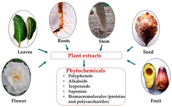
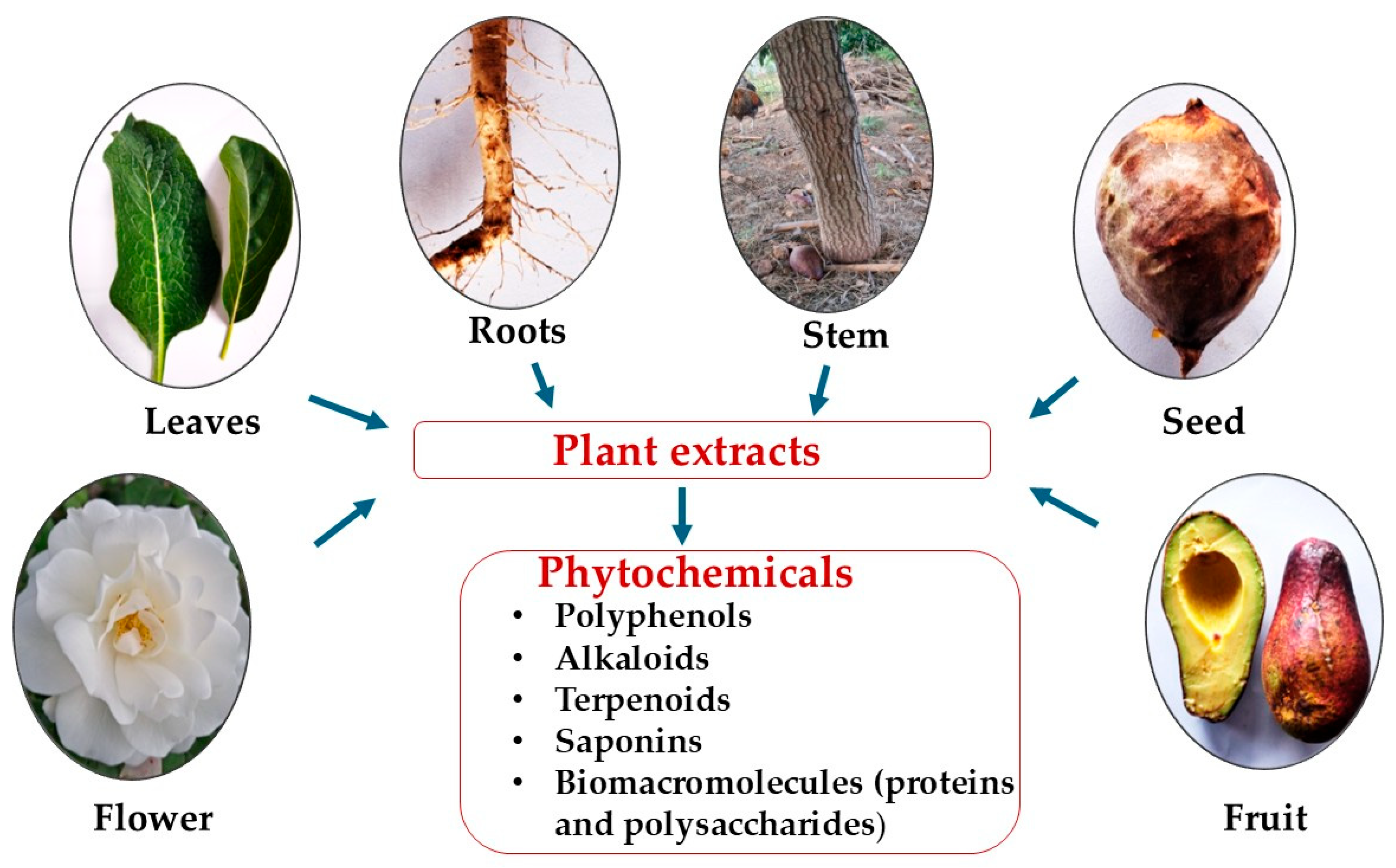
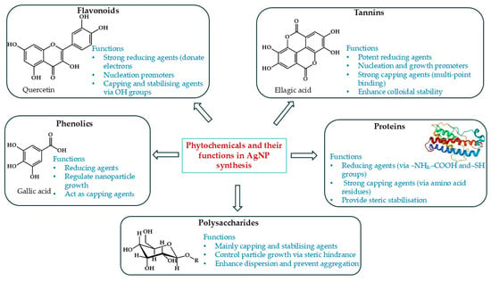
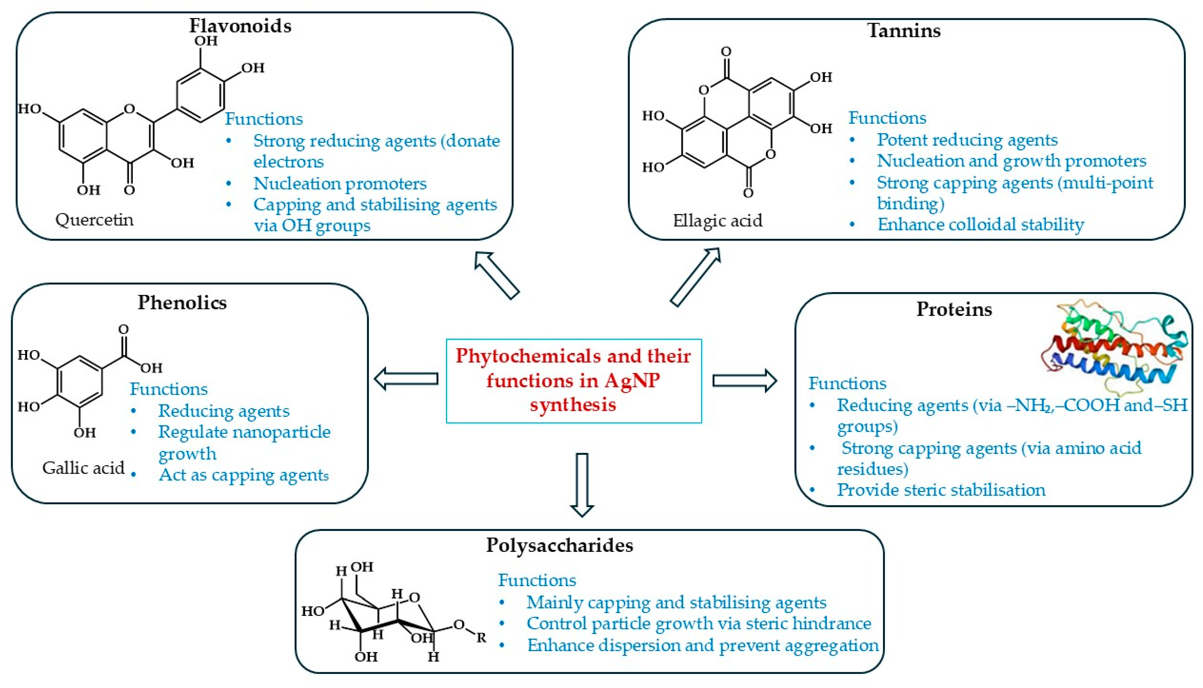
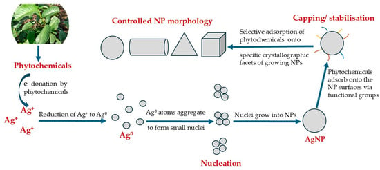
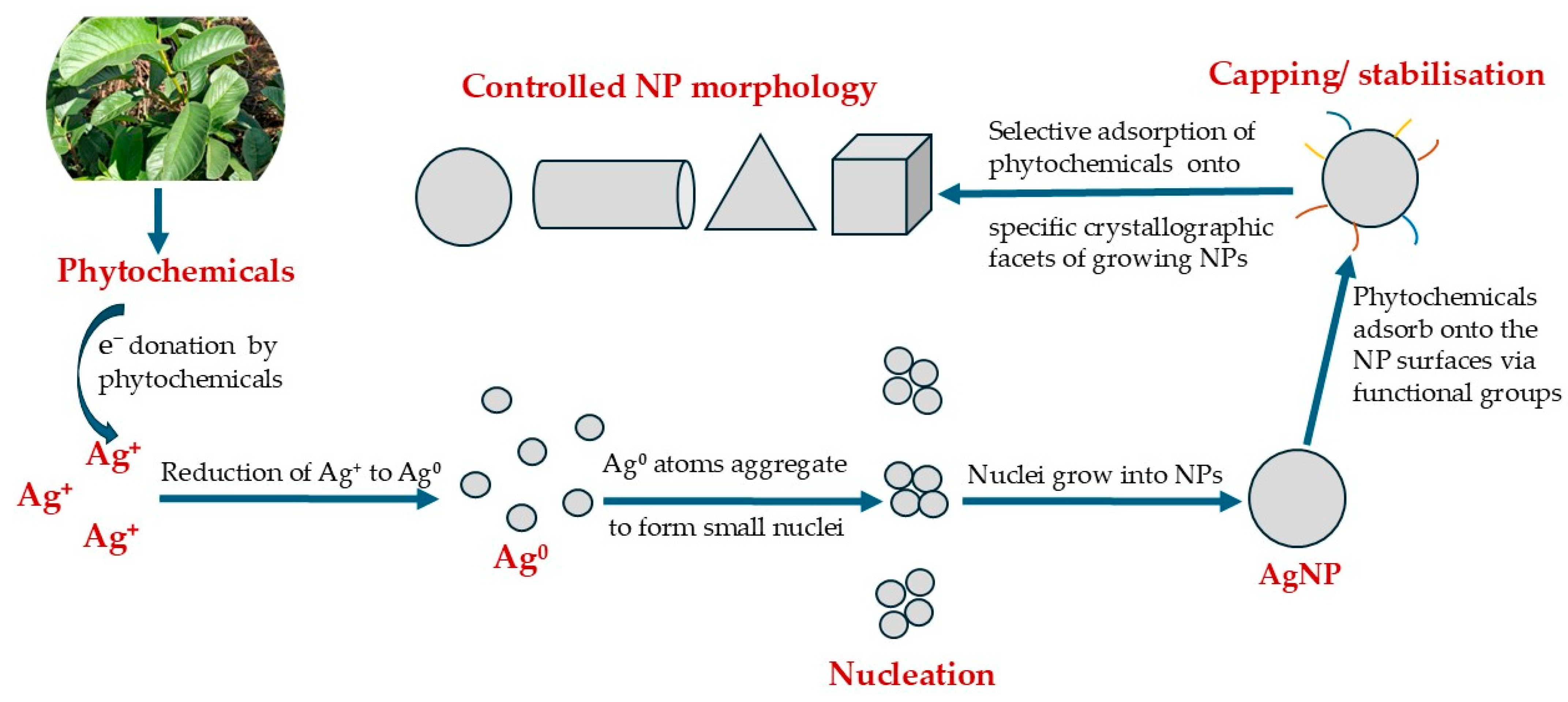
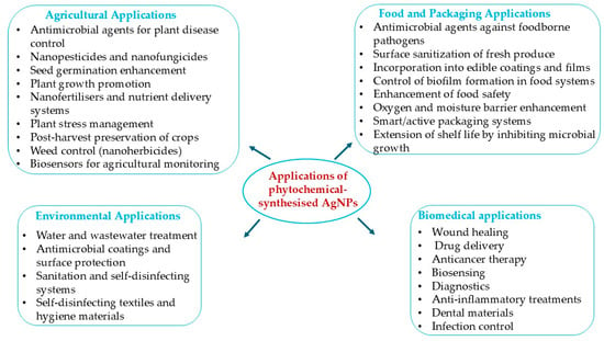
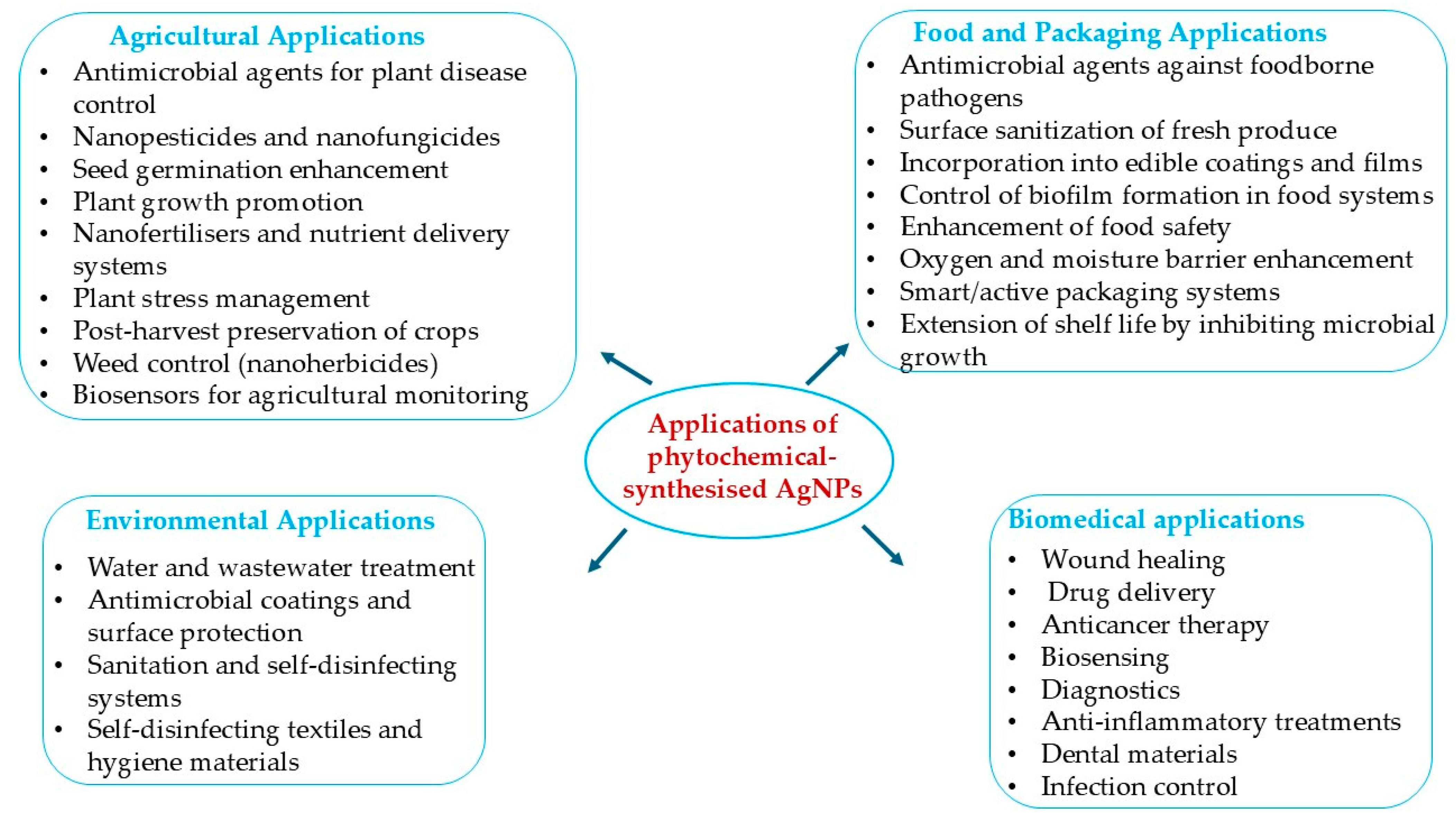
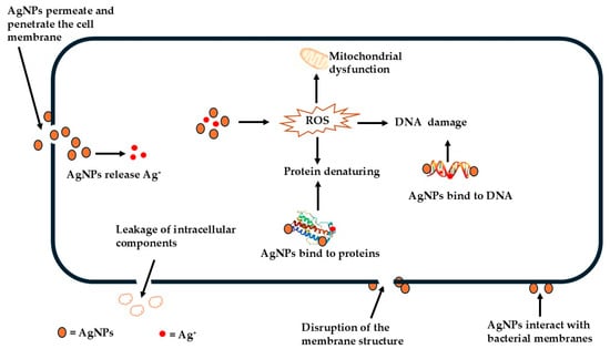
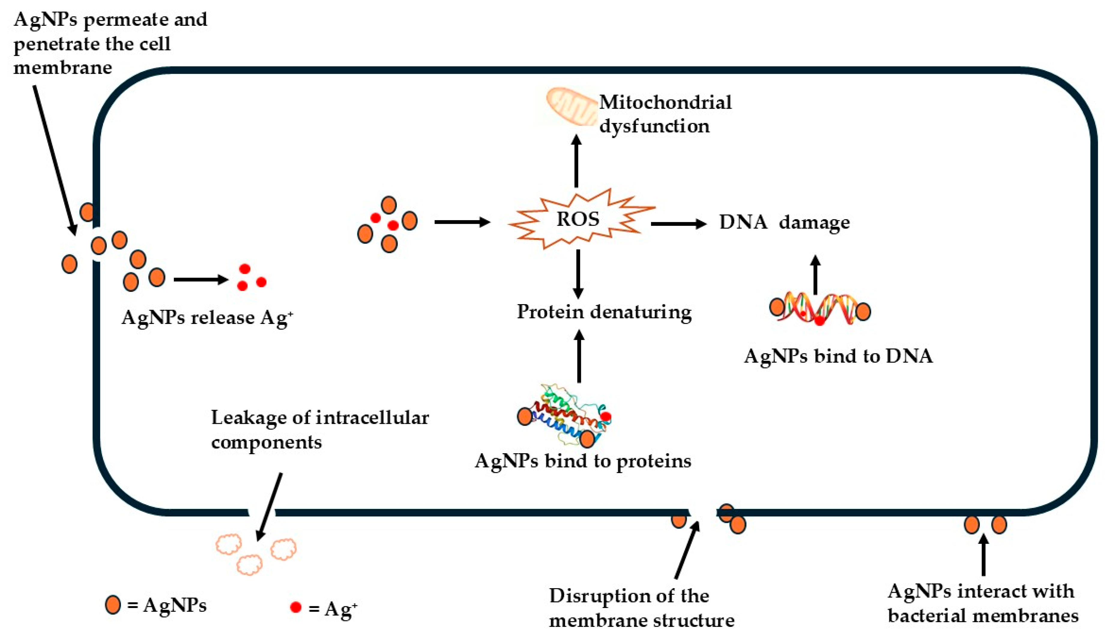

Disclaimer/Publisher’s Note: The statements, opinions and data contained in all publications are solely those of the individual author(s) and contributor(s) and not of MDPI and/or the editor(s). MDPI and/or the editor(s) disclaim responsibility for any injury to people or property resulting from any ideas, methods, instructions or products referred to in the content. |
© 2026 by the author. Licensee MDPI, Basel, Switzerland. This article is an open access article distributed under the terms and conditions of the Creative Commons Attribution (CC BY) license.