


Abstract
The environmental persistence of β-lactam antibiotics represents a growing ecological concern, requiring materials capable of combined adsorption and catalytic degradation. Herein, pure ZnS and 1% Fe-doped ZnS nanoparticles were synthesized via microwave-assisted treatment and evaluated for the removal of ceftaroline fosamil from aqueous media. Transmission electron microscopy revealed quasi-spherical nanoparticles below 10 nm, while selected area electron diffraction confirmed a face-centered cubic structure retained after Fe incorporation. UV-Vis spectroscopy showed similar absorption edges (~316 nm), indicating negligible band-gap variation, whereas photoluminescence analysis demonstrated strong emission quenching in Fe-ZnS, indicating suppressed electron–hole recombination. Point-of-zero charge measurements (pHPZC ≈ 4.6 for ZnS; 4.5 for Fe-ZnS) indicated negatively charged surfaces under circumneutral conditions, influencing interfacial interactions with the antibiotic. Adsorption experiments followed the Langmuir isotherm model, with Fe-ZnS exhibiting a higher maximum adsorption capacity (156 mg g−1) compared to ZnS (115 mg g−1). Under UV irradiation (302 nm), Fe-ZnS achieved near-complete degradation at a catalyst loading of 500 ppm. Liquid chromatography–mass spectrometry analysis revealed the transformation of ceftaroline fosamil (m/z 685.01) into ceftaroline (m/z 605.05) via phosphate group loss, followed by the formation of intermediate fragments at m/z 492.08 and 308.03, associated with cleavage of the thiadiazol-amine moiety and subsequent opening of the cephalosporin ring. After extended irradiation, these intermediates diminished, and a fragment at m/z 356.01 was detected, suggesting further breakdown through thioether bond cleavage. These results support a degradation pathway involving sequential dephosphorylation and fragmentation of the cephalosporin core. Overall, the enhanced performance of Fe-ZnS arises from the synergistic interplay between surface charge characteristics and dopant-modulated charge carrier dynamics, highlighting its potential for antibiotic remediation in aquatic environments.
1. Introduction
Ceftaroline fosamil is a fifth-generation cephalosporin antibiotic approved exclusively for human use and clinically employed in the treatment of complicated skin and soft tissue infections (cSSTIs) and community-acquired bacterial pneumonia (CABP), including infections caused by methicillin-resistant Staphylococcus aureus (MRSA) [1]. Chemically, ceftaroline fosamil is a phosphono prodrug that is rapidly converted in vivo to its active metabolite, ceftaroline, which contains the characteristic β-lactam ring responsible for antibacterial activity through inhibition of bacterial cell wall synthesis [2]. Structural modifications in its side (R) group enhance binding affinity to penicillin-binding proteins, particularly PBP2a, conferring activity against resistant Gram-positive pathogens and distinguishing it from earlier-generation cephalosporins [3]. From a pharmacokinetic perspective, ceftaroline fosamil is administered intravenously and undergoes partial metabolism, with a significant fraction of the active drug excreted unchanged primarily via the renal route [4]. As with many β-lactam antibiotics, incomplete metabolism results in the release of pharmacologically active residues into wastewater systems through hospital effluents and domestic sewage [5]. Conventional wastewater treatment plants are often inefficient at completely removing cephalosporins, allowing these compounds to persist and be transported into surface waters [6]. The continuous discharge and widespread clinical use of antibiotics have led to their recognition as emerging contaminants, raising concerns due to their persistence, ecological toxicity, and contribution to antimicrobial resistance in aquatic environments [7].
The continuous presence of antibiotics such as ceftaroline fosamil in aquatic environments can exert significant adverse ecological and microbiological effects, even at low concentrations. Antibiotics released into surface waters can disrupt native microbial communities, altering essential ecosystem processes such as nutrient cycling and organic matter degradation, while simultaneously exerting selective pressure that promotes the emergence and spread of antibiotic-resistant bacteria and resistance genes (ARGs) [8]. Environmental exposure to β-lactam antibiotics has been shown to facilitate horizontal gene transfer among aquatic microorganisms, creating reservoirs of resistance that may re-enter clinical settings [9]. In addition to microbial effects, chronic antibiotic contamination has been associated with toxic and sublethal effects on aquatic organisms, including oxidative stress, genotoxicity, altered enzyme activity, and impaired growth and reproduction in fish and invertebrates [10]. Antibiotics can also disturb algal and cyanobacterial populations, leading to reduced primary productivity and imbalances in aquatic food webs [11]. Furthermore, the persistence of antibiotics in sediments and water columns allows long-term exposure and potential bioaccumulation, amplifying ecological risks across trophic levels [12]. Collectively, these impacts highlight the importance of effectively removing antibiotics such as ceftaroline fosamil from aquatic systems to protect ecosystem integrity and to mitigate the environmental spread of antimicrobial resistance.
A wide variety of remediation technologies have been developed and evaluated for removing pharmaceutical and antibiotic contaminants from aquatic systems, particularly in wastewater treatment plants and hospital effluents. Among these approaches, adsorption-based processes are among the most extensively studied and implemented due to their operational simplicity and high removal efficiencies for a broad spectrum of pharmaceutical compounds. Activated carbon has been widely used for the adsorption of antibiotics due to its high surface area, porous structure, and strong physicochemical interactions with organic micropollutants [13]. Beyond conventional activated carbon, alternative adsorbent materials such as biochar, carbon nanotubes, graphene-derived materials, modified clays, zeolites, and polymeric resins have gained increasing attention, demonstrating significant adsorption capacities for antibiotics and other emerging contaminants under laboratory and pilot-scale conditions [14,15]. In parallel, membrane-based technologies, including nanofiltration and reverse osmosis, have been implemented to remove pharmaceutical residues via size-exclusion and electrostatic interactions, often achieving high rejection rates for antibiotics in treated effluents [16]. Despite their effectiveness in reducing contaminant concentrations in water, a fundamental limitation of both adsorption and membrane separation processes is that they do not eliminate the contaminant but rather transfer and concentrate it into a solid phase, generating secondary waste that requires further treatment, regeneration, or disposal [17]. As a result, the parent pharmaceutical compound often remains chemically intact, retaining its biological activity and potential environmental risk. This limitation has motivated the exploration of advanced oxidation processes (AOPs), which aim to chemically degrade pharmaceutical contaminants by generating highly reactive species, such as hydroxyl radicals, capable of breaking down complex organic molecules [18]. Among the various AOPs investigated, including ozonation, Fenton and photo-Fenton reactions, and UV/H2O2 systems, heterogeneous photocatalysis has emerged as a particularly promising and sustainable strategy for pharmaceutical remediation [19]. Photocatalytic processes employ semiconductor nanomaterials, such as ZnS, and modified nanocomposites, which, upon light activation, generate reactive oxygen species that can attack and cleave the molecular structure of antibiotics, leading to their transformation into less toxic intermediates or complete mineralization to CO2 and H2O [20]. Importantly, photocatalysis offers the advantage of combining adsorption and degradation mechanisms, allowing contaminants to be initially concentrated on the catalyst surface and subsequently destroyed, thereby overcoming the key limitation of conventional adsorption-based treatments. Consequently, photocatalytic nanomaterials represent a highly attractive remediation approach for pharmaceutical pollutants, as they not only reduce contaminant concentrations in aquatic systems but also minimize long-term persistence, ecological toxicity, and the potential spread of antibiotic resistance associated with residual bioactive compounds.
Selecting a suitable nanomaterial is a key factor in developing an effective photocatalytic system for the degradation of pharmaceutical contaminants in aquatic environments. An ideal photocatalyst must satisfy several fundamental criteria, including small size, rapid electron transport, large surface area, the ability to absorb electromagnetic radiation, and the ability to generate electron–hole pairs upon photoexcitation, while suppressing their recombination, which is one of the primary limitations affecting photocatalytic efficiency [21]. In addition, the material should exhibit chemical and structural stability under irradiation and aqueous conditions, ensuring the preservation of its photocatalytic properties during operation. Semiconductor nanomaterials have been widely investigated for photocatalytic applications due to their tunable electronic structures and high surface-area-to-volume ratios, which promote light absorption and surface redox reactions [22]. Among these materials, zinc sulfide (ZnS) has attracted attention as a potential photocatalyst due to its suitable band structure and its ability to generate electron–hole pairs upon exposure to radiation with energy equal to or greater than its band gap [23]. However, as with many wide-band-gap semiconductors, pure ZnS may suffer from rapid recombination of photogenerated charge carriers, limiting the number of electrons and holes available for photocatalytic reactions [24]. To mitigate this, doping with transition metals has been proposed as a rational strategy to modify ZnS’s electronic properties. The incorporation of metal ions such as iron into the ZnS lattice can introduce defect states or intermediate energy levels within the band gap, which may act as charge carrier traps and thereby reduce electron–hole recombination rates while facilitating charge separation [24,25]. Moreover, transition-metal doping can influence the light-absorption characteristics of ZnS and enhance its interaction with incident radiation, without compromising the host semiconductor’s structural integrity [26]. Based on these physicochemical considerations, both pure and iron-doped ZnS nanoparticles meet the essential requirements for an efficient photocatalytic material, including light absorption, electron–hole pair generation, improved charge-carrier dynamics, and stability in aqueous systems. Given the limitations of conventional remediation techniques, which primarily remove contaminants through concentration rather than destruction, the present study proposes sorption and photocatalysis as alternative approaches to eliminate pharmaceutical pollutants at the molecular level. Accordingly, the objectives of this study were: (a) to synthesize and characterize pure ZnS nanoparticles and iron-doped ZnS nanoparticles, and (b) to evaluate their sorption and photocatalytic performance in the elimination of ceftaroline fosamil in aqueous media.
2. Materials and Methods
2.1. Synthesis of Pure and Doped ZnS Nanoparticles
Zinc sulfate heptahydrate (ZnSO4·7H2O, ≥99.0%, Sigma-Aldrich, St. Louis, MO, USA) and sodium sulfide (Na2S, 3% w/w, Ricca Chemical Company, Arlington, TX, USA) were used as the zinc and sulfur precursors, respectively. Iron doping was achieved using iron (II) sulfate heptahydrate (FeSO4·7H2O, ≥99.0%, Sigma-Aldrich, St. Louis, MO, USA). Deionized water (18.2 MΩ·cm) was used in all experimental procedures. Ceftaroline fosamil acetate hydrate (≥98%, Sigma-Aldrich, St. Louis, MO, USA) was employed as the target contaminant in the sorption and photocatalytic degradation experiments.
2.2. Synthesis of Pure and Doped ZnS Nanoparticles
ZnS nanoparticles were prepared via a hydrothermal method assisted by microwave irradiation, which provides rapid, uniform heating throughout the reaction mixture, promoting uniform nucleation and well-controlled crystal growth. In the synthesis, 5.0 mL of a 0.4 M zinc sulfate heptahydrate solution was mixed with 2.5 mL of a 3% (w/w) sodium sulfide solution under continuous stirring to achieve a homogeneous precursor mixture. The mixture was then transferred to a Teflon-lined vessel, which was tightly sealed and placed in a microwave digestion system (MARS 6, CEM Corporation, Stallings, NC, USA). The reaction was conducted at 140 °C for 15 min using a microwave frequency of 2.45 GHz. Once the heating cycle was complete, the vessel was allowed to return to room temperature naturally. For iron-doped ZnS nanoparticles, 1.0 mL of a 0.15 M iron (II) sulfate heptahydrate solution was added to the zinc-sulfur mixture before microwave treatment, achieving an approximate iron content of 1% relative to zinc. All other conditions, including temperature, irradiation time, and stirring, were kept identical to those used in the synthesis of pure ZnS to maintain consistency between samples. After the reaction, the nanoparticles were isolated by centrifugation at 5000 rpm for 15 min, washed repeatedly with 2-propanol to remove residual reactants, and finally redispersed in deionized water. The purified nanoparticle suspensions were stored for further characterization and use in sorption and photocatalytic studies.
2.3. Characterization Techniques
The structural and morphological features of the synthesized nanoparticles were examined using high-resolution transmission electron microscopy (HRTEM) with a JEM-ARM200cF microscope (JEOL, Tokyo, Japan) operated at 200 kV. Particle size distribution and morphology were determined from the acquired micrographs. Elemental composition and iron incorporation were confirmed by energy-dispersive X-ray spectroscopy (EDS) coupled to the TEM system. Crystallographic properties were studied by electron and X-ray diffraction to confirm the crystalline phase of the ZnS nanostructures. Optical properties were evaluated by UV–visible absorption and photoluminescence spectroscopies, providing information on the electronic structure and light-absorption behavior of both pure and iron-doped ZnS nanoparticles.
2.4. Determination of the Point-of-Zero Charge (pHPZC)
The point-of-zero charge (pHPZC) of pure ZnS and 1% Fe-doped ZnS (Fe-ZnS) was determined using the pH drift (batch equilibration) method [27,28,29,30] in a constant ionic strength medium. A 0.1 M KNO3 solution was used as an inert background electrolyte to minimize specific-ion adsorption and maintain a stable ionic strength throughout the measurements. Aliquots (30 mL) of the electrolyte were transferred into polypropylene centrifuge tubes, and the initial pH (pH0) of each solution was adjusted between 4.0 and 12.0 using standardized 0.1 M HCl or 0.1 M NaOH solutions. Nanoparticles were then added to each tube to obtain a final concentration of 1000 ppm, and the suspensions were homogenized.
The dispersions were agitated at 20 ± 2 °C for 48 h on a rotamix at 20–25 rpm to ensure equilibrium between surface functional groups and the surrounding solution. After equilibration, the suspensions were centrifuged at 5000 rpm for 15 min to separate the solids, and the final pH (pHf) of the supernatant was measured. The change in pH was calculated as ΔpH = pH0 − pHf, and ΔpH values were plotted as a function of pH0. The pHPZC was defined as the pH at which ΔpH = 0 (the intersection with the abscissa), indicating zero net surface charge and no preferential proton exchange.
2.5. Adsorption Experiments with Ceftaroline Fosamil
Stock solutions of ceftaroline fosamil were prepared by dissolving analytical-grade ceftaroline fosamil in deionized water, and working solutions in the 20–140 ppm range were obtained by dilution. Batch adsorption experiments were conducted in 50 mL polypropylene centrifuge tubes containing 5 mL of each ceftaroline solution and ZnS-based nanoparticles at a final concentration of 100 ppm. Both pure ZnS and 1% Fe-doped ZnS nanoparticles were evaluated under identical conditions.
The mixtures were agitated at 350 rpm at 20 ± 2 °C for 3 h. This contact time was selected based on preliminary equilibration tests to ensure that the adsorption equilibrium was reached. After agitation, the suspensions were centrifuged, and the supernatants were filtered through 0.22 μm polyethersulfone (PES) membrane filters (Millex®, Cork, Ireland) to remove residual nanoparticles and obtain particle-free solutions [31].
The equilibrium concentration of ceftaroline in each filtrate was quantified by high-performance liquid chromatography (HPLC) using a Shimadzu LC-2030 system (Shimadzu, Columbia, MD, USA) equipped with a reversed-phase Agilent C8 column (150 mm × 4.6 mm). The mobile phase consisted of 85% aqueous 0.1% formic acid and 15% acetonitrile, delivered at a flow rate of 0.8 mL min−1 at 35 °C. Detection was performed by UV absorbance at 260 nm with an injection volume of 40 μL. External calibration curves were constructed in the 20–140 ppm range and exhibited excellent linearity (R2 > 0.99).
The equilibrium adsorption capacity, Qe (mg g−1), was calculated according to:
where C0 and Ce (ppm) are the initial and equilibrium ceftaroline fosamil concentrations, V is the solution volume (L), and m is the adsorbent mass (g).
Qe = [(C0 − Ce) × V]/m
2.6. Photocatalytic Degradation of Ceftaroline Fosamil
The photocatalytic performance of pure and iron-doped ZnS nanoparticles was evaluated through the degradation of ceftaroline fosamil in aqueous media under UV irradiation. A stock solution of ceftaroline fosamil was prepared in deionized water and used for further experiments. Photocatalytic experiments were carried out by dispersing the ZnS-based photocatalysts into the antibiotic solution at concentrations of 250 ppm and 500 ppm. Prior to irradiation, the suspensions containing 55 ppm of ceftaroline fosamil were magnetically stirred to ensure uniform dispersion and effective sorption of the target compound onto the nanoparticles for 3 h. Two control experiments (55 ppm of antibiotic) were conducted in parallel: a dark control, maintained under identical conditions without light exposure, and a photolysis control, exposed to UV light in the absence of nanoparticles. UV irradiation was provided by a 302 nm ultraviolet lamp with an incident intensity of approximately 10 mW·cm−2. All experiments were performed at ambient temperature under continuous agitation (20–25 rpm) to minimize particle sedimentation. At predetermined time intervals (0, 5, 10, 20, 30, 60, and up to 240 min), aliquots were withdrawn from the reaction mixture and immediately filtered to remove suspended nanoparticles prior to analysis. The residual concentration of ceftaroline fosamil was quantified using high-performance liquid chromatography (HPLC). Chromatographic analysis was performed using a reversed-phase C8 column under isocratic conditions. The mobile phase consisted of 85% aqueous 0.1% formic acid and 15% acetonitrile, delivered at a flow rate of 0.8 mL min−1 at a temperature of 35 °C. Calibration curves were constructed using standard solutions prepared over an appropriate concentration range to ensure accurate quantification. The sorption of ceftaroline fosamil was estimated, and the photocatalytic degradation was calculated by comparing the remaining concentration of ceftaroline fosamil at each irradiation time with the initial concentration. All photocatalytic experiments were conducted in triplicate to ensure reproducibility and statistical reliability of the results.
3. Results
3.1. Morphological and Structural Characterization
Transmission electron microscopy (TEM) was used to evaluate the morphology and size distribution of ZnS and Fe-ZnS nanoparticles synthesized via microwave-assisted hydrothermal treatment at 140 °C for 15 min. The rapid, homogeneous volumetric heating provided by microwave irradiation promotes rapid supersaturation and burst nucleation, thereby limiting prolonged crystal growth. This behavior is consistent with the nanoscale dimensions observed in both materials.
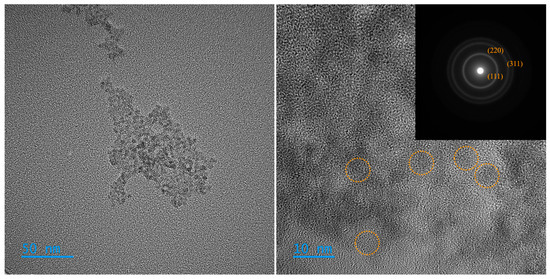
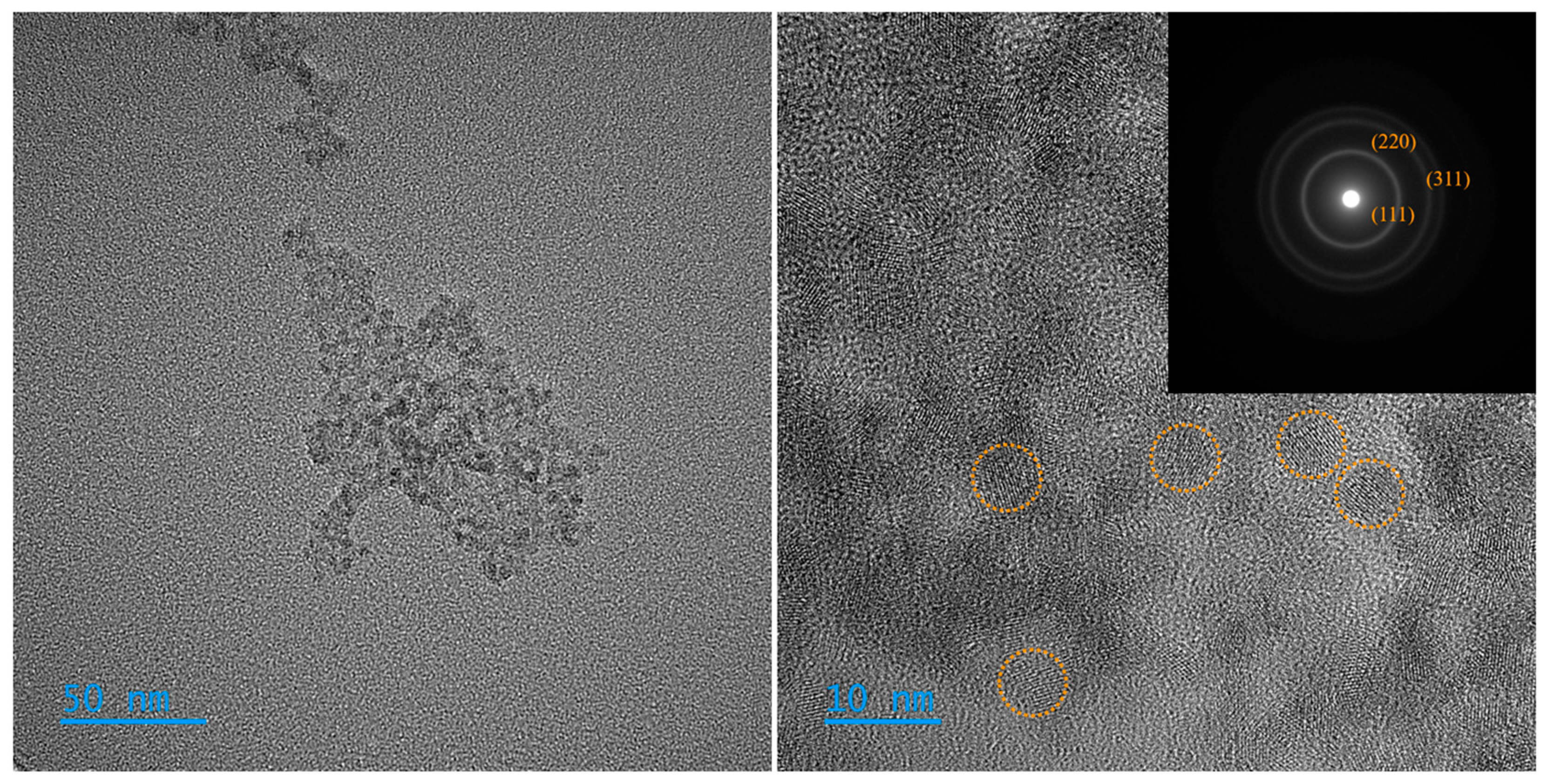
For pure ZnS (Figure 1), the low-magnification image (50 nm scale) reveals the formation of loosely packed aggregates composed of smaller primary particles. Such aggregation is expected due to the high surface energy of ultrasmall nanocrystals and interparticle interactions during post-synthesis processing and solvent evaporation. At higher magnification (10 nm scale), individual nanocrystals become distinguishable, exhibiting predominantly quasi-spherical morphology and sizes generally below 10 nm. The relatively uniform contrast and the absence of large, faceted domains suggest controlled nucleation and short growth times, consistent with the microwave-assisted synthesis conditions. The apparent narrow size distribution indicates that the selected parameters effectively balanced nucleation and growth, preventing excessive coarsening.
Figure 1.
TEM images of ZnS at 50 nm (left) and 10 nm (right). Dashed circles mark representative crystalline nanoparticles. The corresponding SAED pattern is shown as an inset (right).
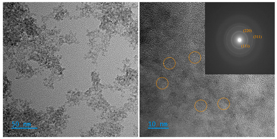
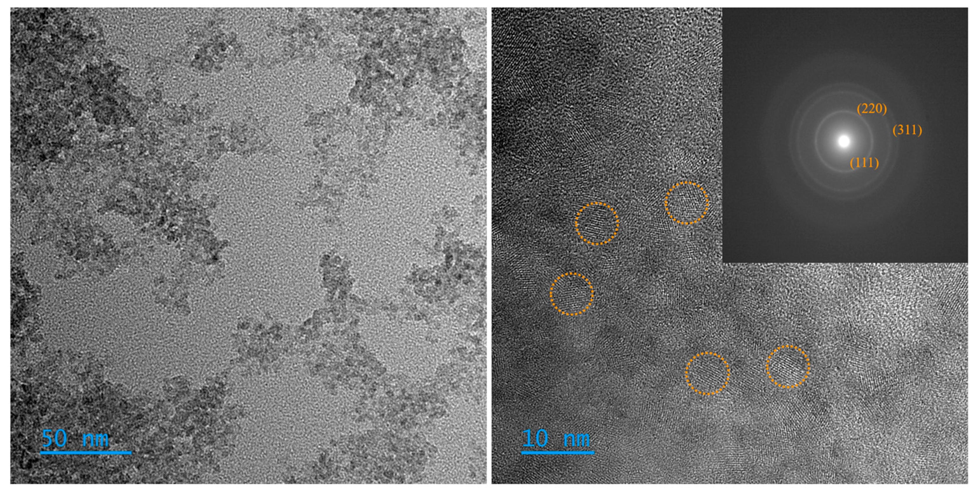
The Fe-ZnS sample (Figure 2), synthesized under identical conditions with approximately 1% Fe2+ relative to Zn2+, exhibits a morphology comparable to that of the ZnS sample, confirming that low-level iron incorporation does not significantly disrupt the dominant nucleation mechanism. At the 50 nm scale, the aggregates appear slightly denser and more compact than those of pure ZnS, which may be associated with subtle changes in surface chemistry or interparticle interactions induced by Fe incorporation. High-magnification images again show nanosized, nearly spherical particles with dimensions predominantly below 10 nm; however, the particle boundaries appear slightly less defined in certain regions, and the size distribution seems marginally broader. These features may reflect lattice distortion or defect formation associated with the substitution of Zn2+ by Fe2+ within the ZnS crystal lattice, introducing local strain and modifying growth kinetics during irradiation.
Figure 2.
TEM images of Fe-ZnS at 50 nm (left) and 10 nm (right). Dashed circles mark representative crystalline nanoparticles. The corresponding SAED pattern is shown as an inset (right).
Overall, both materials consist of sub-10 nm nanocrystals assembled into secondary aggregates, demonstrating that microwave-assisted hydrothermal synthesis enables rapid production of uniformly small ZnS-based nanoparticles. The structural similarity between ZnS and Fe-ZnS indicates that low-concentration iron doping preserves the nanoscale morphology while potentially introducing subtle structural disorder. Such features are particularly relevant for photocatalytic applications, where small particle size enhances the surface-to-volume ratio, and defect-related states introduced by Fe may improve charge separation and surface reactivity.
The selected area electron diffraction (SAED) patterns further confirm the crystalline nature and structural phase of both nanomaterials. The diffraction images display distinct concentric rings that can be indexed to the (111), (220), and (311) planes, which are characteristic of the face-centered cubic (fcc) zinc blende structure of ZnS. The presence of these reflections in both samples indicates that microwave-assisted hydrothermal synthesis promotes the formation of the cubic phase under the selected reaction conditions.
For ZnS nanoparticles (Figure 1-right), the diffraction rings are relatively sharp and well-defined, evidencing a high degree of crystallinity despite the small particle size observed by TEM. In the Fe-ZnS sample (Figure 2-right), the same set of crystallographic planes is preserved, confirming that the incorporation of approximately 1% Fe2+ does not alter the fundamental crystal symmetry or generate detectable secondary phases. A slight broadening of the diffraction rings is observed in the doped material, which can be reasonably attributed to lattice distortion and increased microstrain associated with partial substitution of Zn2+ by Fe2+ within the host lattice.
These results demonstrate that iron incorporation occurs within the ZnS framework rather than forming segregated iron-containing phases. The retention of the fcc structure, combined with subtle dopant-induced structural perturbations, is consistent with a substitutional-doping mechanism and supports the material’s structural integrity for subsequent photocatalytic applications.
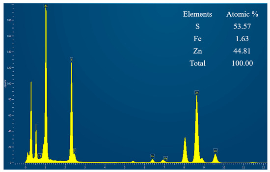
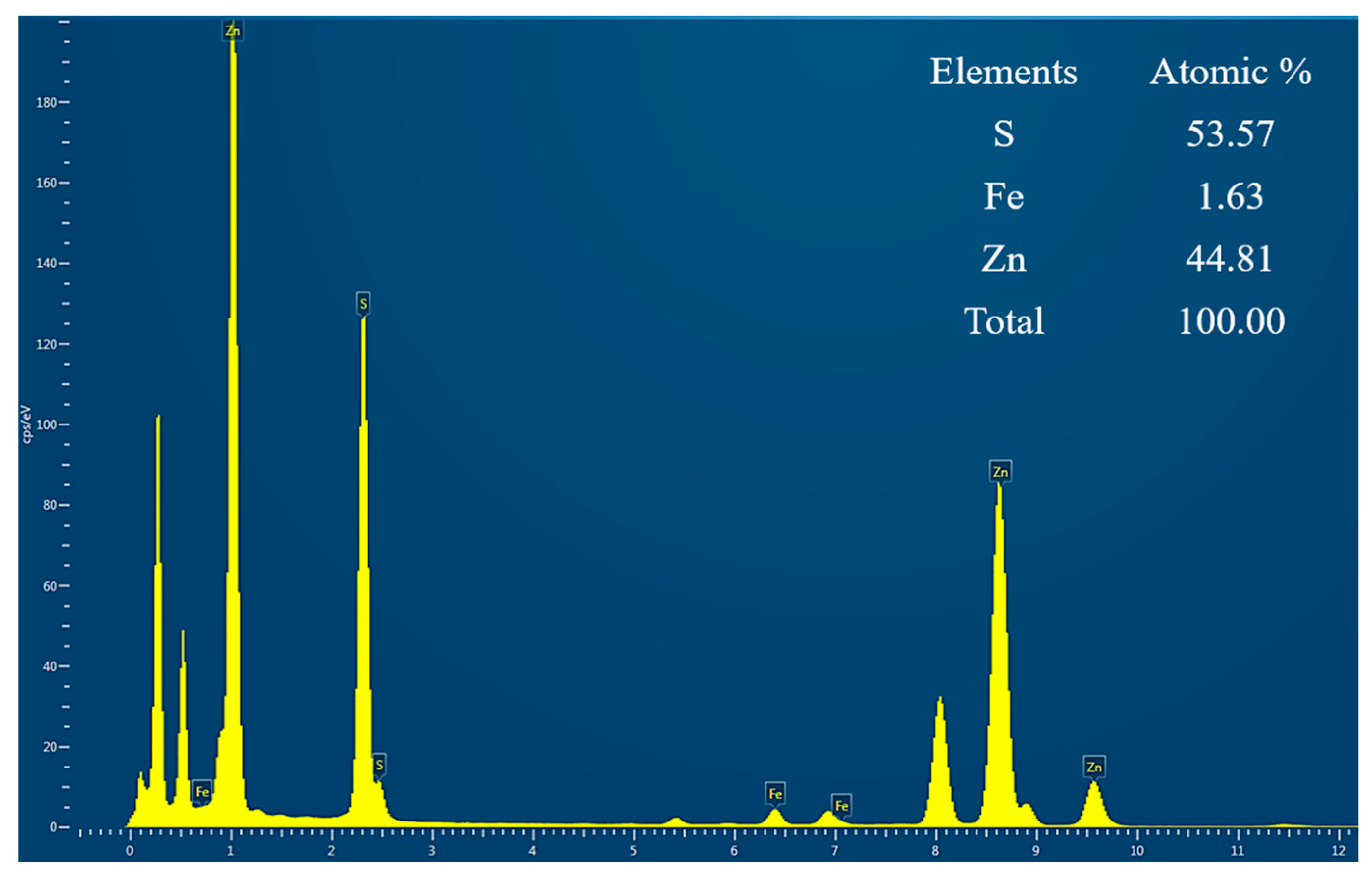
The elemental composition of the Fe-doped ZnS nanoparticles was evaluated by EDS analysis (Figure 3). The results reveal atomic percentages of 53.57% S, 44.81% Zn, and 1.63% Fe, confirming the successful incorporation of iron into the ZnS matrix. The relatively low Fe content is consistent with the intended dopant concentration and suggests that iron is present as a substitutional or interstitial species rather than forming secondary phases. Additionally, the Zn:S ratio remains close to the expected stoichiometry, indicating that the introduction of Fe does not significantly disrupt the host lattice composition.
Figure 3.
EDS spectrum and elemental composition (atomic %) of Fe-ZnS.
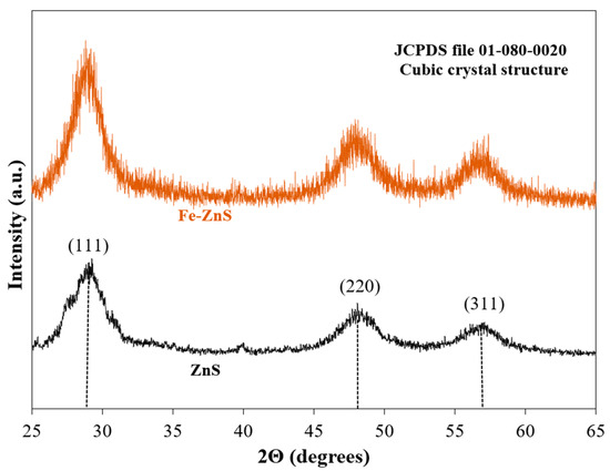
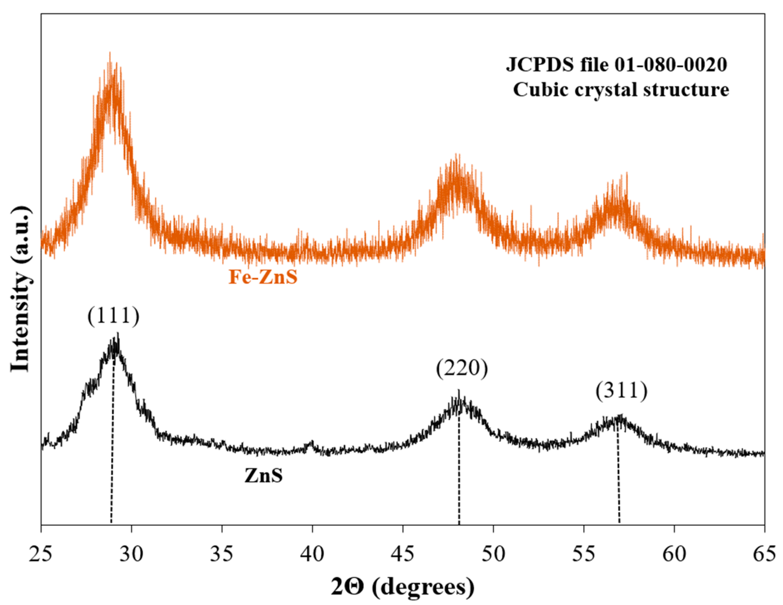
X-ray diffraction (XRD) analysis was further performed to evaluate the crystalline structure and phase purity of the synthesized materials (Figure 4). Both ZnS and Fe-ZnS samples exhibit diffraction peaks centered at approximately 2θ ≈ 28.5°, 47.5°, and 56.3°, corresponding to the (111), (220), and (311) crystallographic planes, respectively, which are consistent with the cubic zinc blende structure (JCPDS file 01-080-0020). The broad nature of the diffraction peaks is indicative of the nanoscale crystallite size, in agreement with the TEM observations. Importantly, no additional peaks associated with secondary iron-containing phases were detected, suggesting that Fe is successfully incorporated into the ZnS lattice rather than forming separate phases.
Figure 4.
XRD pattern for ZnS and Fe-ZnS nanoparticles.
3.2. Optical Properties
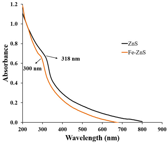
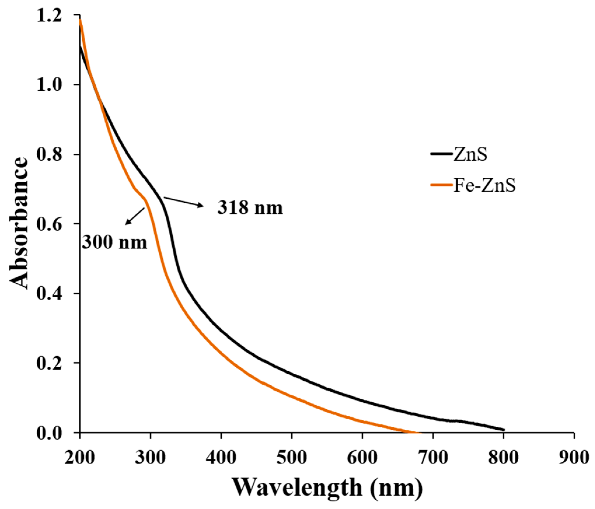
The optical properties of ZnS and 1% Fe-ZnS nanoparticles (Figure 5) were analyzed using UV-Vis absorption spectroscopy, and the optical band gap energies were estimated from Tauc plots derived from the absorption data. Considering ZnS as a direct band gap semiconductor, the band gap values were determined by extrapolating the linear region of versus plots to the energy axis. The calculated band gap for pure ZnS was approximately 3.9 eV, consistent with nanoscale cubic ZnS, while Fe-ZnS exhibited a slightly higher estimated band gap of ~4.1 eV. This modest blue shift corresponds to the absorption feature observed near 300 nm for Fe–ZnS, compared to ~318 nm for undoped ZnS.
Figure 5.
Absorbance spectra of ZnS and Fe-ZnS nanoparticles.
Importantly, no additional absorption bands were detected in the visible region, indicating that Fe incorporation at the 1% level does not introduce optically dominant mid-gap states or secondary phases. The preservation of a well-defined absorption edge suggests that the global electronic structure of ZnS remains largely intact, with Fe substitution inducing only localized perturbations rather than band-gap narrowing.
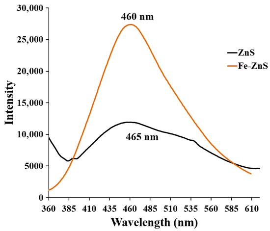
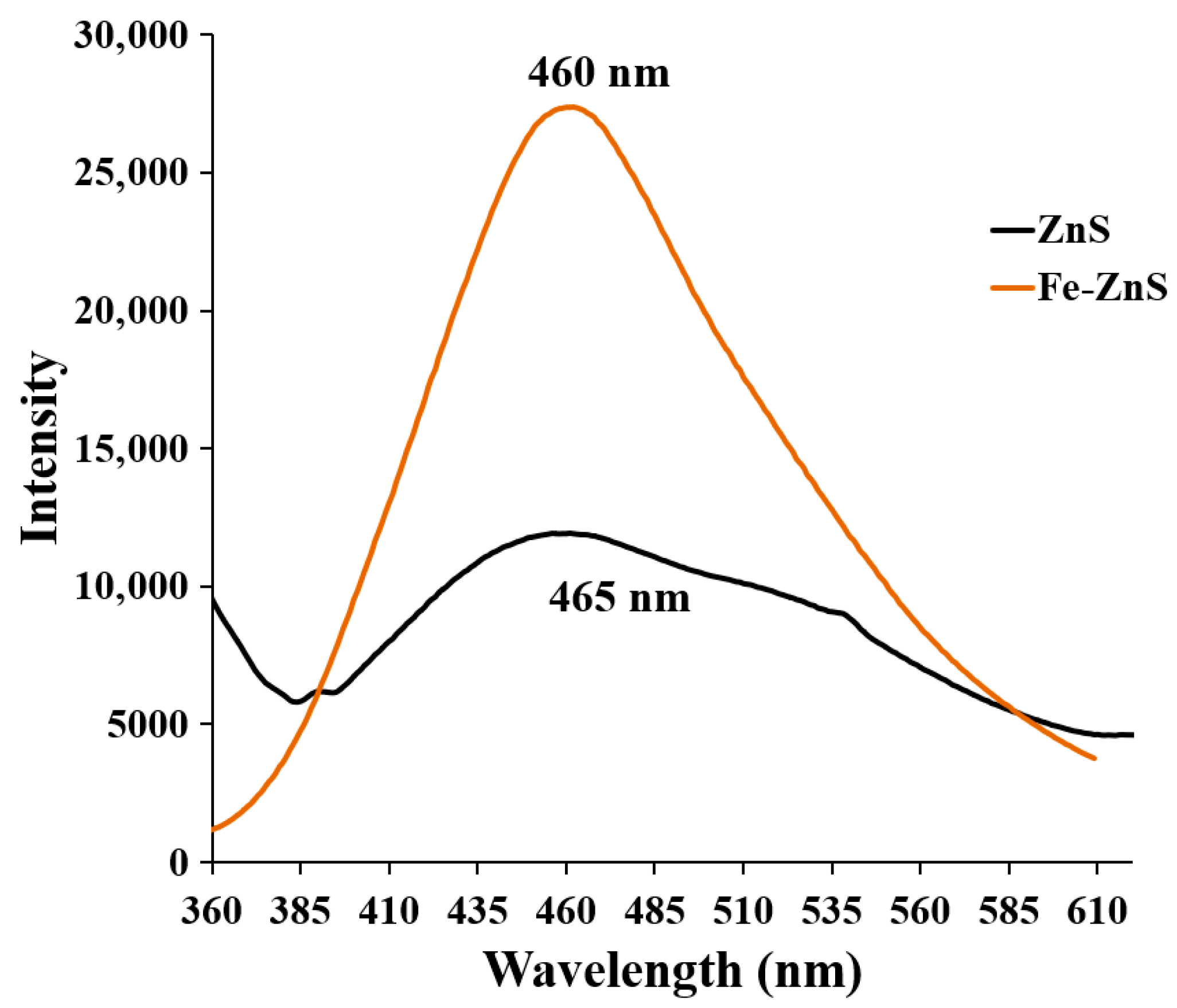
Photoluminescence spectra (Figure 6) further reveal modifications in electronic relaxation pathways. Pure ZnS displays a broad defect-related emission centered at ~465 nm, typically associated with intrinsic vacancies and surface states. In contrast, Fe-ZnS exhibits a slightly shifted emission maximum near ~460 nm, with increased intensity, indicating dopant-induced alterations in the defect landscape and radiative recombination channels. Together, these results demonstrate that low-level Fe incorporation perturbs local electronic states while maintaining the intrinsic direct band gap character of ZnS.
Figure 6.
Photoluminescence spectra of ZnS and Fe-ZnS nanoparticles.
3.3. Point-of-Zero Charge Studies
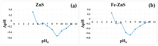
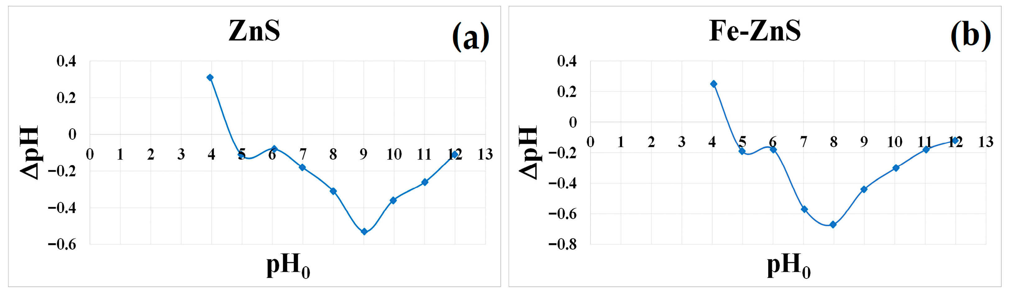
The surface charge properties of pure ZnS and 1% Fe-doped ZnS (Fe-ZnS) were evaluated using the pH drift method to determine the point-of-zero charge (pHPZC), as shown in Figure 7. For all samples, ΔpH (pH0 − pHf) exhibited a clear transition from positive to negative values across the studied pH range, confirming equilibrium proton exchange between surface functional groups and the surrounding electrolyte. The pHPZC values were obtained from the intersection of the ΔpH versus pH0 plots with ΔpH = 0.
Figure 7.
ΔpH vs. pH0 plots for the determination of the point-of-zero charge of (a) ZnS and (b) Fe-ZnS nanoparticles.
Pure ZnS and Fe-ZnS displayed relatively low pHPZC values of 4.6 and 4.5, respectively, indicating that both materials possess predominantly negative surface charge under near-neutral conditions due to deprotonation of amphoteric ≡Zn–OH and ≡Fe–OH groups. The minimal shift in pHPZC upon Fe incorporation suggests that doping does not significantly alter the surface acid–base properties of ZnS, and that surface charge behavior remains largely governed by hydroxyl functional groups.
These results are particularly relevant for antibiotic removal, as surface charge directly influences adsorption affinity [32] and interfacial contact between the nanoparticles and ceftaroline species. At circumneutral pH, both materials are expected to exhibit electrostatic interactions that may affect adsorption performance depending on the speciation of the antibiotic. Materials with pHPZC values below the working pH are anticipated to experience electrostatic repulsion with negatively charged species, potentially limiting adsorption unless other interactions, such as surface complexation or hydrophobic effects, contribute.
Although Fe incorporation produced only a minor change in pHPZC, it is expected to influence removal performance by modifying the semiconductor’s electronic structure. Fe dopants can improve the utilization of photogenerated charge carriers and facilitate interfacial redox reactions, thereby enhancing photocatalytic degradation once the pollutant is adsorbed on the surface [33,34]. Thus, while surface charge governs interfacial interactions and adsorption behavior, Fe doping provides complementary improvements in photochemical efficiency. Together, these findings highlight the importance of considering both surface chemistry and electronic properties when optimizing ZnS-based nanomaterials for antibiotic removal.
3.4. Adsorption Analyses
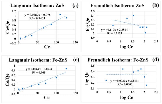
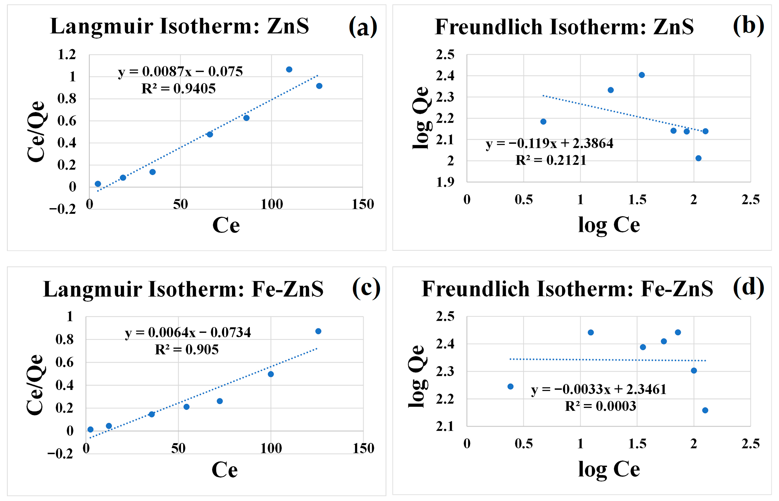
The adsorption behavior of ceftaroline fosamil onto pure ZnS and 1% Fe-doped ZnS nanoparticles was investigated under identical equilibrium conditions to evaluate the influence of Fe incorporation on interfacial affinity and antibiotic removal. HPLC calibration exhibited excellent linearity (R2 = 0.9955), ensuring accurate determination of residual concentrations for both systems. In both materials, the adsorption capacity increased with increasing initial concentration and progressively approached saturation at higher concentrations, indicating occupation of a finite number of surface-active sites. The equilibrium data were fitted using Langmuir and Freundlich isotherm models [35,36] to elucidate the adsorption mechanism, as shown in Figure 8.
Figure 8.
Langmuir and Freundlich isotherms of ZnS (a,b) and Fe-ZnS (c,d) nanoparticles.
For pure ZnS, the Langmuir model showed strong agreement with the experimental data (R2 = 0.9405), while the Freundlich fit was significantly poorer (R2 = 0.2121). The calculated maximum adsorption capacity, qmax, was 115 mg g−1, indicating monolayer adsorption on relatively homogeneous sites. Similarly, the Fe-ZnS sample was better described by the Langmuir model (R2 = 0.905) than by the Freundlich model (R2 = 0.0003), yielding a higher maximum capacity (qmax ≈ 156 mg g−1). The predominance of Langmuir behavior in both materials indicates that adsorption is mainly surface-controlled, monolayer interactions rather than multilayer or highly heterogeneous processes.
Notably, Fe-ZnS exhibited approximately 36% higher adsorption capacity than pure ZnS, demonstrating that Fe incorporation enhances the nanoparticles’ affinity for ceftaroline. This improvement can be attributed to the introduction of additional ≡Fe–OH surface functionalities, which provide supplementary coordination and hydrogen-bonding sites capable of interacting with the β-lactam, amide, and carboxylate groups of the antibiotic [37]. These interactions likely promote stronger surface complexation and increase the density of accessible binding sites, facilitating greater accumulation of the pollutant at the solid–liquid interface.
From a removal perspective, this enhanced adsorption is advantageous because it concentrates ceftaroline molecules near the catalyst surface, increasing the probability of subsequent degradation reactions. Thus, adsorption serves as a preconcentration step, enhancing interfacial contact between the pollutant and reactive sites. When combined with the improved photochemical response associated with Fe incorporation discussed above, the higher surface uptake is expected to accelerate overall degradation kinetics. Together, these results indicate that Fe-ZnS benefits from complementary contributions of stronger adsorption and enhanced catalytic activity, resulting in more efficient antibiotic removal than pure ZnS.
3.5. Photodegradation Experiments
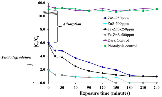
Figure 9 summarizes the photocatalytic degradation of ceftaroline fosamil under UV irradiation (302 nm) in the presence of ZnS and Fe-ZnS nanoparticles at catalyst loadings of 250 and 500 ppm. All assays were performed with an initial antibiotic concentration of 55 ppm. Prior to irradiation, the suspensions were equilibrated in the dark for 3 h to allow adsorption–desorption equilibrium. As a result, the concentrations recorded at irradiation time zero for the catalytic systems are below 55 ppm, reflecting initial adsorption onto nanoparticle surfaces rather than photochemical transformation. Dark and photolysis controls (n = 3 each) remained stable over the 240 min experimental window, confirming that neither UV exposure alone nor adsorption in the absence of irradiation contributed to measurable degradation.
Figure 9.
Photodegradation of ceftaroline fosamil in the presence of ZnS and Fe-ZnS nanoparticles.
At a catalyst loading of 250 ppm, both materials promote progressive degradation; however, Fe-ZnS consistently exhibits faster removal kinetics than undoped ZnS. The enhanced activity of the doped system suggests that Fe incorporation modifies the electronic structure of ZnS in a manner that favors charge carrier separation and suppresses electron–hole recombination. This effect is reflected in the steeper decline in antibiotic concentration during the first 60–120 min of irradiation. While ZnS at 250 ppm shows sustained degradation over time, Fe-ZnS achieves lower residual concentrations within the same period, indicating a clear kinetic advantage.
Increasing the catalyst loading to 500 ppm significantly improves performance for both systems, highlighting the role of available active surface area and photon absorption capacity. Under these conditions, Fe-ZnS reaches near-complete degradation within approximately 150–180 min, whereas pure ZnS, although markedly more efficient than at 250 ppm, remains comparatively slower. The superior performance of Fe-ZnS at higher loadings is attributed to a synergistic effect of increased surface-active sites and dopant-induced electronic effects. The results indicate sustained photocatalytic activity over the 240 min irradiation period, as evidenced by the continuous decrease in ceftaroline fosamil concentration; however, definitive assessment of catalyst stability would require additional post-reaction characterization.
Taken together, the data demonstrate that both catalyst concentration and iron incorporation critically influence photocatalytic efficiency. The stability of the control experiments confirms that the observed removal is due to a true photocatalytic mechanism. The improved performance of Fe-ZnS is consistent with a substitutional-doping model, in which iron introduces localized electronic states that facilitate interfacial redox processes, thereby enhancing the degradation of ceftaroline fosamil in aqueous media.
It is important to note that the photodegradation profiles exhibit slight non-monotonic behavior at intermediate irradiation times. These fluctuations can be attributed to the dynamic nature of heterogeneous photocatalytic systems, where adsorption, surface reaction, and desorption processes occur simultaneously. During irradiation, partially degraded intermediates or weakly adsorbed species may temporarily desorb into the solution, leading to minor increases or plateaus in the measured concentration. Additionally, small variations may arise from sampling and analytical uncertainty, particularly at low concentration levels. Despite these fluctuations, the overall trend remains clearly decreasing, confirming the effective photocatalytic degradation of ceftaroline fosamil over time.
Although the observed photocatalytic activity remained stable throughout the experimental period, a more comprehensive evaluation of catalyst stability and reusability is necessary to assess long-term performance. In heterogeneous photocatalytic systems, potential deactivation pathways may include surface fouling by intermediate species, structural changes under irradiation, or partial dopant leaching, all of which can affect catalytic efficiency over repeated use cycles [38]. Future studies should therefore incorporate recycling experiments combined with post-reaction characterization techniques, such as XRD, TEM, and surface analysis, to determine the structural and functional durability of the materials.
In addition, the performance of ZnS-based photocatalysts may be influenced by the complexity of real environmental matrices. Natural water systems typically contain dissolved organic matter, inorganic ions (e.g., Cl−, SO42−, Ca2+), and variable pH conditions, which can alter adsorption behavior, compete for reactive sites, and affect the generation or scavenging of reactive oxygen species. These factors may ultimately impact photocatalytic efficiency compared to idealized laboratory conditions. Therefore, evaluating the materials under environmentally relevant conditions will be essential to establish their practical applicability in real water treatment scenarios.
Despite these considerations, the results presented here demonstrate the strong intrinsic potential of Fe-ZnS nanoparticles as efficient photocatalysts for antibiotic removal, providing a robust foundation for future investigations focused on material stability, reusability, and performance under realistic environmental conditions.
3.6. Mass Spectrometric Studies
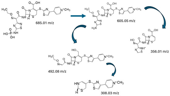
The LC–MS analysis was carried out under the same HPLC conditions used for the photodegradation experiments. The standard solution revealed the presence of ceftaroline fosamil at a retention time of 7.2 min with an m/z value of 685.01. After 3 h of adsorption in the presence of the nanoparticles, ceftaroline fosamil was largely adsorbed onto the NP surface, while the remaining compound was converted into ceftaroline (m/z = 605.05) through the loss of the phosphate group. Additionally, a secondary metabolite with an m/z of 492.08 was detected, which can be attributed to the cleavage of the thiadiazol-amine fragment. The removal of the phosphate group decreases the polarity of ceftaroline, resulting in a shift in the chromatographic peak to a longer retention time (9.4 min). The fragment with m/z 492.08 was observed at a retention time of 8.5 min.
During the first 30 min of irradiation, the intensity of the peaks at 9.4 and 8.5 min gradually decreased, while a new minor peak appeared at 7.9 min with an m/z of 308.03. This species is likely generated from further degradation of the 492 m/z intermediate through cleavage of the cephalosporin ring. After 180 min of irradiation, the signals corresponding to the previous intermediates became almost imperceptible, and a new peak emerged at a retention time of 8.24 min with an m/z of 356.01. This product is likely formed directly from ceftaroline (m/z = 605.05) through cleavage of the thioether bond (C3–S), accompanied by the release of the 2-(thiazolyl)-4-(N-methylpyridinium)-thio fragment. The proposed degradation pathway is illustrated in Figure 10.
Figure 10.
Proposed photodegradation pathway of ceftaroline fosamil.
4. Conclusions
The results demonstrate that both ZnS and Fe-doped ZnS nanoparticles synthesized via microwave-assisted treatment are effective materials for the removal of ceftaroline fosamil from aqueous systems, combining adsorption and photocatalytic degradation mechanisms. Structural and optical characterization confirmed the formation of nanoscale ZnS with a preserved cubic crystal structure after Fe incorporation, while photoluminescence quenching in Fe-ZnS indicated improved charge carrier separation, which contributes to enhanced photocatalytic performance. Surface charge analysis revealed negatively charged nanoparticle surfaces under circumneutral conditions, favoring interfacial interactions with the antibiotic and promoting adsorption consistent with the Langmuir isotherm model, with Fe-ZnS exhibiting a higher adsorption capacity than pure ZnS. Under UV irradiation, the Fe-doped material achieved near-complete degradation, demonstrating that Fe incorporation improves catalytic efficiency without significantly altering the band gap. LC–MS analysis provided insight into the degradation mechanism, revealing the sequential transformation of ceftaroline fosamil into ceftaroline through phosphate group loss, followed by fragmentation of the thiadiazol-amine moiety, opening of the cephalosporin ring, and further cleavage of the thioether bond, leading to smaller intermediates that progressively diminished during irradiation. These findings indicate that Fe doping enhances both the adsorption affinity and photocatalytic activity of ZnS nanoparticles, making Fe-ZnS a promising nanomaterial for the removal and transformation of β-lactam antibiotics in contaminated water systems.
Author Contributions
Conceptualization, S.J.B.-R.; funding acquisition, S.J.B.-R. and Y.C.-M.; methodology, S.J.B.-R., N.C.-D., Y.C.-M., and L.A.-N.; Writing—original draft, S.J.B.-R., N.C.-D., Y.C.-M., and L.A.-N.; writing—review and editing, S.J.B.-R. All authors have read and agreed to the published version of the manuscript.
Funding
This research was supported by the National Science Foundation (NSF) under Award No. 2313252.
Institutional Review Board Statement
Not applicable.
Informed Consent Statement
Not applicable.
Data Availability Statement
The Data presented in this study are available on request from the corresponding author.
Acknowledgments
This research was conducted at the Laboratory of Investigation in Nanotechnology and Characterization (LINC), part of the Department of Chemistry and Physics at UPRP. We acknowledge the support of the National High Magnetic Field Laboratory, supported by the National Science Foundation Cooperative Agreement No. DMR-2128556 and the State of Florida.
Conflicts of Interest
The authors declare that they have no conflicts of interest. The funders had no role in the design of the study, in the collection, analysis, or interpretation of data, in the writing of the manuscript, or in the decision to publish the results.
References
- Alomair, S.M.; Al Sulaiman, K.; Khayyat, T.; Wali, H.; Bosaeed, M. Successful treatment of methicillin-resistant Staphylococcus aureus osteomyelitis and persistent bacteremia using adjunctive ceftaroline fosamil to vancomycin: A case report. IDCases 2025, 42, e02437. [Google Scholar] [CrossRef] [Scilit] [PubMed]
- Corey, G.R.; Wilcox, M.H.; Gonzalez, J.; Jandourek, A.; Wilson, D.J.; Friedland, H.D.; Das, S.; Iaconis, J.; Dryden, M. Ceftaroline fosamil therapy in patients with acute bacterial skin and skin-structure infections with systemic inflammatory signs: A retrospective dose comparison across three pivotal trials. Int. J. Antimicrob. Agents 2019, 53, 830–837. [Google Scholar] [CrossRef] [Scilit]
- Duplessis, C.; Crum-Cianflone, N.F. Ceftaroline: A New Cephalosporin with Activity Against Methicillin-Resistant Staphylococcus aureus (MRSA). Clin. Med. Rev. Ther. 2011, 3, 1–17. [Google Scholar] [CrossRef] [Scilit] [PubMed]
- Kiang, T.K.L.; Wilby, K.J.; Ensom, M.H.H. A Critical Review on the Clinical Pharmacokinetics, Pharmacodynamics, and Clinical Trials of Ceftaroline. Clin. Pharmacokinet. 2015, 54, 915–931. [Google Scholar] [CrossRef] [Scilit]
- Akhter, S.; Bhat, M.A.; Ahmed, S.; Siddiqui, W.A. Antibiotic residue contamination in the aquatic environment, sources and associated potential health risks. Environ. Geochem. Health 2024, 46, 387. [Google Scholar] [CrossRef] [Scilit]
- Alam, M.-U.; Ferdous, S.; Ercumen, A.; Lin, A.; Kamal, A.; Luies, S.K.; Sharior, F.; Khan, R.; Rahman, Z.; Parvez, S.M.; et al. Effective Treatment Strategies for the Removal of Antibiotic-Resistant Bacteria, Antibiotic-Resistance Genes, and Antibiotic Residues in the Effluent From Wastewater Treatment Plants Receiving Municipal, Hospital, and Domestic Wastewater: Protocol for a Systematic Review. JMIR Res. Protoc. 2021, 10, e33365. [Google Scholar] [CrossRef] [Scilit]
- Carvalho, I.T.; Santos, L. Antibiotics in the aquatic environments: A review of the European scenario. Environ. Int. 2016, 94, 736–757. [Google Scholar] [CrossRef] [Scilit]
- Kraemer, S.A.; Ramachandran, A.; Perron, G.G. Antibiotic Pollution in the Environment: From Microbial Ecology to Public Policy. Microorganisms 2019, 7, 180. [Google Scholar] [CrossRef] [Scilit]
- Martinez, J.L. Environmental pollution by antibiotics and by antibiotic resistance determinants. Environ. Pollut. 2009, 157, 2893–2902. [Google Scholar] [CrossRef] [Scilit]
- Li, Z.; Lu, T.; Li, M.; Mortimer, M.; Guo, L.-H. Direct and gut microbiota-mediated toxicities of environmental antibiotics to fish and aquatic invertebrates. Chemosphere 2023, 329, 138692. [Google Scholar] [CrossRef] [Scilit] [PubMed]
- González-Pleiter, M.; Gonzalo, S.; Rodea-Palomares, I.; Leganés, F.; Rosal, R.; Boltes, K.; Marco, E.; Fernández-Piñas, F. Toxicity of five antibiotics and their mixtures towards photosynthetic aquatic organisms: Implications for environmental risk assessment. Water Res. 2013, 47, 2050–2064. [Google Scholar] [CrossRef] [Scilit] [PubMed]
- Belle, G.; Moodley, B.; Moodley, R.; Omotola, E.O.; Truter, C.; Olatunji, O.; Oberholster, P. Occurrence and detection of selected pharmaceuticals of emerging concern: Potential risks for aquatic ecosystems and human health. Discov. Appl. Sci. 2025, 7, 1078. [Google Scholar] [CrossRef] [Scilit]
- Ajiboye, T.O.; Oladoye, P.O.; Omotola, E.O. Adsorptive reclamation of pharmaceuticals from wastewater using carbon-based materials: A review. Kuwait J. Sci. 2024, 51, 100225. [Google Scholar] [CrossRef] [Scilit]
- Peng, B.; Chen, L.; Que, C.; Yang, K.; Deng, F.; Deng, X.; Shi, G.; Xu, G.; Wu, M. Adsorption of Antibiotics on Graphene and Biochar in Aqueous Solutions Induced by π-π Interactions. Sci. Rep. 2016, 6, 31920. [Google Scholar] [CrossRef] [Scilit]
- Ma, R.; Xue, Y.; Ma, Q.; Chen, Y.; Yuan, S.; Fan, J. Recent Advances in Carbon-Based Materials for Adsorptive and Photocatalytic Antibiotic Removal. Nanomaterials 2022, 12, 4045. [Google Scholar] [CrossRef] [Scilit]
- Yang, L.; Xia, C.; Jiang, J.; Chen, X.; Zhou, Y.; Yuan, C.; Bai, L.; Meng, S.; Cao, G. Removal of antibiotics and estrogens by nanofiltration and reverse osmosis membranes. J. Hazard. Mater. 2023, 461, 132628. [Google Scholar] [CrossRef] [Scilit] [PubMed]
- Aldana, J.C.; Agudelo, C.; Álvarez, P.M.; Acero, J.L. Removal of Micropollutants in Water Reclamation by Membrane Filtration: Impact of Pretreatments and Adsorption. Membranes 2024, 14, 146. [Google Scholar] [CrossRef] [Scilit] [PubMed]
- Wang, J.; Zhuan, R. Degradation of antibiotics by advanced oxidation processes: An overview. Sci. Total. Environ. 2019, 701, 135023. [Google Scholar] [CrossRef] [Scilit]
- Friedmann, D. A General Overview of Heterogeneous Photocatalysis as a Remediation Technology for Wastewaters Containing Pharmaceutical Compounds. Water 2022, 14, 3588. [Google Scholar] [CrossRef] [Scilit]
- Kutuzova, A.; Dontsova, T.; Kwapinski, W. Application of TiO2-Based Photocatalysts to Antibiotics Degradation: Cases of Sulfamethoxazole, Trimethoprim and Ciprofloxacin. Catalysts 2021, 11, 728. [Google Scholar] [CrossRef] [Scilit]
- Mahmoud, M.A.; Alsehli, B.R.; Alotaibi, M.T.; Hosni, M.; Shahat, A. A comprehensive review on the application of semiconducting materials in the degradation of effluents and water splitting. Environ. Sci. Pollut. Res. 2023, 31, 3466–3494. [Google Scholar] [CrossRef] [Scilit]
- Chong, M.N.; Jin, B.; Chow, C.W.K.; Saint, C. Recent developments in photocatalytic water treatment technology: A review. Water Res. 2010, 44, 2997–3027. [Google Scholar] [CrossRef] [Scilit]
- Chen, X.; Zhao, C.; Wu, H.; Shi, Y.; Chen, C.; Zhou, X. Two-Dimensional ZnS/SnS2 Heterojunction as a Direct Z-Scheme Photocatalyst for Overall Water Splitting: A DFT Study. Materials 2022, 15, 3786. [Google Scholar] [CrossRef] [Scilit]
- Munawar, T.; Fatima, S.; Batoo, K.M.; Nadeem, M.S.; Mukhtar, F.; Hussain, S.; Khan, S.A.; Koc, M.; Iqbal, F. Enhanced charge carriers separation/transportation via S-scheme ZnCeS–ZnWO heterostructure nanocomposite for photodegradation of synthetic dyes under sunlight. Mater. Chem. Phys. 2024, 314, 128938. [Google Scholar] [CrossRef] [Scilit]
- Geldasa, F.T.; Kebede, M.A.; Shura, M.W.; Hone, F.G. Experimental and computational study of metal oxide nanoparticles for the photocatalytic degradation of organic pollutants: A review. RSC Adv. 2023, 13, 18404–18442. [Google Scholar] [CrossRef] [Scilit]
- Mishra, M.; Kumar, M. Advances in Zinc Oxide nanomaterials and nanocomposites for photocatalytic and biomedical applications. Discov. Mater. 2025, 6, 26. [Google Scholar] [CrossRef] [Scilit]
- Askari, N.; Beheshti, M.; Mowla, D.; Farhadian, M. Synthesis of CuWO4/Bi2S3 Z-Scheme Heterojunction with Enhanced Cephalexin Photodegradation. J. Photochem. Photobiol. A Chem. 2020, 394, 112463. [Google Scholar] [CrossRef] [Scilit]
- Chaukura, N.; Katengeza, G.; Gwenzi, W.; Mbiriri, C.I.; Nkambule, T.T.; Moyo, M.; Kuvarega, A.T. Development and Evaluation of a Low-Cost Ceramic Filter for the Removal of Methyl Orange, Hexavalent Chromium, and Escherichia coli from Water. Mater. Chem. Phys. 2020, 249, 122965. [Google Scholar] [CrossRef] [Scilit]
- Mok, C.F.; Ching, Y.C.; Abu Osman, N.A.; Muhamad, F.; Hai, N.D.; Choo, J.H.; Hassan, C.R. Adsorbents for Removal of Cationic Dye: Nanocellulose Reinforced Biopolymer Composites. J. Polym. Res. 2020, 27, 373. [Google Scholar] [CrossRef] [Scilit]
- Salahuddin, N.A.; El-Daly, H.A.; El Sharkawy, R.G.; Nasr, B.T. Nano-Hybrid Based on Polypyrrole/Chitosan/Grapheneoxide Magnetite Decoration for Dual Function in Water Remediation and Its Application to form Fashionable Colored Product. Adv. Powder Technol. 2020, 31, 1587–1596. [Google Scholar] [CrossRef] [Scilit]
- Bailon-Ruiz, S.J.; Cedeño-Mattei, Y.; Alamo-Nole, L. Fe-Doped ZnS Quantum Dot Photocatalysts for the Degradation of Cefalexin in Water. Micro 2025, 5, 31. [Google Scholar] [CrossRef] [Scilit]
- Zyoud, A.; Zyoud, A.H.; Zyoud, S.H.; Nassar, H.; Zyoud, S.H.; Qamhieh, N.; Hajamohideen, A.; Hilal, H.S. Photocatalytic Degradation of Aqueous Methylene Blue Using Ca-Alginate Supported ZnO Nanoparticles: Point of Zero Charge Role in Adsorption and Photodegradation. Environ. Sci. Pollut. Res. 2023, 30, 68435–68449. [Google Scholar] [CrossRef] [Scilit]
- Nguyen, L.T.; Pham, A.T.; Vo, T.T.; Le, K.T.; Vu, N.H.; Van Pham, V. Promoting the Role of Fe Doping in WO3 toward Enhancing H2O2 Oxidation Ability for Contaminant Degradation. J. Water Process. Eng. 2025, 77, 108503. [Google Scholar] [CrossRef] [Scilit]
- Tan, Y.; Cheng, Z.; Liu, Y.; Gao, X.; Liu, S.; Shen, Z. Quantum Parameter Analysis of the Adsorption Mechanism by Freshly Formed Ferric Hydroxide for Synthetic Dye and Antibiotic Wastewaters. Chemosphere 2021, 280, 130577. [Google Scholar] [CrossRef] [Scilit]
- Alafnan, S.; Awotunde, A.; Glatz, G.; Adjei, S.; Alrumaih, I.; Gowida, A. Langmuir Adsorption Isotherm in Unconventional Resources: Applicability and Limitations. J. Pet. Sci. Eng. 2021, 207, 109172. [Google Scholar] [CrossRef] [Scilit]
- Vigdorowitsch, M.; Pchelintsev, A.; Tsygankova, L.; Tanygina, E. Freundlich Isotherm: An Adsorption Model Complete Framework. Appl. Sci. 2021, 11, 8078. [Google Scholar] [CrossRef] [Scilit]
- Zhu, Y.; Yang, Q.; Lu, T.; Qi, W.; Zhang, H.; Wang, M.; Qi, Z.; Chen, W. Effect of Phosphate on the Adsorption of Antibiotics onto Iron Oxide Minerals: Comparison between Tetracycline and Ciprofloxacin. Ecotoxicol. Environ. Saf. 2020, 205, 111345. [Google Scholar] [CrossRef] [Scilit] [PubMed]
- Zheng, Z.; He, J.; Zhang, Z.; Kumar, A.; Khan, M.; Lung, C.W.; Lo, I.M.C. Magnetically recyclable nanophotocatalysts in photocatalysis-involving processes for organic pollutant removal from wastewater: Current status and perspectives. Environ. Sci. Nano 2024, 11, 1784–1816. [Google Scholar] [CrossRef] [Scilit]
Disclaimer/Publisher’s Note: The statements, opinions and data contained in all publications are solely those of the individual author(s) and contributor(s) and not of MDPI and/or the editor(s). MDPI and/or the editor(s) disclaim responsibility for any injury to people or property resulting from any ideas, methods, instructions or products referred to in the content. |
© 2026 by the authors. Licensee MDPI, Basel, Switzerland. This article is an open access article distributed under the terms and conditions of the Creative Commons Attribution (CC BY) license.