


Abstract
Metal oxide nanoparticles dispersed in water are difficult to recover because of their small size and colloidal stability. In this study, the interfacial adsorption behavior of Fe2O3, CoO, and CuO nanoparticles at hydrophobic ionic liquid (IL)–water interfaces was investigated and compared with that at molecular solvent–water interfaces. When CuO nanoparticle dispersions were shaken with hydrophobic ILs, bis(trifluoromethanesulfonyl)imide ([NTf2]−) salts of 1-butyl-3-methylimidazolium ([BMIm]+) and 1-octyl-3-methylimidazolium ([OMIm]+), the nanoparticles were removed from the aqueous phase and accumulated at the IL–water interface, while negligible Cu was detected in the bulk IL phase. The removal efficiency decreased with increasing ionic strength below 0.05 mol/dm3 and increased with pH, indicating that electrostatic interactions between charged nanoparticles and the IL–water interface contribute to adsorption. Adsorption isotherms were empirically fitted with the Langmuir equation to estimate the maximum adsorption capacity. For negatively charged Fe2O3 and CuO nanoparticles, the maximum adsorption capacities at IL–water interfaces exceeded those at molecular solvent–water interfaces and the theoretical monolayer capacity estimated from nanoparticle size, suggesting multilayer adsorption or aggregation at the interfaces. These results demonstrate the potential of hydrophobic IL–water interfaces for the separation and recovery of metal oxide nanoparticles from aqueous media.
1. Introduction
Metal oxide nanoparticles play important roles in various technological fields such as sensors, catalysts, solar cells, medicine, antimicrobial materials, and environmental remediation [1,2]. Their unique physicochemical properties (e.g., high surface area, reactivity, conductivity, etc.) are a major advantage in these applications. However, metal oxide nanoparticles released into the environment may pose unforeseen health risks and adverse ecological effects, which have become a growing concern [3,4]. For example, CuO and ZnO nanoparticles, which are used to inhibit the growth of bacteria, fungi, and algae in aquatic environments, have been reported to be more toxic to non-target aquatic organisms or mammalian cells than to the target bacteria [5]. Due to their microscopic size, nanoparticles are difficult to degrade in nature and may cause long-term accumulation and environmental pollution.
In nature, heavy metals in groundwater, seawater, and river water often exist as colloidal species with particle sizes on the nanometer scale. Metal oxide colloids suspended in environmental waters are known to capture various metal ions [6,7,8] and are considered to play an important role in the migration of radionuclides in natural waters [9,10,11]. Therefore, identification of the total amount and composition of metal oxide nanoparticles in environmental water is an important issue, because artificial nanomaterials diffused in the environment and colloidal species naturally occurring in environmental water have a significant impact on the distribution and diffusion of biological ions and trace metal ions.
Effective separation and removal technologies are therefore required to determine the concentration of metal oxide nanoparticles in environmental waters and to mitigate their ecological impact. However, inorganic colloids such as metal oxide nanoparticles present in environmental waters are typically on the nanometer scale, making such nanoparticles difficult to efficiently recover and separate from dissolved species. In laboratory studies, ultracentrifugation and ultrafiltration have been used to separate nanoparticles dispersed in water. However, these methods require special equipment such as ultracentrifuges and ultrafiltration devices, and are generally difficult to apply on a larger scale. In recent years, the use of specific coagulants [12,13] and adsorbents [14,15] has been studied for the removal of metal oxide nanoparticles.
In recent years, much research has been conducted using ionic liquids (ILs) as a new extraction solvent for metal ions [16,17,18,19]. ILs are salts that generally exist as liquids at ambient pressure below about 100 °C. They are generally nonflammable and nonvolatile owing to their extremely low vapor pressure. They are also attracting attention for their ability to extract precious metals, which are difficult to extract with conventional organic solvents. As ILs consist entirely of ions, they are expected to interact electrostatically with surface-charged nanoparticles and may therefore provide a new separation medium for metal oxide nanoparticles. Nakashima and Kimizuka reported that anionic fluorescent polystyrene nanospheres (about 500 nm in diameter) spontaneously formed a regular monolayer at the interface between a hydrophobic IL, 1-butyl-3-methylimidazolium hexafluorophosphate, and water, and their adsorption was attributed to electrostatic interaction between the IL–water interface and the nanospheres [20]. If such adsorption also occurs for metal oxide nanoparticles dispersed in water, it could be applied to the separation and removal of metal oxide nanoparticles from water without the need for special equipment.
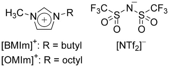
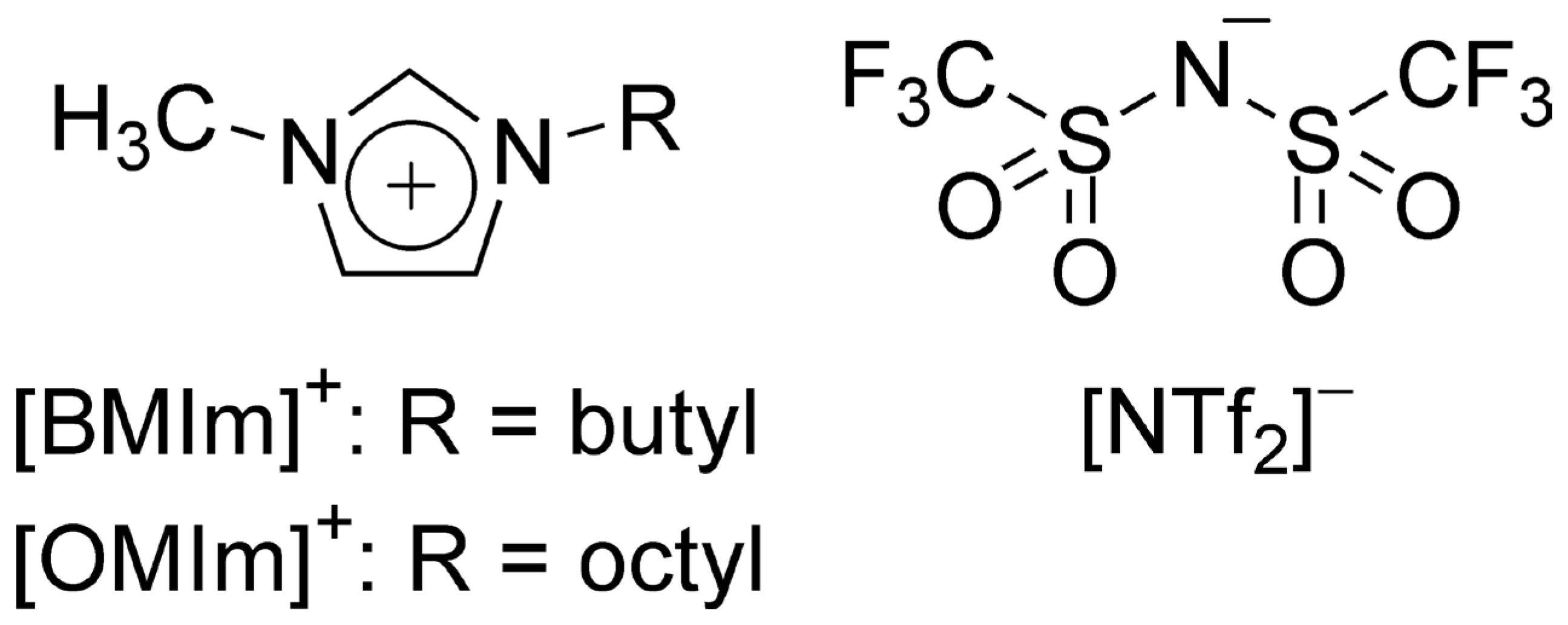
In this study, we investigated the interaction of metal oxide nanoparticles with hydrophobic IL–water interfaces in order to clarify the adsorption behavior of Fe2O3, CoO, and CuO nanoparticles. The ILs used were 1-butyl-3-methylimidazolium bis(trifluoromethanesulfonyl)imide ([BMIm][NTf2]) and 1-octyl-3-methylimidazolium bis(trifluoromethanesulfonyl)imide ([OMIm][NTf2]), which were chosen as representative hydrophobic ILs with the same anion but different alkyl chain lengths, allowing the examination of the effect of cation structure on interfacial adsorption behavior. The structural formulas of the constituent ions of the ILs are shown in Figure 1. For comparison with ILs, the usual hydrophobic organic solvents (molecular solvents), namely benzene, chloroform, hexane, and 1-octanol, were also used.
Figure 1.
Structures of the constituent ions of the ionic liquids used in this study.
2. Materials and Methods
2.1. Reagents and Apparatus
Nanoparticles of Fe2O3 (99.5% purity; average particle size 39 nm, density 5.1 g/cm3), CoO (99.5% purity; average particle size 22 nm, density 5.4 g/cm3), and CuO (99.9% purity; average particle size 48 nm, density 6.3 g/cm3) were purchased from C. I. Kasei Co., Ltd. (Tokyo, Japan). [BMIm][NTf2] and [OMIm][NTf2] were prepared according to the previous reports [21]. Benzene, chloroform, and hexane were purchased from Kanto Chemical Co., Inc. (Tokyo, Japan); benzene and chloroform were washed three times with water; hexane was treated with fuming sulfuric acid, washed three times with water, and distilled. The 1000 mg/dm3 standard solutions of Fe3+, Co2+, and Cu2+ (Kanto Chemical Co., Inc.) were used for atomic absorption spectrometry after being diluted as needed. Water was deionized using a Demiace DX cartridge purification system (Kurita Water Industries Ltd., Tokyo, Japan) and further purified with a Simplicity Water Purification System (Millipore, Billerica, MA, USA). Other reagents were of analytical grade and used without purification.
A Branson table-top ultrasonic cleaner 1510-MT (Emerson Electric Co., St. Louis, MO, USA) was used with transmitting frequency 42 kHz and output 90 W in the preparation of the nanoparticle dispersion water. A table-top centrifuge 2010 and a micro-cooled centrifuge 3700 (Kubota Corp., Tokyo, Japan) were used for centrifugal separation by maximum speed 4000 rpm and 15,000 rpm, respectively. An SR-IN shaking machine (Taitec Co., Koshigaya, Japan) was used for shaking experiments. Hitachi Z-5000 and Z-5200 polarized Zeman atomic absorption spectrophotometers (Hitachi High-Tech Corp., Tokyo, Japan) were used for the graphite furnace atomic absorption analysis (GF-AAS) of metals. The zeta potential and particle size of CuO nanoparticles were measured using an SZ-100 nanoparticle analysis system (Horiba Ltd., Kyoto, Japan) or an ELSZ-1000 zeta potential and particle size measurement system (Otsuka Electronics Co., Ltd., Osaka, Japan).
2.2. Preparation of Metal Oxide Nanoparticle Dispersed Water
Four to 30 mg of metal oxide nanoparticles were placed in a 50-cm3 glass centrifuge tube, and 40 cm3 of water was added. The centrifuge tube was shaken by hand for 1 min, sonicated for 60 min, centrifuged at 3000 rpm (1510× g) for 5 min, and the supernatant was carefully separated with a pipette to make nanoparticle-dispersed water. To quantify the metal oxide contained therein, dilute nitric or hydrochloric acid was added and heated at 60 °C to dissolve all of the metal oxide nanoparticles. The metal concentration in the acid solution was measured by GF-AAS, and the resulting concentration was converted to metal oxide content (mg/dm3). In the experiment to examine the effect of pH, the above metal oxide dispersed water was mixed with the aqueous solutions whose pH was adjusted to 4.7 to 10.3 using aqueous solutions of HCl, 2-morpholinoethanesulfonic acid monohydrate, tris(hydroxymethyl)aminomethane, and NaOH. The ionic strength (I) of the final solutions was adjusted to about 0.01 mol/dm3 by adding NaNO3. In the experiment to study the effect of I, the pH was not adjusted and the final I of the metal oxide nanoparticle-dispersed water was prepared to reach 0.005 to 0.1 mol/dm3 by the addition of NaNO3.
To determine the concentration of dissolved metal in the buffer solution in which the metal oxide was dispersed, the solution in a 50 cm3 centrifuge tube was centrifuged at 15,000 rpm (22,140× g) for 30 min to settle out all nanoparticles dispersed in water; the metal ion concentration in the supernatant solution was measured by GF-AAS. Under the conditions at pH > 6, the amount of metal dissolved was generally negligible compared to the amount of metal oxide dispersed.
When measuring the zeta potential and particle size of nanoparticles in aqueous dispersions, a small amount of hydrophobic ionic liquid was added to the nanoparticle dispersion water with adjusted pH or ionic strength. The amount of ionic liquid added was set so that the ionic liquid concentration in water approached its solubility limit. After thorough mixing, the zeta potential and particle size of the nanoparticles were measured using nanoparticle analysis equipment.
2.3. Adsorption of Metal Oxide Nanoparticles in Nonaqueous Solvent/Water Systems (Typical Conditions)
Six cm3 of metal oxide nanoparticle-dispersed water was placed in a 10 cm3 stoppered glass test tube (15.2 mm i.d.) and 0.6 cm3 of a nonaqueous solvent ([BMIm][NTf2], [OMIm][NTf2], benzene, chloroform, hexane, or 1-octanol) saturated with water was added. The exact volumes of the water-saturated ILs added were calculated from their masses using the densities: 1.4311 g/cm3 [22] for [BMIm][NTf2] and 1.3276 g/cm3 [23] for [OMIm][NTf2]. The test tubes were shaken in a thermostatic chamber at 25 ± 0.2 °C for 60 min, and then left in a thermostatic bath at the same temperature for at least 15 min to separate the two phases, and the aqueous phase was separated. Dilute nitric acid or dilute hydrochloric acid was added to the aqueous phase and heated at 60 °C for 30 min to completely dissolve the metal oxide, and then the metal concentration was measured by GF-AAS. The concentration of metal oxide remaining in the aqueous phase (Cw mg/dm3) was calculated from the metal concentration. The percentage removal (%R) of the metal oxide nanoparticle was calculated from Equation (1):
where Cw° denotes the initial concentration of metal oxide (mg/dm3) in the aqueous phase.
%R = 100(Cw° − Cw)/Cw°,
3. Results and Discussion
3.1. Effect of Shaking Time and IL–Water Interface Area on Interfacial Adsorption of CuO Nanoparticles in [BMIm][NTf2]/Water Biphasic System
First, the effect of the shaking time on %R of CuO from CuO nanoparticle-dispersed water (CuO 47 mg/dm3, 6 cm3) to [BMIm][NTf2] (0.6 cm3) was investigated. No reagent other than CuO nanoparticles was added to the dispersion. Shaking for 15–120 min resulted in constant %R value (mean = 97.7%, standard deviation = 0.7%, n = 4). The settling time after shaking (15–60 min) did not affect the %R value.
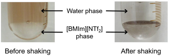
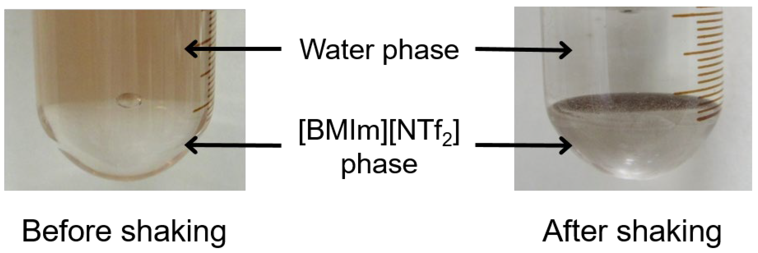
As shown in the photographs in Figure 2, the nanoparticles that were initially dispersed in the aqueous phase appeared to accumulate at the IL–water interface rather than in either bulk phase after shaking. To confirm the presence of CuO in the bulk IL phase, a portion of the IL phase was taken, shaken with 0.1 mol/dm3 HNO3 solution to dissolve CuO, and then analyzed for the determination of Cu in the aqueous phase by AAS; no Cu was detected in the bulk IL phase.
Figure 2.
Photographs of the CuO nanoparticle dispersion before and after shaking with [BMIm][NTf2].
Under the same conditions as above, a magnetic stirrer was used instead of shaking to slowly stir the IL–water interface so as not to disturb the interface, and the change over time of %R was examined. In this case, %R increased slowly with increasing stirring time and was still 46% after 180 min of stirring. This confirms that shaking is necessary for the rapid adsorption of nanoparticles. In the following experiments, the shaking time and subsequent settling time were set to 60 min and 15 min, respectively.
The [BMIm][NTf2]–water interfacial area dependence of %R for CuO nanoparticles was investigated by changing the two-phase interfacial area using test tubes with different inner diameters, 15.2 mm, 18.7 mm, and 35.4 mm whose interfacial areas are 181 mm2, 275 mm2, and 984 mm2, respectively. The conditions of the adsorption experiments were as follows: initial CuO concentration = 45 mg/dm3; volume of aqueous phase = 4 cm3; volume of [BMIm][NTf2] phase = 4 cm3; pH and I adjustment was not done. The obtained %R were 97%, 98%, and 99% for the interfacial area 181 mm2, 275 mm2, and 984 mm2, respectively. This shows that the %R increases slightly with an increase in the IL–water interface area, but quantitative adsorption is achieved in all the interfacial area conditions. In other experiments, only test tubes with an inner diameter of 15.2 mm were used.
Next, the volume of the aqueous phase containing CuO (initial concentration, 17 mg/dm3) was fixed at 8 cm3, and the volume of the [BMIm][NTf2] phase was varied between 0.4 cm3 and 2 cm3 to examine the IL phase volume dependence of %R. As a result, the %R values were nearly constant (mean = 96.9%, standard deviation = 0.5%, n = 4), independently of the volume of the IL phase. If the nanoparticles are extracted into the bulk IL phase, one would expect the %R to increase as the volume of the IL phase increases, but this is not the case in this study and it is consistent with the idea that the CuO nanoparticles partition between the IL–water interface and the bulk aqueous phase. In subsequent experiments, the volumes of the aqueous and IL phases were fixed to 6 cm3 and 0.6 cm3, respectively.
3.2. Effect of Ionic Strength (I) on Interfacial Adsorption of CuO Nanoparticles in [BMIm][NTf2]/Water Biphasic System
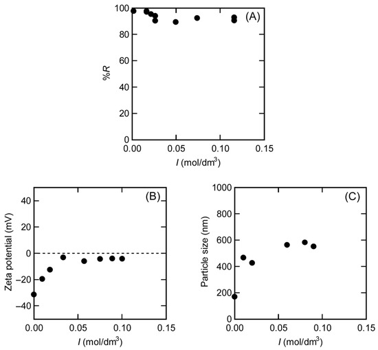
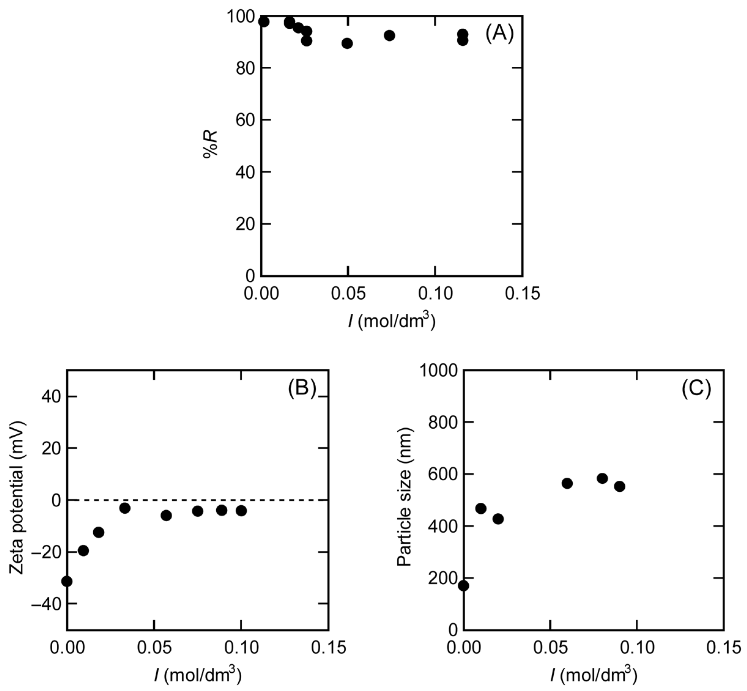
CuO nanoparticle-dispersed water (CuO 28.5 or 45.1 mg/dm3) containing different concentrations of NaNO3 as a modifier of ionic strength I was equilibrated with [BMIm][NTf2]. The concentration of dissolved copper in the aqueous phase was negligible under the present experimental conditions; therefore, the decrease in Cu concentration reflects the removal of CuO nanoparticles from the aqueous phase. The results of I dependence of %R are shown in Figure 3A, where the contribution of dissolved IL components to the ionic strength was not considered. The zeta potential and particle size of the CuO nanoparticles were also measured as a function of I, and the results are shown in Figure 3B and Figure 3C, respectively.
Figure 3.
Percentage removal (A), zeta potential (B), and particle size (C) of CuO nanoparticles in the [BMIm][NTf2]/water biphasic system as a function of the ionic strength of the aqueous phase. Experimental conditions: volume of IL phase = 0.6 cm3; volume of aqueous phase = 6 cm3; Cw° = 28.5 or 45.1 mg/dm3. The dotted line in (B) indicates zero zeta potential.
The %R, i.e., adsorption of CuO nanoparticles to the IL–water interface, is almost 100% under conditions where the I of the aqueous phase is close to 0 mol/dm3. The %R value decreases with increasing I in the region where I is smaller than 0.05 mol/dm3, where the zeta potential of the nanoparticles is negative. As I increases, the zeta potential approaches zero and the particle size increases, indicating the aggregation of nanoparticles due to charge neutralization. The %R becomes almost constant (about 90%) when I exceeds 0.05 mol/dm3. At this time, the zeta potential of the nanoparticles is almost zero and the particle size is also constant (about 600 nm). These results indicate that CuO nanoparticles adsorb at the IL–water interface regardless of the sign of their surface charge, but especially when the nanoparticles are negatively charged, they show high adsorption. This suggests that electrostatic interaction contributes to the interfacial adsorption.
3.3. Effect of pH on Interfacial Adsorption of Fe2O3, CoO, and CuO Nanoparticles in [BMIm][NTf2]/Water Biphasic System
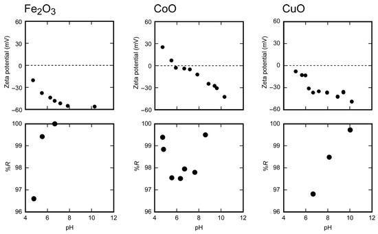
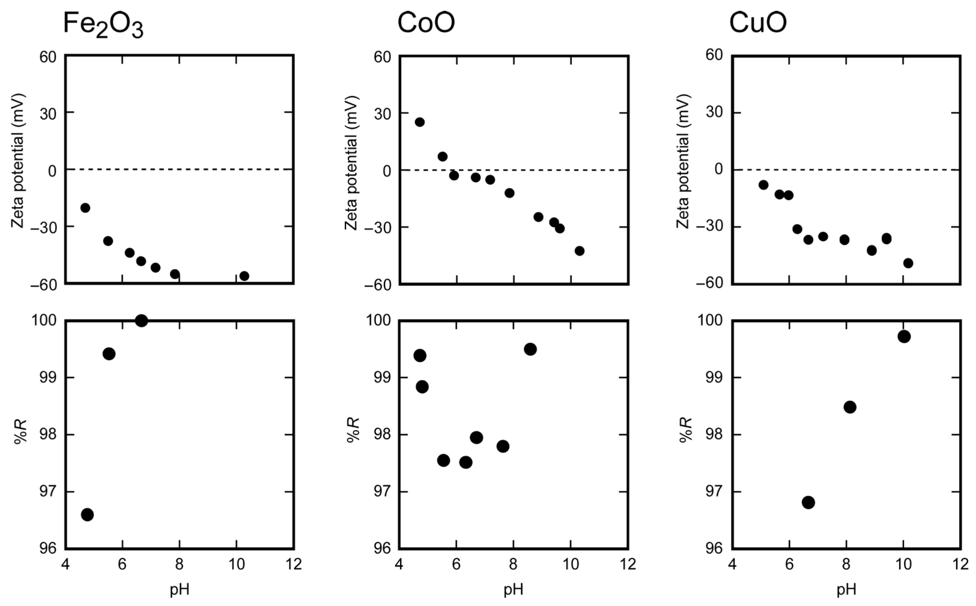
To investigate the relationship between pH and %R in the aqueous phase, nanoparticle-dispersed water of Fe2O3 (7.4 mg/dm3), CoO (22 mg/dm3), and CuO (15 mg/dm3) prepared at different pH and constant I (0.01 mol/dm3) was equilibrated with [BMIm][NTf2]. The obtained %R plotted against pH is shown in Figure 4. In this experiment, Fe2O3 was confirmed to be essentially insoluble in water, whereas CoO and CuO showed slight dissolution in the weakly acidic region; however, the extent of the dissolution did not affect the pH-dependent order of %R.
Figure 4.
Percentage removal and zeta potential of Fe2O3, CoO, and CuO nanoparticles in the [BMIm][NTf2]/water biphasic system as a function of the pH of the aqueous phase. The zeta potential data of Fe2O3 and CoO are replotted from Ref. [8]. Experimental conditions: volume of IL phase = 0.6 cm3; volume of aqueous phase = 6 cm3; I = 0.01 mol/dm3; Cw° = 7.4, 22, and 15 mg/dm3 for Fe2O3, CoO, and CuO, respectively. The dotted lines indicate zero zeta potential.
For Fe2O3 and CuO nanoparticles, the higher the pH, the larger the %R. The zeta potentials of these oxides in water are negative and become more negative with increasing pH in this range. Therefore, the increase in %R with increasing pH can be attributed to the increase in the magnitude of the surface charge of the nanoparticles. On the other hand, in the case of CoO nanoparticles, %R becomes minimum near the neutral pH where the zeta potential of CoO approaches 0 mV. This relationship between zeta potential and %R suggests that adsorption at the IL–water interface occurs not only for negatively charged nanoparticles but also for positively charged ones. Therefore, charged metal oxide nanoparticles, regardless of the polarity of their surface charge, are stabilized at the IL–water interface.
3.4. Interfacial Adsorption Isotherms of Fe2O3, CoO, and CuO Nanoparticles in Several IL/Water Biphasic Systems
CuO nanoparticle-dispersed water with the initial concentration of CuO (Cw°) ranging from 1.7 to 410 mg/dm3 was equilibrated with [BMIm][NTf2]. For Cw° lower than or equal to 87 mg/dm3, CuO nanoparticles were almost completely adsorbed at the [BMIm][NTf2]–water interface (%R = ca. 97%). In the range of Cw° higher than 87 mg/dm3, %R decreased progressively with increasing Cw° and decreased to 93% at Cw° = 410 mg/dm3. The reason for the decrease in %R may be explained in terms of interface saturation. Therefore, we evaluated the adsorption isotherm, i.e., the relationship between the amount of nanoparticles adsorbed per unit area at the liquid–liquid interface, W (mg/dm2), and the concentration of nanoparticles in the aqueous phase, Cw (mg/dm3), at equilibrium.
The values of W were calculated from Equation (2):
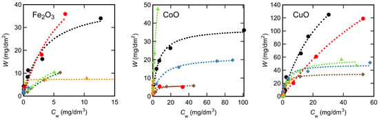
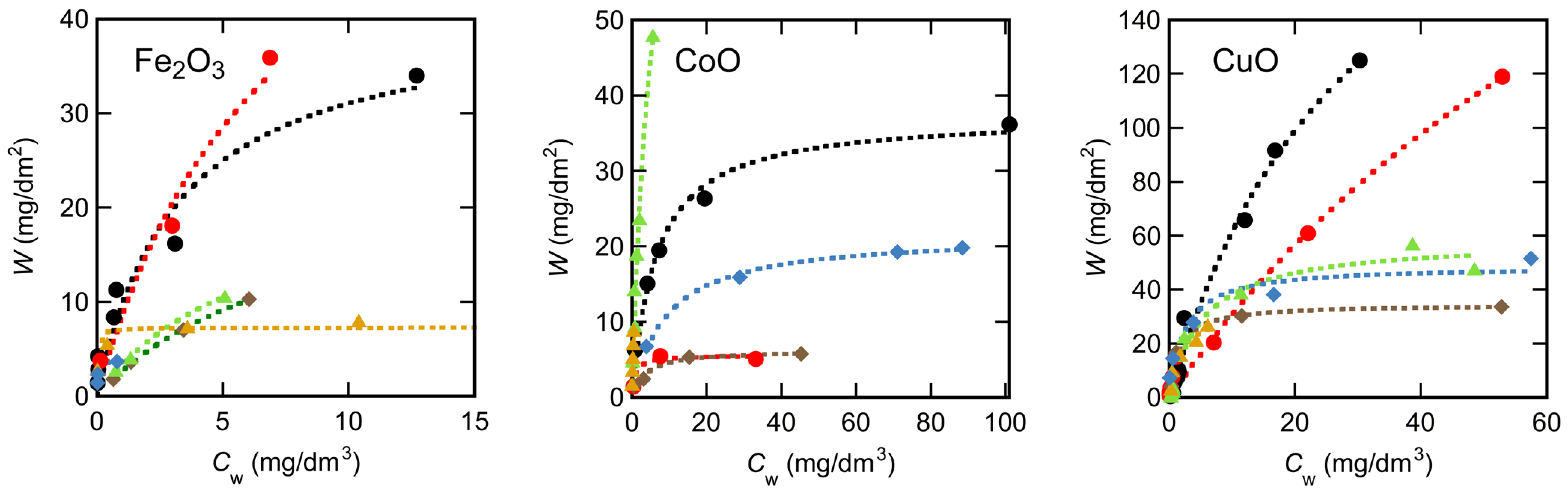
where Vw (dm3) and S (dm2) are the volume of the aqueous phase and the area of the interface, respectively. Adsorption experiments were also conducted for other metal oxides (Fe2O3 and CoO) and nonaqueous solvents ([OMIm][NTf2], benzene, chloroform, hexane, and 1-octanol). All the adsorption isotherms obtained are shown in Figure 5.
W = (Vw/S)(Cw° − Cw),
Figure 5.
Adsorption isotherms of Fe2O3, CoO, and CuO nanoparticles at hydrophobic solvent–water interfaces. Solvents: [BMIm][NTf2] (black circles); [OMIm][NTf2] (red circles); benzene (brown diamonds); chloroform (green triangles); hexane (yellow triangles); 1-octanol (blue diamonds).
In most systems, W increases with increasing Cw and approaches a constant value at higher concentrations. The adsorption isotherms were empirically fitted with the Langmuir equation to estimate the adsorption parameters. The Langmuir equation used is given by Equation (3):
where Wmax (mg/dm2) is the maximum adsorption capacity and K (dm3/mg) is the adsorption equilibrium constant. Equation (3) fits all of the plots in Figure 5 well. The values of Wmax and K obtained by nonlinear least-square analysis are summarized in Table 1.
W = KWmaxCw/(1 + KCw),
Table 1.
Langmuir adsorption parameters for metal oxide nanoparticles at various solvent–water interfaces.
Fe2O3 and CuO nanoparticles have a negative surface charge, around pH 6, whereas CoO nanoparticles are nearly neutral as shown in Figure 4. For Fe2O3 and CuO, the Wmax value decreases in the solvent order [OMIm][NTf2] > [BMIm][NTf2] > molecular solvents (benzene, chloroform, hexane, and 1-octanol); the differences among the molecular solvents are relatively small. The reason for the higher adsorption efficiency at the IL–water interface than at the molecular solvent–water interface can be attributed to the electrostatic interaction between charged nanoparticles and the IL–water interface. On the other hand, for CoO nanoparticles, the order of Wmax is chloroform > [BMIm][NTf2] > hexane ≈ 1-octanol > benzene ≈ [OMIm][NTf2]; no specific adsorption property is observed for the IL–water interface for electrically neutral CoO nanoparticles.
In general, interfacial adsorption is often discussed in relation to interfacial tension, where higher interfacial tension can favor adsorption due to a larger reduction in interfacial free energy, as described by classical interfacial thermodynamics and the Gibbs adsorption framework [24]. In the molecular solvent–water systems investigated in this study, the interfacial tension values were 35.0 mN/m for benzene–water, 32.8 mN/m for chloroform–water, 51.1 mN/m for hexane–water, and 8.52 mN/m for 1-octanol–water [25]. However, no clear correlation was observed between interfacial tension and the Wmax values in Table 1 for these systems. For ionic liquid–water systems, the interfacial tension at the [OMIm][NTf2]–water interface has been reported to be 11.7 mN/m [26], which is comparable to those of the molecular solvent–water systems. Therefore, interfacial tension alone cannot explain the significantly larger Wmax values observed in ionic liquid–water systems. This suggests that additional factors, such as interfacial structure and ion-specific interactions, play important roles.
Furthermore, in the cases of Fe2O3 and CuO, [OMIm][NTf2] exhibits a higher Wmax than [BMIm][NTf2]. This may be related to the longer alkyl chain of the IL cation, which enhances hydrophobic interactions at the interface; however, the detailed mechanism remains unclear.
The diameters of Fe2O3, CoO, and CuO nanoparticles in water dispersion measured by dynamic light scattering were (3.9 ± 2.4) × 102 nm, (2.7 ± 1.4) × 102 nm, and (1.74 ± 0.65) × 102 nm, respectively. The larger particle sizes compared to the manufacturer-reported values in Section 2.1 are attributed to the aggregation of nanoparticles in aqueous dispersion, as reflected in the hydrodynamic diameters measured by dynamic light scattering. From these particle sizes, the mass of a single particle, M (mg) can be calculated from the volume of the particle calculated from its radius r (m) and its density ρ (g/cm3) using the following formula:
M = (4/3)π(100r)3(1000ρ) = (4/3) × 109 πr3ρ.
Assuming that the nanoparticles form a monolayer in the form of a fine packing at the liquid–liquid interface, the number of nanoparticles N that can adsorb on the interface and the number density of nanoparticles N’ (dm−2) that can adsorb on the unit interface area were calculated as
and
where SIL/W is the area of the liquid–liquid interface, which is 0.0181 dm2 using a test tube with a diameter of 15.2 mm; 0.907 is the filling factor for circles of the same size that are placed most closely on a two-dimensional plane. Using these values, the theoretical value of Wmax, Wmaxt (mg/dm2), was calculated using the following equation:
N = 0.907SIL/W/(100πr2)
N’ = N/SIL/W = 0.907/(100πr2),
Wmaxt = MN’ = (4/3) × 0.907 × 107 rρ.
The values of M, N’, and Wmaxt are summarized in Table 2. The Wmaxt values are in 10 mg/dm2 order for all the metal oxide nanoparticles and are generally comparable to the Wmax values obtained for molecular solvent/water biphasic systems (Table 1), suggesting that the nanoparticles form approximately a monolayer at those interfaces while maintaining their particle size in the dispersed state. In contrast, for Fe2O3 and CuO nanoparticles, which possess negative surface charge near neutral pH, the experimentally obtained Wmax values in the IL/water biphasic systems (41 to 350 mg/dm2) are one order of magnitude larger than the theoretical monolayer capacity (ca. 10 mg/dm2). This large difference strongly suggests that the nanoparticles aggregate at the IL–water interface and form multilayer structures rather than a simple monolayer. The metal oxide nanoparticles with high surface charge adsorb multilayers at the IL–water interface, and thus their adsorption is likely to be larger than at molecular solvent–water interfaces.
Table 2.
Calculated parameters for metal oxide nanoparticles used to estimate the theoretical monolayer adsorption capacity.
Concerning the K value, no influence from specific interactions between charged nanoparticles and the IL–water interface was observed; rather, the K value was larger in the molecular solvent/water systems than in the IL system/water systems. Even in comparing nanoparticles within the IL/water biphasic systems, the electrically neutral CoO exhibited a larger K value than the charged CuO or Fe2O3.
4. Conclusions
In this study, the interfacial adsorption behavior of Fe2O3, CoO, and CuO nanoparticles at hydrophobic solvent–water interfaces was investigated, with particular attention to hydrophobic ionic liquid–water systems. Shaking experiments in the [BMIm][NTf2]–water biphasic system showed that CuO nanoparticles were rapidly removed from the aqueous phase and accumulated mainly at the IL–water interface rather than being transferred into the bulk IL phase. The removal efficiency depended on the ionic strength and pH of the aqueous phase: adsorption was enhanced at low ionic strength and increased with increasing pH, consistent with stronger adsorption for nanoparticles having larger surface charge and indicating the contribution of electrostatic interactions at the IL–water interface.
Adsorption isotherms obtained for Fe2O3, CoO, and CuO nanoparticles at various solvent–water interfaces were successfully described by the Langmuir equation, allowing comparison of adsorption capacities and affinities among solvents. For Fe2O3 and CuO nanoparticles, which possess negative surface charge near neutral pH, the maximum adsorption capacity at IL–water interfaces was markedly higher than that at molecular solvent–water interfaces. Comparison with a monolayer packing model indicated that adsorption at molecular solvent–water interfaces is close to monolayer coverage, whereas adsorption at IL–water interfaces greatly exceeds the theoretical monolayer capacity, suggesting multilayer adsorption or aggregation of nanoparticles at ionic liquid interfaces. In contrast, CoO nanoparticles, which are nearly uncharged under similar conditions, did not exhibit specific enhancement at IL–water interfaces, highlighting the importance of nanoparticle surface charge in determining interfacial adsorption behavior.
These findings indicate that hydrophobic ionic liquid–water interfaces can serve as an effective and simple platform for the recovery and separation of metal oxide nanoparticles dispersed in water. This approach may be useful not only for the removal of nanoparticles from environmental or industrial waters but also as a preconcentration technique for the analytical determination of colloidal species. In practical applications, the presence of coexisting ions and natural organic matter in environmental or industrial waters may influence the adsorption behavior through competitive interactions and the modification of nanoparticle surfaces. Evaluation of such matrix effects is an important subject for future study.
Author Contributions
Conceptualization, S.K.; methodology, S.K.; validation, C.T., N.K. and S.K.; formal analysis, C.T., N.K. and S.K.; investigation, C.T. and N.K.; resources, K.B. and S.K.; data curation, C.T. and S.K.; writing—original draft preparation, C.T.; writing—review and editing, S.K.; visualization, C.T. and S.K.; supervision, S.K.; project administration, S.K.; funding acquisition, S.K. All authors have read and agreed to the published version of the manuscript.
Funding
This research was funded by the Japan Society for the Promotion of Science, grant number 26410145 and 22K05151.
Data Availability Statement
The original contributions presented in this study are included in the article. Further inquiries can be directed to the corresponding author.
Acknowledgments
The authors thank the technical assistance of the Center for Analytical Instrumentation, Chiba University, for measurements of zeta potential and particle size of nanoparticles with an Otsuka Electronics ELSZ-1000 instrument.
Conflicts of Interest
The authors declare no conflicts of interest.
References
- Chavali, M.S.; Nikolova, M.P. Metal oxide nanoparticles and their applications in nanotechnology. SN Appl. Sci. 2019, 1, 607. [Google Scholar] [CrossRef] [Scilit]
- Hajra, K.; Maity, D.; Saha, S. Recent advancements of metal oxide nanoparticles and their potential applications: A review. Adv. Mater. Lett. 2024, 15, 24011740. [Google Scholar] [CrossRef] [Scilit]
- Soares, E.V.; Soares, H.M.V.M. Harmful effects of metal(loid) oxide nanoparticles. Appl. Microbiol. Biotechnol. 2021, 105, 1379–1394. [Google Scholar] [CrossRef] [Scilit]
- Xiao, N.; Li, Y.; Sun, P.; Zhu, P.; Wang, H.; Wu, Y.; Bai, M.; Li, A.; Ming, W. A comparative review: Biological safety and sustainability of metal nanomaterials without and with machine learning assistance. Micromachines 2025, 16, 15. [Google Scholar] [CrossRef] [Scilit]
- Bondarenko, O.; Juganson, K.; Ivask, A.; Kasemets, K.; Mortimer, M.; Kahru, A. Toxicity of Ag, CuO and ZnO nanoparticles to selected environmentally relevant test organisms and mammalian cells in vitro: A critical review. Arch. Toxicol. 2013, 87, 1181–1200. [Google Scholar] [CrossRef] [Scilit]
- Brown, G.E., Jr.; Parks, G.A. Sorption of trace elements on mineral surfaces: Modern perspectives from spectroscopic studies, and comments on sorption in the marine environment. Int. Geol. Rev. 2001, 43, 963–1073. [Google Scholar] [CrossRef] [Scilit]
- Bessho, K.; Kanaya, N.; Shimada, S.; Katsuta, S.; Monjushiro, H. Adsorption behavior of beryllium (II) on copper-oxide nanoparticles dispersed in water: A model for 7Be colloid formation in the cooling water for electromagnets at high-energy accelerator facilities. Anal. Sci. 2014, 30, 1069–1074. [Google Scholar] [CrossRef] [Scilit] [PubMed]
- Katsuta, S.; Kanaya, N.; Bessho, K.; Monjushiro, H. Adsorption behavior of trace beryllium (II) onto metal oxide nanoparticles dispersed in water. Int. J. Chem. 2017, 9, 62–70. [Google Scholar] [CrossRef] [Scilit]
- Kurosawa, S.; Ueta, S. Effect of colloids on radionuclide migration for performance assessment of HLW disposal in Japan. Pure Appl. Chem. 2001, 73, 2027–2037. [Google Scholar] [CrossRef] [Scilit]
- Malkovsky, V.I.; Pek, A.A. Effect of colloids on transfer of radionuclides by subsurface water. Geol. Ore Depos. 2009, 51, 79–92. [Google Scholar] [CrossRef] [Scilit]
- Deb, D.; Chakma, S. Colloid and colloid-facilitated contaminant transport in subsurface ecosystem—A concise review. Int. J. Environ. Sci. Technol. 2023, 20, 6955–6988. [Google Scholar] [CrossRef] [Scilit]
- Surawanvijit, S.; Liu, H.H.; Kim, M.; Cohen, Y. Removal of metal oxide nanoparticles from aqueous suspensions. Sep. Sci. Technol. 2014, 49, 161–170. [Google Scholar] [CrossRef] [Scilit]
- You, Z.; Zhao, C.; Sun, Y.; Zhuang, C. Application of PAFC/CPAM for the removal of ZnO nanoparticles by enhanced coagulation. Water Sci. Technol. 2021, 84, 484–498. [Google Scholar] [CrossRef] [Scilit]
- Li, M.; Wang, C.; O’Connell, M.J.; Chan, C.K. Carbon nanosphere adsorbents for removal of arsenate and selenate from water. Environ. Sci. Nano 2015, 2, 245–250. [Google Scholar] [CrossRef] [Scilit]
- Jardiel, T.; Peiteado, M.; Castellanos-Aliaga, A.; Caballero, A.C.; Calatayud, D.G. Peptide-driven bio-assisted removal of metal oxide nanoparticles from an aqueous suspension: A novel strategy for water remediation. J. Clean. Prod. 2021, 285, 124852. [Google Scholar] [CrossRef] [Scilit]
- Wongsawa, T.; Traiwongsa, N.; Pancharoen, U.; Nootong, K. A review of the recovery of precious metals using ionic liquid extractants in hydrometallurgical processes. Hydrometallurgy 2020, 198, 105488. [Google Scholar] [CrossRef] [Scilit]
- Firmansyah, M.L.; Yoshida, W.; Hanada, T.; Goto, M. Application of ionic liquids in solvent extraction of platinum group metals. Solvent Extr. Res. Dev. Jpn. 2020, 27, 1–24. [Google Scholar] [CrossRef] [Scilit]
- Okamura, H.; Hirayama, N. Recent progress in ionic liquid extraction for the separation of rare earth elements. Anal. Sci. 2021, 37, 119–130. [Google Scholar] [CrossRef] [Scilit]
- Inman, G.; Nlebedim, I.C.; Prodius, D. Application of ionic liquids for the recycling and recovery of technologically critical and valuable metals. Energies 2022, 15, 628. [Google Scholar] [CrossRef] [Scilit]
- Nakashima, T.; Kimizuka, N. Water/ionic liquid interfaces as fluid scaffolds for the two-dimensional self-assembly of charged nanospheres. Langmuir 2011, 27, 1281–1285. [Google Scholar] [CrossRef] [Scilit]
- Hamamoto, T.; Okai, M.; Katsuta, S. The laws governing ionic liquid extraction of cations: Partition of 1-ethylpyridinium monocation and paraquat dication in ionic liquid/water biphasic systems. J. Phys. Chem. B 2015, 119, 6317–6325. [Google Scholar] [CrossRef] [Scilit] [PubMed]
- Nakamura, K.; Kudo, Y.; Takeda, Y.; Katsuta, S. Partition of substituted benzenes between hydrophobic ionic liquids and water: Evaluation of interactions between substituents and ionic liquids. J. Chem. Eng. Data 2011, 56, 2160–2167. [Google Scholar] [CrossRef] [Scilit]
- Katsuta, S.; Nakamura, K.; Kudo, Y.; Takeda, Y.; Kato, H. Partition behavior of chlorophenols and nitrophenols between hydrophobic ionic liquids and water. J. Chem. Eng. Data 2011, 56, 4083–4089. [Google Scholar] [CrossRef] [Scilit]
- Adamson, A.W.; Gast, A.P. Physical Chemistry of Surfaces, 6th ed.; Wiley-Interscience: New York, NY, USA, 1997. [Google Scholar]
- Demond, A.H.; Lindner, A.S. Estimation of Interfacial Tension between Organic Liquids and Water. Environ. Sci. Technol. 1993, 27, 2318–2331. [Google Scholar] [CrossRef] [Scilit]
- Fitchett, B.D.; Rollins, J.B.; Conboy, J.C. Interfacial Tension and Electrocapillary Measurements of the Room Temperature Ionic Liquid/Aqueous Interface. Langmuir 2005, 21, 12179–12186. [Google Scholar] [CrossRef] [Scilit] [PubMed]

Disclaimer/Publisher’s Note: The statements, opinions and data contained in all publications are solely those of the individual author(s) and contributor(s) and not of MDPI and/or the editor(s). MDPI and/or the editor(s) disclaim responsibility for any injury to people or property resulting from any ideas, methods, instructions or products referred to in the content. |
© 2026 by the authors. Licensee MDPI, Basel, Switzerland. This article is an open access article distributed under the terms and conditions of the Creative Commons Attribution (CC BY) license.