


Abstract
Lactobacilli are Gram-positive fermentative bacteria with GRAS (“Generally Recognized As Safe”) status. Since 2017, it has been reported that many lactobacilli strains can form extracellular vesicles. Extracellular vesicles (EVs) are spherical bilayered membrane structures of nanometric size that carry cytoplasmic components. Bacterial EVs are considered important mediators between host and commensal bacteria, but their biological effects have not been widely studied. The purification of EVs involves methodological difficulties. Due to their nanometric size, EVs are prone to being co-purified with media compounds, which interfere with their characterization. The present work aimed to obtain high-purity EVs and to determine their physicochemical properties. To achieve the objectives, we developed a minimal synthetic medium (MSM) suitable for the growth of Lactiplantibacillus plantarum CIDCA 83114 and also designed a protocol to concentrate and obtain its EVs. The medium was free of high-molecular-weight compounds and surfactants, and the purification protocol included EV concentration via a tangential flow filtration step. The EVs obtained had an average size of 110 nm ± 40 nm and a negative zeta potential. They exhibited a mean protein content of 100 µg/mL with a clear protein profile absent in the culture media. EVs also contain RNA and scarce DNA. Our work contributed to improving the purification of lactobacilli EVs by replacing the traditional medium used in lactic acid bacteria growth (MRS) with a synthetic minimal medium (MSM). The design of the MSM allows for high-purity vesicles to be obtained and accurately characterized.
1. Introduction
Extracellular vesicles (EVs) are lipid bilayer structures of 20–500 nm in diameter present in all life domains and acting as a cargo system of diverse biomolecules, such as phospholipids, proteins, and nucleic acids [1]. EVs are actively involved in bacterial communication and also in the crosstalk between bacteria and the host, modulating diverse biological processes. Bacterial EVs were first studied fifty years ago, with outer membrane vesicles (OMVs) derived from Gram-negative bacteria [2,3]. However, the discovery of EVs derived from Gram-positive bacteria did not occur until 2009 [4]. Despite their thick cell wall, Gram-positive bacteria can produce EVs, probably through the action of cell-wall-degrading enzymes that weaken the peptidoglycan layer and permit their release [1]. Lactobacilli are Gram-positive bacteria that have been used for fermentation and food production since ancient times. Several lactobacilli strains are part of the human microbiota and are considered probiotic. Probiotic bacteria interact with the host, conferring health benefits [5]. Recently, there has been increasing information about the production of EVs from lactobacilli strains [6,7,8]. Lactobacilli EVs have drawn significant attention due to belonging to bacteria with a GRAS status, the absence of LPS in their composition, and their potential to exert positive effects. Indeed, several beneficial effects were ascribed to the action of lactobacilli EVs, such as increasing the epithelial adhesion of probiotic bacteria, preventing pathogen attachment, enhancing anti-inflammatory responses, and bacteriocin delivery that could regulate the composition of the microbiota [9,10,11,12].
The study and characterization of EVs is not free of methodological problems. It is known that Gram-positive bacteria have a relatively low production rate of vesicles compared to Gram-negative bacteria [13]. In addition, EVs are of nanometer size, and several particles and aggregates present in the culture broth are prone to be co-purified with the vesicles. The MISEV committee (Minimal Information for Studies of Extracellular Vesicles Committee) established that controls of culture media should be processed and characterized to assess their contribution to putative EV measurements [14]. Despite the expansion of bacterial EV studies, only a few contained the controls suggested by the MISEV committee [9,12]. Particles and compounds present in the growth media could interfere with the physicochemical determinations and the biological effects assigned to the EVs. In particular, MRS broth, which constitutes the standard complex medium for lactobacilli growth, contains peptones and surfactants that could form aggregates or micelles. Therefore, it is essential to reduce medium interference in EV studies. To circumvent those problems, we designed a minimal synthetic medium (MSM) lacking surfactants and high-molecular-weight compounds able to support L. plantarum growth. Furthermore, following the MISEV committee suggestions, we included a control of MSM processed by the same EV purification protocol. Considering the growing interest in lactobacilli EVs and the importance of having more accurate descriptions of their properties, this study aimed to obtain and characterize higher purity L. plantarum vesicles from an MSM.
2. Materials and Methods
2.1. Cultures and EV Isolation
Stock cultures of L. plantarum strain CIDCA 83114 were preserved at −80 °C. Cultures were reactivated twice in De Man–Rogosa–Sharpe (MRS, Biokar Diagnostics, Paris, France) medium at 37 °C for 24 h before experiments. The EVs were obtained from MRS broth and from a new minimal synthetic medium (MSM) developed in our lab. The MSM composition was designed based on the MRS formula, removing yeast extract, casein peptone, and Tween 80. The MSM composition was as follows: glucose 20 g/L; K2HPO4 2 g/L; sodium acetate 5 g/L; ammonium citrate 2 g/L; MgSO4 0.24 g/L; MnSO4 0.05 g/L; KCl 0.2 g/L; CaCl2 0.02 g/L; proline 0.2 g/L; glutamic acid 0.6 g/L and cysteine 0.04 g/L. Both media were inoculated with L. plantarum at 0.1% and incubated at 37 °C for 24 (MRS) or 48 h (MSM). The cultures were centrifuged at 6000 × g for 10 min at 4 °C. Supernatants were filtered (0.45 µm) under vacuum, and then they were concentrated using a tangential flow filtration system (TFF) (Vivaflow© 200, Sartorius, Göttingen, Germany) with a cut-off membrane of 10 kDa. The resulting concentrated supernatants were ultracentrifuged at 138,000 × g and 4 °C for 2 h in a SW 40 swinging rotor (Beckman Coulter, Mississauga, ON, Canada). Pellets were finally suspended in 500 μL PBS (10 mM, pH 7.4) for every 15 mL of ultracentrifuged supernatant. In all the experiments, an equal volume of MSM or MRS medium was processed as described above and used as the control. Prior to the measurements, samples were filtered again through a 0.45 µm membrane to eliminate contaminants and reduce the presence of external particles from the procedures.
2.2. Particle Size and Zeta (ζ) Potential Measurement
Measurements were recorded at 25 °C on a Horiba SZ-100 nanoparticle analyzer (Horiba, Kyoto, Japan). Average hydrodynamic particle size (Z-average) and polydispersive index (PDI) were determined by using dynamic light scattering (DLS). The ζ potential was determined by laser Doppler electrophoresis using the Smulochowski model. For each sample, the particle size and zeta potential were calculated from the mean of 10 measurements using the Horiba nanoPartica SZ-100/SZ-100V2 series software.
2.3. Electron Microscopy
Samples were fixed with 2% glutaraldehyde in PBS (10 mM). Drops were deposited onto formvar-carbon coated grids and incubated for 10 min. Excess liquid was blotted, samples were negatively stained with 1% aqueous uranyl acetate incubated for 1 min, then blotted and dried by spontaneous evaporation. TEM imaging was performed using a FEI F200X TALOS microscope (FEI Company, Hillsboro, OR, USA) operated at 200 kV. The high-angle annular dark-field (HAADF) STEM (Scanning-TEM) images were captured by a HAADF detector with a collection angle from 22 to 136 mrad.
2.4. Nanoparticle Tracking Analysis
EVs were characterized by nanoparticle tracking analysis (NTA) using a ZetaView PMX-230 instrument (Particle Metrix GmbH, Ammersee, Germany) according to the manufacturer’s instructions. Aliquots of EV suspensions were diluted 1:5000 in particle-free deionized water to achieve 30–160 particles per frame, and then infused continuously through a 488 nm laser beam using a syringe pump. Size distribution video data were acquired using the following settings: temperature 25.1 °C; 30 frames/second; duration: 2 s; cycles: 1; positions: 11; camera sensitivity: 80 and shutter value: 100. Data were analyzed using ZetaView NTA software (version 8.06.01).
2.5. DNA, RNA and Protein Content
DNA, RNA and protein were extracted from EVs using Bio-Zol reagent (PB-L Productos Bio-Lógicos, Quilmes, Argentina) following the manufacturer’s instructions. DNA and RNA content were determined using a microvolume spectrophotometer (NanoDrop, Thermofisher, Waltham, MA, USA). DNA and RNA samples were subjected to 1.5% agarose gel electrophoresis. The electrophoresis was carried out at 100 V for 1 h in 1X TAE buffer. A 100 bp DNA molecular weight marker was used to estimate the product size. To verify RNA and DNA identity, samples were incubated with DNase and RNAase (30 min, 37 °C). After incubation, they were also subjected to agarose gel electrophoresis. The resulting bands were visualized and documented using a UV gel documentation system.
Total protein was determined by Bradford assay. Protein profile was determined by SDS-PAGE (12.5% acrylamide-bis acrylamide) in denaturing conditions. Gels were revealed by silver staining.
2.6. Fourier Transform Infrared Spectroscopy (FTIR) Analysis
FTIR spectroscopy was performed as described by Gamba et al. (2025) [15], with modifications. Approximately 5 µL of each sample was placed directly onto the ATR crystal of an Attenuated Total Reflectance FTIR (ATR-FTIR) Thermo Nicolet iS10 spectrometer (Thermo Scientific, Waltham, MA, USA) and dried under a stream of cold air prior to spectral acquisition. Spectra were registered in the 4000–600 cm−1 range by co-adding 32 scans with 4 cm−1 spectral resolution, using OMNIC software (version 8.3, Thermo Scientific, Waltham, MA, USA). At least 4 spectra were recorded for each sample. Advanced ATR correction was performed after the collection of spectra. To correct for residual culture medium interference, the spectrum of the dried film was subjected to spectral subtraction using the culture medium spectrum as a background control.
2.7. Statistical Analysis
The obtained results were evaluated by one-way analysis of variance (ANOVA), using InfoStat software v2008 (Grupo InfoStat, FCA, Universidad Nacional de Córdoba, Córdoba, Argentina). When this analysis expressed statistical differences (p < 0.05), intragroup comparisons were tested using Tukey’s test.
3. Results and Discussion
3.1. Optimization of Culture Conditions
L. plantarum CIDCA 83114 EVs were obtained from cultures in MRS broth and in the MSM. Both media exhibited different bacterial yields and pH after culture (Table 1). MRS broth showed a yield ten times higher than MSM (2 × 10⁹ CFU/mL), with a final pH of 4 after 24 h of incubation. However, viability in this medium suffered a drastic drop after 48 h (<2 × 10³ CFU/mL). MSM exhibited a lower lactobacilli yield (2 × 10⁸ CFU/mL) and reached a pH of 5.5 with constant viability after 48 h (Table 1). It has been reported that the late log or early stationary phase of bacteria is the optimum harvest time to maximize the EV yield [16]. In the case of lactobacilli, this phase corresponds to 24 to 48 h.
Table 1.
Growth parameters of L. plantarum in MRS and MSM broth.
If the bacteria culture is kept in the stationary phase for too long, extracellular proteases that break down EVs proteins may be released [17]. Moreover, bacterial cell death processes liberate cytoplasmic contents into the medium, increasing the concentration of debris and high-molecular-weight compounds, which might interfere with EV purification [18]. Considering the different yields of both media and the optimum strain viability, the volume and harvest time selected to obtain EVs were 600 mL and 24 h for MRS and 6000 mL and 48 h for MSM.
3.2. Particle Size and ζ Potential
The analysis of DLS results showed that EVs derived from MRS cultures had a particle size of 63.04 ± 3.42 nm, whereas EVs derived from MSM had a higher size of 118.22 ± 4.12 nm. Zeta potential values were similar for both EVs (Table 2). Control MRS broth contained particles with a size of 52.37 ± 1.92 nm and a zeta potential of −11.07 mV, which were very similar to lactobacilli EVs. Size measurements of the control MSM showed erratic signals and elevated polydispersity indices, indicating extremely low particle content. The particle concentration in control MSM samples is probably below the limit of detection. In the presence of scarce particles, the autocorrelation function to calculate particle diameter did not reflect the actual size distribution, showing Z-average diameters even higher than the equipment upper detection limit (10 µm). It is known that the intensity of scattered light scales with the sixth power of the particle diameter, meaning the presence of a few large aggregates or dust in the sample heavily skews the results [19]. This explains the high variability of Zeta average values obtained from MSM control; some measures were extremely high whereas other measures were null.
Table 2.
Size, Polydispersive index (PDI) and ζ -potential of lactobacilli EVs and medium controls, as measured by DLS. (<LOD: below limit of detection).
No Zeta potential values could be measured in the MSM control (Table 2). DLS results suggested a higher purification quality of EVs obtained from MSM, with a lower contribution of culture media particles.
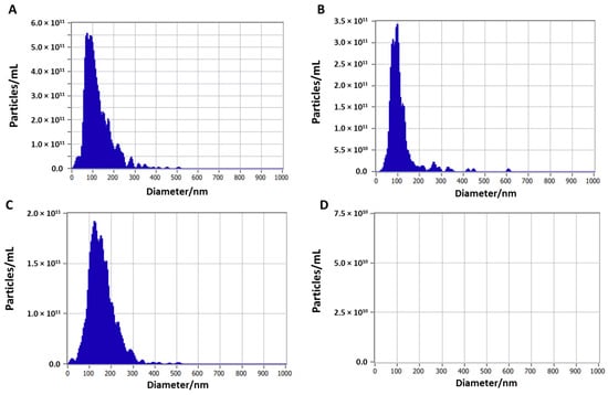
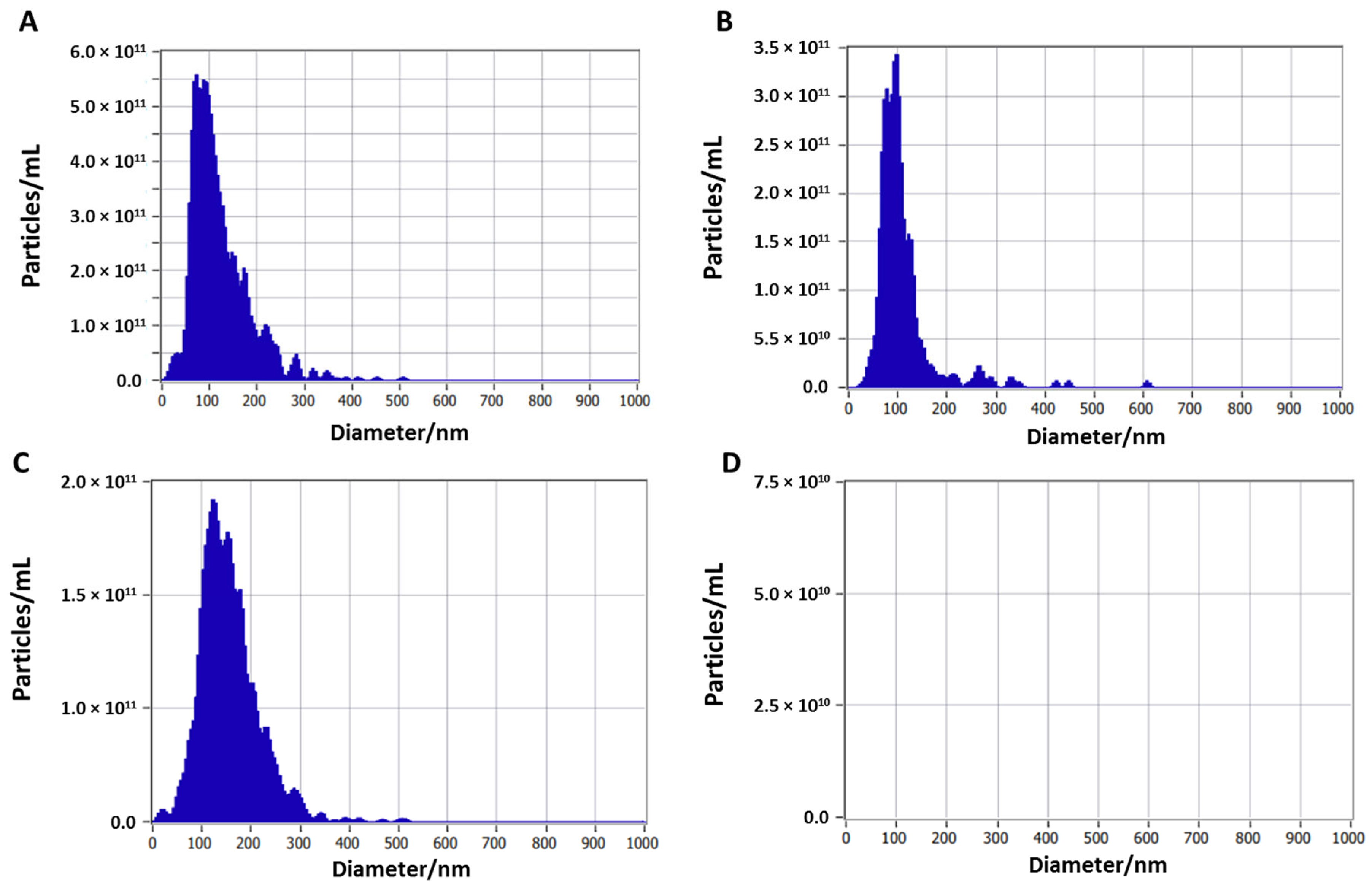
NTA is a high-resolution technique that allows the determination of not only the size and charge but also the absolute concentration of particles. The purification protocol used in this work resulted in a high EV content, reaching a concentration of 11 logs of vesicles per mL in both media (Figure 1 and Table 3). NTA results revealed that lactobacilli EVs derived from MRS and MSM cultures were very similar. EVs purified from MRS had a mean size of 103.0 ± 62.8 nm and a zeta potential of −43.3 ± 2.5 mV, whereas EVs derived from MSM had a mean size of 144.8 ± 59.2 nm and a zeta potential of −55.2 ± 5.4 mV (Figure 1 and Table 3). Several authors reported similar size values of EVs derived from L. plantarum or other lactobacilli strains [20,21,22]. Zeta potential values were higher in the NTA determination compared to DLS. The increased sensitivity of the NTA requires samples to be diluted in water. The use of distinct solvents (10 mM PBS for DLS vs. deionized water for NTA) explained the value differences. NTA measurements in MRS control media indicated that MRS broth had a significant particle contribution compared to MSM. Indeed, MRS control broth showed an elevated particle concentration (2.2 × 10¹¹ particles/mL) with a similar size to EVs, whereas MSM exhibited no particle signal (Figure 1 and Table 3). These results confirmed that MSM is a better medium for vesicle isolation.
Figure 1.
Size distributions of L. plantarum EVs obtained by nanoparticle tracking analysis (NTA). (A) EVs from MRS medium; (B) control of MRS medium; (C) EVs from MS medium; (D) control of MS medium.
Table 3.
Size, concentration and ζ -potential of lactobacilli EVs and medium controls, as measured by NTA.
3.3. Protein Content
The protein content of lactobacilli EVs and control media was quantified by Bradford assay. EVs isolated from MRS showed a protein concentration of 303.60 ± 22.63 μg/mL. Control MRS broth showed similar values, indicating co-purification of medium proteins with the EVs (Table 4). The protein content of lactobacilli EVs isolated from MSM was 105.60 ± 14.14 μg/mL, and proteins were not detected in the control of MSM. Our results demonstrate that EV purification from MSM avoids contamination with culture medium proteins. Moreover, the use of MSM avoids the co-purification of EVs with Tween 80. The MSM was based on MRS but excludes proteins or Tween 80. The addition of Tween 80 to the synthetic medium caused detergent recovery after the purification process (Table 4). Tween 80 (polysorbate 80) is a non-ionic surfactant used in MRS broth, primarily as a source of oleic acid, a fatty acid necessary for many lactobacilli. The standard concentration of Tween 80 in MRS is 1 g/L, and the compatible concentration with the Bradford assay is 0.6 g/L [23]. The addition of Tween 80 to the MSM, even at concentrations as low as 0.02 g/L, interferes with EV protein quantification, indicating that the detergent was concentrated and recovered in the process. Tween 80 is a small molecule (1.3 kDa) that could pass the 10 kDa membrane of the tangential flow filter. However, the detergent forms micelles of approximately 10 nm at concentrations as low as 0.014 g/L that might be recovered after the purification process [24]. To sum up, the use of MSM allows high-purity EVs to be obtained, avoiding contamination with culture media proteins or Tween 80 micelles. Considering these results, further experiments were performed with EVs isolated from MSM.
Table 4.
Protein concentration of lactobacilli EVs and medium controls. Measurements were performed by Bradford assay. Different letters indicate significant differences in ANOVA test (p < 0.05).
3.4. Protein Profile, RNA, and DNA Content
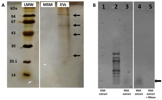
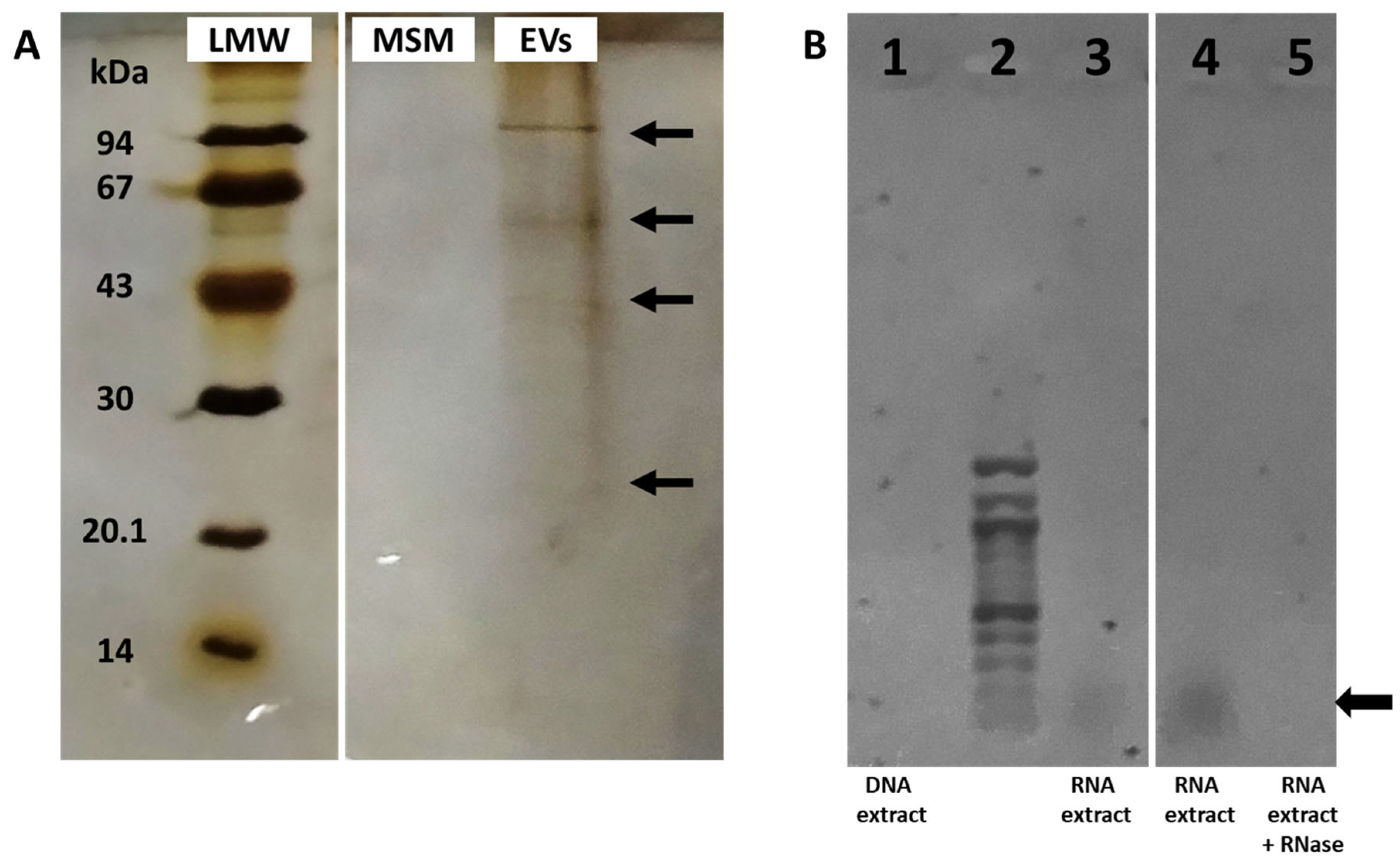
EVs purified from MSM showed a protein profile containing bands of approximately 94, 57, 43, and 23 kDa. Control MSM exhibited no bands (Figure 2A). Proteomic analysis of L. plantarum EVs revealed that the most abundant proteins correspond to cell membrane proteins or enzymes involved in general metabolic pathways [25].
Figure 2.
(A) SDS-PAGE profile of L. plantarum EVs from MS medium. Lane 1: LMW marker; Lane 2: control of MSM; Lane 3: L. plantarum EVs from MSM. Proteins were separated by SDS-PAGE using 12.5% gel and visualized by silver staining. (B) Agarose gel electrophoresis of the products of nucleic acid extraction from EVs. Lane 1: DNA extract; Lane 2: molecular marker; Lanes 3 and 4: RNA extract; Lane 5: RNA extract after RNase treatment. The arrows point to the most prominent bands.
The analysis of the nucleic acid content of EVs revealed an elevated concentration of RNA (1115 ng/µg of protein), whereas the DNA content was very low (67 ng/µg of protein). It has been reported that RNA cargo of lactobacilli EVs usually contains small non-coding RNA, rRNA (5 S, 16 S and 23 S) and lower quantities of mRNA and tRNA [26]. Agarose gel electrophoresis confirmed these results, showing clear RNA bands at the gel end, while DNA was not detected (Figure 2B). RNase treatment degraded the bands present at the gel end, corroborating the identity of the isolated nucleic acid.
The study of biomolecules showed that lactobacilli EVs contain moderate quantities of protein and RNA, but DNA was scarce. These results were consistent with the biomolecule content found in other lactobacilli strains in which RNA and proteins were present, whereas DNA was rarely detected [7,8].
3.5. FTIR Analysis
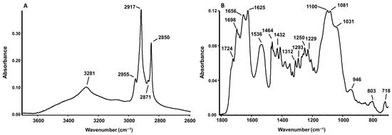
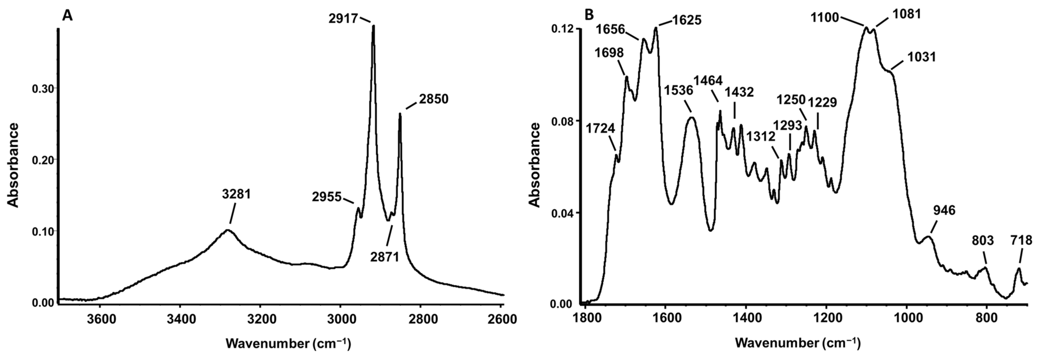
In Figure 3, the ATR-FTIR spectrum of EVs obtained from MS medium cultures is reported. Band assignments were carried out according to Talari et al. (2016) [27].
Figure 3.
ATR-FTIR spectrum of EVs from L. plantarum CIDCA 83114 grown in MSM, in the 3700–2600 cm−1 (A) and 1800–800 cm−1 (B) ranges. Numbers illustrate the wavenumbers at which the main bands are centered.
The spectrum displays a broad band centered at 3280 cm−1, which can be ascribed to the stretching vibrations of the N-H and O-H bonds from proteins and carbohydrates.
The region from 3000 cm−1 to 2800 cm−1 is dominated by the stretching of C-H bonds from aliphatic chains of fatty acids. Bands centered at 2917 and 2850 cm−1 are caused by the asymmetric and symmetric stretching mode of the CH2 groups, respectively, while bands at 2955 and 2871 cm−1 arise from CH3 groups (Figure 3A). These signals suggest the presence of lipidic acyl chains.
The peak located around 1724 cm−1 represents the contribution of the stretching mode of the ester C=O group from phospholipids and triglycerides, while the peaks around 1464 and 1432 cm−1 are attributed to the bending and scissoring modes of the CH3 and CH2 groups from lipids and proteins (Figure 3B). The bands at 1250–1229 cm−1 and 1100–1081 cm−1 can be attributed to the asymmetric and symmetric stretching of PO2 groups of phosphodiester linkages, respectively, typical of phospholipids and nucleic acids.
Additionally, the broad band observed between 1150 and 1000 cm−1 receives contributions from the C-OH and C-O-C stretching vibrations of carbohydrates (Figure 3B).
The primary protein features include the amide I band at 1655 and 1625 cm−1, which arises from the C=O stretching vibrations of peptide bonds in alpha-helix and beta-sheet conformations, respectively. This is accompanied by the amide II band at 1536 cm−1 (N-H bending coupled with C-N stretching), and the amide III bands at 1312 and 1293 cm−1 (C-N stretching and N-H in-plane bending).
3.6. Transmission Electron Microscopy
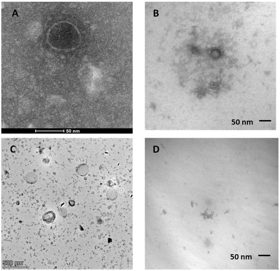
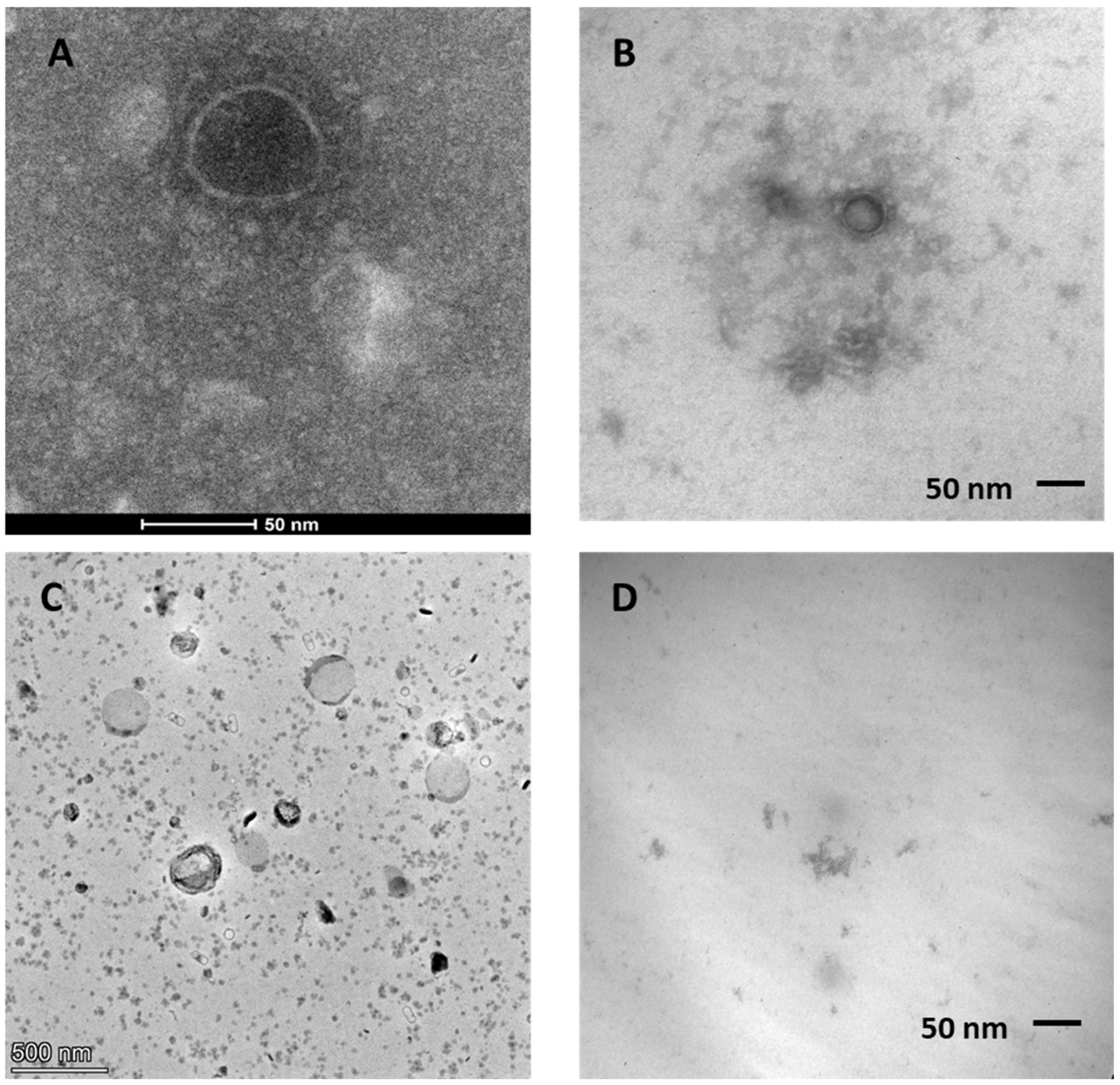
Microscopy observations revealed that L. plantarum produced vesicle-like structures of various sizes from approximately 80 nm to 150 nm (Figure 4A–C). These results were consistent with the size distribution obtained by DLS and NTA (Figure 1 and Figure 2) and were similar to EVs produced by other L. plantarum strains [28,29,30]. TEM images of control MSM showed a clear field with low particle content, confirming minimal presence of raw material and particles in the medium after the purification protocol (Figure 4D).
Figure 4.
TEM images of lactobacilli EVs from MS medium. (A–C) EVs with different magnification. (D) Control of MS medium. Samples were stained with 1% uranyl acetate.
4. Conclusions
A novel minimal synthetic medium (MSM) was developed based on the MRS formula, excluding high molecular weight compounds and surfactants. Although MSM exhibited lower EV yields than MRS, a purification protocol handling large culture volumes and using a tangential flow filtration system enabled the recovery of high-concentration EVs with low impurity levels. The EVs obtained exhibited a mean size from 80 to 150 nm and a zeta potential of −52.2 ± 5.4 mV in pure water. Chemical composition analysis confirmed the presence of proteins, DNA, and RNA within the purified samples, with RNA being the most abundant. FTIR analysis complemented these data, confirming the presence of characteristic functional groups of lipids, proteins, carbohydrates, and compounds containing phosphodiester bonds within these samples. When these spectroscopic data are integrated with NTA and TEM observations, which corroborated the formation of discrete, spherical vesicular structures, the FTIR results provide biochemical evidence for the composition of these nano-assemblies. Furthermore, the presence of specific carbohydrate and phosphate signals in the FTIR spectra, when correlated with the nucleic acids detected through biochemical quantification, suggests that these components are integral to the vesicle structure. Although FTIR on dried films is primarily used to characterize the bulk molecular composition and does not allow definitive conclusions regarding membrane phase or precise spatial organization, the results are consistent with the presence of phospholipid-rich membranes in vesicles. Furthermore, they support the potential presence of cell-wall-derived molecules associated with their surface and a cargo composed of nucleic acids.
To sum up, our improved methodology using MSM and TFF permitted the isolation and accurate characterization of high-purity lactobacilli EVs.
Author Contributions
Conceptualization: A.A.H. and P.M.; Data curation: A.A.H. and P.M.; Funding acquisition: A.A.H.; Investigation: A.A.H. and P.M.; Methodology: A.A.H., P.M. and M.A.F.A.; Writing and manuscript revision: A.A.H., P.M. and M.A.F.A. All authors have read and agreed to the published version of the manuscript.
Funding
This research was funded by the National University of La Plata. Project for Research and Development X985 (2023) “Potencial pro y postbiótico de bacterias benéficas deshidratadas”. Project manager Pérez, P.F.
Institutional Review Board Statement
Not applicable.
Informed Consent Statement
Not applicable.
Data Availability Statement
The data presented in this study are available on request from the corresponding author.
Acknowledgments
Acknowledgments go to the microbiology group of CIDCA, and to Susana Jurado and Fernanda Faisal of the Microscopy Service of the Veterinary Faculty from the National University of La Plata. We would also like to thank Marcela Cucher from the NTA service of the IMPAM Institute CONICET.
Conflicts of Interest
Author María Alejandra Floridia Addato was employed by the company YPF Tecnología (Y-TEC,YPF-CONICET). The remaining authors declare that the research was conducted in the absence of any commercial or financial relationships that could be construed as a potential conflict of interest.
Abbreviations
The following abbreviations are used in this manuscript:
| MSM | Minimal Synthetic Medium |
| DLS | Dynamic Light Scattering |
| NTA | Nanotracking Analysis |
| TFF | Tangential Flow Filtration |
References
- Liu, Y.; Defourny, K.A.Y.; Smid, E.J.; Abee, T. Gram-Positive Bacterial Extracellular Vesicles and Their Impact on Health and Disease. Front. Microbiol. 2018, 9, 1502. [Google Scholar] [CrossRef] [Scilit] [PubMed]
- Chatterjee, S.N.; Das, J. Electron Microscopic Observations on the Excretion of Cell-wall Material by Vibrio cholerae. J. Gen. Microbiol. 1967, 49, 1–11. [Google Scholar] [CrossRef] [Scilit] [PubMed]
- Devoe, I.W.; Gilchrist, J.E. Release of endotoxin in the form of cell wall blebs during in vitro growth of Neisseria meningitidis. J. Exp. Med. 1973, 138, 1156–1167. [Google Scholar] [CrossRef] [Scilit] [PubMed]
- Lee, E.; Choi, D.Y.; Kim, D.K.; Kim, J.W.; Park, J.O.; Kim, S.; Kim, S.H.; Desiderio, D.M.; Kim, Y.K.; Kim, K.P.; et al. Gram-positive bacteria produce membrane vesicles: Proteomics-based characterization of Staphylococcus aureus-derived membrane vesicles. Proteomics 2009, 9, 5425–5436. [Google Scholar] [CrossRef] [Scilit] [PubMed]
- Dean, S.N.; Leary, D.H.; Sullivan, C.J.; Oh, E.; Walper, S.A. Isolation and characterization of Lactobacillus-derived membrane vesicles. Sci. Rep. 2019, 9, 877. [Google Scholar] [CrossRef] [Scilit] [PubMed]
- Domínguez Rubio, A.P.; Martínez, J.H.; Martínez Casillas, D.C.; Coluccio Leskow, F.; Piuri, M.; Pérez, O.E. Lactobacillus casei BL23 Produces Microvesicles Carrying Proteins That Have Been Associated with Its Probiotic Effect. Front. Microbiol. 2017, 8, 1783. [Google Scholar] [CrossRef] [Scilit] [PubMed]
- Kurata, A.; Kiyohara, S.; Imai, T.; Yamasaki-Yashiki, S.; Zaima, N.; Moriyama, T.; Kishimoto, N.; Uegaki, K. Characterization of extracellular vesicles from Lactiplantibacillus plantarum. Sci. Rep. 2022, 12, 13330. [Google Scholar] [CrossRef] [Scilit] [PubMed]
- Grande, R.; Celia, C.; Mincione, G.; Stringaro, A.; Di Marzio, L.; Colone, M.; Di Marcantonio, M.C.; Savino, L.; Puca, V.; Santoliquido, R.; et al. Detection and Physicochemical Characterization of Membrane Vesicles (MVs) of Lactobacillus reuteri DSM 17938. Front. Microbiol. 2017, 8, 1040. [Google Scholar] [CrossRef] [Scilit] [PubMed]
- Croatti, V.; Parolin, C.; Giordani, B.; Foschi, C.; Fedi, S.; Vitali, B. Lactobacilli extracellular vesicles: Potential postbiotics to support the vaginal microbiota homeostasis. Microb. Cell Fact. 2022, 21, 237. [Google Scholar] [CrossRef] [Scilit] [PubMed]
- Choi, J.H.; Moon, C.M.; Shin, T.S.; Kim, E.K.; McDowell, A.; Jo, M.K.; Joo, Y.H.; Kim, S.E.; Jung, H.K.; Shim, K.N.; et al. Lactobacillus paracasei-derived extracellular vesicles attenuate the intestinal inflammatory response by augmenting the endoplasmic reticulum stress pathway. Exp. Mol. Med. 2020, 52, 423–437. [Google Scholar] [CrossRef] [Scilit] [PubMed]
- Kim, W.; Lee, E.J.; Bae, I.H.; Myoung, K.; Kim, S.T.; Park, P.J.; Lee, K.H.; Pham, A.V.; Ko, J.; Oh, S.H.; et al. Lactobacillus plantarum-derived extracellular vesicles induce anti-inflammatory M2 macrophage polarization in vitro. J. Extracell. Vesicle 2020, 9, 1793514. [Google Scholar] [CrossRef] [Scilit] [PubMed]
- Dean, S.N.; Rimmer, M.A.; Turner, K.B.; Phillips, D.A.; Caruana, J.C.; Hervey, W.J., IV; Leary, D.H.; Walper, S.A. Lactobacillus acidophilus Membrane Vesicles as a Vehicle of Bacteriocin Delivery. Front. Microbiol. 2020, 11, 710. [Google Scholar] [CrossRef] [Scilit] [PubMed]
- Sangiorgio, G.; Nicitra, E.; Bivona, D.; Bonomo, C.; Bonacci, P.; Santagati, M.; Musso, N.; Bongiorno, D.; Stefani, S. Interactions of Gram-Positive Bacterial Membrane Vesicles and Hosts: Updates and Future Directions. Int. J. Mol. Sci. 2024, 25, 2904. [Google Scholar] [CrossRef] [Scilit] [PubMed]
- MISEV Consortium. Minimal information for studies of extracellular vesicles (MISEV2023): From basic to advanced approaches. J. Extracell. Vesicle 2024, 13, e12404, Erratum in J. Extracell. Vesicles 2024, 13, e12451. https://doi.org/10.1002/jev2.12451. [Google Scholar] [CrossRef] [Scilit] [PubMed]
- Gamba, R.R.; Ibáñez, A.; Sampaolesi, S.; Mobili, P.; Golowczyc, M.A. Fermentation Strategies to Improve Argentinian Kefir Quality: Impact of Double Fermentation on Physicochemical, Microbial, and Functional Properties. Fermentation 2025, 11, 584. [Google Scholar] [CrossRef] [Scilit]
- Cannizzaro, F.; Gallo, A.; La Scala, S.; Gallo, G.; Faddetta, T. Bacterial Extracellular Vesicles in Biotechnology: Current Challenges and Strategies for Production Enhancement. Fermentation 2026, 12, 86. [Google Scholar] [CrossRef] [Scilit]
- Dai, K.; Liao, B.; Huang, X.; Liu, Q. Consistency in bacterial extracellular vesicle production: Key to their application in human health. Extracell. Vesicles Circ. Nucleic Acids 2025, 6, 1–20. [Google Scholar] [CrossRef] [Scilit] [PubMed]
- Allocati, N.; Masulli, M.; Di Ilio, C.; De Laurenzi, V. Die for the community: An overview of programmed cell death in bacteria. Cell Death Dis. 2015, 6, e1609. [Google Scholar] [CrossRef] [Scilit] [PubMed]
- Malm, A.V.; Corbett, J.C.W. Improved Dynamic Light Scattering using an adaptive and statistically driven time resolved treatment of correlation data. Sci. Rep. 2019, 9, 13519, Erratum in Sci. Rep. 2024, 14, 2021. https://doi.org/10.1038/s41598-024-51806-0. [Google Scholar] [CrossRef] [Scilit] [PubMed]
- Hao, H.; Zhang, X.; Tong, L.; Liu, Q.; Liang, X.; Bu, Y.; Gong, P.; Liu, T.; Zhang, L.; Xia, Y.; et al. Effect of Extracellular Vesicles Derived from Lactobacillus plantarum Q7 on Gut Microbiota and Ulcerative Colitis in Mice. Front. Immunol. 2021, 12, 777147. [Google Scholar] [CrossRef] [Scilit] [PubMed]
- Gong, S.; Zeng, R.; Liu, L.; Wang, R.; Xue, M.; Dong, H.; Wu, Z.; Zhang, Y. Extracellular vesicles from a novel Lactiplantibacillus plantarum strain suppress inflammation and promote M2 macrophage polarization. Front. Immunol. 2024, 15, 1459213. [Google Scholar] [CrossRef] [Scilit] [PubMed]
- Park, B.-O.; Lee, H.W.; Song, C.-H.; Yeom, M.; Shin, S.; Wang, H.; Sim, J.; Cho, E.; Park, D.; Jung, E. Extracellular Vesicles from Lactobacillus rhamnosus BS-Pro-08, Kefir Grain, Suppress Adipogenesis and Enhance Lipolysis in Adipocytes. Int. J. Mol. Sci. 2025, 26, 11732. [Google Scholar] [CrossRef] [Scilit] [PubMed]
- Stutzenberger, F.J. Interference of the detergent Tween 80 in protein assays. Anal. Biochem. 1992, 207, 249–254. [Google Scholar] [CrossRef] [Scilit] [PubMed]
- Bide, Y.; Fashapoyeh, M.A.; Shokrollahzadeh, S. Structural investigation and application of Tween 80-choline chloride self-assemblies as osmotic agent for water desalination. Sci. Rep. 2021, 11, 17068. [Google Scholar] [CrossRef] [Scilit] [PubMed]
- Zhu, L.; Hu, W.; Liu, D.; Tian, W.; Yu, G.; Liu, X.; Wang, J.; Feng, E.; Zhang, X.; Chen, B.; et al. A reference proteomic database of Lactobacillus plantarum CMCC-P0002. PLoS ONE 2011, 6, e25596. [Google Scholar] [CrossRef] [Scilit] [PubMed]
- da Silva, D.R.; Gonzalez, C.F.; Lorca, G.L. Internalization of extracellular vesicles from Lactobacillus johnsonii N6.2 elicit an RNA sensory response in human pancreatic cell lines. J. Extracell. Biol. 2023, 2, e101. [Google Scholar] [CrossRef] [Scilit] [PubMed]
- Talari, A.C.S.; Martinez, M.A.G.; Movasaghi, Z.; Rehman, S.; Rehman, I.U. Advances in Fourier transform infrared (FTIR) spectroscopy of biological tissues. Appl. Spectrosc. Rev. 2017, 52, 456–506. [Google Scholar] [CrossRef] [Scilit]
- Bajic, S.S.; Cañas, M.A.; Tolinacki, M.; Badia, J.; Sánchez, B.; Golic, N.; Margolles, A.; Baldomá, L.; Ruas-Madiedo, P. Proteomic profile of extracellular vesicles released by Lactiplantibacillus plantarum BGAN8 and their internalization by non-polarized HT29 cell line. Sci. Rep. 2020, 10, 21829. [Google Scholar] [CrossRef] [Scilit] [PubMed]
- Kim, M.-H.; Choi, S.J.; Choi, H.I.; Choi, J.P.; Park, H.K.; Kim, E.K.; Kim, M.J.; Moon, B.S.; Min, T.K.; Rho, M.; et al. Lactobacillus plantarum-derived Extracellular Vesicles Protect Atopic Dermatitis Induced by Staphylococcus aureus-derived Extracellular Vesicles. Allergy Asthma Immunol. Res. 2018, 10, 516. [Google Scholar] [CrossRef] [Scilit] [PubMed]
- Li, M.; Lee, K.; Hsu, M.; Nau, G.; Mylonakis, E.; Ramratnam, B. Lactobacillus-derived extracellular vesicles enhance host immune responses against vancomycin-resistant enterococci. BMC Microbiol. 2017, 17, 66. [Google Scholar] [CrossRef] [Scilit] [PubMed]

Disclaimer/Publisher’s Note: The statements, opinions and data contained in all publications are solely those of the individual author(s) and contributor(s) and not of MDPI and/or the editor(s). MDPI and/or the editor(s) disclaim responsibility for any injury to people or property resulting from any ideas, methods, instructions or products referred to in the content. |
© 2026 by the authors. Licensee MDPI, Basel, Switzerland. This article is an open access article distributed under the terms and conditions of the Creative Commons Attribution (CC BY) license.