1. Introduction
The gut microbiota is essential for maintaining human and animal health. Furthermore, the qualitative and quantitative composition of the gut microbiota affects the overall quality and duration of life in humans and animals. This influence is largely attributed to its critical role in regulating metabolism, maintaining immune homeostasis, and enhancing resistance to age-related physiological changes [
1]. To exert such a broad and systematic influence on human and animal physiology, microorganisms possess an extensive arsenal of biologically active metabolites and biochemical pathways that mediate complex interactions with host cells. These metabolites include short-chain fatty acids, bacteriocins, vitamins, signaling molecules, and other bioactive compounds produced by probiotic microorganisms [
2].
Conversely, disturbances or imbalances in the gut microbiota (dysbiosis) are the key contributors to the initiation and progression of numerous diseases. These include inflammatory bowel diseases, metabolic disorders such as obesity and type 2 diabetes, autoimmune and rheumatic diseases, and neurological and cardiovascular pathologies [
3,
4,
5].
Among the intestinal microbiota, lactic acid bacteria (LAB) are recognized as leading contributors to the positive effects on the health of both humans and animals, serving as permanent and functionally important inhabitants of the gastrointestinal tract [
6]. Lactic acid bacteria are not only closely associated with the physiology of their host organisms but also possess considerable biotechnological significance. They play a crucial role in the production of fermented foods, probiotic preparations, and other industrially valuable bioproducts with wide applications in food, agriculture, and health biotechnology [
7,
8].
Genus Enterococcus occupies a distinctive position among LAB due to its dual importance in the food and biotechnology industries. It is involved in the fermentation of various traditional foods and is increasingly explored for its probiotic potential, production of bacteriocins, and other bioactive compounds of industrial relevance [
9]. Firstly, enterococci produce biologically active compounds such as enterocins (bacteriocins)—cationic, hydrophobic, and heat-stable low-molecular-weight peptides (20–60 amino acids). Enterocins possess broad-spectrum antimicrobial activity against a variety of microorganisms, including putrefactive bacteria, foodborne pathogens, and
Listeria spp. Enterocins are generally susceptible to degradation by digestive proteolytic enzymes in the gastrointestinal tract, which makes them particularly attractive for use in the food industry for food preservation and safety applications [
10]. In contrast, probiotic enterococcal strains must survive gastrointestinal transit to exert beneficial effects within the host.
Secondly, selected enterococcal species, including
Enterococcus faecium,
E. durans, and
E. lactis, are widely used as components of probiotic preparations for humans and farm animals, as well as starter or adjunct cultures in the food industry, particularly in the production of fermented dairy products. Their metabolic versatility, ability to survive in harsh environmental conditions, and contribution to flavor and texture make them valuable in both probiotic and technological applications [
11].
However, despite these advantages, the use of enterococci as probiotic candidates requires careful safety evaluation because some members of this genus may carry virulence factors, antimicrobial resistance determinants, or transferable genetic elements. Therefore, each new strain must be assessed individually before being considered for functional or industrial application.
To explore and harness the biotechnological potential of lactic acid bacteria and expand their use in functional food products, researchers are increasingly employing strategies focused on isolating strains from unique ecological niches. Such environments are often characterized by specific physicochemical conditions and microbial interactions that can stimulate the evolution of distinctive metabolic traits and the biosynthesis of potentially novel bioactive metabolites. Advances in modern molecular biology, including whole-genome sequencing, comparative genomics, and bioinformatics analysis, now allow for deep exploration of these strains at the genomic level, facilitating the identification of genes and pathways responsible for probiotic properties, stress resistance, and the production of valuable metabolites [
12].
In our previous studies, we isolated a bacterial strain from traditional Carpathian “brynza,” a naturally fermented cheese typical of the region. The microbiota of this product originates from the indigenous microbial communities of the milk and the cheesemaking environment, where enterococci are commonly encountered as members of the natural lactic acid bacterial population. Whole-genome sequencing combined with bioinformatics analysis revealed genomic features consistent with probiotic potential, including the absence of functional toxin genes and transferable antibiotic resistance determinants. In addition, based on its favorable technological characteristics such as robust growth, acidification capacity, and stability under processing conditions, this strain was identified as a promising industrial candidate for the development of fermented dairy products [
13].
Although the genomic and technological characteristics of Enterococcus sp. SB12 indicate probiotic potential, its biological effects under in vivo conditions remain insufficiently understood. In particular, there is a lack of experimental evidence regarding its ability to modulate the intestinal microbiome, influence fungal and bacterial communities, and affect biochemical and oxidative stress parameters in the host organism. This scientific gap limits the evaluation of SB12 as a safe and functional probiotic candidate.
Based on these findings, the present study was designed to evaluate the safety and physiological effects of Enterococcus sp. SB12 under in vivo conditions. This study included characterization of the vitamin and amino acid composition of Enterococcus sp. SB12 biomass by HPLC, together with assessment of its effects on the intestinal microbiota, blood biochemical parameters, and oxidative stress markers in a mouse model. This integrative approach was intended to provide preliminary evidence of the strain’s safety and probiotic potential and to support its possible application as a functional and industrially relevant culture.
2. Materials and Methods
2.1. Bacterial Culture and Growth Conditions
The Enterococcus sp. SB12 strain, previously isolated from traditional Carpathian cheese “brynza,” was maintained in a lyophilized form at 4 °C in sealed ampoules until use (stored in the Collection of Cultures of Microorganisms—Producers of Antibiotics at the Ivan Franko National University). To restore the culture, sterile physiological saline was aseptically added directly into the ampoule, and the suspension was streaked onto De Man–Rogosa–Sharpe (MRS) agar medium (Merck, Darmstadt, Germany). The plates were incubated at 37 °C for 24 h under aerobic conditions. Then, one colony was transferred to liquid MRS broth and used for further experiments.
To obtain a sufficient amount of microorganisms for the in vivo experiment, MRS medium was used. For this purpose, the microorganisms were inoculated into liquid medium at an initial concentration of 104–105 CFU/mL and incubated at 37 °C for 18 h. The bacterial biomass was harvested by centrifugation at 6000 rpm for 10 min. The pellet was resuspended in a cryoprotective medium containing 10% sucrose, and the number of viable cells was determined using the serial tenfold dilution method. The bacterial suspension was aliquoted, stored at −20 °C, and thawed immediately before use for daily administration during the 29-day experiment (maximum storage period).
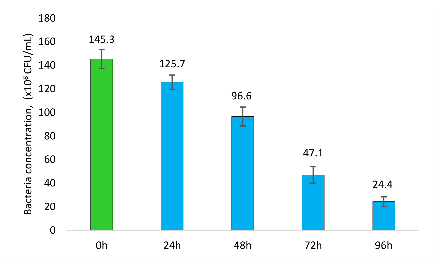
To assess bacterial survival in drinking water, an experiment was performed using an initial bacterial concentration of 1.5 × 10
10 CFU/mL. The appropriate amount of microorganisms was diluted in water, and viable cell counts (CFU/mL) were determined after 24, 48, 72, and 96 h at 20 °C using the serial dilution method followed by colony enumeration on MRS agar plates. The results are presented in
Supplementary Figure S1.
2.2. Analysis of Vitamin and Amino Acid Profiles of Enterococcus sp. SB12
To obtain sufficient biomass, Enterococcus sp. SB12 was cultivated in MRS medium for 18 h at 37 °C. The biomass was harvested by centrifugation at 4000× g for 10 min, washed with sterile distilled water to remove residual culture medium components, and resuspended in a sterile 10% sucrose solution. Viable cell counts were determined by serial tenfold dilution and plating on MRS agar, followed by colony enumeration. Biomass yield was determined by gravimetric dry cell weight analysis.
For metabolite analysis, bacterial biomass, cell-free conditioned medium obtained after cultivation, and fresh MRS medium (control) were subjected to extraction followed by high-performance liquid chromatography (HPLC).
For vitamin analysis, bacterial biomass was separated from the medium by centrifugation at 4000× g for 10 min at 4 °C. The cells were disrupted by ultrasonication for 15 min without additional heating, and the obtained cell suspension was diluted at a ratio of 1 g of biomass per 10 mL of solvent. The solvent consisted of phosphoric acid solution, pH adjusted to 3.0 with 1 M NaOH.
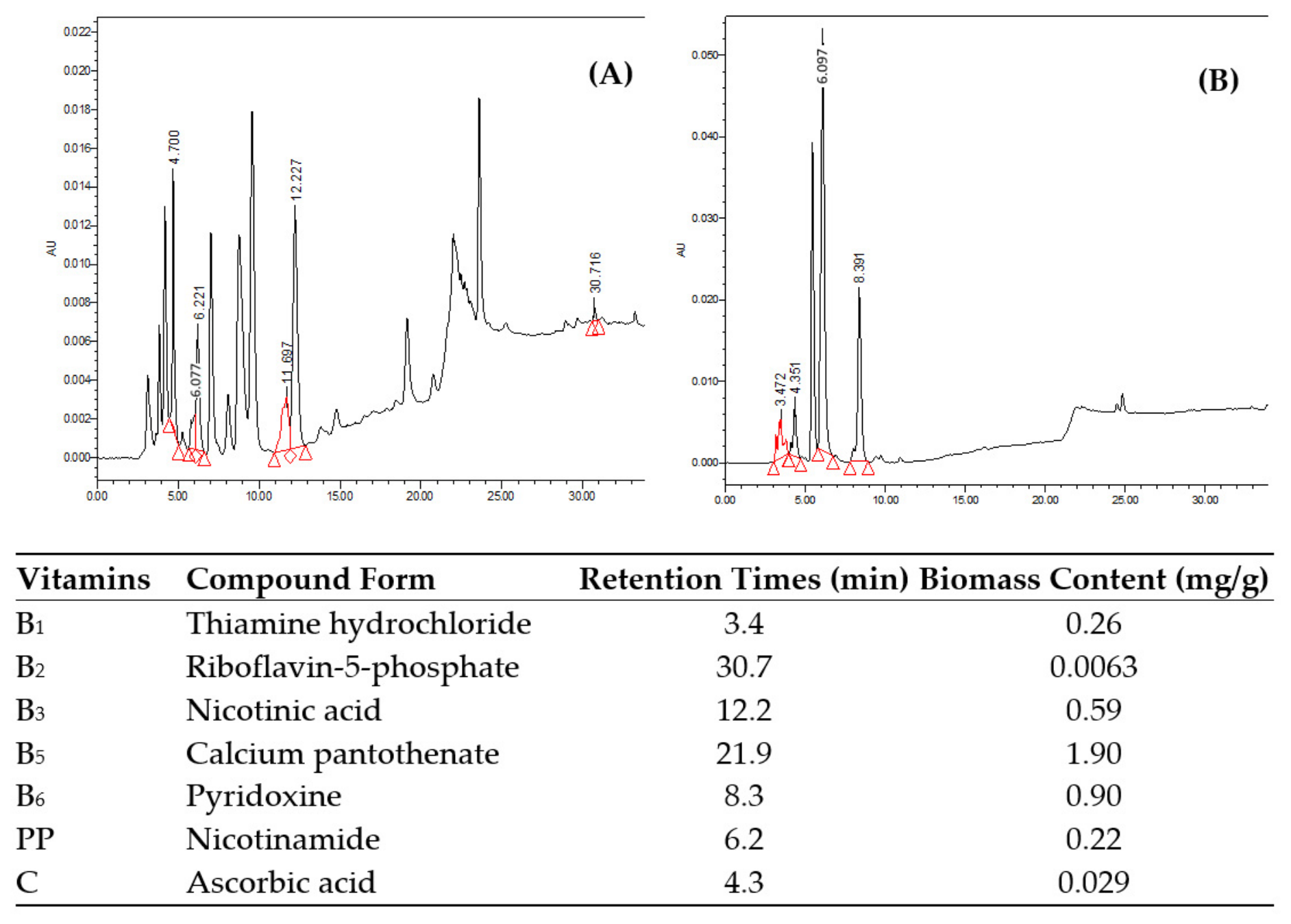
Water-soluble vitamins, including thiamine hydrochloride, riboflavin-5-phosphate, nicotinamide, nicotinic acid, pyridoxine, ascorbic acid, and calcium pantothenate, were identified and quantified using an HPLC system equipped with a reverse-phase C18 column (250 × 4.6 mm, Luna® Omega, Phenomenex, Torrance, CA, USA, 5 μm particle size) and UV detection at 200–265 nm, and quantitative analysis was carried out at 265 nm. Empower 2.0 was used for vitamin analysis, and Chromeleon 7.4.2 was used for amino acid analysis. Chromatographic separation of vitamins was performed at a flow rate of 1.0 mL/min, column thermostat temperature of 25 °C, and injection volume of 10 µL. Separation was performed using a gradient elution system with acetonitrile and phosphate buffer (pH 3.0).
Quantification was carried out using certified HPLC-grade vitamin standards. Standard stock solutions were prepared in the mobile-phase buffer and diluted to the following concentrations: vitamin B1, 30 µg/mL; vitamin B2, 0.63 µg/mL; vitamin B3/nicotinic acid, 59 µg/mL; vitamin B5, 190 µg/mL; vitamin B6, 90 µg/mL; nicotinamide, 22 µg/mL; and ascorbic acid, 2.2 µg/mL. Five-point calibration curves were prepared in the range of 80–120% of these concentrations, and vitamin concentrations in the samples were calculated from the corresponding calibration curves. The HPLC gradient program is presented in
Table S11.
Amino acid composition was determined using HPLC after acid hydrolysis of bacterial biomass (15% HCl, 140 °C, 7 h). The hydrolysates were neutralized and derivatized using 2,4-dinitrofluorobenzene, and separated on a reverse-phase column (250 × 3.0 mm). Detection was performed at 350 nm. Identification and quantification were carried out by comparison with certified amino acid standards.
Standard solutions were prepared at 0.1 mg/mL in 0.05 M sodium tetraborate solution and diluted to 1, 3, 5, 7, and 10 µg/mL for calibration. Derivatization was performed using 0.5 M dinitrofluorobenzene in acetonitrile as the derivatization reagent and 0.05 M sodium tetraborate solution as the alkalizing agent. The reaction was carried out in a water bath at 60 °C for 60 min. Chromatographic separation was performed at a flow rate of 1.0 mL/min and column thermostat temperature of 30 °C. The mobile phase consisted of 0.05 M sodium dihydrogen phosphate buffer, adjusted to pH 2.0 with phosphoric acid, and acetonitrile. The amino acid gradient program is presented in
Table S12.
One biological sample was prepared for each experiment. Each sample underwent a single extraction procedure, and each extract was analyzed by four repeated chromatographic injections. The mean value of the injections was used for quantitative analysis. The relative standard deviation (RSD) of peak areas was below 2%, and the RSD of retention times was below 1%. Standard solutions remained stable for 24 h, with differences in peak areas at 0, 12, and 24 h below 2%. Calibration curves showed good linearity, with coefficients of determination (R2) ≥ 0.997. Accuracy was evaluated by spiking bacterial suspensions with standard compounds, and recovery values ranged from 90% to 107%.
2.3. In Vivo Experiment
All experimental procedures with animals in vivo were approved by the Bioethics Committee of the Institute of Animal Biology of the National Academy of Agrarian Sciences of Ukraine (Protocol №132 from 2 December 2024) in accordance with the current legislation of Ukraine and European Union.
One-month-old female mice obtained from the vivarium of Ivan Franko National University of Lviv underwent a one-week acclimatization period in the animal facility of the Institute of Animal Biology of the National Academy of Agrarian Sciences of Ukraine prior to the experiment. Female mice were selected to minimize aggression-related behavioral variability and to ensure uniform experimental conditions.
Throughout the experiment, mice had access to a standard laboratory rodent diet and drinking water. The animals were fed a commercially available pelleted maintenance diet formulated to meet the nutritional requirements of laboratory mice.
Throughout the experimental period, the mice had unlimited (ad libitum) access to water and standard pelleted feed (Vita, Obuhiv, Ukraine). The composition and nutritional characteristics of the diet used in this study are presented in
Supplementary Table S13. The animals were housed in a climate-controlled facility under standard laboratory conditions (22–24 °C, 50–65% relative humidity, and a 12 h light/dark cycle).
A total of 20 mice with an average initial body weight of 20 ± 0.74 g were included in this study. The mice were randomly assigned to two experimental groups, each housed in two cages (0.35 m2 each) containing five animals per cage (M1–M2: control group; M3–M4: experimental group). The control group (n = 10) received a standard laboratory diet and drinking water, whereas the experimental group (n = 10) received the same diet and drinking water supplemented daily with freshly prepared suspensions of Enterococcus sp. SB12 at a target dose of 1 × 10⁸ CFU/g body weight/day. The bacterial concentration in the drinking water was calculated based on the mean initial body weight of the animals and their average daily water consumption (4.71 ± 0.45 mL per mouse, corresponding to 23.6 ± 1.3 mL per cage during the acclimatization period), resulting in a final bacterial concentration of approximately 4.25 × 10⁸ CFU/mL. Each cage was provided with 100 mL of freshly prepared bacterial suspension daily, while the average daily consumption was 23.6 ± 1.3 mL per cage. Drinking bottles were replaced every 24 h, and water consumption was monitored at the cage level throughout the experiment.
On the 29th day of the experiment, all animals were humanely euthanized by rapid displacement of the cervical vertebrae. Body weight as well as the weights of the liver, heart, kidneys, and total intestinal fat were recorded for each animal. Blood, tissue, and organ samples from both the experimental and control groups were collected for genetic and biochemical analyses. Tissue samples were homogenized in 1 M Tris-HCl buffer (pH 7.4) to obtain homogenates, which were stored at −20 °C and analyzed within six months of sample collection.
2.4. Metataxonomic Library Preparation, Sequencing, and Bioinformatic Analysis
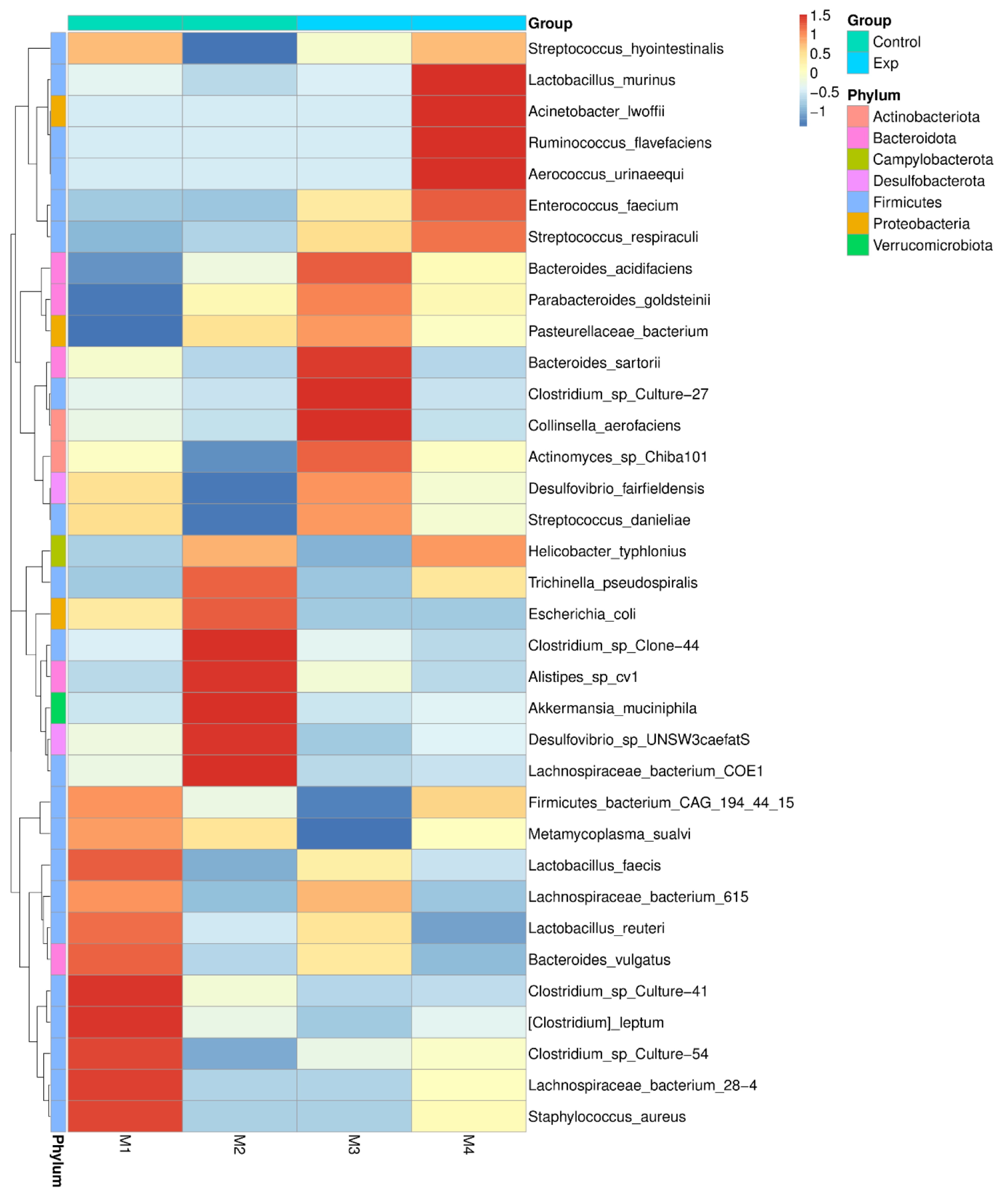
Intestinal content samples were collected from control and experimental mice on day 29 following euthanasia. Samples from animals housed within the same cage were pooled prior to DNA extraction, resulting in four composite samples: M1 and M2 (control group) and M3 and M4 (experimental group). Therefore, each cage represented a single microbiome sample.
DNeasy PowerSoil Pro Kit (Qiagen, Hilden, Germany) was used to extract DNA. The V3-V4 16S rRNA region was amplified using primers 341F (5′-CCTACGGGNGGCWGCAG-3′) and 806R (5′-GGACTACNVGGGTWTCTAAT-3′). The internal transcribed spacer 2 (ITS2) region was amplified using primers ITS3-2024F (5′-GCATCGATGAAGAACGCAGC-3′) and ITS4-2409R (5′-TCCTCCGCTTATTGATATGC-3′). PCR products were purified using the QIAquick Gel Extraction Kit (Qiagen, Hilden, Germany). Sequencing was performed on an Illumina NovaSeq 6000 platform using paired-end 2 × 250 bp chemistry, generating more than 100,000 reads per sample (Novogene (UK) Company Limited, Cambridge, UK). Paired-end reads were merged using FLASH (version 1.2.11) [
14]. Sequence analysis was performed using UPARSE (version 7.0.1001). All sequences with ≥97% similarity were assigned to the same operational taxonomic units (OTUs). A representative sequence from each OTU was selected for taxonomic annotation. Further annotation of OTUs was performed using Mothur software v1.48.1 in the SSU rRNA SILVA database for taxonomic assignment of bacterial taxa [
14] and the Unite database [
15] based on the blast algorithm for taxonomic assignment of fungal taxa. Multiple sequence alignment was performed using the MUSCLE algorithm (version 3.8.31). Alpha-diversity and beta-diversity analyses were performed using the OTU abundance tables and are presented in the
Supplementary Materials (Tables S1 and S2, and Figures S3 and S4).
2.5. Analysis of Biochemical Parameters
2.5.1. Determination of HDL Cholesterol, LDL Cholesterol, Glucose, Protein Content
The levels of high-density lipoprotein (HDL) cholesterol, low-density lipoprotein (LDL) cholesterol, glucose, and total protein were determined using commercial assay kits (Filisit, Dnipro, Ukraine): CHOLESTEROL-HDL F (53,391), CHOLESTEROL-LDL F (HP026.05), GLUCOSE F (53,301), and Total Protein (61,900), in accordance with the manufacturer’s instructions.
2.5.2. Determining the Content of Thiobarbituric Acid-Reactive Substances
A 0.01 mL sample homogenate (test) and 2 mL distilled H
2O (control) were incubated for an hour at 37 °C. Then, 1 mL of trichloroacetic acid and 1 mL of 0.8% thiobarbituric acid were added. The samples were boiled in a water bath for 10 min, and then centrifuged at 3000 rpm for another 10 min and the supernatant was collected. The absorbance of the samples was measured at 535 nm against the control. The content of TBARS was determined by the following formula:
where C is the concentration of TBARS, E
sample is the absorbance of the sample, V
total is the total volume of the reaction mixture, 1.56 is the molar extinction coefficient used in the assay, and V
sample is the volume of the analyzed sample.
TBARS concentration was expressed as nmol of thiobarbituric acid-reactive substances per milliliter of sample (nmol/mL) [
16].
2.5.3. Determination of Oxidative Modification of Proteins
For each test and control sample, 0.1 mL of homogenate and 900 μL of 20% trichloroacetic acid were added. For the test sample, 1 mL of 0.1 M 2,4-dinitrophenylhydrazine solution was added, whereas for the control sample, 1 mL of 2 M HCl was added. The tubes were incubated for 1 h at 37 °C and then centrifuged for 45 min at 3000 rpm. The precipitate was washed three times with an ethanol–ethyl acetate mixture, and 2.5 mL of 8 M urea was added to each tube. The samples were then boiled for 5 min in a water bath until the precipitate was completely dissolved.
Absorbance was measured at 274, 370, and 430 nm to assess different products of oxidative protein modification. The concentration was calculated using the following equation:
where C is the concentration of oxidative modification of proteins, E
sample is the absorbance of the sample, V
total is the total volume of the reaction mixture, C
protein is the protein concentration in the sample, 22 is the extinction coefficient used for the assay, and V
sample is the volume of the analyzed sample.
The obtained values were expressed as nmol of oxidatively modified protein products per milligram of protein (nmol/mg protein) [
16].
2.5.4. Determination of Catalase Activity
To the test and control samples, 100 μL of blood serum (or tissue homogenate) and 2.0 mL of 0.03% hydrogen peroxide (H
2O
2) solution were added. After incubation for 10 min, 1.0 mL of 4% ammonium molybdate solution was added to terminate the reaction. Subsequently, 1.0 mL of 30% trichloroacetic acid was added, and the mixture was centrifuged at 3000 rpm for 10 min. The absorbance of the supernatant was measured spectrophotometrically at 410 nm. The calculation was performed according to the following formula:
where C is the catalase activity, E
control is the absorbance of the control, E
sample is the absorbance of the sample, V
total is the total volume of the reaction mixture, 22.2 is the molar extinction coefficient of the hydrogen peroxide-ammonium molybdate complex, C
protein is the protein concentration in the sample, V
sample is the volume of the analyzed sample, and t is the time of reaction.
Catalase activity was expressed as μmol of H
2O
2 decomposed per minute per milligram of protein (μmol/min/mg protein) [
16].
2.6. Statistical Analysis
Results are presented as mean ± SD. Statistical analysis was performed using Microsoft Excel. Diagrams were also created in Microsoft Excel. For biochemical and morphometric parameters, comparisons between the control and experimental groups were performed using Student’s t-test. Statistical significance was set at p < 0.05, p < 0.01, and p < 0.001.
For microbiome analysis, intestinal samples from animals housed in the same cage were pooled prior to DNA extraction, resulting in two biological replicates per treatment group. Therefore, formal differential abundance testing was not performed, and microbiome results are presented as descriptive comparisons of taxonomic composition and diversity metrics.
4. Discussion
The present study provides a comprehensive evaluation of the probiotic potential and safety profile of Enterococcus sp. SB12 isolated from traditional Carpathian cheese “brynza” through analysis of bacterial survival, metabolic characteristics, intestinal microbiome modulation, and physiological effects in a mouse model. The obtained results demonstrated that Enterococcus sp. SB12 retained sufficient viability under drinking-water administration conditions, was associated with changes in the composition of bacterial and fungal intestinal communities, and contained several biologically relevant vitamins and amino acids in its biomass. Furthermore, administration of the strain for 29 days was associated with increased body weight and did not result in detectable alterations in biochemical parameters, oxidative stress markers, antioxidant defense indicators, or organ weights in mice.
The observed time-dependent decline in
Enterococcus sp. SB12 viability in drinking water is consistent with previous reports demonstrating that enterococci and other lactic acid bacteria maintain sufficient short-term survival in aqueous environments despite gradual loss of culturability. However, during the first 24 h of storage, the number of viable microorganisms remained sufficient to maintain a sufficiently high viable cell count for the in vivo experiment. Similar studies have shown that enterococci can retain viability in drinking water long enough to ensure effective oral administration in animal models, supporting the applicability of this route for probiotic delivery [
17].
The vitamin profile of
Enterococcus sp. SB12 revealed the presence of several physiologically important water-soluble vitamins, including B-group vitamins and ascorbic acid. The highest concentrations were detected for calcium pantothenate (B5), pyridoxine (B6), and nicotinic acid (B3). Similar vitamin profiles have been reported for probiotic and food-associated lactic acid bacteria and are considered desirable functional characteristics because such microorganisms may contribute to the nutritional value of fermented foods [
18].
Analysis of amino acid composition showed the high concentrations of valine, glutamine, cysteine, and proline in bacterial biomass. Increased intracellular level of glutamine and branched-chain amino acids is frequently associated with microbial growth, protein synthesis, and stress resistance as well as accumulation of cysteine may contribute to antioxidant protection due to its role as a precursor of thiol-containing compounds [
19].
Interestingly, a marked increase in taurine concentration was observed in the conditioned medium after cultivation. Taurine is involved in osmoregulation, membrane stabilization, and antioxidant processes in animal organisms. Although taurine metabolism by lactic acid bacteria remains insufficiently characterized, changes in taurine concentration may indicate active transformation of sulfur-containing compounds and deserve further mechanistic investigation [
20].
The administration of
Enterococcus sp. SB12 for 29 days resulted in a significant increase in body weight without affecting the relative weights of internal organs or visceral fat. The absence of significant differences in liver, heart, kidney, and adipose tissue weights suggests that SB12 supplementation was not associated with detectable alterations in organ mass. Previous studies investigating probiotic enterococci in animal models have reported similar observations, where administration of selected
Enterococcus strains improved growth performance without negatively affecting organ morphology or biochemical parameters [
21,
22].
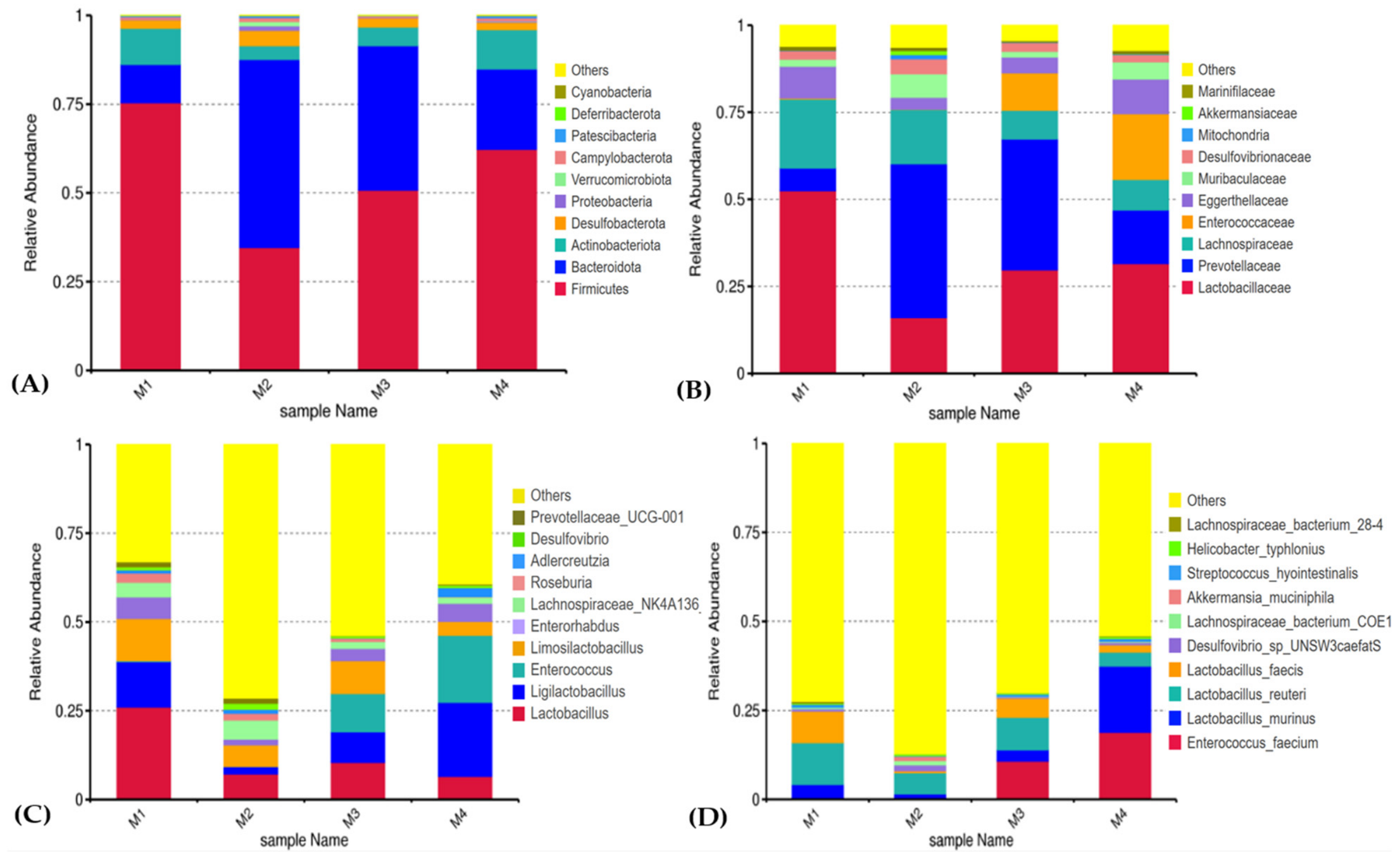
This metataxonomy profile obtained in our study is considered with reference datasets describing the “baseline” bacteria in the guts of healthy laboratory mice, where Firmicutes and Bacteroidota typically represent the largest fractions, and families such as Lachnospiraceae and Lactobacillaceae are commonly observed [
23]. Because the microbiome analysis was based on two pooled intestinal samples per group, no differential abundance testing was possible. Therefore, all microbiome observations presented below should be interpreted as descriptive trends rather than statistically confirmed differences. In the pooled intestinal samples, administration of
Enterococcus sp. SB12 was associated with a descriptively higher relative abundance of representatives belonging to the phyla Firmicutes, Actinobacteriota, Campylobacterota, Patescibacteria, Cyanobacteria, and Spirochaetota, as well as enrichment of bacterial genera including
Enterococcus,
Limosilactobacillus,
Ligilactobacillus,
Carnobacterium,
Bacteroides, and
Alistipes.
The observed increase in
Enterococcus abundance in the experimental group may be consistent with persistence of the administered strain within the gastrointestinal tract. However, because the sequencing approach used in this study does not provide strain-level resolution, these sequences cannot be unequivocally attributed to the administered strain. A similar study reported a continuous increase in the relative abundance of Firmicutes in the gut microbiota of male C57BL/6 mice following 28 days of
Enterococcus faecalis DH9003 administration. Furthermore, the authors demonstrated that
E. faecalis DH9003 was able to colonize the mouse gastrointestinal tract after intragastric administration, resulting in a regulatory effect on the intestinal microbiota composition [
24].
The administration of Enterococcus sp. SB12 was associated with a reduction or complete disappearance of several microbial groups, including members of the phyla Desulfobacterota, Proteobacteria, Chloroflexi, and Deferribacterota. Although members of this taxonomic group have been associated with intestinal inflammation in previous studies, the present observation is based on pooled samples and should be regarded as descriptive rather than as evidence of a statistically confirmed effect of Enterococcus sp. SB12.
This is because sulfate-reducing bacteria are often considered to be factors contributing to intestinal inflammation through the production of hydrogen sulfide, and they have been shown to exacerbate DSS-induced colitis in C57BL/6 model mice [
25,
26]. Similarly, a reduction in the relative abundance of Proteobacteria-associated taxa, including members of the genus
Helicobacter, was observed in the experimental group. Increased abundance of Proteobacteria is frequently considered a marker of gut microbiota imbalance and inflammation. However, unlike
Helicobacter pylori, which is a recognized human pathogen associated with chronic gastritis and gastric cancer [
27], several
Helicobacter species commonly occur in laboratory and wild mice as part of the normal gut microbiota and can play an important role in maintaining of it homeostasis [
28]. Therefore, the lower relative abundance of
Helicobacter observed in the pooled experimental samples represents a descriptive observation of microbial community composition. Because no differential abundance testing was possible, its biological significance cannot be determined from the present study. However, it cannot be considered direct evidence of a beneficial health effect, especially in humans or animal species other than mice.
Beyond bacterial communities, administration of
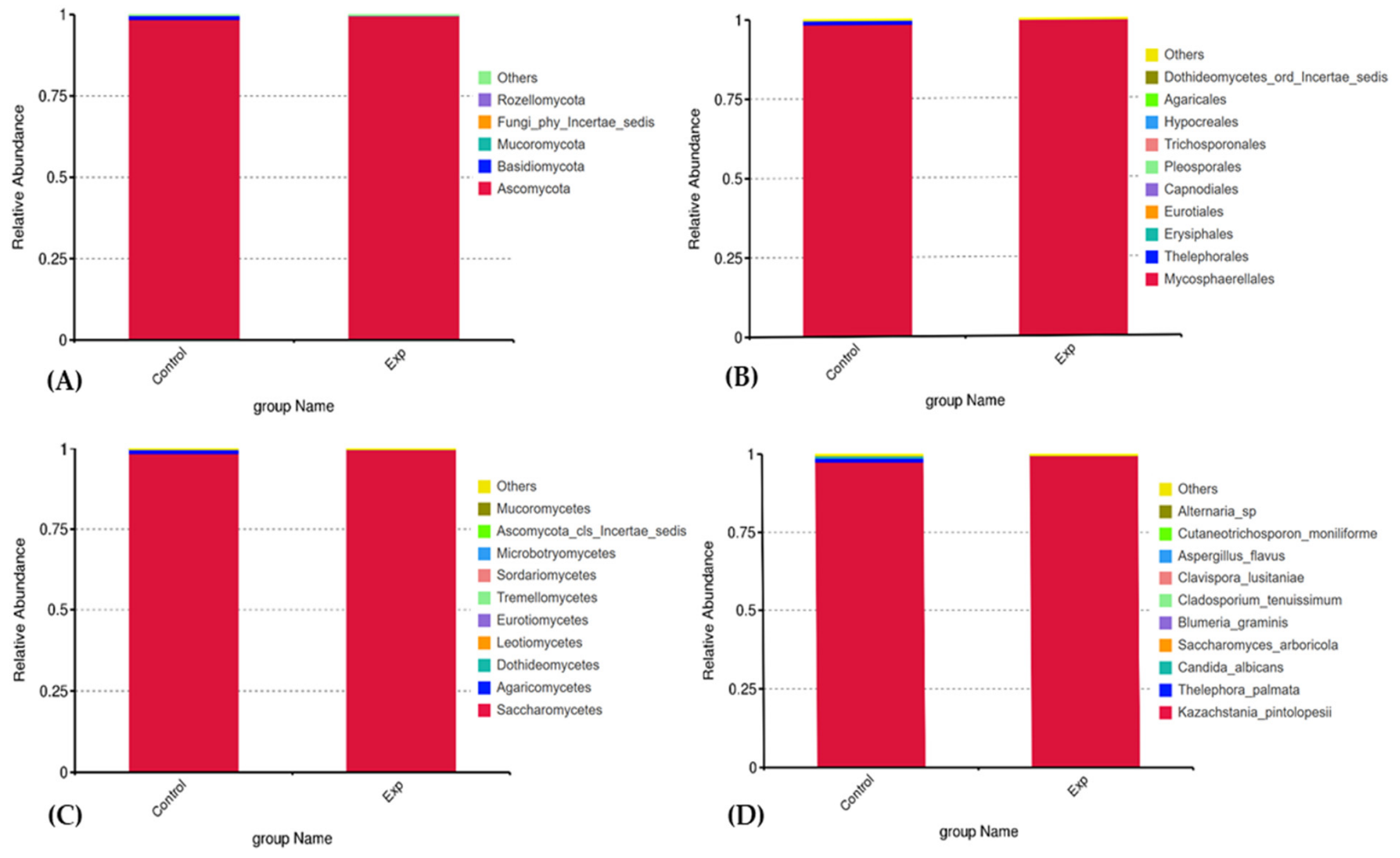
Enterococcus sp. SB12 was also associated with alterations in fungal populations within the gut microbiome. The experimental group showed decreased representation of fungal genera including Candida, Aspergillus, Fusarium, Pichia, Alternaria, and Saccharomyces, whereas increased abundance of
Lichtheimia and
Psathyrella was observed (
Tables S7–S10). According to literature on the gut microbiome, fungal communities typically have lower biomass, are more variable than bacterial populations, and are highly dependent on diet, host genetics, and interactions between bacteria and fungi [
29]. Such changes support the concept of cross-kingdom interactions between bacterial probiotics and intestinal fungi, although the functional implications of these shifts require further targeted investigation.
The metataxonomic findings indicate that administration of Enterococcus sp. SB12 was associated with changes in both bacterial and fungal intestinal communities without evidence of major dysbiosis-associated shifts. The observed changes included increased relative abundance of several taxa commonly reported in healthy murine microbiota and decreased representation of some microorganisms previously associated with inflammatory or gastrointestinal disorders. Nevertheless, the overall microbial community shifts observed in this study provide preliminary support for the probiotic potential of Enterococcus sp. SB12.
The present study aimed to evaluate the physiological safety of the newly isolated Enterococcus sp. SB12 strain through assessment of biochemical parameters, including HDL and LDL cholesterol, glucose, total protein, TBARS, oxidative modification of proteins, and catalase activity, which are commonly used to assess metabolic stability and potential toxicological and beneficial effects of probiotic in animal models.
HDL and LDL cholesterol concentrations are widely recognized as indicators of lipid metabolism and overall metabolic health. Some enterococcal strains have been reported to exhibit cholesterol-lowering properties, potentially mediated through bile salt hydrolase (BSH) activity, which promotes bile salt deconjugation and influences cholesterol metabolism [
30]. Similarly, blood glucose levels reflect carbohydrate metabolism, whereas total protein concentrations—particularly albumin—serve as indicators of protein metabolism, transport functions, and general physiological status [
6]. In the present study, administration of
Enterococcus sp. SB12 did not result in significant changes in blood glucose, cholesterol, or total protein levels, suggesting that prolonged supplementation with the strain did not disrupt major metabolic processes in mice. These findings indicate metabolic stability and are consistent with a favorable safety profile of the investigated strain.
Interestingly, mice receiving
Enterococcus sp. SB12 exhibited a significantly higher body weight than control animals at the end of the experiment, despite the absence of significant differences in blood glucose, total protein, HDL, and LDL concentrations. Moreover, no changes were detected in visceral fat accumulation or organ weights. The increase in body weight was not accompanied by adverse alterations in biochemical parameters or oxidative stress markers, including lipid peroxidation, oxidative protein modification, and catalase activity. These findings suggest that administration of
Enterococcus sp. SB12 did not induce detectable metabolic disturbances under the conditions of this study. The mechanism underlying the observed increase in body weight remains unclear; however, it may be associated with microbiota-mediated effects on nutrient utilization, energy harvest, or growth performance. Since these parameters were not directly investigated, no definitive conclusions can be drawn regarding the underlying mechanism. Therefore, the observed increase in body weight should be interpreted as a physiological response associated with strain administration rather than as direct evidence of improved metabolic status. Similar observations have been reported for probiotic enterococci and other lactic acid bacteria, where increased growth performance occurred without pathological changes or adverse metabolic effects [
31].
An important indicator of potential toxic effects of a microbial strain on the host organism is the induction of oxidative stress, which is associated with the excessive formation of reactive oxygen species (ROS). Elevated levels of ROS can damage cell membranes, organelles, and nucleic acids, ultimately leading to cell dysfunction and death. In addition, the decay products released from necrotic cells can trigger inflammatory processes and autoimmune reactions in the body [
32]. One of the main markers of oxidative stress is the level of malondialdehyde, which increases in response to an increase in the amount of ROS. It is a mutagen and has pronounced cytotoxicity, leading to changes in the structure of the cell membrane and potentially causing its destruction [
33]. In our study, concentrations of TBARS in blood, liver, and kidney tissues did not differ significantly between the experimental and control groups, interpreted as evidence of metabolic and toxicological safety following administration of
Enterococcus sp. SB12. Similar results were obtained for other strains of enterococci with probiotic potential, where stable levels of malondialdehyde and preserved redox balance were interpreted as evidence of metabolic and toxicological safety in animal models [
34].
Reactive oxygen species additionally promote oxidative modification of proteins (OMP), resulting in carbonyl formation, structural changes, and potential loss of protein function. Accumulation of oxidatively modified proteins contributes to cellular damage and may amplify oxidative stress through secondary molecular injury [
35]. However, OMP values measured at different wavelengths corresponding to early and advanced stages of protein oxidation did not differ significantly between the experimental and control groups. These findings suggest that administration of
Enterococcus sp. SB12 was not associated with detectable oxidative protein damage in the examined tissues.
The intracellular antioxidant defense system, comprising enzymes such as superoxide dismutase, catalase, and glutathione peroxidase, represents a primary mechanism protecting tissues from ROS-mediated injury. Catalase is particularly important because of its ability to rapidly decompose hydrogen peroxide into water and oxygen, thereby limiting oxidative damage [
36]. Reduced catalase activity has been associated with several pathological conditions, including neurodegenerative disorders and chronic oxidative stress [
37]. In the present study, catalase activity did not differ significantly between the experimental and control groups, indicating preserved antioxidant defense mechanisms and unchanged redox homeostasis.
Limitations
In our opinion, the interpretation of the microbiome data should take into account that gut microbiome analysis was performed using pooled intestinal samples from animals housed within the same cage, resulting in four composite samples (M1-M4) rather than individual microbiome profiles. Consequently, cage-associated effects could not be separated from treatment-associated effects, and the microbiome findings should be regarded as descriptive evidence of community shifts rather than statistically robust individual-animal comparisons.
Another limitation is the absence of food consumption and feed conversion efficiency measurements, which limits the interpretation of the observed increase in body weight. Therefore, it was not possible to determine whether the weight gain was associated with differences in feed intake, nutrient utilization efficiency, or other physiological mechanisms.
Several additional limitations should also be acknowledged. The present study did not include histopathological examination of tissues, bacterial translocation assays, or phenotypic confirmation of antimicrobial resistance and virulence traits, although previous whole-genome analysis of
Enterococcus sp. SB12 indicated the absence of clinically relevant antimicrobial resistance and virulence genes [
13]. Furthermore, the experiment was limited to a single treatment period using healthy mice of one sex and a similar age; therefore, long-term safety, sex- and age-related responses, and additional functional and immunological outcomes were not evaluated. Future studies addressing these aspects will provide a more comprehensive assessment of the
Enterococcus sp. SB12 safety profile.
5. Conclusions
The present study demonstrated that Enterococcus sp. SB12, isolated from traditional Carpathian artisanal cheese, exhibits promising probiotic characteristics and showed no adverse effects in mice following 29 days of oral administration. No significant changes were observed in body organ indices, biochemical parameters, or oxidative stress markers, supporting the safety of the strain under the experimental conditions.
Administration of Enterococcus sp. SB12 was associated with descriptive changes in the composition of the intestinal microbiota in pooled samples, including higher relative abundances of members of the phyla Firmicutes and Actinobacteriota and lower relative abundance of several potentially undesirable taxa. The pooled intestinal sample from the treated mice also showed a descriptively higher relative abundance of the genus Enterococcus, which may be consistent with the presence of the administered strain at the time of sampling. Because the microbiome analysis was based on pooled samples, these observations should be considered preliminary and require confirmation in future studies using individual biological replicates and strain-specific approaches.
HPLC analysis revealed the presence of several biologically important vitamins in the bacterial biomass, including vitamins B1, B3, B5, B6, and C, as well as amino acids such as taurine, proline, and valine, demonstrating its metabolic activity and potential nutritional value.
Administration of Enterococcus sp. SB12 for 29 days was associated with a moderate increase in body weight compared with the control group, while no significant changes were observed in organ morphology, visceral fat mass, blood glucose, total protein, HDL, or LDL concentrations. Furthermore, no significant alterations were detected in TBARS, OMP, or catalase activity in blood, liver, or kidney tissues, indicating the absence of detectable oxidative stress or impairment of antioxidant defense mechanisms.
Overall, administration of Enterococcus sp. SB12 was associated with compositional changes in the intestinal microbiome and did not induce detectable alterations in biochemical or oxidative stress parameters. These findings provide preliminary evidence supporting the probiotic potential and safety of Enterococcus sp. SB12 under the experimental conditions used; however, additional studies are required to confirm its long-term safety profile and functional effects on the host.




 ,
,