


Abstract
High fructose intake rapidly induces protein glycation, oxidative stress, inflammation, and disturbances in the cultivable fraction of gut bacteria, contributing to early metabolic impairment. This study examined whether selected plant-derived bioactives and a prebiotic–probiotic formulation could mitigate fructose-induced biochemical and microbial alterations. Male Wistar rats were fed fructose for 45 days, and advanced glycation end product (AGE)-associated fluorescence, oxidative stress markers, and lipid peroxidation were measured to assess metabolic changes. Culture-dependent enumeration of intestinal and fecal bacteria was performed to evaluate shifts in cultivable aerobic and facultative bacterial counts, while limonene, eugenol, and emodin were tested for antibacterial activity against aerobic and facultative bacterial isolates obtained from fructose-fed rats during our previous study. The prebiotic–probiotic formulation was assessed alone and in combination with these bioactives. Fructose feeding increased protein glycation, oxidative stress, lipid peroxidation, and reduced counts of cultivable gut bacteria. Treatment with the bioactives and the formulation lowered glycation-related fluorescence, reduced oxidative stress, and decreased lipid peroxidation. The bioactives exhibited antioxidant and antiglycation activity and inhibited growth of selected cultivable bacterial isolates, including Corynebacterium stationis. While emodin contributed primarily through its known α-glucosidase inhibitory and antiglycation properties rather than antibacterial activity. Combined treatment partially restored cultivable bacterial counts and improved metabolic parameters. Overall, the interventions attenuated fructose-induced biochemical disturbances and modulated the cultivable gut bacterial counts, suggesting a complementary approach to managing early metabolic changes in rats associated with high fructose intake.
1. Introduction
Diabetes mellitus is characterized by chronic hyperglycemia arising from impaired insulin secretion or action, and long-standing elevation of blood glucose is closely linked to the development of secondary complications [1]. A major biochemical contributor to these complications is non-enzymatic protein glycation, a process in which reducing sugars such as glucose or fructose react with amino groups of proteins through the Maillard reaction, ultimately forming irreversible advanced glycation end products (AGEs) [2]. Accumulation of AGEs alters protein structure and function and has been associated with microvascular and metabolic complications of diabetes [3,4]. AGEs also promote oxidative stress and cytokine signaling, thereby contributing to inflammation and tissue injury [5]. Thus, glycation, oxidative stress, and immune-mediated interactions represent interconnected drivers of metabolic deterioration.
It is well established that the gut microbial ecosystem plays an important role in regulating host metabolism, inflammation, and nutrient processing. Disturbances in this community are increasingly linked to early metabolic changes associated with high-glycemic diets. Although the majority of gut bacteria are obligate anaerobes that require sequencing-based approaches for comprehensive characterization, fructose-rich diets have also been shown to influence the aerobic and facultative fraction that can be captured through culture-dependent methods. These cultivable bacteria represent a small but metabolically relevant subset that can respond to luminal glycation, oxidative stress, and changes in nutrient availability. Recent observations suggest that high-glycemic diets may also influence the cultivable fraction of the gut bacterial community by promoting glycation of intestinal proteins and altering the microbial environment [6,7,8,9]. Our earlier work showed that fructose feeding in experimental rats induces changes in gut protein structure and alters cultivable bacterial counts, changes that resemble early pre-diabetic alterations [10,11]. Specifically, glycation of intestinal α-glucosidase was found to reduce its activity, followed by compensatory overexpression, contributing to exaggerated postprandial glucose excursions [10]. Dietary patterns rich in high-glycemic foods and fructose syrups, combined with sedentary behavior, are well-recognized contributors to pre-diabetes and its progression to overt diabetes [12]. We also observed that glycated gut proteins may influence shifts in cultivable bacterial populations, potentially reinforcing metabolic stress [13]. These findings raise the question of whether glycation inhibitors, together with prebiotic–probiotic formulations, could interrupt this cycle by limiting glycation and supporting the cultivable bacterial fraction.
The present investigation builds upon our earlier observations that short-term fructose feeding increases oxidative stress, accelerates protein glycation, alters serum biochemical indices, and promotes early metabolic disturbances [11]. In that work, aminoguanidine and a selected prebiotic–probiotic formulation effectively limited these changes; however, due to the clinical limitations associated with aminoguanidine, it was not included in the present study. Instead, the current work focuses on phytochemicals previously identified for antiglycation, antioxidant, or glucose-modulating properties as potential alternatives. Our laboratory has long investigated plant-derived bioactive compounds, identifying several with antidiabetic potential. Limonene [14], emodin [15], and eugenol [16] were previously isolated as active constituents from Aegle marmelos, Rheum emodi, and Piper betle, respectively. Limonene and eugenol exhibit antiglycation activity, while emodin is a potent α-glucosidase inhibitor and has been reported to influence gastric motility, which may affect microbial clearance. Independent studies have described emodin as antihyperglycemic and renoprotective [17,18,19], limonene as anti-inflammatory [20] and protective against diabetic complications [21,22], and eugenol as anti-inflammatory, renoprotective, and beneficial in insulin resistance [23,24].
Given these properties, it is relevant to examine whether these phytochemicals can modulate rat cultivable gut bacterial counts altered by fructose feeding and whether their combined antiglycation, antioxidant, and culture-dependent microbial modulatory effects, with or without prebiotic–probiotic supplementation, can mitigate fructose-induced biochemical and microbial disturbances. The present study was undertaken to evaluate these possibilities and to assess the potential of these interventions in limiting early metabolic changes associated with high fructose intake in rats.
Recent studies have shown that high fructose intake can disrupt gut microbiota and contribute to metabolic dysfunction [25,26,27,28,29]. What remains unclear is how fructose affects the cultivable bacterial fraction and how these early microbial shifts relate to oxidative stress, barrier changes, and metabolic imbalance. Our study adds new insight by focusing specifically on the cultivable component of the gut microbiota and by testing a combined antiglycation and prebiotic–probiotic strategy designed to counter these early disturbances. This integrated approach provides a new perspective on how dietary sugars influence host–microbe interactions and identifies a potential intervention framework for mitigating fructose-induced metabolic stress.
2. Materials and Methods
2.1. Chemicals and Materials
Analytical-grade reagents including sodium azide, nitro blue tetrazolium (NBT), hydrogen peroxide, and other routine chemicals were obtained from authorized local distributors (HiMedia Laboratories Pvt. Ltd., Mumbai, India). Streptozotocin and the purified phytochemicals (eugenol, emodin, and limonene) were purchased from Sigma-Aldrich (St. Louis, MO, USA). The antibiotics streptomycin, ampicillin, and kanamycin were procured from HiMedia (HiMedia Laboratories Pvt. Ltd., Mumbai, India). Blood glucose measurements were performed using an ACCU-CHEK glucometer (Roche Diagnostics, Mannheim, Germany).
A commercially available prebiotic–probiotic formulation, i.e., EUGI™ (WALLACE Pharmaceuticals Pvt. Ltd., Mumbai, India), was used for supplementation. EUGI™ composition (per 1 g): fructo-oligosaccharides (300 mg), Lactobacillus acidophilus, L. rhamnosus, Bifidobacterium longum, B. bifidum, Streptococcus thermophilus (0.24 billion CFU each), and Saccharomyces boulardii (0.05 billion CFU).
2.2. In Vitro Assessment of Bioactive Compounds and Antibiotics Against Cultivable Gut Bacteria
Antibacterial activity of the phytochemicals and antibiotics was evaluated against cultivable bacterial isolates obtained from fructose-fed rats [13] by spreading 100 µL suspension (OD at 530 nm/600 nm ≥ 0.050) of each isolate on sterile nutrient agar plates. Streptomycin (100 µL of 10 µg) served as the positive control, while 100 µL ethanol was used as the solvent control. Ampicillin and kanamycin were tested at 10 µg (100 µL). Limonene, eugenol, and emodin were tested at 10 µg and 20 µg (100 µL). Phytochemicals were dissolved in ethanol, and equivalent volumes of solvent were used as vehicle controls. All antibiotics were sourced from HiMedia, and phytochemicals from Sigma-Aldrich. The assay conditions were kept uniform across all treatments. The bacterial isolates used in this study were previously identified to the species level in our earlier work [13] using 16S rRNA gene sequencing supported by cultural and biochemical characterization. These isolates represent the aerobic and facultative cultivable fraction accessible through nutrient agar culture; testing antimicrobials directly on mixed dilutions of the cultivable community may identify additional resistant types, and this is acknowledged as a methodological limitation.
2.3. Experimental Animals and Grouping
Male Wistar rats (Rattus norvegicus), 6–7 weeks old and weighing 180–200 g, were housed individually under controlled temperature (21–25 °C) and a 12:12 h light–dark cycle, with free access to standard chow diet (Nutrivet Life Sciences, Pune, India) and filtered water. All procedures followed institutional ethical guidelines (Registration No. 1825/PO/EReBi/S/15/CPCSEA).
Animals (n = 4 per group) were assigned to eight groups:
Group I: Control;
Group II: Diabetic;
Group III: Fructose (3 g twice daily);
Group IV: Fructose + Emodin (0.5 mg twice daily) + EUGI™, i.e., prebiotic–probiotic formulation (50 mg twice daily);
Group V: Fructose + Eugenol (0.5 mg twice daily) + EUGI™ (50 mg twice daily);
Group VI: Fructose + Limonene (0.5 mg twice daily) + EUGI™ (50 mg twice daily);
Group VII: Fructose + [Emodin + Eugenol + Limonene (0.5 mg twice daily)];
Group VIII: Fructose + [Emodin + Eugenol + Limonene (0.5 mg twice daily)] + EUGI™ (50 mg twice daily).
A fructose + EUGI™ group was not included because the independent effects of EUGI™ have been characterized in our earlier work [11], and the present design focused on testing phytochemicals as aminoguanidine alternatives and determining whether their actions are enhanced when combined with a prebiotic–probiotic formulation. Further, four animals per group (n = 4) were used in this study, reflecting its preliminary nature and consistent with prior work in this model as well as institutional ethical guidelines aimed at reducing animal use while obtaining meaningful data. We believe that this sample size is adequate for detecting large, treatment-related effects in key biochemical and oxidative stress parameters. The diabetic control group was included to provide a reference arm for metabolic severity.
All treatments were administered by oral gavage. EUGI™ was administered 30 min after phytochemicals to avoid interaction during absorption. All doses used in this study were determined based on our earlier experimental work and standard laboratory pharmacological practices.
Animals were treated for 45 days and euthanized for biochemical analyses.
2.4. Induction of Diabetes
Diabetes was induced in overnight-fasted male Wistar rats (200–220 g) by intraperitoneal injection of freshly prepared streptozotocin (65 mg/kg body weight) in 0.1 M citrate buffer (pH 4.5). Blood glucose was monitored for 14 days, and rats with glucose levels >200 mg/dL were considered diabetic.
2.5. Oral Glucose Tolerance Test (OGTT) Procedure
For the OGTT, rats were fasted overnight with free access to water. Baseline glucose was recorded, followed by oral administration of glucose (3 mg/g body weight in 0.85% saline). Blood samples were collected from the tail vein at 0, 30, 60, and 120 min, and glucose levels were measured using an ACCU-CHEK glucometer.
2.6. Body Weight, Blood Parameters, and Liver Function Tests
Body weight was recorded after a 12-h fast on day 0 and day 46. Serum triglycerides and high-density lipoprotein (HDL) were measured using commercial kits (Span Diagnostics, Surat, India). Serum creatinine and total cholesterol were assessed using kits from Creast Biosynthesis (Goa, India) and Biolab Diagnostics (Boisar, India), respectively. Very-low-density lipoprotein (VLDL) was calculated from total cholesterol and HDL values. Serum urea was measured using a modified Berthelot method (YUCCA Diagnostics, India). Alanine Aminotransferase (ALT) and Aspartate Aminotransferase (AST) were quantified using ELISA kits (MyBioSource, San Diego, CA, USA; Catalog Nos. MBS264333 and MBS2022384), respectively.
2.7. Antioxidant Enzyme Assays
Liver and kidney tissues (10% w/v) were homogenized in 100 mM Tris-HCl buffer (pH 7.4). The homogenate was centrifuged at 270× g for 10 min, and the supernatant was further centrifuged at 7270× g for 30 min at 4 °C. The resulting supernatant was used to determine superoxide dismutase (SOD) and catalase (CAT) activities. Lipid peroxidation was assessed by measuring malondialdehyde (MDA) using the thiobarbituric acid (TBA) reaction. Protein concentration was estimated using the Lowry method [30].
2.7.1. Superoxide Dismutase (SOD) Assay
SOD activity was determined using the method of Misra and Fridovich [31]. Briefly, 0.1 mL of tissue supernatant was added to a reaction mixture containing 3.5 mL phosphate buffer (50 mM, pH 7.4), 0.3 mL ethylenediaminetetraacetate (EDTA; 10 mM), 1.2 mL methionine (130 mM), and 0.6 mL nitro blue tetrazolium (NBT; 150 μM) prepared in 100 mM sodium carbonate buffer (pH 10.35). A blank was prepared by replacing the sample with distilled water. The reaction was initiated by adding 0.4 mL riboflavin (60 μM), and tubes were exposed to illumination from an 18 W fluorescent lamp for 30 min. The reaction was stopped by placing tubes in the dark, and absorbance was recorded at 560 nm. One unit of SOD activity was defined as the amount of enzyme required to inhibit 50% of riboflavin-mediated NBT reduction.
2.7.2. Catalase (CAT) Assay
Catalase activity in liver and kidney was measured according to Beers and Sizer [32]. The reaction mixture contained 2.1 mL phosphate buffer (100 mM, pH 7.4) and 50 μL tissue homogenate. The reaction was initiated by adding 0.5 mL freshly prepared hydrogen peroxide (30 mM). Decomposition of H2O2 was monitored immediately at 240 nm using a UV-visible spectrophotometer (Eppendorf BioSpectrometer® basic, Hamburg, Germany). Catalase activity was expressed as units per mg protein using the formula:
The extinction coefficient for H2O2 was taken as 43.6 M−1 cm−1.
2.7.3. Thiobarbituric Acid Reactive Substances (TBARSs)
Lipid peroxidation was assessed by measuring TBARSs following Ohkawa et al. [33]. A 0.2 mL aliquot of homogenate was mixed with 0.2 mL SDS (0.3% w/v), 1.5 mL glacial acetic acid (20%), and 1.5 mL thiobarbituric acid (0.8%). A blank was prepared by substituting homogenate with buffer. Samples were heated in a boiling water bath for 30 min, cooled, and extracted with 5 mL n-butanol:pyridine (15:1). After centrifugation at 1160× g for 15 min at 25 °C, absorbance of the organic layer was measured at 532 nm. TBARS levels were expressed as nmol/mg protein using the malondialdehyde extinction coefficient (1.56 × 10⁵ M−1 cm−1).
2.8. Tissue Fluorescence Measurement (AGEs)
General and specific AGEs (pentosidine) were quantified following Nakagawa et al. [34]. Tissues were excised, washed with saline, and delipidated overnight in chloroform:methanol (2:1). Delipidated tissues were homogenized in 0.1 N NaOH and centrifuged at 7270× g for 30 min at 4 °C. Fluorescence of the supernatant was measured using a Cary Eclipse spectrofluorometer (Agilent Technologies, Santa Clara, CA, USA). General AGEs were recorded at excitation/emission 370/440 nm, and pentosidine at 335/385 nm. Results were expressed as arbitrary units per mg protein (AU/mg).
2.9. Intestinal AGE-Associated Fluorescence Measurement
The small intestine was removed, washed with saline, and divided into duodenum, jejunum, and ileum. One gram of each segment was delipidated overnight in chloroform:methanol (2:1), homogenized in 0.1 N NaOH, and centrifuged at 7270× g for 30 min at 4 °C. General and specific AGEs were measured as described above, and fluorescence intensities were expressed as AU/mg protein.
2.10. Enumeration of Intestinal and Fecal Cultivable Bacterial Community
The intestinal and fecal cultivable bacterial counts were assessed using the standard plate count (SPC) method. Immediately after euthanasia, one gram of duodenal tissue was aseptically removed and homogenized in sterile saline (first tube of the SPC serial dilution). The homogenate was mixed thoroughly, and 1 mL of the suspension was subjected to serial ten-fold dilutions under aseptic conditions. Aliquots from appropriate dilutions were spread onto sterile nutrient agar plates (nutrient agar is a non-selective aerobic medium and therefore supports only the growth of aerobic and facultative bacteria; this culture-dependent approach does not capture obligate anaerobes or the full diversity of the gut microbiota, but it allows quantification of the cultivable bacterial fraction relevant to fructose-associated changes).
Fecal samples were collected fresh from each animal prior to euthanasia. Approximately 1 g of fecal material was suspended in sterile saline, homogenized, and processed identically through serial dilution and plating.
All plates were incubated aerobically (at 37 °C for 24–48 h), acknowledging that this method captures only aerobic and facultative cultivable bacteria rather than the complete gut microbiota. Colony-forming units (CFU) were calculated based on dilution factors to estimate viable cultivable bacterial load in both intestinal tissue and fecal samples and expressed as CFU per gram of sample. Because all samples were processed under identical conditions and analyzed using non-parametric Mann–Whitney test, original untransformed CFU/g values provide a consistent and appropriate basis for comparison across experimental groups. All procedures were performed under strict aseptic conditions to avoid contamination.
2.11. Statistical Analysis
Data are presented as Mean ± SD from four independent experiments (n = 4). Because the sample size was small and data distribution could not be assumed normal, non-parametric tests were used. Multi-group comparisons were performed using the Kruskal–Wallis test followed by Dunn’s post-hoc test. Comparisons between two selected groups were performed using the Mann–Whitney test. The diabetic control group served only as a reference arm and was not included in any statistical comparisons. Significance levels were set at * p < 0.05. Details are provided in the corresponding tables and figure legends. Statistical analysis and all graphs in this study were generated using GraphPad Prism (version 9.0 and 10.5).
3. Results
3.1. Antibiotic Sensitivity of Gut Isolates
The antibacterial activity of streptomycin, ampicillin, and kanamycin was evaluated against cultivable bacterial isolates obtained from fructose-fed rats (Table A1). Bacillus subtilis strain SBR 333 F11 showed resistance to streptomycin, Bacillus cereus strain TAM 333 F1 was resistant to ampicillin, and Bacillus kochii strain RY 333 was resistant to kanamycin. All remaining isolates (F2–F6, RS 333, F9, F10) were sensitive to the antibiotics at 10 µg. These findings indicate variable antibiotic susceptibility among the cultivable fructose-associated bacterial isolates.
3.2. Antimicrobial Activity of Bioactive Compounds
The phytochemicals limonene, eugenol, and emodin were tested for antibacterial activity against the same cultivable isolates (Table A1). Eugenol showed the strongest inhibitory effect at both 10 µg and 20 µg. Limonene demonstrated moderate activity, whereas emodin did not inhibit growth at either concentration. Paenibacillus lautus strain MLN 333 F4 was resistant to all bioactives at 10 µg, but both limonene and eugenol inhibited this strain at 20 µg. Corynebacterium stationis strain VRD 333 N7 was inhibited by eugenol even at 10 µg. These observations supported the selection of bioactives for subsequent in vivo evaluation. Though emodin did not exhibit antibacterial activity, its inclusion was based on its α-glucosidase inhibitory and antiglycation properties.
3.3. Oral Glucose Tolerance Test Results
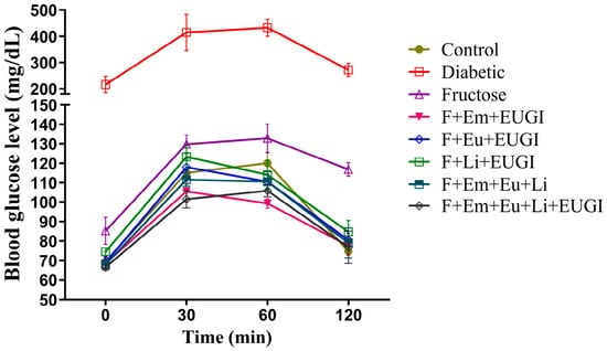
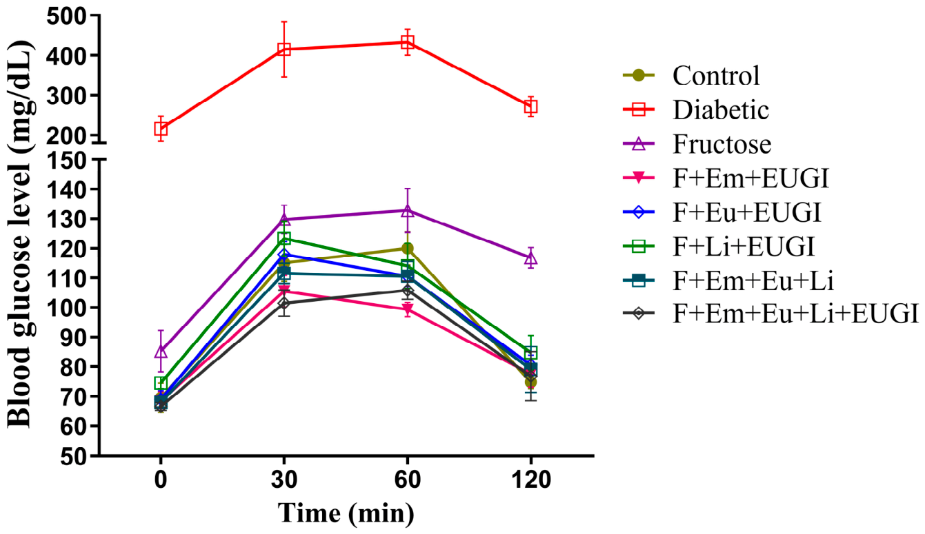
Fructose-fed rats exhibited elevated fasting glucose (85.25 ± 6.99 mg/dL) compared with controls (69.5 ± 5 mg/dL). Emodin with prebiotic–probiotic formulation (Emodin +EUGI™) reduced postprandial glucose at 30 min (105.5 ± 5.26 mg/dL; p < 0.05 vs. fructose). The mixture of all three bioactives, with or without the prebiotic–probiotic formulation, also lowered postprandial glucose (111.5 ± 3.42 and 101.5 ± 4.44 mg/dL, respectively; p < 0.05 vs. fructose) and reduced 120 min glucose values (79 ± 7.75 and 76.75 ± 8.30 mg/dL, respectively; p < 0.05 vs. fructose) (Figure 1).
Figure 1.
Oral glucose tolerance in fructose-fed and treated rats. Fasting and postprandial blood glucose levels in control, fructose-fed, and treatment groups on day 43. Fructose-fed rats showed elevated fasting glucose compared with controls. Emodin + EUGI™ reduced the 30-min postprandial glucose rise. The bioactive mixture, with or without prebiotic–probiotic formulation, lowered postprandial glucose and produced the greatest reduction in 120-min glucose values. Data are presented as mean ± SD (n = 4 per group). F = fructose; Em = emodin; Eu = eugenol; Li = limonene; EUGI = prebiotic–probiotic formulation.
3.4. Effects on Body Weight and Serum Biochemical Parameters
Rats receiving fructose alone showed increased body weight and fasting glucose compared with other non-diabetic groups (Table A2). Emodin + EUGI™ limited weight gain. The bioactive mixture, with or without the prebiotic–probiotic formulation, effectively normalized fasting glucose.
Fructose feeding also elevated serum lipids. The bioactive mixture and Limonene + EUGI™ reduced serum creatinine compared with fructose-fed rats. Limonene + EUGI™ also reduced serum urea compared with fructose-fed rats. Significant differences for these parameters are indicated in Table A2, and the corresponding p-values for each comparison are referenced directly in the table.
3.5. Effects on Urine Parameters and Fecal Output
Fructose feeding induced urinary abnormalities, including increased glucose excretion. Although Eugenol + EUGI™ did not improve urine creatinine, it reduced urinary glucose excretion compared to fructose alone. The bioactive mixture, with or without the prebiotic–probiotic formulation, showed protective effects on kidney-related parameters (Table A2).
Fructose feeding increased fecal output, whereas treated groups showed values closer to controls. The corresponding p-values for urine parameters and fecal output are provided in Table A2, and the exact rows indicating significant differences are referenced directly in the table.
3.6. Antioxidant Enzyme Status in Liver and Kidney
3.6.1. Superoxide Dismutase
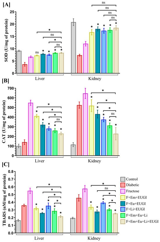
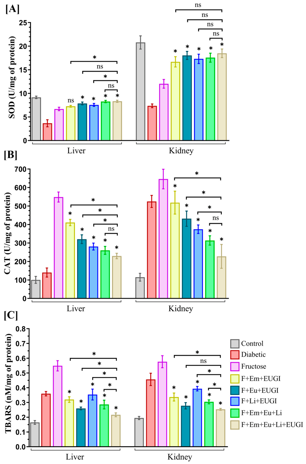
Fructose feeding reduced SOD activity in both liver and kidney. All treatment groups showed improved SOD activity relative to fructose-fed rats, with the bioactive mixture—either alone or combined with the prebiotic–probiotic formulation—producing the most pronounced restoration (Figure 2A).
Figure 2.
Antioxidant enzyme status in liver and kidney of experimental rats. (A) Superoxide dismutase (SOD) activity increased in both tissues across all treatment groups compared with fructose-fed rats, with the bioactive mixture, with or without the prebiotic–probiotic formulation, showing the greatest enhancement. (B) Catalase activity increased in all treated groups relative to fructose-fed rats. (C) TBARS levels decreased in all treatment groups, with the bioactive mixture + prebiotic–probiotic formulation producing the greatest reduction. Values are presented as mean ± SD (n = 4 per group). * p < 0.05 vs. fructose-fed; ns = not significant.
3.6.2. Catalase
Catalase activity in liver and kidney was also impaired by fructose feeding. All treated groups showed restoration of catalase activity relative to fructose-fed rats (Figure 2B). The bioactive mixture with the prebiotic–probiotic formulation produced the most consistent recovery.
3.6.3. TBARS (Lipid Peroxidation)
Fructose feeding increased lipid peroxidation in both tissues. All treatment groups reduced TBARS levels relative to fructose-fed rats, and the bioactive mixture with EUGI™ produced the greatest reduction (Figure 2C).
3.7. Effects on Tissue Fluorescence (AGE Accumulation)
3.7.1. Liver and Kidney
Fructose feeding increased liver AGE-associated general and specific fluorescence by 10.3-fold and 5.7-fold compared with controls (0.431 ± 0.052 and 1.047 ± 0.218, respectively). Eugenol, limonene, and the bioactive mixture with the prebiotic–probiotic formulation markedly reduced both parameters (Table A3).
Kidney AGE-associated fluorescence increased 3.89-fold (general) and 4.4-fold (specific) with fructose feeding (control: 1.386 ± 0.133 and 4.028 ± 0.342, respectively). Treatments, particularly the bioactive mixture with the prebiotic–probiotic formulation, substantially reduced AGE-related fluorescence.
3.7.2. Heart, Lungs, Lens, and Soleus Muscle
Fructose feeding increased AGE-associated general and specific fluorescence in all tissues examined. All treatments reduced fluorescence, except that Eugenol + EUGI™ and Limonene + EUGI™ did not significantly reduce specific fluorescence in heart tissue (Table A3). The bioactive mixture with the prebiotic–probiotic formulation was consistently the most effective.
3.7.3. Intestinal Fluorescence
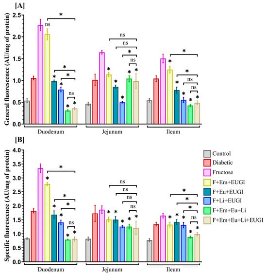
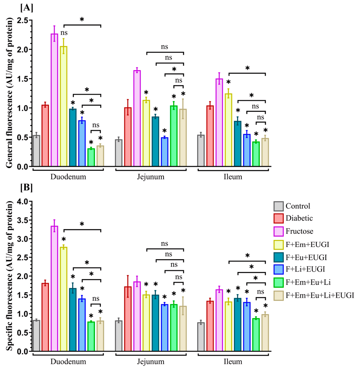
Fructose feeding increased AGE-associated glycation in duodenum, jejunum, and ileum. Limonene + EUGI™ produced the greatest reduction among individual bioactives. The bioactive mixture with the prebiotic–probiotic formulation showed the most pronounced improvement in intestinal AGE-associated fluorescence (Figure 3A,B).
Figure 3.
AGE-associated fluorescence in intestinal segments of experimental rats. (A) General AGE-associated fluorescence (370/440 nm) and (B) specific AGE-associated fluorescence (335/385 nm) were measured in the duodenum, jejunum, and ileum of all experimental groups. Results are expressed in AU/mg protein. Fructose feeding increased both general and specific AGE-associated fluorescence across all intestinal segments. Limonene + EUGI™ showed the greatest reduction among individual treatments, while the bioactive mixture, with or without the prebiotic–probiotic formulation, produced the most marked improvement. Values are presented as mean ± SD (n = 4 per group). * p < 0.05 vs. fructose-fed; ns = not significant.
3.8. Intestinal and Fecal Cultivable Bacterial Counts
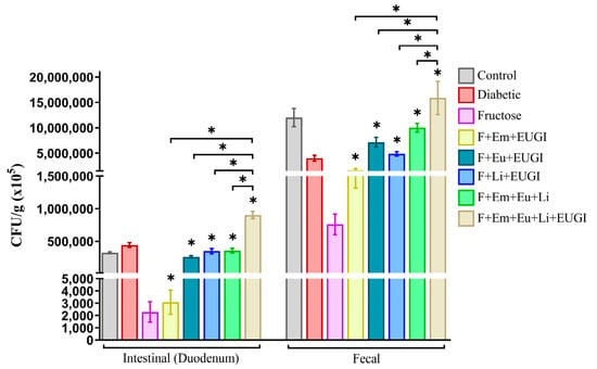
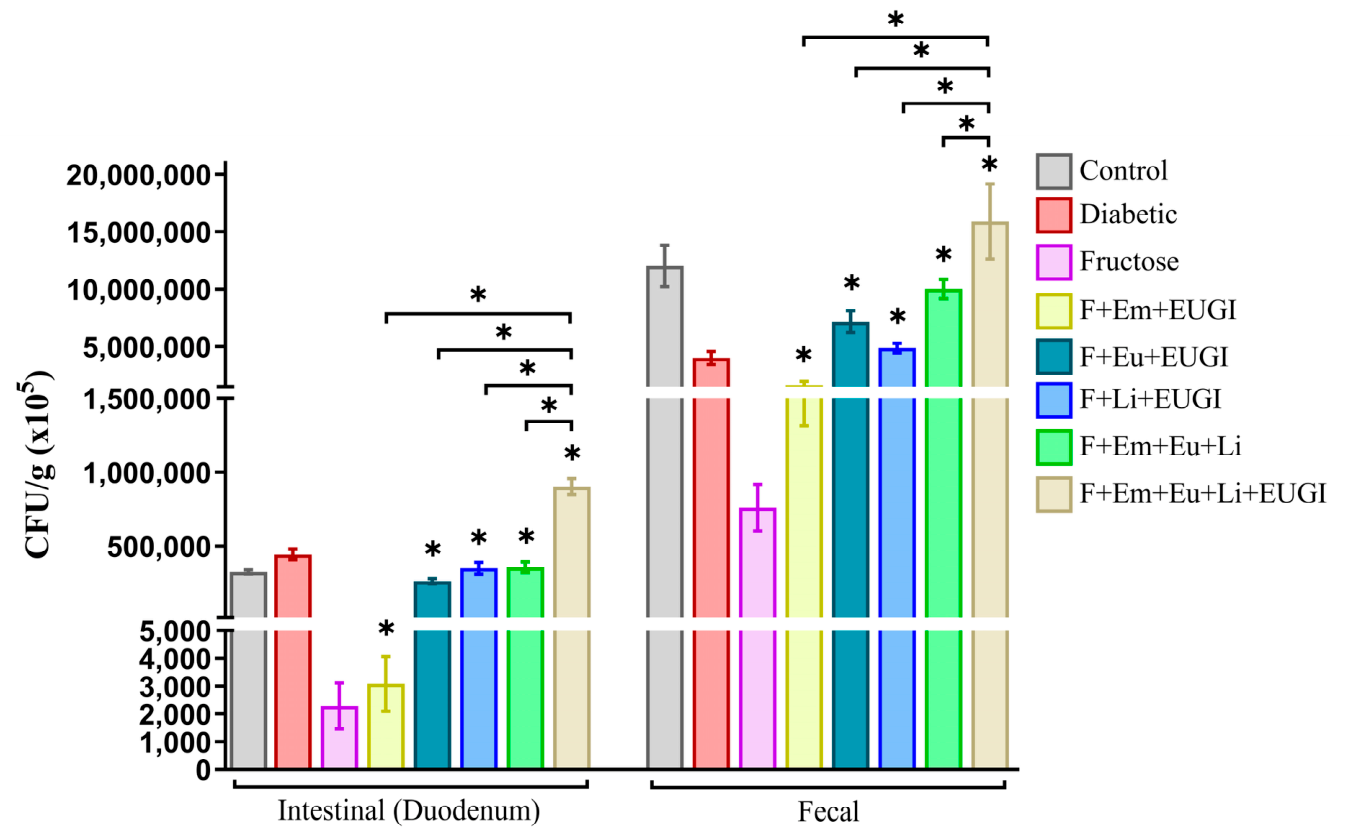
Fructose feeding markedly reduced cultivable intestinal bacterial abundance and altered the distribution of aerobic and facultative cultivable bacteria. Emodin treatment reduced cultivable intestinal bacterial counts but increased cultivable fecal bacterial counts, likely reflecting its known effects on gastric motility.
Eugenol and limonene, both antimicrobial agents, supported modulation of cultivable gut bacterial populations by limiting selected cultivable opportunistic isolates.
The bioactive mixture, particularly with the prebiotic–probiotic formulation, produced the most substantial improvement in both intestinal and fecal cultivable bacterial counts (Figure 4).
Figure 4.
Intestinal and fecal cultivable bacterial counts in fructose-fed and treated rats. Fructose feeding markedly reduced intestinal cultivable bacterial abundance and altered the distribution of aerobic and facultative anaerobic cultivable bacteria. Individually, emodin decreased intestinal cultivable bacteria but increased fecal cultivable bacteria, consistent with its reported effects on gastric motility. Eugenol and limonene supported modulation of cultivable gut bacterial populations by limiting selected cultivable opportunistic isolates. The bioactive mixture, especially when combined with the prebiotic–probiotic formulation, produced the greatest improvement in both intestinal and fecal cultivable bacterial distribution. All samples were processed identically, and comparisons were performed using non-parametric Mann–Whitney test appropriate for untransformed CFU data. Values are presented as mean ± SD (n = 4 per group). * p < 0.05 vs. fructose-fed.
4. Discussion
The present findings build upon our earlier observations that short-term fructose feeding increases oxidative stress, accelerates protein glycation, alters serum biochemical parameters, and promotes early metabolic disturbances [11]. In that work, the protein glycation inhibitor aminoguanidine and selected prebiotic–probiotic formulations were effective in limiting these changes. In the current study, we evaluated the phytochemicals limonene, eugenol, and emodin, compounds previously identified for their antiglycation, antioxidant, or glucose-modulating properties, as potential alternatives to aminoguanidine, which is limited by toxicity concerns. These bioactives have a long history of safe use, and earlier reports indicate their ability to inhibit glycation, modulate glucose metabolism, and reduce oxidative stress [15,35,36]. Their evaluation in combination with a prebiotic–probiotic formulation was based on the rationale that restoration of the cultivable gut bacterial fraction is essential for metabolic stability.
Across biochemical and cultivable microbial parameters, the combined bioactives and prebiotic–probiotic formulation improved oxidative stress indices, reduced AGE-related fluorescence, and supported healthier serum and lipid profiles. Emodin showed glucose-lowering effects during OGTT when administered with the prebiotic–probiotic formulation, while mixtures containing all three bioactives demonstrated consistent improvements in metabolic markers. The combinations also increased antioxidant enzyme activity and reduced lipid peroxidation, indicating broad protection against fructose-induced oxidative stress. In addition, eugenol and limonene exhibited antibacterial activity against cultivable gut isolates, including selected cultivable opportunistic isolates such as Corynebacterium stationis, suggesting a role in limiting succession of such isolates and supporting modulation of cultivable bacterial populations. Several studies have been carried out on the role of antibiotics in the development of obesity [37], and a number of studies have examined antibiotic resistance and the use of antibiotics in diabetes [38,39,40]. In this context, identifying effective, natural, and safe compounds is of critical importance.
Fructose feeding again produced marked alterations in cultivable bacterial populations, consistent with earlier reports linking high-glycemic diets to microbial imbalance, increased protein glycation, and accumulation of reactive dicarbonyls [13,41]. The bioactive mixture, with or without the prebiotic–probiotic formulation, helped counter these changes and supported modulation of cultivable bacterial counts rather than full re-establishment of a healthier microbial community.
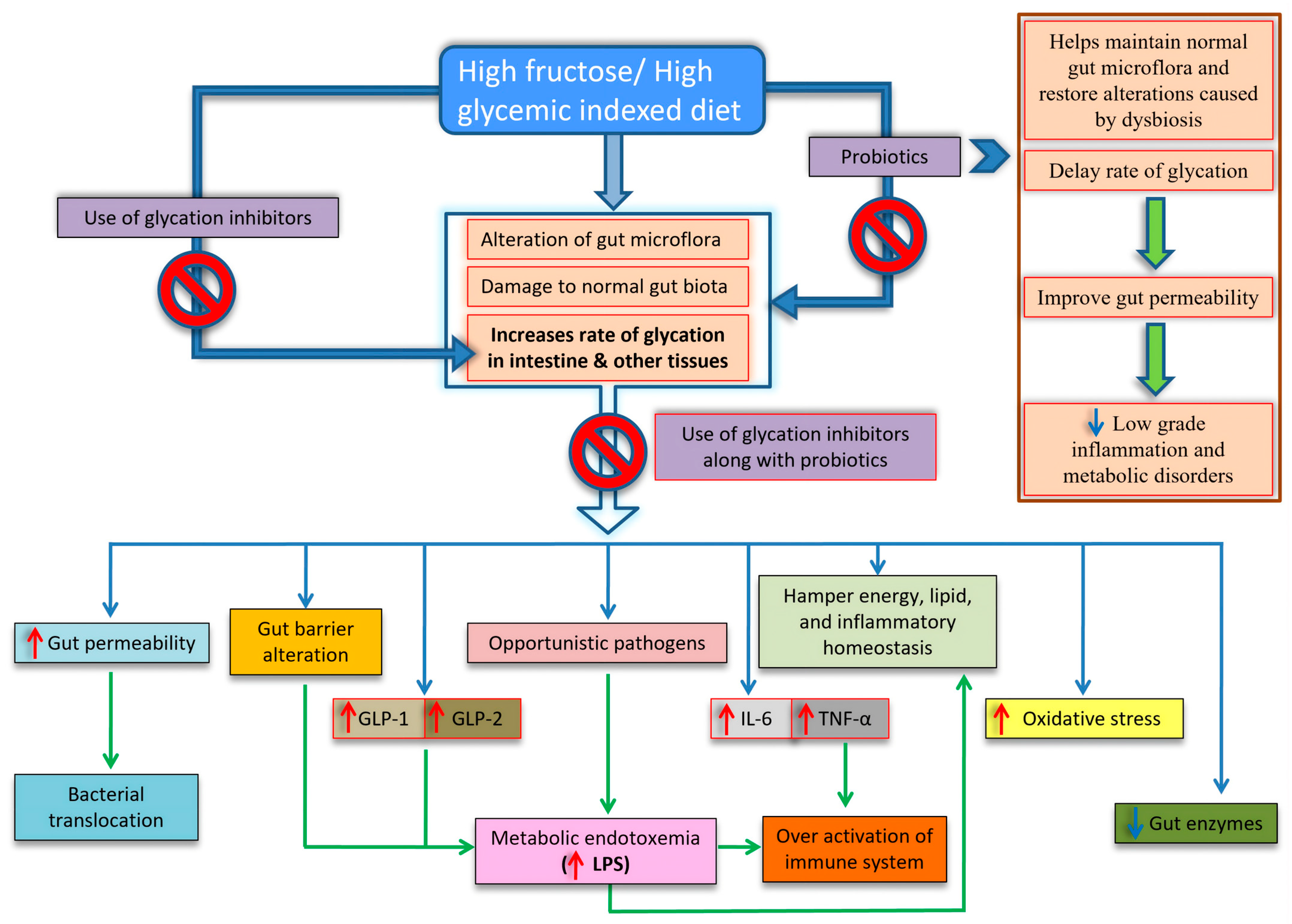
High-glycemic diets such as fructose exert a broad influence on gut physiology by altering cultivable bacterial distribution, weakening epithelial barriers, and increasing gut permeability [42]. These changes favor the dominance of selected cultivable opportunistic isolates and contribute to oxidative stress, disturbances in lipid and energy homeostasis, and heightened inflammatory signaling, ultimately promoting metabolic endotoxemia and excessive immune activation [43]. Glycation inhibitors such as aminoguanidine, limonene, and eugenol, and the α-glucosidase inhibitor emodin, limit protein glycation but show limited ability to modulate cultivable gut bacteria on their own. When these bioactives are combined with a prebiotic–probiotic formulation, their antiglycation and antioxidant actions are complemented by microbial support within the cultivable fraction, resulting in more effective protection against fructose-induced alterations in cultivable bacterial populations and metabolic disturbances.
Several recent studies from independent groups support the broader relevance of our findings. Human data show that high fructose intake is associated with depletion of key commensal taxa such as Eubacterium and Streptococcus [29], while work on added sugars highlights consistent shifts in gut microbial composition linked to metabolic outcomes [27]. Experimental studies further demonstrate that excessive fructose intake aggravates dysbiosis and promotes macrophage-driven inflammatory responses [25], and that microbiota from metabolically compromised donors can worsen fructose-induced metabolic dysfunction [26]. Additional recent work shows that dietary fiber-adapted microbiomes can enhance fructose clearance and reverse hepatic steatosis [44], and comprehensive reviews emphasize fructose-driven dysbiosis as a contributor to chronic inflammation and biological aging [45]. Together, these studies broaden the context of our observations and demonstrate that the mechanisms described here align with a wider body of current research on fructose-induced microbial and metabolic disturbances.
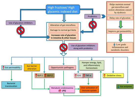
Therefore, drawing on the published literature [25,26,27,28,29,44,45,46], our earlier work [5,10,11,13,14,15,16,35,41], and the present preliminary data, we have summarized a conceptual mechanism and research scope in Figure 5 to illustrate how the combined intervention may help counteract the cascade of events initiated by a high-glycemic diet. A detailed description is provided in the Figure 5 legend.
Figure 5.
Proposed mechanism and scope linking a high-glycemic diet to alterations in cultivable bacteria and metabolic disturbances. A high-glycemic diet such as fructose can influence the cultivable gut bacterial fraction by increasing gut permeability, altering barrier function, and permitting the expansion of cultivable opportunistic isolates. These changes contribute to increased oxidative stress and disturbances in energy, lipid, and inflammatory homeostasis, ultimately promoting metabolic endotoxemia through elevated lipopolysaccharide levels and heightened immune activation. Glycation inhibitors such as aminoguanidine (used in our prior studies [11,35]), limonene, and eugenol, along with the α-glucosidase inhibitor emodin, help limit protein glycation but show limited ability to modulate the cultivable bacterial fraction when administered individually. When combined with a prebiotic–probiotic formulation, their antiglycation and antioxidant actions are complemented by support of the cultivable bacterial fraction, resulting in more effective protection against fructose-induced alterations in cultivable bacterial populations and associated metabolic disturbances. As a future direction, it will be interesting to investigate and validate this proposed mechanism.
We would like to address some limitations of the current study. This study was conducted in a rat model, and the findings may not fully translate to human physiology. The doses of phytochemicals were selected based on earlier experimental work and may not reflect dietary exposure levels. Microbial identification relied on culture-based methods, which capture only cultivable aerobic and facultative bacteria and do not represent the full diversity of the gut microbiota. The study duration was limited to 45 days, and long-term effects of these interventions were not assessed. Advanced mechanistic studies are required to validate Figure 5. Our study used a small sample size (n = 4 per group), which limits the strength of statistical comparisons and the generalizability of the findings. Larger, adequately powered studies will be important to confirm these preliminary observations as well as to define the effects of the intervention. Further work using metagenomic approaches, dose–response analyses, and extended treatment periods will be necessary to clarify mechanisms and evaluate translational potential.
5. Conclusions
Inhibitors of protein glycation such as eugenol and limonene demonstrated antibacterial activity against selected cultivable opportunistic isolates observed in the gut of fructose-fed rats, a condition characterized by elevated glycation and alterations in cultivable bacterial populations. These findings suggest that such phytochemicals may offer leads for drug development aimed at limiting the progression toward diabetes and its associated complications. The combined administration of these bioactive compounds with a prebiotic–probiotic formulation supported modulation of intestinal cultivable bacterial counts, restricted the growth of selected cultivable opportunistic isolates, reduced oxidative stress, and slowed the rate of protein glycation, thereby improving metabolic disturbances associated with the pre-diabetic state.
It is well reported that AGEs, oxidative stress, dyslipidemia, and metabolic endotoxemia are shared drivers of insulin resistance, endothelial dysfunction, and vascular inflammation. Notably, the bioactive mixture also had beneficial effects in the absence of the prebiotic–probiotic formulation. Their antiglycation properties, together with their antibacterial action, facilitated the modulation of altered cultivable bacterial populations and supported a shift toward a more favorable cultivable bacterial distribution. Additionally, these results also indicate that the bioactive mixture improved antioxidant enzyme activity as well as markers associated with oxidative and inflammatory stress. Taken together, these preliminary findings suggest that the bioactive mixture may serve as a supportive strategy alongside conventional pre- and probiotic approaches for maintaining intestinal metabolic stability and preventing early metabolic deterioration associated with diabetes mellitus and cardiometabolic dysfunction.
Author Contributions
Conceptualization, Formal analysis, Data curation, Resources, R.S.P. and A.U.A.; Methodology, R.S.P., S.B.R., M.L.N., S.P.K. and L.N.B.; Investigation, Visualization, Software, R.S.P.; Validation, R.S.P., S.B.R., M.L.N., S.P.K., L.N.B. and A.U.A.; Writing—original draft preparation, R.S.P.; Writing—review and editing, S.B.R., M.L.N., S.P.K., L.N.B., H.H.D. and A.U.A.; supervision, project administration, A.U.A.; funding acquisition, R.S.P. and A.U.A. All authors have read and agreed to the published version of the manuscript.
Funding
This research was supported by Department of Science and Technology, Government of India (DST/INSPIRE Fellowship/IF160187; Rahul Patil), Rajiv Gandhi Science and Technology Commission (RGSTC/File-2013/DPP-114/CR-36), Government of Maharashtra and Rashtriya Uchchatar Shiksha Abhiyan (RUSA/Order/R&I/2016-17/270&284), Government of India (Akalpita Arvindekar).
Institutional Review Board Statement
The animal study protocol and the methods were approved by the ethical committee of SHIVAJI UNIVERSITY, registered under the ethical committee certification (Registration no. 1825/PO/EReBi/S/15/CPCSEA; dated: 14 September 2015; Last approval date: 14 January 2021). All protocols and methods were carried out in accordance with relevant guidelines and regulations by the institutional ethical committee.
Informed Consent Statement
Not applicable.
Data Availability Statement
The original contributions presented in this study are included in the article. Further inquiries can be directed to the corresponding authors.
Acknowledgments
Rahul Patil acknowledges the Department of Science and Technology, Government of India, for the DST-INSPIRE fellowship. Authors also acknowledge Rajiv Gandhi Science and Technology Commission (RGSTC), Government of Maharashtra and Rashtriya Uchchatar Shiksha Abhiyan (RUSA), Government of India, for major research projects funded to Akalpita Arvindekar.
Conflicts of Interest
The authors declare no conflicts of interest.
Abbreviations
The following abbreviations are used in this manuscript:
| AGEs | advanced glycation end products |
| ALT | alanine aminotransferase |
| AMG | aminoguanidine |
| ANOVA | analysis of variance |
| AST | aspartate aminotransferase |
| CAT | catalase |
| CFU | colony-forming units |
| ELISA | enzyme-linked immunosorbent assay |
| GLP-1 | glucagon-like peptide-1 |
| GLP-2 | glucagon-like peptide-2 |
| HDL | high-density lipoprotein |
| IL-6 | interleukin-6 |
| LPS | lipopolysaccharide |
| MDA | malondialdehyde |
| NBT | nitro blue tetrazolium |
| OD | optical density |
| OGTT | oral glucose tolerance test |
| SOD | superoxide dismutase |
| SPC | standard plate count |
| TBARS | thiobarbituric acid reactive substances |
| TNF-α | tumor necrosis factor-alpha |
| VLDL | very-low-density lipoprotein |
Appendix A
Table A1.
Antibacterial activity of protein glycation inhibitors (limonene, eugenol, and emodin) against cultivable bacteria isolated from fructose-fed rats [13].
Table A2.
Effects of the bioactive mixture and prebiotic–probiotic formulation on body weight and metabolic parameters in experimental rats.
Table A3.
AGE-associated tissue fluorescence levels in experimental rats.
References
- Mathis, D.; Vence, L.; Benoist, C. β-Cell death during progression to diabetes. Nature 2001, 414, 792–798. [Google Scholar] [CrossRef] [Scilit] [PubMed]
- Maillard, L.C. Formation of melanoidins in a methodical way. Comptes Rendus 1912, 154, 66–68. [Google Scholar]
- Monnier, V.; Vishwanath, V.; Frank, K.; Elmets, C.; Dauchot, P.; Kohn, R.R. Relation between complications of Type I diabetes mellitus and collagen-linked fluorescence. N. Engl. J. Med. 1986, 314, 403–408. [Google Scholar] [CrossRef] [Scilit] [PubMed]
- Fioretto, P.; Steffes, M.; Sutherland, D.; Mauer, M. Sequential renal biopsies in insulin-dependent diabetic patients: Structural factors associated with clinical progression. Kidney Int. 1995, 48, 1929–1935. [Google Scholar] [CrossRef] [Scilit] [PubMed]
- Bavkar, L.N.; Patil, R.S.; Rooge, S.; Nalawade, M.L.; Arvindekar, A.U. Acceleration of protein glycation by oxidative stress and comparative role of antioxidant and protein glycation inhibitor. Mol. Cell. Biochem. 2019, 459, 61–71. [Google Scholar] [CrossRef] [Scilit] [PubMed]
- Pei, Y.; Wang, R.; Chen, W.; Yi, S.; Huang, C.; Liang, S.; Cao, H.; Xu, Y.; Tan, B. Impaired colonic motility in high-glycemic diet-induced diabetic mice is associated with disrupted gut microbiota and neuromuscular function. Endocr. Connect. 2023, 12, e230078. [Google Scholar] [CrossRef] [Scilit] [PubMed]
- Hullar, M.A.; Kahsai, O.; Curtis, K.R.; Navarro, S.L.; Zhang, Y.; Randolph, T.W.; Levy, L.; Shojaie, A.; Kratz, M.; Neuhouser, M.L.; et al. Metabolic plasticity of the gut microbiome in response to diets differing in glycemic load in a randomized, crossover, controlled feeding study. Am. J. Clin. Nutr. 2025, 122, 780–792. [Google Scholar] [CrossRef] [Scilit] [PubMed]
- Fu, Y.; Gou, W.; Zhong, H.; Tian, Y.; Zhao, H.; Liang, X.; Shuai, M.; Zhuo, L.B.; Jiang, Z.; Tang, J.; et al. Diet–gut microbiome interaction and its impact on host blood glucose homeostasis: A series of nutritional n-of-1 trials. eBioMedicine 2025, 111, 105483. [Google Scholar] [CrossRef] [Scilit] [PubMed]
- Rabbani, S.A.; El-Tanani, M.; Janić, M.; Janež, A.; Tanani, Y.E.; Hajeer, E.; Matalka, M.I.; Rizzo, M.; Kumar, R. Modulating metabolic health and physiological functions: Advances in dietary interventions targeting gut microbiota. Ther. Clin. Risk Manag. 2025, 21, 1701–1733. [Google Scholar] [CrossRef] [Scilit] [PubMed]
- Nalawade, M.L.; Patil, R.S.; Bavkar, L.N.; Rooge, S.; Haldavnekar, V.S.; Arvindekar, A.U. Early metabolic changes in the gut lead to higher expression of intestinal α-glucosidase and thereby enhanced postprandial spikes. Life Sci. 2019, 218, 8–15. [Google Scholar] [CrossRef] [Scilit] [PubMed]
- Patil, R.S.; Jagdale, A.D.; Nalawade, M.L.; Bavkar, L.N.; Arvindekar, A.U. Glycation inhibitors and probiotics can ameliorate the changes caused by high fructose feed. Int. J. Pharm. Pharm. Sci. 2018, 10, 28–32. [Google Scholar] [CrossRef] [Scilit]
- Hu, F.; Satija, A.; Manson, J. Curbing the diabetes pandemic: The need for global policy solutions. JAMA 2015, 313, 2319–2320. [Google Scholar] [CrossRef] [Scilit] [PubMed]
- Patil, R.S.; Arvindekar, A.U. Glycation of gut proteins initiates microbial dysbiosis and can promote establishment of diabetes in experimental animals. Microb. Pathog. 2021, 152, 104589. [Google Scholar] [CrossRef] [Scilit] [PubMed]
- Panaskar, S.N.; Joglekar, M.M.; Taklikar, S.S.; Haldavnekar, V.S.; Arvindekar, A.U. Aegle marmelos Correa leaf extract prevents secondary complications in streptozotocin-induced diabetic rats and demonstration of limonene as a potent antiglycating agent. J. Pharm. Pharmacol. 2013, 65, 884–894. [Google Scholar] [CrossRef] [Scilit] [PubMed]
- Arvindekar, A.; More, T.; Payghan, P.V.; Laddha, K.; Ghoshal, N.; Arvindekar, A. Evaluation of anti-diabetic and α-glucosidase inhibitory action of anthraquinones from Rheum emodi. Food Funct. 2015, 6, 2693–2700. [Google Scholar] [CrossRef] [Scilit] [PubMed]
- Nalawade, M.L.; Arvindekar, A.U. Study of Metabolic Changes in Prediabetes and Prevention of Progression to Diabetes Using Plant Components. Ph.D. Thesis, Shivaji University, Kolhapur, India, 2019. [Google Scholar]
- Hou, K.; Chen, C.; Wang, X.; Guo, Y. Effects of emodin on blood glucose and body weight in type 1 diabetic rats. Univers. J. Pharm. Res. 2017, 2, 11–14. [Google Scholar] [CrossRef] [Scilit]
- Jing, D.; Bai, H.; Yin, S. Renoprotective effects of emodin against diabetic nephropathy in rat models are mediated via PI3K/Akt/GSK-3β and Bax/caspase-3 signaling pathways. Exp. Ther. Med. 2017, 14, 5163–5169. [Google Scholar] [PubMed]
- Song, Y.; Fan, X.; Guo, Z.; Fan, Y.; Yang, J. Therapeutic effects of emodin in type 2 diabetes mellitus in KKAy mouse model. Int. J. Clin. Exp. Med. 2017, 10, 14408–14413. [Google Scholar]
- Yılmaz, B.S.; Özbek, H. Investigation of the anti-inflammatory, hypoglycemic activity and median lethal dose (LD50) level of limonene in mice and rats. Acta Pharm. Sci. 2018, 56, 85–94. [Google Scholar] [CrossRef] [Scilit]
- Bacanlı, M.; Anlar, H.G.; Aydın, S.; Çal, T.; Arı, N.; Ündeğer Bucurgat, Ü.; Başaran, A.A.; Başaran, N. D-Limonene ameliorates diabetes and its complications in streptozotocin-induced diabetic rats. Food Chem. Toxicol. 2017, 110, 434–442. [Google Scholar] [CrossRef] [Scilit] [PubMed]
- Paarakh, M.P. In silico antidiabetic activity of limonene isolated from Coriandrum sativum Linn fruit. ASPS 2018, 2, 2–6. [Google Scholar]
- Al-Trad, B.; Alkhateeb, H.; Alsmadi, W.; Al-Zoubi, M. Eugenol ameliorates insulin resistance, oxidative stress and inflammation in high-fat diet/streptozotocin-induced diabetic rats. Life Sci. 2019, 216, 183–188. [Google Scholar] [CrossRef] [Scilit] [PubMed]
- Garud, M.S.; Kulkarni, Y.A. Eugenol ameliorates renal damage in streptozotocin-induced diabetic rats. Flavour Fragr. J. 2017, 32, 54–62. [Google Scholar]
- Wu, K.; Yu, H.; Cao, K.; Dai, B.; Yuan, Y.; Qian, X.; Zhong, H.; Qu, Y.; Jiang, H.; Chen, T. Overconsumption of fructose aggravates acute GVHD by inducing gut dysbiosis and promoting macrophage-mediated inflammatory response. Gut Microbes 2026, 18, 2642459. [Google Scholar] [CrossRef] [Scilit] [PubMed]
- Valdez-Palomares, F.; Noriega, L.G.; Reyes-Romo, D.; Canizales-Quinteros, S.; Nambo-Venegas, R.; Salinas-Lara, C.; Tovar-Palacio, A.; Menjivar, M.; Peña-Espinoza, B.; Ortiz, G.; et al. Human fecal transplantation from stunted children promotes metabolic dysfunction in mice fed with a high-fat and high-fructose corn syrup diet. Gut Microbes 2026, 18, 2651984. [Google Scholar] [CrossRef] [Scilit] [PubMed]
- Zhang, Y.; Walker, R.W.; Kaplan, R.C.; Qi, Q. Added sugars, gut microbiota, and host health. Gut Microbes 2025, 17, 2592431. [Google Scholar] [CrossRef] [Scilit] [PubMed]
- Simões, C.D.; Sousa, A.S.; Fernandes, S.; Sarmento, A. Fructose Malabsorption, Gut Microbiota and Clinical Consequences: A Narrative Review of the Current Evidence. Life 2025, 15, 1720. [Google Scholar] [CrossRef] [Scilit] [PubMed]
- Jones, R.B.; Alderete, T.L.; Kim, J.S.; Millstein, J.; Gilliland, F.D.; Goran, M.I. High intake of dietary fructose in overweight/obese teenagers associated with depletion of Eubacterium and Streptococcus in gut microbiome. Gut Microbes 2019, 10, 712–719. [Google Scholar] [CrossRef] [Scilit] [PubMed]
- Lowry, O.H.; Rosebrough, N.J.; Farr, A.L.; Randall, R.J. Protein measurement with the Folin phenol reagent. J. Biol. Chem. 1951, 193, 265–275. [Google Scholar] [CrossRef] [Scilit]
- Misra, H.P.; Fridovich, I. The role of superoxide anion in the autoxidation of epinephrine and a simple assay for superoxide dismutase. J. Biol. Chem. 1972, 247, 3170–3175. [Google Scholar] [CrossRef] [Scilit]
- Beers, R.F.; Sizer, I.W. A spectrophotometric method for measuring the breakdown of hydrogen peroxide by catalase. J. Biol. Chem. 1952, 195, 133–140. [Google Scholar] [CrossRef] [Scilit]
- Ohkawa, H.; Ohishi, N.; Yagi, K. Assay for lipid peroxides in animal tissues by thiobarbituric acid reaction. Anal. Biochem. 1979, 95, 351–358. [Google Scholar] [CrossRef] [Scilit] [PubMed]
- Nakagawa, T.; Yokozawa, T.; Terasawa, K.; Shu, S.; Juneja, L.R. Protective activity of green tea against free radical and glucose-mediated protein damage. J. Agric. Food Chem. 2002, 50, 2418–2422. [Google Scholar] [CrossRef] [Scilit] [PubMed]
- Joglekar, M.M.; Bavkar, L.N.; Sistla, S.; Arvindekar, A.U. Effective inhibition of protein glycation by combinatorial usage of limonene and aminoguanidine through differential and synergistic mechanisms. Int. J. Biol. Macromol. 2017, 99, 563–569. [Google Scholar] [CrossRef] [Scilit] [PubMed]
- Singh, P.; Jayaramaiah, R.H.; Agawane, S.; Vannuruswamy, G.; Korwar, A.M.; Anand, A.; Dhaygude, V.S.; Shaikh, M.L.; Joshi, R.; Boppana, R.; et al. Potential dual role of eugenol in inhibiting advanced glycation end products in diabetes: Proteomics and mechanistic insights. Sci. Rep. 2016, 6, 18798. [Google Scholar] [CrossRef] [Scilit] [PubMed]
- Leong, K.S.W.; Derraik, J.G.B.; Hofman, P.L.; Cutfield, W.S. Antibiotics, gut microbiome and obesity. Clin. Endocrinol. 2018, 88, 185–200. [Google Scholar] [CrossRef] [Scilit] [PubMed]
- Demetriou, M.; Papanas, N.; Panagopoulos, P.; Panopoulou, M.; Maltezos, E. Antibiotic resistance in diabetic foot soft tissue infections: A series from Greece. Int. J. Low. Extrem. Wounds 2017, 16, 255–259. [Google Scholar] [CrossRef] [Scilit] [PubMed]
- Ko, J.S.; Zwiebel, S.; Wilson, B.; Becker, D.B. Perioperative antibiotic use in diabetic patients: A retrospective review of 670 surgeries. J. Plast. Reconstr. Aesthet. Surg. 2017, 70, 1629–1634. [Google Scholar] [CrossRef] [Scilit] [PubMed]
- Mission, J.F.; Catov, J.; Deihl, T.; Feghali, M.; Scifres, C. Antibiotic use in pregnancy, abnormal fetal growth, and development of gestational diabetes mellitus. Am. J. Perinatol. 2019, 36, 243–251. [Google Scholar] [CrossRef] [Scilit] [PubMed]
- Patil, R.S.; Rooge, S.B.; Damame, H.H.; Haldavnekar, V.S.; Arvindekar, A.U. Role of curd and yogurt in establishment and progression of diabetes through protein glycation and induction of inflammation. Food Biosci. 2021, 39, 100829. [Google Scholar] [CrossRef] [Scilit]
- Guney, C.; Bal, N.B.; Akar, F. The impact of dietary fructose on gut permeability, microbiota, abdominal adiposity, insulin signaling and reproductive function. Heliyon 2023, 9, e18896. [Google Scholar] [CrossRef] [Scilit] [PubMed]
- Di Vincenzo, F.; Del Gaudio, A.; Petito, V.; Lopetuso, L.R.; Scaldaferri, F. Gut microbiota, intestinal permeability, and systemic inflammation: A narrative review. Intern. Emerg. Med. 2024, 19, 275–293. [Google Scholar] [CrossRef] [Scilit] [PubMed]
- Jung, S.; Bae, H.; Song, W.S.; Chun, Y.; Le, J.; Alam, Y.; Verlande, A.; Chun, S.K.; Kim, J.; Kelly, M.E.; et al. Dietary fibre-adapted gut microbiome clears dietary fructose and reverses hepatic steatosis. Nat. Metab. 2025, 7, 1801–1818. [Google Scholar] [CrossRef] [Scilit] [PubMed]
- Xu, X.Z.; Dong, M.S.; Dai, J.Y.; Wang, Q.Y.; Dong, X.C.; Jin, Y. High-fructose diet–induced gut dysbiosis as a mechanistic driver of chronic inflammation and accelerated biological aging: A comprehensive review. Front. Microbiol. 2026, 1865281. [Google Scholar]
- Yakovlieva, M.; Tacheva, T.; Mihaylova, S.; Tropcheva, R.; Trifonova, K.; Tolekova, A.; Danova, S.; Vlaykova, T. Influence of Lactobacillus brevis 15 and Lactobacillus plantarum 13 on blood glucose and body weight in rats after high-fructose diet. Benef. Microbes 2015, 6, 505–512. [Google Scholar] [CrossRef] [Scilit] [PubMed]
Disclaimer/Publisher’s Note: The statements, opinions and data contained in all publications are solely those of the individual author(s) and contributor(s) and not of MDPI and/or the editor(s). MDPI and/or the editor(s) disclaim responsibility for any injury to people or property resulting from any ideas, methods, instructions or products referred to in the content. |
© 2026 by the authors. Licensee MDPI, Basel, Switzerland. This article is an open access article distributed under the terms and conditions of the Creative Commons Attribution (CC BY) license.