


Abstract
Endophytic bacteria from medicinal plants are increasingly recognised as sources of antimicrobial metabolites. However, the endophytic bacterial community of Mitrephora heyneana remains poorly explored. In the present study, endophytic bacteria were isolated from the leaves of M. heyneana, collected from the Western Ghats of Tamil Nadu, India. Among seven isolates, the plant endophyte strain from leaf 6th strain (PEL6) was identified as Paenibacillus lactis through 16S rRNA gene sequencing. The ethyl acetate extract of PEL6 (EAE-PEL6) was subjected to gas chromatography–mass spectrometry (GC-MS) analysis, which putatively identified 32 metabolites based on GC-MS library matching, including pyrrolo [1,2-a] pyrazine-1,4-dione derivatives and triazole compounds as major constituents. The EAE-PEL6 demonstrated significant in vitro antibacterial activity against Staphylococcus aureus. In silico ADMET (absorption, distribution, metabolism, excretion, and toxicity), profiling predicted drug-likeness and pharmacokinetic properties of selected candidate compounds. Molecular docking suggested favourable binding of selected metabolites to S. aureus target proteins; however, these interactions require experimental validation. Density Functional Theory calculations indicated that CID 70504 had the lowest Highest Occupied Molecular Orbital (HOMO)–Lowest Unoccupied Molecular Orbital (LUMO) energy gap, reflecting higher electronic reactivity. Molecular electrostatic potential mapping further supported its enhanced binding propensity. Molecular dynamics simulations suggested structural stability, with Root Mean Square Deviation, Solvent Accessible Surface Area, radius of gyration, and hydrogen-bond analyses indicating stable interactions throughout the 100 ns trajectory. Overall, this study identifies P. lactis PEL6 as a promising endophytic source of anti-S. aureus metabolites and provides candidates for future purification, structural confirmation, and biological validation.
1. Introduction
Medicinal plants, rich in diverse bioactive compounds, play a crucial role in promoting human health and advancing sustainable healthcare systems. They serve as vital resources for treating various diseases, strengthening immunity, and providing affordable, accessible therapeutic options [1]. Beyond healthcare, medicinal plants play an important role in preserving indigenous ethnomedicinal knowledge, supporting rural livelihoods through their cultivation, sustainable harvesting, and commercialisation and contributing to biodiversity conservation by promoting the sustainable management and protection of medicinal plant species and their natural habitats. Their bioactive constituents remain essential in modern pharmaceutical research, driving drug discovery and innovation, thereby underscoring their ongoing significance in global medicine [2,3]. Despite this potential, only limited information is available on endophytic bacteria associated with Mitrephora heyneana and their antimicrobial metabolites.
Plants are closely associated with and influenced by a wide range of microorganisms and other multicellular organisms, both above and below ground [4]. Among these, microorganisms are considered some of the most important organisms capable of forming beneficial associations with plants [5]. Medicinal plants are increasingly recognised as being significantly influenced by their associations with specific bacterial endophytes [6]. Endophytes, which include bacteria and fungi, reside within the internal tissues of plants without causing any detrimental effects. They contribute significantly to plant development by enhancing growth, improving resistance to environmental stresses and pest attacks, and promoting the biosynthesis of valuable bioactive compounds, including therapeutic agents and industrial enzymes [7,8]. These symbiotic interactions indirectly support plant health through mechanisms such as antibiotic production, modulation of ethylene levels, and induction of systemic resistance to pathogens [9]. Notably, the bioactive metabolites synthesised by endophytic microorganisms often demonstrate greater biological activity than those produced by their host plants [10]. Owing to their broad-spectrum antimicrobial properties, these metabolites have attracted considerable interest as potential therapeutic agents against clinically important skin pathogens [11]. Among these, Staphylococcus aureus is a predominant pathogen associated with skin infections and is particularly implicated in the pathogenesis and exacerbation of atopic dermatitis (AD). Colonisation by S. aureus is significantly more common in individuals with AD than in healthy individuals. The bacterium aggravates AD through multiple mechanisms, including disruption of the skin barrier, dysregulation of immune responses, alteration of the skin microbiome and biofilm formation [12].
M. heyneana, a medicinal tree belonging to the family Annonaceae, is recognised for its rich phytochemical profile and therapeutic potential. Although there are currently no published reports describing the antimicrobial activity of M. heyneana, several species within the genus Mitrephora have been extensively investigated for their pharmacological properties. For instance, bioactive diterpenoids and polyacetylene derivatives isolated from M. celebica have demonstrated antibacterial activity against methicillin-resistant S. aureus (MRSA) and Mycobacterium smegmatis [13]. Likewise, phytochemical studies on M. glabra have identified biologically active diterpenoids, polyacetylenes, and the aporphine alkaloid liriodenine, which exhibit antimicrobial potential [14]. Furthermore, investigations into M. tomentosa, M. diversifolia, and M. sirikitiae have revealed a diverse array of secondary metabolites, including alkaloids, diterpenoids, and polyacetylene compounds, associated with antimicrobial, antimalarial, cytotoxic, and anti-inflammatory activities [15]. Collectively, these findings highlight the genus Mitrephora as an important reservoir of pharmacologically active natural products and provide a scientific basis for exploring M. heyneana and its endophytic microorganisms as potential sources of novel antibacterial metabolites.
Paenibacillus is a genus of small, Gram-positive bacteria currently classified under the family Paenibacillaceae, which comprises eight genera. This genus includes more than 150 identified species, many of which play important roles in agriculture, particularly in plant growth promotion and biological control [16]. Several Paenibacillus species produce antimicrobial compounds with applications in both medicine and agriculture, as well as enzymes valuable for bioremediation and the synthesis of industrial chemicals [17]. Members of this genus are also known to synthesise lipopeptides, polyketides, and other secondary metabolites with antimicrobial activities. Despite the well-documented antimicrobial potential of several Paenibacillus species, the antibacterial activity of endophytic P. lactis associated with M. heynenana has not been previously investigated. Furthermore, no studies have comprehensively integrated experimental antibacterial evaluation with metabolite profiling and computational analyses to identify potential antibacterial candidates from this endophyte.
Therefore, the present study aimed to isolate and identify antibacterial endophytic bacteria from M. heyneana, evaluate the anti-S. aureus activity of the EAE-PEL6, characterise its putative metabolites using GC-MS, and prioritise potential antibacterial compounds through ADMET prediction, molecular docking, density functional theory (DFT), and molecular dynamics simulation.
2. Materials and Methods
2.1. Plant Collection and Identification
Healthy, mature, disease-free leaves of plants were collected from the Palamalai Hills in the Coimbatore district, Tamil Nadu, India (11.7355° N, 77.7494° E; elevation: 1623 m) in January 2021. The collected specimen was taxonomically identified and authenticated as M. heyneana (Hook.f. & Thomson). Authentication was carried out by the Botanical Survey of India (BSI), Southern Regional Centre, TNAU Campus, Coimbatore, Tamil Nadu, India, under reference number BSI/SRC/5/23/2022/Tech/282. A voucher specimen was deposited at the BSI herbarium for future reference and assigned the barcode number 178485.
2.2. Explant Sterilisation and Endophyte Isolation
Endophytic bacteria were isolated from surface-sterilised leaf segments of M. heyneana. Surface sterilisation was performed to eliminate epiphytic microorganisms from the leaf surface while preserving the endophytic bacteria residing within the internal tissues. Healthy leaves were first washed thoroughly under running tap water to remove adhering dust and debris. The leaves were then surface sterilised using a slightly modified protocol described by Deepa et al. [18]. Briefly, the leaf samples were immersed in 70% ethanol for 1 min followed by 2% sodium hypochlorite solution for 2 min, and rinsed three times with sterile distilled water to remove residual sterilising agents. The sterilised leaves were then aseptically blotted dry on sterile filter paper, cut into 10 explants and transferred onto Murashige and Skoog (MS) medium for plant tissue culture under controlled culture conditions. Following incubation, bacterial colonies emerged exclusively from internal leaf tissues of sterilised leaf explants. In parallel, an MS medium without plant explants was maintained under identical culture conditions as a control and showed no bacterial growth throughout the incubation period. The emergence of bacterial colonies only from the internal leaf tissues, together with the absence of microbial growth in the control medium, supported the endophytic origin of the isolates. Subsequently, the bacterial colonies were isolated on nutrient agar plates and incubated at 37 °C for 24 h, as this temperature provided satisfactory growth under laboratory conditions. Colonies exhibiting distinct morphological characteristics were repeatedly subcultured to obtain pure cultures and assigned individual isolate codes for further characterisation.
2.3. Morphological and Molecular Identification of Endophyte
Pure isolates from 24 h-old cultures were grown on nutrient agar medium, and Gram staining was performed to examine their morphological characteristics [19]. For molecular identification, 16S rRNA gene sequencing was carried out using a thermocycler with universal primers 27F (5′-AGAGTTTGATCCTGGCTCAG-3′) and 1492R (5′-TACCTTGTTACGACTT-3′), followed by Sanger’s dideoxy sequencing method. The PCR reaction mixture included a Taq Master Mix containing Taq DNA polymerase in a 2× buffer, 0.4 mM dNTPs, 3.2 mM MgCl2, and 0.02% bromophenol blue, which served as a tracking dye during gel electrophoresis. The thermal cycling programme consisted of an initial denaturation at 95 °C for 2 min, followed by 25 cycles of denaturation at 95 °C for 30 s, annealing at 50 °C for 30 s, and extension at 72 °C for 2 min, with a final extension at 72 °C for 10 min. The reaction was then maintained at 4 °C indefinitely. The amplified 16S rRNA gene sequence was analysed using the BLAST algorithm (https://blast.ncbi.nlm.nih.gov/Blast.cgi; accessed on 29 April 2023) by comparing it against reference sequences in the NCBI GenBank database. The validated sequence was subsequently submitted to GenBank, and an accession number was obtained.
2.4. Extraction of EAE-PEL6
Secondary metabolites from the Plant Endophyte from Leaf 6th strain (PEL6) were extracted using the solvent partition method [20]. The strain was cultured in nutrient broth medium at 30 °C with agitation at 150 rpm for 5–6 days. After incubation, the culture was centrifuged at 10,000 rpm for 10 min at 4 °C. The resulting supernatant was collected and filtered through a 0.2 μm syringe filter. For extraction, an equal volume of ethyl acetate was added to the filtrate in a separating funnel, and the mixture was shaken vigorously. The organic (ethyl acetate) phase, containing the secondary metabolites, was separated from the aqueous phase. The organic solvent was then evaporated to dryness, yielding the EAE-PEL6. Approximately 1000 mg of the crude ethyl acetate extract of plant endophyte from the leaf’s 6th strain (EAE-PEL6) was reconstituted in an ethyl acetate solvent for further analysis.
2.5. GC-MS Analysis
EAE-PEL6 (100 mg/mL) was analysed to identify its bioactive constituents using GC-MS. A 1 µL aliquot of EAE-PEL6 was injected into the instrument. Helium was utilised as the carrier gas at a constant flow rate of 1 mL/min. The GC-MS analysis was carried out under electron-impact ionisation conditions at an ionisation energy of 70 eV. The injector temperature was maintained at 250 °C. The oven temperature programme was initiated at 50 °C and held for 3 min, then ramped at 10 °C/min to 280 °C with a 3 min hold, and finally increased to 300 °C, where it was maintained for 10 min [21].
2.6. In Vitro Antibacterial Assay
The antibacterial activity of EAE-PEL6 was evaluated against a clinical isolate of S. aureus using the agar well diffusion method. The bacterial strain was obtained from the Department of Microbiology, PSG College of Arts and Science, Coimbatore, Tamil Nadu, India. The microorganism was cultured overnight at 37 °C in Mueller–Hinton broth to obtain an actively growing inoculum for the antibacterial assay.
2.6.1. Preparation of Inoculum
Bacterial isolates from the stock culture were streaked onto agar plates and incubated at 37 °C for 24 h [22]. After incubation, colonies grown for 24 h were suspended in sterile 0.9% (w/v) sodium chloride solution to a final concentration of 108 CFU/mL, corresponding to the 0.5 McFarland turbidity standard, for use as an inoculum. This suspension was subsequently diluted with sterile 0.9% sodium chloride to obtain a final working concentration of 107 CFU/mL, thereby providing a standardised inoculum to ensure uniform bacterial growth during the antibacterial assay.
2.6.2. Test for Antibacterial Activity
For antimicrobial screening, EAE-PEL6 was prepared at concentrations of 10, 25, and 50 mg/mL in 10% dimethyl sulfoxide (DMSO) [23]. Approximately 20 mL of sterile Mueller–Hinton medium was poured into Petri dishes and allowed to solidify. A 100 µL aliquot of bacterial suspension was uniformly spread over the agar surface using a sterile cotton swab, and the plates were left to dry for 5 min. Wells of 6 mm diameter were aseptically created in the agar using a sterile cork borer [24]. Each well was loaded with 20 µL of EAE-PEL6 at concentrations of 10, 25 or 50 mg/mL using a sterile micropipette and allowed to diffuse at room temperature. Gentamicin was used as a positive control, while 10% DMSO served as a negative control. The plates were sealed with parafilm and incubated upright at 37 °C for 24 h. Antimicrobial activity was evaluated by measuring the diameter of the zones of inhibition surrounding the wells. All antibacterial assays (except the primary antibacterial screening assay) were performed in triplicate. Data are expressed as mean ± standard deviation.
2.6.3. Minimum Inhibitory Concentration (MIC)
The minimum inhibitory concentration of the ethyl acetate extract of the endophytic bacterial isolate P. lactis (EAE-PEL6) against S. aureus was determined using the broth microdilution method in sterile 96-well microtiter plates following the Clinical and Laboratory Standards Institute (CLSI) guidelines with minor modifications [25]. Briefly, 50 µL of Mueller–Hinton broth (MBH) was dispensed into each well of a sterile 96-well microtiter plate, followed by 50 µL of EAE-PEL6 at an initial concentration of 20 mg/mL. Two-fold serial dilutions of the extract were performed across the wells to obtain a range of test concentrations. Subsequently, 100 µL of a standardised S. aureus suspension (approximately 5 × 10⁵ CFU/mL) was added to each well, resulting in a final volume of 200 µL. Gentamicin prepared at an initial concentration of 0.5 mg/mL was similarly subjected to two-fold serial dilution and used as the positive control, while the extract solvent served as the negative control. Growth and sterility controls were included in each assay. The plates were incubated at 37 °C for 24 h, and the bacterial growth was assessed by the visual observation of turbidity. The MIC was defined as the lowest concentration of EAE-PEL6 that completely inhibited visible bacterial growth.
2.6.4. Statistical Analysis
All experiments (except the primary antibacterial screening assay) were performed in triplicate, and the results are expressed as the mean ± standard deviation (SD).
2.7. In Silico Analysis
2.7.1. Identification of Compounds
The mass spectra and retention indices of the detected compounds were compared against the National Institute of Standards and Technology (NIST 17) mass spectral database (http://www.sisweb.com/software/ms/nist.htm#gc; accessed on 3 April 2023) for compound identification. GC-MS identified the compounds present in the EAE-PEL6 based on their retention times, peak areas, molecular structures, and molecular weights. The results were analysed and documented accordingly.
2.7.2. Ligand Preparation
The three-dimensional structures of the bioactive compounds were obtained from the PubChem database [26] in Spatial Data File (SDF) format. These ligands were then converted into Protein Data Bank (PDB) format using Open Babel software version 3.1.1 [27]. Energy minimisation was performed using the MMFF94 force field. For cheminformatics analysis, the SMILES representations of the ligands were extracted and stored.
2.7.3. Toxicity Analysis
The chemical structures of the bioactive compounds were converted into their canonical Simplified Molecular Input Line Entry System (SMILES) format. These SMILES were then submitted to the SwissADME online tool (https://www.swissadme.ch/; accessed on 10 November 2023) as described by Daina et al. [28] to predict pharmacokinetic properties, including ADMET. Additionally, physicochemical properties such as the numbers of hydrogen-bond donors and acceptors, rotatable bonds, topological polar surface area (TPSA), and synthetic accessibility were evaluated. Drug-likeness was analysed with a focus on toxicity and potential adverse effects, which are critical for ensuring patient safety. These properties were examined using the ADMETlab 3.0 web server [29], thereby facilitating early-phase drug discovery by predicting pharmacological suitability and minimising reliance on extensive in vivo experimentation.
2.7.4. Target Preparation
The protein structure of S. aureus bacterial cell-division protein, FtsA bound to ATP (PDB ID: 3WQU; resolution: 2.80 Å), the crystal structure of S. aureus tyrosyl-tRNA synthetases, TyrRS in complex with SB-239629 (PDB ID: 1JIJ; resolution: 3.20 Å) and the structure of S. aureus Clumping Factor A (PDB ID: 1N67; resolution: 1.90 Å) were retrieved from the Protein Data Bank (PDB; http://www.rcsb.org, accessed on 15 May 2023). Initial preprocessing involved removing cofactors, water molecules, and co-crystallised ligands using Molegro Molecular Viewer version 2.5 (http://molexus.io/molegro-molecular-viewer/, accessed on 15 May 2023). In cases where the active-site amino acid sequence was incomplete or missing in the downloaded PDB structures, homology modelling was performed. Accordingly, homology models were generated for all three protein receptors using the SWISS-MODEL [30] platform to ensure structural completeness and suitability for molecular docking. Polar hydrogen atoms were then added to each protein structure, and the finalised models were saved in PDB format for subsequent docking analysis.
2.7.5. Molecular Docking
Molecular docking studies were performed using AutoDock Vina (version 1.1.2) implemented within the PyRx Virtual Screening Tool (version 0.8) to investigate the binding interactions between the target proteins and ligand [31]. PyRx served as the graphical interface for ligand preparation and docking setup, while AutoDock Vina was used as the docking engine. The protein targets and ligand molecules were prepared and converted to PDBQT format prior to docking. Molecular docking was performed using the Iterated Local Search (ILS) algorithm with Broyden–Fletcher–Goldfarb–Shanno (BFGS) local optimisation implemented in Autodock vina to predict the optimal ligand-binding poses and binding affinities. Site-specific docking was performed for TyrRS (PDB ID: 1JIJ) using a grid box centred at X = −12.8884, Y = 4.9554, and Z = 2.9440 with dimensions of 25 × 25 × 25 Å, and for FtsA (PDB ID: 3WQU) using a grid box centred at X = −4.2498, Y = 7.4295, and Z = 17.5671 with dimensions of 25 × 25 × 25. Blind docking was carried out for ClfA (PDB ID: 1N67) using a grid box centred at X = 24.7428, Y = 45.9304, and Z = 68.0001 with dimensions of 75.97 × 106.69 × 73.99 to encompass the entire protein. The exhaustiveness parameter was set to eight for all docking simulations. A total of nine binding modes were generated for each ligand, and the docking pose with the lowest binding energy and the most favourable interactions with key active-site residues was selected for further analysis. The docked complexes were subsequently analysed based on binding affinity and hydrogen bond interactions. Three-dimensional interactions between ligands and target proteins were visualised using PyMOL version 3.1 [32], while two-dimensional interaction profiles were generated using BIOVIA Discovery Studio 2021 [33].
2.8. Density Functional Theory (DFT)
DFT calculations were performed to estimate electronic properties that may influence ligand reactivity and interaction potential; these results were interpreted as supportive physicochemical data rather than direct evidence of antibacterial activity. The compounds exhibiting binding energy were evaluated for physicochemical properties using DFT calculations performed with ORCA software version 5.0.4 [34]. Initial molecular structures were prepared and auto-optimised in Avogadro using the Universal Force Field and the steepest-descent algorithm. Subsequently, single-point energy calculations were carried out, employing Becke’s three-parameter hybrid functional with the Lee–Yang–Parr correlation (B3LYP) [35] in conjunction with the polarised triple-zeta 6-311G** basis set and the Restricted Hartree–Fock method. Additional quantum-chemical analyses included the evaluation of Frontier Molecular Orbitals (FMOs) and the Molecular Electrostatic Potential (MEP). The HOMO-LUMO energies and the energy gap (ΔE) were determined. Visualisation of the computational results, including optimised molecular geometries, HOMO-LUMO energy gaps [36], and electrostatic potential maps, was performed using Avogadro version 1.2.0.
2.9. Molecular Dynamics Simulation
The molecular dynamics simulation study was conducted using GROMACS (https://www.gromacs.org/, accessed on 1 May 2023) [37] with the OPLS-AA/L all-atom force field. The docked complexes were initially cleaned and optimised, and their hydrogen-bond networks were oriented. Using the TIP3P solvation model with periodic boundary conditions, the rhombic dodecahedron simulation cell was constructed. The simulation cells were set to 310 K, pH 7.4, and 0.9% NaCl as their physiological conditions [38]. Using simulated annealing techniques, the steepest-gradient algorithms (5000 Cycles) were employed to minimise energy in the simulation systems. A simulation time step of 2.0 fs was chosen. Using a cutoff radius of 8.0 Å, the Particle Mesh Ewalds algorithm was used to compute the long-range electrostatic interactions. Every 100 ps, the simulation trajectories were saved. Using the constant-pressure and Berendsen thermostats, the simulations were run for 100 ns. The simulation trajectories were used to calculate the root-mean-square deviations, root-mean-square fluctuations, hydrogen-bond counts, solvent-accessible surface area, and radius of gyration (Rg).
3. Results
3.1. Identification of Endophyte
The endophytic bacteria were isolated from the leaves of M. heyneana. A total of seven morphologically distinct strains were obtained and designated as PEL1 to PEL7. All isolates were subjected to primary screening against S. aureus; however, only strain PEL6 produced a clear zone of inhibition (14 mm), whereas the other strains showed no activity. The primary screening assay was performed once as a qualitative screening to identify the most active isolate. Based on these results, PEL6 was selected for further analysis. Gram staining of the isolate revealed that it is a Gram-positive, rod-shaped bacterium. Molecular identification was performed by amplifying and sequencing the 16S rRNA gene. BLAST analysis of the sequence confirmed the identity of the isolate as Paenibacillus lactis. The corresponding 16S rRNA gene sequence has been submitted to the GenBank database and is available under the accession number OQ861110 (https://www.ncbi.nlm.nih.gov/nuccore/OQ861110.1/; accessed on 1 May 2023).
3.2. Metabolite Profiling
The GC-MS analysis of EAE-PEL6 revealed a diverse range of putatively annotated metabolites (Table 1). A total of 32 distinct compounds were detected, varying in molecular weight, chemical structure, and retention time. The most abundant constituents were Pyrrolo [1,2-a] pyrazine-1,4-dione derivatives and 1- [1,2,4] triazol-1-ylethanone, together contributing 44.32% of the total EAE-PEL6. Other major compounds included 2-Decene, 3-methyl-, (Z)-, and L-Proline derivatives, with relative abundances of 8.77% each. Additional notable compounds, such as 3-pyrrolidin-2-ylpropionic acid, cyclopentane derivatives, and 5-isopropylidene-3,3-dimethyl-dihydrofuran-2-one, were identified in moderate proportions, ranging from 4% to 6.9%. Several minor constituents, including Distannoxane hexabutyl and 2,2-dibromocholestanone, were detected at levels below 1%.
Table 1.
GC-MS profile of EAE-PEL6.
3.3. Antibacterial Activity
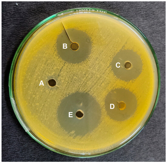
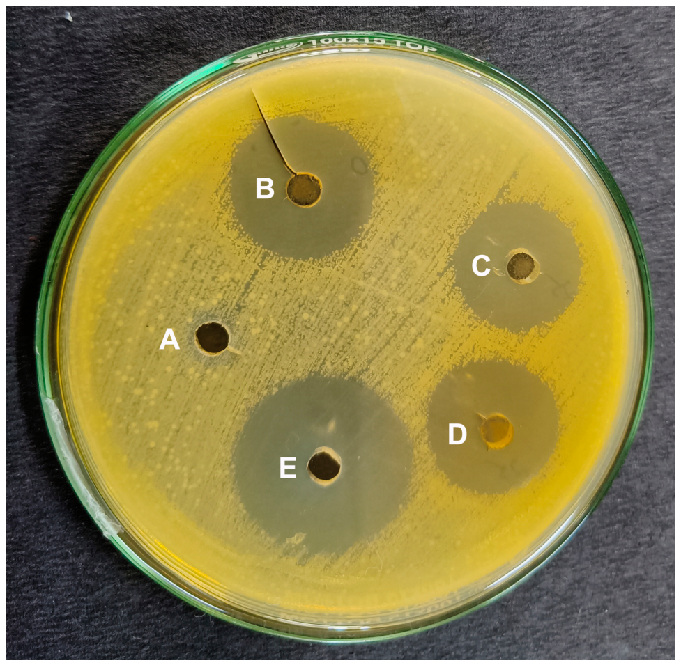
The antibacterial activity of EAE-PEL6 was evaluated against S. aureus using varying concentrations of 10, 25, and 50 mg/mL. EAE-PEL6 showed concentration-dependent inhibition of S. aureus. The inhibition zones were 15.67 ± 1.31, 17.18 ± 0.62, and 19.53 ± 2.01 mm at 10, 25, and 50 mg/mL, respectively. Gentamicin produced a larger inhibition zone of 24.30 ± 0.76 mm, whereas 10% DMSO showed no inhibition. These findings indicate preliminary anti-S. aureus activity of EAE-PEL6. Figure 1 illustrates the obtained results.
Figure 1.
Antibacterial activity of EAE-PEL6 against S. aureus: (A) 10% DMSO negative control, (B) 50 mg/mL, (C) 10 mg/mL, (D) 25 mg/mL, and (E) gentamicin-positive control.
Minimum Inhibitory Concentrations
The antibacterial efficacy of EAE-PEL6 against S. aureus was determined using the broth microdilution method. As shown in Table 2, the extract exhibited an MIC of 0.625 mg/mL, indicating effective inhibition of bacterial growth. In comparison, the positive control gentamicin showed an MIC of 1.95 µg/mL. These findings demonstrated that the crude extract of EAE-PEL6 possesses antibacterial activity against S. aureus.
Table 2.
Minimum inhibitory concentration (MIC) of EAE-PEL6.
3.4. In Silico Toxicity Analysis
The pharmacokinetic and pharmacodynamic profiles for the selected compounds are summarised in Table 3.
Table 3.
In silico analysis of pharmacokinetic and drug-like characteristics of EAE-PEL6 *.
A total of 32 molecules were evaluated using SwissADME and ADMETlab 3.0 for toxicity and drug-likeness, from which four compounds demonstrated significant potential. Compound CID 3467 (gentamicin) was included as a reference for comparative analysis. Among the 32 selected candidates, compounds 262502, 313278, and 575215 exhibited desirable drug-like characteristics. These compounds demonstrated high gastrointestinal absorption, supporting their suitability for oral administration. Notably, only compound 262502 was predicted to cross the blood–brain barrier. Except for 70504 and 3467, all compounds showed favourable lipophilicity, with consensus Log Po/w values ranging between 5.16 and 6.27, indicating good membrane permeability. In terms of solubility, compound 3467 was classified as highly soluble, while 70504 exhibited good solubility, both of which are favourable for drug formulation. Although 262502, 313278, and 575215 exhibited low solubility, they had high molar refractivity and acceptable synthetic accessibility, suggesting potential for chemical optimisation. None of the compounds inhibited key cytochrome P450 enzymes, minimising the risk of metabolic interactions. All compounds were free of PAINS alerts. Despite some Lipinski rule violations, most candidates adhered to Veber’s criteria and showed moderate bioavailability, supporting their further exploration in drug development.
3.5. Molecular Docking
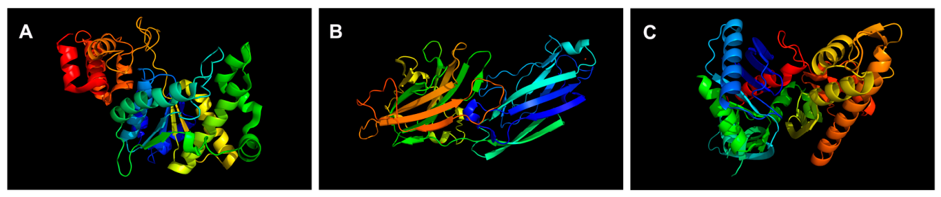
Molecular docking analysis was conducted to evaluate the binding affinity and molecular interactions of selected compounds with the target proteins 1JIJ, 1N67, and 3WQU, using gentamicin as the reference drug. The 3D structures of the targets are presented in Figure 2A–C.
Figure 2.
3D structures of the protein targets. (A) 1JIJ, (B) 1N67, (C) 3WQU.
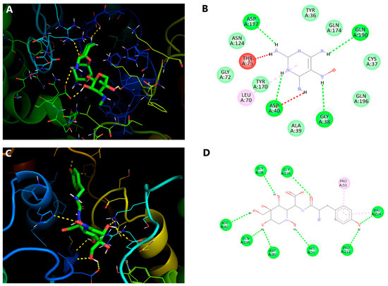
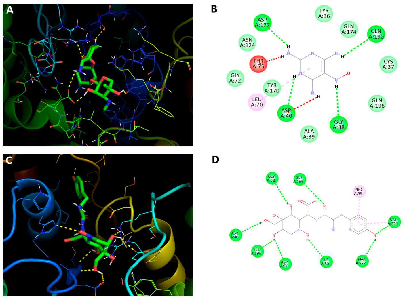
The detailed binding interactions and docking scores are summarised in Table 4. Among the evaluated compounds, solasodine benzoate (CID 313278) showed the highest binding affinity for 1JIJ, with a binding energy of −8.9 kcal/mol, although it formed only a single hydrogen bond with GLN A:196. In comparison, 5-nitroso-2,4,6-triaminopyrimidine (CID 70504) showed a favourable binding energy of −7.0 kcal/mol, forming four hydrogen bonds with ASP A:177, ASP A:40, GLN A:190, and GLY A:38 (Figure 3A,B). The co-crystallised ligand exhibited a binding affinity of −7.9 kcl/mol and formed eight hydrogen bonds with GLY A: 38, GLY A: 193, ASP A: 40, GLN A: 196, ASP A: 80, HIS A: 50, PRO A: 222 and VAL A: 224 (Figure 3C,D). Notably, CID 70504 shared key interactions with ASP A: 40 and GLY A: 38, while solasodine benzoate interacted with GLN A: 196; indicating that these compounds occupy regions within the active site like those of the co-crystallised ligand. These findings suggest that both compounds may have the potential to interact with functionally important residues of TyRS. However, docking scores are predictive and should not be interpreted as direct measures of antibacterial potency. Experimental enzyme inhibition or target-engagement assays are required.
Table 4.
Molecular docking evaluation of EAE-PEL6 targeting S. aureus proteins.
Figure 3.
3D and 2D interactions of the ligands binding with the receptor protein 1JIJ. (A,B) 5-Nitroso-2,4,6-triaminopyrimidine, CID-70504 and (C,D) co-crystalised ligand.
Docking analysis against the crystal structure of S. aureus clumping factor A (PDB ID: 1N67) revealed that solasodine benzoate (CID 313278) exhibited the highest binding affinity, with a binding energy of −10.9 kcal/mol. This compound formed three hydrogen bonds with residues ASP A:139, TYR A:198, and ARG A:194 (Figure 4A,B). Additionally, solasodine ethyl carbonate (CID 575215) and acetylsolasodine (CID 262502) demonstrated substantial binding affinities, with docking scores of −9.2 kcal/mol and −9.8 kcal/mol, respectively. As no co-crystallised ligand was available for this target, blind molecular docking was performed to identify the potential ligand binding regions. Since ClfA is a virulence-associated fibronectin-binding adhesin rather than a conventional enzymatic antibacterial target, the docking analysis was conducted as an exploratory approach to evaluate the potential interactions of the identified compounds with this virulence factor. These computational predictions require further experimental validation to confirm their biological relevance.
Figure 4.
3D and 2D interactions of ligands with 1N67. (A,B) Solasodine benzoate, CID 313278.
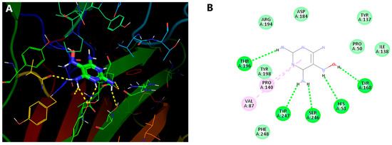
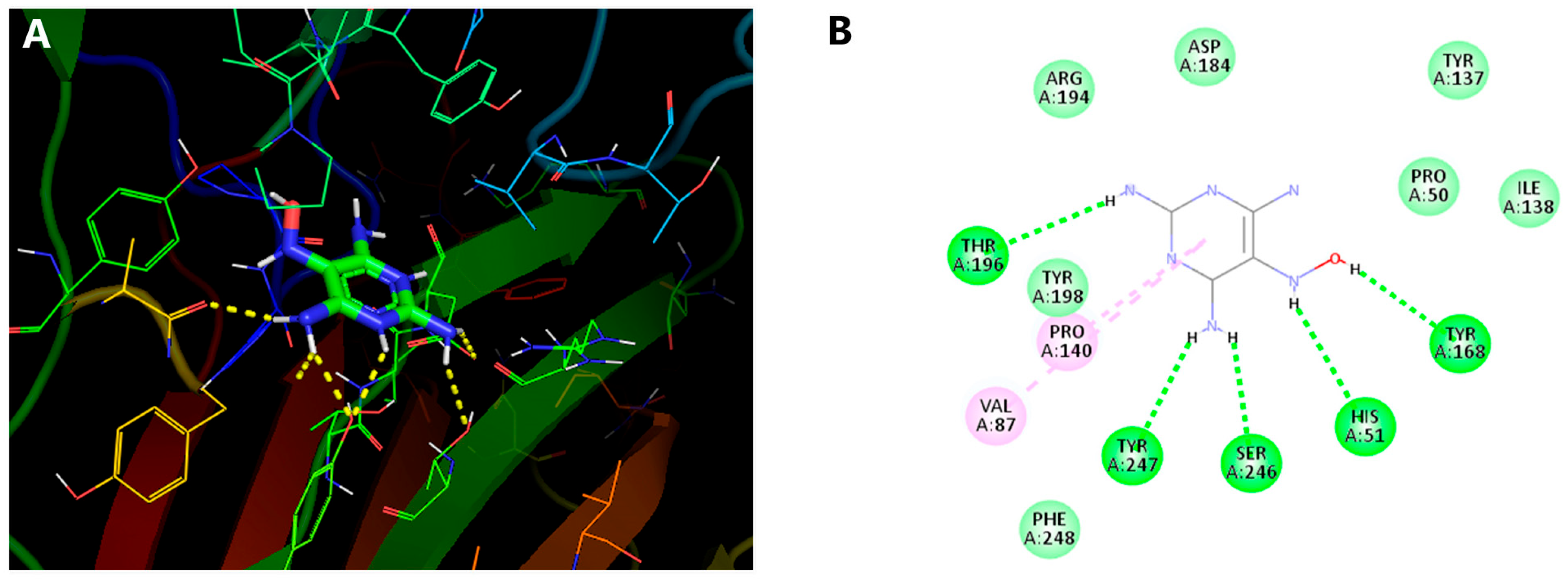
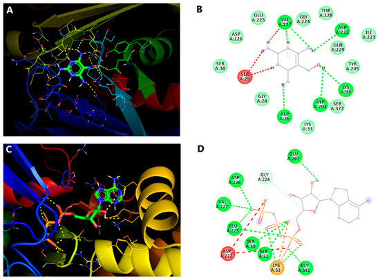
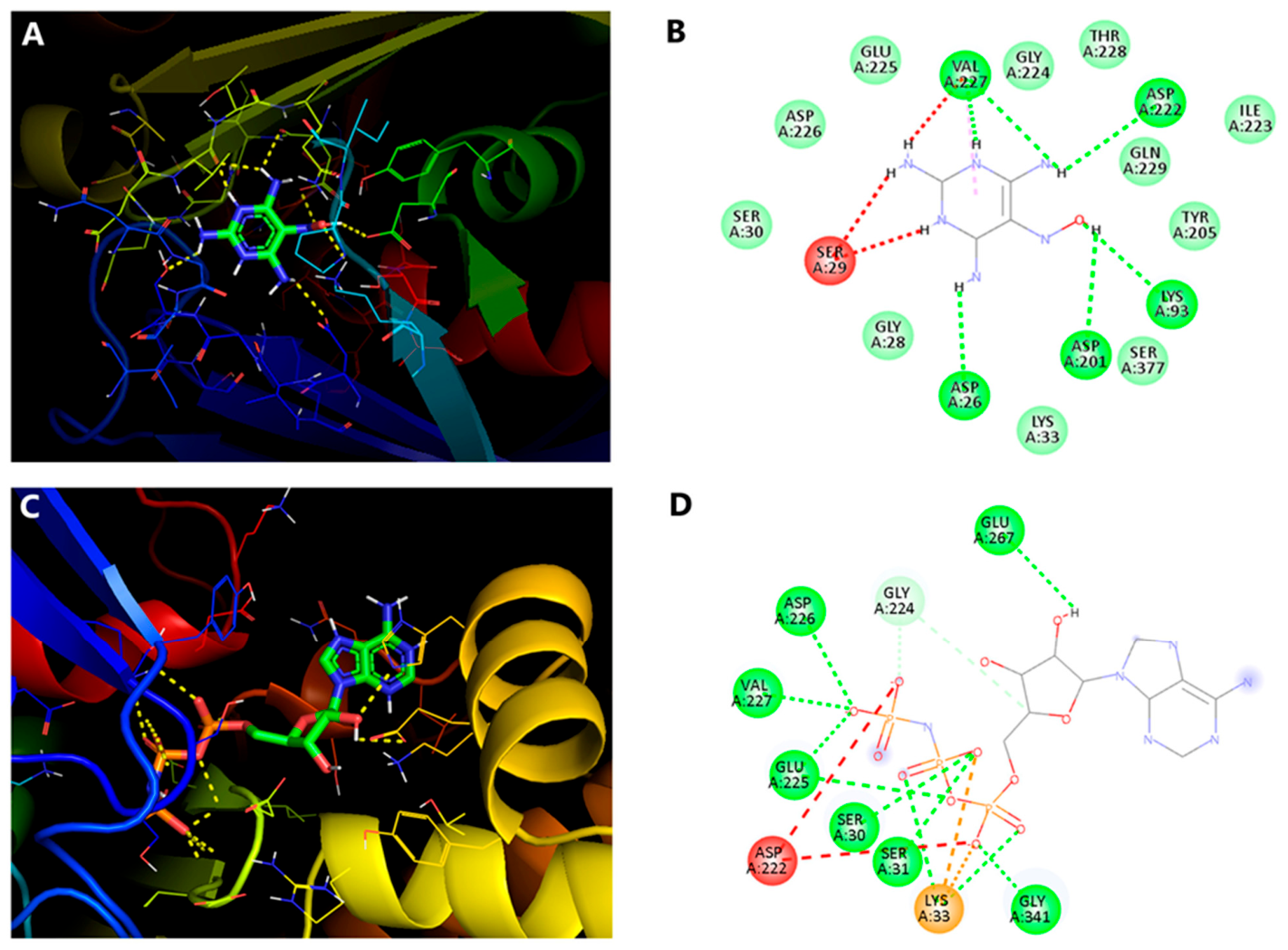
Solasodine benzoate demonstrated the strongest binding affinity towards FtsA (PDB: 3wqu), with a docking score of −8.0 kcal/mol, mediated by two hydrogen bonds. Acetylsolasodine (CID 262502) and 5-nitroso-2,4,6-triaminopyrimidine (CID 70504) also exhibited substantial binding affinities of −7.7 and −7.4 kcal/mol, respectively. Importantly, acetylsolasodine interacted with the same amino acid residue as the co-crystalised ligand (Figure 5A,B). The co-crystallised ligand exhibited the highest binding affinity of −10.7 kcal/mol and formed eight hydrogen bonds with the active site residues GLU A:267, ASP A:226, VAL A:227, GLU A:225, SER A:30, SER A:31, and GLY A:341, thereby validating the selected binding pocket (Figure 5C,D). The shared interaction of acetylsolasodine with the co-crystallised ligand suggests that it occupies a similar region within the FtsA active site and interacts with functionally important residues.
Figure 5.
3D and 2D interactions of the ligands binding with the receptor protein 3WQU. (A,B) 5-Nitroso-2,4,6-triaminopyrimidine CID- 70504 and (C,D) co-crystalised ligand.
3.6. Density Functional Theory
3.6.1. Geometric Optimisation
DFT analysis was performed to optimise the molecular geometries of the selected compounds and assess their stability and reactivity. Initial structures were minimised using Avogadro (v1.2), followed by refinement and single-point energy calculations in ORCA (v5.0.4) with the 6-31G (d, p) basis set. Geometry optimisation identifies the lowest-energy configuration, providing the most stable structural arrangement and reliable insights into molecular properties.
3.6.2. Frontier Molecular Orbitals
The quantum-chemical reactivity descriptors for the studied compounds are presented in Table 5.
Table 5.
Quantum chemical reactivity of selected bioactive compounds of EAE-PEL6.
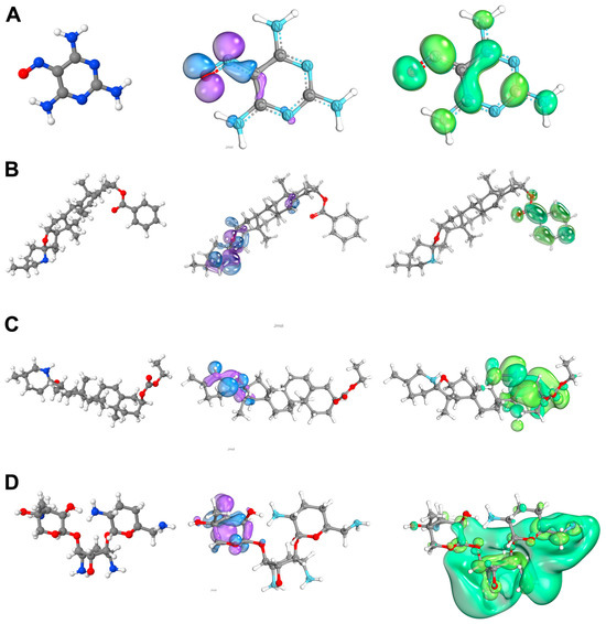
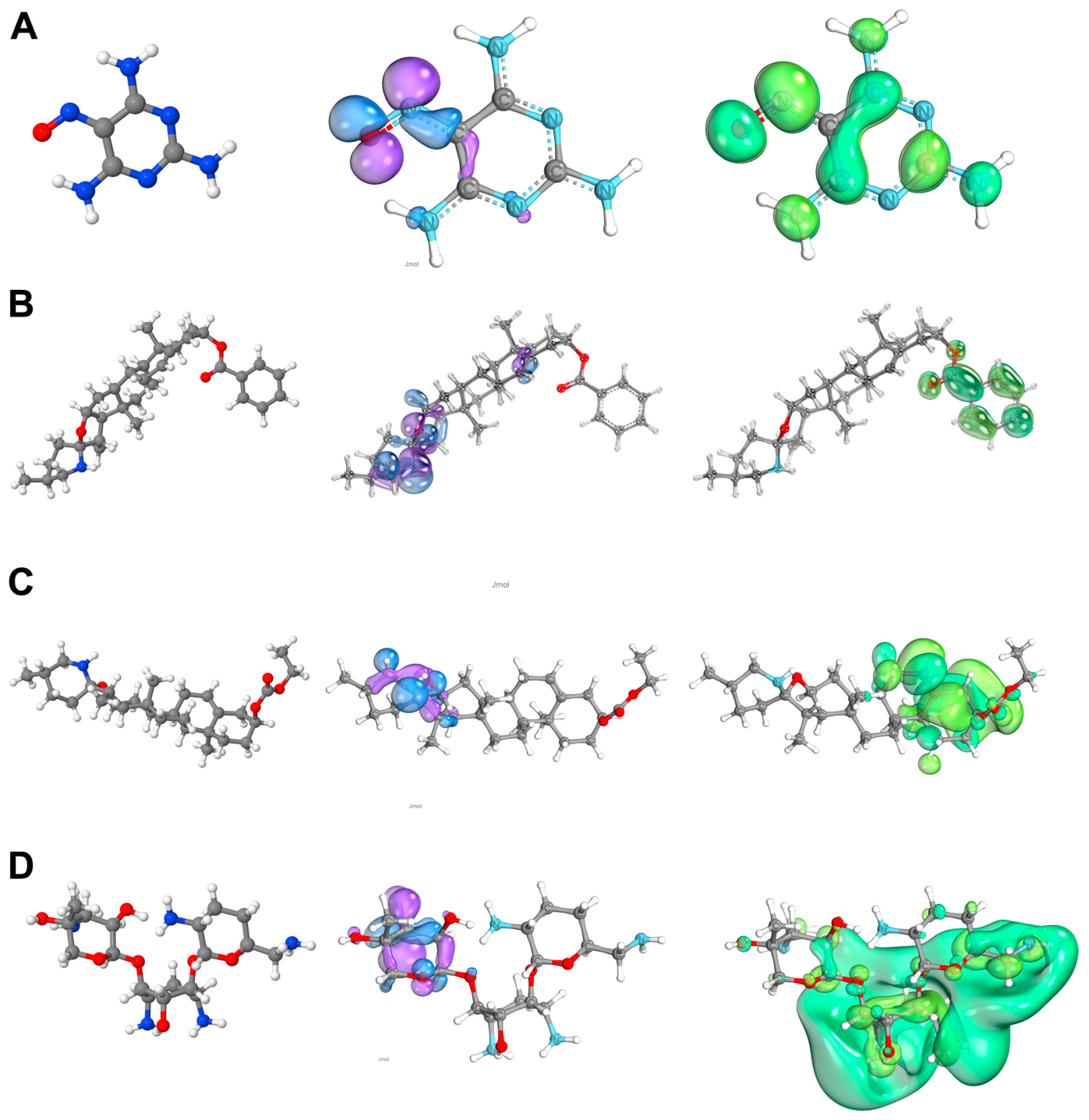
The FMO analysis revealed distinct variations in the HOMO and LUMO energy levels among the studied compounds. CID 70504 (Figure 6A) showed the lowest HOMO-LUMO energy gap (0.1336 eV), indicating higher chemical reactivity and greater potential for charge transfer compared to gentamicin (0.2423 eV) and the other compounds. CID 313278 (Figure 6B) also exhibited a relatively small energy gap (0.1730 eV), suggesting moderate reactivity, while (Figure 6C) CID 575215 (0.2406 eV) displayed a stability pattern like gentamicin (Figure 6D). The higher reactivity of CID 70504 can be attributed to its elevated electron-donating and accepting ability, as reflected in its HOMO and LUMO energy values. Overall, the reduced energy gap of CID 70504 highlights its enhanced electronic reactivity and possible superiority over gentamicin in molecular interactions.
Figure 6.
Analysis of Frontier molecular orbitals—HOMO/LUMO. (A) CID 70504, (B) CID 313278, (C) CID 575215, (D) Gentamicin.
3.6.3. Molecular Electrostatic Potential
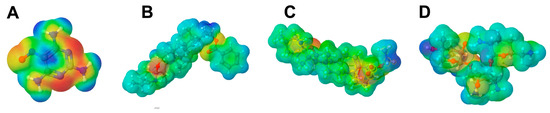
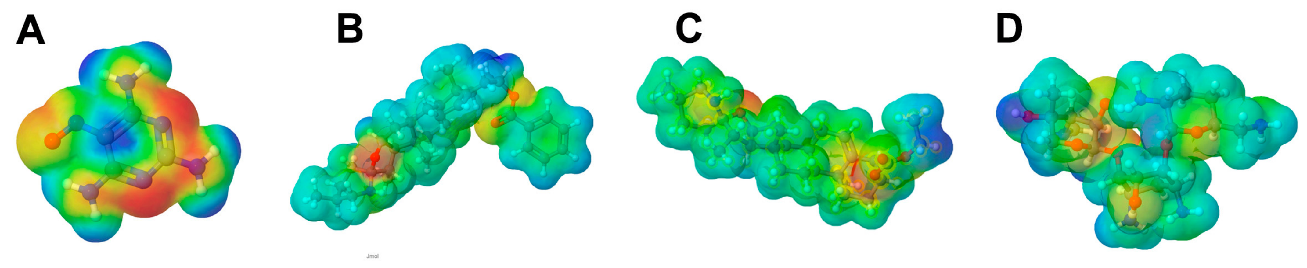
The MEP analysis provided insights into the charge distribution and reactive regions of the studied compounds. In all cases, negative potential regions (red zones) were predominantly localised around electronegative atoms, such as oxygen and nitrogen, indicating favourable sites for electrophilic attack. Conversely, positive potential regions (blue zones) were observed near hydrogen atoms bound to electronegative centres, suggesting that these regions are susceptible to nucleophilic interactions. Among the compounds, CID 70504 (Figure 7A) exhibited more pronounced negative potential regions than gentamicin, reflecting stronger electron-rich domains and a greater tendency for intermolecular interactions. CID 313278 (Figure 7B) and CID 575215 (Figure 7C) showed moderate charge separation, whereas gentamicin (Figure 7D) displayed relatively balanced charge distribution, consistent with its higher stability. These variations in electrostatic potential mapping highlight the greater reactivity of CID 70504, correlating with its lower HOMO-LUMO energy gap, and suggest its potential advantage in molecular binding and biological activity.
Figure 7.
Molecular electrostatic potential of the compounds. (A) CID 70504, (B) CID 313278, (C) CID 575215, (D) Gentamicin.
3.7. Molecular Dynamics Simulation
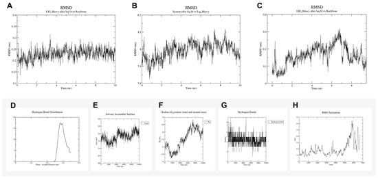
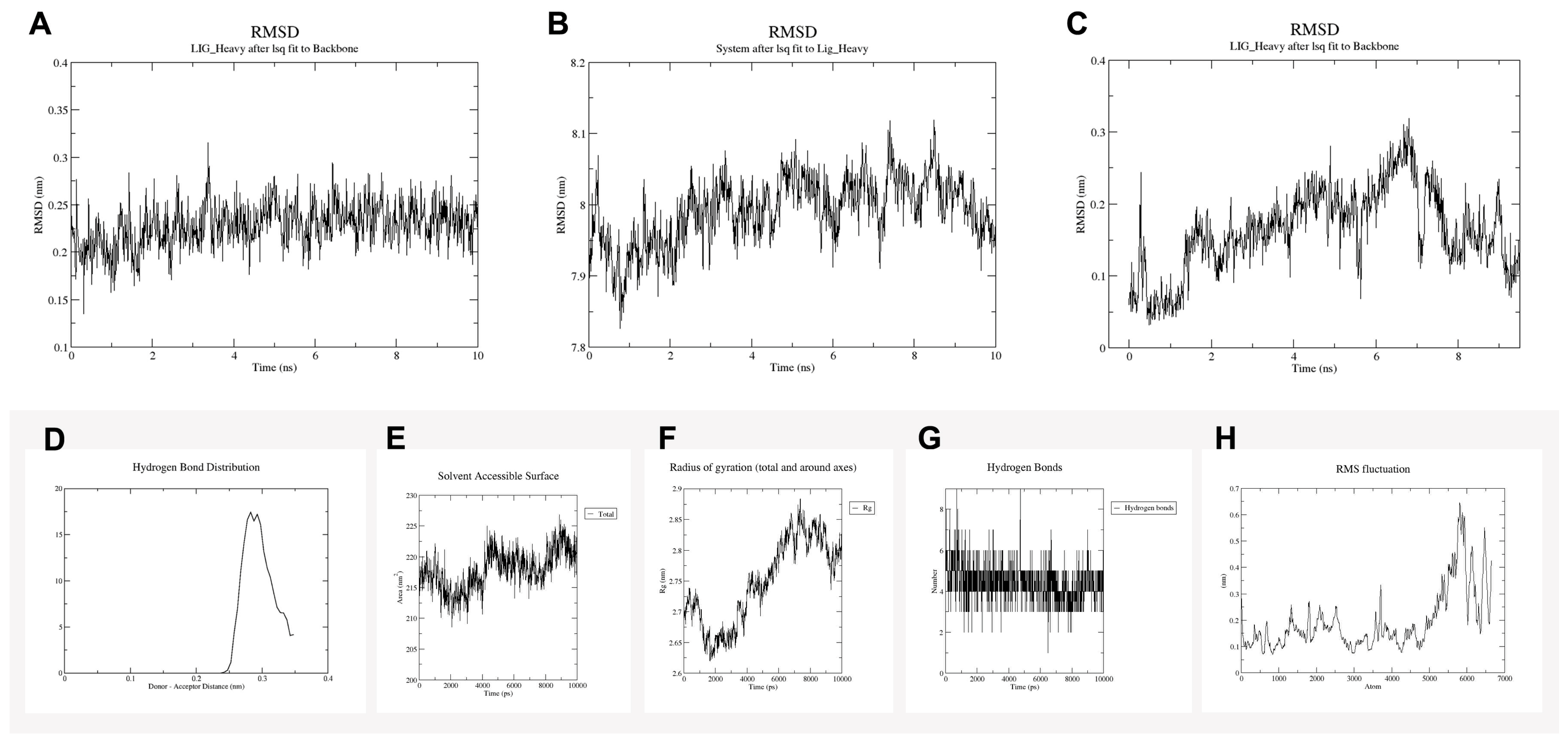
Molecular dynamics simulations of the docked complexes were performed to assess their structural variations and stability. The Root Mean Square Deviation (RMSD) profiles of the three protein–ligand complexes were extracted from the simulation trajectories to assess their rigidity. As shown in Figure 8A, the 1JIJ complex with 5-nitroso-2,4,6-triaminopyrimidine exhibited a nearly linear RMSD profile, indicating better structural stability. Similarly, Figure 8B shows that the 1N67 complex maintained an almost steady RMSD, further suggesting stable binding. In contrast, Figure 8C shows that the 3WQU complex initially exhibited an increase in RMSD, reflecting greater flexibility; however, it reached a stable state after 2 ns of simulation. Overall, the 1JIJ complex exhibited greater flexibility than the other complexes. Both 3WQU and 1JIJ maintained RMSD values between 0 and 0.4 Å throughout the simulation, while the 1N67 complex showed RMSD values ranging between 7.8 and 8.2 Å, confirming their structural stability.
Figure 8.
Graphical depiction of the protein and ligand molecule (CID 70504). (A) RMSD of 1JIJ, (B) RMSD of 1N67, (C) RMSD of 3WQU, (D) hydrogen bond distribution, (E) solvent accessible surface area, (F) radius of gyration, (G) hydrogen bond pattern, and (H) root mean square fluctuation.
Hydrogen-bond analysis of the 1JIJ complex revealed a peak distribution centred at 0.3 nm, indicating consistent intermolecular interactions (Figure 8D). Solvent accessible surface area (SASA) calculations were performed to assess changes in protein volume, with higher SASA values indicating surface expansion and lower values indicating compact conformations. As shown in Figure 8E, the 1JIJ complex exhibited only minor fluctuations in SASA, suggesting negligible volume changes during the simulation. The Rg was further analysed to assess protein compactness; Figure 8F shows that the 1JIJ complex maintained a stable Rg profile throughout the trajectory. Additionally, hydrogen-bond monitoring confirmed stable bonding patterns, as depicted in Figure 8G. Root mean square fluctuation (RMSF) analysis (Figure 8H) showed that almost all residues displayed fluctuations below 0.7 Å, reflecting minimal flexibility and overall stability of the complex.
4. Discussion
Medicinal plants are widely recognised as valuable reservoirs of bioactive compounds, contributing significantly to the development of novel therapeutics and sustainable healthcare practices [1]. Approximately 40% of pharmaceutical drugs originate from natural sources, highlighting the critical role of plant-associated microorganisms, particularly endophytes, in producing pharmacologically relevant metabolites [4,39]. Endophytic bacteria, which inhabit plant tissues without causing harm, not only support plant growth and stress tolerance but also synthesise secondary metabolites that can exhibit stronger bioactivity than those produced by the host plant [7].
In this study, an endophytic bacterium, P. lactis (PEL6), was successfully isolated from the leaves of M. heyneana, a species collected from the Western Ghats. Of the seven initially obtained morphologically distinct strains, only PEL6 remained viable after successive subculturing, suggesting selective adaptation to the leaf environment. This observation aligns with the known ecological behaviour of endophytes, in which microbial survival and dominance are influenced by host factors and inter-microbial interactions [5]. Paenibacillus has emerged as a promising microbial genus with broad applications owing to its production of diverse antimicrobial compounds and industrially significant enzymes. Antimicrobial agents synthesised by Paenibacillus, including peptides, hydrolytic enzymes, volatile organic compounds, and non-volatile metabolites, are effective against destructive phytopathogens such as Fusarium, Rhizoctonia, and Phytophthora, as well as foodborne pathogens such as Salmonella [13,40]. Compared with chemical biocides, these natural products are environmentally friendly and sustainable. Importantly, strain-level diversity, exemplified in P. polymyxa, provides opportunities to identify potent isolates with tailored bioactivity profiles [16]. Beyond antimicrobial potential, Paenibacillus species demonstrate biotechnological versatility. For example, P. lactis PKC5 produces an alkali-stable pectate lyase that has been statistically optimised and functionally characterised for juice clarification, thereby highlighting its industrial relevance [41]. Collectively, such antimicrobial and enzymatic attributes reinforce Paenibacillus as a valuable resource for sustainable agriculture, food safety, and biotechnology.
GC–MS profiling suggested the presence of structurally diverse metabolites in EAE-PEL6. Because these annotations were based on library matching, the identities of major metabolites should be confirmed using authentic standards or LC-MS/MS before assigning biological roles. Among these, pyrrolo[1,2-a] pyrazine-1,4-dione derivatives, belonging to the 2,5-diketopiperazine family, are well documented for their antimicrobial, antifungal, and antioxidant activities [42,43]. Triazole derivatives detected in the extract further strengthen its therapeutic value, as triazole scaffolds are widely recognised pharmacophores in antifungal and antibacterial drug development [44]. In addition, proline-based derivatives and microbial volatile compounds have been linked to suppression of phytopathogens and foodborne bacteria, underscoring their importance in agriculture and food safety [43,45]. The structural diversity of the EAE-PEL6, which also included minor halogenated and cyclic molecules, highlights its potential as a reservoir of novel bioactive agents. The GC–MS-identified compounds were tentatively assigned based on NIST library matching. Confirmation of their identities and biological relevance requires further validation using complementary techniques such as LC–MS/MS, authentic standards, and NMR spectroscopy. These findings align with previous studies showing that endophytes can synthesise unique metabolites not produced by their host plants, thereby expanding opportunities for drug discovery and biocontrol applications [6].
S. aureus frequently colonises the compromised skin of atopic dermatitis patients, producing toxins, enzymes, and superantigens that disrupt the skin barrier and activate immune responses. These factors provoke Th2 and Th17 polarisation, leading to sustained inflammation and exacerbation of skin lesions. This microbial invasion perpetuates a vicious cycle of barrier dysfunction and disease severity, worsening symptoms, and chronic skin damage characteristic of atopic dermatitis [46]. Due to its critical role in disease progression, S. aureus remains a significant focus of research and management in atopic dermatitis. EAE-PEL6 exhibited notable antibacterial activity against S. aureus, producing inhibition zones of approximately 19.5 mm at 50 mg/mL. This level of activity surpasses that reported for other endophytic bacteria, such as Dermacoccus sp. and Paenibacillus sp., isolated from Equisetum arvense, whose methanol extracts produced inhibition zones of approximately 11.30 ± 0.17 mm against S. aureus [47]. A similar trend was observed in plant endophyte extracts from Xerophyta spekei, which produced inhibition zones ranging from 7.67 ± 0.33 mm to 14.67 ± 0.33 mm at concentrations of 50–100 mg/mL [48]. Although conventional antibiotics such as gentamicin or ciprofloxacin generally exhibit stronger antibacterial activity, crude EAE-PEL6 often show more modest activity.
The broth microdilution assay demonstrated that the crude extract of EAE-PEL6 inhibited the growth of S. aureus with an MIC of 0.625 mg/mL, confirming its antibacterial activity. The antimicrobial efficacy of crude natural extracts is influenced by several factors, including the chemical composition of the extract, the abundance of bioactive metabolites, and the susceptibility of the target microorganism. Behbahani et al. [49] reported that the cumin essential oil exhibited lower MIC values against Candida albicans than against Gram-positive and Gram-negative bacterial species, demonstrating that the antimicrobial activity varies among microorganisms. Similarly, Chang and Chong [50] observed that the ethyl acetate fraction of Ganoderma spp. exhibited a lower MIC against MRSA, which was attributed to the enrichment of antibacterial constituents through fractionation. The present findings highlight the potential of EAE-PEL6 as a source of antibacterial metabolites. However, further purification and structural characterisation of the bioactive constituents are required to enhance antibacterial potency and identify the compounds responsible for the observed activity. In addition, future investigations should include determination of the minimum bactericidal concentration (MBC), cytotoxicity assessment, and bioassay-guided fractionation to validate the therapeutic potential of the active metabolites.
MRSA remains a major global health concern because of its resistance to multiple antibiotics and its association with increased mortality. Consequently, the discovery of novel antibacterial agents from natural sources has gained considerable attention. In the present study, the EAE-PEL6 of endophyte P. lactis exhibited antibacterial activity against S. aureus, highlighting the potential of endophytic bacteria as a source of antibacterial metabolites. These findings warrant further investigation to isolate the active constituents and evaluate their efficacy against multidrug-resistant pathogens, including MRSA.
In silico pharmacokinetic and toxicity evaluations offered valuable insights into the drug-like potential of metabolites CID 70504, 313278, and 575215. All three compounds complied with Lipinski’s rule of five, exhibiting physicochemical properties favourable for oral bioavailability [51,52]. ADMET analysis further indicated high gastrointestinal absorption, suitable lipophilicity, minimal cytochrome P450 inhibition, and reasonable synthetic accessibility, collectively reducing the risk of metabolic instability and toxicity [53,54]. Notably, CID 70504 displayed predicted blood–brain barrier permeability, suggesting potential for central nervous system-targeted applications. Compared with endophytic metabolites from Enhydra fluctuans, these compounds showed high absorption and low CYP inhibition but higher lipophilicity and lower solubility [55]. Consistent with previous computational studies of natural products, issues such as solubility and possible immunotoxicity remain relevant [56]. Based on our results, CID 70504 was identified as the most promising lead, highlighting the need for structural refinement to maximise therapeutic efficacy.
Although selected compounds showed favourable predicted binding and some drug-like properties, several ADMET limitations, including poor solubility, high lipophilicity, and predicted toxicity alerts, indicate that these compounds should be considered preliminary lead structures rather than ready drug candidates. The ADMET analysis performed in this study provides only a preliminary prediction of the pharmacokinetic and toxicity profiles of the putatively identified compounds. Therefore, further in vitro and in vivo studies are required to validate the predicted properties, and these investigations will be conducted in future work to establish the safety and therapeutic potential of the identified compounds.
Molecular docking studies of EAE-PEL6 demonstrated strong, highly specific interactions with key S. aureus proteins (1JIJ, 1N67, and 3WQU), highlighting their potential as antibacterial agents. Three S. aureus protein targets were selected based on their essential roles in bacterial physiology and pathogenesis. The bacterial cell-division protein FTsA (PDB ID: 3WQU) was selected because it is indispensable for Z-ring assembly and cell division [57]. Tyrosyl-tRNA synthetase (PDB ID: 1JIJ) was chosen due to its role in protein biosynthesis and its recognition as a validated antibacterial drug target [58]. Clumping Factor (PDB ID: 1N67), a major surface adhesin involved in bacterial adhesion, colonisation and biofilm-associated infection, was included to evaluate the potential of the identified compounds to interfere with S. aureus [59]. Collectively, these proteins represent complementary therapeutic targets associated with bacterial growth, survival and pathogenicity, thereby providing a comprehensive framework for evaluating the antibacterial potential of the identified phytoconstituents.
Among the tested metabolites, CID 313278 showed the most potent binding, with docking scores of −10.9 and −8 kcal/mol, stabilised by multiple hydrogen bonds with crucial active-site residues, confirming both stability and specificity. CID 70504 exhibited consistent affinities (−7.0 to −7.4 kcal/mol) across all targets, interacting with residues including ASP A:177, THR A:196, and VAL A:227, indicating broad binding capability. These results suggest that PEL6-derived compounds may not only replicate but also enhance gentamicin’s antimicrobial effects by targeting overlapping residues. In comparison, hydrazone derivatives act by disrupting cell structures and enzymes, showing moderate affinities (−7.0 to −5.0 kcal/mol), with icaceine binding being slightly stronger (−7.45 kcal/mol) [60,61]. Plant-derived inhibitors, including L-tyrosine methyl ester (−6.7 kcal/mol) from Calotropis gigantea and metabolites of Syzygium samarangense (−5.5 to −7.2 kcal/mol), showed weaker interactions [62,63]. Overall, PEL6 metabolites, particularly CID 70504, exhibited superior binding, highlighting their potential for S. aureus inhibition.
The docking results provide a hypothesis for possible target interactions but do not establish the mechanism of antibacterial action. Enzyme inhibition assays, bacterial target validation, membrane permeability studies, or transcriptomic/proteomic approaches would be needed to confirm the proposed mechanisms.
DFT calculations demonstrated that CID 70504 exhibited the smallest HOMO–LUMO energy gap, reflecting enhanced chemical reactivity and favourable electronic features for molecular binding. MEP analysis further identified electron-rich regions near electronegative atoms, indicating potential interaction sites with bacterial proteins. FMOs, specifically HOMO and LUMO, remain critical descriptors of molecular reactivity and electron transfer processes [64]. In Annona muricata, genistein and kaempferol displayed the greatest reactivity, with energy gaps of 0.16507 and 0.16413 eV, respectively, suggesting their ability to establish stable intermolecular associations with survivin [65]. The quantum-chemical findings validate the docking results and elucidate the mechanism driving the antibacterial activity. The DFT findings provide insight into electronic reactivity and charge distribution. However, a lower HOMO-LUMO gap alone does not guarantee stronger antibacterial activity and should be interpreted alongside experimental validation.
Molecular dynamics simulations were performed to investigate the stability and dynamic behaviour of the protein–ligand complexes. The results confirmed structural stability, as analyses of RMSD, RMSF, hydrogen bonding, solvent-accessible surface area, and Rg showed only minor conformational variations during the 100 ns simulation. These findings highlight stable interactions between EAE-PEL6 and their targets, while the slight flexibility observed in some complexes may enhance ligand adaptability and binding specificity. Molecular dynamics simulations are recognised as valuable tools in drug discovery for evaluating biomolecular stability and conformational dynamics [38]. The RMSD values revealed minimal deviations, consistent with earlier reports of stable protein–ligand complexes [66]. Likewise, the stable Rg profiles demonstrated that the enzyme maintained its compact structure, whereas significant fluctuations are typically associated with unfolding [67]. Notably, these stability patterns are comparable to or exceed those reported for previously studied inhibitors, underscoring the therapeutic promise of the investigated metabolites. Molecular dynamics simulations suggested that selected docked complexes remained relatively stable under simulated conditions, but replicate simulations and binding free-energy calculations, such as MM-PBSA/MM-GBSA, would strengthen confidence in these predictions.
Overall, these results indicate that the endophytic bacterium P. lactis PEL6, isolated from M. heyneana, synthesises a wide spectrum of metabolites with strong antibacterial activity. The integration of experimental findings and computational approaches underscores the value of endophytes as promising reservoirs of novel therapeutic molecules. Future research should focus on the purification, detailed characterisation, and in vivo assessment of individual metabolites to facilitate their translation into effective antimicrobial agents. Taken together, these outcomes highlight the potential of EAE-PEL6 as strong antibacterial leads with significant prospects in drug discovery and sustainable therapeutic development.
This study has several limitations. Although antibacterial activity was confirmed using agar well diffusion and MIC assays, the MBC (Minimum Bacterial Count) was not determined. The compounds identified by GC-MS were putatively assigned based on library matching and require structural confirmation. Moreover, the active constituents responsible for the observed antibacterial activity were not isolated or experimentally validated. The computational analyses represent predictive evidence and require further biological validation. Therefore, the findings should be considered preliminary and additional investigation is required.
5. Conclusions
The present study isolated seven putative endophytic bacterial strains from the leaves of M. heyneana, of which PEL6 was identified as Paenibacillus lactis based on 16S rRNA gene sequencing. The crude ethyl acetate extract of PEL6 exhibited concentration-dependent antibacterial activity against S. aureus. GC–MS analysis provided a tentative profile of 32 metabolites, including diketopiperazine-, triazole-, and solasodine-related compounds. Computational analyses, including ADMET prediction, molecular docking, DFT, and molecular dynamics simulation, identified several putatively annotated compounds with favourable interactions against selected antibacterial targets. However, these computational findings do not establish that the identified compounds are responsible for the observed antibacterial activity of the crude extract. Therefore, further studies involving bioassay-guided fractionation, compound purification, structural confirmation using complementary analytical techniques, cytotoxicity evaluation, and experimental target validation are required to identify and confirm the active antibacterial constituents.
Author Contributions
Conceptualisation, K.S.I.; methodology, S.D., S.M.R.F. and K.S.I.; software, K.S.I., S.V., R.P. and B.S.S.; validation, K.S.I., C.C., S.K. and B.S.S., formal analysis, S.D., B.S.S., S.V., S.M.R.F., R.P. and K.S.I.; investigation, S.D., B.S.S., S.V., S.M.R.F., R.P. and K.S.I.; resources, B.S.S., C.C. and K.S.I.; data curation, S.D., B.S.S., S.V., S.M.R.F., R.P. and K.S.I.; writing—original draft preparation, S.D., B.S.S., S.V., S.M.R.F., R.P. and K.S.I.; writing—review and editing, S.D., B.S.S., S.V., S.M.R.F., R.P., P.F., C.C., S.K. and K.S.I.; visualisation, K.S.I.; supervision, K.S.I.; project administration, K.S.I. and B.S.S.; funding acquisition, K.S.I., B.S.S. and C.C. All authors have read and agreed to the published version of the manuscript.
Funding
This research received funding from Tamil Nadu State Council for Science and Technology (TNSCST) under ref. no. TNSCST/RFRS/06/VM/2021-22.
Institutional Review Board Statement
Not applicable.
Informed Consent Statement
Not applicable.
Data Availability Statement
All the data presented within the manuscript.
Acknowledgments
The study was partially supported by Chiang Mai University, Chiang Mai, Thailand. The author, B.S.S., P.F., C.C., and S.K., gratefully acknowledges Chiang Mai University for its support. We sincerely thank C. S. Shobana, Department of Microbiology, PSG College of Arts & Science, Coimbatore, for kindly providing the clinical isolate of the microbial strain used in this study. NCBI Accession Number OQ861110.
Conflicts of Interest
The authors declare no conflicts of interest.
References
- Asiminicesei, D.M.; Fertu, D.I.; Gavrilescu, M. Impact of heavy metal pollution in the environment on the metabolic profile of medicinal plants and their therapeutic potential. Plants 2024, 13, 913. [Google Scholar] [CrossRef]
- Dailah, H.G. The ethnomedicinal evidences about traditional medicinal herbs used in the treatment of respiratory illnesses and disorders in Saudi Arabia: A review. Saudi J. Biol. Sci. 2022, 29, 103386. [Google Scholar] [CrossRef] [PubMed]
- Lee, E.L.; Richards, N.; Harrison, J.; Barnes, J. Prevalence of use of traditional, complementary and alternative medicine by the general population: A systematic review of national studies published from 2010 to 2019. Drug Saf. 2022, 45, 713–735. [Google Scholar] [CrossRef] [PubMed]
- Hardoim, P.R.; Van Overbeek, L.S.; Berg, G.; Pirttilä, A.M.; Compant, S.; Campisano, A.; Sessitsch, A. The hidden world within plants: Ecological and evolutionary considerations for defining functioning of microbial endophytes. Microbiol. Mol. Biol. Rev. 2015, 79, 293–320. [Google Scholar] [CrossRef] [PubMed]
- Nair, D.N.; Padmavathy, S. Impact of endophytic microorganisms on plants, environment and humans. Sci. World J. 2014, 2014, 250693. [Google Scholar] [CrossRef] [PubMed]
- Ek-Ramos, M.J.; Gomez-Flores, R.; Orozco-Flores, A.A.; Rodríguez-Padilla, C.; González-Ochoa, G.; Tamez-Guerra, P. Bioactive products from plant-endophytic gram-positive bacteria. Front. Microbiol. 2019, 10, 463. [Google Scholar] [CrossRef] [PubMed]
- Zotchev, S.B. Unlocking the potential of bacterial endophytes from medicinal plants for drug discovery. Microb. Biotechnol. 2024, 17, e14382. [Google Scholar] [CrossRef] [PubMed]
- Eshboev, F.; Mamadalieva, N.; Nazarov, P.A.; Hussain, H.; Katanaev, V.; Egamberdieva, D.; Azimova, S. Antimicrobial action mechanisms of natural compounds isolated from endophytic microorganisms. Antibiotics 2024, 13, 271. [Google Scholar] [CrossRef] [PubMed]
- Wu, D.; Wang, W.; Yao, Y.; Li, H.; Wang, Q.; Niu, B. Microbial interactions within beneficial consortia promote soil health. Sci. Total Environ. 2023, 900, 165801. [Google Scholar] [CrossRef] [PubMed]
- Muzzamal, H.; Sarwar, R.; Sajid, I.; Hasnain, S. Isolation, identification and screening of endophytic bacteria antagonistic to biofilm formers. Pak. J. Zool. 2012, 44, 249–257. [Google Scholar]
- Kim, J.; Kim, B.E.; Ahn, K.; Leung, D.Y. Interactions between atopic dermatitis and Staphylococcus aureus infection: Clinical implications. Allergy Asthma Immunol. Res. 2019, 11, 593–603. [Google Scholar] [CrossRef] [PubMed]
- Wang, Z.; Hülpüsch, C.; Traidl-Hoffmann, C.; Reiger, M.; Schloter, M. Understanding the role of Staphylococcus aureus in atopic dermatitis: Strain diversity, microevolution, and prophage influences. Front. Med. 2024, 11, 1480257. [Google Scholar] [CrossRef] [PubMed]
- Zgoda-Pols, J.R.; Freyer, A.J.; Killmer, L.B.; Porter, J.R. Antimicrobial diterpenes from the stem bark of Mitrephora celebica. Fitoterapia 2002, 73, 434–438. [Google Scholar] [CrossRef] [PubMed]
- Li, C.; Lee, D.; Graf, T.N.; Phifer, S.S.; Nakanishi, Y.; Riswan, S.; Oberlies, N.H. Bioactive constituents of the stem bark of Mitrephora glabra. J. Nat. Prod. 2009, 72, 1949–1953. [Google Scholar] [CrossRef] [PubMed]
- Jalil, J.; Afiq, M.; Wahab, S.; Husain, K. Phytoconstituents and pharmacological activities of Mitrephora species (Annonaceae): A review. Trop. J. Pharm. Res. 2025, 24, 413–429. [Google Scholar] [CrossRef]
- Anand, R.; Grayston, S.; Chanway, C. N2-fixation and seedling growth promotion of lodgepole pine by endophytic Paenibacillus polymyxa. Microb. Ecol. 2013, 66, 369–374. [Google Scholar] [CrossRef] [PubMed]
- Grady, E.N.; MacDonald, J.; Liu, L.; Richman, A.; Yuan, Z.C. Current knowledge and perspectives of Paenibacillus: A review. Microb. Cell Fact. 2016, 15, 203. [Google Scholar] [CrossRef] [PubMed]
- Deepa, S.; Prabakaran, R.; Joshi, A.; Ibrahim, K.S.; Rajiv, P. Callus of Mundulea sericea as potential anti-diabetic agents; an in silico approach. S. Afr. J. Bot. 2024, 173, 338–346. [Google Scholar] [CrossRef]
- Razia, S.; Chowdhury, M.S.M.; Aminuzzaman, F.M.; Sultana, N.; Islam, M. Morphological, pathological, biochemical and molecular characterization of Ralstonia solanacearum isolates in Bangladesh. Am. J. Mol. Biol. 2021, 11, 142–164. [Google Scholar] [CrossRef]
- Khan, M.S.; Gao, J.; Chen, X.; Zhang, M.; Yang, F.; Du, Y.; Moe, T.S.; Munir, I.; Xue, J.; Zhang, X. Isolation and characterization of plant growth-promoting endophytic bacteria Paenibacillus polymyxa SK1 from Lilium lancifolium. BioMed Res. Int. 2020, 2020, 8650957. [Google Scholar] [CrossRef] [PubMed]
- Akshatha, J.V.; SantoshKumar, H.S.; Prakash, H.S.; Nalini, M.S. In silico docking studies of α-amylase inhibitors from the anti-diabetic plant Leucas ciliata Benth. and an endophyte, Streptomyces longisporoflavus. 3 Biotech 2021, 11, 51. [Google Scholar] [CrossRef] [PubMed]
- Okla, M.K.; Alatar, A.A.; Al-Amri, S.S.; Soufan, W.H.; Ahmad, A.; Abdel-Maksoud, M.A. Antibacterial and antifungal activity of the extracts of different parts of Avicennia marina (Forssk.) Vierh. Plants 2021, 10, 252. [Google Scholar] [CrossRef] [PubMed]
- Mohamed, E.A.A.; Muddathir, A.M.; Osman, M.A. Antimicrobial activity, phytochemical screening of crude extracts, and essential oils constituents of two Pulicaria spp. growing in Sudan. Sci. Rep. 2020, 10, 17148. [Google Scholar] [CrossRef] [PubMed]
- Baskaran, C.; Velu, S.; Kumaran, K. The efficacy of Carica papaya leaf extract on some bacterial and a fungal strain by well diffusion method. Asian Pac. J. Trop. Dis. 2010, 2, S658–S662. [Google Scholar]
- Savu, M.; Simo, M.K.; Fopokam, G.X.; Olaru, S.M.; Cioanca, O.; Boyom, F.F.; Stefan, M. New Insights into the Antimicrobial Potential of Polyalthia longifolia—Antibiofilm Activity and Synergistic Effect in Combination with Penicillin against Staphylococcus aureus. Microorganisms 2022, 10, 1943. [Google Scholar] [CrossRef] [PubMed]
- Kim, S.; Chen, J.; Cheng, T.; Gindulyte, A.; He, J.; He, S.; Li, Q.; Shoemaker, B.A.; Thiessen, P.A.; Yu, B.; et al. PubChem in 2021: New data content and improved web interfaces. Nucl. Acids Res. 2021, 49, D1388–D1395. [Google Scholar] [PubMed]
- O’Boyle, N.M.; Campbell, C.M.; Hutchison, G.R. Computational design and selection of optimal organic photovoltaic materials. J. Phys. Chem. C 2011, 115, 16200–16210. [Google Scholar] [CrossRef]
- Daina, A.; Michielin, O.; Zoete, V. SwissADME: A free web tool to evaluate pharmacokinetics, drug-likeness and medicinal chemistry friendliness of small molecules. Sci. Rep. 2017, 7, 42717. [Google Scholar] [CrossRef] [PubMed]
- Fu, L.; Shi, S.; Yi, J.; Wang, N.; He, Y.; Wu, Z.; Peng, J.; Deng, Y.; Wang, W.; Wu, C.; et al. ADMETlab 3.0: An updated comprehensive online ADMET prediction platform enhanced with broader coverage, improved performance, API functionality and decision support. Nucl. Acids Res. 2024, 52, W422–W431. [Google Scholar] [CrossRef] [PubMed]
- Robin, X.; Waterhouse, A.M.; Bienert, S.; Studer, G.; Alexander, L.T.; Tauriello, G.; Schwede, T.; Pereira, J. The SWISS-model repository of 3D protein structures and models. In Open Access Databases and Datasets for Drug Discovery: Methods and Principles in Medicinal Chemistry; Daina, A., Przewosny, M., Zoete, V., Eds.; John Wiley & Sons, Inc.: New York, NY, USA, 2024; Volume 83, pp. 175–199. [Google Scholar]
- Dallakyan, S.; Olson, A.J. Small-molecule library screening by docking with PyRx. Methods Mol. Biol. 2015, 1263, 243–250. [Google Scholar] [PubMed]
- Schrodinger LLC. The PyMOL Molecular Graphics System; Version 4.6.0.; Schrodinger LLC.: New York, NY, USA, 2021. [Google Scholar]
- Studio, D. Dassault Systèmes BIOVIA, Discovery Studio Modelling Environment; Release 4.5.; Accelrys Softw Inc.: San Diego, CA, USA, 2015; pp. 98–104. [Google Scholar]
- Neese, F.; Wennmohs, F.; Becker, U.; Riplinger, C. The ORCA quantum chemistry program package. J. Chem. Phys. 2020, 152, 224108. [Google Scholar] [CrossRef] [PubMed]
- Hertwig, R.H.; Koch, W. On the parameterization of the local correlation functional. What is Becke-3-LYP? Chem. Phys. Lett. 1997, 268, 345–351. [Google Scholar] [CrossRef]
- Majeed, A.; Hussain, W.; Yasmin, F.; Akhtar, A.; Rasool, N. Virtual screening of phytochemicals by targeting HR1 domain of SARS-CoV-2 S protein: Molecular docking, molecular dynamics simulations, and DFT studies. BioMed Res. Int. 2021, 2021, 6661191. [Google Scholar] [CrossRef] [PubMed]
- Karplus, M.; McCammon, J.A. Molecular dynamics simulations of biomolecules. Nat. Struct. Biol. 2002, 9, 646–652. [Google Scholar] [CrossRef] [PubMed]
- Rawat, K.; Tewari, D.; Bisht, A.; Chandra, S.; Tiruneh, Y.K.; Hassan, H.M.; Al-Emam, A.; Sindi, E.R.; Al-Dies, A.M. Identification of AChE targeted therapeutic compounds for Alzheimer’s disease: An in-silico study with DFT integration. Sci. Rep. 2024, 14, 30356. [Google Scholar] [CrossRef] [PubMed]
- Calixto, J.B. The role of natural products in modern drug discovery. An. Acad. Bras. Cienc. 2019, 91, e20190105. [Google Scholar] [CrossRef] [PubMed]
- Lorentz, R.H.; Artico, S.; Da Silveira, A.B.; Einsfeld, A.; Corcao, G. Evaluation of antimicrobial activity in Paenibacillus spp. strains isolated from natural environment. Lett. Appl. Microbiol. 2006, 43, 541–547. [Google Scholar] [CrossRef] [PubMed]
- Sheladiya, P.; Kapadia, C.; Prajapati, V.; Ali El Enshasy, H.; Abd Malek, R.; Marraiki, N.; Zaghloul, N.S.S.; Sayyed, R.Z. Production, statistical optimization, and functional characterization of alkali stable pectate lyase of Paenibacillus lactis PKC5 for use in juice clarification. Sci. Rep. 2022, 12, 7564. [Google Scholar] [CrossRef] [PubMed]
- Jia, J.; Yao, J.; Kong, J.; Yu, A.; Wei, J.; Dong, Y.; Song, R.; Shan, D.; Zhong, X.; Lv, F.; et al. 2,5-Diketopiperazines: A Review of Source, Synthesis, Bioactivity, Structure, and MS Fragmentation. Curr. Med. Chem. 2023, 30, 1060–1085. [Google Scholar] [CrossRef] [PubMed]
- Radford, E.J.; Whitworth, D.E.; Allison, G. Identification of secondary metabolites containing a diketopiperazine core in extracts from myxobacterial strains with growth inhibition activity against a range of prey species. Access Microbiol. 2023, 5, 000629-v4. [Google Scholar] [CrossRef] [PubMed]
- Obled, A.M.C.; Hamed, R.B.; Spence, E.; Zacharova, M.K.; Sharma, S.V.; Wang, Y.; Lynch, R.; Connaris, H.; Tatheer, A.; Bourguet-Kondracki, M.L.; et al. Isolation and Identification of Cis-2, 5-Diketopiperazine from a Novel Bacillus Strain and Synthesis of Its Four Stereoisomers. Mar. Drugs 2025, 23, 234. [Google Scholar] [CrossRef] [PubMed]
- Beneš, R.; Koval, D.; Švec, I.; Macůrková, A.; Vrchotová, B.; Honzíková, T.; Kalenchak, K.; Bárta, J.; Bártová, V.; Bedrníček, J.; et al. Antimicrobial and Antifungal Activities of Proline-Based 2,5-Diketopiperazines Occurring in Food and Beverages and Their Synergism with Lactic Acid. ACS Agric. Sci. Technol. 2025, 5, 1681–1692. [Google Scholar] [CrossRef] [PubMed]
- Alexander, H.; Paller, A.S.; Traidl-Hoffmann, C.; Beck, L.A.; De Benedetto, A.; Dhar, S.; Girolomoni, G.; Irvine, A.D.; Spuls, P.; Su, J.; et al. The role of bacterial skin infections in atopic dermatitis: Expert statement and review from the International Eczema Council Skin Infection Group. Br. J. Dermatol. 2020, 182, 1331–1342. [Google Scholar] [CrossRef] [PubMed]
- Das, G.; Patra, J.K.; Baek, K.H. Antibacterial properties of endophytic bacteria isolated from a fern species Equisetum arvense L. against foodborne pathogenic bacteria Staphylococcus aureus and Escherichia coli O157: H7. Foodborne Pathog. Dis. 2017, 14, 50–58. [Google Scholar] [CrossRef] [PubMed]
- Nyalo, P.O.; Omwenga, G.I.; Ngugi, M.P. Antibacterial properties and GC-MS analysis of ethyl acetate extracts of Xerophyta spekei (Baker) and Grewia tembensis (Fresen). Heliyon 2023, 9, e14461. [Google Scholar] [CrossRef] [PubMed]
- Behbahani, B.A.; Noshad, M.; Falah, F. Cumin essential oil: Phytochemical analysis, antimicrobial activity and investigation of its mechanism of action through scanning electron microscopy. Microb. Pathog. 2019, 136, 103716. [Google Scholar] [CrossRef] [PubMed]
- Chan, Y.S.; Chong, K.P. Bioactive compounds of Ganoderma boninense inhibited methicillin-resistant Staphylococcus aureus growth by affecting their cell membrane permeability and integrity. Molecules 2022, 27, 838. [Google Scholar] [CrossRef] [PubMed]
- Benet, L.Z.; Hosey, C.M.; Ursu, O.; Oprea, T.I. BDDCS, the Rule of 5 and drugability. Adv. Drug Deliv. Rev. 2016, 101, 89–98. [Google Scholar] [CrossRef] [PubMed]
- Erol, M.; Celik, I.; Kuyucuklu, G. Synthesis, molecular docking, molecular dynamics, DFT and antimicrobial activity studies of 5-substituted-2-(p-methylphenyl) benzoxazole derivatives. J. Mol. Struct. 2021, 1234, 130151. [Google Scholar] [CrossRef]
- Wang, Y.; Xing, J.; Xu, Y.; Zhou, N.; Peng, J.; Xiong, Z.; Liu, X.; Luo, X.; Luo, C.; Chen, K.; et al. In silico ADME/T modelling for rational drug design. Q. Rev. Biophys. 2015, 48, 488–515. [Google Scholar] [CrossRef] [PubMed]
- Pasquereau, S.; Nehme, Z.; Haidar Ahmad, S.; Daouad, F.; Van Assche, J.; Wallet, C.; Schwartz, C.; Rohr, O.; Morot-Bizot, S.; Herbein, G. Resveratrol Inhibits HCoV-229E and SARS-CoV-2 Coronavirus Replication In Vitro. Viruses 2021, 13, 354. [Google Scholar] [CrossRef] [PubMed]
- Bhuiyan, A.A.; Aboul-Soud, M.A.M.; Uddin, M.S.; Mazumder, K.; Akash, A.R.; Aljowaie, R.M.; Giesy, J.P.; Agrawal, M.; Haque, S. GC-MS profiling, in vitro and in silico antibacterial and antioxidant potential of crude secondary metabolites of an endophytic bacterium isolated from Enhydra fluctuans lour. Sci. Rep. 2025, 15, 31876. [Google Scholar] [CrossRef] [PubMed]
- Durán-Iturbide, N.A.; Díaz-Eufracio, B.I.; Medina-Franco, J.L. In silico ADME/Tox profiling of natural products: A focus on BIOFACQUIM. ACS Omega 2020, 5, 16076–16084. [Google Scholar] [CrossRef] [PubMed]
- Fujita, J.; Maeda, Y.; Nagao, C.; Tsuchiya, Y.; Miyazaki, Y.; Hirose, M.; Matsumura, H. Crystal structure of FtsA from Staphylococcus aureus. FEBS Lett. 2014, 588, 1879–1885. [Google Scholar] [CrossRef] [PubMed]
- Qiu, X.; Janson, C.A.; Smith, W.W.; Green, S.M.; McDevitt, P.; Johanson, K.; Jarvest, R.L. Crystal structure of Staphylococcus aureus tyrosyl-tRNA synthetase in complex with a class of potent and specific inhibitors. Protein Sci. 2001, 10, 2008–2016. [Google Scholar] [CrossRef] [PubMed]
- Deivanayagam, C.C.; Wann, E.R.; Chen, W.; Carson, M.; Rajashankar, K.R.; Höök, M.; Narayana, S.V. A novel variant of the immunoglobulin fold in surface adhesins of Staphylococcus aureus: Crystal structure of the fibrinogen-binding MSCRAMM, clumping factor A. EMBO J. 2002, 21, 6660–6672. [Google Scholar] [CrossRef] [PubMed]
- Sony, A.S.; Suresh, X. Molecular Modeling and Docking Studies of Some Benzodiazole Derivatives on the Protein of Staphylococcus aureus. J. Pharm. Res. Int. 2020, 32, 102–110. [Google Scholar] [CrossRef]
- Temitope, T. Icacina Species: A Multi-targeted Molecular Docking of Selected Compounds Against Staphylococcus aureus Proteins. Maghr. J. Pure Appl. Sci. 2023, 9, 30–37. [Google Scholar]
- Beg, M.A.; Ansari, S.; Athar, F. Molecular docking studies of Calotropis gigantea phytoconstituents against Staphylococcus aureus tyrosyl-tRNA synthetase protein. J. Bacteriol. Mycol. 2020, 8, 78–91. [Google Scholar] [CrossRef]
- Philip, A.; Nivetha, L.; Rajan, D. GCMS Analysis and Molecular Docking Studies of Bioactive Compounds in Leaf Extract of Syzgium samarangense. Int. Res. J. Biotech. 2023, 14, 1–5. [Google Scholar]
- Hussein, R.K.; Elkhair, H.M.; Elzupir, A.O.; Ibnaouf, K.H. Spectral, structural, stability characteristics and frontier molecular orbitals of tri-n-butyl phosphate (TBP) and its degradation products: DFT calculations. J. Ovonic. Res. 2021, 17, 23–30. [Google Scholar] [CrossRef]
- Omiyale, B.O.; Oyinloye, B.E.; Ajiboye, B.O.; Ubah, C.S. Curated phytochemicals of Annona muricata modulate proteins linked to type II diabetes mellitus: Molecular docking studies, ADMET and DFT calculation. Inform. Med. Unlocked 2024, 47, 101511. [Google Scholar] [CrossRef]
- Joshi, T.; Joshi, T.; Sharma, P.; Chandra, S.; Pande, V. Molecular docking and molecular dynamics simulation approach to screen natural compounds for inhibition of Xanthomonas oryzae pv. Oryzae by targeting peptide deformylase. J. Biomol. Struct. Dyn. 2021, 39, 823–840. [Google Scholar] [CrossRef] [PubMed]
- Lobanov, M.Y.; Bogatyreva, N.S.; Galzitskaya, O.V. Radius of gyration as an indicator of protein structure compactness. Mol. Biol. 2008, 42, 623–628. [Google Scholar] [CrossRef]

Disclaimer/Publisher’s Note: The statements, opinions and data contained in all publications are solely those of the individual author(s) and contributor(s) and not of MDPI and/or the editor(s). MDPI and/or the editor(s) disclaim responsibility for any injury to people or property resulting from any ideas, methods, instructions or products referred to in the content. |
© 2026 by the authors. Licensee MDPI, Basel, Switzerland. This article is an open access article distributed under the terms and conditions of the Creative Commons Attribution (CC BY) license.