


Abstract
The excessive use of chemical fertilizers has depleted agricultural soils, necessitating a paradigm shift toward eco-friendly alternatives such as plant-beneficial microbes. However, the integration of plant-beneficial bacteria into global agroecosystems requires strategic and comprehensive analyses, as well as the development of optimally designed bioinocula to maximize their benefits. In this study, twenty-one rhizobacteria isolated from the maize rhizosphere were systematically screened for plant-beneficial traits, including phosphate and zinc solubilization, indole-3-acetic acid (IAA) production, and the synthesis of extracellular hydrolytic enzymes, followed by their evaluation for plant growth promotion. Among all bacterial isolates, Pseudomonas sp. NCR2 displayed the most comprehensive plant growth-promoting traits. In a pot-scale experiment, maize plants inoculated with multifaceted Pseudomonas sp. NCR2 showed significantly increased root growth, chlorophyll, soluble proteins, and phenolic contents as compared to untreated plants. This study underscores the significance of systematic screening of host-adaptive rhizobacteria for developing promising and tailored bioinocula. Furthermore, the results of this study also demonstrate the use of multifunctional biofertilizing inoculum for the systematic decrease of chemical inputs while simultaneously maintaining the crop productivity.
1. Introduction
Excessive use of chemical pesticides and fertilizers is a continuous threat to agricultural ecosystems and is leading to severe environmental degradation, biodiversity loss, deterioration of soil health, and soil eutrophication [1]. Concurrently, mounting global population, projected to reach 9.7 billion by 2050, imposes an unprecedented burden on agricultural systems to ensure food security [2]. This paradox necessitates an urgent shift towards climate-resilient agricultural systems capable of fulfilling global food needs without compromising ecological integrity. In this context, microbial inoculants, particularly plant-beneficial bacteria (PGPR/PGPB), offer a promising strategy to revitalize soil fertility while simultaneously maintaining crop productivity [3].
Plant growth-promoting rhizobacteria (PGPR) are associated with the plant rhizosphere, which is the hot spot of microbial activity and is directly influenced by plant roots [4]. These rhizobacteria, being the active colonizers of this niche, influence plant growth through diverse direct and indirect mechanisms for enhanced nutrient acquisition, suppression of phytopathogens, and abiotic stress tolerance. Some of the direct mechanisms include solubilization of inorganic phosphate and zinc salts present in the soil in unavailable forms through the production of organic acids [5,6,7]. Due to the limiting nature of phosphorus and zinc in agricultural soils and their well-documented roles in plant growth promotion, phosphate and zinc solubilization are widely used as important preliminary traits when screening rhizobacterial isolates for plant growth-promoting potential. Likewise, hydrolytic enzyme synthesis, including cellulase, proteases, glucanases, and lipases, is another key feature of plant growth-promoting rhizobacteria. These enzymes participate in organic matter turnover within the rhizosphere and also contribute to pathogen suppression by degrading cell walls of fungal plant pathogens [8]. In addition to these, induction of systemic resistance (ISR) and modulation of stress ethylene are some of the indirect mechanisms of plant-beneficial bacteria to boost plant health and resilience [9].
Maize (Zea mays L.) is one of the world’s most widely cultivated staple cereal crops, grown on an estimated ~206 million hectares worldwide and producing over 1.2 billion tonnes of grain annually, making it a critical component of food, feed, and nutrition security across the globe [10]. However, its productivity is often constrained by abiotic stresses like nutrient deficiency and biotic stresses from soil-borne pathogens [11]. The maize rhizosphere harbors a unique microbial community, and exploring this indigenous reservoir for novel, host-adapted PGPR presents a promising avenue for developing tailored bio-inoculants [12]. Although several studies have characterized plant-beneficial bacteria from maize rhizosphere, the systematic isolation and comprehensive in vitro characterization of corn-associated bacteria with a suite of beneficial traits remain a rich source for discovering potent microbial agents. Hence, this study aimed to isolate and characterize corn rhizobacteria with multiple plant-beneficial traits and to systematically screen them for the production of different hydrolytic enzymes, phosphate, and zinc solubilization. The objective was to select unique PGPR with multifaceted plant-beneficial traits for developing effective biofertilizers, thereby reducing the dependency on agrochemicals.
2. Materials and Methods
2.1. Sample Collection and Isolation of Rhizobacteria
Plant specimens and their associated rhizosphere soil were collected from maize fields in Nainan Kot, Narowal, Pakistan [32.1843° N, 75.2509° E] during the spring maize growing season. Rhizosphere samples were collected from [n = 3] randomly selected, healthy maize plants at the active growth stage. Sampling was carried out using a random field sampling scheme, with plants collected from different locations within the field to minimize spatial bias. Whole maize plants were gently uprooted, and their roots were aseptically excised using a sterile blade. Root sections with adhering rhizosphere soil were transferred to sterile containers and transported to the Biotechnology Laboratory at the University of Central Punjab (UCP), Pakistan. Rhizosphere soil was carefully separated from the roots, and 1 g of this soil was suspended in sterile 1% peptone water, following incubation in a shaking incubator at 37 °C for three hours to enrich the microbial population. Following this, the suspension was serially diluted (10−3, 10−4, 10−5) and 100 µL aliquots of these dilutions were spread on LB agar plates to isolate single bacterial colonies [13]. Plates were incubated for 48 h at 37 °C and observed for bacterial growth.
2.2. In Vitro Assessment for Hydrolytic Enzymes Production
To perform all plant growth-promoting (PGP) assays, bacterial inocula were standardized to ensure uniform cell density. Each isolate was grown in Luria–Bertani (LB) broth at 37 °C with shaking (180 rpm) until reaching the exponential growth phase. Cultures were adjusted to an optical density of OD600 ≈ 0.8, corresponding to approximately 108 CFU mL−1, using sterile LB broth. Equal volumes of the standardized cultures were used for spot inoculation in all in vitro assays.
For the determination of cellulolytic activity, bacterial isolates were individually spot inoculated on CMC agar plates [g/100 mL: 0.2 g NaNO3, 0.1 g K2HPO4, 0.05 g MgSO4, 0.05 g KCl, 0.2 g carboxymethylcellulose (CMC) sodium salt, 0.02 g peptone, and 1.7 g agar] and incubated for 72 h at 37 °C [14]. Plates were flooded with Gram’s iodine solution for 5–10 min with frequent swirling. Staining solution was decanted, and bacterial colonies with yellowish clear zones around them were monitored.
Lipolytic activity of bacterial isolates was determined using Tween 80 medium. Briefly, fresh bacterial cultures were spot inoculated on Tween 80 agar plates [g/L: 10 g peptone, 5 g NaCl, 0.1 g CaCl2·2H2O, 15 g agar, 10 mL Tween 80] and incubated for 48 h at 37 °C [15]. Plates were observed for opaque white precipitates or halos around bacterial colonies.
Starch agar medium [g/L: 10 g soluble starch, 5 g peptone, 3 g yeast extract, 5 g NaCl, 15 g agar] plates were used to determine the amylase production ability of bacterial isolates [16]. Plates were incubated for 48 h and flooded with Gram’s iodine solution (Iodine 1 g, Potassium iodide 2 g, water 100 mL) for 10 min and observed for clear halos around the bacterial growth.
Proteolytic activity of maize rhizobacteria was assessed on skim milk agar plates [g/L: 5 g peptone, 3 g yeast extract, 5 g NaCl, 10 g skim milk powder, 15 g agar]. Plates with spot-inoculated bacterial cultures were incubated for 48 h and observed for clear zones [17]. All experiments were repeated in triplicate, and the diameter of the halos was recorded for the semi-quantitative measure of the activity.
2.3. Screening for Inorganic Phosphate Solubilization
Phosphate solubilization potential of isolates was checked on Pikovskaya’s agar medium and on National Botanical Research Institute’s Phosphate (NBRIP) medium supplemented with tri-calcium phosphate (Ca3(PO4)2) as an insoluble phosphate source [18,19]. Bacterial isolates were spot inoculated on the plates and incubated for 14 days at 37 °C. Formation of transparent halo zones around bacterial colonies indicated phosphate solubilization potential. The experiment was repeated three times, and the phosphate solubilization index was calculated using the following formula:
Quantitative phosphate solubilization was assessed using NBRIP liquid broth. A single colony of the fresh bacterial culture was inoculated into 20 mL of NBRIP broth and incubated at 37 °C for 7 days on a rotary shaker at 200 rpm. After incubation, the cultures were centrifuged at 8000× g for 10 min, and the soluble phosphorus in the supernatant was measured using the vanado-molybdate yellow colorimetric method at 430 nm [20].
2.4. In Vitro Zinc Solubilization Assay
Tris-minimal agar medium [g/L: 6.06 g Tris base, 1.0 g NH4Cl, 0.5 g NaCl, 0.24 g MgSO4·7H2O, 0.1 g KCl, 15 g agar] supplemented with D-glucose (0.2%) and insoluble ZnCO3 (1.728 g/L) was used to assess zinc solubilization potential of rhizobacteria [21]. Briefly, fresh bacterial cultures were spot inoculated on zinc agar plates and incubated in the dark for 14 days. Formation of clear halo zones around the bacterial colony was considered a positive test result. The zinc solubilization index was calculated according to Equation (1).
2.5. Qualitative Assessment of Indole-3-Acetic Acid (IAA) Production
To detect indole-3-acetic acid-like substances, a single bacterial colony of each isolate was inoculated in 5 mL LB broth supplemented with L-tryptophan (0.1%). Cultures were incubated for 7 days and harvested at 8000× g for 10 min. From each culture, 1 mL of supernatant was mixed with 2 mL of Salkowski’s reagent and incubated in the dark for 30 min to observe the color change to pink [22].
2.6. Morphological and Biochemical Characterization of Rhizobacteria
Colony morphology of each bacterial isolate was examined on LB agar medium after 48 h of growth [23]. Colony characteristics, including color, shape (round, irregular), size (small, medium, large), margin (entire, undulate, filamentous), elevation, and opacity (opaque, translucent, transparent), were recorded. For biochemical characterization, QTS-24 (Quick Test System 24) miniaturized identification kits (DESTO Labs, Karachi, Pakistan) were used according to standard guidelines. Freshly grown bacterial isolates were inoculated in 5 mL of LB broth overnight following centrifugation. Supernatant of each culture was discarded, and pellets were resuspended in sterile 0.85% saline until the turbidity matched a 0.5 McFarland standard. The suspensions were used to inoculate all the wells, and mineral oil was added to specific cells to create an anaerobic environment. All strips were incubated for 24 h and processed for documentation of data.
2.7. Molecular Characterization of Selected Isolate NCR2
Among the isolated rhizobacteria, isolate NCR2 exhibited the highest plant growth–promoting potential based on preliminary screening of multiple plant-beneficial traits and biochemical characterization. Therefore, NCR2 was subjected to 16S rRNA amplification. Genomic DNA of isolate NCR2 was extracted using a genomic DNA purification kit (Thermo Scientific, K0721, Vilnius, Lithuania) according to the manufacturer’s instructions. The 16S rRNA gene was amplified in a 25 μL PCR reaction containing 12.5 μL of DreamTaq PCR Master Mix (2×), 2.5 μL of template DNA, 4 μL each (20 pmol; final concentration 0.8 μM) of the forward primer P15 (5′-CGGGATCCAGAGTCAGAACGAACGCT-3′) and reverse primer P65 (5′-CGGGATCCTACGGACGACTTCACCCC-3′), and 2 μL of nuclease-free water [24]. PCR amplification was performed with an initial denaturation at 94 °C for 2 min, followed by 35 cycles of denaturation at 94 °C for 1 min, annealing at 55 °C for 1 min, and extension at 72 °C for 3 min, with a final extension at 72 °C for 10 min [25]. The amplified PCR products were analyzed on a 1% (w/v) agarose gel and commercially sequenced by Macrogen (Korea).
For phylogenetic analysis, the 16S rRNA gene sequence of isolate NCR2 was compared with closely related sequences retrieved from the GenBank database. Reference sequences were selected based on BLAST (v2.15.0) similarity and included type strains and representative species within the same genus and related clade. The phylogenetic tree was constructed using MEGA12 to elucidate the evolutionary relationship and taxonomic placement of isolate NCR2.
2.8. Pot-Scale Experiment with Rhizobacteria NCR2
Bacterial isolate NCR2 was selected for pot-scale experiments based on its superior plant growth-promoting traits observed during preliminary screening, including phosphorus and zinc solubilization, production of hydrolytic enzymes, and biochemical characterization. Corn (Zea mays L.) seeds of variety DK-9108 were surface sterilized with 70% ethanol for two minutes, followed by 5 min sterilization in 2% NaClO. The seeds were rinsed thoroughly with sterile distilled water five times and dried in a biosafety cabinet. Sterile seeds were aseptically transferred to 1% water-agar plates and germinated in the dark at 25 °C for 3–5 days. Uniformly grown seeds with 1–2 cm long radicles were used for the pot experiment.
Isolate NCR2 was grown overnight in 10 mL LB broth at 30 °C with shaking. After 24 h, the culture was centrifuged at 8000× g for 10 min. The pellet was washed once and resuspended in sterile 0.85% saline. The optical density at 600 nm (OD600) was adjusted corresponding to approximately 108 CFU/mL and verified by the plate count method. This standardized suspension was used as the inoculum.
Seedlings were transferred individually to plastic pots containing 250 g of sterile soil. The experiment consisted of two treatments: (i) NCR2-inoculated plants, which received 1 mL of bacterial suspension applied near the root zone using a soil drenching method, and (ii) uninoculated control plants, which received 1 mL of sterile 0.85% saline solution. Each treatment included nine biological replicates (one plant per pot).
Pots were arranged in a completely randomized design under controlled environmental conditions. Plants were watered weekly and supplemented with half-strength Hoagland’s nutrient solution [26] once per week. After eight weeks of growth, plants were harvested, and growth parameters, including plant biomass and fresh and dry weights of shoots and roots, were recorded. The entire experiment was repeated three times to ensure consistency and reproducibility of results. Data from the three experiments was subsequently pooled, and outliers were removed and subjected to statistical analysis.
2.9. Estimation of Chlorophyll Content
For determination of total chlorophyll content, 0.1 g of fresh leaf tissue was placed in test tubes containing 10 mL of dimethyl sulfoxide (DMSO). Test tubes were incubated in a water bath at 65 °C for an hour and observed for the decolorization of the tissue. Samples were cooled at room temperature for 30 min and filtered using sterile filter papers. Spectrophotometric observations were recorded at 665 nm and 648 nm, respectively, using 1 cm optical path length. Blank DMSO was used as control [27]. Chlorophyll concentrations were measured according to the following formulas:
where: A665 = absorption value at 665 nm, A648 = absorption value at 648 nm.
The chlorophyll content was then converted to mg g−1 fresh weight (F.W) using the following formula:
where: C is the chlorophyll concentration in mg L−1, V is the extraction volume (10 mL), and W is the fresh weight of tissue (0.1 g). No additional dilution was performed.
Chlorophyll (mg g−1 F.W) = C × V/1000 × W
2.10. Estimation of Total Proteins and Phenolics
Total protein content was measured by homogenizing 100 mg of corn leaf tissue in 50 mM potassium phosphate buffer. The extract was centrifuged at 4480× g for 20 min, and protein concentration was quantified following the Lowry procedure as outlined by Sarkar et al. [28]. Total phenolics were assessed using the Folin–Ciocalteu assay. The reaction mixture consisted of 250 μL Folin–Ciocalteu reagent, 3 mL deionized water, 0.75 mL of 20% Na2CO3, and 100 μL of leaf extract. Phenolic concentration was expressed as gallic acid equivalents using a gallic acid calibration curve [29].
2.11. Statistical Analysis
All the results were evaluated from mean values of a triplicate set of experiments. Error bars indicate standard errors, and the data was analyzed using one-way analysis of variance (ANOVA), followed by Tukey’s test with IBM SPSS version 23.0.
3. Results
3.1. Corn Rhizobacteria
In total, twenty-one bacteria were isolated from the corn rhizosphere. Morphologically distinct, comparatively abundant, and purified bacterial isolates were picked from LB agar plates and separately streaked for subsequent analysis. All bacterial isolations and functional assays were conducted at 37 °C to reflect the prevailing environmental conditions at the time of sample collection. As the rhizosphere samples were obtained from spring maize fields in Narowal, Pakistan, during June, when soil temperatures commonly exceed 30 °C, the incubation at 37 °C was selected to recover bacteria adapted to higher temperature conditions relevant to the target agroecosystem.
3.2. Assessment of Hydrolytic Enzyme Production
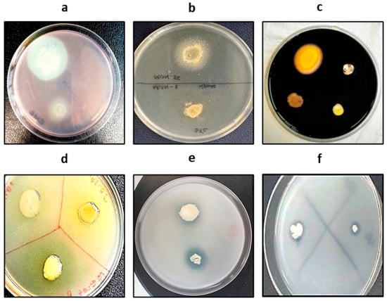
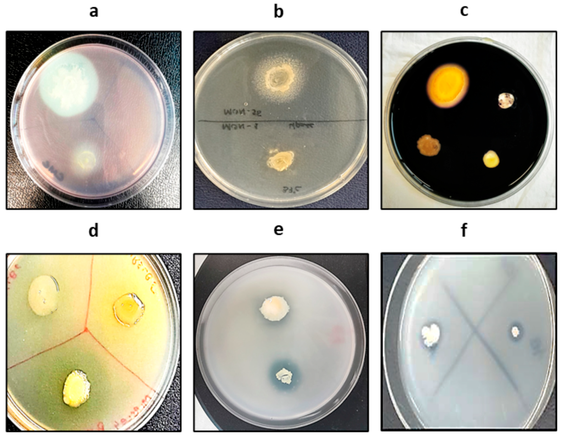
Carboxymethyl cellulose agar plates treated with Gram’s iodine solution were observed for yellowish halos around bacterial growth. Only four isolates, NCR2, NCR9, NCR10, and NCR 11 showed positive test results. The biggest zone was shown by NCR2 with 2.97 ± 0.09 mm (Figure 1a). Isolates NCR2, NCR8, NCR13, and NCR16 showed lipolytic activity, and accumulated whitish precipitates around bacterial growth were noted as a positive test result. The biggest zone size was observed around isolate NCR2 of 2.60 ± 0.06 mm (Figure 1b). Only three isolates, NCR2, NCR4, and NCR20, were positive for amylase production, with NCR2 showing the maximum halo diameter of 1.1 ± 0.06 mm (Figure 1c). Proteolytic activity was observed only in four isolates, including NCR2, NCR4, NCR13, and NCR20 (Figure 1d). The rest of the strains did not show the production of any of these hydrolytic enzymes (Table 1).
Figure 1.
Plant growth-promoting attributes of maize rhizobacterium NCR2: (a) cellulase, (b) lipase, (c) amylase, (d) protease production, (e) phosphate, and (f) zinc solubilization.
Table 1.
In vitro screening of plant growth-promoting traits in isolated rhizobacterial strains.
3.3. Phosphate and Zinc Solubilization Activities
On Pikovskaya’s agar medium, six isolates, including NCR1, NCR2, NCR3, NCR13, NCR15, and NCR18, were positive for inorganic phosphate solubilization; however, on NBRIP medium, only three isolates showed solubilization of tri-calcium phosphate (Figure 1e). Among these, isolate NCR2 was common and showed clear halo zones on both media (Table 1). The highest phosphate solubilization in NBRIP liquid broth was shown by isolate NCR2 (836 µg/mL) followed by NCR13, which showed 617 µg/mL of p solubilization.
Four isolates were positive for zinc solubilization and showed clear halos on tris minimal agar medium supplemented with ZnCO3. These isolates included NCR1, NCR2, NCR13, and NCR18; however, all of these isolates showed mild solubilization of zinc (Figure 1f).
3.4. Indole-3-Acetic Acid (IAA) Production
A qualitative test for the detection of indole-3-acetic acid production showed only three isolates showing a pink color of culture supernatants on the addition of Salkowski’s reagent. Only three isolates, including NCR2, NCR13, and NCR18, were positive for IAA synthesis (Table 1).
3.5. Morphological and Biochemical Characterization of Maize Rhizobacteria
The morphological characterization of the bacterial isolates revealed variation in colony size, color, margin, shape, elevation, and opacity. Colony sizes ranged from small to large, with most isolates presenting as small. The colors of the colonies varied from white, creamy, and off-white to yellow and light yellow, with a few showing dusty or greyish hues. The majority of colonies exhibited an entire (smooth) margin, though a few showed wavy or undulate margins. All isolates were predominantly round in shape, with only one displaying an irregular form. Colony elevation varied, with most being slightly raised, several flat, one raised, and one convex. Regarding opacity, colonies were mostly opaque or translucent, with a few showing transparent or shiny/mucoid appearances. Overall, these observations indicate a predominance of small, round colonies with entire margins and variable color, elevation, and opacity among the isolates, highlighting morphological diversity within the bacterial population (Table 2).
Table 2.
Morphological characterization of corn rhizobacteria.
Based upon the results of plant growth-promoting attributes possessed by bacterial isolates, only five isolates were biochemically screened using QTS-24 bacterial identification kits. The biochemical characterization of the bacterial isolates revealed distinct metabolic profiles, reflecting their taxonomic diversity. All isolates were positive for Voges-Proskauer and glucose and arabinose fermentation, indicating a common capacity for certain carbohydrate metabolism. Variations were observed in other tests: O-nitrophenyl-β-D-galactopyranoside activity, citrate utilization, malonate utilization, and lysine and ornithine decarboxylase activity differed among the isolates. Indole production and urea hydrolysis were generally negative, except in Escherichia coli (NCR4), which showed positive results. Sugar fermentation patterns varied, with differences in maltose, sucrose, rhamnose, inositol, melibiose, and raffinose utilization, while mannitol and adonitol were not fermented by any of the isolates. Gelatin hydrolysis was positive in NCR1 and NCR4, and cytochrome oxidase activity was detected in NCR1, NCR2, and NCR13. Based on these biochemical characteristics, the isolates were tentatively identified as Providencia sp. (NCR1), Pseudomonas sp. (NCR2 and NCR13), Escherichia coli (NCR4), and Acinetobacter sp. (NCR18), highlighting the metabolic diversity within the bacterial population (Table 3).
Table 3.
Biochemical characteristics and tentative identification of selected bacterial isolates using QTS-24 kits.
3.6. Phylogeny Confirms Evolutionary Relationship of Pseudomonas sp. NCR2
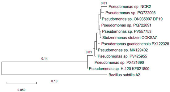
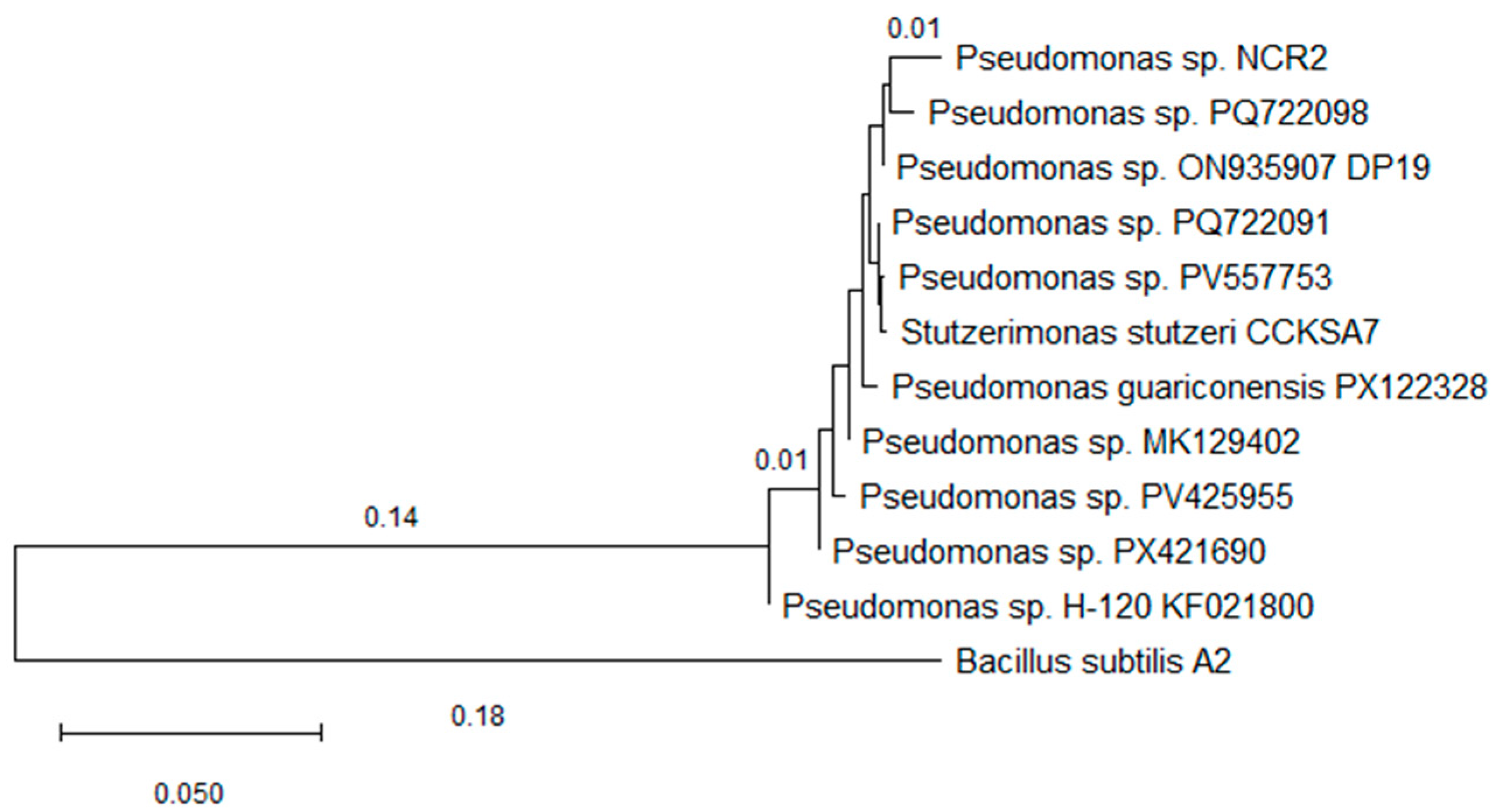
A maximum likelihood phylogenetic tree was constructed to elucidate the evolutionary relationships among eleven 16S rRNA gene sequences affiliated with the genus Pseudomonas (Figure 2). The 16S rRNA gene sequence of the isolate NCR2 was aligned with eleven closely related reference sequences retrieved from the NCBI database using ClustalW. After trimming ambiguous and poorly aligned terminal regions, the final alignment comprised 937 nucleotide positions.
Figure 2.
A phylogenetic tree of Pseudomonas strain NCR2. The evolutionary history was inferred by using the Maximum Likelihood method and Tamura-Nei model. Evolutionary analyses were conducted in MEGA12.
Phylogenetic analysis was performed using MEGA version 12. Model selection based on the Bayesian Information Criterion (BIC) identified the Tamura–Nei nucleotide substitution model as the best-fit model, which was subsequently used to infer the maximum likelihood tree. The tree was rooted using Bacillus subtilis A2 as an outgroup to provide phylogenetic context outside the genus Pseudomonas. Bootstrap resampling was attempted; however, due to the high sequence similarity and low divergence among the analyzed 16S rRNA sequences, reliable bootstrap support values could not be generated. The resulting tree topology demonstrates that the query sequence Pseudomonas sp. NCR2 (Accession No. PX716695) clusters within a well-defined Pseudomonas clade, grouping closely with Pseudomonas sp. ON935907 DP19, Pseudomonas sp. H-120 KF021800, Pseudomonas sp. PQ722098, and Pseudomonas guariconensis PX122328. Other reference sequences, including Pseudomonas sp. MK129402, Pseudomonas sp. PX421690, and Pseudomonas sp. PV425955, formed a separate but related clade within the genus. The consistent placement of NCR2 within the Pseudomonas lineage supports its taxonomic affiliation at the genus level.
3.7. Maize Growth-Promoting Potential of Pseudomonas sp. NCR2
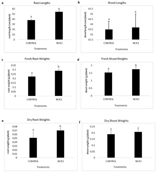
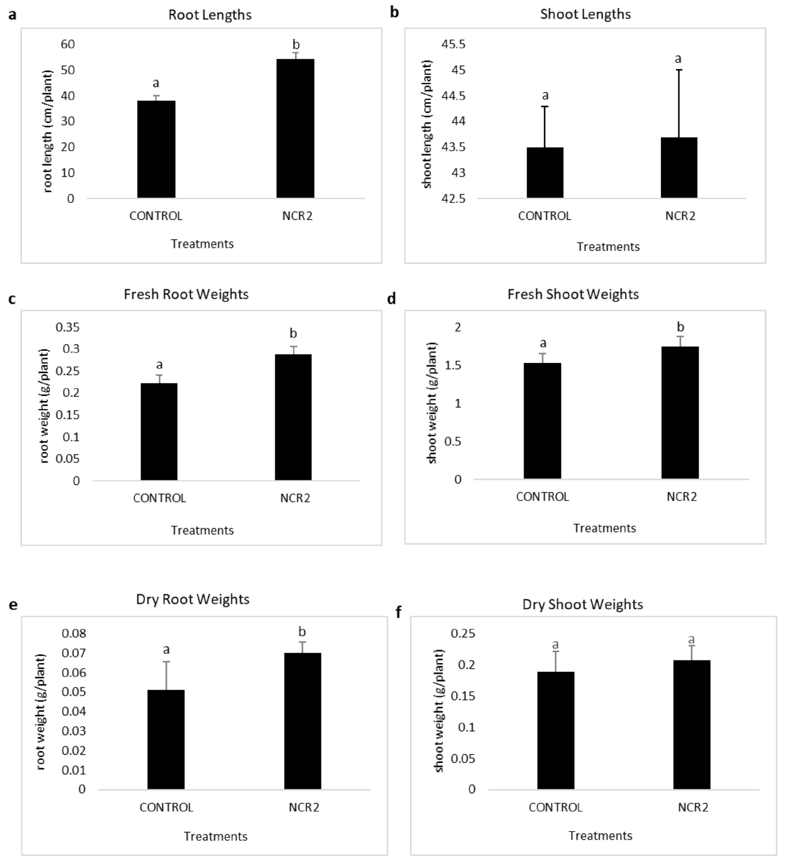
Maize plants were inoculated with the Pseudomonas NCR2 strain to check the effect of inoculation on maize growth parameters. Inoculated plants with NCR2 displayed markedly enhanced plant growth compared with the uninoculated control. Treated plants showed higher root lengths and significantly enhanced fresh and dry root weights as compared to the untreated control group. Although the shoot lengths and fresh shoot weights of Pseudomonas-treated plants were also high, nonetheless, they were not statistically significant (Figure 3).
Figure 3.
Growth parameters of maize plants inoculated with Pseudomonas strain NCR2. Bars represent mean ± SE (n = 9), and different letters indicate significant differences according to Tukey’s test (p < 0.05). Fresh shoot and root weights show significant differences between treatments, while dry shoot weights did not differ significantly, indicating that variations in fresh weight were mainly due to water content. (a) Root length, (b) Shoot length, (c) Root fresh weight, (d) Shoot fresh weight, (e) Root dry weight, and (f) Shoot dry weight.
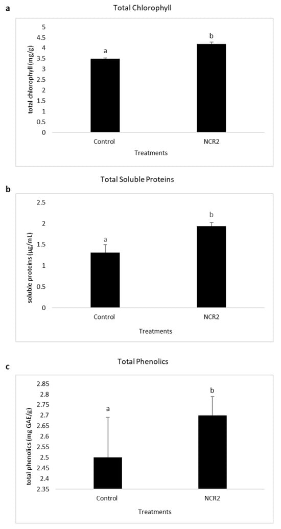
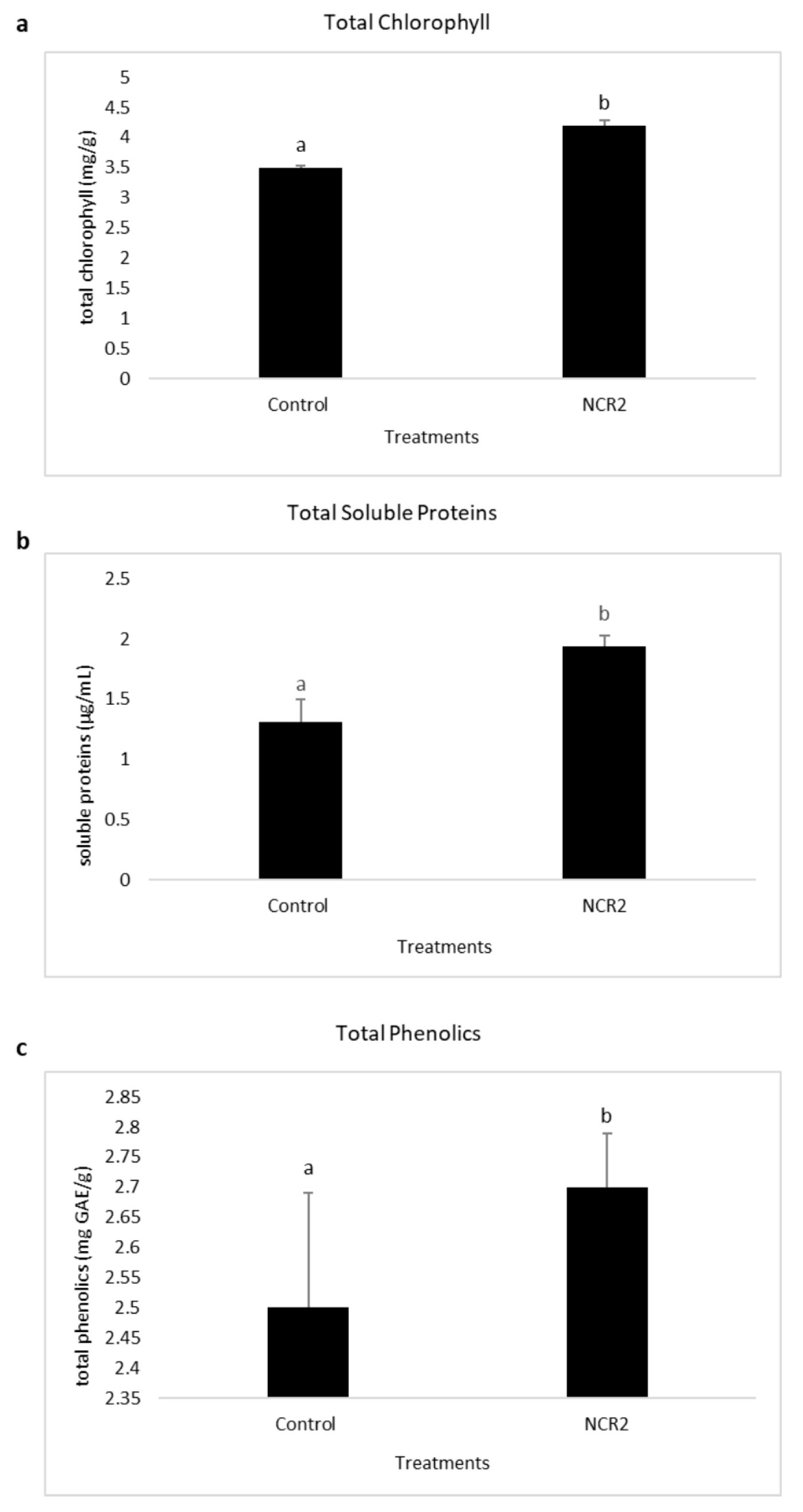
Physiological and biochemical analysis showed that key metabolic indicators, including total chlorophyll, soluble proteins, and phenolics, were significantly high in the Pseudomonas NCR2-treated group as compared to uninoculated controls (Figure 4). A substantial elevation of ~14% total chlorophyll content (mg/g fresh weight) was observed in comparison to the untreated control group, suggesting improved photosynthetic efficiency. Similarly, total soluble proteins showed an increase of ~33%, and a moderate increase in total phenolic content of NCR2-treated plants was also observed, reflecting the potential upregulation of secondary metabolism and antioxidant capacity of maize plants.
Figure 4.
Physiological indicators of maize plants inoculated with Pseudomonas strain NCR2. Bars represent mean ± SE (n = 9), and different letters indicate significant differences according to Tukey’s test (p < 0.05). (a) Total chlorophyll content (mg g−1 F.W), (b) Soluble protein content (µg mL−1), and (c) Total phenolic content (mg GAE g−1 F.W).
4. Discussion
The daunting challenge of deteriorating agricultural ecosystems has led to the characterization of countless plant growth-promoting rhizobacteria (PGPR). Nonetheless, the poor ecological adaptation of the introduced strains and persistent gaps between in vitro potential and field performance require diligence. Hence, a rigorous host-specific rhizosphere assessment of the native microbiome can prime for success and consistent performance of these isolates. To address this challenge, our study focuses on systematic isolation and characterization of the maize rhizomicrobiome and its integrated validation on maize growth promotion.
The initial screening of twenty-one rhizobacterial isolates demonstrated a wide spectrum of plant-beneficial profiles, with Pseudomonas sp. NCR2 as the most promising candidate. The strain showed the potential of producing a suite of hydrolytic enzymes, including cellulase, lipase, protease, and amylase. These extracellular enzymes are critical for nutrient cycling and organic matter decomposition in the rhizosheath area and also suppress the approaching phytopathogens [30]. For instance, cellulases can critically cleave β-1,4-D glucan linkages in cellulose, leading to cell wall degradation generating cello-oligosaccharides [31]. For example, cellulases from P. fluorescens have been reported for the successful biocontrol of Phytophthora capsici and showed notable inhibition of several phytopathogens, including Fusarium oxysporum, Rhizoctonia sp., Botrytis cineria, and Alternaria alternata [32]. Likewise, the management of verticillium wilt of olive caused by V. dahliae showed Pseudomonas sp. cellulases as the main biocontrol agents [33].
In addition to cellulases, proteases are equally important in the maintenance of rhizosphere competition and for the induction of systemic resistance in plants [34]. Proteases can protect the plants from disease-causing organisms by degrading cell wall proteins into peptides and amino acids. Usually, the proteases produced by beneficial rhizobacteria are extracellular and active under various environmental conditions, thereby successfully inhibiting the action of pathogenic proteins on plant cells [35]. For example, serine protease AprA and zinc-metalloproteases, like PrtA/PrtB produced by Pseudomonas sp., can directly lyse fungal hyphae and are established biocontrol agents against Fusarium oxysporum, Pythium spp., and Botrytis cinerea [36,37]. Moreover, bacterial proteases can be the potential microbe-associated molecular patterns (MAMPs), which are recognized by the plant root pattern-recognition receptors, leading to triggering jasmonate/ethylene-dependent immunity in plants [38]. In addition, proteolytic activity of several Pseudomonas spp. strains has been shown to contribute to nitrogen mineralization by breaking down complex organic proteins into plant-available forms, improving overall plant nutrition [39].
Ecological fitness and plant-beneficial potential of Pseudomonas sp. NCR2 is further enhanced by its lipolytic activity. Lipases produced by rhizobacteria can cleave the ester linkages and possess diverse catalytic characteristics, including transesterification, decarboxylation, and alcoholysis [40]. For example, lipases produced by P. aeruginosa FG106 were the main biocontrol agents in the suppression of Xanthomonas euvesicatoria pv. perforans, R. solani, P. infestans, A. alternata, B. cinerea, Clavibacter michiganensis subsp. Michiganensis, and P. colocasiae [41]. Likewise, P. putida strain AUS15 showed considerable biocontrol efficacy against common bean rust-causing Uromyces appendiculatus through its lipolytic potential [42]. In addition to antagonistic potential, hydrolysis of lipids and complex triglycerides into readily available fatty acids plays a crucial role in rhizosphere nutrient dynamics and competitive colonization [43].
Another significant hydrolytic enzyme produced by Pseudomonas sp. NCR2 was α-amylase, which can cleave glycosidic bonds in starch and related polysaccharides and has documented antimicrobial potential. A study by Mohammad et al. [44] reported the successful inhibition of R. solanacearum by amylase-producing P. syringae, P. fluorescens and P. aeruginosa. In addition, several pseudomonads have been shown to deplete glycogen reserves in the spermosphere, reducing disease incidence [45]. Furthermore, the oligosaccharides generated during starch hydrolysis modulate defense-related gene expression and promote root growth. Recent studies have highlighted the mechanisms of Pseudomonas spp. to utilize starch-derived sugars for the synthesis of exopolysaccharides (EPS) critical for root colonization and biofilm formation [46].
Moreover, Pseudomonas sp. NCR2 also demonstrated phosphate solubilization potential on both Pikovskaya’s and NBRIP growth media alongside zinc solubilization and indole-3-acetic acid production. Indole-3-acetic acid is a fundamental phytohormone, which directly promotes root proliferation and growth, thereby facilitating nutrient foraging and water retention [47]. A study evaluating the effect of two IAA-producing pseudomonads, including P. putida and P. fluorescens, on plant growth promotion reported significantly increased seed germination and vigor indices as compared to controls [48]. In another study, drought-stressed Vigna radiata inoculated with IAA-producing P. aeruginosa strains demonstrated significantly improved response as compared to untreated ones [49]. Alongside phytohormone synthesis, several mineral-solubilizing pseudomonads have been reported for notable plant growth. For example, zinc- and phosphate-solubilizing Pseudomonas sp. strain AZ5 led to significantly increased grain yield in two chickpea varieties [50]. In another study, P. protegens RY2 contributed to chickpea growth and enhanced its zinc content [51]. Similarly, a drought-adaptive phosphorus solubilizing P. libanensis EU-LWNA-33 was shown to alleviate drought stress in wheat plants and contributed to growth promotion [52]. In our study too, notably increased plant growth and chlorophyll and protein contents were observed in Pseudomonas sp. NCR2 inoculated plants as compared to controls, validating the translation of its plant-beneficial attributes. The pronounced increase in the physiological parameters is indicative of improved nutrient acquisition and an overall metabolic vigor.
From an ecological perspective, the application of PGPR may influence native rhizosphere microbial assemblages; however, growing evidence suggests that inoculation with indigenous and host-adapted Pseudomonas spp. predominantly results in positive modulation of the resident microbiome [4,53]. Several recent studies have demonstrated that Pseudomonas-based inoculants can enhance microbial functional diversity, nutrient cycling, and disease suppressiveness by reinforcing beneficial microbial networks rather than disrupting community stability [54,55]. Moreover, pseudomonads can also be central to horizontal gene transfer within the rhizosphere, enhancing microbial adaptation and functional redundancy in a complex environment [56]. Given that Pseudomonas sp. NCR2 was isolated from the maize rhizosphere itself, its reintroduction is expected to strengthen existing beneficial interactions and ecological functions while minimizing risks associated with non-native microbial introduction. Moreover, while the strain demonstrated multiple plant growth-promoting traits through qualitative and functional assays, quantitative estimation of certain metabolites, particularly indole-3-acetic acid (IAA), was beyond the scope of the current study due to resource constraints. Detailed quantitative analyses and metabolite profiling in future investigations will provide deeper mechanistic insight into the contribution of phytohormone production to maize growth enhancement.
5. Conclusions
This study strongly aligns with the growing paradigm shift towards microbial solutions for compromised agricultural systems. Pseudomonas sp. NCR2, isolated from the maize rhizosphere, has been shown to substantially contribute to maize growth metrics and physiological health under controlled conditions, providing a strong foundation for its use as a maize-specific biofertilizer. The follow-up research should focus on bioformulation development, elucidation of the precise mechanisms of growth promotion, and evaluation of the strain’s performance under diverse environmental conditions. This work strengthens the overall research aiming to foster microbial solutions for sustainable maize production.
Author Contributions
Conceptualization, I.S.; methodology, I.S.; software, I.S.; validation, I.S. and M.U.N.; formal analysis, I.S.; investigation, M.U.N., N.N., F.M. and M.A.; resources, I.S.; data curation, N.N. and F.M.; writing—original draft preparation, M.U.N., N.N., F.M. and M.A.; writing—review and editing, I.S.; visualization, I.S.; supervision, I.S.; project administration, I.S. All authors have read and agreed to the published version of the manuscript.
Funding
This research received no external funding.
Data Availability Statement
The original data presented in the study are openly available in NCBI GenBank with the Accession No. PX716695.
Conflicts of Interest
The authors declare no conflicts of interest.
Abbreviations
The following abbreviations are used in this manuscript:
| PGPR | Plant growth-promoting rhizobacteria |
| PGPB | Plant growth-promoting bacteria |
| EPS | Exopolysaccharides |
| ISR | Induced systemic resistance |
| CMC | Carboxymethyl cellulose |
| NBRIP | National Botanical Research Institute’s Phosphate growth medium |
References
- Raimi, A.; Roopnarain, A.; Adeleke, R. Biofertilizer production in Africa: Current status, factors impeding adoption and strategies for success. Sci. Total Environ. 2021, 769, 144626. [Google Scholar] [CrossRef] [Scilit]
- United Nations, Department of Economic and Social Affairs, Population Division. World Population Prospects 2022; United Nations: New York, NY, USA, 2022. Available online: https://population.un.org/wpp/ (accessed on 13 November 2025).
- Saeed, Q.; Wang, X.; Haider, F.U.; Kučerik, J.; Mumtaz, M.Z.; Holatko, J.; Naseem, M.; Kintl, A.; Ejaz, M.; Naveed, M.; et al. Rhizosphere bacteria in plant growth promotion, biocontrol, and bioremediation of contaminated sites: A Comprehensive Review of Effects and Mechanisms. Int. J. Mol. Sci. 2021, 22, 10529. [Google Scholar] [CrossRef] [Scilit]
- Trivedi, P.; Leach, J.E.; Tringe, S.G.; Sa, T.; Singh, B.K. Plant-microbiome interactions: From community assembly to plant health. Nat. Rev. Microbiol. 2021, 19, 72–86. [Google Scholar] [CrossRef] [Scilit]
- Kalayu, G. Phosphate solubilizing microorganisms: Promising approach as biofertilizers. Int. J. Agron. 2021, 2021, 1–11. Available online: https://onlinelibrary.wiley.com/doi/epdf/10.1155/2019/4917256 (accessed on 13 November 2025). [CrossRef] [Scilit]
- Alori, E.T.; Dare, M.O.; Babalola, O.O. Microbial Inoculants for Soil Quality and Plant Health. In Sustainable Agriculture Reviews; Lichtfouse, E., Ed.; Springer: Cham, Switzerland, 2017; Volume 22. [Google Scholar] [CrossRef] [Scilit]
- Vaid, S.K.; Kumar, B.; Sharma, A.; Shukla, A.K.; Srivastava, P.C. Effect of zinc solubilizing bacteria on growth promotion and zinc nutrition of rice. J. Soil Sci. Plant Nutr. 2021, 21, 922–935. Available online: https://www.scielo.cl/scielo.php?pid=S0718-95162014000400011&script=sci_arttext (accessed on 13 November 2025).
- Suman, J.; Rakshit, A.; Ogireddy, S.D.; Singh, S.; Gupta, C.; Chandrakala, J. Microbiome as a key player in sustainable agriculture and human health. Front. Soil Sci. 2022, 2, 821589. [Google Scholar] [CrossRef] [Scilit]
- Goswami, D.; Thakker, J.N.; Dhandhukia, P.C. Portraying mechanics of plant growth promoting rhizobacteria (PGPR): A review. Cogent Food Agric. 2016, 2, 1127500. [Google Scholar] [CrossRef] [Scilit]
- Erenstein, O.; Jaleta, M.; Sonder, K.; Mottaleb, K.; Prasanna, B.M. Global maize production, consumption and trade: Trends and R&D implications. Food Secur. 2022, 14, 1295–1319. [Google Scholar] [CrossRef] [Scilit]
- Kumar, A.; Singh, S.; Mukherjee, A.; Rastogi, R.P.; Verma, J.P. Salt-tolerant plant growth-promoting Bacillus pumilus strain JPVS11 to enhance plant growth attributes of rice and improve soil health under salinity stress. Microbiol. Res. 2021, 242, 126616. [Google Scholar] [CrossRef] [Scilit]
- Elnahal, A.S.; El-Saadony, M.T.; Saad, A.M.; Desoky, E.S.M.; El-Tahan, A.M.; Rady, M.M.; AbuQamar, S.F.; El-Tarabily, K.A. The use of microbial inoculants for biological control, plant growth promotion, and sustainable agriculture: A review. Eur. J. Plant Pathol. 2022, 162, 759–792. [Google Scholar] [CrossRef] [Scilit]
- White, L.J.; Brözel, V.S.; Subramanian, S. Isolation of rhizosphere bacterial communities from soil. Bio-Protocol 2015, 5, e1569. [Google Scholar] [CrossRef] [Scilit]
- Kasana, R.C.; Salwan, R.; Dhar, H.; Dutt, S.; Gulati, A. A rapid and easy method for the detection of microbial cellulases on agar plates using Gram’s iodine. Curr. Microbiol. 2008, 57, 503–507. [Google Scholar] [CrossRef] [Scilit]
- Plou, F.J.; Ferrer, M.; Nuero, O.M.; Calvo, M.V.; Alcalde, M.; Reyes, F.; Ballesteros, A. Analysis of Tween 80 as an esterase/lipase substrate for lipolytic activity assay. Biotech. Tech. 1998, 12, 183–186. [Google Scholar] [CrossRef] [Scilit]
- Kumari, P.; Khanna, V.; Kumar, P. Multifaceted rhizobacteria-mediated growth augmentation in chickpea. Agric. Res. 2017, 6, 368–377. [Google Scholar] [CrossRef] [Scilit]
- Alnahdi, H.S. Isolation and screening of extracellular proteases produced by new isolated Bacillus sp. J. Appl. Pharm. Sci. 2012, 2, 71–74. [Google Scholar] [CrossRef] [Scilit]
- Pikovskaya, R.I. Mobilization of phosphorus in soil in connection with the vital activity of some microbial species. Microbiologiya 1948, 17, 362–370. [Google Scholar]
- Nautiyal, C.S. An efficient microbiological growth medium for screening phosphate-solubilizing microorganisms. FEMS Microbiol. Lett. 1999, 170, 265–270. [Google Scholar] [CrossRef]
- Dipak, P.; Sankar, N.S. Isolation and characterization of phosphate solubilizing bacterium Pseudomonas aeruginosa KUPSB12 with antibacterial potential from river Ganga, India. Ann. Agrar. Sci. 2017, 15, 130–136. [Google Scholar] [CrossRef] [Scilit]
- Sharma, S.K.; Sharma, M.P.; Ramesh, A.; Joshi, O.P. Characterization of zinc-solubilizing Bacillus isolates and their potential to influence zinc assimilation in soybean seeds. J. Microbiol. Biotechnol. 2012, 22, 352–359. [Google Scholar] [CrossRef] [Scilit]
- Gordon, A.S.; Weber, R.P. Colorimetric estimation of indole acetic acid. Plant Physiol. 1950, 26, 192–195. [Google Scholar] [CrossRef] [Scilit]
- Weaver, P.F.; Wall, J.D.; Gest, H. Characterization of Rhodopseudomonas capsulata. Arch. Microbiol. 1975, 105, 207–216. [Google Scholar] [CrossRef] [Scilit]
- Tan, Z.Y.; Xu, X.D.; Wang, E.T.; Gao, J.I.; Romero, E.M.; Chen, W.X. Phylogenetic and genetic relationships of Mesorhizobium tinshanense and related rhizobia. Int. J. Syst. Bacteriol. 1997, 47, 874–879. [Google Scholar] [CrossRef] [Scilit]
- Chun, J.; Goodfellow, M. A phylogenetic analysis of the genus Nocardia with 16S rRNA gene sequences. Int. J. Syst. Bacteriol. 1995, 45, 240–245. [Google Scholar] [CrossRef] [Scilit] [PubMed]
- Arnon, D.I.; Hoagland, D.R. A Comparison of Water Culture and Soil as Media for Crop Production; Circular 346; California Agricultural Experiment Station: Berkeley, CA, USA, 1938; p. 26. [Google Scholar]
- Manolopoulou, E.; Varzakas, T.H.; Petsalaki, A. Chlorophyll determination in green pepper using two different extraction methods. Curr. Res. Nutr. Food Sci. J. 2016, 4, 52–60. [Google Scholar] [CrossRef] [Scilit]
- Sarkar, S.; Mondal, M.; Ghosh, P.; Saha, M.; Chatterjee, S. Quantification of total protein content from some traditionally used edible plant leaves: A comparative study. J. Med. Plant Stud. 2020, 8, 166–170. [Google Scholar] [CrossRef] [Scilit]
- Ainsworth, E.A.; Gillespie, K.M. Estimation of total phenolic content and other oxidation substrates in plant tissues using Folin–Ciocalteu reagent. Nat. Protoc. 2007, 2, 875–877. [Google Scholar] [CrossRef] [Scilit]
- Kumar, S.; Stecher, G.; Suleski, M.; Sanderford, M.; Sharma, S.; Tamura, K. MEGA12: Molecular evolutionary genetic analysis version 12 for adaptive and green computing. Mol. Biol. Evol. 2024, 41, msae263. [Google Scholar] [CrossRef] [Scilit]
- Reddy, E.C.; Reddy, G.S.; Goudar, V.; Sriramula, A.; Swarnalatha, G.V.; Al Tawaha, A.R.M.; Sayyed, R.Z. Hydrolytic enzyme producing plant growth-promoting rhizobacteria (PGPR) in plant growth promotion and biocontrol. In Secondary Metabolites and Volatiles of PGPR in Plant-Growth Promotion; Springer International Publishing: Cham, Switzerland, 2022; pp. 303–312. Available online: https://link.springer.com/chapter/10.1007/978-3-031-07559-9_15#citeas (accessed on 13 November 2025).
- Köhl, J.; Kolnaar, R.; Ravensberg, W.J. Mode of action of microbial biological control agents against plant diseases: Relevance beyond efficacy. Front. Plant Sci. 2019, 10, 845. [Google Scholar] [CrossRef] [Scilit] [PubMed]
- Admassie, M.; Woldehawariat, Y.; Alemu, T. In vitro evaluation of extracellular enzyme activity and its biocontrol efficacy of bacterial isolates from pepper plants for the management of Phytophthora capsici. BioMed Res. Int. 2022, 2022, 6778352. [Google Scholar] [CrossRef] [Scilit]
- Gómez-Lama Cabanás, C.; Legarda, G.; Ruano-Rosa, D.; Pizarro-Tobías, P.; Valverde-Corredor, A.; Niqui, J.L.; Triviño, J.C.; Roca, A.; Mercado-Blanco, J. Indigenous Pseudomonas spp. strains from the olive (Olea europaea L.) rhizosphere as effective biocontrol agents against Verticillium dahliae: From the host roots to the bacterial genomes. Front. Microbiol. 2018, 9, 277. [Google Scholar] [CrossRef] [Scilit]
- Pieterse, C.M.J.; Zamioudis, C.; Berendsen, R.L.; Weller, D.M.; Van Wees, S.C.M.; Bakker, P.A.H.M. Induced systemic resistance by beneficial microbes. Annu. Rev. Phytopathol. 2014, 52, 347–375. [Google Scholar] [CrossRef] [Scilit]
- Bhatia, R.K.; Ullah, S.; Hoque, M.Z.; Ahmad, I.; Yang, Y.H.; Bhatt, A.K.; Bhatia, S.K. Psychrophiles: A source of cold-adapted enzymes for energy efficient biotechnological industrial processes. J. Environ. Chem. Eng. 2021, 9, 104607. [Google Scholar] [CrossRef] [Scilit]
- Dunne, C.; Moënne-Loccoz, Y.; McCarthy, J.; Higgins, P.; Powell, J.; Dowling, D.N.; O’Gara, F. Combined use of rhizospheric bacteria and bacterial fungicides for improved biocontrol of a fungal root pathogen. Microbiology 1998, 144, 1085–1097. [Google Scholar] [CrossRef] [Scilit]
- Loper, J.E.; Kobayashi, D.Y.; Paulsen, I.T. The genomic sequence of Pseudomonas fluorescens Pf-5: Insights into biological control. ISME J. 2008, 2, 119–130. [Google Scholar] [CrossRef] [Scilit]
- Van der Ent, S.; Van Wees, S.C.M.; Pieterse, C.M.J. Jasmonate signaling in plant interactions with resistance-inducing beneficial microbes. Phytochemistry 2009, 70, 1581–1588. [Google Scholar] [CrossRef] [Scilit]
- Luo, L.; Meng, H.; Gu, J.-D. Microbial extracellular enzymes in biogeochemical cycling of ecosystems. J. Environ. Manag. 2017, 197, 539–549. [Google Scholar] [CrossRef] [Scilit]
- Agobo, K.; Arazu, V.; Uzo, K.; Igwe, C. Microbial lipases: A prospect for biotechnological industrial catalysis for green products—A review. J. Ferment. Technol. 2017, 6, 2. [Google Scholar] [CrossRef]
- Ghadamgahi, F.; Tarighi, S.; Taheri, P.; Saripella, G.V.; Anzalone, A.; Kalyandurg, P.B.; Catara, V.; Ortiz, R.; Vetukuri, R.R. Plant growth-promoting activity of Pseudomonas aeruginosa FG106 and its ability to act as a biocontrol agent against potato, tomato and taro pathogens. Biology 2022, 11, 140. [Google Scholar] [CrossRef] [Scilit]
- Abo-Elyousr, K.A.M.; Abdel-Rahim, I.R.; Almasoudi, N.M.; Alghamdi, S.A. Native endophytic Pseudomonas putida as a biocontrol agent against common bean rust caused by Uromyces appendiculatus. J. Fungi 2021, 7, 745. [Google Scholar] [CrossRef] [Scilit]
- Mhetras, N.; Mapare, V.; Gokhale, D. Cold-Active Pseudomonas lipases: Catalysis and applications. Biochem. Eng. J. 2021, 176, 108203. [Google Scholar] [CrossRef] [Scilit]
- Mohammed, A.F.; Oloyede, A.R.; Odeseye, A.O. Biological control of bacterial wilt of tomato caused by Ralstonia solanacearum using Pseudomonas species isolated from the rhizosphere of tomato plants. Arch. Phytopathol. Plant Prot. 2020, 53, 1–16. [Google Scholar] [CrossRef] [Scilit]
- Sindhu, S.S.; Sehrawat, A.; Glick, B.R. The role of microbial enzymes in the biocontrol of plant diseases and plant growth promotion. In Microbial Biocontrol: Food Security and Post Harvest Management; Dubey, R.C., Maheshwari, D.K., Eds.; Springer: Cham, Switzerland, 2021; pp. 257–284. [Google Scholar] [CrossRef] [Scilit]
- Alonso, L.E.; de la Torre, M.J.; Sampedro, I. Carbon source utilization patterns in Pseudomonas spp.: Impact on biofilm formation and root colonization. Appl. Soil Ecol. 2023, 182, 104698. [Google Scholar] [CrossRef] [Scilit]
- Spaepen, S.; Vanderleyden, J. Auxin and plant–microbe interactions. Cold Spring Harb. Perspect. Biol. 2011, 3, a001438. [Google Scholar] [CrossRef] [Scilit]
- Meliani, A.; Bensoltane, A.; Benidire, L.; Oufdou, K. Plant growth-promotion and IAA secretion with Pseudomonas fluorescens and Pseudomonas putida. Res. Rev. J. Bot. Sci. 2017, 6, 16–24. [Google Scholar]
- Uzma, M.; Iqbal, A.; Hasnain, S. Drought tolerance induction and growth promotion by indole acetic acid producing Pseudomonas aeruginosa in Vigna radiata. PLoS ONE 2022, 17, e0262932. [Google Scholar] [CrossRef] [Scilit] [PubMed]
- Zaheer, A.; Malik, A.; Sher, A.; Qaisrani, M.M.; Mehmood, A.; Khan, S.U.; Ashraf, M.; Mirza, Z.; Karim, S.; Rasool, M. Isolation, characterization, and effect of phosphate-zinc-solubilizing bacterial strains on chickpea (Cicer arietinum L.) growth. Saudi J. Biol. Sci. 2019, 26, 1061–1067. [Google Scholar] [CrossRef] [Scilit] [PubMed]
- Yasmin, R.; Hussain, S.; Rasool, M.H.; Siddique, M.H.; Muzammil, S. Isolation and characterization of Zn-solubilizing bacterium (Pseudomonas protegens RY2) and its contribution in the growth of chickpea (Cicer arietinum L.) as deciphered by improved growth parameters and Zn content. Dose-Response 2021, 19, 15593258211036791. [Google Scholar] [CrossRef] [Scilit]
- Kour, D.; Rana, K.L.; Sheikh, I.; Kumar, V.; Yadav, A.N.; Dhaliwal, H.S.; Saxena, A.K. Alleviation of drought stress and plant growth promotion by Pseudomonas libanensis EU-LWNA-33, a drought-adaptive phosphorus-solubilizing bacterium. Proc. Natl. Acad. Sci. India Sect. B Biol. Sci. 2020, 90, 785–795. [Google Scholar] [CrossRef] [Scilit]
- Berg, G.; Rybakova, D.; Fischer, D.; Cernava, T.; Vergès, M.-C.C.; Charles, T.; Chen, X.; Cocolin, L.; Eversole, K.; Corral, G.H.; et al. The plant microbiome explored: Implications for experimental botany. J. Exp. Bot. 2021, 72, 6993–7007. [Google Scholar] [CrossRef] [Scilit]
- Nishisaka, C.S.; Quevedo, H.D.; Ventura, J.P.; Andreote, F.D.; Mauchline, T.H.; Mendes, R. Soil microbial diversity: A key factor in pathogen suppression and inoculant performance. Geoderma 2025, 460, 117444. [Google Scholar] [CrossRef] [Scilit]
- Cotta, S.R.; Dias, A.C.F.; Mendes, R.; Andreote, F.D. Role of horizontal gene transfer and cooperation in rhizosphere microbiome assembly. Braz. J. Microbiol. 2025, 56, 225–236. [Google Scholar] [CrossRef] [Scilit] [PubMed]

Disclaimer/Publisher’s Note: The statements, opinions and data contained in all publications are solely those of the individual author(s) and contributor(s) and not of MDPI and/or the editor(s). MDPI and/or the editor(s) disclaim responsibility for any injury to people or property resulting from any ideas, methods, instructions or products referred to in the content. |
© 2026 by the authors. Licensee MDPI, Basel, Switzerland. This article is an open access article distributed under the terms and conditions of the Creative Commons Attribution (CC BY) license.