


Abstract
Damping-off caused by Fusarium spp. limits tomato seedling establishment, while chemical control is constrained by resistance development and environmental risks. As a result, biological alternatives and nanomaterials have attracted increasing interest. This study aimed to quantify the in vitro inhibition and in vivo control of Fusarium spp. associated with tomato damping-off using the marine strains KN1 and KN2 of Stenotrophomonas rhizophila and copper oxide nanoparticles (CuO-NPs). Nine fungal isolates were recovered from symptomatic plants; the most virulent isolate (3DR23HA) caused 60% disease incidence and was identified as Fusarium oxysporum. In dual-culture assays, both bacterial strains inhibited mycelial growth, with percentage inhibition of radial growth (PIRG) values exceeding 65% in several isolates, whereas KN1 showed greater inhibition of conidial germination. CuO-NPs exhibited a concentration-dependent response, reaching near-complete suppression at 0.75–1.0 mg mL−1. In seedlings, the inoculated control showed 100% incidence and a disease severity index (DSI) of 85%, whereas KN1, KN1 + CuO-NPs at 0.75 mg mL−1, and KN2 + CuO-NPs at 0.75 mg mL−1 achieved a DSI of 0 and 100% control efficiency, while also improving growth and biomass. Overall, S. rhizophila, particularly strain KN1, and CuO-NPs at 0.75 mg mL−1 represent a promising strategy for the integrated management of tomato damping-off in nurseries.
1. Introduction
Tomato (Solanum lycopersicum L.) is one of the most important horticultural crops worldwide. Recent FAO statistics indicate that, in 2024, global production reached 1.2 billion tonnes of vegetables and 954 million tonnes of fruits, with tomato accounting for 188 million tonnes [1]. Given the global importance of tomato cultivation, constraints affecting seedling establishment can compromise crop uniformity, yield, and economic returns. In this context, damping-off, commonly associated with soil-borne phytopathogenic fungi such as Fusarium spp., is one of the most important diseases affecting tomato because it can severely reduce seedling emergence and stand establishment, with consequent effects on crop uniformity and productivity [2,3]. Because the disease occurs during germination and early seedling development, it increases the need for re-sowing or re-transplanting and further aggravates production losses [4]. In commercial nurseries, high humidity, dense seedling trays, and continuous production cycles create favorable microclimatic conditions for infection, thereby increasing the economic impact of the disease [5,6]. Management of damping-off still relies largely on synthetic fungicides; however, their repeated use can favor the selection of resistant pathogen populations, contribute to environmental contamination, and pose risks to human and animal health [7,8]. These limitations support the evaluation of alternative strategies that are effective against soil-borne phytopathogens and compatible with sustainable nursery production systems.
Biological control has emerged as a relevant alternative for suppressing phytopathogens through the use of beneficial microorganisms, mainly bacteria and fungi [9,10]. In this context, marine bacteria represent an important source of antimicrobial compounds, as adaptation to saline and fluctuating environments may confer distinctive physiological and metabolic traits that favor the production of bioactive metabolites with potential for plant disease suppression [11,12]. The use of marine bacteria as biocontrol agents has reduced several plant diseases in crops such as mango [13], melon [14], strawberry [15], and chickpea [16], highlighting genera such as Bacillus [17], Stenotrophomonas [18], and Micrococcus [19]. Their antagonistic activity against phytopathogens has been demonstrated both in vitro and in vivo through multiple mechanisms, including competition for space and nutrients, siderophore production, biofilm formation, secretion of hydrolytic enzymes, and induction of host resistance [20,21,22]. Collectively, this evidence provides a scientific basis for exploring marine bacterial strains as biological agents against damping-off-associated phytopathogens in tomato.
Among these bacterial taxa, Stenotrophomonas rhizophila, a plant-associated species within the genus Stenotrophomonas of the phylum Pseudomonadota, has emerged as a promising bacterium for damping-off management because of its ability to colonize the rhizosphere and persist under competitive soil conditions [23,24,25]. This ecological competence is particularly relevant in damping-off pathosystems, in which disease establishment is largely determined during the earliest stages of seed and root colonization; infection of the seed or emerging radicle may result in pre-emergence damping-off, whereas colonization of the hypocotyl or root-collar region after emergence may cause water-soaked lesions, tissue constriction, and seedling collapse [2,5,26]. Therefore, evaluating a marine strain of S. rhizophila is biologically justified, as its rhizosphere competence and multiple antagonistic mechanisms may restrict phytopathogen establishment during the most susceptible phase of seedling development, both in vitro and under nursery conditions.
In parallel, nanomaterials have gained increasing attention as tools for plant disease management because of their distinctive physicochemical and biological properties [27,28]. Among metallic nanoparticles with antimicrobial activity, copper-based nanoparticles are particularly relevant because of their relatively low cost, high surface reactivity, and reported efficacy against a broad spectrum of phytopathogen [29,30,31,32]. In addition to their antimicrobial potential, copper is an essential micronutrient involved in plant growth and in the function of proteins associated with defense responses, such as plastocyanin, peroxidases, and oxidases [33,34]. Although the precise antimicrobial mechanism of copper oxide nanoparticles (CuO-NPs) remains under investigation, available evidence suggests that their activity may involve reactive oxygen species (ROS) generation and damage to essential cellular targets, including deoxyribonucleic acid (DNA) [35,36]. However, their efficacy is strongly influenced by dose, formulation, and exposure route [37,38]. For example, in tomato plants challenged with Fusarium oxysporum f. sp. lycopersici, chitosan-loaded CuO nanoparticles applied at 1 mg L−1 reduced Fusarium wilt severity by 91.5% after 75 days, whereas applications at 25 and 50 mg L−1 reduced disease severity by 96.0% and 98.0%, respectively, outperforming the conventional fungicide Kocide 2000, which achieved about 90% disease reduction at 2.5 g L−1 [39]. This aspect is particularly relevant in nursery systems, where CuO-NPs may be applied as foliar sprays [40,41]. When applied as foliar sprays, copper-based nanoparticles may also act as elicitors of plant defense responses. These effects have been associated with enhanced antioxidant capacity, including the upregulation or activation of superoxide dismutase (SOD), ascorbate peroxidase (APX), and glutathione peroxidase (GPX); stimulation of the phenylpropanoid pathway, including increased phenylalanine ammonia-lyase (PAL) and, in some cases, polyphenol oxidase (PPO) activity; and induction of pathogenesis-related (PR) genes [42,43,44,45]. Therefore, defining effective foliar CuO-NP application strategies under nursery conditions is relevant for their practical use against damping-off-associated phytopathogens.
From a phytopathological perspective, integrating microbial antagonists with nanomaterials represents a rational strategy because both components may operate through complementary mechanisms. Beneficial bacteria can reduce phytopathogen establishment at the root-substrate interface through ecological competition and metabolite- or enzyme-mediated inhibition [20,21], whereas foliar-applied CuO-NPs may contribute through direct antimicrobial activity and by enhancing host defense responses [38,42,43]. Thus, combining a rhizosphere-competent antagonistic bacterium with a nanoparticle-based treatment may provide broader protection than either component applied individually. Despite this conceptual and practical relevance, evidence on the combined use of Stenotrophomonas rhizophila and CuO-NPs against Fusarium-associated damping-off in tomato seedlings remains limited. Therefore, the objective of this work was to evaluate the in vitro and in vivo effects of the marine bacterium Stenotrophomonas rhizophila and foliar-applied copper oxide nanoparticles (CuO-NPs) on the suppression of Fusarium spp. associated with damping-off in tomato seedlings, and to determine whether the integration of both components could improve disease suppression compared with their individual application.
2. Materials and Methods
2.1. Plant Material and Isolation of Phytopathogens
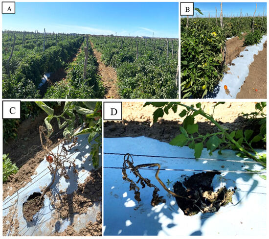
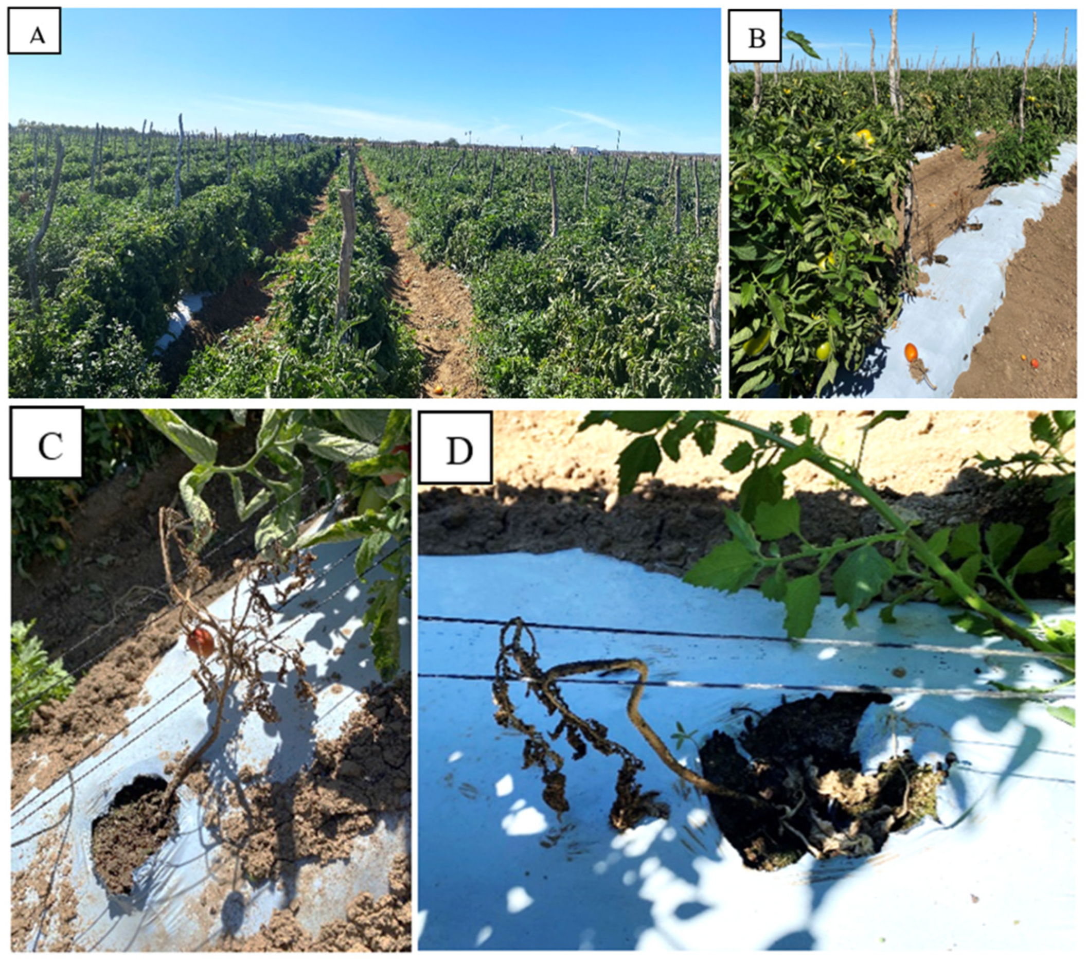
Symptomatic saladette tomato plants (cv. Río Grande) showing stem base rot and/or root rot were collected on 7 March 2023 from a commercial field in Baja California Sur, Mexico (23°47′00″ N, 110°17′40″ W) (Figure 1). Samples were individually labeled, placed in polyethylene bags, transported in a cooler, and processed upon arrival at the Microbial Biotechnology Laboratory (CIBNOR). Diseased tissues were rinsed under running water, and small fragments were excised from the lesion margins, including the transition zone between symptomatic and healthy tissue. The fragments were surface-disinfected with 3% sodium hypochlorite for 30 s, rinsed three times with sterile distilled water, blotted dry on sterile paper, and plated onto potato dextrose agar (PDA) amended with Bactrol® (BASF Mexicana, Ciudad de México, Mexico) at 0.5 g L−1 to suppress bacterial growth. Plates were incubated at 28 °C and monitored until fungal growth emerged. Hyphal tips from representative colonies were transferred to fresh PDA plates to obtain pure cultures and incubated at 28 °C for 7 days [21]. Each recovered fungal isolate was then assigned an internal alphanumeric code (e.g., 3DR23HA, 3DR23, 12DR23, 11R23, and 05DR23) for laboratory tracking and subsequent analyses.
Figure 1.
Symptomatic saladette tomato plants (cv. Río Grande) collected from a commercial field in Baja California Sur, Mexico. (A) View of “El Chilerito” Ranch. (B–D) Plants showing stem base and root rot symptoms.
2.2. Morphological Identification of Fusarium spp. Isolates
Morphological identification of Fusarium spp. isolates was performed based on the taxonomic keys and diagnostic criteria described by Summerell et al. [46]. Pure cultures were grown on PDA at 28 °C for 7 days under a 12 h light/12 h dark photoperiod to promote sporulation. Macroscopic traits were recorded, including colony pigmentation (surface and reverse), texture, and margin characteristics. For microscopic examination, sporulating structures were mounted in lactophenol cotton blue and observed under a light microscope at 400×–1000× magnification. The diagnostic characters evaluated included the morphology of macroconidia and microconidia, as well as the presence and distribution of chlamydospores [46].
2.3. Pathogenicity Test
Each fungal isolate was cultured on PDA at 28 °C for 7 days. Conidia were harvested by flooding the plate surface with sterile distilled water and gently scraping the colony to dislodge them. The resulting suspension was filtered through sterile gauze to remove mycelial fragments, and the conidial concentration was adjusted to 1 × 10⁴ conidia mL−1 using a hemocytometer. Tomato seedlings (cv. Río Grande) at the 3–4 true-leaf stage, with no visible symptoms of damping-off or root rot at the time of inoculation, were inoculated by applying 5 mL of each conidial suspension as a soil drench around the root zone. Control plants received 5 mL of sterile distilled water following the same procedure. Plants were arranged in a completely randomized design with fifteen seedlings per isolate (n = 15) and maintained in an environmental chamber for 20 days at 28 ± 2 °C and 70% relative humidity under a 12 h light/12 h dark photoperiod.
Disease severity was assessed using a modified ordinal scale based on Boehm et al. [47], adapted for post-emergence damping-off in transplanted tomato seedlings. Because the assay was conducted with seedlings at the 3–4 true-leaf stage, pre-emergence damping-off was not applicable under these conditions. Disease severity was scored as follows: 0 = symptomless seedlings; 1 = mild symptoms (wilting, chlorosis, and/or small lesions at the hypocotyl/root-collar region, without collapse); 2 = moderate symptoms (evident wilting, chlorosis, and/or lesions accompanied by noticeable growth reduction, with seedlings still remaining upright); 3 = severe symptoms (pronounced wilting and/or extensive lesions associated with marked growth reduction, with seedlings still remaining upright); and 4 = post-emergence damping-off (seedling collapse or death after emergence).
Disease incidence (DI, %) was calculated as the proportion of plants showing visible symptoms consistent with post-emergence damping-off (e.g., chlorosis, wilting, lesions in the hypocotyl/root-collar region, or stunting) relative to the total number of evaluated plants, using the following expression:
where FI is the number of infected plants and TF is the total number of plants [48].
DI (%) = (FI/TF) × 100
At the end of the experiment, roots were gently washed to remove the substrate, and root fresh weight (g) was recorded using a precision balance (OHAUS Adventurer® AX423N, OHAUS Corp., Parsippany, NJ, USA; ±0.002 g). Root dry weight (g) was determined after oven-drying at 60 °C to constant mass in a benchtop drying oven (Model 9025H). To fulfill Koch’s postulates, the phytopathogen was re-isolated from symptomatic collar and root tissues onto PDA, purified, and identified by comparing its colony and microscopic morphology with those of the original isolate [49].
2.4. Molecular Identification and Phylogenetic Analysis
All nine isolates were initially characterized morphologically and evaluated in the pathogenicity assay. Because isolate 3DR23HA showed the highest disease incidence and was considered the most virulent isolate, it was selected for molecular identification. Genomic DNA was extracted following the method of Raeder and Broda [50]. The ITS1–5.8S–ITS2 rDNA region was amplified using primers ITS1 (5′-TCCGTAGGTGAACCCTGCGG-3′) and ITS4 (5′-TCCTCCGCTTATTGATATGC-3′). PCR amplification was performed in a thermal cycler (GeneAmp® PCR System 9700, Applied Biosystems, Foster City, CA, USA) with an initial denaturation at 95 °C for 3 min, followed by 30 cycles of denaturation at 95 °C for 1 min, annealing at 50 °C for 30 s, and extension at 72 °C for 1 min, with a final extension at 72 °C for 10 min. PCR products were sequenced by MACROGEN (Seoul, Republic of Korea), and the resulting ITS sequence of isolate 3DR23HA was deposited in the NCBI GenBank database under accession number PZ256382.
Reference ITS sequences were retrieved from the NCBI GenBank database and aligned with the obtained sequence using MUSCLE v. 3.8.31 [51]. The multiple alignment was curated using the Gblocks program v. 0.91b [52], implemented in the Phylogeny.fr platform [53], to remove highly variable positions and gap-containing regions while retaining conserved blocks. Based on the curated alignment, the best-fit nucleotide substitution model was determined in MEGA v. 12.0 (MEGA Software, Tempe, AZ, USA) [54]. Phylogenetic analysis was then performed in MEGA v. 12.0 using the Maximum Likelihood (ML) method under the Kimura 2-parameter model with gamma-distributed rate variation among sites (K2P + G). Branch support was assessed by bootstrap analysis with 10,000 replicates. Molecular identification was established based on the phylogenetic clustering of isolate 3DR23HA with reference sequences retrieved from the NCBI GenBank database.
2.5. Bacterial Strains
Marine strains KN1 and KN2 of Stenotrophomonas rhizophila were provided by the Microbial Biotechnology Laboratory at CIBNOR. Each strain was cultured in tryptic soy broth (TSB) at 28 °C for 24 h with shaking at 200 rpm. Cell density was adjusted spectrophotometrically to an OD660 of 1.0, corresponding to approximately 1 × 10⁸ CFU mL−1.
2.6. In Vitro Antifungal Activity of Marine Bacteria
Dual-culture assays were performed on PDA plates. A 5 mm mycelial plug taken from the actively growing margin of each 7-day-old fungal culture was placed at the center of each Petri dish. Bacterial suspensions were spot-inoculated by depositing 5 µL at a distance of 2 cm from the fungal plug. Control plates received only the fungal plug. Ten replicate plates were prepared for each bacterial strain–fungal isolate combination. Plates were incubated at 28 °C for 7 days. Fungal radial growth (mm) was measured along two perpendicular axes and averaged. The percentage inhibition of radial growth (PIRG) [55] was calculated as follows:
where Rc is the mean radial growth of the control colony and Rt is the mean radial growth of the treated colony.
PIRG (%) = ((Rc − Rt)/Rc) × 100,
2.7. Spore Germination Inhibition Assay
Spore germination inhibition was evaluated using conidial suspensions of four Fusarium spp. isolates (3DR23, 3DR23HA, 4DR23, and 12DR23), each adjusted to 1 × 10⁴ conidia mL−1. Equal volumes of each conidial suspension and each bacterial suspension (1 × 10⁸ cells mL−1) were mixed (1:1, v/v) in sterile microtubes and incubated at 28 °C for 12 h. A control treatment consisted of conidia mixed with sterile distilled water under the same conditions. After incubation, an aliquot of each mixture was placed on a microscope slide, and at least 100 conidia were examined per replicate under a light microscope at 400× magnification. Conidia were considered germinated when the germ tube length was equal to or greater than the conidial length [56]. Spore germination (%) was calculated as follows:
Germination (%) = (number of germinated conidia/total conidia counted) × 100.
Spore germination inhibition (%) was calculated relative to the control as:
where G_c is germination in the control and G_t is germination in the bacterial treatment.
Inhibition (%) = ((G_c − G_t)/G_c) × 100,
2.8. In Vitro Antifungal Activity of Nanoparticles
Copper oxide nanoparticles (CuO-NPs) were purchased from Sigma-Aldrich/Aldrich (St. Louis, MO, USA; Product No. 544868; CAS No. 1317-38-0). According to the supplier’s specifications, the material was provided as a black nanopowder with a nominal particle size of <50 nm. The certificate of analysis for the product lot used in this study reported an average particle size of 40 nm as determined by transmission electron microscopy (TEM), X-ray diffraction consistent with the expected CuO structure, and a copper content of 77.2%. CuO-NPs were evaluated at concentrations of 0.1, 0.25, 0.5, 0.75, and 1.0 mg mL−1. A stock suspension of CuO-NPs was prepared in sterile distilled water and dispersed by sonication in three 15 min cycles (45 min total). The nanoparticle suspensions were added directly to molten PDA (50–55 °C), thoroughly homogenized, and poured into Petri dishes. Control plates contained PDA without CuO-NPs. A 5 mm mycelial plug taken from the actively growing margin of each fungal isolate grown on PDA for 7 days was placed at the center of each plate. Plates were incubated at 28 °C for 7 days. Ten replicate plates were prepared for each concentration–isolate combination. Radial growth (mm) was measured along two perpendicular axes and averaged. The percentage inhibition of radial growth (PIRG) was calculated using the same equation described in Section 2.6, where Rc corresponds to the mean radial growth of the control colony (PDA without CuO-NPs) and Rt corresponds to the mean radial growth of the treated colony (PDA amended with CuO-NPs) [57].
2.9. In Vivo Experiment
Tomato seedlings at the 3–4 true-leaf stage were transplanted into pots containing 500 g of a sterilized substrate mixture of soil, sand, and vermiculite (2:1:1, v/v/v). The substrate was sterilized by autoclaving at 121 °C and 15 psi for 60 min on two consecutive days. Seedlings were inoculated with isolate 3DR23HA (Fusarium oxysporum) by applying 5 mL of a conidial suspension (1 × 10⁴ conidia mL−1) as a soil drench around the root zone.
The experiment was conducted under phytopathogen challenge in a completely randomized design with a 3 × 3 factorial arrangement. The factors were bacterial treatment (none, KN1, and KN2) and CuO-NP concentration (0, 0.50, and 0.75 mg mL−1), including their interaction. For CuO-NP treatments, either alone or in combination with bacteria, suspensions prepared in sterile water were applied as a foliar spray 24 h after phytopathogen inoculation until the foliage of each seedling was uniformly wetted. For bacterial treatments, either alone or in combination with CuO-NPs, 1 mL of each Stenotrophomonas rhizophila suspension (1 × 10⁸ cells mL−1) was applied to the stem base (root-collar region) at the soil line. In addition, a synthetic fungicide reference treatment was included, consisting of phytopathogen-inoculated plants treated with carbendazim at 2 mL kg−1 of substrate. Each treatment included 15 seedlings (n = 15). Pots were maintained in an environmental chamber for 15 days at 28 ± 2 °C and 70% relative humidity under a 12 h light/12 h dark photoperiod.
At 15 days after treatment application, morphological and disease-related variables were recorded for each seedling. Stem length (cm) was measured from the substrate surface to the apical meristem using a graduated ruler. Root length (cm) was measured from the root-collar region to the tip of the main root after gently washing the root system to remove adhering substrate. Stem diameter (mm) was measured at the soil line using a digital caliper. The number of leaves was recorded as the total number of fully expanded true leaves per plant. Fresh biomass (g) was determined as the total fresh weight of each plant immediately after harvest using an analytical balance (OHAUS Adventurer® AX423N; ±0.002 g). Disease incidence (DI, %) was calculated using the equation described in Section 2.3. Disease severity (DS) was assessed using the ordinal scale described in Section 2.3, adapted from Boehm et al. [47]. Disease severity was expressed as the disease severity index (DSI, %), calculated as:
where ni is the number of seedlings in severity class i, si is the numerical value assigned to class i, N is the total number of assessed seedlings per treatment, and K is the maximum severity score (4).
2.10. Control Efficiency (Abbott, %)
Control efficiency (E, %) was calculated from final disease severity, expressed as the disease severity index (DSI, %), using the Abbott formula [58]. The inoculated control treatment was used solely as the reference for calculating E:
where DSIcontrol is the mean DSI (%) of the inoculated control (3DR23HA) and DSItreatment is the mean DSI (%) of each treatment. For statistical comparisons of control efficiency among treatments, the reference control was excluded from the analysis.
E(%) = ((DSIcontrol − DSItreatment)/DSIcontrol) × 100
2.11. Statistical Analysis
All statistical analyses were performed in R (version 4.4.2). For the in vitro assays, radial growth inhibition (PIRG, %) in the dual-culture test was evaluated using a two-way model with bacterial strain (KN01, KN02), fungal isolate (3DR23, 3DR23HA, 4DR23, and 12DR23), and their interaction as fixed effects; when the interaction was significant, simple-effects pairwise contrasts were conducted and p-values were adjusted using the Holm method (p ≤ 0.05). For CuO-NP dose–response assays within each Fusarium spp. isolate (0.1–1 mg mL−1), treatment effects were tested using the Kruskal–Wallis test, followed, when significant, by Dunn’s post hoc test with Holm adjustment (p ≤ 0.05). For seedling responses, growth variables were analyzed under a CRD with a 3 × 3 factorial arrangement (bacterial treatment: none, KN1, KN2); CuO-NPs concentration (0, 0.50, 0.75 mg mL−1), including the A × B interaction; because several variables did not meet parametric assumptions, factorial effects were evaluated using the Scheirer–Ray–Hare two-way nonparametric ANOVA on ranks, and significant interactions were decomposed via simple-effects analyses (Kruskal–Wallis) with Dunn–Holm multiple comparisons (p ≤ 0.05), whereas the synthetic fungicide was treated as a non-factorial reference and compared separately using rank-based pairwise tests with Holm adjustment. Disease incidence (severity > 0) was analyzed as a binary outcome, reported as x/n (%) with 95% Wilson confidence intervals, and factorial effects were assessed using Firth-corrected binomial logistic regression. Disease severity was analyzed using Scheirer–Ray–Hare, with significant interactions followed by simple-effects Dunn–Holm comparisons (p ≤ 0.05), and severity was additionally summarized as DSI (%). Control efficiency (Abbott, %) was compared using the same factorial framework (Scheirer–Ray–Hare with simple effects and Dunn–Holm, p ≤ 0.05).
3. Results
3.1. Isolation, Morphological Characterization, and Pathogenicity of Fungal Isolates
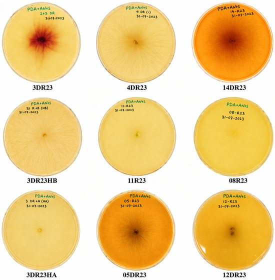
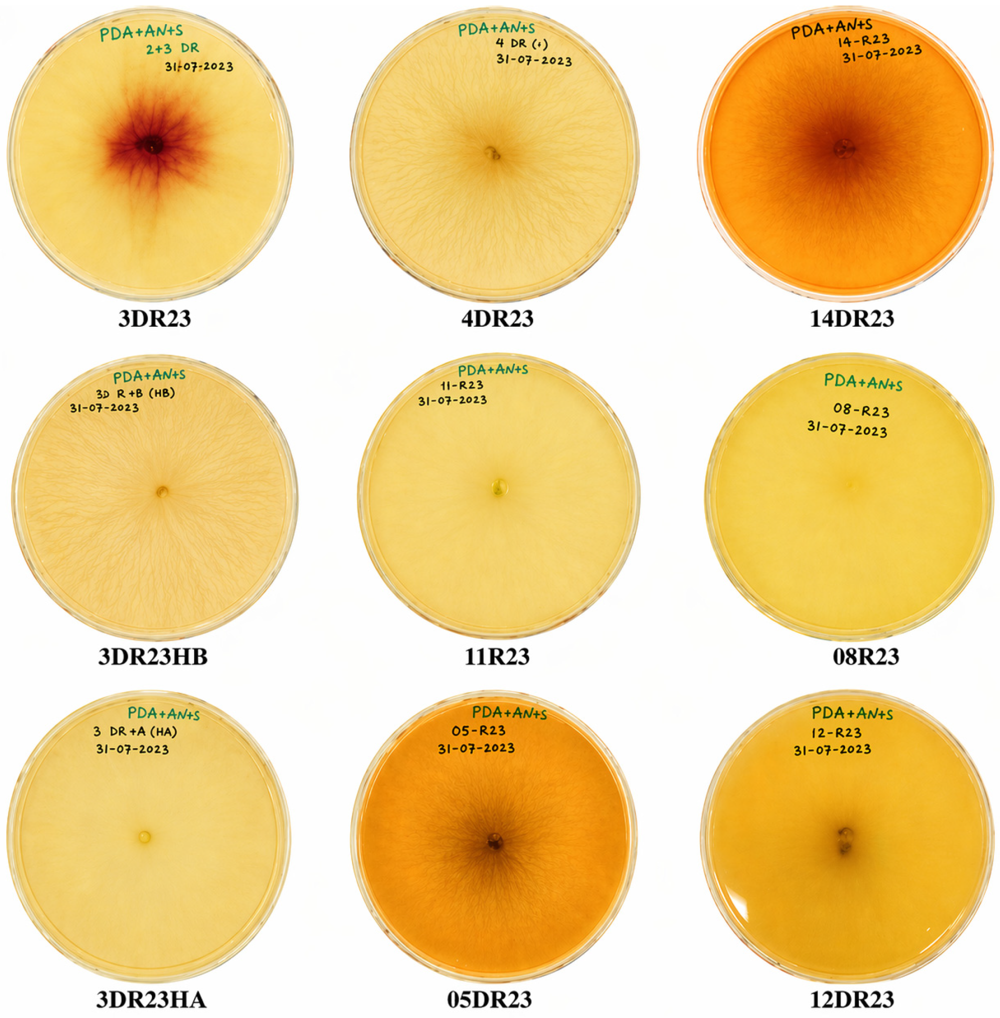
Nine fungal isolates were recovered on PDA from tomato plants exhibiting damping-off symptoms. After incubation on PDA at 28 °C for 7 days under a 12 h light/12 h dark photoperiod, colonies showed abundant aerial mycelium with a cottony to floccose texture and variable pigmentation among isolates (Figure 2). During early growth, colonies were predominantly white to pale pink; as incubation progressed, some isolates developed more intense pigmentation, ranging from pinkish-violet to brownish, often concentrated in the central zone, whereas others remained cream to whitish with a paler center. Colony margins were generally entire to slightly undulate, and reverse pigmentation ranged from pale yellow to pinkish-violet or brown.
Figure 2.
Isolates of Fusarium spp. recovered from tomato plants with symptoms of damping-off. Colonies were cultured on PDA and photographed in 90 mm-diameter Petri dishes. Alphanumeric designations correspond to internal isolate codes assigned during fungal isolation and laboratory tracking.
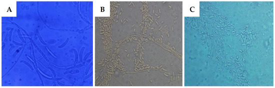
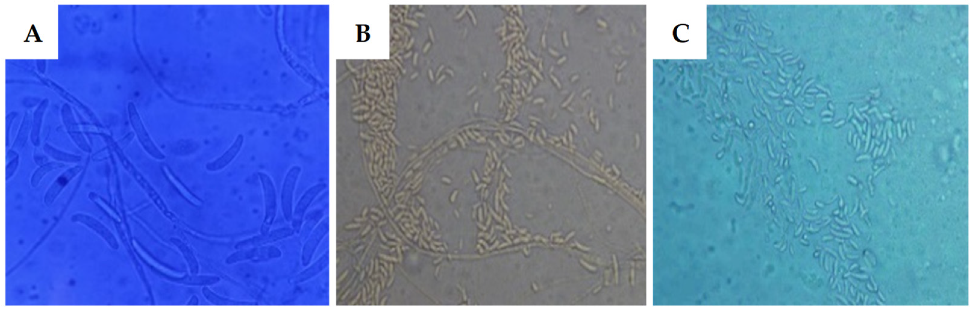
In the microscopic characterization, lactophenol cotton blue-stained preparations revealed sporulating structures consistent with Fusarium spp. Hyaline macroconidia were observed, characterized by an elongated, slightly curved shape and evident septation (Figure 3A), together with hyaline microconidia of ovoid to ellipsoidal shape, grouped in masses around hyphae or conidiophores (Figure 3B,C). These macroscopic and microscopic features supported the assignment of the isolates to the genus Fusarium.
Figure 3.
Microscopic structures of Fusarium spp. isolates from tomato plants with symptoms of stem softening and/or root rot (damping-off). (A) Macroconidia isolate 11R23. (B,C) Microconidia isolates 12DR23 and 05DR23, respectively. Alphanumeric designations correspond to internal isolate codes assigned during fungal isolation and laboratory tracking. Observations were made at 400× magnification.
The highest observed disease incidence in tomato seedlings was recorded for isolate 3DR23HA (60%), followed by isolates 3DR23 and 12DR23, each with 50% (Table 1). The lowest incidence was observed for isolates 05DR23 and 08R23 (10% each). Symptom severity was similar among most isolates, which were classified as severity grade 3, whereas isolate 05DR23 produced the lowest severity response (grade 2).
Table 1.
Pathogenicity of Fusarium spp. isolates and biomass of diseased tomato seedlings with damping off.
Regarding root biomass, inoculation reduced root fresh weight relative to the non-inoculated control (4.79 g), with the greatest reduction caused by isolate 12DR23 (3.46 g; 27.8%), followed by isolates 3DR23HA (3.60 g; 24.8%) and 4DR23 (3.63 g; 24.2%). Root dry weight also decreased relative to the control (2.90 g), with the largest reductions recorded for isolates 3DR23HA and 14DR23 (1.63 g; 43.8%) and 12DR23 (1.64 g; 43.4%). The phytopathogen was re-isolated from symptomatic seedlings, thereby supporting Koch’s postulates.
3.2. Molecular Identification and Phylogenetic Analysis of Isolate 3DR23HA
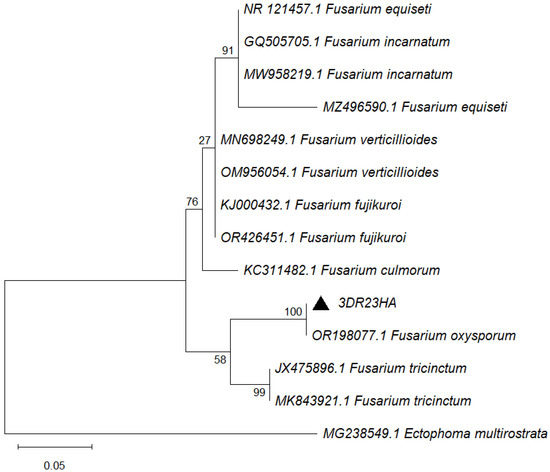
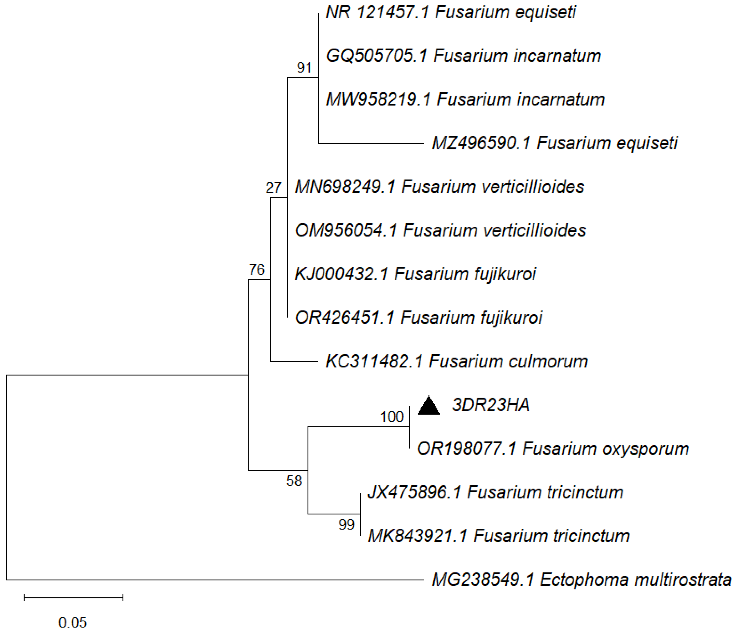
The ITS rDNA sequence generated for isolate 3DR23HA was deposited in the NCBI GenBank database under accession number PZ256382. BLASTn (v. 2.17.0+) analysis against the NCBI GenBank database showed that its best match was Fusarium oxysporum (accession OR198077.1), with 100% query coverage and 99.02% nucleotide identity. Consistently, in the ITS-based phylogenetic analysis (Figure 4), isolate 3DR23HA (GenBank accession PZ256382; ▲) clustered within the F. oxysporum clade together with OR198077.1, with strong bootstrap support (100%), and was clearly separated from clades comprising other Fusarium species included in the analysis.
Figure 4.
Phylogenetic tree based on ITS rDNA sequences of Fusarium spp., showing the placement of isolate 3DR23HA from this study (GenBank accession PZ256382; ▲). Numbers at the nodes indicate bootstrap support values (%) computed from 10,000 replicates. GenBank accession numbers are shown before each species name. Ectophoma multirostrata was used as the outgroup.
3.3. In Vitro Antifungal Activity of Stenotrophomonas rhizophila
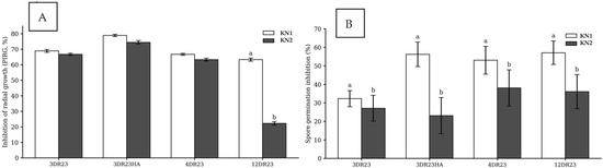
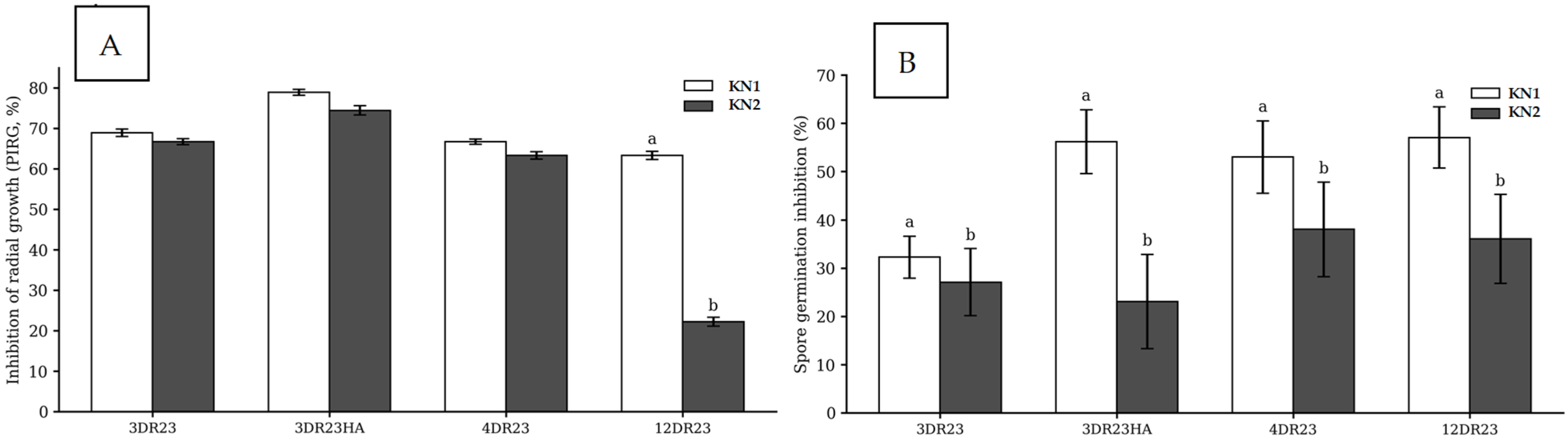
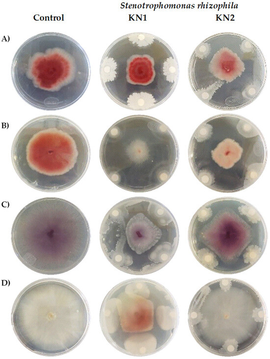
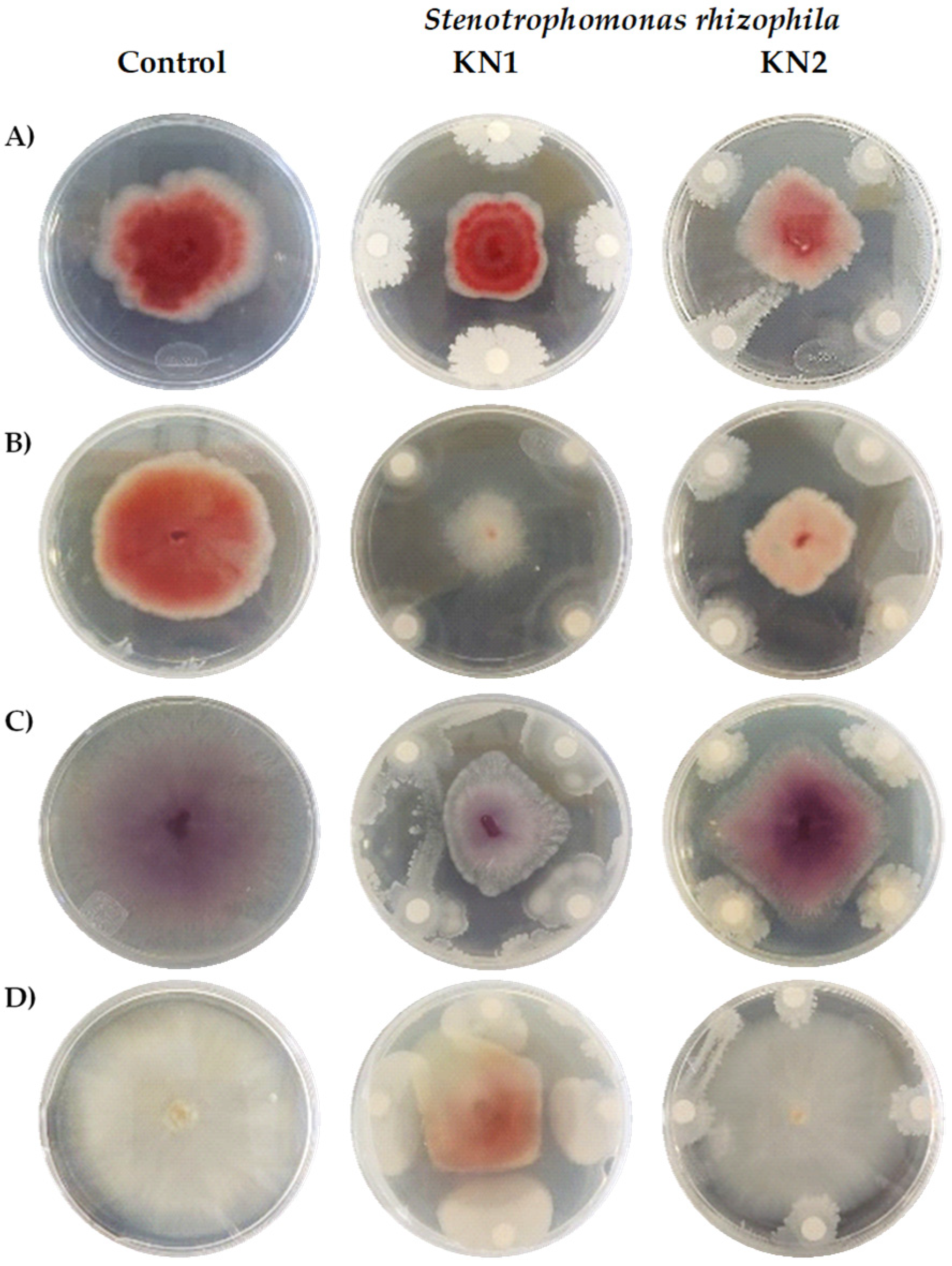
Dual-culture assays showed that both marine Stenotrophomonas rhizophila strains inhibited the Fusarium spp. isolates in vitro, although the magnitude of inhibition depended on the bacterial strain and the fungal isolate (Figure 5 and Figure 6). Regarding mycelial growth, inhibition of radial growth (PIRG, %) exceeded 65% for isolates 3DR23, 3DR23HA, and 4DR23 with both KN1 and KN2, with no significant differences between strains within these isolates (Holm-adjusted p > 0.05; Figure 5A). In contrast, for isolate 12DR23, KN1 produced a higher PIRG than KN2 (Holm-adjusted p ≤ 0.05; Figure 5A and Figure 6).
Figure 5.
Antagonistic activity of marine Stenotrophomonas rhizophila strains (KN1 and KN2) against four Fusarium spp. isolates (3DR23, 3DR23HA, 4DR23, and 12DR23). (A) Inhibition of radial growth (PIRG, %). (B) Spore germination inhibition (%). Within each isolate, different lowercase letters indicate significant differences between bacterial strains (KN1 vs. KN2) according to simple-effects pairwise contrasts with Holm-adjusted p ≤ 0.05. Bars without letters indicate that no significant differences were detected between strains within that isolate. Data are shown as mean ± standard deviation.
Figure 6.
In vitro inhibition of Fusarium spp. isolates by two marine strains of Stenotrophomonas rhizophila (KN1 and KN2). (A) 3DR23. (B) 3DR23HA. (C) 4DR23. (D) 12DR23.
When spore germination was assessed, KN1 significantly inhibited conidial germination compared with KN2 across all four isolates (3DR23, 3DR23HA, 4DR23, and 12DR23) (Holm-adjusted p ≤ 0.05; Figure 5B). Overall, these results indicate that KN1 exhibits stronger antagonistic activity than KN2, particularly by suppressing early developmental stages (spore germination) and, in the case of isolate 12DR23, also by restricting mycelial growth.
3.4. In Vitro Antifungal Activity of CuO-NPs
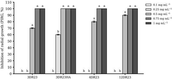
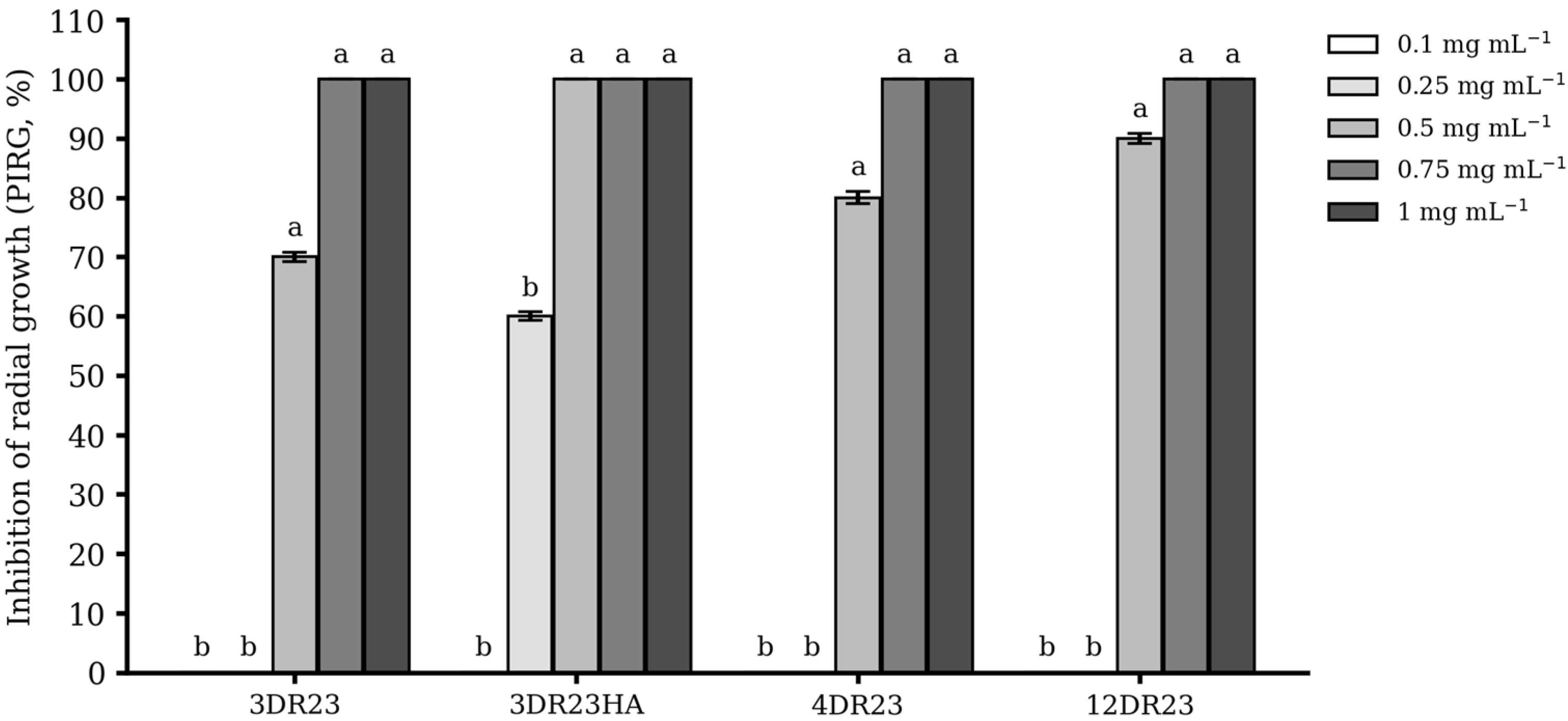
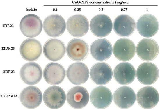
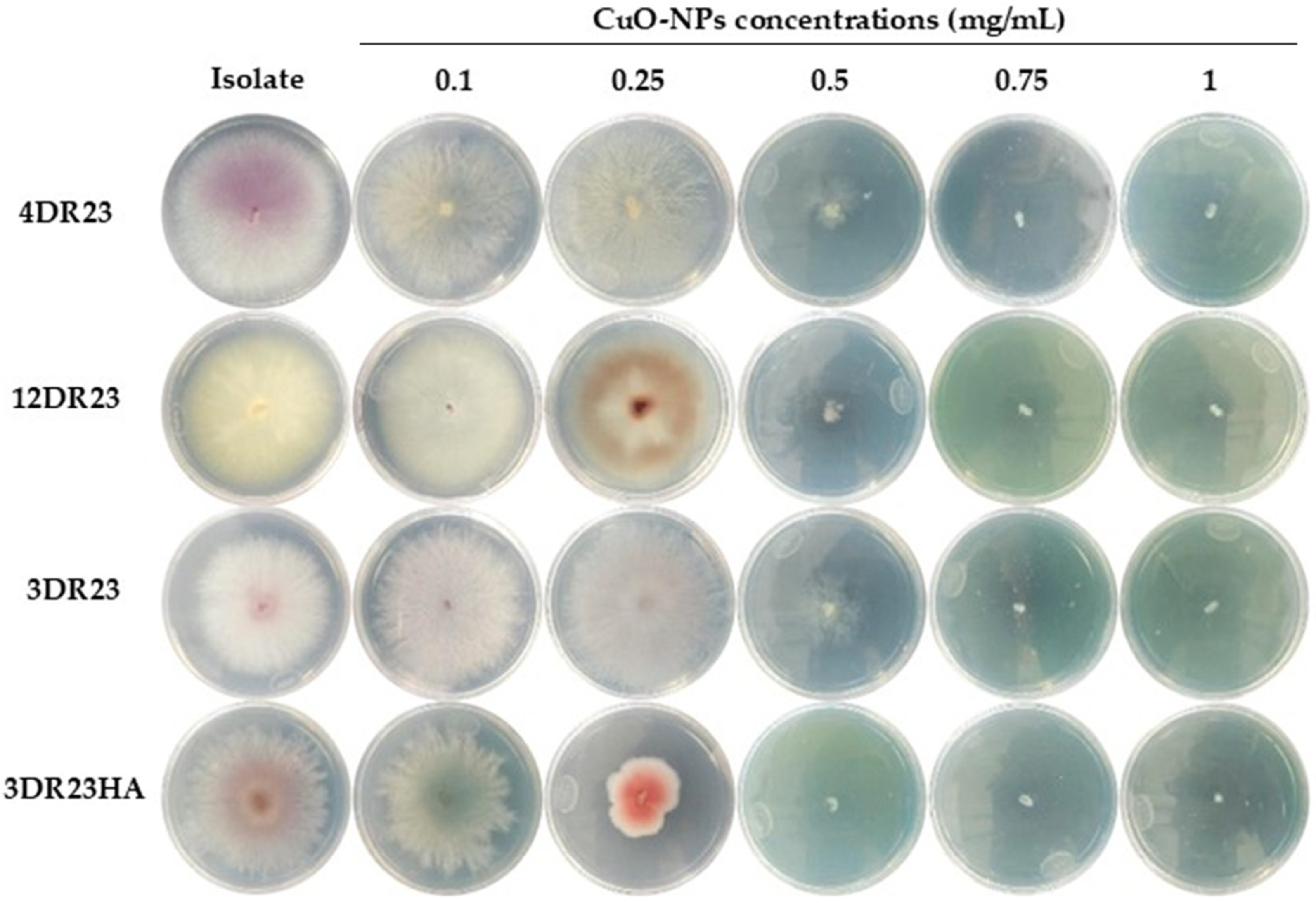
Copper oxide nanoparticles (CuO-NPs) inhibited mycelial growth of all four Fusarium spp. isolates in a concentration-dependent manner (Figure 7 and Figure 8). At the lowest concentrations (0.1–0.25 mg mL−1), inhibition was minimal, whereas increasing the concentration to 0.5 mg mL−1 resulted in significantly higher PIRG values in all isolates. The greatest inhibition was observed at 0.75 and 1.0 mg mL−1, reaching complete suppression of radial growth in most isolates. Within each isolate, PIRG differed significantly among CuO-NP concentrations (Dunn–Holm, p ≤ 0.05), indicating that higher CuO-NP levels enhanced antifungal activity.
Figure 7.
In vitro inhibition of radial growth (PIRG, %) by CuO-NPs against four Fusarium spp. isolates (3DR23, 3DR23HA, 4DR23, and 12DR23). Within each isolate, different lowercase letters indicate significant differences among CuO-NPs concentrations based on Kruskal–Wallis followed by Dunn’s multiple comparisons with Holm adjustment (p ≤ 0.05) ± Standard deviation.
Figure 8.
In vitro inhibition of Fusarium spp. by CuO-NPs. Isolates of phytopathogenic fungi: 4DR23, 12DR23, 3DR23, and 3DR23HA.
3.5. Effects of Integrated Treatments on Tomato Seedling Growth Under Phytopathogen Challenge
Table 2 shows that F. oxysporum (3DR23HA) reduced seedling growth, resulting in the lowest stem length (15.9 cm) and the lowest number of leaves (5.4 leaves plant−1). In contrast, the synthetic fungicide, the bacterial strains (KN1 and KN2), and CuO-NPs applied individually generally improved growth relative to the phytopathogen-only treatment, with stem length values ranging from 24.1 to 26.9 cm (p ≤ 0.05).
Table 2.
Effects of Stenotrophomonas rhizophila (KN1, KN2), CuO-NPs, and their combinations on the growth of tomato seedlings infected with Fusarium oxysporum (3DR23HA).
Stem diameter also increased relative to 3DR23HA, particularly in the synthetic fungicide and KN1 treatments (3.4–3.6 mm), whereas the phytopathogen-only treatment showed the greatest reduction (2.2 mm) (p ≤ 0.05). Root length showed a positive numerical response to some individual treatments; however, statistically significant increases relative to the phytopathogen-only treatment were observed only for CuO-NPs at 0.50 mg mL−1 (18.4 cm) and KN1 (16.4 cm), compared with 9.6 cm in the phytopathogen-only treatment (p ≤ 0.05).
Among the combined treatments, KN1 + CuO-NPs 0.75 and KN2 + CuO-NPs 0.75 maintained stem length values statistically comparable to those of the best-performing individual treatments and the synthetic fungicide, whereas KN1 + CuO-NPs 0.50 and KN2 + CuO-NPs 0.50 produced shorter stems (16.8–17.0 cm), similar to the phytopathogen-only treatment (p ≤ 0.05). Overall, these results indicate that KN1 and CuO-NPs, particularly at 0.50–0.75 mg mL−1, can mitigate the growth suppression caused by 3DR23HA, although the magnitude of the response depends on the specific bacterium–nanoparticle combination.
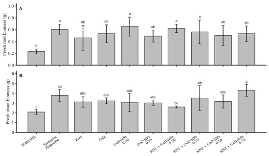
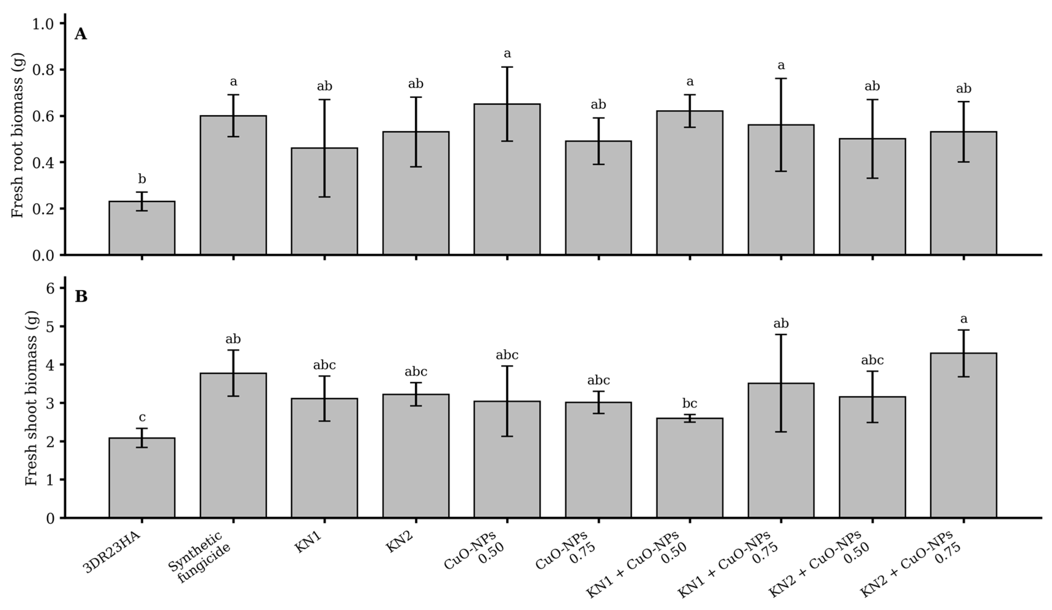
Figure 9 shows that inoculation with Fusarium oxysporum (3DR23HA) resulted in the lowest mean values for both fresh root biomass and fresh shoot biomass among the treatments evaluated. For fresh root biomass (Figure 9A), statistically significant increases relative to the phytopathogen-only treatment were observed for the synthetic fungicide, CuO-NPs at 0.50 mg mL−1, KN1 + CuO-NPs 0.50 mg mL−1, and KN1 + CuO-NPs 0.75 mg mL−1 (p ≤ 0.05). The remaining treatments did not differ significantly from 3DR23HA. For fresh shoot biomass (Figure 9B), statistically significant increases relative to the phytopathogen-only treatment were detected only for the synthetic fungicide, KN1 + CuO-NPs 0.75 mg mL−1, and KN2 + CuO-NPs 0.75 mg mL−1 (p ≤ 0.05), with KN2 + CuO-NPs 0.75 mg mL−1 showing the highest mean value. Overall, treatments involving S. rhizophila and CuO-NPs mitigated the biomass reduction caused by F. oxysporum, although the magnitude of the response varied according to the variable evaluated and the treatment applied.
Figure 9.
Effects of Stenotrophomonas rhizophila (KN1, KN2), CuO-NPs, and their combinations on the biomass of tomato seedlings infected with Fusarium oxysporum (3DR23HA). (A) Fresh root biomass (g) and (B) fresh shoot biomass (g). Different lowercase letters above bars indicate significant differences among treatments (Dunn’s test with Holm adjustment, p ≤ 0.05) ± Standard deviation.
3.6. Effects of Integrated Treatments on Disease Incidence, Severity, and Control Efficiency
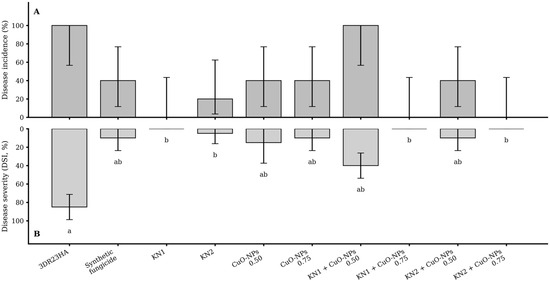
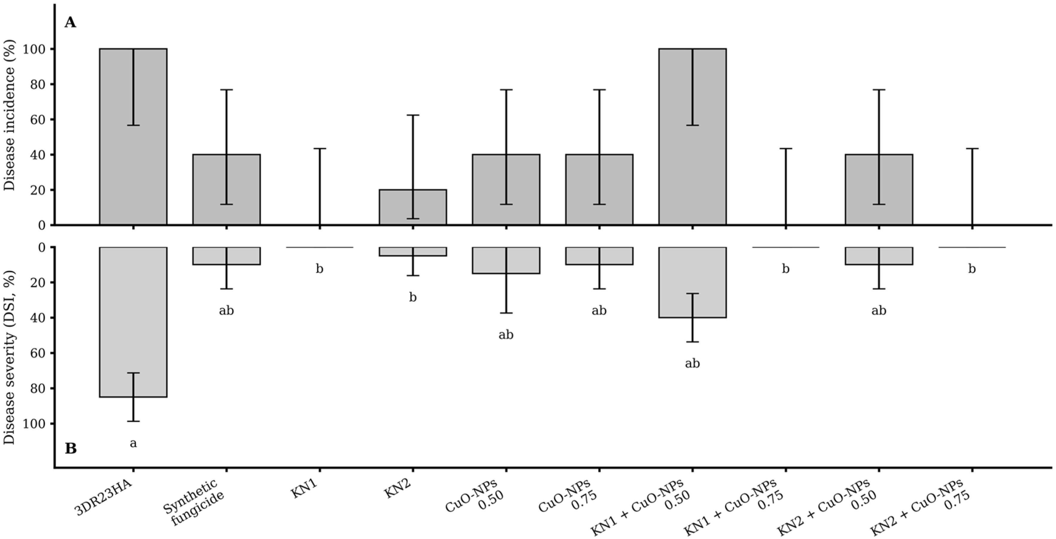
In the phytopathogen-only control (3DR23HA), damping-off incidence was 100% and severity reached a DSI of 85.0%. Most treatments reduced incidence to 0–40% (relative reductions of 60–100%), and the KN1, KN1 + CuO-NPs 0.75, and KN2 + CuO-NPs 0.75 treatments achieved 0% incidence. However, no significant differences in incidence were detected among treatments (p > 0.05) (Figure 10A).
Figure 10.
Disease incidence and disease severity (DSI, %) of tomato damping-off under integrated treatments with Stenotrophomonas rhizophila (KN1, KN2) and CuO-NPs. (A) Disease incidence (%) with 95% Wilson confidence intervals. (B) Disease severity expressed as disease severity index (DSI, %) (mean ± standard deviation). Severity bars are plotted downward for visual comparison with incidence. In panel B, different lowercase letters indicate significant differences in disease severity among treatments based on the Scheirer–Ray–Hare factorial test, followed by simple-effects comparisons and Dunn’s test with Holm adjustment (p ≤ 0.05). Treatments without letters did not show significant pairwise differences in the post hoc comparisons.
In contrast, severity (DSI, %) showed a significant treatment effect (p = 0.00016). The most effective treatments—KN1, KN1 + CuO-NPs 0.75, and KN2 + CuO-NPs 0.75—reached DSI = 0%, corresponding to a 100% relative reduction compared with the phytopathogen-only control (3DR23HA) and the synthetic fungicide. The KN1 + CuO-NPs 0.50 combination increased severity relative to KN1 alone (DSI = 40.0 ± 13.7%, ≈52.9% reduction vs. the control), suggesting an antagonistic effect at that dose/mixture, whereas at 0.75 the combination maintained complete control (DSI = 0%) (Figure 10B).
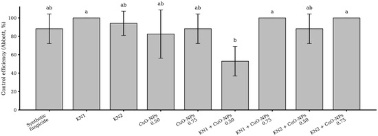
Control efficiency (Abbott, %) varied significantly among treatments (p = 0.0071). The highest efficiencies were observed for KN1, KN1 + CuO-NPs 0.75, and KN2 + CuO-NPs 0.75, all of which reached 100.0%. In contrast, the KN1 + CuO-NPs 0.50 combination showed the lowest efficiency (52.9%) and was significantly lower than KN1, KN1 + CuO-NPs 0.75, and KN2 + CuO-NPs 0.75 (Dunn–Holm, p = 0.0092). In terms of control efficiency (Abbott, %), none of the evaluated treatments differed significantly from the synthetic fungicide (Dunn–Holm, p > 0.05). Notably, several alternatives achieved maximum efficiency (100%), indicating performance comparable to the chemical treatment and supporting their potential as substitutes or complements within integrated damping-off management (Figure 11).
Figure 11.
Control efficiency (Abbott, %) of integrated treatments based on disease severity in tomato damping-off. Different lowercase letters indicate significant differences among treatments (Dunn’s test with Holm adjustment; p ≤ 0.05) ± Standard deviation.
4. Discussion
Fusarium spp. are widely distributed in tomato production systems worldwide and represent a phylogenetically diverse group of fungi frequently associated with damping-off [59,60]. In the present study, the macroscopic and microscopic features of the isolates were consistent with the taxonomic descriptions of Fusarium spp. provided by Summerell et al. [46] and Nelson et al. [61]. The colony traits observed, including cottony aerial mycelium and variable pigmentation ranging from whitish to pink, violet, or brown, agree with previous descriptions for this genus [62]. Likewise, the morphological variation recorded among isolates is consistent with the phenotypic plasticity reported for Fusarium spp., particularly with respect to colony pigmentation and structure [63,64].
The differential incidence and severity caused by the isolates are also biologically plausible, since aggressiveness in Fusarium spp. may vary according to isolate genetics, host susceptibility, and environmental conditions [65,66]. After conidia adhere to and invade plant roots, fungal colonization of the vascular system can disrupt water transport through the xylem, thereby promoting wilting, growth restriction, and, in severe cases, plant death [67]. Disease symptoms are associated with vascular blockage caused by fungal hyphae and by host–pathogen interaction processes, including the accumulation of toxins, gels, and tyloses, which ultimately lead to epinasty, chlorosis, wilting, defoliation, and death of the host plant [68,69]. At the early stages of disease development, growth and biomass reduction are expected because vascular dysfunction restricts the movement of water and nutrients through the xylem [70,71].
Our results support this pathophysiological framework. Isolate 3DR23HA showed the highest incidence (60%) and was associated with the greatest reductions in root biomass. Although severity clustered at the same ordinal level (grade 3 for most isolates), biomass, particularly root dry weight, provided greater sensitivity for discriminating damage intensity. This is consistent with damping-off pathosystems, in which external symptoms may appear similar among isolates, whereas the functional deterioration of the root system and the associated limitation in water and nutrient uptake may differ substantially [72,73].
In vitro confrontation assays are widely used as an initial screening step to identify microorganisms with potential for the biological control of phytopathogens [74]. In the present study, both S. rhizophila strains inhibited Fusarium spp., although the magnitude of antagonism depended on both the bacterial strain and the fungal isolate. Hernández-Montiel et al. [75] likewise reported strong inhibition of the mycelial growth of several phytopathogenic fungi, including Colletotrichum gloeosporioides, Penicillium italicum, P. digitatum, Alternaria solani, Fusarium oxysporum, Neoscytalidium dimidiatum, Alternaria alternata, F. solani, and Curvularia sp., by marine strains of S. rhizophila, with inhibition values ranging from 90 to 98% and from 88 to 97%, respectively, thereby supporting the high antifungal potential of this bacterial species. In our study, both S. rhizophila strains inhibited radial growth, but the magnitude of inhibition depended on the fungal isolate. KN1 inhibited radial growth by 63–79% across the four isolates, whereas KN2 produced inhibition values of 63–74% in three isolates but only 22% against isolate 12DR23. KN1 also showed greater inhibition of conidial germination than KN2. This distinction is epidemiologically relevant because conidial germination and early establishment are critical stages in disease initiation; therefore, an antagonist that interferes with these early events may reduce effective inoculum pressure even when subsequent mycelial expansion is not proportionally reduced [76,77].
The antagonistic activity observed for S. rhizophila may be related to mechanisms previously reported for this species, including hydrolytic enzymes, antibiosis, competition for nutrients and space, siderophore production, surfactant-associated effects, and volatile organic compounds [78,79,80]. From a microbiological standpoint, hydrolytic enzymes such as chitinases, glucanases, and proteases are of particular interest because they may act directly on structural components of the fungal cell wall, including chitin, β-glucans, and wall-associated proteins. The hydrolysis of these polymers may weaken cell wall integrity, compromise hyphal stability, and generate smaller oligosaccharides that can subsequently be assimilated by bacteria as carbon sources [81]. Although these mechanisms were not directly measured in the present study, the stronger suppression of conidial germination by KN1 suggests that strain-dependent traits may influence the earliest stages of phytopathogen establishment, which is particularly relevant in damping-off, where disease onset depends heavily on successful infection shortly after seedling emergence.
The response of F. oxysporum to CuO-NPs was clearly concentration dependent. At 0.1–0.25 mg mL−1, inhibition was limited, whereas concentrations from 0.5 mg mL−1 onward markedly reduced radial growth, with complete suppression in most isolates at 0.75–1.0 mg mL−1. This result indicates that the antifungal effect of CuO-NPs was dose-dependent and that a threshold concentration was required to achieve strong inhibition. At the lowest concentration, changes in mycelial pigmentation were observed, which may reflect fungal stress responses, including the production of protective pigments such as melanin and carotenoids [82]. Previous studies have associated the antifungal activity of CuO-NPs with copper ion release, membrane perturbation, oxidative stress, and damage to cellular macromolecules [83,84,85]. At the cellular level, copper ions released from CuO-NPs may alter membrane permeability and redox balance, thereby promoting oxidative damage to lipids, proteins, and nucleic acids. Once the antioxidant and repair capacity of the fungal cell is exceeded, these alterations may become incompatible with normal hyphal growth and viability [84,85]. Although those processes were not directly measured here, the concentration-dependent inhibition observed in our assays is consistent with progressive cellular stress that becomes increasingly damaging at higher nanoparticle concentrations.
The in vivo results further support the role of S. rhizophila in damping-off management. In the individual treatments, both bacterial strains improved growth relative to the phytopathogen-only control, suggesting that their effect extended beyond pathogen suppression. This interpretation is consistent with previous reports describing S. rhizophila as a multifunctional plant-associated bacterium with potential roles in growth promotion, nutrient mobilization, and enhanced seedling performance under biotic stress [86,87,88]. In our study, this broader effect was reflected in the recovery of seedling vigor under phytopathogen challenge, which is agronomically relevant because damping-off management should not only reduce visible symptoms but also preserve seedling quality, biomass accumulation, and subsequent establishment capacity.
The integrated treatments provide the most important biological and practical outcome of the study. Under challenge with the most virulent isolate, the combinations containing CuO-NPs at 0.75 mg mL−1 (KN1 + CuO-NPs 0.75 and KN2 + CuO-NPs 0.75) achieved complete control of severity (DSI = 0%) and maximum control efficiency (Abbott = 100%), with performance comparable to that of the synthetic fungicide. Although incidence was numerically reduced to 0–40%, those differences were not statistically significant, whereas severity clearly discriminated among treatments. This pattern is consistent with the lower discriminatory power of incidence as a binary variable, whereas DSI integrates symptom intensity and is therefore more sensitive for separating treatment effects under high disease pressure [89].
The superior performance of the 0.75 mg mL−1 combinations may reflect complementary effects between S. rhizophila and CuO-NPs operating at different stages of the host-phytopathogen interaction. A biologically consistent interpretation is that S. rhizophila reduced the probability of early inoculum establishment in the rhizosphere, whereas foliar-applied CuO-NPs enhanced host defensive capacity through mechanisms previously reported in the literature. In the case of S. rhizophila, suppression of early infection may involve antibiosis and the production of lytic enzymes such as chitinases, glucanases, and proteases, which can hydrolyze major structural components of the fungal cell wall, including chitin, β-glucans, and wall-associated proteins, thereby weakening hyphal integrity and restricting phytopathogen establishment [79,80,81]. This antagonistic effect may be further reinforced by competition for nutrients and space in the rhizosphere, siderophore-mediated iron sequestration, and the action of diffusible and volatile metabolites, all of which may reduce the likelihood of successful colonization during the earliest stages of infection [14,78,79,80,81].
Copper-based nanoparticles have also shown disease-suppressive effects in tomato–Fusarium pathosystems. In tomato plants challenged with Fusarium oxysporum f. sp. lycopersici, chitosan-loaded CuO nanoparticles reduced wilt severity by 91.5% at 1 mg L−1 and by 96.0–98.0% at 25–50 mg L−1 after 75 days; the same study also reported increases in plant height, dry weight, flowering, photosynthetic pigments, and the activity of chitinase and peroxidase [39]. Likewise, in tomato plants affected by Fusarium solani root rot, CuO nanoparticles derived from Zizyphus spina leaf extract reduced disease severity from 80.5% in the infected control to 9.17 ± 2.89% at 250 mg L−1, lowered disease incidence to 4.17 ± 3.80%, and improved seedling vigor, chlorophyll pigments, plant growth, and enzymatic activity [90].
In our study, the superior performance of the integrated treatments is consistent with the possibility that CuO-NPs contributed to disease suppression not only through direct antimicrobial activity, but also through host-mediated responses. Previous studies have associated copper-based nanoparticles with increased activity and/or expression of antioxidant enzymes such as SOD, APX, and GPX, which contribute to redox homeostasis and attenuation of infection-associated oxidative stress [42,43]. CuO-NPs have also been linked to activation of the phenylpropanoid pathway, reflected in increased PAL activity and, in some cases, PPO activity, which may promote the accumulation of phenolic compounds and flavonoids involved in chemical defense and reinforcement of structural barriers [42,44]. In addition, CuO-NPs have been associated with signaling-mediated resistance, including jasmonate (JA) modulation and activation of pathogenesis-related (PR) genes, responses consistent with SAR/ISR-like states and a priming effect that may enable the host to respond more rapidly and effectively to phytopathogen challenge [45,91]. Although these physiological and molecular responses were not measured in the present study, they provide a plausible literature-based framework for interpreting the superior performance of the integrated treatments observed here.
Overall, our results suggest that integrating S. rhizophila with CuO-NPs at 0.75 mg mL−1 may provide an effective strategy, with performance comparable to that of the synthetic fungicide, for managing tomato damping-off. However, validation under nursery and field conditions, together with further compatibility and safety assessments, remains necessary.
5. Conclusions
The present study demonstrated that both Stenotrophomonas rhizophila strains and CuO-NPs have strong potential for managing tomato damping-off associated with Fusarium oxysporum. In vitro, KN1 and KN2 inhibited mycelial growth of the evaluated isolates, whereas KN1 showed greater suppression of conidial germination. CuO-NPs exhibited a clear concentration-dependent antifungal effect, with complete inhibition of radial growth in most isolates at 0.75 and 1.0 mg mL−1. In vivo, the phytopathogen reduced seedling growth and biomass, whereas individual applications of KN1, KN2, and CuO-NPs improved several growth traits relative to the phytopathogen-only treatment. The most effective integrated treatments were KN1 + CuO-NPs 0.75 mg mL−1 and KN2 + CuO-NPs 0.75 mg mL−1, which achieved complete suppression of disease severity (DSI = 0%) and 100% control efficiency, with performance comparable to that of the synthetic fungicide. Overall, these findings indicate that the integration of S. rhizophila with CuO-NPs, particularly at 0.75 mg mL−1, represents a promising strategy for managing tomato damping-off, although validation under nursery and field conditions is still required before practical recommendation.
Author Contributions
Conceptualization, R.G.C.-C. and L.G.H.M.; methodology, R.G.C.-C., C.R.-M. and L.G.H.M.; software, J.J.R.-P., J.A.T.-R. and R.J.H.-F.; validation, R.G.C.-C., L.H.-A. and L.G.H.M.; formal analysis, R.J.H.-F. and J.A.T.-R.; investigation, L.G.H.M.; resources, J.J.R.-P., L.H.-A. and R.J.H.-F.; writing—original draft preparation, R.G.C.-C., C.R.-M. and L.G.H.M.; writing—review and editing, R.G.C.-C. and L.G.H.M.; supervision, R.G.C.-C.; project administration, R.G.C.-C. and L.G.H.M. All authors have read and agreed to the published version of the manuscript.
Funding
This research received no external funding.
Data Availability Statement
The raw data supporting the conclusions of this article will be made available by the authors on request.
Conflicts of Interest
The authors declare no conflicts of interest.
References
- FAO. Agricultural Production Statistics 2010–2024; FAOSTAT Analytical Briefs; FAO: Rome, Italy, 2025. [Google Scholar]
- Intana, W.; Promwee, A.; Wijara, K.; Nguyen, H.H. Enhancement of Damping-Off Disease Control in Tomatoes Using Two Strains of Trichoderma Asperellum Combined with a Plant Immune Stimulant. Agronomy 2024, 14, 1655. [Google Scholar] [CrossRef] [Scilit]
- Madlhophe, S.; Ogugua, U.V.; Makhubu, F.N.; Figlan, S. Use of Biological Control Agents for Managing Fungal Pathogens in Solanaceae Crops: Progress and Future Perspectives—A Review. Discov. Appl. Sci. 2025, 7, 83. [Google Scholar] [CrossRef] [Scilit]
- Lamichhane, J.R.; Dürr, C.; Schwanck, A.A.; Robin, M.-H.; Sarthou, J.-P.; Cellier, V.; Messéan, A.; Aubertot, J.-N. Integrated Management of Damping-off Diseases. A Review. Agron. Sustain. Dev. 2017, 37, 10. [Google Scholar] [CrossRef] [Scilit]
- Delai, C.; Muhae-Ud-Din, G.; Abid, R.; Tian, T.; Liu, R.; Xiong, Y.; Ma, S.; Ghorbani, A. A Comprehensive Review of Integrated Management Strategies for Damping-off Disease in Chili. Front. Microbiol. 2024, 15, 1479957. [Google Scholar] [CrossRef] [Scilit] [PubMed]
- Arora, H.; Sharma, A.; Sharma, S.; Haron, F.F.; Gafur, A.; Sayyed, R.Z.; Datta, R. Pythium Damping-Off and Root Rot of Capsicum annuum L.: Impacts, Diagnosis, and Management. Microorganisms 2021, 9, 823. [Google Scholar] [CrossRef] [Scilit] [PubMed]
- Kawamoto, K.; Shimotsuma, Y.; Maeda, K.; Matsunaga, K.; Asano, H.; Cohen, S.M.; Yamada, T. An Evaluation of the Human Relevance of the Hepatocellular Tumors Observed in Mice Treated with Synthetic Fungicide, Pyridachlometyl, Based on Mode of Action. Arch. Toxicol. 2025, 99, 3803–3824. [Google Scholar] [CrossRef] [Scilit]
- Sharma, D.; Gautam, H.R.; Chandel, S.; Thakur, K.; Sharma, P.L. Seed Biopriming with Antagonistic Microbes and Their Defense Activity against Damping-off Disease in Solanaceous Vegetable Crops. Indian Phytopathol. 2024, 77, 483–489. [Google Scholar] [CrossRef] [Scilit]
- Fenta, L.; Mekonnen, H. Microbial Biofungicides as a Substitute for Chemical Fungicides in the Control of Phytopathogens: Current Perspectives and Research Directions. Scientifica 2024, 2024, 5322696. [Google Scholar] [CrossRef] [Scilit]
- Etesami, H.; Jeong, B.R.; Glick, B.R. Biocontrol of Plant Diseases by Bacillus spp. Physiol. Mol. Plant Pathol. 2023, 126, 102048. [Google Scholar] [CrossRef] [Scilit]
- Carmona-Hernandez, S.; Reyes-Pérez, J.J.; Chiquito-Contreras, R.G.; Rincon-Enriquez, G.; Cerdan-Cabrera, C.R.; Hernandez-Montiel, L.G. Biocontrol of Postharvest Fruit Fungal Diseases by Bacterial Antagonists: A Review. Agronomy 2019, 9, 121. [Google Scholar] [CrossRef] [Scilit]
- Halsey, K.H.; Giovannoni, S.J. Biological Controls on Marine Volatile Organic Compound Emissions: A Balancing Act at the Sea-Air Interface. Earth-Sci. Rev. 2023, 240, 104360. [Google Scholar] [CrossRef] [Scilit]
- Reyes-Perez, J.J.; Hernandez-Montiel, L.G.; Vero, S.; Noa-Carrazana, J.C.; Quiñones-Aguilar, E.E.; Rincón-Enríquez, G. Postharvest Biocontrol of Colletotrichum Gloeosporioides on Mango Using the Marine Bacterium Stenotrophomonas rhizophila and Its Possible Mechanisms of Action. J. Food Sci. Technol. 2019, 56, 4992–4999. [Google Scholar] [CrossRef] [Scilit]
- Rivas-Garcia, T.; Murillo-Amador, B.; Reyes-Pérez, J.J.; Chiquito-Contreras, R.G.; Preciado-Rangel, P.; Ávila-Quezada, G.D.; Lara-Capistran, L.; Hernandez-Montiel, L.G. Debaryomyces Hansenii, Stenotrophomonas rhizophila, and Ulvan as Biocontrol Agents of Fruit Rot Disease in Muskmelon (Cucumis melo L.). Plants 2022, 11, 184. [Google Scholar] [CrossRef] [Scilit] [PubMed]
- Yang, W.; Wang, M.; Wang, H.; Zhang, C.; Zhang, Q.; Xiao, H. Exploitation of the Biocontrol Potential of a Marine-Derived Bacillus Velezensis and Its Application on Postharvest Strawberry. Food Control 2024, 161, 110311. [Google Scholar] [CrossRef] [Scilit]
- Rathod, K.; Rana, S.; Dhandhukia, P.; Thakker, J.N. Marine Bacillus as a Potent Biocontrol Agent against Fusarium oxysporum f. sp. Ciceris. Plant Stress 2023, 10, 100289. [Google Scholar] [CrossRef] [Scilit]
- Rathod, K.; Rana, S.; Dhandhukia, P.; Thakker, J.N. Marine Bacillus Subtilis as an Effective Biocontrol Agent against Fusarium oxysporum f. sp. Ciceris. Eur. J. Plant Pathol. 2023, 167, 759–770. [Google Scholar] [CrossRef] [Scilit]
- Lara-Capistran, L.; Zulueta-Rodriguez, R.; Castellanos-Cervantes, T.; Reyes-Perez, J.J.; Preciado-Rangel, P.; Hernandez-Montiel, L.G. Efficiency of Marine Bacteria and Yeasts on the Biocontrol Activity of Pythium Ultimum in Ancho-Type Pepper Seedlings. Agronomy 2020, 10, 408. [Google Scholar] [CrossRef] [Scilit]
- Patel, P.; Patel, K.; Dhandhukia, P.; Thakker, J.N. Plant Growth Promoting Traits of Marine Micrococcus Sp. with Bio-Control Ability against Fusarium in Chickpea Plant. Vegetos 2021, 34, 94–101. [Google Scholar] [CrossRef] [Scilit]
- Sheng, M.; Jia, H.; Zhang, G.; Zeng, L.; Zhang, T.; Long, Y.; Lan, J.; Hu, Z.; Zeng, Z.; Wang, B.; et al. Siderophore Production by Rhizosphere Biological Control Bacteria Brevibacillus Brevis GZDF3 of Pinellia Ternata and Its Antifungal Effects on Candida Albicans. J. Microbiol. Biotechnol. 2020, 30, 689–699. [Google Scholar] [CrossRef] [Scilit]
- Torres-Rodriguez, J.; Reyes-Pérez, J.; Castellanos, T.; Quiñones-Aguilar, E.; Hernandez-Montiel, L. Identification and Morphological Characterization of Marine Actinomycetes as Biocontrolagents of Fusarium Solani in Tomato. RevFacAgron 2022, 39, e223915. [Google Scholar] [CrossRef] [Scilit]
- Guan, L.; Wang, H.; Chen, J.; Yang, F.; Yang, J.; Li, J.; Jin, L. Isolation and Identification of Culturable Bacteria from South China Seawater and Preliminary Screening of Marine Biocontrol Bacteria. Microorganisms 2023, 11, 2933. [Google Scholar] [CrossRef] [Scilit]
- Freese, H.M.; Meier-Kolthoff, J.P.; Sardà Carbasse, J.; Afolayan, A.O.; Göker, M. TYGS and LPSN in 2025: A global core biodata resource for genome-based classification and nomenclature of prokaryotes within DSMZ Digital Diversity. Nucleic Acids Res. 2026, 54, D884–D891. [Google Scholar] [CrossRef] [Scilit]
- Zhang, X.; Liao, H.; Cai, P.; Cai, T.; Si, H.; Yuan, J.; Yu, Y.; Zhao, M.; Li, P.; Zhang, X.; et al. Isolation and Characterization of Stenotrophomonas rhizophila T3E: A Multifunctional Rhizobacterium Enhancing Tomato Growth and Soil Health. Front. Plant Sci. 2025, 16, 1692957. [Google Scholar] [CrossRef] [Scilit] [PubMed]
- Garin, T.; Brault, A.; Marais, C.; Briand, M.; Préveaux, A.; Bonneau, S.; Simonin, M.; Barret, M.; Sarniguet, A. T6SS-Mediated Competition by Stenotrophomonas rhizophila Shapes Seed-Borne Bacterial Communities and Seed-to-Seedling Transmission Dynamics. mSystems 2025, 10, e00457-25. [Google Scholar] [CrossRef] [Scilit] [PubMed]
- Takeuchi, K.; Ogiso, M.; Ota, A.; Nishimura, K.; Nishino, C.; Omori, Y.; Maeda, M.; Mizui, R.; Yamanaka, H.; Ogino, T.; et al. Pseudomonas Rhodesiae HAI-0804 Suppresses Pythium Damping off and Root Rot in Cucumber by Its Efficient Root Colonization Promoted by Amendment with Glutamate. Front. Microbiol. 2024, 15, 1485167. [Google Scholar] [CrossRef] [Scilit] [PubMed]
- Dávila Costa, J.S.; Romero, C.M. Nano-Biofungicides and Bio-Nanofungicides: State of the Art of Innovative Tools for Controlling Resistant Phytopathogens. Biophysica 2025, 5, 15. [Google Scholar] [CrossRef] [Scilit]
- Ribeiro, L.G.; Roque, G.S.C.; Conrado, R.; De Souza, A.O. Antifungal Activity of Mycogenic Silver Nanoparticles on Clinical Yeasts and Phytopathogens. Antibiotics 2023, 12, 91. [Google Scholar] [CrossRef] [Scilit]
- Manzoor, M.A.; Shah, I.H.; Ali Sabir, I.; Ahmad, A.; Albasher, G.; Dar, A.A.; Altaf, M.A.; Shakoor, A. Environmental Sustainable: Biogenic Copper Oxide Nanoparticles as Nano-Pesticides for Investigating Bioactivities against Phytopathogens. Environ. Res. 2023, 231, 115941. [Google Scholar] [CrossRef] [Scilit]
- Kamel, S.M.; Elgobashy, S.F.; Omara, R.I.; Derbalah, A.S.; Abdelfatah, M.; El-Shaer, A.; Al-Askar, A.A.; Abdelkhalek, A.; Abd-Elsalam, K.A.; Essa, T.; et al. Antifungal Activity of Copper Oxide Nanoparticles against Root Rot Disease in Cucumber. J. Fungi 2022, 8, 911. [Google Scholar] [CrossRef] [Scilit]
- Parada, J.; Tortella, G.; Seabra, A.B.; Fincheira, P.; Rubilar, O. Potential Antifungal Effect of Copper Oxide Nanoparticles Combined with Fungicides against Botrytis Cinerea and Fusarium oxysporum. Antibiotics 2024, 13, 215. [Google Scholar] [CrossRef] [Scilit]
- Mir, A.R.; Pichtel, J.; Hayat, S. Copper: Uptake, Toxicity and Tolerance in Plants and Management of Cu-Contaminated Soil. Biometals 2021, 34, 737–759. [Google Scholar] [CrossRef] [Scilit] [PubMed]
- López-Ortiz, M.; Zamora, R.A.; Giannotti, M.I.; Gorostiza, P. The Protein Matrix of Plastocyanin Supports Long-Distance Charge Transport with Photosystem I and the Copper Ion Regulates Its Spatial Span and Conductance. ACS Nano 2023, 17, 20334–20344. [Google Scholar] [CrossRef] [Scilit] [PubMed]
- Roy, D.; Adhikari, S.; Adhikari, A.; Ghosh, S.; Azahar, I.; Basuli, D.; Hossain, Z. Impact of CuO Nanoparticles on Maize: Comparison with CuO Bulk Particles with Special Reference to Oxidative Stress Damages and Antioxidant Defense Status. Chemosphere 2022, 287, 131911. [Google Scholar] [CrossRef] [Scilit] [PubMed]
- Letchumanan, D.; Sok, S.P.M.; Ibrahim, S.; Nagoor, N.H.; Arshad, N.M. Plant-Based Biosynthesis of Copper/Copper Oxide Nanoparticles: An Update on Their Applications in Biomedicine, Mechanisms, and Toxicity. Biomolecules 2021, 11, 564. [Google Scholar] [CrossRef] [Scilit]
- Alavi, M.; Hamblin, M.R. Interaction of Copper Oxide Nanoparticles with Bacterial Nucleic Acids: A Mini-Review. Micro Nano Bio Asp. 2023, 2, 20–25. [Google Scholar] [CrossRef]
- Amin, N.; Aziz, K. Copper Oxide-Based Nanoparticles in Agro-Nanotechnology: Advances and Applications for Sustainable Farming. Agric. Food Secur. 2025, 14, 7. [Google Scholar] [CrossRef] [Scilit]
- Gudkov, S.V.; Burmistrov, D.E.; Fomina, P.A.; Validov, S.Z.; Kozlov, V.A. Antibacterial Properties of Copper Oxide Nanoparticles (Review). Int. J. Mol. Sci. 2024, 25, 11563. [Google Scholar] [CrossRef] [Scilit]
- Mosa, M.A.; El-Abeid, S.E. Chitosan-Loaded Copper Oxide Nanoparticles: A Promising Antifungal Nanocomposite against Fusarium Wilt Disease of Tomato Plants. Sustainability 2023, 15, 14295. [Google Scholar] [CrossRef] [Scilit]
- Fetyan, N.A.H.; Essa, T.A.; Salem, T.M.; Taha, A.A.; Elgobashy, S.F.; Tharwat, N.A.; Elsakhawy, T. Promising Eco-Friendly Nanoparticles for Managing Bottom Rot Disease in Lettuce (Lactuca sativa Var. longifolia). Microbiol. Res. 2024, 15, 196–212. [Google Scholar] [CrossRef] [Scilit]
- Hussain, I.; Zhao, T.; Wang, Y.; Lei, N.; Liu, K.; Yu, H.; Zhang, Y.; Muhammad, U.; Ullah, H.; Yu, X. Melatonin and Copper Oxide Nanoparticles Synergistically Mitigate Clubroot Disease and Enhance Growth Dynamics in Brassica Rapa. Plant Physiol. Biochem. 2024, 215, 109020. [Google Scholar] [CrossRef] [Scilit]
- Quiterio-Gutiérrez, T.; Ortega-Ortiz, H.; Cadenas-Pliego, G.; Hernández-Fuentes, A.D.; Sandoval-Rangel, A.; Benavides-Mendoza, A.; Cabrera-de la Fuente, M.; Juárez-Maldonado, A. The Application of Selenium and Copper Nanoparticles Modifies the Biochemical Responses of Tomato Plants Under Stress by Alternaria Solani. Int. J. Mol. Sci. 2019, 20, 1950. [Google Scholar] [CrossRef] [Scilit]
- Pérez-Labrada, F.; López-Vargas, E.R.; Ortega-Ortiz, H.; Cadenas-Pliego, G.; Benavides-Mendoza, A.; Juárez-Maldonado, A. Responses of Tomato Plants Under Saline Stress to Foliar Application of Copper Nanoparticles. Plants 2019, 8, 151. [Google Scholar] [CrossRef] [Scilit]
- Sarkar, J.; Chakraborty, N.; Chatterjee, A.; Bhattacharjee, A.; Dasgupta, D.; Acharya, K. Green Synthesized Copper Oxide Nanoparticles Ameliorate Defence and Antioxidant Enzymes in Lens Culinaris. Nanomaterials 2020, 10, 312. [Google Scholar] [CrossRef] [Scilit]
- Hernández-Hernández, H.; Juárez-Maldonado, A.; Benavides-Mendoza, A.; Ortega-Ortiz, H.; Cadenas-Pliego, G.; Sánchez-Aspeytia, D.; González-Morales, S. Chitosan-PVA and Copper Nanoparticles Improve Growth and Overexpress the SOD and JA Genes in Tomato Plants Under Salt Stress. Agronomy 2018, 8, 175. [Google Scholar] [CrossRef] [Scilit]
- Summerell, B.A.; Salleh, B.; Leslie, J.F. A Utilitarian Approach to Fusarium Identification. Plant Dis. 2003, 87, 117–128. [Google Scholar] [CrossRef] [Scilit] [PubMed]
- Boehm, M.J.; Madden, L.V.; Hoitink, H.A.J. Effect of Organic Matter Decomposition Level on Bacterial Species Diversity and Composition in Relationship to Pythium Damping-Off Severity. Appl. Environ. Microbiol. 1993, 59, 4171–4179. [Google Scholar] [CrossRef] [Scilit] [PubMed]
- Saravanakumar, K.; Yu, C.; Dou, K.; Wang, M.; Li, Y.; Chen, J. Synergistic Effect of Trichoderma-Derived Antifungal Metabolites and Cell Wall Degrading Enzymes on Enhanced Biocontrol of Fusarium oxysporum f. sp. Cucumerinum. Biol. Control 2016, 94, 37–46. [Google Scholar] [CrossRef] [Scilit]
- Agrios, G.N. Plant Pathology; Elsevier: Amsterdam, The Netherlands, 2005. [Google Scholar]
- Raeder, U.; Broda, P. Rapid Preparation of DNA from Filamentous Fungi. Lett. Appl. Microbiol. 1985, 1, 17–20. [Google Scholar] [CrossRef] [Scilit]
- Edgar, R.C. MUSCLE: Multiple Sequence Alignment with High Accuracy and High Throughput. Nucleic Acids Res. 2004, 32, 1792–1797. [Google Scholar] [CrossRef] [Scilit]
- Talavera, G.; Castresana, J. Improvement of Phylogenies after Removing Divergent and Ambiguously Aligned Blocks from Protein Sequence Alignments. Syst. Biol. 2007, 56, 564–577. [Google Scholar] [CrossRef] [Scilit]
- Dereeper, A.; Guignon, V.; Blanc, G.; Audic, S.; Buffet, S.; Chevenet, F.; Dufayard, J.-F.; Guindon, S.; Lefort, V.; Lescot, M.; et al. Phylogeny.Fr: Robust Phylogenetic Analysis for the Non-Specialist. Nucleic Acids Res. 2008, 36, W465–W469. [Google Scholar] [CrossRef] [Scilit] [PubMed]
- Kumar, S.; Stecher, G.; Li, M.; Knyaz, C.; Tamura, K. MEGA X: Molecular Evolutionary Genetics Analysis across Computing Platforms. Mol. Biol. Evol. 2018, 35, 1547–1549. [Google Scholar] [CrossRef] [Scilit] [PubMed]
- Nzabanita, C.; Zhang, L.; Wang, Y.; Wang, S.; Guo, L. The Wheat Endophyte Epicoccum Layuense J4-3 Inhibits Fusarium Graminearum and Enhances Plant Growth. J. Fungi 2023, 10, 10. [Google Scholar] [CrossRef] [Scilit] [PubMed]
- Park, J.S.; Ryu, G.R.; Kang, B.R. Target Mechanism of Iturinic Lipopeptide on Differential Expression Patterns of Defense-Related Genes against Colletotrichum Acutatum in Pepper. Plants 2022, 11, 1267. [Google Scholar] [CrossRef] [Scilit]
- Ibarra-Laclette, E.; Blaz, J.; Pérez-Torres, C.-A.; Villafán, E.; Lamelas, A.; Rosas-Saito, G.; Ibarra-Juárez, L.A.; García-Ávila, C.d.J.; Martínez-Enriquez, A.I.; Pariona, N. Antifungal Effect of Copper Nanoparticles against Fusarium Kuroshium, an Obligate Symbiont of Euwallacea Kuroshio Ambrosia Beetle. J. Fungi 2022, 8, 347. [Google Scholar] [CrossRef] [Scilit]
- Abbott, W.S. A Method of Computing the Effectiveness of an Insecticide. J. Econ. Entomol. 1925, 18, 265–267. [Google Scholar] [CrossRef] [Scilit]
- Sehim, A.E.; Hewedy, O.A.; Altammar, K.A.; Alhumaidi, M.S.; Abd Elghaffar, R.Y. Trichoderma Asperellum Empowers Tomato Plants and Suppresses Fusarium oxysporum through Priming Responses. Front. Microbiol. 2023, 14, 1140378. [Google Scholar] [CrossRef] [Scilit]
- Ribeiro, J.A.; Albuquerque, A.; Materatski, P.; Patanita, M.; Varanda, C.M.R.; Félix, M.D.R.; Campos, M.D. Tomato Response to Fusarium spp. Infection Under Field Conditions: Study of Potential Genes Involved. Horticulturae 2022, 8, 433. [Google Scholar] [CrossRef] [Scilit]
- Nelson, P.E.; Dignani, M.C.; Anaissie, E.J. Taxonomy, Biology, and Clinical Aspects of Fusarium Species. Clin. Microbiol. Rev. 1994, 7, 479–504. [Google Scholar] [CrossRef]
- Chauhan, P.; Tiwari, R.K.; Saini, A.K.; Kumar, A.; Rithesh, L. Deciphering the Morphological, Molecular, and Pathogenic Variability in Fusarium Species Associated with Potato Dry Rot Disease. Front. Microbiol. 2025, 16, 1478798. [Google Scholar] [CrossRef] [Scilit]
- Suanda, I.W.; Widnyana, I.K.; Mirah Dharmadewi, A. Isolation and identification of pathogenic Fusarium oxysporum isolated from soybean leaves. Biodiversitas J. Biol. Divers. 2025, 26, 2261. [Google Scholar] [CrossRef] [Scilit]
- Namisy, A.; Wu, Y.-C.; Lin, K.-H.; Hsu, W.-C.; Chung, W.-H. Fusarium Incarnatum: A Paradigm for One Health Pathogen Dynamics across Humans, Animals, and the Environment. Microbiol. Spectr. 2026, 14, e01919-25. [Google Scholar] [CrossRef] [Scilit] [PubMed]
- Chitwood-Brown, J.; Vallad, G.E.; Lee, T.G.; Hutton, S.F. Breeding for Resistance to Fusarium Wilt of Tomato: A Review. Genes 2021, 12, 1673. [Google Scholar] [CrossRef] [Scilit] [PubMed]
- Hernández-Aparicio, F.; Lisón, P.; Rodrigo, I.; Bellés, J.M.; López-Gresa, M.P. Signaling in the Tomato Immunity against Fusarium oxysporum. Molecules 2021, 26, 1818. [Google Scholar] [CrossRef] [Scilit]
- Ji, H.-M.; Mao, H.-Y.; Li, S.-J.; Feng, T.; Zhang, Z.-Y.; Cheng, L.; Luo, S.-J.; Borkovich, K.A.; Ouyang, S.-Q. Fol-milR1, a Pathogenicity Factor of Fusarium oxysporum, Confers Tomato Wilt Disease Resistance by Impairing Host Immune Responses. New Phytol. 2021, 232, 705–718. [Google Scholar] [CrossRef] [Scilit]
- Fernandes, L.B.; Ghag, S.B. Molecular Insights into the Jasmonate Signaling and Associated Defense Responses against Wilt Caused by Fusarium oxysporum. Plant Physiol. Biochem. 2022, 174, 22–34. [Google Scholar] [CrossRef] [Scilit]
- Namisy, A.; Chen, S.-Y.; Huang, J.-H.; Unartngam, J.; Thanarut, C.; Chung, W.-H. Histopathology and Quantification of Green Fluorescent Protein-Tagged Fusarium oxysporum f. sp. Luffae Isolate in Resistant and Susceptible Luffa Germplasm. Microbiol. Spectr. 2024, 12, e03127-23. [Google Scholar] [CrossRef] [Scilit]
- Srinivas, C.; Nirmala Devi, D.; Narasimha Murthy, K.; Mohan, C.D.; Lakshmeesha, T.R.; Singh, B.; Kalagatur, N.K.; Niranjana, S.R.; Hashem, A.; Alqarawi, A.A.; et al. Fusarium oxysporum f. sp. Lycopersici Causal Agent of Vascular Wilt Disease of Tomato: Biology to Diversity– A Review. Saudi J. Biol. Sci. 2019, 26, 1315–1324. [Google Scholar] [CrossRef] [Scilit]
- Sevillano-Caño, J.; Córdoba-Galván, C.; Sánchez-Rodríguez, A.R.; Romera, F.J.; Trapero, A.; García-del Rosal, M.J.; Agustí-Brisach, C. The Non-Pathogenic Strain Fusarium oxysporum FO12 Mitigates Simultaneous Verticillium Dahliae and Iron Deficiency Stresses in Arabidopsis Thaliana. Physiol. Plant. 2025, 177, e70550. [Google Scholar] [CrossRef] [Scilit]
- Hossain, M.M.; Sultana, F.; Mostafa, M.; Rubayet, M.T.; Mishu, N.J.; Khan, I.; Mostofa, M.G. Biological Management of Soil-Borne Pathogens Through Tripartite Rhizosphere Interactions with Plant Growth-Promoting Fungi. Appl. Microbiol. 2025, 5, 123. [Google Scholar] [CrossRef] [Scilit]
- Djalali Farahani-Kofoet, R.; Witzel, K.; Graefe, J.; Grosch, R.; Zrenner, R. Species-Specific Impact of Fusarium Infection on the Root and Shoot Characteristics of Asparagus. Pathogens 2020, 9, 509. [Google Scholar] [CrossRef] [Scilit]
- Guato, J.; Auhing, J.; Crespo-Ávila, J.; Esmeraldas García, G.; Mendoza-León, A.; Canchignia-Martínez, H. Plant Growth Promoting Bacteria with Potential Biocontrol Agent of Fusarium oxysporum f. sp. Lycopersici, and Moniliophthora Roreri. Sci. Agropecu. 2019, 10, 393–402. [Google Scholar] [CrossRef] [Scilit]
- Hernández Montiel, L.G.; Rivas García, T.; Romero Bastidas, M.; Chiquito Contreras, C.J.; Ruiz Espinoza, F.H.; Chiquito Contreras, R.G.; Hernández Montiel, L.G.; Rivas García, T.; Romero Bastidas, M.; Chiquito Contreras, C.J.; et al. Potencial antagónico de bacterias y levaduras marinas para el control de hongos fitopatógenos. Rev. Mex. Cienc. Agrícolas 2018, 9, 4311–4321. [Google Scholar] [CrossRef] [Scilit]
- Deng, G.-M.; Yang, Q.-S.; He, W.-D.; Li, C.-Y.; Yang, J.; Zuo, C.-W.; Gao, J.; Sheng, O.; Lu, S.-Y.; Zhang, S.; et al. Proteomic Analysis of Conidia Germination in Fusarium oxysporum f. sp. Cubense Tropical Race 4 Reveals New Targets in Ergosterol Biosynthesis Pathway for Controlling Fusarium Wilt of Banana. Appl. Microbiol. Biotechnol. 2015, 99, 7189–7207. [Google Scholar] [CrossRef] [Scilit] [PubMed]
- van Dijk, K.; Nelson, E.B. Inactivation of Seed Exudate Stimulants of Pythium Ultimum Sporangium Germination by Biocontrol Strains of Enterobacter Cloacae and Other Seed-Associated Bacteria. Soil Biol. Biochem. 1998, 30, 183–192. [Google Scholar] [CrossRef] [Scilit]
- Ghosh, R.; Chatterjee, S.; Mandal, N.C. Stenotrophomonas. In Beneficial Microbes in Agro-Ecology; Academic Press: Cambridge, MA, USA, 2020; pp. 427–442. [Google Scholar]
- Martínez Cano, B.; Toledano-Ayala, M.; Alberto, C.; Olvera, O.; Nava, R.; Soto-Zarazúa, G. International Journal of Agriculture & Biology full Length Article Effect of Two Strains of Stenotrophomonas Rhizophila Isolated from Compost on the Growth of Tomato, Lettuce, and Jalapeno Pepper. Int. J. Agric. Biol. 2022, 27, 423–430. [Google Scholar] [CrossRef] [Scilit]
- Yin, Y.; Cheng, Q.; Shi, Y.; Du, Z.; Luo, X.; Chen, X.; Hu, X.; Wang, Y.; Wan, C.; Shen, X. Healthy Cotton Roots Assemble a Pseudomonas sp. Contributing to Disease Resistance against Verticillium Wilt and Cotton Seedlings Growth Promotion. Ind. Crops Prod. 2025, 233, 121451. [Google Scholar] [CrossRef] [Scilit]
- Köhl, J.; Kolnaar, R.; Ravensberg, W.J. Mode of Action of Microbial Biological Control Agents Against Plant Diseases: Relevance Beyond Efficacy. Front. Plant Sci. 2019, 10, 845. [Google Scholar] [CrossRef] [Scilit]
- Malandrakis, A.A.; Kavroulakis, N.; Chrysikopoulos, C.V. Use of Copper, Silver and Zinc Nanoparticles against Foliar and Soil-Borne Plant Pathogens. Sci. Total Environ. 2019, 670, 292–299. [Google Scholar] [CrossRef] [Scilit]
- Shah, I.H.; Ashraf, M.; Khan, A.R.; Manzoor, M.A.; Hayat, K.; Arif, S.; Sabir, I.A.; Abdullah, M.; Niu, Q.; Zhang, Y. Controllable Synthesis and Stabilization of Tamarix Aphylla-Mediated Copper Oxide Nanoparticles for the Management of Fusarium Wilt on Musk Melon. 3 Biotech 2022, 12, 128. [Google Scholar] [CrossRef] [Scilit]
- Yaakoub, H.; Mina, S.; Calenda, A.; Bouchara, J.-P.; Papon, N. Oxidative Stress Response Pathways in Fungi. Cell Mol. Life Sci. 2022, 79, 333. [Google Scholar] [CrossRef] [Scilit]
- Mattila, H.; Österman-Udd, J.; Mali, T.; Lundell, T. Basidiomycota Fungi and ROS: Genomic Perspective on Key Enzymes Involved in Generation and Mitigation of Reactive Oxygen Species. Front. Fungal Biol. 2022, 3, 837605. [Google Scholar] [CrossRef] [Scilit]
- Wang, L.; Xi, N.; Lang, D.; Zhou, L.; Zhang, Y.; Zhang, X. Potential Biocontrol and Plant Growth Promotion of an Endophytic Bacteria Isolated from Glycyrrhiza Uralensis Seeds. Egypt. J. Biol. Pest. Control 2022, 32, 55. [Google Scholar] [CrossRef] [Scilit]
- Raio, A.; Brilli, F.; Neri, L.; Baraldi, R.; Orlando, F.; Pugliesi, C.; Chen, X.; Baccelli, I. Stenotrophomonas rhizophila Ep2.2 Inhibits Growth of Botrytis Cinerea through the Emission of Volatile Organic Compounds, Restricts Leaf Infection and Primes Defense Genes. Front. Plant Sci. 2023, 14, 1235669. [Google Scholar] [CrossRef] [Scilit]
- Abdel-Monaim, M.F. Improvement of Biocontrol of Damping-off and Root Rot/Wilt of Faba Bean by Salicylic Acid and Hydrogen Peroxide. Mycobiology 2013, 41, 47–55. [Google Scholar] [CrossRef] [Scilit]
- Madden, L.V.; Hughes, G. Sampling for Plant Disease Incidence. Phytopathology 1999, 89, 1088–1103. [Google Scholar] [CrossRef] [Scilit] [PubMed]
- El-Abeid, S.E.; Mosa, M.A.; El-Tabakh, M.A.M.; Saleh, A.M.; El-Khateeb, M.A.; Haridy, M.S.A. Antifungal Activity of Copper Oxide Nanoparticles Derived from Zizyphus Spina Leaf Extract against Fusarium Root Rot Disease in Tomato Plants. J. Nanobiotechnol. 2024, 22, 28. [Google Scholar] [CrossRef] [Scilit] [PubMed]
- Abbasirad, S.; Ghotbi-Ravandi, A.A. Toxicity of Copper Oxide Nanoparticles in Barley: Induction of Oxidative Stress, Hormonal Imbalance, and Systemic Resistances. BMC Plant Biol. 2025, 25, 187. [Google Scholar] [CrossRef] [Scilit] [PubMed]

Disclaimer/Publisher’s Note: The statements, opinions and data contained in all publications are solely those of the individual author(s) and contributor(s) and not of MDPI and/or the editor(s). MDPI and/or the editor(s) disclaim responsibility for any injury to people or property resulting from any ideas, methods, instructions or products referred to in the content. |
© 2026 by the authors. Licensee MDPI, Basel, Switzerland. This article is an open access article distributed under the terms and conditions of the Creative Commons Attribution (CC BY) license.