


Abstract
Gliomas are generally readily detected and broadly characterized using conventional MRI; however, substantial challenges remain in accurately delineating tumor extent, grading heterogeneous disease, and translating imaging findings into consistent, reproducible clinical decisions. Despite reported Dice coefficients of 0.85–0.91 for whole-tumor segmentation and classification AUC values exceeding 0.90 for glioma grading in curated datasets, most AI systems remain limited by validation design, dataset bias, and inadequate external generalizability. This narrative review synthesizes current AI applications for MRI-based glioma detection and segmentation, highlighting the evolution from radiomics-based classical machine learning approaches relying on handcrafted features to deep learning models capable of end-to-end representation learning. Commonly used MRI sequences, algorithmic paradigms, and reported performance trends are reviewed, with particular emphasis on tumor segmentation as a foundational enabling task. Key limitations that hinder clinical translation are examined, including limited dataset diversity, validation practices that inflate reported performance, domain shift across institutions, acquisition-related bias, and inadequate model interpretability. Emerging strategies to address these challenges, such as multi-institutional training, harmonization techniques, explainable AI frameworks, and workflow-integrated validation, are also discussed. While AI-based models demonstrate strong technical performance in research settings, their clinical impact will depend on rigorous external validation, transparency, and alignment with real-world neuro-oncology workflows.
1. Introduction
Gliomas represent the most common primary malignant brain tumors in adults, with an annual incidence of approximately six cases per 100,000 individuals worldwide [1]. The WHO 2021 [2] classification defines gliomas based on integrated histological and molecular criteria, including markers such as IDH mutation status, 1p/19q codeletion, MGMT promoter methylation, and CDKN2A/B homozygous deletion [2]. Complementary to this classification framework, the National Comprehensive Cancer Network (NCCN) guidelines provide clinical recommendations for diagnostic evaluation, molecular testing, and treatment planning rather than defining tumor classification itself [2]. Together, these frameworks have made molecular characterization essential for diagnostic accuracy and therapeutic decision-making [2,3].
Conventional MRI can grossly distinguish low-grade from high-grade gliomas; however, exact identification of tumor subtypes and distinction from tumor mimics is limited by glioma heterogeneity [4]. Moreover, identification of molecular features noninvasively, selection of optimal biopsy targets, identification of precise surgical margins, and differentiation of tumor progression from pseudoprogression remain challenging [4]. During follow-up, the breakdown of the blood–brain barrier as indicated by contrast enhancement can be nonspecific and may represent treatment-related changes (e.g., pseudoprogression) rather than malignancy [5]. Furthermore, low-grade gliomas commonly show no contrast enhancement in early stages of progression to higher grades, leading to underestimation of tumor severity [5]. Conventional MRI also demonstrates limited specificity despite excellent anatomic resolution, particularly in the posttreatment setting [6].
Artificial intelligence (AI) in neuro-oncology may address these diagnostic challenges and improve precision in brain tumor management [7,8]. AI methods, particularly machine learning (ML) and deep learning (DL), can extract quantitative features from routine neuroimaging that are imperceptible to human observers, enabling noninvasive assessment of tumor characteristics, molecular markers, and treatment response [7,9]. ML and DL have the potential to delineate infiltrating tumor margins, differentiate pseudoprogression from true progression, predict recurrence and survival, and provide systems-level understanding of heterogeneous cellular and molecular processes through radio genomics [7]. By providing in vivo markers of spatial and molecular heterogeneity, AI-based radiomic and radio genomic tools may stratify patients into more precise diagnostic and therapeutic pathways and enable better dynamic treatment monitoring in the era of personalized medicine [7,8]. The exponential growth in computational algorithms, combined with the large volume and complexity of neuroimaging data acquired for routine clinical purposes, has created both the need and opportunity for AI-assisted analysis [9,10].
Recent years have seen rapid growth in artificial intelligence applications for glioma imaging, with numerous studies demonstrating promising results for tumor detection, segmentation, grading, and molecular prediction using MRI-based machine learning and deep learning approaches. Despite these advances, translation of such systems into routine clinical practice remains limited. The present review therefore focuses not only on summarizing methodological developments but also on critically examining barriers to clinical implementation, including challenges related to dataset heterogeneity, validation strategies, and interpretability of AI-based imaging models. To contextualize these developments, a narrative literature search was conducted across PubMed, Scopus, and Google Scholar using combinations of terms such as “glioma,” “MRI,” “machine learning,” “deep learning,” and “radiomics.” The search prioritized English-language, peer-reviewed studies published between 2015 and 2025, with selective inclusion of earlier foundational works where relevant. Studies were included if they reported on AI- or ML-based approaches applied to MRI-based glioma detection, segmentation, grading, or molecular prediction. Studies focused exclusively on non-glioma brain tumors, non-MRI imaging modalities, or purely theoretical modeling without imaging data were excluded. Approximately 34 representative studies were selected to illustrate key methodological advances, recurring limitations, and emerging directions in MRI-based glioma analysis. As this is a narrative rather than systematic review, the search and selection process was guided by the Scale for the Assessment of Narrative Review Articles (SANRA) to ensure methodological transparency and balanced coverage [11].
2. MRI in Glioma Evaluation
2.1. Segmentation Using Conventional MRI Sequences
Conventional MRI sequences remain central to AI-based glioma segmentation because their combined use provides quantifiable performance advantages rather than descriptive radiologic differences. Large benchmark analyses using the BraTS dataset demonstrate that multimodal MRI inputs outperform single-sequence models across tumor subregions. State-of-the-art models trained with all four standard sequences (T1, post-contrast T1, T2, and T2-FLAIR) achieve Dice scores approaching 0.88–0.91 for whole tumor, 0.78–0.85 for tumor core, and 0.72–0.80 for enhancing tumor, whereas restricting models to single modalities results in substantial performance degradation, particularly for enhancing tumor segmentation [9,12].
Importantly, sequence contributions are subregion-specific rather than interchangeable. Contrast-enhanced T1-weighted imaging provides the dominant signal for enhancing tumor delineation, while T2-weighted and FLAIR sequences better preserve sensitivity to infiltrative and non-enhancing tumor components. Modality ablation analyses within the BraTS framework demonstrate that omission of contrast-enhanced T1 disproportionately degrades enhancing tumor segmentation performance, whereas omission of T2/FLAIR more strongly affects whole-tumor segmentation [9,10,11,12]. These findings provide quantitative justification for multimodal conventional MRI as the default input strategy in AI-based glioma segmentation workflows.
This disproportionate emphasis on conventional MRI reflects its role as the backbone of most current AI pipelines, where sequences such as T1-weighted, T1-contrast, T2, and FLAIR are consistently available across datasets and form the primary inputs for model training and validation [13,14,15].
2.2. Diffusion-Based Imaging
Diffusion-weighted imaging (DWI) and diffusion tensor imaging (DTI) provide complementary information to conventional MRI sequences in glioma evaluation. DWI-derived apparent diffusion coefficient (ADC) maps reflect cellularity differences between tumor grades, while DTI-based metrics characterize white matter tract disruption and infiltration patterns relevant to surgical planning [7]. However, diffusion-derived imaging features show substantial overlap between recurrent tumors, pseudoprogression, radiation necrosis, and postoperative inflammation, limiting classification specificity, particularly in the early post-treatment period [2,7]. Pseudoprogression occurs in an estimated 20–30% of glioblastoma patients following standard chemoradiation, with a recent cohort study reporting an incidence of approximately 29% [5,7,16]. It remains one of the most clinically consequential diagnostic challenges in post-treatment glioma imaging.
Despite these inherent radiological limitations, AI models incorporating diffusion features have demonstrated improved classification performance compared to conventional interpretation alone. DWI-derived ADC metrics are commonly incorporated as input features in supervised tumor grading and classification models, with reported AUC values of approximately 0.85–0.92 for glioma grading in selected studies [13,17]. In a CNN-based grading study, Zhuge et al. achieved classification accuracies exceeding 90% for differentiating high- from low-grade gliomas using conventional MRI with diffusion inputs [13]. Similarly, machine learning pipelines that integrate ADC metrics show higher sensitivity to high-grade disease than models relying on intensity features alone [17]. Multiple AI-based approaches have also been investigated for differentiating pseudoprogression from true progression. Patel et al. reported an AUC of 0.80 using a radiomics-based model combining clinical and imaging features [18]. Yadav et al. demonstrated that an SVM classifier using multiparametric MRI features derived from diffusion and perfusion sequences achieved classification accuracy of approximately 85–90% [19]. Jang et al. developed a CNN-LSTM model combining MRI data with clinical features across multiple institutions, achieving AUC values up to 0.83, though performance was limited when applied to external datasets [20]. A recent meta-analysis of 26 studies (1972 patients) reported pooled sensitivity of 88% and specificity of 75% for AI algorithms differentiating pseudoprogression from true progression in high-grade gliomas, though with substantial heterogeneity across studies [21]. Despite these efforts, diagnostic specificity remains a challenge, with most models achieving specificity in the range of 0.60–0.75, and no AI-based approach has yet been validated in a prospective clinical setting. These findings suggest that diffusion features are most effective when incorporated into multimodal frameworks rather than used in isolation, and further prospective multi-institutional validation is needed.
2.3. Perfusion-Weighted Imaging
Perfusion-weighted imaging, most commonly dynamic susceptibility contrast (DSC) MRI, is incorporated into AI-based glioma models as a source of vascular-related features such as relative cerebral blood volume (rCBV). These features are primarily used in machine learning and deep learning frameworks for glioma grading and treatment-response classification rather than for tumor detection alone.
Multimodal models integrating perfusion with conventional MRI inputs have demonstrated incremental improvements in classification performance. Reported AUC values typically range from approximately 0.80 to 0.90 for distinguishing high- from low-grade gliomas or differentiating tumor progression from treatment-related effects [22,23]. However, these estimates are derived from heterogeneous, predominantly single-institution studies summarized across the literature rather than from large, standardized cohorts, which limits direct comparability across models. In this context, perfusion features generally function as complementary inputs that enhance model performance when combined with structural imaging, rather than as dominant predictors.
Despite these advantages, the clinical utility of perfusion-derived features in AI models is constrained by variability in acquisition and processing. rCBV measurements are highly sensitive to factors such as contrast leakage, sequence parameters, and post-processing techniques, leading to inconsistent feature distributions across datasets [22,24]. This variability contributes to reduced model generalizability, particularly in multi-institutional settings, and remains a key barrier to the broader clinical translation of perfusion-based AI approaches.
2.4. Susceptibility-Based Imaging
Susceptibility-weighted imaging (SWI) has been incorporated into selected AI-based glioma studies as an auxiliary input sequence to capture hemorrhagic components and susceptibility-related signal heterogeneity. Radiomics and machine learning studies that include SWI features report moderate discriminatory performance, with classification metrics typically in the range of AUC 0.75–0.85 for glioma grading or phenotypic characterization tasks in small, single-institution datasets [25,26].
In these models, SWI-derived features contribute to texture and intensity descriptors associated with intratumoral hemorrhage and vascular heterogeneity, but they are not used as standalone predictors. Instead, SWI features are combined with conventional MRI inputs to provide complementary information, and their incremental contribution to overall model performance is generally modest [25].
Gradient echo (GRE) imaging provides susceptibility contrast with broader clinical availability but lower sensitivity than SWI. GRE-based features have been included in limited AI workflows for identifying hemorrhagic or calcified tumor components, but reported performance gains are small and inconsistent across studies [26]. As a result, susceptibility-based imaging remains a supplementary rather than core input modality in current AI-based glioma models.
3. Evolution of AI Approaches in Glioma Detection
The transition to the WHO 2021 classification framework has important implications for artificial intelligence model development in glioma imaging. Unlike prior systems based primarily on histopathological grading, the current framework emphasizes integrated molecular–histological diagnoses [3,27].
This shift alters the definition of ground truth labels used in AI training. Models developed using legacy datasets labeled solely by histological grade may not align with contemporary diagnostic standards, limiting clinical applicability. As a result, emerging AI approaches increasingly aim to predict molecular features directly from imaging data (radiogenomics) or generate outputs consistent with WHO 2021 criteria [28,29].
These changes introduce challenges for both training and evaluation. High-quality molecular annotations are often incomplete, and model performance must extend beyond grading accuracy to include molecular subtype prediction and concordance with integrated diagnoses.
Notably, a substantial proportion of existing AI models spanning both classical machine learning and deep learning approaches have been developed using datasets labeled according to pre-2021 histological grading systems, which further contributes to the gap between reported performance and current clinical applicability [22,30].
3.1. Classical Machine Learning
3.1.1. Radiomics-Based Frameworks and Workflow
Early glioma detection systems that utilized artificial intelligence approaches were primarily grounded in radiomics and handcrafted feature extraction. In these approaches, preset quantitative features were manually engineered from segmented tumor regions on MRI [31,32]. These features included intensity histograms, texture descriptors, shape metrics, and wavelet-based features, designed to capture tumor heterogeneity, margin irregularity, and variations in signal intensity associated with tumor presence and grade.
Radiomics-based workflows typically involve a multistep pipeline, including image preprocessing, tumor segmentation (manual or semi-automated), feature extraction and selection, dimensionality reduction, and downstream classification or regression modeling [33]. While these approaches enabled interpretable, hypothesis-driven analysis of imaging biomarkers, they were highly dependent on segmentation quality and sensitive to variability in MRI acquisition protocols.
Classical machine learning algorithms commonly applied in this context included support vector machines, random forests, k-nearest neighbors, logistic regression, and naïve Bayes classifiers [17,34,35]. These models were often trained on relatively small, single-center datasets and required extensive feature selection to mitigate overfitting associated with high-dimensional radiomic inputs.
3.1.2. Performance Characteristics
Across multiple studies, classical machine learning approaches demonstrated moderate to high internal classification accuracy, particularly when applied to well-curated datasets with standardized imaging protocols and clearly defined tumor labels [22,36,37]. Performance was generally strongest for binary classification tasks, such as tumor detection or differentiation between high- and low-grade gliomas, while accuracy declined for more heterogeneous datasets or more granular subtype classification tasks [36,37].
Systematic reviews of radiomics-based machine learning studies report classification accuracies for glioma grading tasks frequently approaching 0.85–0.90 in curated datasets, with support vector machines among the most commonly utilized algorithms [22,33]. Individual studies demonstrate comparable findings; for example, Tian et al. reported 96.8% accuracy (AUC 0.987) for differentiating low-grade from high-grade gliomas using an SVM classifier based on multiparametric MRI radiomic features [38]. These results highlight the strong internal performance achievable with classical radiomics pipelines under controlled conditions.
3.1.3. Limitations and Clinical Implications
Despite these findings, reported performance varied substantially depending on feature selection strategies, preprocessing methods, and validation approaches. Variability was observed not only in classification accuracy but also in the specific radiomic features identified as most informative, with different studies emphasizing distinct intensity-, texture-, or shape-based predictors for similar tasks [31].
Importantly, reported performance metrics were predominantly derived from internal validation schemes, and generalizability to independent cohorts remains limited, as discussed in detail in Section 5.1 and Section 5.3. From a clinical perspective, these limitations directly impact reliability and deployment. Reduced generalizability across institutions, dependence on segmentation quality, and variability in feature extraction pipelines introduce uncertainty into model outputs, limiting their integration into real-world neuro-oncology workflows.
At the same time, classical machine learning approaches retain key advantages, particularly in interpretability and transparency. Handcrafted radiomic features can often be linked to underlying imaging characteristics, facilitating clinical insight and hypothesis generation [31]. In addition, these methods typically require fewer computational resources and smaller datasets compared to deep learning models. Despite these strengths, the reliance on handcrafted feature engineering and sensitivity to dataset variability have contributed to the growing adoption of deep learning approaches, which are capable of automatically learning hierarchical imaging representations and are discussed in the following section.
3.2. Deep Learning
3.2.1. Deep Learning Architectures
Deep learning approaches have become the primary model in glioma detection research over the last ten years. In particular, convolutional neural networks (CNNs) are the dominant paradigms utilized. CNNs take advantage of hierarchical convolutional filters in order to automatically learn spatial and contextual features directly from imaging data; this ultimately eliminates the need for explicit manual feature engineering [13,39,40].
Initial CNN-based models were applied to two-dimensional MRI slices. Recently developed variations of these models have been able to increasingly incorporate three-dimensional volumetric inputs to more accurately detect spatial tumor context and inter-slice dependencies [14,15]. These frameworks have been employed across a wide range of tasks, such as tumor detection, tumor grade classification, and the prediction of molecular subtypes. Several deep learning architectural families have been applied to MRI-based glioma analysis, with task-specific strengths. Classification models commonly use convolutional backbones (e.g., VGGNet, ResNet, DenseNet, EfficientNet), which provide hierarchical feature extraction and achieve accuracies exceeding 85–90% in curated MRI datasets [13,40]. Segmentation tasks are dominated by U-Net-based architectures (e.g., 3D U-Net, Attention U-Net, nnU-Net), which preserve spatial resolution through skip connections and achieve Dice coefficients approaching 0.85–0.90 in BraTS benchmarks [9,12]. Transformer-based architectures (e.g., UNETR, Swin-UNETR, TransBTS) have been introduced to capture long-range spatial dependencies in volumetric MRI data [41]. Although these models perform well in benchmark datasets, they require larger training sets and greater computational resources, and their advantage over optimized CNN architectures remains uncertain.
3.2.2. Multimodal MRI Integration and End-to-End Learning
A critical feature of deep learning-based glioma detection systems is their transition toward end-to-end learning. This type of learning involves raw or minimally processed MRI data directly mapping to predictive outputs. Deep learning paradigms minimize dependence on subjective, manual feature selection and provide models with the opportunity to be trained to grasp complex, non-linear representations that may better reflect underlying tumor biology [24,42]. End-to-end learning has also allowed for integration across varying imaging modalities. Multimodal MRI sequences are incorporated into deep learning frameworks using data integration strategies such as early and late fusion. In early-fusion architectures, multiple MRI sequences (e.g., T1, post-contrast T1, T2, FLAIR) are combined at the input level, enabling shared spatial feature learning. In contrast, late-fusion approaches process each modality independently before merging higher-level representations. Multimodal CNN frameworks have demonstrated improved tumor grading and segmentation performance compared to single-modality approaches across several studies [13,23,39,43].
3.2.3. Emerging Deep Learning Paradigms
Beyond architectural developments, recent work has focused on training strategies to address the limited availability of annotated medical imaging data. Self-supervised and semi-supervised learning enable models to learn feature representations from large collections of unlabeled images before fine-tuning on smaller annotated datasets, improving performance in data-constrained settings [25,41]. These approaches have shown promise in neuroimaging tasks such as tumor segmentation and molecular subtype prediction, where curated labels are often scarce [25,26].
Emerging foundation models for medical imaging, including adaptations of large vision models (e.g., Segment Anything) and multimodal representation models such as BiomedCLIP, aim to provide generalizable features transferable across imaging tasks [41]. Related strategies such as transfer learning and domain adaptation further improve robustness across institutions with varying MRI acquisition protocols and patient populations [25,44]. Collectively, these approaches represent a key direction for improving scalability and generalizability in glioma imaging AI.
In addition to these approaches, strategies for learning from limited labeled data have gained increasing attention. Few-shot learning aims to enable models to generalize from small, annotated datasets, which is particularly relevant in glioma imaging, where expert labeling is resource-intensive. Although applications in glioma remain limited, related work in medical imaging suggests that transfer learning and pretrained models can improve performance in low-data settings [41]. Self-supervised learning further addresses this challenge by leveraging large volumes of unlabeled imaging data, with prior work demonstrating improved downstream performance compared to models trained from scratch in data-constrained settings [25,41].
3.2.4. Performance and Comparison with Classical Machine Learning
Deep learning models have demonstrated high internal classification accuracy for glioma detection and grading tasks, often outperforming classical machine learning approaches in curated datasets with standardized imaging protocols [13,14]. Performance is typically highest for binary classification tasks, such as tumor detection and differentiation between high- and low-grade gliomas, and decreases for more complex tasks such as molecular subtype prediction or heterogeneous tumor characterization [15,42]. These models are particularly effective at capturing subtle spatial patterns and imaging heterogeneity that are difficult to encode using handcrafted features. However, these performance gains are largely derived from internal validation schemes, and the implications for real-world applicability are examined in Section 5.3 [8,10,41].
Representative deep learning studies report classification accuracies exceeding 90–95% for glioma grading [13,39] and Dice coefficients of 0.90 for whole-tumor segmentation [23], suggesting performance comparable to or exceeding classical radiomics-based approaches when large annotated datasets are available. However, these results should be interpreted in the context of validation limitations discussed in Section 5.3.
In comparison, classical radiomics-based machine learning studies typically report classification accuracies in the range of 0.80–0.90 in curated datasets [34,35], whereas deep learning models trained on multiparametric MRI datasets frequently report accuracies exceeding 0.90–0.95 for glioma grading and detection tasks [13,39,40]. These findings illustrate the incremental performance improvements associated with end-to-end feature learning in deep neural networks when sufficiently large annotated datasets are available.
4. Tumor Segmentation as a Foundational Task
Tumor segmentation plays a major role in glioma detection systems driven by artificial intelligence. In fact, it is widely held to be a core enabling task instead of an independent clinical endpoint. Accurate representation of tumor subregions, which include enhancing tumor, non-enhancing core, and peritumoral edema, is necessary for precise feature extraction, volumetric analysis, and downstream detection or classification tasks [23].
In both classical and deep learning workflows, tumor segmentation acts as the spatial framework on which detection and characterization tasks are built. Detection based on radiomics depends directly on segmented regions for feature extraction, while several deep learning pipelines integrate segmentation either implicitly or explicitly to map out relevant regions of tumors [22,31].
Segmentation errors, however, propagate downstream, exacerbating uncertainty of tumor grade classification and threatening model reliability. Consequently, the accuracy of segmentation heavily impacts the overall performance and clinical utilization of detection systems, specifically in heterogeneous or infiltrative gliomas where the boundaries of tumors are unclear.
Recently, U-Net-style architectures have become the primary approach for glioma segmentation due to their encoder–decoder structure with skip connections that maintain spatial resolution while concurrently allowing for hierarchical feature learning [23,39]. Across large benchmarking initiatives such as the Brain Tumor Segmentation (BraTS) challenges, which have evolved from standardized multi-institutional datasets in early iterations to more recent efforts emphasizing heterogeneous data sources, expanded tumor types, and out-of-distribution generalization, modern deep learning segmentation models have achieved Dice similarity coefficients approaching 0.85–0.90 for whole tumor segmentation, including in self-configuring architectures such as nnU-Net [9,12].
While the Dice similarity coefficient is the most commonly reported metric, it is sensitive to tumor size and may not fully capture boundary accuracy; therefore, complementary metrics such as the 95th percentile Hausdorff distance are often used to better assess segmentation quality [22].
These performance levels have been reported to approach inter-observer agreement among expert human annotators in selected studies; however, inter-observer variability itself is inconsistent and task-dependent and therefore represents an imperfect benchmark for model performance [22,23].
5. Limitations in Clinical Applications of Machine Learning
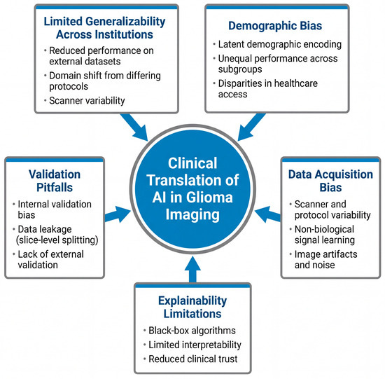
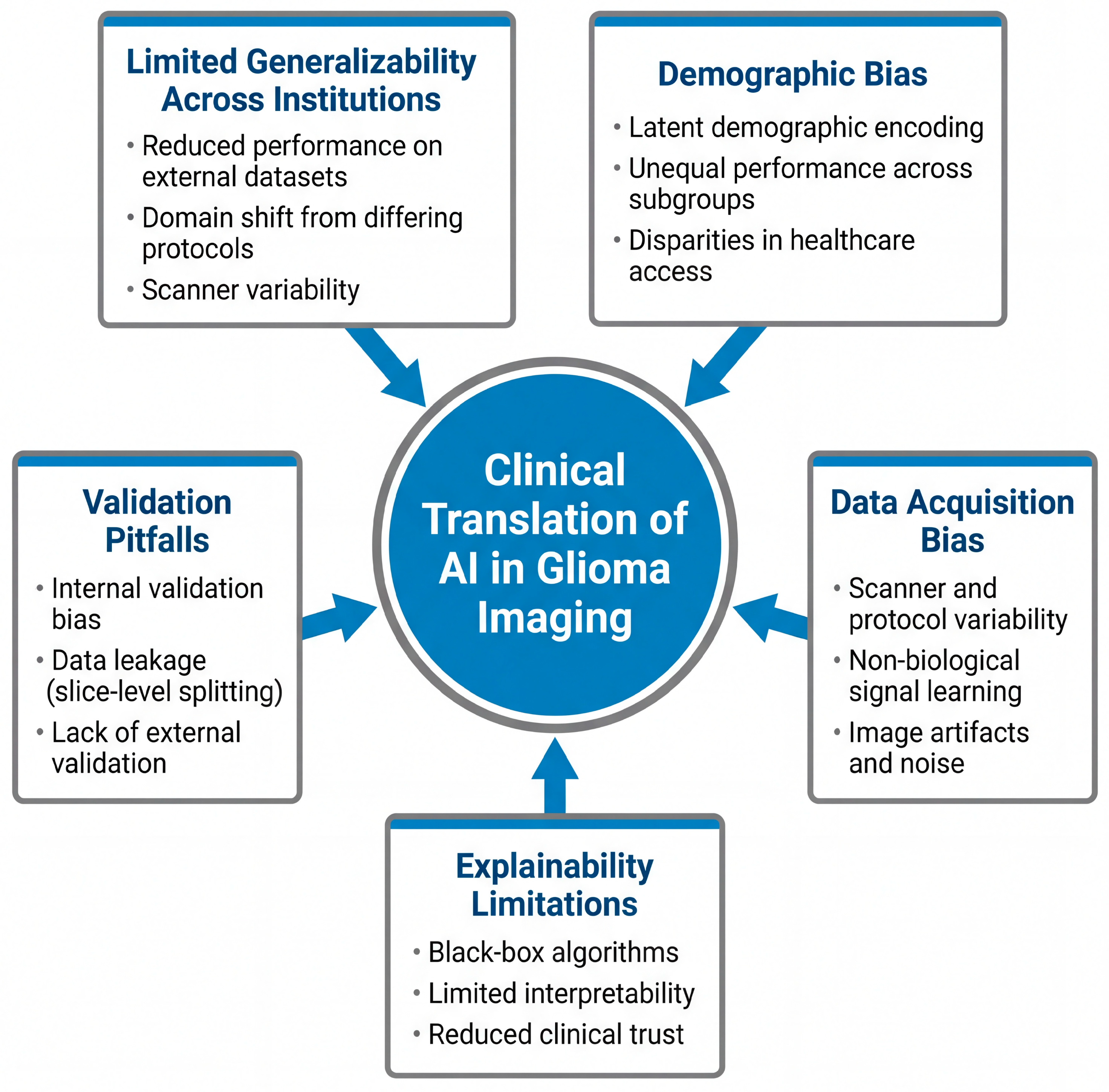
Despite strong performance reported in controlled research settings, several critical limitations hinder the reliable clinical translation of AI-based glioma imaging systems. These challenges span technical, methodological, and ethical domains, including issues related to generalizability, validation, data bias, and interpretability. Collectively, these factors contribute to discrepancies between experimental performance and real-world applicability. These major limitations are summarized in Figure 1.
Figure 1.
Key Limitations in the Clinical Translation of AI-Based Glioma Imaging. Key limitations affecting the clinical translation of artificial intelligence (AI)-based glioma imaging systems. Major barriers include limited generalizability across institutions due to domain shift and scanner variability; demographic bias arising from latent encoding of patient characteristics; data acquisition bias related to differences in imaging protocols and artifact burden; validation pitfalls such as data leakage and lack of external validation; and limited explainability of deep learning models. Collectively, these factors contribute to discrepancies between high performance in controlled research settings and reduced reliability in real-world clinical environments.
5.1. Limited Generalizability Across Institutions
Limited generalizability does not necessarily imply complete loss of model function, particularly for models evaluated and applied within a single institution. In a multi-dataset study, AlBadawy et al. reported that convolutional neural networks for brain tumor segmentation achieved Dice coefficients of 0.72 ± 0.17 and 0.76 ± 0.12 when trained and tested within the same institution, but showed statistically significant degradation (p < 0.0001) when applied cross-institutionally, with Dice coefficients decreasing to 0.68 ± 0.19 and 0.59 ± 0.19 [44].
Similar findings have been reported across neuro-oncologic imaging studies. Park et al. [22] demonstrated that model performance varies significantly across institutions due to differences in MRI acquisition protocols, while Buchlak et al. [33] identified limited external validation as a major barrier to clinical translation. Zech et al. [45] further showed that deep learning models can learn institution-specific features, resulting in reduced performance in new clinical environments.
These findings indicate that strong internal performance does not reliably translate to consistent performance across institutions. As a result, reported accuracy should be interpreted in the context of dataset origin and validation design, with clear distinction between institution-specific performance and true cross-institution generalizability.
To address this limitation, approaches such as domain adaptation, data harmonization, and multi-institutional training have been proposed to reduce sensitivity to inter-institutional variability [46,47,48]. However, these strategies remain inconsistently applied and are not yet standardized in clinically validated glioma imaging workflows.
5.2. Demographic Signal Encoding and Algorithmic Bias
Using multi-institutional imaging datasets, Gichoya et al. demonstrated that deep learning models trained on medical images can predict patient race at performance levels substantially above chance, even when race is not explicitly provided as an input variable. Across multiple imaging modalities, race prediction achieved AUC values exceeding 0.80 and, in some settings, approached 0.90–0.99, indicating the presence of strong latent demographic signals within imaging data [49].
The ability to predict demographic attributes from imaging data suggests that latent signals may influence model decision boundaries, potentially creating disparities in false-positive and false-negative rates across patient subgroups if not controlled [49]. While the example above highlights race as a demonstrative case, similar concerns extend to other demographic and structural variables, including age, sex, socioeconomic status, and healthcare access, which may also be implicitly encoded in imaging datasets through differences in disease prevalence, referral patterns, and imaging acquisition practices. These findings underscore the importance of evaluating AI models for potential bias across multiple demographic dimensions during development and validation.
5.3. Validation Pitfalls and Performance Inflation
A major methodological concern in glioma imaging AI is reliance on internal validation, where training and testing data originate from the same dataset. Although this approach often yields strong reported performance, it may not generalize to independent datasets. Differences in scanner hardware, acquisition protocols, and patient populations across institutions can introduce domain shifts that significantly affect model performance when applied to external cohorts. Robust multi-institutional validation is therefore essential before AI-based imaging systems can be reliably integrated into clinical practice [22,33].
The reported performance of machine learning models in glioma imaging is highly dependent on the validation strategy. A common pitfall is image-level data splitting, in which MRI slices from the same patient appear in both training and test sets. Because slices from a single patient are highly correlated, this practice allows models to learn patient-specific features, leading to inflated performance estimates that do not reflect true predictive performance on unseen patients [33].
In addition to this form of leakage, longitudinal datasets introduce further challenges. A related but distinct source of bias arises when multiple timepoints from the same patient are available. Temporal data leakage can occur if scans from different timepoints of a single patient are distributed across training and test sets, allowing models to exploit patient-specific or disease trajectory features rather than learning generalizable patterns. This issue is particularly relevant in glioma imaging, where serial MRI is routinely used for monitoring progression, and improper handling of longitudinal data has been recognized as a source of inflated model performance in neuroimaging studies [22,45,50].
Strong quantitative evidence for this effect is provided by AlBadawy et al., who demonstrated that brain tumor segmentation models with high internal performance experienced substantial absolute Dice score reductions of approximately 0.04–0.17 when evaluated using cross-institutional testing rather than slice-level splitting [44]. This finding indicates that a meaningful proportion of reported accuracy in some studies reflects data leakage rather than robust feature learning.
Similar patterns have been reported across broader glioma imaging AI literature. Buchlak et al. found that fewer than 15% of studies incorporated independent external validation, despite frequently reporting high internal performance [33]. Xu et al. and Bahar et al. further demonstrated substantial variability in reported model performance, with AUC values ranging from approximately 0.70 to 0.95 across studies, reflecting differences in validation strategy and dataset composition [36,42]. In addition, Park et al. emphasized that models developed using single-institution datasets often demonstrate reduced performance when applied to external cohorts due to domain shift [22]. Collectively, these findings, together with cross-institutional studies such as AlBadawy et al., suggest that meaningful performance degradation commonly occurs when transitioning from internal to external validation.
5.4. Data Acquisition Bias and Hidden Confounding
Another contributor to performance inflation and reduced model robustness in glioma imaging AI is data acquisition bias, in which image characteristics reflect institutional acquisition practices rather than intrinsic tumor biology [51]. In MRI-based datasets, scanner manufacturer, field strength, coil configuration, pulse sequence parameters, contrast timing, and reconstruction workflows may vary systematically across institutions. When such acquisition-related factors correlate with diagnostic labels, models may exploit these contextual cues instead of learning biologically meaningful tumor features. To address such variability, several harmonization approaches have been proposed, including statistical normalization methods such as ComBat and deep learning-based image translation techniques such as CycleGAN, which aim to reduce scanner-related differences and improve model robustness across multi-institutional imaging datasets [52,53].
Deep learning systems have demonstrated the ability to encode non-pathologic signals embedded within medical images, including demographic attributes that are not visually apparent to human observers, underscoring the capacity of models to capture latent contextual information [49]. Variations in intensity normalization, noise, spatial resolution, and imaging artifacts (e.g., motion degradation and susceptibility distortions) can alter feature distributions and reduce model robustness when applied to real-world clinical imaging, thereby inflating internal performance without improving true disease discrimination [49,51].
Quantitative evidence supports the impact of this bias. Using controlled experiments that isolated acquisition-related shortcut signals, Ly et al. demonstrated that conventional training and evaluation pipelines overestimated classification accuracy by approximately 20% when hidden shortcut features were present, with performance declining substantially under simulated external conditions [51]. These findings suggest that reported model performance may partly reflect acquisition context rather than tumor-specific information.
In addition to acquisition-related bias, variability in image quality and artifact burden represents an important source of model instability in MRI-based glioma AI. Clinical MRI data frequently include motion-related degradation, susceptibility artifacts, and variation in signal-to-noise ratio, all of which can alter intensity distributions and spatial features used for both radiomic analysis and deep learning. Prior work has shown that such non-biologic imaging characteristics can influence model behavior, with deep learning systems capable of encoding latent signals unrelated to underlying pathology [49,51].
These effects are particularly relevant in real-world settings, where imaging quality is less controlled than in curated research datasets. Radiomics pipelines are sensitive to noise and preprocessing variation, which can affect feature reproducibility, while deep learning models may rely on spurious correlations associated with image quality or artifact patterns. This contributes to reduced robustness during external validation and may partially explain discrepancies between internal and cross-institutional performance observed in prior studies [44,51].
Addressing this limitation requires training on heterogeneous datasets that reflect real-world imaging variability, along with preprocessing and augmentation strategies designed to improve robustness to noise and artifacts.
5.5. Explainability as a Clinical Limitation
Explainable artificial intelligence (XAI) techniques aim to address this limitation by providing insight into which image regions influence model predictions. Visualization-based approaches, including saliency mapping and class-discriminative localization, are commonly used explainability methods that generate heatmaps over the input image to highlight regions most influential to a model’s prediction, allowing assessment of whether algorithmic attention aligns with known tumor anatomy [54]. Gradient-based saliency methods such as vanilla gradients, guided backpropagation, integrated gradients, and SmoothGrad highlight influential input features but often emphasize edges or textures rather than anatomically meaningful regions [54,55,56,57]. In contrast, localization-based approaches such as Gradient-weighted Class Activation Mapping (Grad-CAM) produce spatial heatmaps that are generally more intuitive for clinical interpretation [54].
Despite their promise, no standardized framework exists for selecting, evaluating, or reporting explainability methods. Saliency outputs are sensitive to model architecture, preprocessing choices, and input perturbations, and visually plausible heatmaps do not necessarily reflect reliable or causal reasoning [54,55,56,57]. In a systematic evaluation of clinical AI research, Nagendran et al. found that fewer than 10% of studies used structured or quantitative approaches to interpretability assessment, with most relying solely on qualitative visualization [58]. Proposed tools such as Saliency Cards seek to improve transparency by providing structured documentation of how saliency methods are generated and interpreted, including model assumptions, implementation choices, and known sources of variability [59]. Without interpretable and auditable decision-making processes, AI systems risk remaining confined to experimental settings regardless of technical performance [60,61].
Beyond saliency-based visualization methods, additional interpretability approaches have been explored to improve the transparency of model decision-making. Concept-based explanation frameworks, such as Testing with Concept Activation Vectors (TCAV), aim to link network predictions to clinically meaningful imaging features by quantifying the influence of predefined semantic concepts on model outputs. In glioma imaging, such methods may allow models to associate predictions with recognizable tumor characteristics such as necrosis, edema, or contrast enhancement patterns [62]. Counterfactual explanation techniques represent another emerging strategy, identifying minimal changes in an input image that would alter the model’s prediction and thereby revealing the decision boundary used by the algorithm. Uncertainty quantification approaches, including Bayesian neural networks and Monte Carlo dropout, have been proposed to estimate prediction confidence [22,62]. This is particularly relevant in glioma imaging, where tumor heterogeneity, post-treatment effects, and variability in image quality introduce inherent uncertainty in both classification and segmentation tasks. In this context, uncertainty estimates may help distinguish high-confidence predictions from cases requiring closer clinical review. However, despite their potential clinical value, these methods remain infrequently implemented in glioma imaging studies and are not routinely reported alongside performance metrics, representing an important gap in current model evaluation and a barrier to clinical trust [22,62]. Table 1 provides a concise summary of the key limitations discussed, highlighting representative findings from some of the most frequently cited studies as supporting evidence.
Table 1.
Key limitations in clinical applications of machine learning for glioma imaging.
6. Clinical Implications and Knowledge Gaps
From a clinical perspective, artificial intelligence systems for MRI-based glioma detection should be evaluated not solely on benchmark accuracy, but on their ability to support real-world neuro-oncology decision-making. Potential applications include assisting with tumor detection, informing biopsy target selection, improving consistency in surgical and radiation planning, and supporting longitudinal assessment of treatment response, as reflected by the limitations discussed in Section 5.
The methodological challenges identified above have direct clinical consequences. Models that generalize poorly across institutions may be unreliable in multi-center or community practice settings, while segmentation inaccuracies can propagate into volumetric response assessment, surgical margin estimation, and radiation treatment planning, increasing the risk of misclassification of tumor progression versus treatment-related change. In addition, limited interpretability and poorly defined failure modes hinder clinician trust and accountability in routine clinical workflows.
Accordingly, current AI systems for glioma imaging should be viewed as decision-support tools rather than autonomous diagnostic solutions. Meaningful clinical adoption will require robust external validation, transparent reporting of limitations, and evidence that AI integration improves diagnostic confidence, workflow efficiency, or treatment decision consistency.
A major unresolved gap lies in the lack of prospective, workflow-integrated evaluations. Few studies assess how AI outputs influence clinician behavior, diagnostic confidence, or treatment decisions in real-world settings. Additionally, standardized frameworks for reporting failure modes, fairness auditing, and interpretability remain inconsistently applied. Several implementation frameworks have been proposed to evaluate AI systems within routine clinical environments. For example, prospective reader studies may compare radiologist performance with and without AI assistance to assess changes in diagnostic accuracy, reading efficiency, and interobserver agreement. Pragmatic clinical trials or prospective observational deployments within radiology workflows may further evaluate how AI-supported interpretation influences treatment planning or diagnostic confidence in neuro-oncology practice. Such workflow-centered validation approaches have been highlighted as critical for determining whether AI systems provide measurable benefit beyond retrospective performance metrics [22,58,60].
In addition, several reporting and evaluation frameworks have been proposed to improve the rigor and transparency of clinical artificial intelligence studies. TRIPOD-AI extends traditional prediction model reporting guidelines to machine learning-based systems, while CLAIM provides a standardized checklist for reporting artificial intelligence studies in medical imaging. More recently, the FUTURE-AI framework has emphasized key principles for trustworthy clinical AI, including fairness, robustness, explainability, and reproducibility, highlighting the need for structured evaluation of model performance across diverse clinical settings [63,64,65].
7. Future Directions
7.1. Emerging Modeling Strategies for Glioma Imaging AI
Future directions in AI-based glioma detection are increasingly focused on improving clinical applicability, rather than further optimizing model architecture alone. Recent reviews emphasize that models trained on isolated MRI timepoints provide limited insight into real-world disease dynamics, underscoring the need for approaches that better reflect routine clinical practice [8].
In this context, growing attention has shifted toward multimodal and longitudinal modeling approaches that integrate conventional MRI with advanced imaging sequences, molecular markers, histopathology, and clinical data [25,66]. These strategies aim to characterize how gliomas evolve over time rather than relying on static imaging features, with potential to improve earlier and more reliable detection of tumor recurrence.
These approaches require architectures capable of capturing temporal dependencies across sequential imaging timepoints. Recurrent neural networks (RNNs), including long short-term memory (LSTM) and gated recurrent unit (GRU) models, have been explored to model temporal disease progression using serial MRI scans. More recently, transformer-based architectures have gained attention due to their ability to model long-range dependencies without the limitations of sequential processing inherent to RNNs. In neuro-oncology imaging, early studies integrating longitudinal MRI data into deep learning frameworks have demonstrated improved performance in tumor progression prediction and survival modeling compared to single timepoint approaches although these methods remain limited by data availability and heterogeneity [8,10]. Despite these advances, longitudinal modeling in glioma imaging remains underdeveloped, with few studies incorporating fully temporal architectures in clinically validated settings.
To address data-sharing constraints, federated learning and other privacy-preserving frameworks have emerged as strategies for multi-institutional model development without centralized data pooling. In neuro-oncology imaging, early work by Sheller et al. demonstrated that federated learning for brain tumor segmentation achieved Dice similarity coefficients of approximately 0.85–0.88 across tumor subregions, comparable to centrally trained models while maintaining data locality [48]. Similarly, Li et al. reported that federated deep learning models can maintain performance within approximately 1–3% of centrally trained approaches under controlled multi-institutional settings [67].
Beyond these approaches, several emerging directions may influence the next generation of glioma imaging AI. Large-scale pre-trained foundation models, including vision and multimodal architectures, have shown strong performance across medical imaging tasks through transfer learning, particularly when labeled datasets are limited, although their application in neuro-oncology remains in early stages [68,69]. In parallel, integration of imaging with genomic and molecular data often referred to as radiogenomics has demonstrated potential for predicting key molecular alterations such as IDH mutation status and 1p/19q co-deletion from MRI features, supporting the biological relevance of multi-modal approaches [28,29].
Additionally, digital twin and simulation-based frameworks have been proposed to model tumor growth and treatment response using patient-specific data, although current implementations remain largely experimental and limited to small-scale studies [70]. Finally, large language models (LLMs) have recently been explored for clinical text synthesis and multimodal reasoning, with early evidence suggesting potential utility in integrating radiology reports and clinical documentation, though their application in neuro-oncology workflows remains preliminary [71]. Collectively, these approaches represent promising but still developing directions that require further validation in clinically representative, multi-institutional settings.
7.2. Practical Challenges in Multi-Institutional Implementation and Clinical Translation
Among these approaches, longitudinal modeling and federated learning represent the most clinically actionable and immediately translatable strategies due to their alignment with existing clinical workflows and data availability. In contrast, emerging paradigms such as foundation models, digital twin systems, and large language model integration remain exploratory and face greater challenges related to data requirements, validation, and regulatory implementation [8,10].
Despite these promising developments, several practical challenges limit real-world implementation. A major limitation is data heterogeneity, as imaging datasets across institutions are typically non-independent and non-identically distributed (non-IID). This has been shown to impair convergence and reduce model performance compared to idealized training conditions, particularly when institutional datasets differ substantially in acquisition protocols or patient populations [72].
In addition, federated learning introduces non-trivial communication overhead, as repeated transmission of model parameters is required across training rounds. In large deep learning models, communication costs can represent a substantial proportion of total training time, particularly in bandwidth-constrained environments [72].
Further challenges include imbalance in dataset size across participating institutions, which can bias model updates, as well as partial client participation and system-level instability during training. Moreover, although federated learning reduces the need for direct data sharing, it does not fully eliminate privacy risks, as model updates may still encode sensitive information. Techniques such as differential privacy and secure aggregation can mitigate this risk but may introduce additional computational burden and potential reductions in model performance [72].
Collectively, these findings indicate that while federated learning represents a promising direction for improving generalizability in glioma imaging AI, its implementation remains constrained by statistical heterogeneity, communication costs, and privacy–performance trade-offs.
Parallel efforts to improve standardization and transparency are also expected to shape future progress. Continued development of AI-specific reporting and validation guidelines may support reproducibility, regulatory evaluation, and large-scale evidence synthesis across studies [8,10].
Finally, meaningful clinical translation will require a shift beyond retrospective accuracy metrics derived from curated datasets toward prospective, workflow-integrated validation. Future studies should evaluate how AI systems influence diagnostic confidence, efficiency, and clinical decision-making in routine practice, rather than performance metrics alone [8,42].
8. Conclusions
AI-based methods for MRI glioma detection, segmentation, and classification have shown strong technical performance in research settings. Techniques ranging from radiomics-based machine learning to deep learning have enabled automated extraction of imaging features that may complement expert interpretation [31,34].
Despite these advances, most existing models are not yet ready for routine clinical use. Common limitations include dataset bias, MRI protocol heterogeneity, overfitting, limited external validation, and inconsistent reporting, all of which reduce robustness and generalizability [10,33,41]. Importantly, high internal accuracy does not consistently translate to reliable performance across institutions or patient populations, limiting clinical trust [8].
Currently, AI should be considered a decision-support tool rather than a replacement for clinical judgment. Meaningful clinical adoption will require standardized reporting, multi-center and prospective validation, improved interpretability, and clear evidence of benefit in real-world workflows. Among these priorities, prospective multi-center validation represents the most critical and immediate next step, as it directly addresses the generalizability gap that underlies most other limitations identified in this review. Without rigorous evidence that AI models perform reliably across diverse institutions and patient populations, progress in reporting standards, interpretability, and workflow integration will remain difficult to translate into clinical practice.
Author Contributions
Conceptualization, A.S., M.P., N.Y. and M.K.; validation, N.Y., M.P. and M.K.; investigation, M.K.; resources, A.S., I.Z. and C.S.; writing—original draft preparation, A.S., I.Z., C.S., E.D. and V.N.; writing—review and editing, A.S., I.Z., C.S., E.D., M.P. and M.K.; visualization, A.S.; supervision, M.K., N.Y. and M.P.; project administration, A.S. All authors have read and agreed to the published version of the manuscript.
Funding
This research received no external funding.
Informed Consent Statement
Not applicable.
Data Availability Statement
No new data were created or analyzed in this study. Data sharing is not applicable to this article.
Conflicts of Interest
The authors declare no conflicts of interest.
Abbreviations
The following abbreviations are used in this manuscript:
| AI | Artificial Intelligence |
| ML | Machine Learning |
| DL | Deep Learning |
| CNN | Convolutional Neural Network |
| MRI | Magnetic Resonance Imaging |
| DWI | Diffusion-Weighted Imaging |
| ADC | Apparent Diffusion Coefficient |
| DTI | Diffusion Tensor Imaging |
| DSC | Dynamic Susceptibility Contrast |
| rCBV | Relative Cerebral Blood Volume |
| SWI | Susceptibility-Weighted Imaging |
| GRE | Gradient Echo |
| WHO | World Health Organization |
| IDH | Isocitrate Dehydrogenase |
| AUC | Area Under the Curve |
| ROC | Receiver Operating Characteristic |
| XAI | Explainable Artificial Intelligence |
| Grad-CAM | Gradient-weighted Class Activation Mapping |
| BraTS | Brain Tumor Segmentation Challenge |
References
- Weller, M.; van den Bent, M.; Tonn, J.C.; Stupp, R.; Preusser, M.; Cohen-Jonathan-Moyal, E.; Henriksson, R.; Le Rhun, E.; Balana, C.; Chinot, O.; et al. European Association for Neuro-Oncology (EANO) guideline on the diagnosis and treatment of adult astrocytic and oligodendroglial gliomas. Lancet Oncol. 2017, 18, e315–e329. [Google Scholar] [CrossRef] [Scilit]
- Horbinski, C.; Solomon, D.A.; Lukas, R.V.; Reuss, D.E.; Sahm, F.; Capper, D.; Jones, D.T.W.; Pfister, S.M.; Aldape, K.D.; Louis, D.N.; et al. Molecular testing for the World Health Organization classification of central nervous system tumors: A review. JAMA Oncol. 2025, 11, 317–328. [Google Scholar] [CrossRef] [Scilit] [PubMed]
- National Comprehensive Cancer Network. Central Nervous System Cancers (Version 1.2024). Available online: https://www.nccn.org/professionals/physician_gls/pdf/cns.pdf (accessed on 11 January 2026).
- Bonm, A.V.; Ritterbusch, R.; Throckmorton, P.; Graber, J.J. Clinical imaging for diagnostic challenges in the management of gliomas: A review. J. Neuroimaging 2020, 30, 139–145. [Google Scholar] [CrossRef] [Scilit] [PubMed]
- Herholz, K.; Coope, D.; Jackson, A. Metabolic and molecular imaging in neuro-oncology. Lancet Neurol. 2007, 6, 711–724. [Google Scholar] [CrossRef] [Scilit] [PubMed]
- Ivanidze, J.; Shih, R.Y.; Utukuri, P.S.; Patel, N.D.; Burns, J.; Corey, A.S.; Policeni, B.A.; Rosenow, J.M.; Reddy, K.; Expert Panel on Neurologic Imaging; et al. ACR Appropriateness Criteria® brain tumors. J. Am. Coll. Radiol. 2025, 22, S108–S135. [Google Scholar] [CrossRef] [Scilit]
- Rudie, J.D.; Rauschecker, A.M.; Bryan, R.N.; Davatzikos, C.; Mohan, S. Emerging applications of artificial intelligence in neuro-oncology. Radiology 2019, 290, 607–618. [Google Scholar] [CrossRef] [Scilit]
- Khalighi, S.; Reddy, K.; Midya, A.; Sharma, A.; Gupta, R.; Shukla, G.; Singh, A.; Kumar, S.; Banerjee, I.; Bhattacharya, S.; et al. Artificial intelligence in neuro-oncology: Advances and challenges in brain tumor diagnosis, prognosis, and precision treatment. NPJ Precis Oncol. 2024, 8, 80. [Google Scholar] [CrossRef] [Scilit]
- Menze, B.H.; Jakab, A.; Bauer, S.; Kalpathy-Cramer, J.; Farahani, K.; Kirby, J.; Burren, Y.; Porz, N.; Slotboom, J.; Wiest, R.; et al. The Multimodal Brain Tumor Image Segmentation Benchmark (BRATS). IEEE Trans. Med. Imaging 2015, 34, 1993–2024. [Google Scholar] [CrossRef] [Scilit]
- Luo, J.; Pan, M.; Mo, K.; Mao, Y.; Zou, D. Emerging role of artificial intelligence in diagnosis, classification and clinical management of glioma. Semin. Cancer Biol. 2023, 91, 110–123. [Google Scholar] [CrossRef] [Scilit]
- Baethge, C.; Golber, S.; Hegerl, U. SANRA—A scale for the quality assessment of narrative review articles. Res. Integr. Peer Rev. 2019, 4, 5. [Google Scholar] [CrossRef] [Scilit]
- Isensee, F.; Jaeger, P.F.; Kohl, S.A.A.; Petersen, J.; Maier-Hein, K.H. nnU-Net: A self-configuring method for deep learning-based biomedical image segmentation. Nat. Methods 2021, 18, 203–211. [Google Scholar] [CrossRef] [Scilit] [PubMed]
- Zhuge, Y.; Ning, H.; Mathen, P.; Cheng, J.Y.; Krauze, A.V.; Camphausen, K.; Miller, R.W.; Shao, W.; Wang, Y.; Liu, T.; et al. Automated glioma grading on conventional MRI images using deep convolutional neural networks. Med. Phys. 2020, 47, 3044–3053. [Google Scholar] [CrossRef] [Scilit] [PubMed]
- Chen, T.; Hu, L.; Lu, Q.; Wu, F.; Wang, Y.; Zhang, J.; Li, X.; Zhao, Z.; Liu, H.; Sun, C.; et al. A computer-aided diagnosis system for brain tumors based on artificial intelligence algorithms. Front. Neurosci. 2023, 17, 1120781. [Google Scholar] [CrossRef] [Scilit] [PubMed]
- Bhuiyan, E.H.; Khan, M.M.; Hossain, S.A.; Rahman, M.A.; Islam, M.T.; Uddin, M.S.; Alam, M.A.; Hossain, M.S.; Hasan, M.M.; Karim, M.R.; et al. Classification of glioma grade and Ki-67 level prediction in MRI data: A SHAP-driven interpretation. Comput. Med. Imaging Graph 2025, 124, 102578. [Google Scholar] [CrossRef] [Scilit]
- Blakstad, H.; Mendoza Mireles, E.E.; Heggebø, L.C.; Solheim, O.; Jakola, A.S.; Berntsen, E.M.; Salvesen, Ø.; Unsgård, G.; Torp, S.H.; Rygh, O.M.; et al. Incidence and outcome of pseudoprogression after radiation therapy in glioblastoma patients: A cohort study. Neuro Oncol. Pract. 2024, 11, 36–45. [Google Scholar] [CrossRef] [Scilit]
- Ahammed Muneer, K.V.; Rajendran, V.R.; Paul Joseph, K. Glioma Tumor Grade Identification Using Artificial Intelligent Techniques. J. Med. Syst. 2019, 43, 113. [Google Scholar] [CrossRef] [Scilit]
- Patel, M.; Zhan, J.; Natarajan, K.; Gupta, A.; Singh, R.; Kumar, S.; Sharma, P.; Mehta, R.; Das, S.; Verma, N.; et al. Machine learning-based radiomic evaluation of treatment response prediction in glioblastoma. Clin. Radiol. 2021, 76, 628.e17–628.e27. [Google Scholar] [CrossRef] [Scilit]
- Yadav, V.K.; Mohan, S.; Agarwal, S.; Gupta, R.K.; Kumar, R.; Saini, J.; Jaiswal, A.K.; Verma, R.K.; Singh, A.; Srivastava, C.; et al. Distinction of pseudoprogression from true progression in glioblastomas using machine learning based on multiparametric magnetic resonance imaging and O6-methylguanine-methyltransferase promoter methylation status. Neurooncol. Adv. 2024, 6, vdae159. [Google Scholar] [CrossRef] [Scilit]
- Jang, B.S.; Jeon, S.H.; Kim, I.H.; Kim, I.A. Machine learning model to predict pseudoprogression versus progression in glioblastoma using MRI: A multi-institutional study (KROG 18-07). Cancers 2020, 12, 2706. [Google Scholar] [CrossRef] [Scilit]
- Palavani, L.B.; Nogueira, B.V.; Mitre, L.P.; Silva, A.C.; Teixeira, B.C.; Santos, F.A.; Oliveira, R.M.; Souza, J.R.; Costa, R.F.; Pereira, M.L.; et al. Artificial intelligence algorithms for differentiating pseudoprogression from true progression in high-grade gliomas: A systematic review and meta-analysis. Neurosurg. Rev. 2025, 48, 591. [Google Scholar] [CrossRef] [Scilit]
- Park, J.E.; Kickingereder, P.; Kim, H.S. Radiomics and Deep Learning from Research to Clinical Workflow: Neuro-Oncologic Imaging. Korean J. Radiol. 2020, 21, 1126–1137. [Google Scholar] [CrossRef] [Scilit] [PubMed]
- Decuyper, M.; Bonte, S.; Deblaere, K.; Van Holen, R. Automated MRI based pipeline for segmentation and prediction of grade, IDH mutation and 1p19q co-deletion in glioma. Comput. Med. Imaging Graph. 2021, 88, 101831. [Google Scholar] [CrossRef] [Scilit] [PubMed]
- Lohmann, P.; Galldiks, N.; Kocher, M.; Heinzel, A.; Filss, C.P.; Stegmayr, C.; Mottaghy, F.M.; Fink, G.R.; Shah, N.J.; Langen, K.J.; et al. Radiomics in neuro-oncology: Basics, workflow, and applications. Methods 2021, 188, 112–121. [Google Scholar] [CrossRef] [Scilit]
- Fan, H.; Luo, Y.; Gu, F.; Zhang, Y.; Li, X.; Wang, J.; Liu, Z.; Chen, W.; Zhao, Q.; Sun, H.; et al. Artificial intelligence-based MRI radiomics and radiogenomics in glioma. Cancer Imaging 2024, 24, 36. [Google Scholar] [CrossRef] [Scilit] [PubMed]
- Alongi, P.; Arnone, A.; Vultaggio, V.; Laudicella, R.; Albano, D.; Bruno, A.; Palumbo, B.; Midiri, F.; Stefano, A.; Toia, P.; et al. Artificial intelligence analysis using MRI and PET imaging in gliomas: A narrative review. Cancers 2024, 16, 407. [Google Scholar] [CrossRef] [Scilit]
- Louis, D.N.; Perry, A.; Wesseling, P.; Brat, D.J.; Cree, I.A.; Figarella-Branger, D.; Hawkins, C.; Ng, H.K.; Pfister, S.M.; Reifenberger, G.; et al. The 2021 WHO classification of tumors of the central nervous system: A summary. Neuro Oncol. 2021, 23, 1231–1251. [Google Scholar] [CrossRef] [Scilit]
- Chang, K.; Bai, H.X.; Zhou, H.; Su, C.; Bi, W.L.; Agbodza, E.; Kavouridis, V.K.; Senders, J.T.; Boaro, A.; Beers, A.; et al. Residual convolutional neural network for the determination of IDH status in low- and high-grade gliomas from MR imaging. Clin. Cancer Res. 2018, 24, 1073–1081. [Google Scholar] [CrossRef] [Scilit]
- Kickingereder, P.; Bonekamp, D.; Nowosielski, M.; Kratz, A.; Sill, M.; Burth, S.; Wick, A.; Eidel, O.; Schlemmer, H.P.; Radbruch, A.; et al. Radiogenomics of glioblastoma: Machine learning-based classification of molecular characteristics by using multiparametric and multiregional MR imaging features. Radiology 2016, 281, 907–918. [Google Scholar] [CrossRef] [Scilit]
- Bakas, S.; Reyes, M.; Jakab, A.; Bauer, S.; Rempfler, M.; Crimi, A.; Shinohara, R.T.; Berger, C.; Ha, S.M.; Rozycki, M.; et al. Identifying the best machine learning algorithms for brain tumor segmentation, progression assessment, and overall survival prediction in the BraTS challenge. arXiv 2019, arXiv:1811.02629. [Google Scholar]
- Gao, M.; Huang, S.; Pan, X.; Liao, X.; Yang, R.; Liu, J. Machine Learning-Based Radiomics Predicting Tumor Grades and Expression of Multiple Pathologic Biomarkers in Gliomas. Front. Oncol. 2020, 10, 1676. [Google Scholar] [CrossRef] [Scilit]
- Zacharaki, E.I.; Wang, S.; Chawla, S.; Yoo, D.S.; Wolf, R.; Melhem, E.R.; Davatzikos, C. Classification of brain tumor type and grade using MRI texture and shape in a machine learning scheme. Magn Reson Med. 2009, 62, 1609–1618. [Google Scholar] [CrossRef] [Scilit] [PubMed]
- Buchlak, Q.D.; Esmaili, N.; Leveque, J.C.; Bennett, C.; Farrokhi, F.; Piccardi, M. Machine learning applications to neuroimaging for glioma detection and classification: An artificial intelligence augmented systematic review. J. Clin. Neurosci. 2021, 89, 177–198. [Google Scholar] [CrossRef] [Scilit] [PubMed]
- Gore, S.; Chougule, T.; Jagtap, J.; Saini, J.; Ingalhalikar, M. A Review of Radiomics and Deep Predictive Modeling in Glioma Characterization. Acad. Radiol. 2021, 28, 1599–1621. [Google Scholar] [CrossRef] [Scilit] [PubMed]
- Jin, W.; Fatehi, M.; Abhishek, K.; Mallya, M.; Toyota, B.; Hamarneh, G. Artificial intelligence in glioma imaging: Challenges and advances. J. Neural Eng. 2020, 17, 021002. [Google Scholar] [CrossRef] [Scilit]
- Bahar, R.C.; Merkaj, S.; Cassinelli Petersen, G.I.; Smith, J.; Brown, T.; Lee, H.; Nguyen, P.; Patel, A.; Johnson, M.; Williams, K.; et al. Machine learning models for classifying high- and low-grade gliomas: A systematic review and quality of reporting analysis. Front. Oncol. 2022, 12, 856231. [Google Scholar] [CrossRef] [Scilit]
- Subramanian, H.; Dey, R.; Brim, W.R.; Gupta, S.; Kumar, R.; Shah, V.; Patel, D.; Singh, P.; Mehta, A.; Rao, N.; et al. Trends in development of novel machine learning methods for the identification of gliomas in datasets that include non-glioma images: A systematic review. Front. Oncol. 2021, 11, 788819. [Google Scholar] [CrossRef] [Scilit]
- Tian, Q.; Yan, L.F.; Zhang, X.; Hu, Y.C.; Han, Y.; Liu, Z.C.; Nan, H.Y.; Sun, Q.; Wang, W.; Yang, Y.; et al. Radiomics strategy for glioma grading using texture features from multiparametric MRI. J. Magn. Reson. Imaging 2018, 48, 1518–1528. [Google Scholar] [CrossRef] [Scilit]
- Naser, M.A.; Deen, M.J. Brain tumor segmentation and grading of lower-grade glioma using deep learning in MRI images. Comput. Biol. Med. 2020, 121, 103758. [Google Scholar] [CrossRef] [Scilit]
- Wong, Y.; Su, E.L.M.; Yeong, C.F.; Holderbaum, W.; Yang, C. Brain tumor classification using MRI images and deep learning techniques. PLoS ONE 2025, 20, e0322624. [Google Scholar] [CrossRef] [Scilit]
- Missaoui, R.; Hechkel, W.; Saadaoui, W.; Helali, A.; Leo, M. Advanced deep learning and machine learning techniques for MRI brain tumor analysis: A review. Sensors 2025, 25, 2746. [Google Scholar] [CrossRef] [Scilit]
- Xu, J.; Meng, Y.; Qiu, K.; Zhang, Y.; Liu, X.; Wang, J.; Li, H.; Chen, F.; Zhao, Z.; Sun, L.; et al. Applications of artificial intelligence based on medical imaging in glioma: Current state and future challenges. Front. Oncol. 2022, 12, 892056. [Google Scholar] [CrossRef] [Scilit] [PubMed]
- Khalil, Y.A.; Ayaz, A.; Lorenz, C.; Weese, J.; Pluim, J.; Breeuwer, M. Multi-modal brain tumor segmentation via conditional synthesis with Fourier domain adaptation. Comput. Med. Imaging Graph 2024, 112, 102332. [Google Scholar] [CrossRef] [Scilit] [PubMed]
- AlBadawy, E.A.; Saha, A.; Mazurowski, M.A. Deep learning for segmentation of brain tumors: Impact of cross-institutional training and testing. Med. Phys. 2018, 45, 1150–1158. [Google Scholar] [CrossRef] [Scilit] [PubMed]
- Zech, J.R.; Badgeley, M.A.; Liu, M.; Costa, A.B.; Titano, J.J.; Oermann, E.K. Variable generalization performance of a deep learning model to detect pneumonia in chest radiographs: A cross-sectional study. PLoS Med. 2018, 15, e1002683. [Google Scholar] [CrossRef] [Scilit]
- Dinsdale, N.K.; Jenkinson, M.; Namburete, A.I.L. Deep learning with domain randomization for optimal suppression of scanner effects in brain MRI data. NeuroImage 2021, 224, 117400. [Google Scholar] [CrossRef] [Scilit]
- Glocker, B.; Robinson, R.; Castro, D.C.; Dou, Q.; Konukoglu, E. Machine learning with multi-site imaging data: An empirical study on the impact of scanner effects. arXiv 2019, arXiv:1910.04597. [Google Scholar] [CrossRef] [Scilit]
- Sheller, M.J.; Edwards, B.; Reina, G.A.; Martin, J.; Pati, S.; Kotrotsou, A.; Bakas, S.; Milchenko, M.; Xu, W.; Marcus, D.; et al. Federated learning in medicine: Facilitating multi-institutional collaborations without sharing patient data. Sci. Rep. 2020, 10, 12598. [Google Scholar] [CrossRef] [Scilit]
- Gichoya, J.W.; McCoy, L.G.; Celi, L.A.; Oakden-Rayner, L.; Yeh, R.W.; Ross, C.; Ranschaert, E.; Jaremko, J.L.; Kim, J.; Kim, S.; et al. AI recognition of patient race in medical imaging: A modelling study. Lancet Digit Health 2022, 4, e406–e414. [Google Scholar] [CrossRef] [Scilit]
- Wen, J.; Thibeau-Sutre, E.; Diaz-Melo, M.; Samper-González, J.; Routier, A.; Bottani, S.; Dormont, D.; Durrleman, S.; Burgos, N.; Colliot, O.; et al. Convolutional neural networks for classification of Alzheimer’s disease: Overview and reproducible evaluation. Med. Image Anal. 2020, 63, 101694. [Google Scholar] [CrossRef] [Scilit]
- Ly, C.O.; Unnikrishnan, B.; Tadic, T.; Lee, S.; Kim, H.; Park, J.; Choi, Y.; Nguyen, T.; Tran, L.; Pham, D.; et al. Shortcut learning in medical AI hinders generalization: Method for estimating AI model generalization without external data. npj Digit Med. 2024, 7, 124. [Google Scholar] [CrossRef] [Scilit]
- Fortin, J.P.; Cullen, N.; Sheline, Y.I.; Taylor, W.D.; Aselcioglu, I.; Cook, P.A.; Adams, P.; Cooper, C.; Fava, M.; McGrath, P.J.; et al. Harmonization of cortical thickness measurements across scanners and sites. Neuroimage 2018, 167, 104–120. [Google Scholar] [CrossRef] [Scilit] [PubMed]
- Zhu, J.Y.; Park, T.; Isola, P.; Efros, A.A. Unpaired image-to-image translation using cycle-consistent adversarial networks. In Proceedings of the IEEE International Conference on Computer Vision, Venice, Italy, 22–29 October 2017; pp. 2223–2232. [Google Scholar] [CrossRef] [Scilit]
- Selvaraju, R.R.; Cogswell, M.; Das, A.; Vedantam, R.; Parikh, D.; Batra, D. Grad-CAM: Visual explanations from deep networks via gradient-based localization. In Proceedings of the IEEE International Conference on Computer Vision, Venice, Italy, 22–29 October 2017; pp. 618–626. [Google Scholar] [CrossRef] [Scilit]
- Simonyan, K.; Vedaldi, A.; Zisserman, A. Deep inside convolutional networks: Visualising image classification models and saliency maps. arXiv 2014, arXiv:1312.6034. [Google Scholar] [CrossRef] [Scilit]
- Zeiler, M.D.; Fergus, R. Visualizing and understanding convolutional networks. In Computer Vision—ECCV 2014; Lecture Notes in Computer Science; Springer: Cham, Switzerland, 2014; Volume 8689, pp. 818–833. [Google Scholar] [CrossRef] [Scilit]
- Sundararajan, M.; Taly, A.; Yan, Q. Axiomatic attribution for deep networks. In Proceedings of the 34th International Conference on Machine Learning, Sydney, Australia, 6–11 August 2017; Volume 70, pp. 3319–3328. [Google Scholar]
- Nagendran, M.; Chen, Y.; Lovejoy, C.A.; Gordon, A.C.; Komorowski, M.; Harvey, H.; Topol, E.J.; Ioannidis, J.P.A.; Collins, G.S.; Maruthappu, M. Artificial intelligence versus clinicians: Systematic review of design, reporting standards, and claims of deep learning studies in medical imaging. BMJ 2020, 368, m689. [Google Scholar] [CrossRef] [Scilit] [PubMed]
- Boggust, A.W.; Carter, S.; Satyanarayan, A. Saliency cards: A framework for documenting saliency methods. arXiv 2022, arXiv:2206.02702. [Google Scholar]
- Ho, C.W.L.; Soon, D.; Caals, K.; Kapur, J. Governance of automated image analysis and artificial intelligence analytics in healthcare. Clin. Radiol. 2019, 74, 329–337. [Google Scholar] [CrossRef] [Scilit]
- Stead, W.W. Clinical implications and challenges of artificial intelligence and deep learning. JAMA 2019, 320, 1107–1108. [Google Scholar] [CrossRef] [Scilit]
- Ayaz, H.; Khosravi, H.; McLoughlin, I.; Tormey, D.; Özsunar, Y.; Unnikrishnan, S. A random graph-based neural network approach to assess glioblastoma progression from perfusion MRI. Biomed. Signal Process. Control. 2023, 86, 105286. [Google Scholar] [CrossRef] [Scilit]
- Collins, G.S.; Dhiman, P.; Andaur Navarro, C.L.; Ma, J.; Hooft, L.; Reitsma, J.B.; Logullo, P.; Beam, A.L.; Peng, L.; Van Calster, B.; et al. Protocol for development of a reporting guideline (TRIPOD-AI) and risk of bias tool (PROBAST-AI). BMJ Open 2021, 11, e048008. [Google Scholar] [CrossRef] [Scilit]
- Mongan, J.; Moy, L.; Kahn, C.E., Jr. Checklist for Artificial Intelligence in Medical Imaging (CLAIM). Radiol. Artif. Intell. 2020, 2, e200029. [Google Scholar] [CrossRef] [Scilit]
- Lekadir, K.; Osuala, R.; Gallin, C.; Lazari, A.; Zanca, D.; Munoz-Gonzalez, E.; Balaguer-Ballester, E.; Zubizarreta, I.; Sanz, F.; Garcia, J.; et al. FUTURE-AI: International consensus guideline for trustworthy and deployable artificial intelligence in healthcare. arXiv 2021, arXiv:2109.12325. [Google Scholar]
- Jensen, M.A.; Ferretti, V.; Grossman, R.L.; Staudt, L.M. The NCI Genomic Data Commons as an engine for precision medicine. Blood 2017, 130, 453–459. [Google Scholar] [CrossRef] [Scilit]
- Li, W.; Milletari, F.; Xu, D.; Rieke, N.; Hancox, J.; Zhu, W.; Baust, M.; Cheng, Y.; Ourselin, S.; Cardoso, M.J.; et al. Privacy-preserving federated brain tumour segmentation. In MLMI 2019; Lecture Notes in Computer Science; Springer: Cham, Switzerland, 2019; Volume 11861, pp. 133–141. [Google Scholar]
- Zhou, Y.; Chia, M.A.; Wagner, S.K.; Ayhan, M.S.; Williamson, D.J.; Struyven, R.R.; Liu, X.; Sim, D.A.; Patel, P.J.; Teo, J.T.H.; et al. A foundation model for generalizable disease detection from retinal images. Nature 2023, 622, 156–163. [Google Scholar] [CrossRef] [Scilit]
- Azizi, S.; Mustafa, B.; Ryan, F.; Beaver, Z.; Freyberg, J.; Deaton, J.; Loh, A.; Caine, B.; Corrado, G.S.; Gardner, J.; et al. Big self-supervised models advance medical image classification. In Proceedings of the IEEE/CVF International Conference on Computer Vision, Montreal, BC, Canada, 11–17 October 2021; pp. 3478–3488. [Google Scholar]
- Viceconti, M.; Pappalardo, F.; Rodriguez, B.; Horner, M.; Bischoff, J.; Musuamba Tshinanu, F.; Walczak, K.; Alimohammadi, M.; Gray, R.A.; Viceconti, A.; et al. In silico trials: Verification, validation and uncertainty quantification of predictive models. Methods 2021, 185, 120–127. [Google Scholar] [CrossRef] [Scilit]
- Singhal, K.; Azizi, S.; Tu, T.; Mahdavi, S.S.; Wei, J.; Chung, H.W.; Scales, N.; Tanwani, A.K.; Cole-Lewis, H.; Pfohl, S.; et al. Large language models encode clinical knowledge. Nature 2023, 620, 172–180. [Google Scholar] [CrossRef] [Scilit]
- Kairouz, P.; McMahan, H.B.; Avent, B.; Bellet, A.; Bennis, M.; Bhagoji, A.N.; Bonawitz, K.; Charles, Z.; Cormode, G.; Cummings, R.; et al. Advances and open problems in federated learning. Found. Trends Mach. Learn. 2021, 14, 1–210. [Google Scholar] [CrossRef] [Scilit]

Disclaimer/Publisher’s Note: The statements, opinions and data contained in all publications are solely those of the individual author(s) and contributor(s) and not of MDPI and/or the editor(s). MDPI and/or the editor(s) disclaim responsibility for any injury to people or property resulting from any ideas, methods, instructions or products referred to in the content. |
© 2026 by the authors. Licensee MDPI, Basel, Switzerland. This article is an open access article distributed under the terms and conditions of the Creative Commons Attribution (CC BY) license.