


Abstract
The zinc oxide nanoparticles (ZnO NP) were synthesized from zinc acetate using potato starch as a capping agent through alkaline sol–gel precipitation, without calcination. The produced nanoparticles were characterized using various analytical techniques, including UV–visible absorption spectroscopy (UV-vis), zeta potential analysis, dynamic light scattering (DLS), X-ray diffraction (XRD), Fourier transform infrared spectroscopy (FTIR) and transmission electron microscopy (TEM). The ZnO NP exhibited an absorption maximum at 361 nm in the UV-vis spectrum, a z-average value of 138.1 nm and a zeta potential value of −23.8 mV as measured by DLS. The XRD pattern revealed distinctive diffraction peaks corresponding to the hexagonal wurtzite crystal structure characteristic of ZnO. FTIR analysis indicated that NP were capped with hydroxyl functional groups from the starch. The produced NP were quasi spherical, with sizes ranging from 20.7 to 38.1 nm and a mean particle size of 30.6 ± 5.1 nm. The potential application of ZnO NP as a photocatalyst was studied under UV light at 365 nm for the decolourization of tartrazine, a model azo food dye. The effects of varying concentrations of the catalyst (900–4500 µg/mL), tartrazine (2.5–10 µg/mL) and reaction time (30–120 min) on tartrazine removal were monitored using UV-vis at 426 nm. Under optimum conditions of 2700 µg/mL NP, 7.5 µg/mL tartrazine and a reaction time of 90 min, a 92% removal was achieved, with a rate constant (k) of 0.0135 min−1. Additionally, the bactericidal activity of the NP against Escherichia coli and Bacillus subtilis was investigated using the resazurin broth method. The minimum inhibitory concentration (MIC) and minimum bactericidal concentration (MBC) values against E. coli and B. subtilis were 2700 and 1800 µg/mL and 3600 and 2700 µg/mL, respectively. Thus, the current study highlights the calcination-free, potato starch-capped sol–gel synthesis and the bifunctionality of ZnO NP as a recyclable photocatalyst and a bactericide for the decolourization of dyes and pigments, as well as for the bacterial disinfection of food industry wastewater effluents.
1. Introduction
Effluents generated by food industries are characterized by high levels of organic contaminants and dyes [1,2]. Among these, tartrazine is a lemon yellow, synthetic azo dye identified by the E number E102. Its excellent solubility, cost effectiveness and good colour fixation properties make it widely accepted and utilized as a colourant and food additive across multiple sectors in many countries. In the food and beverage industry, tartrazine is extensively incorporated into confectionery and desserts (such as ice creams, candies, jellies, chewing gums, puddings, cakes, pastries, biscuits and cookies), beverages (including soft, sports and flavoured drinks), condiments (like sauces and jams) and snacks (such as chips and popcorn). Furthermore, it is used as a colourant in cosmetic products (including moisturizers, soaps, shampoos and hand sanitizers) and pharmaceutical formulations (such as antacids, vitamins and cough syrups) [1,2,3,4,5,6]. The US Food and Drug Administration (USFDA) has established an acceptable daily intake (ADI) of 7.5 mg/kg of body weight/day for tartrazine as a food additive [2]. However, it has been reported to cause an array of undesirable effects, including urticaria, eczema, allergies, bronchitis, rhinitis, asthma, hypersensitivity, immune suppression and hyperactivity. Moreover, studies have indicated that tartrazine is toxic, cytotoxic, ecotoxic, mutagenic and carcinogenic across plant, animal and human cellular systems [2,3,4].
In addition to dye enrichment, the effluents produced by food industries are laden with high loads of microbes, attributable to the abundance of organic nutrients such as sugars, proteins and lipids. The discharge of these effluents laden with dyes and microbes into various water bodies, including rivers, streams and lakes pose a serious threat to aquatic flora and fauna. Such effluents lead to high degree of water colouration, which diminishes sunlight diffusion and consequently reduces photosynthesis in aquatic environments. Additionally, elevated biochemical oxygen demand, chemical oxygen demand and total organic carbon levels contribute to a depletion of dissolved oxygen. These combined effects disrupt the food chain and biodiversity, triggering the rapid growth of both indigenous and pathogenic microbes. This increases the likelihood of encountering metabolic degradation, chlorination products and secondary metabolites that may act as mutagens and carcinogens [2,5,7,8,9]. Tartrazine azo dye possesses a complex aromatic molecular structure that demonstrates high persistence, water solubility and chemical stability. It also exhibits resistance to variations in light, humidity, pH and oxygen along with low biodegradability. Consequently, conventional methods such as adsorption, sedimentation, coagulation and flocculation are neither efficient and nor cost effective. Furthermore, these methods generate large volumes of sludge and result in incomplete removal and degradation [2,7,8].
Zinc oxide nanoparticles (ZnO NP) are widely utilized in various applications due to their outstanding piezoelectric, pyroelectric, optical, catalytic, luminescent and antibacterial properties [10,11]. They demonstrate excellent thermal, mechanical and photochemical stability, which is attributed to their high electron hole binding energy, broad direct band gap and UV absorption capabilities. Further, they are characterized by low toxicity, biocompatibility, cost effectiveness, simpler synthesis process and reusability. ZnO NP have been designated as generally recognized as a safe (GRAS) by the USFDA [8,10,11,12,13,14,15,16,17]. Consequently, these properties position them as excellent candidates for use as photocatalysts and bactericides in the reclamation of wastewater from the food industry, which is contaminated with dissolved dyes [12,15,17,18], anthropogenic chemicals [18] and waterborne bacteria [8,14,19].
In the traditional sol–gel method for synthesizing ZnO NP, starch derived from various plant sources, such as corn [10,20], tapioca [21] and potato [20,22] has been utilized as a capping and stabilizing agent. Most of these protocols require an energy-intensive calcination step at 500 °C to produce crystalline ZnO NP [10,20,22]. Even with calcination, potato starch [20,22] efficiently caps and yields smaller ZnO NP compared to those produced with corn [10,20] and tapioca [21] starches. Potato starch is distinct from other starches due to its unique physical properties, such as large granules, high peak viscosity, increased paste transparency and strong gel formation [23]. In the present study, we produced ZnO NP capped with potato starch that are reasonably smaller in size without the need for calcination. The production of starch-capped ZnO NP and their photocatalytic and antibacterial applications have been documented in previous studies [6,10,16]. However, this current work introduces highly stable, non leaching, green synthesized ZnO NP stabilized by potato starch, showcasing an enhanced photostability and photocatalytic recyclability. Uncapped ZnO NP, synthesized via the sol–gel method without any stabilizing agent [3], typically suffer from rapid photolytic dissolution and particle aggregation. In contrast, potato starch-capped ZnO NP exhibit a negative zeta potential, which enhances their colloidal dispersion and structural integrity. Notably, the biopolymer-stabilized ZnO NP demonstrated remarkable durability under continuous UV irradiation, maintaining their wurtzite crystalline structure with negligible zinc ion leaching and no degradation of the starch matrix over five consecutive photocatalytic cycles. Thus, this study highlights the significance of synthesizing potato starch-capped ZnO NP, which eliminates the need for calcination and underscores their dual functionality as both a bactericide and a reusable photocatalyst.
In this study, starch-capped zinc oxide nanoparticles (ZnO NP) were synthesized using the sol–gel method with potato as the starch source. The resulting NP were characterized using various techniques, including UV–visible absorption spectroscopy, zeta potential measurements, dynamic light scattering, X-ray diffraction, Fourier transform infrared spectroscopy and transmission electron microscopy. The applicability of the ZnO NP as a photocatalyst was assessed for the decolourization of azo food dye tartrazine under different reaction conditions. The mechanism underlying the ZnO NP-mediated photocatalytic decolourization of tartrazine was investigated through spectrophotometric and photoluminescence methods. Additionally, the bactericidal potential of the NP against Escherichia coli and Bacillus subtilis was determined using the resazurin broth method.
2. Materials and Methods
2.1. Materials
Potato (Solanum tuberosum L.) soluble starch [22], zinc acetate dihydrate, sodium hydroxide, tartrazine, nitroblue tetrazolium chloride, dimethyl sulfoxide and isopropyl alcohol (SD Fine Chemicals, Mumbai, India); terephthalic acid (Otto Chemie, Mumbai, India); dichlorodihydrofluorescein diacetate (Sigma-Aldrich, Bengaluru, India); hydroquinone (Merck, Mumbai, India); sodium phosphate buffer pH 7.2 (Becton Dickinson, Gurugram, India); N-acetylcysteine, HiCrome coliform agar, nutrient agar, nutrient broth, resazurin and ciprofloxacin hydrochloride monohydrate (HiMedia, Mumbai, India) were utilized in this report. All solutions were prepared using ultrapure water from a water polishing system (Purelab Flex 3, Elga, High Wycombe, UK). Glassware, plasticware and bacterial growth media used in this report were sterilized using a domestic pressure cooker (Popular, TTK Prestige, Bengaluru, India).
2.2. Production of Starch-Capped ZnO NP
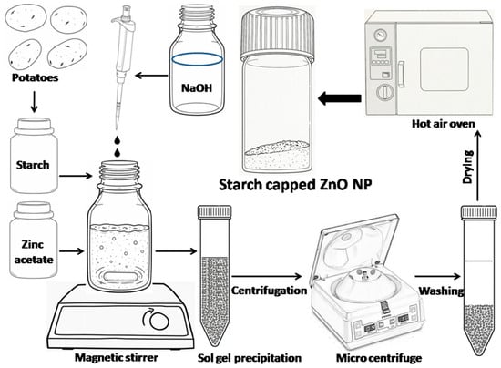
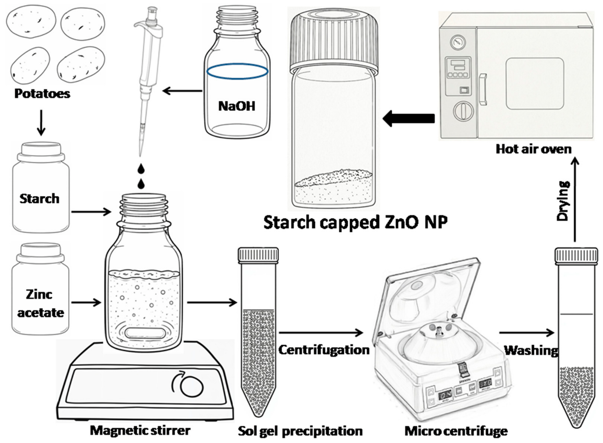
A starch solution (0.5 wt%) was prepared by dissolving it in hot ultrapure water. Zinc acetate (0.1 M) was then added to the starch solution and stirred for 30 min. ZnO NP were produced using the sol–gel method [16,24] by the dropwise addition (5 mL/min) of 100 mL of sodium hydroxide solution (0.2 M) into 100 mL of the zinc acetate amended starch solution while continuously stirring at 1000 rpm for 2 h at ambient temperature. The resulting starch-capped ZnO NP precipitate (pH 9) was separated by centrifugation at 10,000 rpm for 10 min, washed extensively with ultrapure water and dried at 80 °C for 3 h (Figure 1) [6,13,25]. The obtained ZnO NP powder (~50% yield) was stored in a glass bottle, wrapped in aluminum foil and sealed with a screw cap, at room temperature.
Figure 1.
Schematic illustration of the steps involved in synthesizing ZnO NP using the sol–gel technique. This process employs zinc acetate as the zinc precursor, sodium hydroxide as the precipitating agent and potato starch as the stabilizer.
2.3. Characterization of the Produced ZnO NP
The powdered NP (25 μg/mL) were dispersed in ultrapure water using sonication for 30 min. Their UV–visible absorption (UV-vis) spectrum was recorded against an aqueous blank over the wavelength range of 200–800 nm with a spectrophotometer (Specord 200 Plus, Analytik Jena AG, Jena, Germany). The optical band gap (Eg) of the synthesized ZnO NP was determined from the UV-vis absorption spectra using the Tauc relation. The Eg value was obtained by extrapolating the linear region of the (αhν)2 versus hν plot to the energy axis, where (αhν)2 = 0 [5].
(αhν)2 = A (hν − Eg)
- α = Absorption coefficient = 2.303 × AAbs/d
- AAbs = Absorbance
- d = Path length
- h = Planck’s constant
- ν = Frequency = c/λ
- c = Speed of light
- λ = Wavelength
- A = Band edge sharpness constant
- Eg = Optical band gap
The z-average hydrodynamic diameter, polydispersity index (PDI) and zeta potential of the produced nanoparticle solution was determined using a dynamic light scattering (DLS) device (Zetasizer Nano ZS90, Malvern, Malvern, UK). Measurements were conducted at 25 °C with a 90° scattering angle, using a 25 μg/mL concentration of NP dispersed in ultrapure water. The X-ray diffraction (XRD) analysis of the ZnO NP solution, which was drop-coated on to a glass slide, was performed with a diffractometer utilizing Cu Kα radiation (λ = 1.5406 Å) operating at 40 kV and 30 mA, over a 2θ range of 3° to 70° at a scan rate of 1°/min (Ultima IV diffractometer, Rigaku, Tokyo, Japan). Fourier transform infrared (FTIR) spectra of the starch and NP powders were recorded over the wave number range of 1000–4000 cm−1 (Alpha, Bruker, Ettlingen, Germany). Transmission electron microscope (TEM) images of the generated NP solution, which was drop-coated onto carbon-coated copper grids, were captured at 100 kV (JEM-3010, JEOL, Tokyo, Japan). The specific surface area and pore volume of the NP were quantified using N2 adsorption desorption isotherms analyzed with a Brunauer–Emmett–Teller (BET) surface area analyzer (Sorptomatic 1990, Thermo Electron Corporation, Mundelein, IL, USA).
2.4. Photocatalytic Activity of ZnO NP
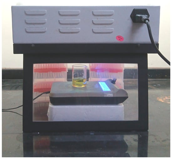
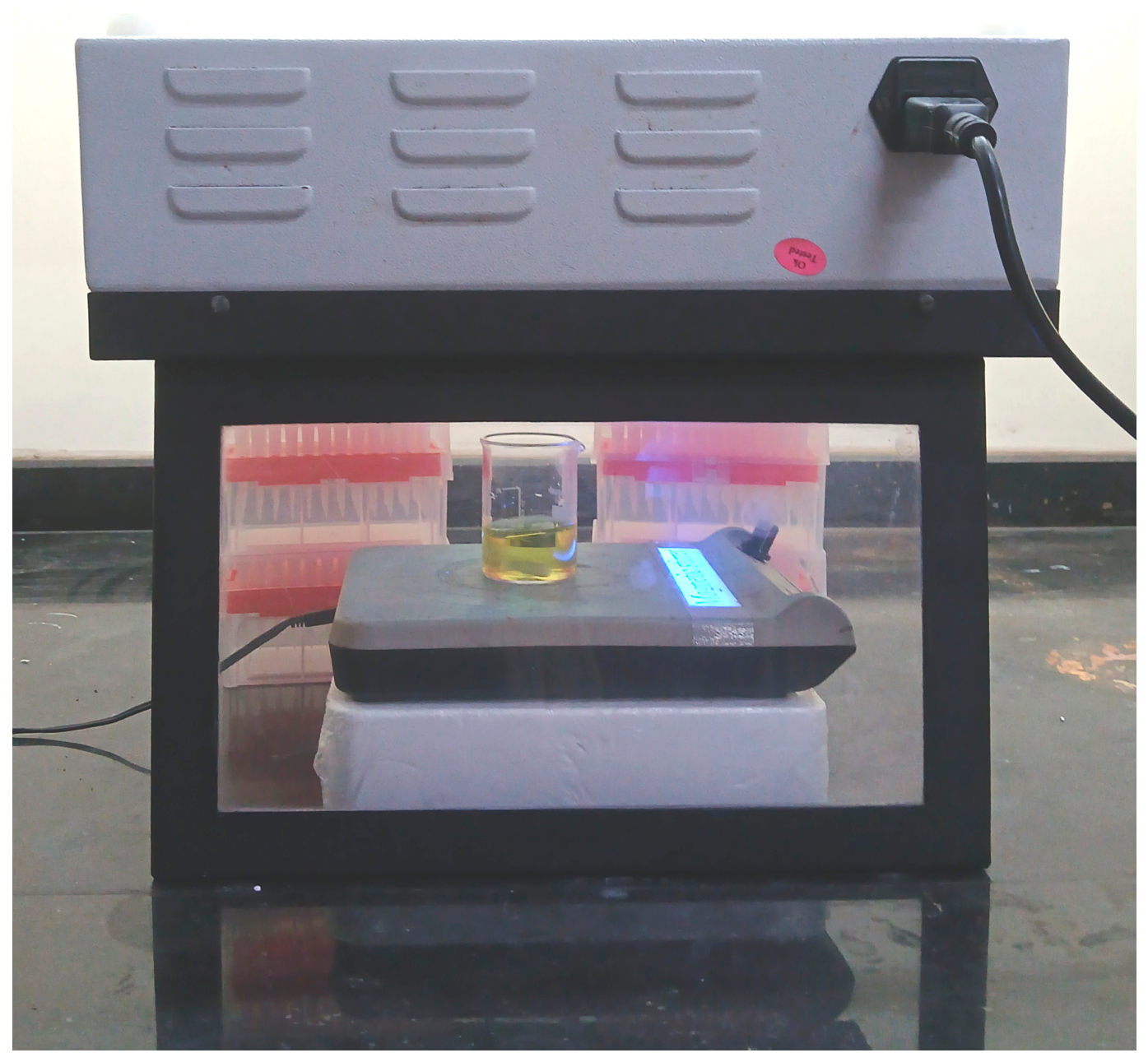
The applicability of ZnO NP as a photocatalyst for the decolourization of dye was examined using tartrazine as a model azo food dye in a custom-designed top irradiation assembly measuring 35 cm (L) × 28 cm (W) × 30 cm (H). The radiation source comprised four UV tubes (8W, λ = 365 nm, irradiance = 0.77 mW/cm2). The reaction vessel was placed on a magnetic stirrer directly beneath the centre of assembly, positioned at a vertical distance of 10 cm from the UV light source to the base of the vessel (Figure 2). Various control experiments were conducted out, including (i) dye solution subjected to UV irradiation without the presence of NP, (ii) dye and NP suspension that was stirred in the dark and (iii) dye solution containing starch that was exposed to UV irradiation. All solutions were stirred in the dark for 30 min before exposure to UV light to achieve adsorption desorption equilibrium. A 20 mL reaction mixture with a pH 6.5–7.0 was continuously stirred at 300 rpm at room temperature while being exposed to UV light. Samples were collected at regular intervals and the catalyst was separated using centrifugation at 10,000 rpm for 10 min before spectral measurements were taken [26]. The influence of varying concentrations of catalyst (900–4500 µg/mL), tartrazine (2.5–10 µg/mL) and irradiation time (30–120 min) on dye removal was investigated by recording the UV-vis absorption spectra in the range of 300 to 600 nm [27].
Figure 2.
Photograph of the custom top irradiation UV reactor assembly utilized for the photocatalytic decolourization of tartrazine.
The calculation of tartrazine decolourization (%) [28] and the pseudo first-order reaction rate constant values [29] is based on the following equations.
Decolourization (%) = (C0 − Ct)/C0 × 100
In this equation, C0 represents the tartrazine concentration at time zero, while Ct denotes the tartrazine concentration at a specific time t (min).
The pseudo first-order rate constant (k) can be obtained from the slope of the pseudo first-order reaction equation.
ln Ct/C0 = −kt
The photostability and recyclability of the ZnO NP (2700 µg/mL) for the decolourization of tartrazine (7.5 µg/mL) were investigated over five consecutive cycles of 30 min each, under UV irradiation. Following each cycle, the catalyst was quantitatively recovered via high-speed centrifugation (20,000 rpm, 10 min) and thoroughly washed with ultrapure water. The recovered wet mass was monitored by weighing the centrifuge tube that contained the catalyst pellet after decanting the supernatant, confirming minimal mass loss across runs. The wet catalyst was then reintroduced into a freshly prepared dye solution for the subsequent cycles [29,30]. The supernatant obtained after centrifugation of the catalyst during each cycle was analyzed for released zinc ions using inductively coupled plasma optical emission spectroscopy (ICP-OES) (Horiba, Ultima Expert, Longjumeau, France), while the release of starch was qualitatively verified using Gram’s iodine reagent.
2.5. Detection of Free Radical Generation During ZnO NP Mediated Photocatalysis
Hydroxyl radical (·OH) production was detected through photoluminescence using terephthalic acid (TA) as a ·OH-selective probe. A 5 mM solution of TA in 10 mM sodium hydroxide was combined with ZnO NP (2700 µg/mL) and irradiated with UV light for 120 min. Control reactions included TA exposed to UV irradiation and TA mixed with ZnO NP in the dark. Additionally, isopropyl alcohol (IPA, 10 mM) was introduced as an ·OH scavenger during the UV photocatalytic generation of ·OH by ZnO NP. Samples were withdrawn at specified time intervals (5, 10, 20, 30, 60, 90 and 120 min), then centrifuged and the supernatants were collected. The photoluminescence (PL) intensity of the supernatants was recorded at an excitation wavelength of 315 nm [12,27,31,32] using a multimode plate reader (Synergy H1, BioTek, Winooski, VT, USA). Superoxide radical (O2·−) production was assessed using the nitroblue tetrazolium (NBT) reduction assay. A 0.15 mM solution of NBT, prepared in 15 mM sodium hydroxide and 50% (v/v) dimethyl sulfoxide, was combined with ZnO NP (2700 µg/mL) and exposed to UV light for 120 min. Control reactions included NBT under UV irradiation and NBT with ZnO NP in the dark. In addition, hydroquinone (HQ, 1 mM) was utilized as a scavenger for O2·− during the UV photocatalytic generation of O2·− by ZnO NP. Samples collected at regular time intervals were centrifuged and the UV-vis absorption spectra of the resulting supernatants were recorded over the wavelength range of 450–850 nm [31,33]. Reactive oxygen species (ROS) production during the ZnO NP-mediated photocatalysis was assessed using dichlorodihydrofluorescein diacetate (DCFH2-DA), a non specific probe for intracellular and acellular ROS. A 10 µM solution of DCFH2-DA prepared in 100 mM sodium phosphate buffer (pH 7.2) was mixed with ZnO NP at a concentration of 2700 µg/mL and exposed to UV light for 120 min. Control reactions included DCFH2-DA under UV irradiation and DCFH2-DA with ZnO NP in the dark. In addition, N-acetylcysteine (NAC, 10 mM) was utilized as a general ROS scavenger during the UV photocatalytic generation of ROS by ZnO NP. At specified time intervals, samples were centrifuged and the PL intensity of the resulting supernatants was recorded at an excitation wavelength of 490 nm [34,35,36].
2.6. Assessment of the Antibacterial Potential of ZnO NP
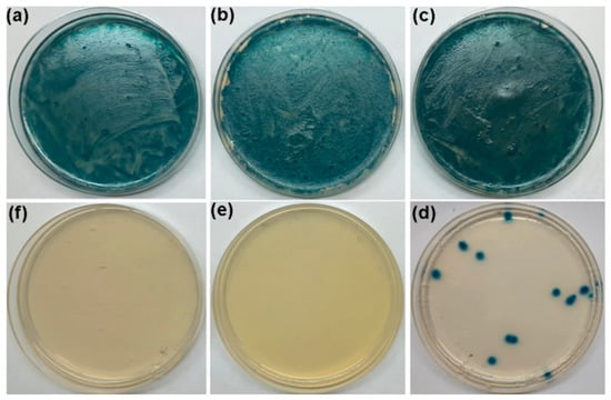
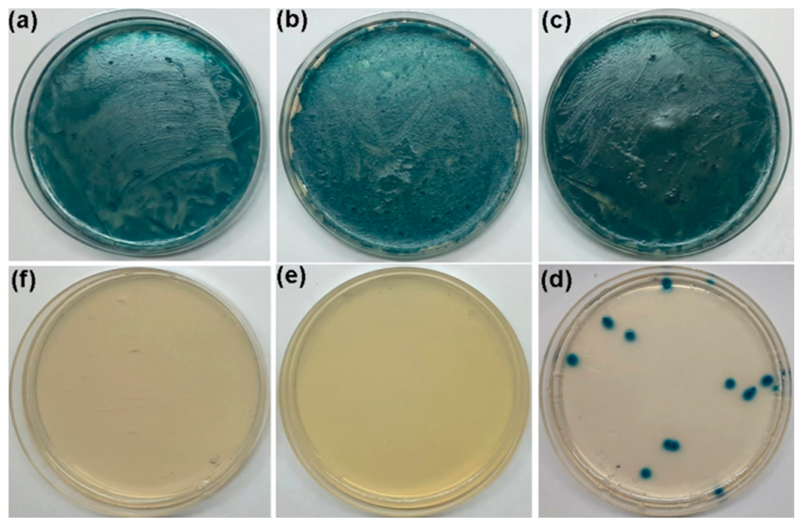
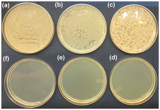
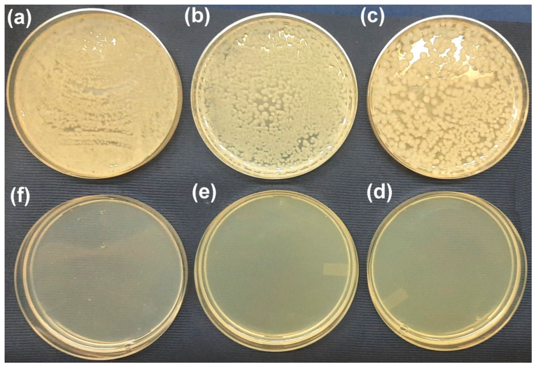
The bactericidal potential of the starch-capped ZnO NP was evaluated using an in vitro test method known as resazurin broth dilution [37,38]. The bacterial strains used in the study included Escherichia coli ATCC 25922 (Gram-negative) and Bacillus subtilis ATCC 6633 (Gram-positive), which served as representative test bacteria [38,39,40,41]. The bacterial suspension was obtained by culturing the pure strains overnight in nutrient broth and then adjusting the turbidity to match the 0.5 McFarland standard at 600 nm. The inoculum concentration was confirmed through viable colony counts on nutrient agar at serial dilutions of 10−5 and 10−6. Plates with colony counts ranging from 30 to 300 were chosen for bacterial quantification, adhering to the standard plate count detection limits. The glass tubes containing 5 mL of nutrient broth (NB) at pH 7.4 and varying levels of ZnO NP (900–4500 µg/mL) were inoculated with bacterial suspensions of 1.1 × 10⁶ CFU/mL for E. coli and 1.0 × 10⁶ CFU/mL for B. subtilis. The NB tubes containing starch and ciprofloxacin (10 μg/mL) inoculated with bacteria served as negative and positive controls, respectively. The cell-free NB tubes amended with ZnO NP served as an abiotic control. After incubating for 24 h at 37 °C, 62.5 μL of resazurin solution (400 μg/mL) was added to achieve a final concentration of 5 μg/mL and the tubes were incubated for an additional 48 h. The NB tubes were visually examined for changes in turbidity and colour. A pink colouration indicated bacterial growth, while a blue or purple colouration signified growth inhibition. The minimum inhibitory concentration (MIC) of NP was established as the lowest concentration at which blue or purple colouration appeared in the NB tube. The minimum bactericidal concentration (MBC) of NP was defined as the lowest concentration at which no viable counts were observed in blue or purple-coloured NB tubes. The bacterial growth inhibition (%) at various NP concentrations, along with the MBC value of ZnO NP, was determined by counting viable colonies on HiCrome coliform agar (CA) for E. coli and nutrient agar (NA) for B. subtilis, following suitable serial dilutions [38].
2.7. Statistical Analysis
All the UV photocatalytic and antibacterial experiments were conducted in triplicate (n = 3). The data for photocatalytic decolourization (%) were presented as the mean ± standard deviation (SD), with error bars included in the respective graphs. Linear regression and kinetic modelling analyses were conducted using Origin software (OriginPro 8.5, OriginLab Corporation, Northampton, MA, USA). The pseudo first-order kinetic constant (k) and its corresponding uncertainty (standard error) were calculated from the slope of the linear fits, with the correlation quality indicated by the coefficient of determination (R2). In the antibacterial assays, the MIC and MBC values were established as discrete threshold concentrations based on three independent experimental replicates. The bacterial growth reduction (%) were reported as mean ± SD derived from the triplicate experiments.
3. Results and Discussion
3.1. Production and Capping of ZnO NP
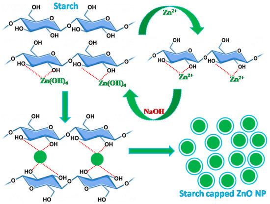
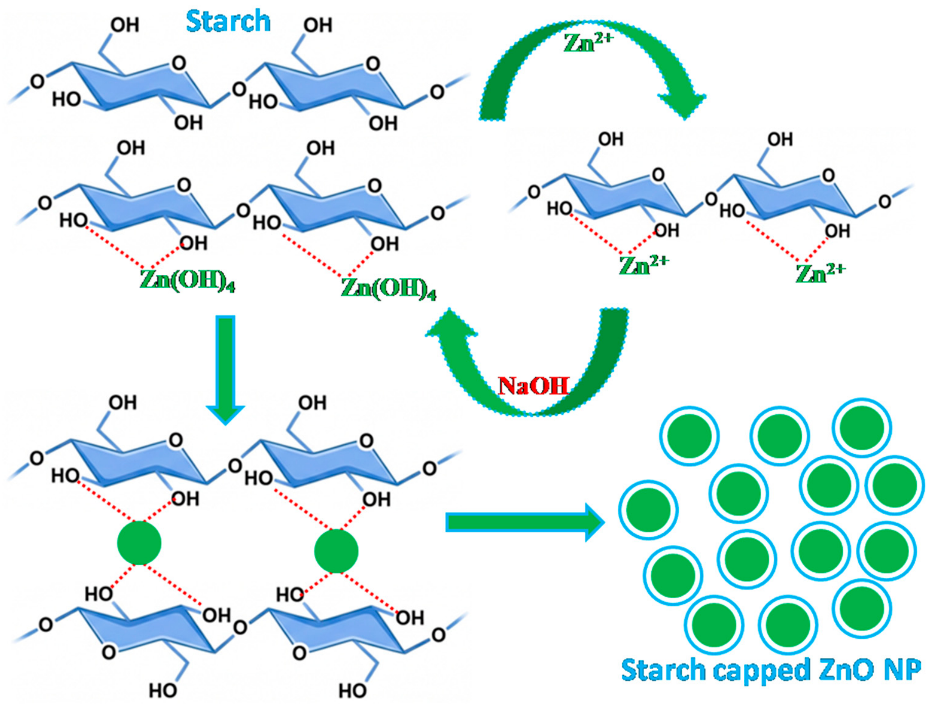
In the present study, zinc oxide nanoparticles (ZnO NP) were produced using the sol–gel technique, with zinc acetate as the zinc precursor, sodium hydroxide as the precipitating agent and starch as a stabilizer (Figure 1) [24]. When zinc acetate is mixed into the starch solution, the released zinc ions coordinate with the surface hydroxyl groups of the starch molecules through hydrogen bonding. The starch molecules act as a template ensuring the uniform dispersion of zinc ions within the biopolymer network and limiting their mobility. With the dropwise addition of sodium hydroxide under continuous stirring, zinc ions react with hydroxide ions to form zinc hydroxide in alkaline conditions. Further addition of sodium hydroxide converts the zinc hydroxide into soluble zincate ions, which then further interact with the hydroxyl groups of biopolymer. Consequently, starch functions as both a template and a capping agent. Surface-adsorbed starch molecules restrict the particle growth and prevent nanoparticle agglomeration through steric hindrance. Finally, upon thermal drying, the hydroxide complex undergoes dehydration resulting in the formation of stabilized ZnO NP (Figure 3) [6,13,16,25,27,42,43]. A similar capping mechanism is reported with other biopolymers and polysaccharide gums [44] such as rhamnolipid [45], carboxy methyl cellulose [13], gum tragacanth [46], gum kondagogu [38], etc. The chemical reactions involved in the synthesis of ZnO NP are represented by the following equations [6,13,29,35].
Zn(CH3COO)2⋅2H2O + 2NaOH → Zn(OH)2 + 2CH3COONa + 2H2O
Zn(OH)2 + 2NaOH → Na2[Zn(OH)4]
Figure 3.
Schematic illustration depicting the capping mechanism of ZnO NP by the starch molecules.
3.2. Characterization of Starch Stabilized ZnO NP
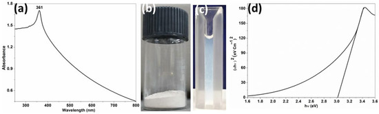
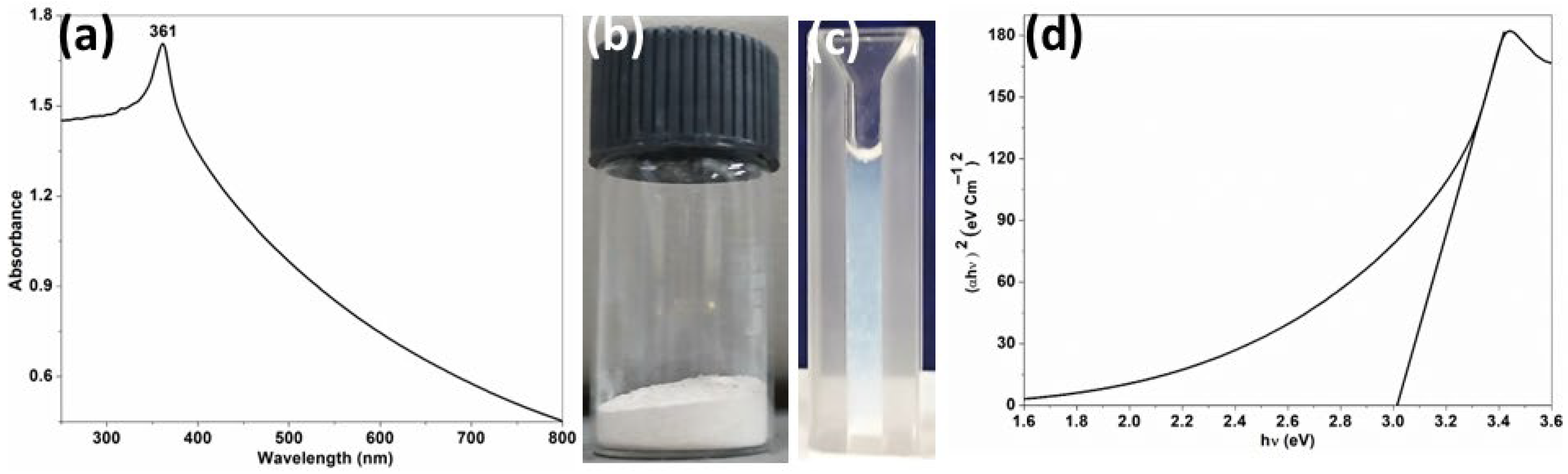
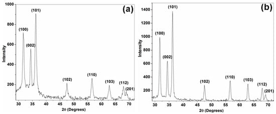
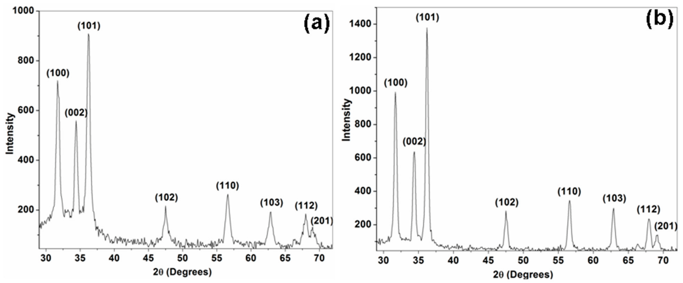
The aqueous solution of starch-stabilized ZnO NP at a concentration of 25 μg/mL exhibited a characteristic absorption maximum at 361 nm in the UV-vis spectrum, confirming the successful synthesis of ZnO NP [Figure 4a] [6]. Visual representations of the powdered sample and aqueous solutions of the polymer stabilized ZnO NP are presented in Figure 4b,c, respectively. The observed absorption peak of the biopolymer-capped ZnO NP aligns well with previous studies on ZnO NP synthesized using various capping agents, including starch [6], Pseudomonas aeruginosa rhamnolipid [45] and Anacardium occidentale leaf extract [47]. The optical band gap (Eg) value obtained from the Tauc plot was 3.01 eV for the synthesized ZnO NP, which aligns with the previously reported values for starch-stabilized ZnO NP [Figure 4d] [5,27]. The ZnO NP demonstrated a specific surface area of 8.99 m2/g and a pore volume of 0.015 cm3/g. The diffractogram of the synthesized ZnO NP exhibited pronounced diffraction peaks at angles of 31.7°, 34.4°, 36.2°, 47.5°, 56.6°, 62.9°, 68°, and 69°. These peaks correspond to the (100), (002), (101), (102), (110), (103), (112) and (201) lattice planes of hexagonal wurtzite crystal structure of ZnO, respectively [Figure 5a]. The calculated interplanar d spacing (dhkl) values were 2.820, 2.604, 2.479, 1.912, 1.624, 1.476, 1.377, 1.359 Å, while the lattice constants were determined to be a = b = 3.256 Å, c = 5.208 Å. These values closely match the standard data for ZnO (JCPDS 00-036-1451) [25,46,48]. The average crystallite size, calculated using Scherrer’s equation, was 25.6 nm. Additionally, the observed widening of the diffraction peaks further corroborated the nanoscale nature of the synthesized ZnO [6,46]. The XRD pattern of the starch-capped ZnO NP aligns well with that of ZnO NP synthesized using gum tragacanth, a polysaccharide root exudate obtained from Astragalus gummifer [46]. The sharp diffraction peaks observed at 31.7° and 36.2°, which corresponds to the (100) and (101) crystal planes, indicate a preferential crystallographic orientation in the synthesized ZnO NP [10,25].
Figure 4.
Characterization of starch-capped zinc oxide nanoparticles (ZnO NP), (a) UV–visible absorption spectra of aqueous solution at a concentration of 25 μg/mL, highlighting an absorption peak at 361 nm, (b) photograph of the synthesized ZnO NP powder, (c) photograph of the ZnO NP aqueous solution contained in a cuvette and (d) Tauc plot used for determining the optical band gap of the ZnO NP.
Figure 5.
XRD patterns illustrating the hexagonal wurtzite crystal structure of (a) pristine and (b) recycled starch-capped ZnO NP.
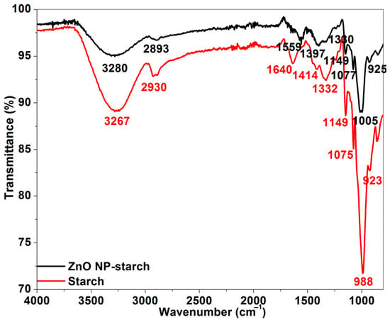
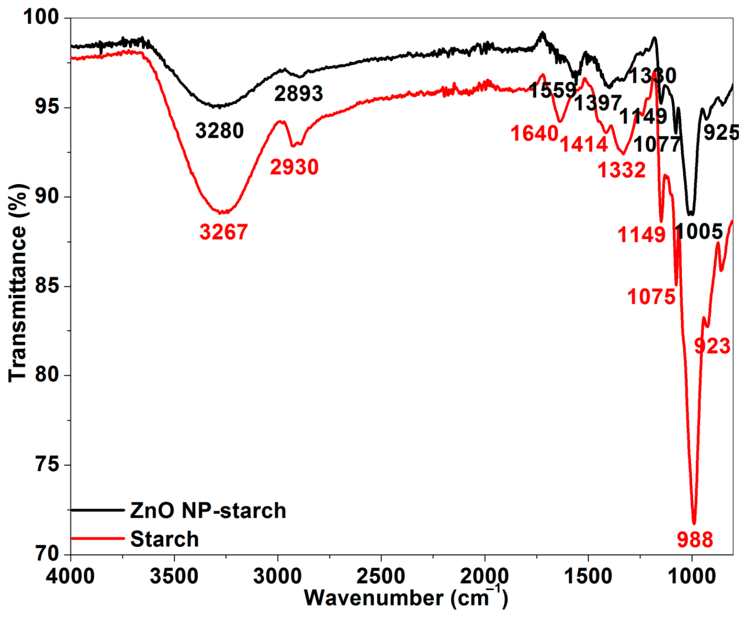
The IR spectrum of starch exhibited distinctive absorption peaks at 3267 (O-H group stretching vibration), 2930 (methylene group C-H stretching vibration), 1640 (H-O-H bending, C=O stretch), 1414, 1332 (C-H, O-H groups bending vibrations), 1149 (C-O-C asymmetric stretching of glycosidic linkage), 1075 (C-O stretching), 988 and 923 (C-O-H bending). In comparison, the starch-capped ZnO NP displayed absorbance peaks at 3280, 2893, 1559, 1397, 1330, 1149, 1077, 1005 and 925 cm−1 (Figure 6). A slight peak shift from 3267 to 3280 cm−1 indicates the surface coordination of OH groups of starch with the zinc ions, leading to a modified O-H stretching environment. An intermediate shift in C-H stretching vibrations from 2930 to 2893 cm−1 suggests steric interactions resulting from adsorption of starch molecules on the surface of ZnO NP. A pronounced shift from 1640 to 1559 cm−1 signifies strong coordination between the zinc ions and oxygen donor atoms of hydroxyl or carbonyl groups in starch. The intermediate shift from 1414 to 1397 cm−1 supports the involvement of C-H/O-H bending vibrations. The shift observed from 988 to 1005 cm−1 and 923 to 925 cm−1 confirm the interaction between starch and the ZnO NP surface, suggesting the formation of Zn-O-C or Zn-O-H bonds. Therefore, the observed variations in the FTIR spectra substantiate the successful capping and steric stabilization of ZnO NP through the formation of Zn-O linkages [6,13,16,25].
Figure 6.
FTIR spectra for starch and starch-stabilized ZnO NP.
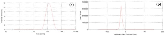
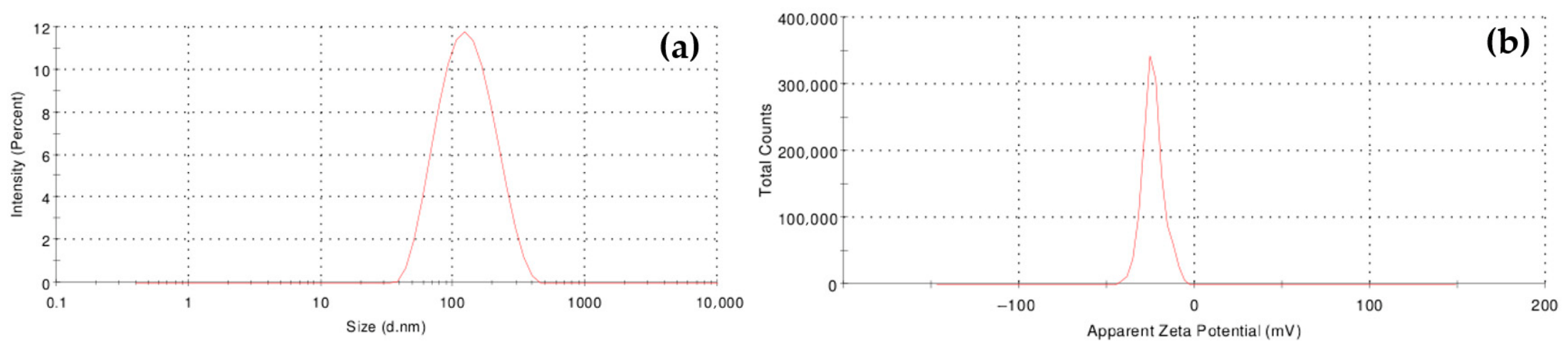
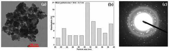
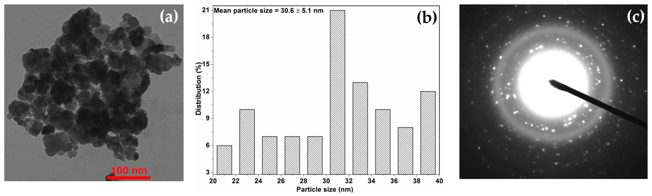
The DLS technique was employed to measure the hydrodynamic diameter and zeta potential values of the synthesized ZnO NP solution. The prepared NP exhibited a z-average value of 138.1 ± 4.5 nm with a polydispersity index (PDI) of 0.278, indicating a moderately polydisperse distribution [Figure 7a]. The measured zeta potential value was −23.8 ± 1.3 mV [Figure 7b] and the noted negative zeta potential suggests sufficient stability of the produced ZnO NP [13]. The synthesized NP were nearly spherical in shape, with sizes varying from 20.7 to 38.1 nm and the average particle size was 30.6 ± 5.1 nm [Figure 8a,b]. The nanoparticle size distribution recorded using TEM in this current study matches with earlier reports on starch-capped ZnO NP [6,13,16,25,42]. The presence of both diffraction spots and continuous rings in the selected area electron diffraction (SAED) pattern indicates the polycrystalline nature of the produced ZnO NP [Figure 8c]. The DLS analysis of the starch-capped ZnO NP indicated a higher particle size of 138.1 nm when compared to the 30.6 nm measured by TEM. The TEM technique quantifies the true physical radius of the solid, electron-dense inorganic core of the NP under ultra-high vacuum conditions. In contrast, DLS measures the hydrodynamic diameter of the NP in an aqueous environment, which includes the inorganic core, the surrounding hydration shell and the starch capping layer. When dispersed in ultrapure water at 25 °C, the hydroxylated starch molecules swell due to hydrogen bonding with water molecules, thus increasing the apparent particle volume.
Figure 7.
DLS and surface characterization of starch-capped ZnO NP, (a) distribution of hydrodynamic diameter and (b) distribution of zeta potential.
Figure 8.
TEM characterization of starch-capped ZnO NP, (a) representative TEM image at 100 nm scale, (b) histogram of particle size distribution and (c) the corresponding selected area electron diffraction (SAED) pattern.
3.3. ZnO NP-Mediated Photocatalytic Decolourization of Tartrazine
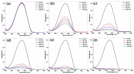
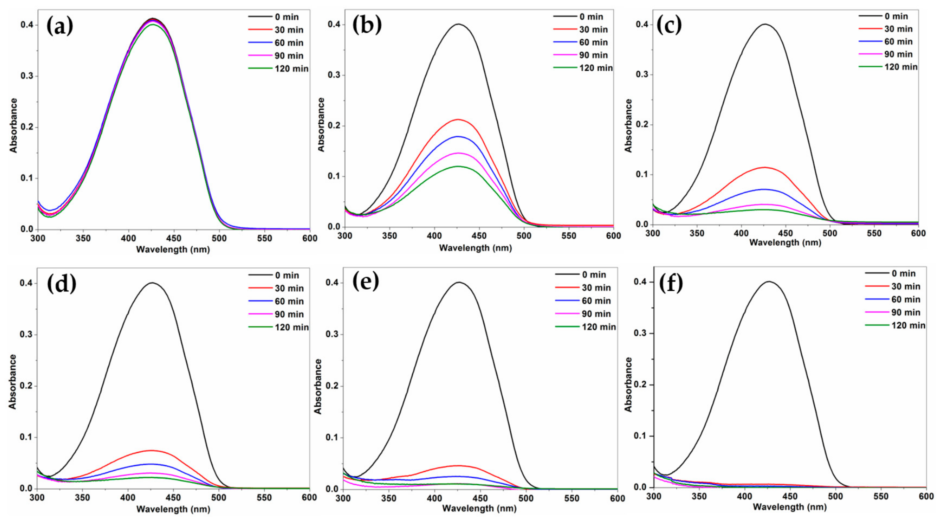
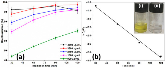
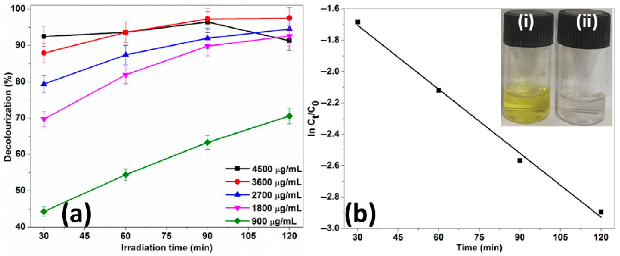
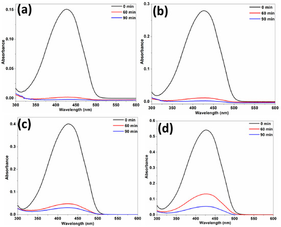
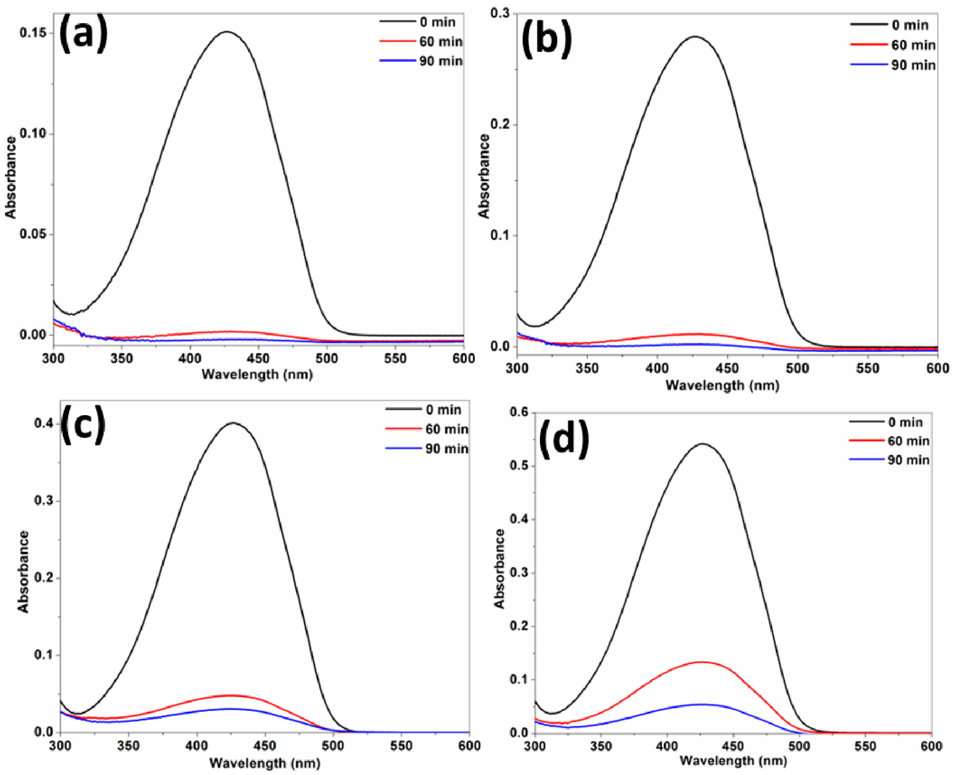
The strong UV absorption capability of the starch-capped ZnO NP was utilized for the decolourization of tartrazine [26,46]. The tartrazine removal was investigated at various concentrations of the catalyst (900–4500 µg/mL), tartrazine (2.5–10 µg/mL) and reaction time (30–120 min) under UV light irradiation at 365 nm. The typical absorption spectrum of tartrazine exhibits a peak at 426 nm. Controls for both photolysis (dye under UV without NP) and capping (dye with starch under UV) showed no decrease in absorbance, indicating a lack of dye photodegradation and starch mediated dye uptake. A negligible change in dye absorbance was observed during the dark adsorption phase, likely due to electrostatic repulsion between the negatively charged starch-capped ZnO NP and the anionic tartrazine dye. Without the NP catalyst, UV irradiation alone did not result in decolourization of tartrazine over the entire reaction period of 120 min [28]. This was evident from the absorption spectra of tartrazine at a concentration of 7.5 μg/mL at various time intervals (30 min, 60 min, 90 min and 120 min) [Figure 9a]. At a concentration of 7.5 μg/mL of tartrazine, the NP-mediated decolourization was monitored over a duration of 30 to 120 min, with varying catalyst concentrations (900–4500 µg/mL). Analysis of the absorption spectra confirmed that as the irradiation time increased from 30 to 120 min, the absorbance of tartrazine decreased across different concentrations of NP [Figure 9b–f]. The dye decolourization (%) at various catalyst concentrations during different durations of irradiation are presented in Figure 10a. The tartrazine decolourization (%) values were 63.3%, 89.3%, 92%, 97.2% and 96.4% at concentrations of 900 µg/mL, 1800 µg/mL, 2700 µg/mL, 3600 µg/mL and 4500 µg/mL, respectively, under 90 min of UV irradiation. The higher concentration of 3600 µg/mL resulted in only a marginal improvement in decolourization (4.9%). Beyond a catalyst dosage 2700 µg/mL, the system reached a plateau phase. Therefore, a catalyst concentration of 2700 µg/mL, a dye concentration of 7.5 µg/mL and a UV irradiation time of 90 min were selected as the optimum conditions for tartrazine decolourization [Figure 10a]. The inset of [Figure 10b] clearly indicates the decolourization of the dye by ZnO NP, showing the yellow-coloured solution of tartrazine before UV irradiation and the colourless solution afterwards. The decolourization of tartrazine followed pseudo first-order kinetics, evidenced by an R2 value of 0.993. The reaction rate constant (k) value calculated at optimal conditions was 0.0135 ± 0.0006 min−1 [Figure 10b]. Additionally, at an optimized ZnO NP dosage of 2700 µg/mL, the decolourization of varying concentrations of tartrazine (2.5–10 µg/mL) were studied over a duration of 60 to 90 min (Figure 11). It was noted that at lower concentrations of tartrazine, such as 2.5 µg/mL [Figure 11a] and 5 µg/mL [Figure 11b], the dye was completely decolourized within 90 min. In contrast, at higher concentrations of 7.5 µg/mL [Figure 11c] and 10 µg/mL [Figure 11d], the decolourization were 92% and 89.9%, respectively.
Figure 9.
Time-dependent (30–120 min) photocatalytic decolourization of tartrazine (7.5 µg /mL) at varying concentrations of ZnO NP, (a) 0 µg/mL, (b) 900 µg/mL, (c) 1800 µg/mL, (d) 2700 µg/mL, (e) 3600 µg/mL and (f) 4500 µg/mL.
Figure 10.
(a) The decolourization (%) of tartrazine (7.5 µg/mL) at different ZnO NP concentrations (900–4500 µg/mL) over different irradiation durations (30–120 min) and (b) rate constant (k) value determination at 2700 µg/mL of ZnO NP and 7.5 µg/mL of tartrazine. Inset: Colour of the tartrazine solution (i) before and (ii) after UV irradiation.
Figure 11.
Time-dependent (60–90 min) photocatalytic dye decolourization using 2700 µg/mL of ZnO NP at various concentrations of tartrazine: (a) 2.5 µg/mL, (b) 5 µg/mL, (c) 7.5 µg/mL and (d) 10 µg/mL.
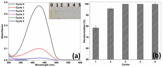
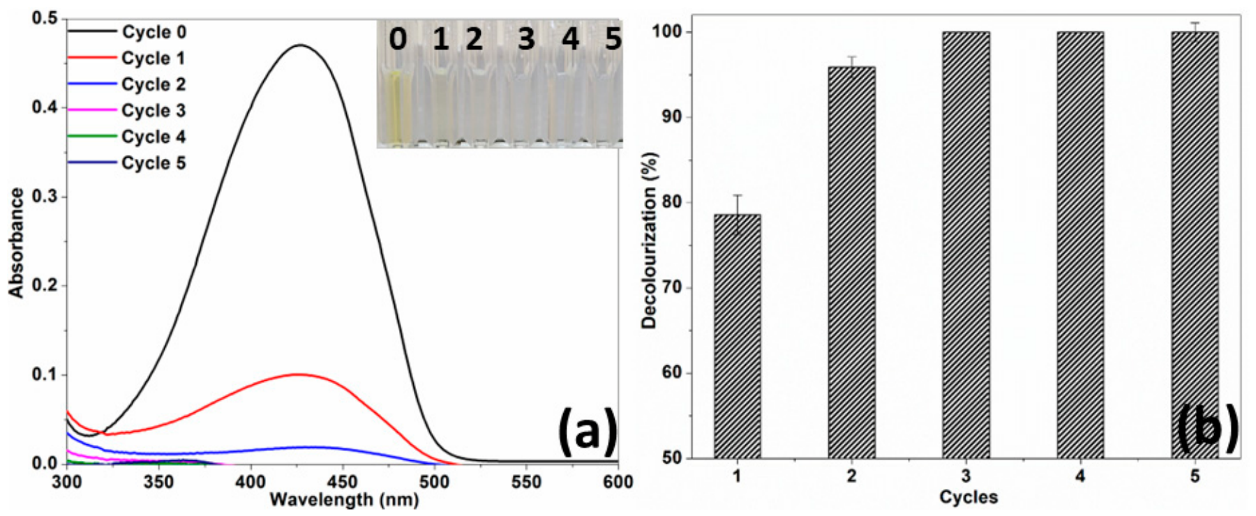
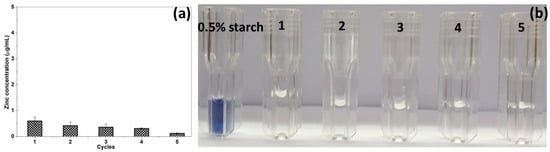
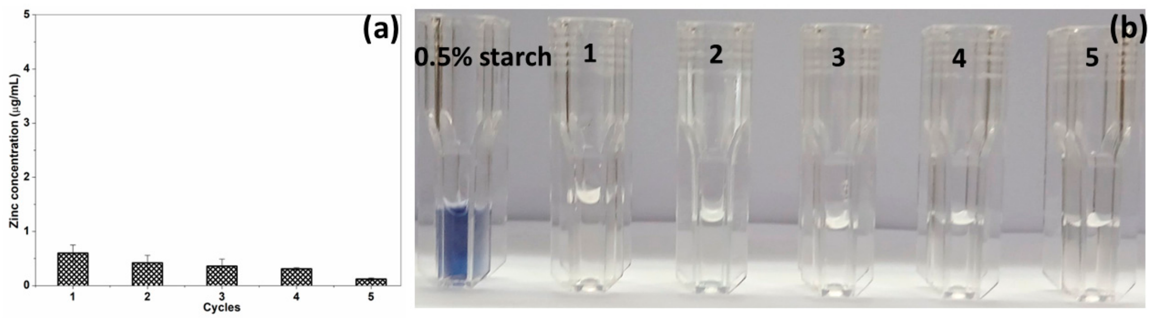
The recyclability of the catalyst was studied over five cycles, during which it successfully decolourized tartrazine within 30 min of UV exposure, as evident from the recorded absorption spectra at each cycle [Figure 12a]. This was further confirmed by the colour change observed in the tartrazine solution, from the tartrazine solution colouration, given in the inset of Figure 12a, which was initially yellow at cycle 0 and became colourless with each subsequent catalytic cycle. The observed phenomenon was further supported by the dye decolourization (%) values obtained across the different cycles [Figure 12b]. The values recorded were 78.6%, 95.9%, 100%, 100% and 100% at 1, 2, 3, 4 and 5 cycles, respectively, under conditions of 7.5 µg/mL of tartrazine, 2700 µg/mL of ZnO NP and 30 min of UV irradiation. Interestingly, as the number of cycles increased, the catalytic efficiency of ZnO NP was also increased. Consequently, the starch-capped ZnO NP exhibited superior UV photostability and impressive recycling capabilities [31,49] over a span of five cycles. Also, the structural stability was confirmed by the hexagonal crystal structure observed in the XRD pattern of ZnO NP recycled for five cycles [Figure 5b]. These results were validated by the concentration of released zinc ions determined by ICP-OES. At each cycle, the NP released negligible quantities of zinc ions: 0.60, 0.42, 0.36, 0.31 and 0.12 µg/mL during cycles 1 through 5 respectively, at a catalyst concentration of 2700 µg/mL [Figure 13a]. Also, the starch leaching into the supernatant during each cycle was monitored with Gram’s iodine qualitative testing. As shown in Figure 13b, the supernatants after each cycle showed no blue–black colouration, confirming the absence of detectable starch leaching. In contrast, a 0.5% starch solution produced a distinct blue–black colouration. The observed increase in photocatalytic activity from cycle 1 to 5 could be attributed to the removal of residual surface contaminants and steric stabilization of NP due to starch capping, which effectively prevents the agglomeration of ZnO NP upon reuse. Agglomeration is a common cause for activity loss in recycled catalysts. These findings align with previous studies on ZnO NP stabilized with biomolecules from Eucalyptus grandis leaf extract [24] and carrageenan, a marine polysaccharide derived from red alga, Kappaphycus striatus [44].
Figure 12.
The recyclability of ZnO NP (2700 µg/mL) in the decolourization of tartrazine (7.5 µg/mL) under UV exposure (30 min) across five cycles, in terms of (a) UV absorption spectra. Inset: Corresponding tartrazine solutions colours and (b) decolourization (%).
Figure 13.
The release of (a) zinc ions and (b) starch from ZnO NP during five consecutive photocatalytic cycles.
Also, a comparison on photocatalytic decolourization of tartrazine at a UV wavelength of 365 nm by ZnO NP was made with earlier published reports with reference to average particle size, NP concentration, UV lamp power and number, tartrazine concentration, reaction time, decolourization (%) and rate constant (Table 1). The quasi-spherical, uncapped ZnO NP of 68 nm size at a concentration of 10000 µg/mL, were able to decolourize 10 µg/mL of tartrazine by 87% over a period of 1440 min using a 20 W UV lamp [3]. The cassava starch-capped, 58 nm sized, spherical and hexagonal-shaped ZnO NP (500 µg/mL) decolourized 94% of the dye (10 µg/mL) in 180 min, employing a photoreactor with five 9 W UV lamps yielding a k value of 0.0179 min−1 [5]. In the case of uncapped, hexagonal, 56 nm sized ZnO NP (1000 µg/mL), a k value of 0.04 min−1 was achieved at a concentration of 0.26 µg/mL of tatrazine after 90 min of UV irradiation at 150 W [29]. Using 20–50 nm sized ZnO NP of 500 µg/mL and 10 µg/mL of tartrazine, 23.2% decolourization and k value of 0.008 min−1 were obtained within 30 min of irradiation utilizing a UV light of 15 W [26]. With a 15 W UV lamp, the spherical, citrate-capped ZnO NP sized 30–40 nm at a concentration of 500 µg/mL completely decolourized 10 µg/mL of tartrazine within 70 min of irradiation, exhibiting a k value of 0.04 min−1 [49]. Using similar UV lamp power and irradiation duration, citrate-capped ZnO NP of 20–30 nm size completely decolourized 20 µg/mL of tartrazine, achieving a k value of 0.052 min−1 [28]. The 30 nm sized ZnO NP at 200 µg/mL concentration decolourized 93.7% of 25 µg/mL of tartrazine within 60 min of irradiation, yielding a k value of 0.0459 min−1 in a photoreactor equipped with fifteen 8 W UV lamps [50].
Table 1.
Comparison of the photocatalytic decolourization of tartrazine using ZnO NP in terms of mean particle size, NP concentration, UV lamp power and number, tartrazine concentration, reaction time, decolourization (%) and rate constant.
In the current study, starch-capped, nearly spherical ZnO NP with a mean particle size of 30.6 nm and a catalyst loading of 2700 µg/mL achieved 92% decolourization of 7.5 µg/mL of tartrazine within 90 min of irradiation, resulting in a k value of 0.0135 min−1. The photocatalysis was conducted in a custom-built assembly equipped with four 8 W UV lamps. It is important to observe that the previous studies on ZnO NP-based photocatalytic decolourization were carried out using NP with larger particle size [3,5,29], at a very low dye concentration [29] and with high catalyst concentration [3], for extended irradiation durations [3,5]. These studies employed either high-power UV lamps [29] or compact UV photoreactors designed with optimized cylindrical [29] or annular [5] geometries, featuring integrated internal light reflection for uniform light distribution, localized irradiation and maximum light intensity [50]. Also, the ZnO NP that demonstrated high reported k values and enhanced photocatalytic activity were utilized in either cylindrical UV photoreactor equipped with a high-power UV lamp [29] or a compact UV photoreactor featuring a large number of UV lamps [50]. These configurations exhibited higher surface areas of 26.2 m2/g [49] and 26.4 m2/g [28], 14.05 m2/g [29] or combinations thereof [29,50]. In the current report, the starch-capped ZnO NP, with a mean particle size of 30.6 nm and a surface area of 8.99 m2/g showed a reasonably good decolourization of tartrazine (92% at 7.5 µg/mL) and a k value of 0.0135 min−1 over a duration of 90 min. This was achieved using a custom-built assembly equipped with four 8 W UV lamps. Thus, the study confirms the interplay of various factors, including photocatalyst particle size, capping agent, concentration, surface area, dye concentration and operational features such as lamp power, number, photoreactor geometry and irradiation time in the UV photocatalytic dye decolourization process.
3.4. Proposed Mechanism of ZnO NP-Mediated Photocatalysis
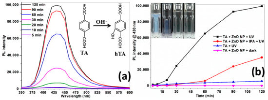
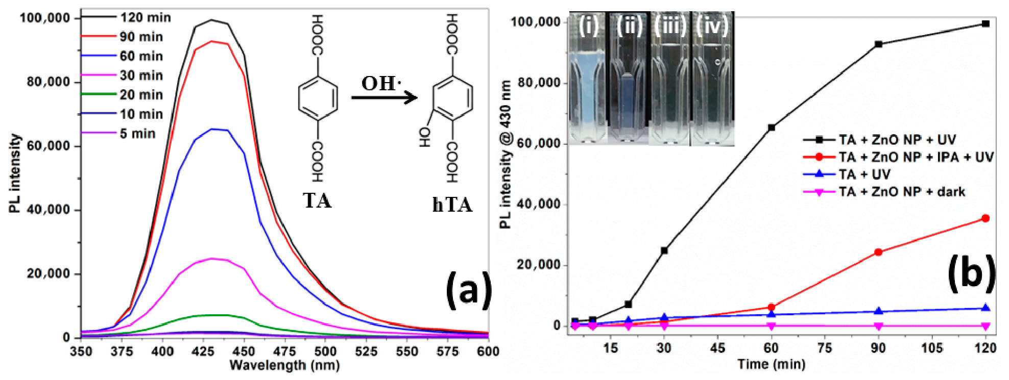
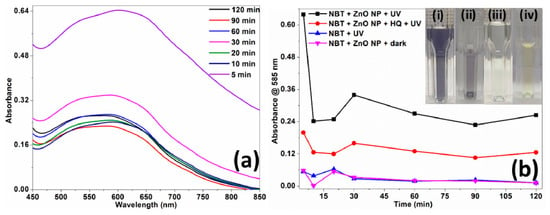
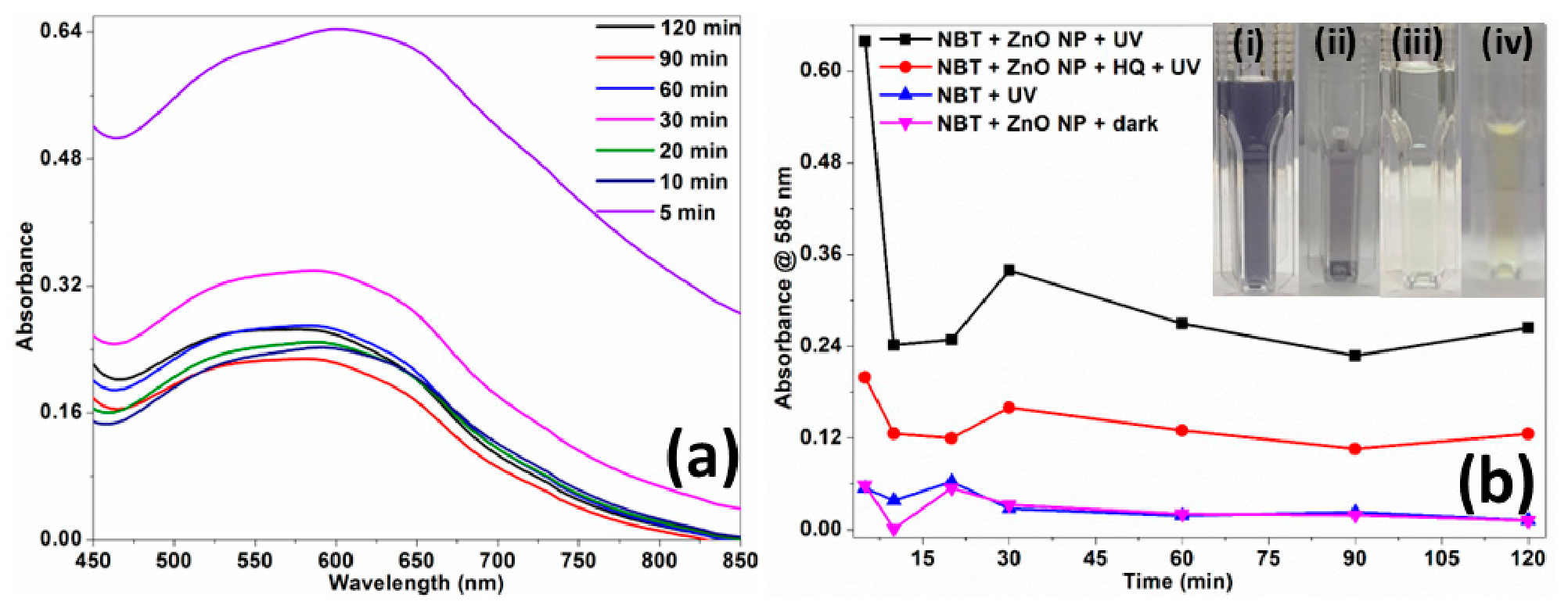
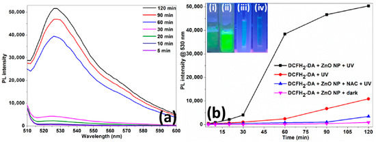
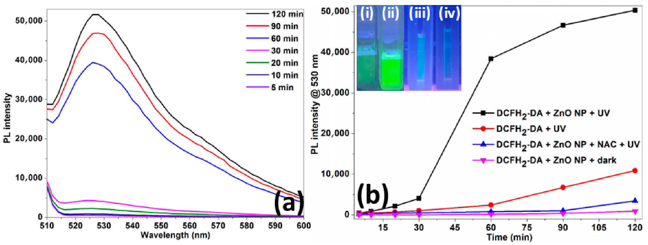
The generation of hydroxyl radicals (·OH) during UV photocatalysis by ZnO NP was investigated by measuring the conversion of terephthalic acid (TA) to 2-hydroxy terephthalic acid (hTA), a fluorescent product [Inset of Figure 14a]. As the irradiation time increased from 5 min to 120 min, the photoluminescence (PL) intensity of the produced hTA at 430 nm enhanced concomitantly [Figure 14a]. The time-dependent PL intensity of the ZnO NP-catalyzed reaction increased at a much faster rate compared to the control reactions, which included TA under UV irradiation and TA with ZnO NP in the dark. In addition, the introduction of IPA as a scavenger, diminished the production of hTA, leading to subsequent decrease in PL intensity [Figure 14b] [32,51]. The appearance of the TA solutions indicates that they remained colourless in the control reactions (TA under UV irradiation and TA with ZnO NP in the dark). In contrast, the produced hTA exhibited a bluish white colour in the NP catalyzed reaction and was only faintly coloured in the IPA-scavenged reaction [Inset of Figure 14b]. Thus, the data confirms the generation of hydroxyl radicals during the photocatalysis mediated by ZnO NP [27,31,32,51]. The production of superoxide radicals (O2·−) during the ZnO NP-mediated photocatalysis was quantified by measuring the reduction of nitroblue tetrazolium (NBT) to diformazan in an alkaline environment [Figure 15a] [31,33]. The absorbance of diformazan at 585 nm peaked at 5 min of UV exposure, followed by 30 min. In the control reactions (NBT under UV irradiation and NBT with ZnO NP in the dark), the absorbance remained relatively constant over time. The addition of HQ served as a scavenger for O2·−, which decreased the production of diformazan and as a result decreased the absorbance [Figure 15b]. The appearance of the NBT solutions exemplifies this, as they remained pale yellow in the control reactions (NBT under UV irradiation and NBT with ZnO NP in the dark). The produced diformazan showed a purple colour in the NP catalyzed reaction, while it appeared only faintly coloured in the HQ-scavenged reaction [Inset of Figure 15b]. The vast range of reactive oxygen species (ROS) generated during ZnO NP-mediated photocatalysis was measured in terms of production of dichlorodihydrofluorescein (DCF) from dichlorodihydrofluorescein diacetate (DCFH2-DA). The PL intensity of the generated DCF at 530 nm showed a slight increase from 5 min to 30 min of irradiation. A significant and sudden enhancement in intensity was observed from 60 min to 120 min [Figure 16a]. The control reaction such as DCFH2-DA under UV irradiation produced in measurable quantities of DCF due to the photooxidation of DCFH2-DA by UV light [Figure 16b] [34]. A clear difference in DCF production is evident from the elevated fluorescence of the NP catalyzed reaction compared to the uncatalyzed [Inset of Figure 16b]. In contrast, the DCFH2-DA with ZnO NP in the dark produced negligible fluorescence. Also, the added NAC functioned as a scavenger, which reduced the fluorescence intensity. The results were clearly indicated by the colours of the solutions shown in the inset of Figure 16b. The data indicates that the hydroxyl and superoxide radicals are the major ROS involved in the photocatalysis of ZnO NP [31,51,52].
Figure 14.
(a) Time-dependent photoluminescence (PL) intensity of 2-hydroxyterephthalic acid produced by hydroxyl radicals generated during the ZnO NP photocatalysis. Inset: Conversion of terephthalic acid to 2-hydroxyterephthalic acid by hydroxyl radicals, and (b) time-dependent PL intensity at 430 nm for NP catalyzed, uncatalyzed and scavenging reactions. Inset: Corresponding solution colours, (i) TA + ZnO NP + UV, (ii) TA + ZnO NP + IPA + UV, (iii) TA + UV and (iv) TA + ZnO NP + dark.
Figure 15.
(a) Time-dependent absorbance of the diformazan produced by superoxide radicals generated during the ZnO NP photocatalysis and (b) time-dependent diformazan absorbance at 585 nm for NP catalyzed, uncatalyzed and scavenging reactions. Inset: Corresponding solution colours, (i) NBT + ZnO NP + UV, (ii) NBT + ZnO NP + HQ + UV, (iii) NBT + UV and (iv) NBT + ZnO NP + dark.
Figure 16.
(a) Time-dependent PL intensity of the dichlorodihydrofluorescein produced by reactive oxygen species generated during the ZnO NP photocatalysis and (b) time-dependent PL intensity at 530 nm for NP catalyzed, uncatalyzed and scavenging reactions. Inset: Corresponding solution colours, (i) DCFH2-DA + ZnO NP + UV, (ii) DCFH2-DA + ZnO NP + NAC + UV, (iii) DCFH2-DA + UV and (iv) DCFH2-DA + ZnO NP + dark.
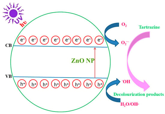
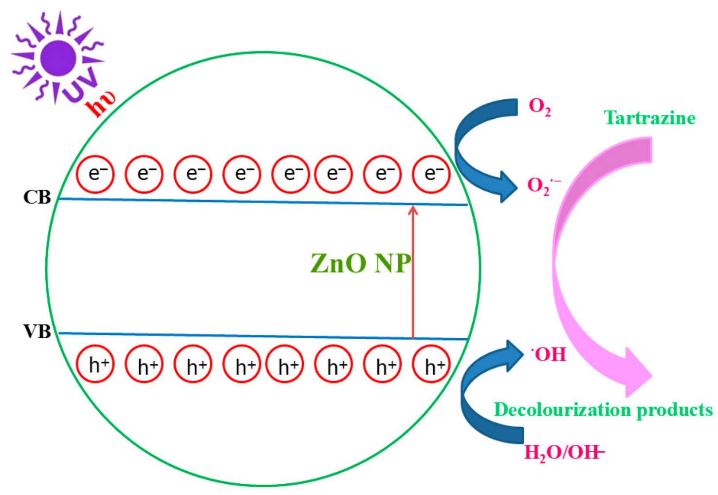
Upon UV exposure of ZnO NP, electrons (e−) in the valence band (VB) become excited and move to the conduction band (CB), leaving holes (h+) in the VB. The created holes in the VB react with either water or hydroxyl groups to generate hydroxyl radicals. Meanwhile, the electrons in the CB generate superoxide radicals from oxygen. Consequently, the produced ROS, such as hydroxyl and superoxide radicals, attack the tartrazine molecules, leading to their decolourization and degradation into smaller intermediate products. The proposed mechanism of UV photocatalysis by ZnO NP is shown in Figure 17 [5,12,26,31,44,49,52].
Figure 17.
A proposed mechanism for the photocatalytic decolourization of tartrazine using ZnO NP.
3.5. Bactericidal Activity of ZnO NP
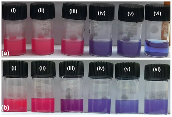
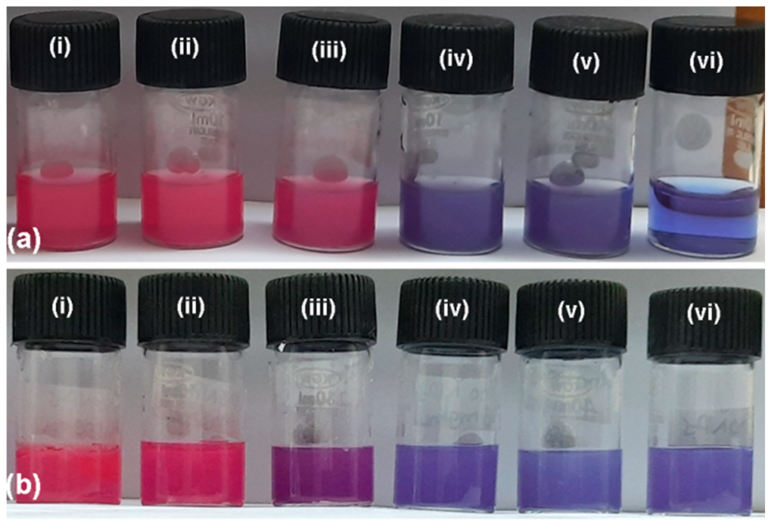
The bactericidal action of starch-capped ZnO NP against Gram-negative E. coli and Gram-positive B. subtilis was investigated using the resazurin broth dilution technique. Inoculated NB tubes containing starch (negative control) and those without NP showed pink colouration, indicating bacterial growth. In contrast, the ciprofloxacin-positive control inhibited bacterial growth, as indicated by the blue colour. The abiotic control tubes consisting of cell-free NB amended with ZnO NP showed a blue colouration. This observation confirms that there was no abiotic reduction or bleaching of resazurin by the ZnO NP. A separate control for dissolved zinc ions was not included in this study, as negligible leaching of zinc ions from the photocatalyst was observed during the recyclability studies [Figure 13a]. Based on the blue/purple colouration of the inoculated NB tubes amended with ZnO NP, the resulting MIC values against E. coli and B. subtilis were 2700 µg/mL [Figure 18a] and 1800 µg/mL [Figure 18b], respectively. The corresponding MBC values (Table 2) determined from the viable colony counts of the blue/purple coloured inoculated NB tubes amended with ZnO NP were 3600 µg/mL (Figure 19) and 2700 µg/mL (Figure 20). The dose-dependent bactericidal action of ZnO NP was shown through viable colony counts and bacterial growth reduction (%) (Table 3). For E. coli, the bacterial growth reduction (%) values were 0, 0, 0, 99.98 and 100 at 0, 900, 1800, 2700 and 3600 µg/mL of ZnO NP, respectively. For B. subtilis, the bacterial growth reduction (%) values were 0, 98.3, 99.12, 100 and 100 at 0, 900, 1800, 2700 and 3600 µg/mL of ZnO NP, respectively. The superior bactericidal action of ZnO NP against the Gram-positive B. subtilis in comparison with Gram-negative E. coli can be attributed to differential bacterial cell wall structure, in terms of thickness, composition and organization. Gram-negative bacteria are protected by a cell wall that contains a complex outer membrane composed of phospholipids and lipopolysaccharides. On the contrary, the cell wall of Gram-positive bacteria is dominated by a peptidoglycan layer [19,38,53].
Figure 18.
Bactericidal activity of ZnO NP against (a) Escherichia coli ATCC 25922 and (b) Bacillus subtilis ATCC 6633 as studied using the resazurin broth method at (i) 0 µg/mL, (ii) 900 µg/mL, (iii) 1800 µg/mL, (iv) 2700 µg/mL, (v) 3600 µg/mL and (vi) ciprofloxacin (10 µg/mL).
Table 2.
Bactericidal action of ZnO NP against E. coli and B. subtilis in terms of minimum inhibitory concentration (MIC) and minimum bactericidal concentration (MBC) values.
Figure 19.
Validation of bactericidal action in terms of viable bacterial growth and colony counts of E. coli at (a) 0 µg/mL, (b) 900 µg/mL, (c) 1800 µg/mL, (d) 2700 µg/mL, (e) 3600 µg/mL of ZnO NP, and (f) ciprofloxacin (10 µg/mL).
Figure 20.
Validation of bactericidal action in terms of viable bacterial growth and colony counts of B. subtilis at (a) 0 µg/mL, (b) 900 µg/mL, (c) 1800 µg/mL, (d) 2700 µg/mL, (e) 3600 µg/mL of ZnO NP, and (f) ciprofloxacin (10 µg/mL).
Table 3.
Bacterial growth reduction (%) of E. coli and B. subtilis treated with different concentrations of ZnO NP.
Based on the nature of the capping agent and particle size of ZnO NP, the MIC and MBC values for Gram-negative and Gram-positive bacteria are compared (Table 4). The ZnO NP of 60 nm size, capped with biomolecules from Pseudomonas hibiscicola culture supernatant (alcohols, amines, alkanes) showed higher MIC values of 10,000 µg/mL, 10,000 µg/mL and 5000 µg/mL against the Gram-negative bacteria, E. coli, Klebsiella pneumoniae and Salmonella typhimurium respectively. The values against the Gram-positive bacteria B. cereus, Staphylococcus aureus and Staphylococcus epidermis were 10,000 µg/mL, 5000 µg/mL and 5000 µg/mL, correspondingly [54]. The 50 nm sized ZnO NP capped with biomolecules from Bacillus haynesii culture free supernatant exhibited elevated MIC and MBC values of 8000 µg/mL, and 16,000 µg/mL against E. coli and 4000 µg/mL and 8000 µg/mL against S. aureus [53]. The uncapped ZnO NP, with a smaller particle size of 3 nm showed higher MIC and MBC values of 3100 µg/mL and 16,000 µg/mL against E. coli and 1500 µg/mL and 8000 µg/mL against S. aureus, ascribed to the absence of surface capping [55]. The ZnO NP of 40–50 nm size and capped with biomolecules (epicatechin, catechin, ellagic acid, rhamnoside) from Eucalyptus grandis extract exhibited lower MIC values of 625 µg/mL against both Gram-negative (E. coli, K. pneumoniae, P. aeruginosa) and Gram-positive bacteria (S. aureus), which could be accounted to the combined action of the ZnO NP and inherent antibacterial properties of the capping agents present in the leaf extract [30]. The 24.8 nm sized, capped with polysaccharides and proteins of gum kondagogu demonstrated lower MIC and MBC values of 900 µg/mL and 1800 µg/mL against E. coli and 900 µg/mL and 900 µg/mL against B. subtilis, which was accounted for the exceptional stability of the produced NP [38]. The ZnO NP, with a lower particle size of 4.7 nm and capped with proteins from Weissella cibaria culture supernatant showed lower MIC and MBC values of 625 µg/mL and 1250 µg/mL against S. aureus, attributed to the smaller particle size and presence of protein capping [56]. Similarly, citrate-capped, 4.4 nm sized ZnO NP showed a lower MIC value of 1500 µg/mL against E. coli [57]. The ZnO NP of 25–30 nm size, capped with phytochemicals (proteins, phenolics, flavonoids, alkaloids, terpenoids) from Moringa oleifera extract exhibited lower MIC and MBC values of 700 µg/mL and 700 µg/mL against E. coli and 120 µg/mL and 120 µg/mL against B. subtilis, accounted to the additive antibacterial activity of the ZnO NP and the antibacterial capping molecules from the leaf extract [19]. The 2-mercaptoethanol capped ZnO NP of 47 nm size showed an MBC value of 4000 µg/mL against E. coli, which is comparable to the findings reported in the present study [58]. The tabulated data clearly indicates that higher MIC/MBC values are associated with either capped ZnO NP of larger particle size [53,54] or uncapped ZnO NP of smaller particle size [55]. If the values are lower, they could be attributed to the additional inherent antibacterial action of the capping biomolecules [19,30], the efficient capping [38,56] and the smaller particle size [56,57]. In this study, the ZnO NP of 30.6 nm size, capped with a non toxic, non antibacterial polymer, starch showed sufficiently good bactericidal action against E. coli (3600 µg/mL) and B. subtilis (2700 µg/mL). Interestingly, the water-soluble, tetramethyl ammonium hydroxide-capped, 5.4 nm sized ZnO NP were found to be non mutagenic in the AMES bacterial reverse mutation test [17]. The present report highlights the importance of interaction between particle size, NP concentration, surface stabilization and the nature of capping agent in determining the bactericidal properties of ZnO NP.
Table 4.
Comparison of the bactericidal action of ZnO NP in terms of capping/stabilizing agent, NP size, used bacterial strain, MIC and MBC values.
The production of biopolymer-stabilized ZnO NP through the sol–gel method using potato starch in an aqueous medium exemplifies the implementation of sustainable development goals (SDG), particularly SDG 12 (Responsible Consumption and Production). A renewable, biodegradable and non-toxic capping agent was utilized without the use of harsh solvents. This study also aligns with SDG 6 (Clean Water and Sanitation) by employing dual functional ZnO NP for the photocatalyzed decolourization of tartrazine and disinfection of bacterial contamination in water [9,19,30]. Also, the free radical mediated decolourization mechanism, along with the photostability and recyclability of the catalyst, helps reduce the environmental footprint of wastewater treatment and promotes a circular economy. Therefore, it addresses SDG 9 (Industry, Innovation and Infrastructure) and offers a cost-effective, sustainable solution for treating complex effluents to safeguard aquatic ecosystems and public health.
4. Conclusions
The present study reports the calcination-free, sol–gel synthesis of ZnO NP stabilized with potato starch, a renewable feedstock. The ZnO NP serve as a bifunctional bactericide and a recyclable photocatalyst for the decolourization of tartrazine. The optimum conditions for decolourization were 2700 µg/mL of ZnO NP, 7.5 µg/mL of tartrazine and a UV exposure time of 90 min. The ROS involved in the ZnO NP-mediated photocatalytic removal of tartrazine were detected employing both spectrophotometric and photoluminescence methods. The 30.6 nm sized, starch-capped ZnO NP exhibited significant bactericidal effect on both E. coli and B. subtilis. Thus, the multifunctional properties of ZnO NP can be utilized for the treatment of wastewater derived from food industries, which are often enriched with food dyes, nutrients and microbes. Further studies are needed on bulk-scale photocatalyst production, photocatalyst doping, photocatalytic efficiency improvement, detection of photocatalytic degradation products, decolourization and degradation mechanism prediction, applicability to other industrial dyes and pigments, integration with other techniques and ecotoxicity assessment, among others [9,10,11,14,44].
Author Contributions
Conceptualization, A.J.K.; methodology, A.J.K. and V.B.K.M.; software, A.J.K.; validation, A.J.K. and V.B.K.M.; formal analysis, A.J.K. and V.B.K.M.; investigation, A.J.K.; resources, A.J.K. and V.B.K.M.; data curation, A.J.K. and V.B.K.M.; writing—original draft preparation, A.J.K.; writing—review and editing, A.J.K. and V.B.K.M.; visualization, A.J.K.; supervision, A.J.K. and V.B.K.M.; project administration, A.J.K. and V.B.K.M.; funding acquisition, A.J.K. and V.B.K.M. All authors have read and agreed to the published version of the manuscript.
Funding
This research received no external funding.
Institutional Review Board Statement
Not applicable.
Informed Consent Statement
Not applicable.
Data Availability Statement
The original contributions presented in this study, including all the primary data (spectra, DLS, kinetics, bacterial counts) are included in the Article. Further inquiries can be directed to the corresponding author.
Acknowledgments
The authors are thankful to Kulamani Dash, Head, NCCCM, BARC, for his continuous support and encouragement throughout the study. The authors acknowledge A. A. Sukumar, NCCCM, BARC, for XRD characterization of the catalyst.
Conflicts of Interest
The authors declare no conflicts of interest.
Abbreviations
The following abbreviations are used in this manuscript:
| ·OH | Hydroxyl radicals |
| BET | Brunauer–Emmett–Teller |
| CA | HiCrome coliform agar |
| CB | Conduction band |
| DCF | Dichlorodihydrofluorescein |
| DCFH2-DA | Dichlorodihydrofluorescein diacetate |
| DLS | Dynamic light scattering |
| FTIR | Fourier transform Infrared spectroscopy |
| GRAS | Generally recognized as a safe |
| hTA | Hydroxy terephthalic acid |
| HQ | Hydroquinone |
| IPA | Isopropyl alcohol |
| MBC | Minimum bactericidal concentration |
| MIC | Minimum inhibitory concentration |
| NA | Nutrient agar |
| NAC | N-acetylcysteine |
| NB | Nutrient broth |
| NBT | Nitroblue tetrazolium |
| O2·− | Superoxide radicals |
| PL | Photoluminescence |
| ROS | Reactive oxygen species |
| SAED | Selected area electron diffraction |
| TA | Terephthalic acid |
| TEM | Transmission electron microscopy |
| USFDA | US Food and Drug Administration |
| UV-vis | UV–visible absorption spectroscopy |
| VB | Valence band |
| XRD | X-ray diffraction |
| ZnO NP | Zinc oxide nanoparticles |
References
- Hussain, M.; Farid, M.; Sumbal; Ali, N.; Karam, S.; Khan, M.A. Removal of food dye tartrazine (E102) from aqueous solution using ZnO and CuO-TiO2 as photocatalysts. J. Chem. Soc. Pak. 2023, 45, 279–287. [Google Scholar] [CrossRef] [Scilit]
- Silva, J.d.; Fracacio, R. Toxicological and ecotoxicological aspects of tartrazine yellow food dye: A literature review. Rev. Bras. De Ciências Ambient. 2021, 56, 137–151. [Google Scholar] [CrossRef] [Scilit]
- Alcantara-Cobos, A.; Gutiérrez-Segura, E.; Solache-Ríos, M.; Amaya-Chávez, A.; Solís-Casados, D.A. Tartrazine removal by ZnO nanoparticles and a zeolite-ZnO nanoparticles composite and the phytotoxicity of ZnO nanoparticles. Microporous Mesoporous Mater. 2020, 302, 110212. [Google Scholar] [CrossRef] [Scilit]
- dos Santos, J.R.; de Sousa Soares, L.; Soares, B.M.; de Gomes Farias, M.; de Oliveira, V.A.; de Sousa, N.A.B.; Negreiros, H.A.; da Silva, F.C.C.; Peron, A.P.; Pacheco, A.C.L.; et al. Cytotoxic and mutagenic effects of the food additive tartrazine on eukaryotic cells. BMC Pharmacol. Toxicol. 2022, 23, 95. [Google Scholar] [CrossRef] [Scilit] [PubMed]
- Macruz, P.D.; Nippes, R.P.; de Matos Jorge, L.M.; dos Santos, O.A.A. Photocatalytic removal of persistent pollutants using eco-friendly ZnO. J. Sol-Gel Sci. Technol. 2022, 104, 387–400. [Google Scholar] [CrossRef] [Scilit]
- Vigneshwaran, N.; Kumar, S.; Kathe, A.A.; Varadarajan, P.V.; Prasad, V. Functional finishing of cotton fabrics using zinc oxide–soluble starch nanocomposites. Nanotechnology 2006, 17, 5087–5095. [Google Scholar] [CrossRef] [Scilit]
- Berradi, M.; Hsissou, R.; Khudhair, M.; Assouag, M.; Cherkaoui, O.; El Bachiri, A.; El Harfi, A. Textile finishing dyes and their impact on aquatic environs. Heliyon 2019, 5, e02711. [Google Scholar] [CrossRef] [Scilit] [PubMed]
- Mutukwa, D.; Taziwa, R.T.; Khotseng, L. Antibacterial and photodegradation of organic dyes using lamiaceae-mediated ZnO nanoparticles: A review. Nanomaterials 2022, 12, 4469. [Google Scholar] [CrossRef] [Scilit] [PubMed]
- Periyasamy, A.P. Recent advances in the remediation of textile-dye-containing wastewater: Prioritizing human health and sustainable wastewater treatment. Sustainability 2024, 16, 495. [Google Scholar] [CrossRef] [Scilit]
- Ashaduzzaman, M.; Al Muhit, M.A.; Dey, S.C.; Rahaman, M.M.; Mahmudul Hasan, H.N.; Mustary, N.; Hossain, M.K.; Das, M.K. Microwave assisted starch stabilized green synthesis of zinc oxide nanoparticles for antibacterial and photocatalytic applications. Sci. Rep. 2025, 15, 28288. [Google Scholar] [CrossRef] [Scilit] [PubMed]
- Akter, L.; Nabi, M.H.B.; Chowdhury, Y.; Any, M.A.; Rayhan, M.A.; Arnab, A.H.; Zzaman, W. Zinc oxide nanoparticles for food applications: From synthesis and functional properties to safety and sustainability. Food Front. 2026, 7, e70311. [Google Scholar] [CrossRef] [Scilit]
- Ghamarpoor, R.; Fallah, A.; Jamshidi, M. A review of synthesis methods, modifications, and mechanisms of ZnO/TiO2-based photocatalysts for photodegradation of contaminants. ACS Omega 2024, 9, 25457–25492. [Google Scholar] [CrossRef] [Scilit] [PubMed]
- Hassanein, T.F.; Mohammed, A.S.; Mohamed, W.S.; Sobh, R.A.; Zahran, M.K. Optimized synthesis of biopolymer-based zinc oxide nanoparticles and evaluation of their antibacterial activity. Egypt. J. Chem. 2021, 64, 3767–3790. [Google Scholar] [CrossRef] [Scilit]
- Mahlaule-Glory, L.M.; Hintsho-Mbita, N.C. Green derived zinc oxide (ZnO) for the degradation of dyes from wastewater and their antimicrobial activity: A review. Catalysts 2022, 12, 833. [Google Scholar] [CrossRef] [Scilit]
- Rajiv, P.; Rajeshwari, S.; Venckatesh, R. Bio-fabrication of zinc oxide nanoparticles using leaf extract of Parthenium hysterophorus L. and its size-dependent antifungal activity against plant fungal pathogens. Spectrochim. Acta. Part A Mol. Biomol. Spectrosc. 2013, 112, 384–387. [Google Scholar] [CrossRef] [Scilit] [PubMed]
- Sagadevan, S.; Vennila, S.; Lett, J.A.; Marlinda, A.R.; Hamizi, N.A.B.; Johan, M.R. Tailoring the structural, morphological, optical, thermal and dielectric characteristics of ZnO nanoparticles using starch as a capping agent. Results Phys. 2019, 15, 102543. [Google Scholar] [CrossRef] [Scilit]
- Yoshida, R.; Kitamura, D.; Maenosono, S. Mutagenicity of water-soluble ZnO nanoparticles in Ames test. J. Toxicol. Sci. 2009, 34, 119–122. [Google Scholar] [CrossRef] [Scilit] [PubMed][Green Version]
- Rastogi, L.; Beedu, S.R.; Kora, A.J. Facile synthesis of palladium nanocatalyst using gum kondagogu (Cochlospermum gossypium): A natural biopolymer. IET Nanobiotechnology 2015, 9, 362–367. [Google Scholar] [CrossRef] [Scilit] [PubMed]
- Natrayan, L.; Janardhan, G.; Nadh, V.S.; Srinivas, C.; Kaliappan, S.; Velmurugan, G. Eco-friendly zinc oxide nanoparticles from Moringa oleifera leaf extract for photocatalytic and antibacterial applications. Clean Technol. Environ. Policy 2024, 27, 5761–5773. [Google Scholar] [CrossRef] [Scilit]
- de Almeida, W.L.; Freisleben, L.C.; Brambilla, B.C.; Isoppo, V.G.; Rodembusch, F.S.; de Sousa, V.C. Influence of starch used in the sol-gel synthesis of ZnO nanopowders. J. Nanoparticle Res. 2023, 25, 75. [Google Scholar] [CrossRef] [Scilit]
- Almeida, W.L.d.; Rodembusch, F.S.; Ferreira, N.S.; Caldas de Sousa, V. Eco-friendly and cost-effective synthesis of ZnO nanopowders by Tapioca-assisted sol-gel route. Ceram. Int. 2020, 46, 10835–10842. [Google Scholar] [CrossRef] [Scilit]
- Buazar, F.; Bavi, M.; Kroushawi, F.; Halvani, M.; Khaledi-Nasab, A.; Hossieni, S.A. Potato extract as reducing agent and stabiliser in a facile green one-step synthesis of ZnO nanoparticles. J. Exp. Nanosci. 2016, 11, 175–184. [Google Scholar] [CrossRef] [Scilit]
- Saiprabhath, T.; Samuel, M.; Duraiyan, D.; Basavegowda, N.; Somu, P.; Baek, K.-H.; Haldar, D. Structural insights, modification techniques and multisectoral applications of starch from various plant sources: A review. J. Nat. Fibers 2026, 23, 2634408. [Google Scholar] [CrossRef] [Scilit]
- Hasnidawani, J.N.; Azlina, H.N.; Norita, H.; Bonnia, N.N.; Ratim, S.; Ali, E.S. Synthesis of ZnO nanostructures using sol-gel method. Procedia Chem. 2016, 19, 211–216. [Google Scholar] [CrossRef] [Scilit]
- Lahariya, V.; Pandey, K.K.; Dhoble, S.J. Structural characterization of starch capped ZnO nanoparticles. J. Phys. Conf. Ser. 2021, 1913, 012043. [Google Scholar] [CrossRef] [Scilit]
- Vu, A.-T.; Pham, T.A.T.; Do, X.T.; Tran, V.A.; Le, V.D.; Truong, D.D.; Nguyen, T.H.; Nguyen, M.V.; Kumar, A. Preparation of hierarchical structure Au/ZnO composite for enhanced photocatalytic performance: Characterization, effects of reaction parameters, and oxidizing agent investigations. Adsorpt. Sci. Technol. 2021, 2021, 5201497. [Google Scholar] [CrossRef] [Scilit]
- Sultana, K.A.; Hernandez Ortega, J.; Islam, M.T.; Dorado, Z.N.; Alvarado-Tenorio, B.; Galindo-Esquivel, I.R.; Noveron, J.C. Saccharide-derived zinc oxide nanoparticles with high photocatalytic activity for water decontamination and sanitation. Sustain. Chem. 2023, 4, 321–338. [Google Scholar] [CrossRef] [Scilit]
- Vu Anh, T.; Pham, T.A.T.; Mac, V.H.; Nguyen, T.H. Facile controlling of the physical properties of zinc oxide and its application to enhanced photocatalysis. J. Anal. Methods Chem. 2021, 2021, 5533734. [Google Scholar] [CrossRef] [Scilit] [PubMed]
- Mbrouk, O.; Hafez, H.; Mozia, S.; Othman, A.M.; Abdel Mottaleb, M.S.A. Stimulated generation of photobiogas by morphologically tuned nanostructured ZnO and ZnO/TiO2. BMC Chem. 2022, 16, 74. [Google Scholar] [CrossRef] [Scilit] [PubMed]
- Wouters, R.D.; Muraro, P.C.L.; Druzian, D.M.; Viana, A.R.; de Oliveira Pinto, E.; da Silva, J.K.L.; Vizzotto, B.S.; Ruiz, Y.P.M.; Galembeck, A.; Pavoski, G.; et al. Zinc oxide nanoparticles: Biosynthesis, characterization, biological activity and photocatalytic degradation for tartrazine yellow dye. J. Mol. Liq. 2023, 371, 121090. [Google Scholar] [CrossRef] [Scilit]
- Achouri, F.; Corbel, S.; Aboulaich, A.; Balan, L.; Ghrabi, A.; Ben Said, M.; Schneider, R. Aqueous synthesis and enhanced photocatalytic activity of ZnO/Fe2O3 heterostructures. J. Phys. Chem. Solids 2014, 75, 1081–1087. [Google Scholar] [CrossRef] [Scilit]
- Islam, M.T.; Dominguez, A.; Alvarado-Tenorio, B.; Bernal, R.A.; Montes, M.O.; Noveron, J.C. Sucrose-mediated fast synthesis of zinc oxide nanoparticles for the photocatalytic degradation of organic pollutants in water. ACS Omega 2019, 4, 6560–6572. [Google Scholar] [CrossRef] [Scilit] [PubMed]
- Matyszczuk, K.; Krzepiłko, A. Model study for interaction of sublethal doses of zinc oxide nanoparticles with environmentally beneficial bacteria Bacillus thuringiensis and Bacillus megaterium. Int. J. Mol. Sci. 2022, 23, 11820. [Google Scholar] [CrossRef] [Scilit] [PubMed]
- Boyles, M.; Murphy, F.; Mueller, W.; Wohlleben, W.; Jacobsen, N.R.; Braakhuis, H.; Giusti, A.; Stone, V. Development of a standard operating procedure for the DCFH2-DA acellular assessment of reactive oxygen species produced by nanomaterials. Toxicol. Mech. Methods 2022, 32, 439–452. [Google Scholar] [CrossRef] [Scilit] [PubMed]
- Kora, A.J.; Sashidhar, R.B. Antibacterial activity of biogenic silver nanoparticles synthesized with gum ghatti and gum olibanum: A comparative study. J. Antibiot. 2015, 68, 88–97. [Google Scholar] [CrossRef] [Scilit] [PubMed]
- Peter, A.; Jose, J.; Bhat, S.G.; K, A. A modified fluorescent probe protocol for evaluating the reactive oxygen species generation by metal and metal oxide nanoparticles in Gram-positive and Gram-negative organisms. Results Eng. 2024, 24, 102925. [Google Scholar] [CrossRef] [Scilit]
- Kora, A.J. Synthesis, characterization and in vitro antifungal action of gum ghatti capped copper oxide nanoparticles. Biointerface Res. Appl. Chem. 2022, 13, 138. [Google Scholar] [CrossRef] [Scilit]
- Kora, A.J.; Mullapudi, V.B.K. Synthesis and characterization of gum kondagogu stabilized zinc oxide nanoparticles and its application as an antibacterial agent against wastewaterborne bacteria. APMIS 2025, 133, e70046. [Google Scholar] [CrossRef] [Scilit] [PubMed]
- Kora, A.J. Antibacterial and antioxidant activities of aqueous extract of soapnuts (Sapindus mukorossi). Curr. Trends Biotechnol. Pharm. 2020, 14, 388–395. [Google Scholar] [CrossRef] [Scilit]
- Kora, A.J. Biosynthesis of reduced graphene oxide and its functionality as an antibacterial template. In Graphene from Natural Sources: Synthesis, Characterization, and Applications, 1st ed.; Al-Ahmed, A., Inamuddin, Eds.; CRC Press: Boca Raton, FL, USA, 2022; pp. 193–205. [Google Scholar] [CrossRef] [Scilit]
- Kora, A.J. Recycled wastewater from sewage treatment plant for sustainable agriculture: An Indian scenario. In Sewage and Biomass from Wastewater to Energy: Possibilities and Technology; Inamuddin, Altalhi, T., Luqman, M., Bwapwa, J.K., Eds.; Wiley-Scrivener Publishing: Austin, TX, USA, 2024; pp. 391–412. [Google Scholar] [CrossRef] [Scilit]
- Chandramouleeswaran, S.; Mhaske, S.T.; Kathe, A.A.; Varadarajan, P.V.; Prasad, V.; Vigneshwaran, N. Functional behaviour of polypropylene/ZnO–soluble starch nanocomposites. Nanotechnology 2007, 18, 385702. [Google Scholar] [CrossRef] [Scilit]
- Nair, S.; Sasidharan, A.; Divya Rani, V.V.; Menon, D.; Nair, S.; Manzoor, K.; Raina, S. Role of size scale of ZnO nanoparticles and microparticles on toxicity toward bacteria and osteoblast cancer cells. J. Mater. Sci. Mater. Med. 2009, 20, S235–S241. [Google Scholar] [CrossRef] [Scilit] [PubMed]
- Calderon Moreno, J.M.; Chelu, M.; Popa, M. Eco-friendly bioinspired synthesis and environmental applications of zinc oxide nanoparticles mediated by natural polysaccharide gums: A sustainable approach to nanomaterials fabrication. Nanomaterials 2026, 16, 407. [Google Scholar] [CrossRef] [Scilit] [PubMed]
- Singh, B.N.; Rawat, A.K.; Khan, W.; Naqvi, A.H.; Singh, B.R. Biosynthesis of stable antioxidant ZnO nanoparticles by Pseudomonas aeruginosa rhamnolipids. PLoS ONE 2014, 9, e106937. [Google Scholar] [CrossRef] [Scilit] [PubMed]
- Darroudi, M.; Sabouri, Z.; Kazemi Oskuee, R.; Khorsand Zak, A.; Kargar, H.; Hamid, M.H.N.A. Sol–gel synthesis, characterization, and neurotoxicity effect of zinc oxide nanoparticles using gum tragacanth. Ceram. Int. 2013, 39, 9195–9199. [Google Scholar] [CrossRef] [Scilit]
- Droepenu, E.K.; Asare, E.A.; Wee, B.S.; Wahi, R.B.; Ayertey, F.; Kyene, M.O. Biosynthesis, characterization, and antibacterial activity of ZnO nanoaggregates using aqueous extract from Anacardium occidentale leaf: Comparative study of different precursors. Beni-Suef Univ. J. Basic Appl. Sci. 2021, 10, 1. [Google Scholar] [CrossRef] [Scilit]
- Vijayakumar, S.; Vinoj, G.; Malaikozhundan, B.; Shanthi, S.; Vaseeharan, B. Plectranthus amboinicus leaf extract mediated synthesis of zinc oxide nanoparticles and its control of methicillin resistant Staphylococcus aureus biofilm and blood sucking mosquito larvae. Spectrochim. Acta. Part A Mol. Biomol. Spectrosc. 2015, 137, 886–891. [Google Scholar] [CrossRef] [Scilit] [PubMed]
- Tu, V.A.; Tuan, V.A. A facile and fast solution chemistry synthesis of porous ZnO nanoparticles for high efficiency photodegradation of tartrazine. Vietnam J. Chem. 2018, 56, 214–219. [Google Scholar] [CrossRef] [Scilit]
- Türkyılmaz, Ş.Ş.; Güy, N.; Özacar, M. Photocatalytic efficiencies of Ni, Mn, Fe and Ag doped ZnO nanostructures synthesized by hydrothermal method: The synergistic/antagonistic effect between ZnO and metals. J. Photochem. Photobiol. A Chem. 2017, 341, 39–50. [Google Scholar] [CrossRef] [Scilit]
- Haque, M.J.; Bellah, M.M.; Hassan, M.R.; Rahman, S. Synthesis of ZnO nanoparticles by two different methods & comparison of their structural, antibacterial, photocatalytic and optical properties. Nano Express 2020, 1, 010007. [Google Scholar] [CrossRef] [Scilit]
- Bouarroudj, T.; Aoudjit, L.; Djahida, L.; Zaidi, B.; Ouraghi, M.; Zioui, D.; Mahidine, S.; Shekhar, C.; Bachari, K. Photodegradation of tartrazine dye favored by natural sunlight on pure and (Ce, Ag) co-doped ZnO catalysts. Water Sci. Technol. A J. Int. Assoc. Water Pollut. Res. 2021, 83, 2118–2134. [Google Scholar] [CrossRef] [Scilit] [PubMed]
- Rehman, S.; Jermy, B.R.; Akhtar, S.; Borgio, J.F.; Abdul Azeez, S.; Ravinayagam, V.; Al Jindan, R.; Alsalem, Z.H.; Buhameid, A.; Gani, A. Isolation and characterization of a novel thermophile; Bacillus haynesii, applied for the green synthesis of ZnO nanoparticles. Artif. Cells Nanomed. Biotechnol. 2019, 47, 2072–2082. [Google Scholar] [CrossRef] [Scilit] [PubMed]
- Punjabi, K.; Mehta, S.; Chavan, R.; Chitalia, V.; Deogharkar, D.; Deshpande, S. Efficiency of biosynthesized silver and zinc nanoparticles against multi-drug resistant pathogens. Front. Microbiol. 2018, 9, 2207. [Google Scholar] [CrossRef] [Scilit] [PubMed]
- Zarrindokht, E.-K. Antibacterial activity of ZnO nanoparticle on Gram-positive and Gram-negative bacteria. Afr. J. Microbiol. Res. 2012, 5, 1368–1373. [Google Scholar] [CrossRef] [Scilit]
- Al-Tameemi, A.I.; Masarudin, M.J.; Abdul Rahim, R.; Mills, T.; Timms, V.J.; Neilan, B.A.; Mat Isa, N. Biosynthesis of zinc oxide nanoparticles using the supernatant of Weissella cibaria UPM22MT06 and its antibacterial and cytotoxicity properties. Biologia 2023, 78, 3315–3328. [Google Scholar] [CrossRef] [Scilit]
- Saliani, M.; Jalal, R.; Kafshdare Goharshadi, E. Effects of pH and temperature on antibacterial activity of zinc oxide nanofluid against Escherichia coli O157: H7 and Staphylococcus aureus. Jundishapur J. Microbiol. 2015, 8, e17115. [Google Scholar] [CrossRef] [Scilit] [PubMed]
- Padmavathy, N.; Vijayaraghavan, R. Enhanced bioactivity of ZnO nanoparticles-an antimicrobial study. Sci. Technol. Adv. Mater. 2008, 9, 035004. [Google Scholar] [CrossRef] [Scilit] [PubMed]

Disclaimer/Publisher’s Note: The statements, opinions and data contained in all publications are solely those of the individual author(s) and contributor(s) and not of MDPI and/or the editor(s). MDPI and/or the editor(s) disclaim responsibility for any injury to people or property resulting from any ideas, methods, instructions or products referred to in the content. |
© 2026 by the authors. Licensee MDPI, Basel, Switzerland. This article is an open access article distributed under the terms and conditions of the Creative Commons Attribution (CC BY) license.