


Abstract
The conformational composition and ultraviolet photochemistry of neutral valine isolated in solid parahydrogen were investigated using high-resolution infrared spectroscopy, ultraviolet photolysis, and density functional theory calculations. The observed infrared spectrum was analyzed by combining calculated vibrational frequencies, relative infrared intensities, and conformer-specific photolysis kinetics. Four distinct photolysis-rate categories were identified experimentally, providing direct evidence for the presence of at least four valine conformers in the parahydrogen matrix. The combined spectroscopic and kinetic analysis enabled assignment of the major absorption bands to the six lowest-energy conformers and revealed pronounced conformer-dependent photostability of valine. Upon irradiation at 213 nm, valine undergoes predominantly -carbonyl C–C bond cleavage, producing the hydrocarboxyl (HOCO) radical and 2-methylpropan-1-imine as the major photoproducts. The formation of HOCO is consistent with previous studies of amino acids isolated in solid parahydrogen and supports a common photodissociation pathway among aliphatic amino acids. The hydrogen-bonded Type II conformer exhibits significantly slower photodepletion than the Type I conformers, indicating that intramolecular hydrogen bonding enhances ultraviolet photostability of valine. These results establish a direct relationship between molecular conformation and photochemical stability in isolated amino acids while further demonstrating the unique capability of solid parahydrogen matrix isolation for conformer-specific photochemical investigations.
1. Introduction
Amino acids, the essential building blocks of proteins, play important roles in biological processes and can adopt multiple conformations stabilized by different intramolecular hydrogen-bonding interactions. Characterizing these conformers is important because they often exhibit distinct spectroscopic signatures and different chemical and photochemical reactivities. The occurrence of amino acids in extraterrestrial environments has been the subject of extensive investigation [1,2], motivated by the detection of amino acids in carbonaceous meteorites [3,4] and the prospect that prebiotic molecules may be synthesized in space. While in condensed phases intermolecular interactions stabilize the zwitterionic form, amino acids are expected to exist predominantly in their non-zwitterionic neutral form under interstellar gas-phase conditions [5]. Consequently, laboratory studies that preserve and characterize non-zwitterionic neutral amino acids under isolated conditions are important for understanding their intrinsic properties and behavior in extraterrestrial environments [6].
Valine is one of the twenty standard amino acids and has been identified in several carbonaceous meteorites [3,4]. Owing to its branched side chain and multiple internal degrees of freedom, valine exhibits several low-energy conformers stabilized by different intramolecular hydrogen-bonding interactions [7]. This conformational complexity makes valine an attractive model system for investigating relationships among molecular structure, vibrational spectra, and photochemical reactivity. The conformational landscape of valine has been investigated theoretically using ab initio and high-level electronic structure calculations [7,8], while condensed-phase, crystalline, and jet-cooled rotational spectroscopic studies have identified several of its low-energy conformers [9,10,11,12]. However, high-resolution infrared spectroscopic studies of non-zwitterionic neutral valine under isolated conditions remain relatively limited, particularly those aimed at characterizing individual conformers.
Photodissociation and fragmentation of valine have been investigated using several gas-phase techniques. Collision-induced dissociation studies of valine cluster cations revealed preferential loss of the carboxyl group [13], while electron-ionization and vacuum-ultraviolet experiments identified numerous ionic fragmentation channels through mass spectrometric detection [14,15]. These studies have provided valuable information on the fragmentation pathways of ionized valine. However, much less is known about the ultraviolet photochemistry of non-zwitterionic neutral valine. In particular, whether different conformers exhibit distinct photodissociation behaviors or photostabilities has not yet been established.
Matrix-isolation spectroscopy provides an effective approach for studying isolated non-zwitterionic neutral amino acids under cryogenic conditions [16,17]. In particular, solid parahydrogen provides an exceptionally weakly perturbing environment, yielding narrow infrared absorption bands with minimal matrix-induced frequency shifts [18,19,20]. The resulting narrow spectral features enable vibrational bands of different conformers to be resolved more readily than in conventional matrix-isolation experiments. Furthermore, compared with conventional rare-gas matrices [21], the weak intermolecular interactions and quantum nature of solid parahydrogen substantially reduce cage effects, allowing photofragments to separate rather than remain trapped within a rigid matrix environment [22,23]. Consequently, photodissociation processes that are strongly suppressed in rare-gas matrices can often be observed in solid parahydrogen.
Recent studies from our laboratory have shown that near-200 nm ultraviolet photolysis of several non-zwitterionic neutral amino acids isolated in solid parahydrogen proceeds predominantly through cleavage of the -carbonyl C–C bond, producing hydrocarboxyl (HOCO) radicals together with amino acid-specific coproducts [24,25]. This reaction pathway has been observed for glycine, alanine, leucine, serine, and proline, suggesting that -carbonyl C–C bond cleavage is a general photodissociation pathway of proteinogenic amino acids under near-200 nm ultraviolet irradiation. The HOCO radical is of particular astrochemical interest because it has been proposed as an important intermediate in the formation of interstellar CO2 [26,27,28]. These studies therefore establish a connection between amino acid photochemistry and interstellar carbon chemistry. While the dominant photodissociation pathway appears to be common among amino acids, the influence of molecular conformation on ultraviolet photochemistry has not been systematically investigated. Because different conformers possess distinct intramolecular hydrogen-bonding arrangements and electronic structures, they are expected to exhibit different photochemical stabilities under ultraviolet irradiation. Recent theoretical calculations have suggested that ultraviolet photolysis of amino acids may proceed through triplet-state pathways, leading to facile -C–C bond cleavage and formation of HOCO and aminoalkyl radical intermediates [29]. The present work provides experimental evidence relevant to these proposed reaction pathways through conformer-resolved photochemistry and photoproduct identification.
In the present work, non-zwitterionic neutral valine was isolated in solid parahydrogen and investigated using high-resolution infrared spectroscopy, ultraviolet photolysis, and density functional theory calculations. The observed infrared spectrum was assigned by combining calculated vibrational frequencies, relative infrared intensities, and conformer-specific photolysis kinetics. The photolysis kinetics provide direct experimental evidence for the presence of at least four valine conformers in the solid parahydrogen matrix and serve as an independent constraint for the conformer assignments. The resulting photoproducts were subsequently characterized, and the relationship between molecular conformation and ultraviolet photostability was investigated.
2. Materials and Methods
The experimental apparatus has been described previously [30]. Normal hydrogen gas (99.99%, Praxair Canada Inc., Mississauga, ON, Canada) was converted to >99.99% parahydrogen in a converter containing (FeOH)O catalyst maintained at 14 K using a closed-cycle helium refrigerator [31]. The converted parahydrogen was stored in a 2 L Teflon-lined stainless-steel cylinder to prevent back-conversion.
DL-valine (99%, Sigma-Aldrich, Oakville, ON, Canada) was loaded into a homemade Knudsen cell and heated to 403 K [32]. The sublimation conditions were optimized to minimize the formation of valine clusters. At higher sublimation temperatures, additional peaks and broadening were observed, indicating contributions from aggregated species. The sublimation temperature was therefore reduced to 403 K, at which these additional features were no longer observed and the spectrum consisted of sharp, well-resolved lines. This optimized condition was used for all measurements reported here. A comparison of spectra obtained under a higher sublimation temperature and optimized conditions is shown in Figure S3 in the Supplementary Information (SI). The vaporized sample was co-deposited with parahydrogen onto a BaF2 window maintained at 3.5 K at a flow rate of 8 sccm [33].
Infrared absorption spectra of valine isolated in solid parahydrogen were recorded at a resolution of 0.1 cm−1 using a Fourier-transform infrared spectrometer (Bruker IFS 125HR). Ultraviolet irradiation was carried out at 213 nm using the fifth harmonic of a Nd:YAG laser (Litron Lasers Ltd., Rugby, UK, Nano-LG 130-50, 50 Hz repetition rate) for 4 h. The pulse energy before the chamber entrance window was approximately μJ at 50 Hz. To minimize two-photon excitation, the beam was expanded using a mm concave UV-grade fused-silica lens, thereby reducing the photon fluence per pulse.
Geometry optimizations and harmonic vibrational frequency calculations were performed at the B97X-D/def2-TZVPP level of theory using the Gaussian 09 package [34]. The B97X-D functional was selected because its range-separated hybrid formulation and treatment of dispersion interactions are suitable for describing the intramolecular interactions and relative energetics of different conformers. The def2-TZVPP basis set provides triple- valence flexibility with extensive polarization functions, giving a practical balance between accuracy and computational cost for geometry optimizations and vibrational-frequency calculations. Accordingly, final geometries and harmonic vibrational wavenumbers of valine and its proposed photoproducts were calculated consistently at the B97X-D/def2-TZVPP level. M06-L/6-311+G(2d,p) was used for the initial potential-energy-surface survey of the photoproducts, after which all identified minima were reoptimized and characterized at the B97X-D/def2-TZVPP level.
Anharmonic vibrational frequency calculations of valine conformers were also carried out for comparison and are listed in the SI. Unscaled harmonic frequencies are listed in the SI, whereas scaled frequencies (0.9564 for modes below 1500 cm−1, 0.9575 for modes between 1500–3100 cm−1, and 0.9289 for modes above 3100 cm−1) were used for comparison with the experimental spectra for valine conformers. These scaling factors were empirically determined to improve agreement with the observed valine bands. For 2-methylpropan-1-imine and the aminoalkyl radical photoproducts, a standard method-dependent scaling factor of 0.957 [35] was applied uniformly to the harmonic frequencies calculated at the B97X-D/def2-TZVPP level.
The conformer assignments were based primarily on the scaled harmonic frequencies because they provided more consistent agreement with the experimental spectra over the entire spectral range, while the anharmonic frequencies were used as additional support. Relative Gibbs free energies and Boltzmann populations at 403 K were obtained from the thermochemical analysis. Calculated vibrational frequencies together with photolysis rate constants obtained by fitting the decay profiles of selected absorption bands as a function of photon fluence [30] were used for conformer assignments.
3. Results and Discussion
3.1. Valine Conformational Analysis in Parahydrogen Matrix
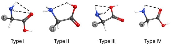
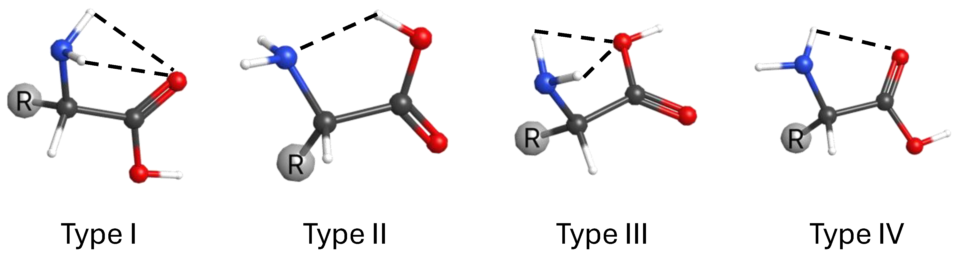
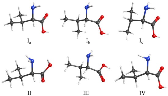
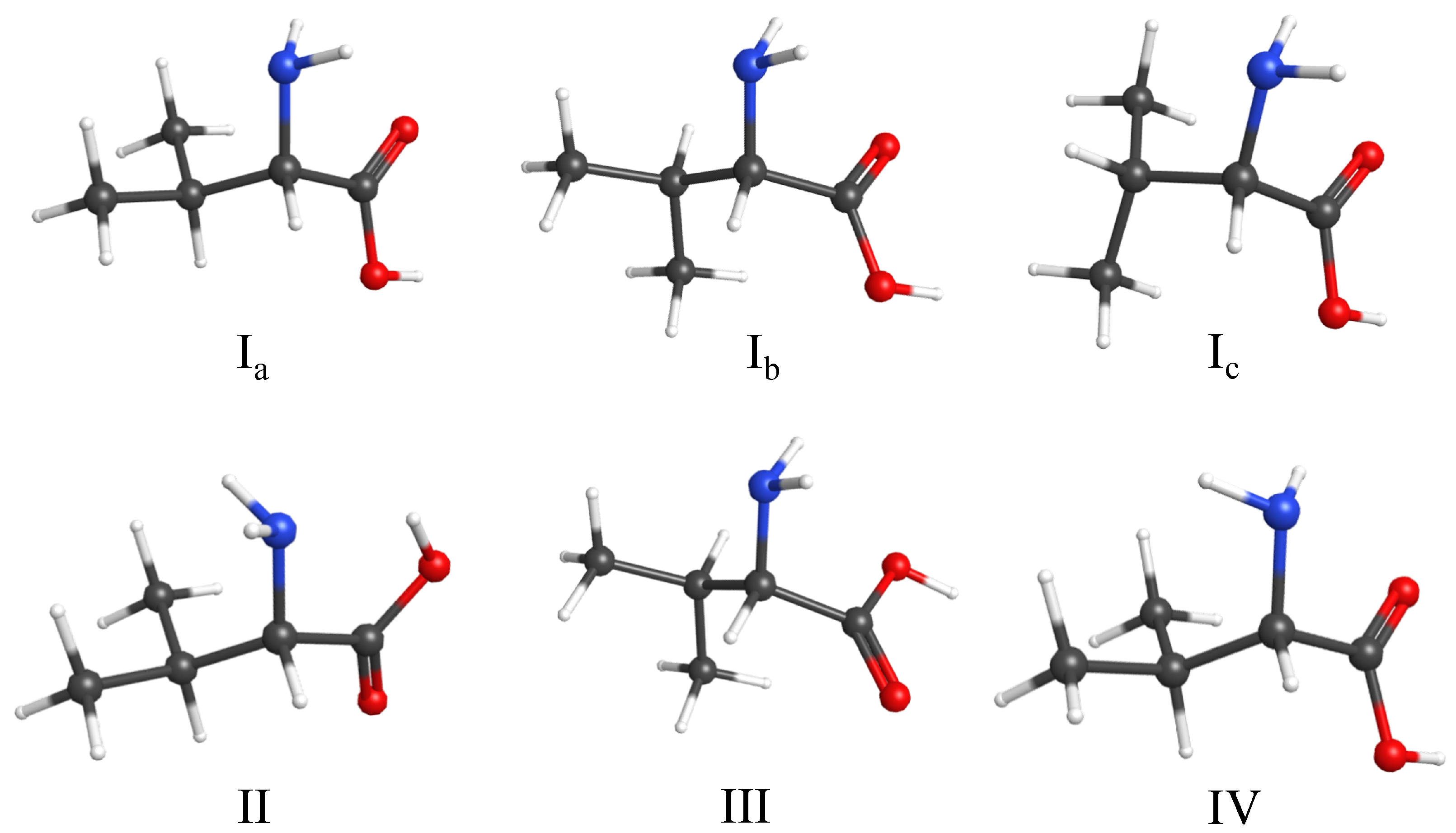
Previous theoretical studies identified six low-energy conformers of non-zwitterionic neutral valine that are stabilized by different intramolecular hydrogen-bonding interactions [8]. These conformers were used as the initial structural candidates for the present spectroscopic analysis. Geometry optimizations and vibrational frequency calculations were performed at the B97X-D/def2-TZVPP level of theory. Based on their hydrogen-bonding arrangements, the conformers can be classified into four structural types, Type I, II, III and IV, as illustrated in Figure 1. The optimized structures of the six conformers are shown in Figure 2, and their relative energies and calculated populations at the sublimation temperature (403 K) are summarized in Table 1.
Figure 1.
Four types of valine conformers classified according to their intramolecular hydrogen-bonding arrangements. Dashed lines denote intramolecular hydrogen bonds. Oxygen, nitrogen, carbon, and hydrogen atoms are shown in red, blue, black, and white, respectively.
Figure 2.
Optimized structures of the six lowest-energy valine conformers calculated at the B97X-D/def2-TZVPP level of theory. Oxygen, nitrogen, carbon, and hydrogen atoms are shown in red, blue, black, and white, respectively. The Type I conformers (Ia, Ib, and Ic) are shown in the top row, and conformers II, III, and IV are shown in the bottom row.
Table 1.
Relative electronic energies (E), zero-point-energy-corrected relative energies (EZPE), Gibbs free energies at 403 K (G403K), and calculated Boltzmann populations of valine conformers calculated at the B97X-D/def2-TZVPP level of theory.
Type I conformers (Ia, Ib, and Ic) are stabilized by an N–H⋯O=C hydrogen bond between the amino and carboxyl groups. Type II contains an O–H⋯N hydrogen bond, while Type III is characterized by an N–H⋯O–H interaction. Type IV also exhibits an N–H⋯O=C hydrogen bond but differs from Type I in the orientation of the NH2 group relative to the carboxyl group. Conformers Ia, Ib, and Ic differ primarily in the orientation of the isopropyl side chain [17]. The conformer nomenclature follows the amino-acid backbone classification introduced by Lesarri and co-workers [12]. The Type II, III, and IV structures shown in Figure 2 correspond to conformers IIa, IIIb, and IVa, respectively. Although each of these conformational families contains multiple conformers, IIa, IIIb, and IVa are the lowest-energy members and are therefore expected to be the most populated under the present experimental conditions. For simplicity, these conformers are referred to throughout this work as conformers II, III, and IV, respectively. Complete optimized geometrical parameters, including bond lengths, bond angles, and dihedral angles, are provided in Table S8 of the SI.
As shown in Table 1, although conformer II is calculated to be the global minimum on the potential-energy surface, conformer Ia becomes the most populated species at the sublimation temperature (403 K) because of entropic contributions to the Gibbs free energy. As a result, Type I conformers are predicted to account for approximately 56% of the equilibrium population, followed by Type II (28%), whereas Types III and IV contribute only minor fractions. These calculated populations suggest that the strongest experimental absorption bands are likely to originate predominantly from the Type I conformers and conformer II, providing a useful starting point for the spectroscopic assignments discussed below.
3.2. Observed Infrared Spectra of Valine in Solid Parahydrogen
3.2.1. As-Deposited Infrared Spectrum of Valine
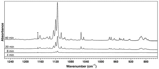
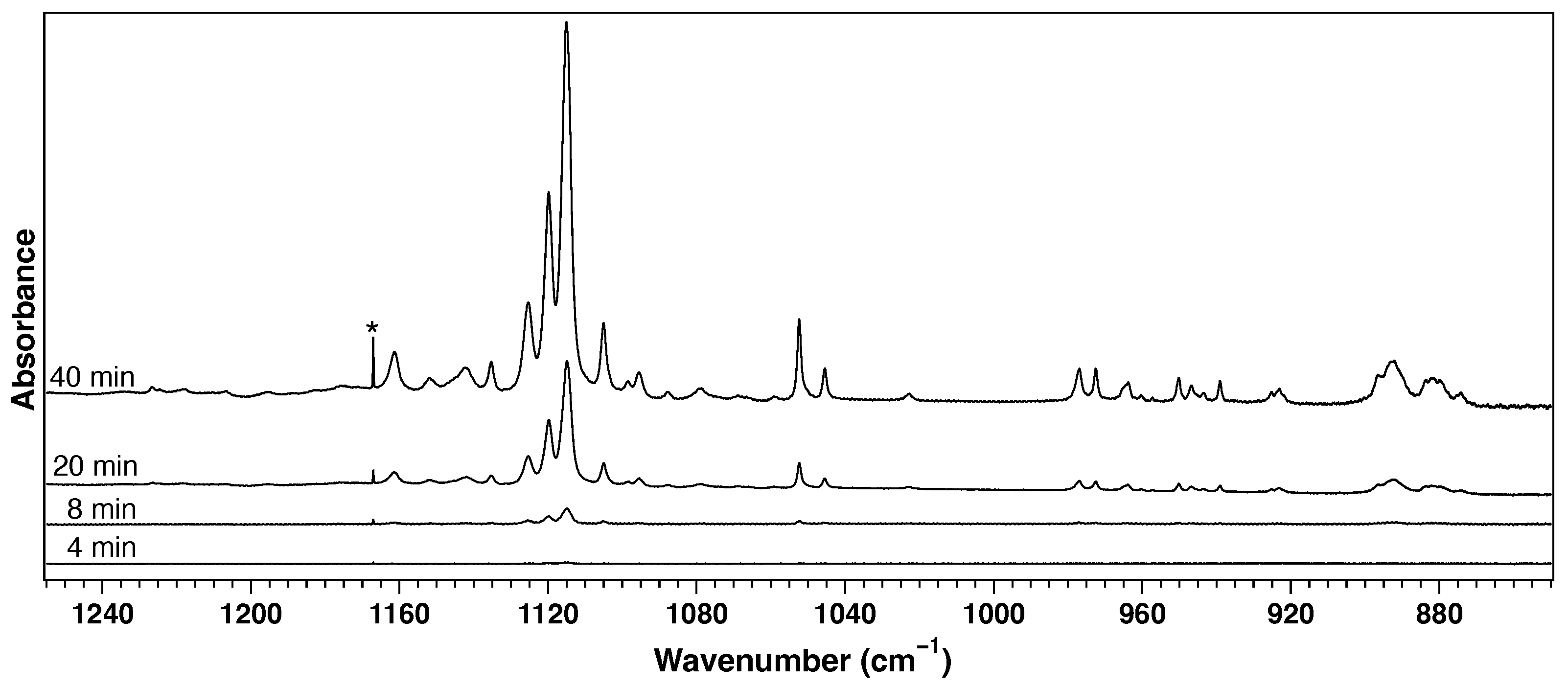
Figure 3 shows the infrared spectra recorded during deposition of valine in solid parahydrogen in the spectral region from 850 to 1250 cm−1. The absorbance increases continuously with deposition time, indicating steady accumulation of valine molecules in the matrix. No new absorption bands or noticeable changes in the spectral pattern are observed during deposition, suggesting that the deposited molecules remain chemically stable under the present experimental conditions.
Figure 3.
Infrared spectra of valine isolated in solid parahydrogen recorded during deposition after 4, 8, 20, and 40 min. The feature marked with an asterisk (*) arises from solid parahydrogen and is not associated with valine.
The deposited sample exhibits numerous sharp absorption bands with excellent signal-to-noise ratios. The most prominent feature is a group of intense absorptions centered near 1120 cm−1, accompanied by several weaker but well-resolved bands in the 1040–1170 cm−1 region. Additional well-resolved absorptions are observed in the low-frequency fingerprint region (700–900 cm−1), including prominent bands near 880 cm−1. The sharp linewidths and reproducible spectral pattern demonstrate the excellent spectral resolution afforded by the solid parahydrogen matrix, providing sufficient spectral resolution to distinguish contributions from multiple conformers. The sharpness of the observed bands also supports their assignment to isolated valine monomers. At higher sublimation temperatures, additional broadened features appeared, consistent with aggregation, whereas these features were absent under the optimized deposition conditions used in the present experiments (Figure S3 in the SI).
3.2.2. Ultraviolet Irradiation and Difference Spectra
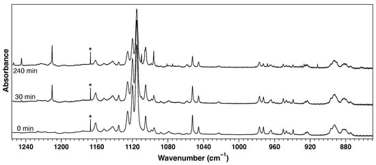
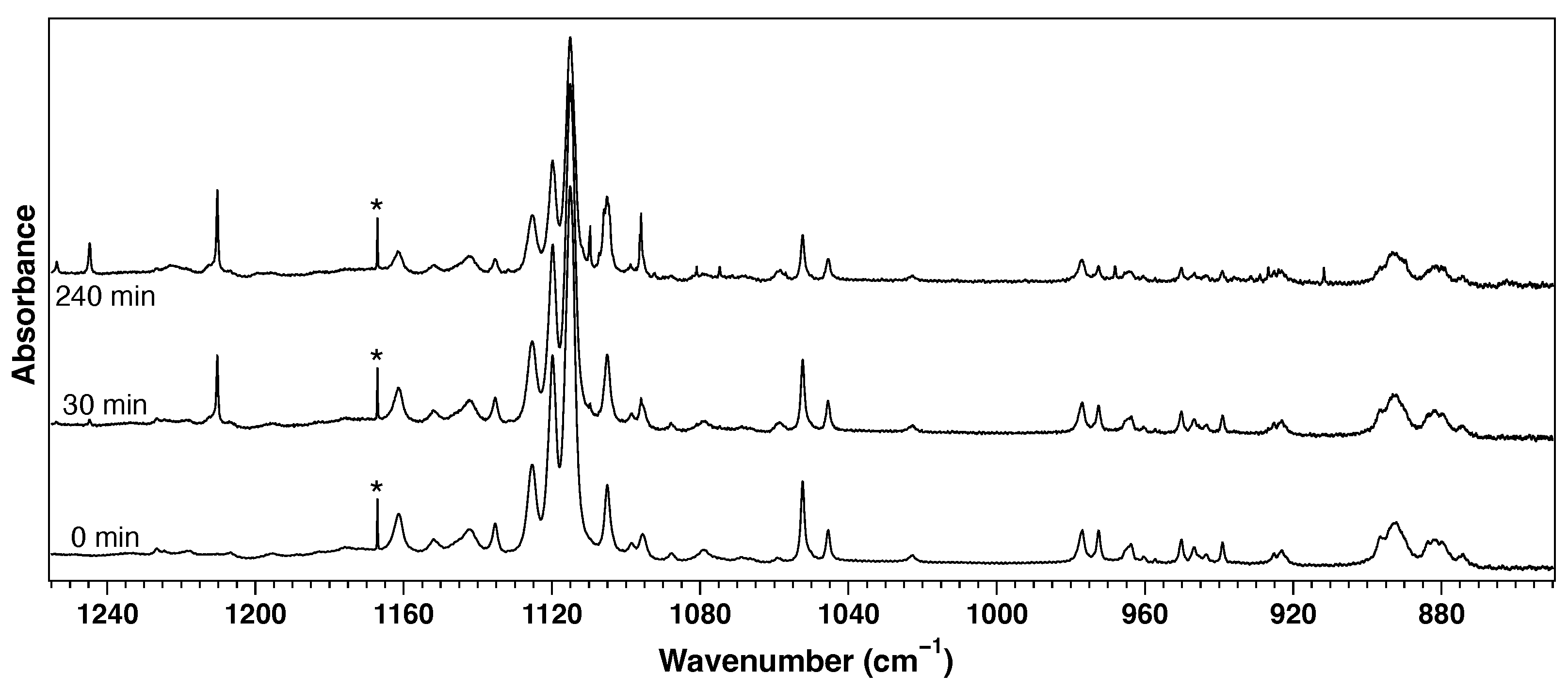
The deposited valine sample was irradiated at 213 nm, and the resulting spectral changes were monitored by infrared spectroscopy. Representative spectra recorded before irradiation and after 30 and 240 min of irradiation are shown in Figure 4. Upon irradiation, the valine absorption bands decrease progressively in intensity, while new absorption bands gradually appear throughout the spectrum.
Figure 4.
Infrared spectra of valine isolated in solid parahydrogen before irradiation (0 min) and after 30 and 240 min of irradiation at 213 nm. The feature marked with an asterisk (*) arises from solid parahydrogen and is not associated with valine.
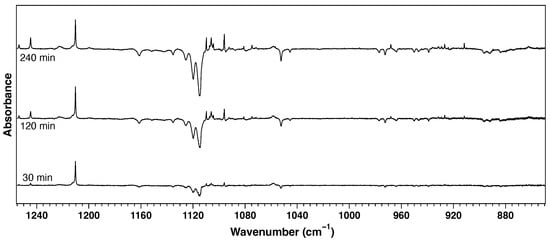
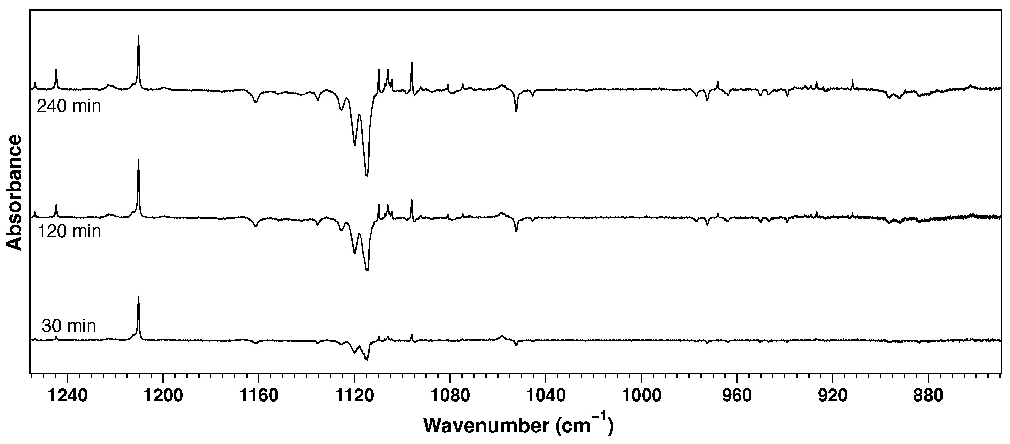
The corresponding difference spectra are shown in Figure 5, where negative features represent depletion of valine and positive features correspond to formation of photoproducts. The intensities of both the negative and positive bands increase systematically with irradiation time, demonstrating continuous photodissociation of valine and accumulation of photoproducts during irradiation.
Figure 5.
Difference spectra obtained after 30, 120, and 240 min of irradiation at 213 nm. Negative features correspond to depletion of valine, while positive features correspond to photoproduct formation.
Careful inspection of Figure 4 shows that the depletion of the valine absorption bands is not uniform. These differences become evident upon quantitative analysis of the decay profiles, as discussed in the following section.
3.3. Photolysis Kinetics
To investigate the photochemical behavior of matrix-isolated valine, infrared spectra were recorded repeatedly at approximately 4 min intervals during ultraviolet irradiation. Simultaneously, the laser pulse energy was monitored for every pulse by measuring a fraction of the incident beam reflected immediately before the vacuum chamber. The accumulated photon fluence was calculated from the integrated pulse energy and used as the independent variable for the kinetic analysis. The raw integrated areas of individual infrared absorption bands were fitted with a single-exponential decay function including a residual intensity,
where N is the cumulative photon fluence and k is the photolysis rate constant (cm2 photon−1). A single-exponential fit was used to determine an effective photolysis rate constant for each observed absorption band. For bands containing unresolved contributions from multiple conformers, the measured decay may, in principle, contain multiple exponential components. However, because the observed conformer-dependent rates differ by only a factor of a few, double- or multiexponential fits yielded large uncertainties and did not permit reliable separation of the individual components. We therefore applied the single-exponential model consistently to all bands. The resulting effective rate constants were used primarily to classify the bands into four reproducible photolysis-rate categories, very fast (VF), fast (F), medium (M), and slow (S), rather than being interpreted as precise conformer-specific rate constants.
The decay of each absorption band was measured independently in two photolysis experiments, and the resulting rate constants were averaged. The complete fitting results are summarized in Table S1 in the SI. For clarity, the decay curves shown in Figure 6 were normalized by subtracting the fitted residual intensity and scaling the initial intensity to unity. This normalization facilitates direct comparison of the decay profiles and minimizes the influence of baseline offsets arising from overlapping absorption bands.
Figure 6.
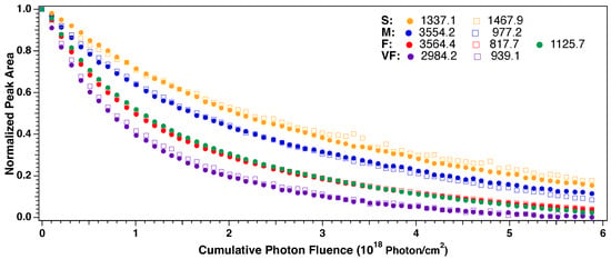
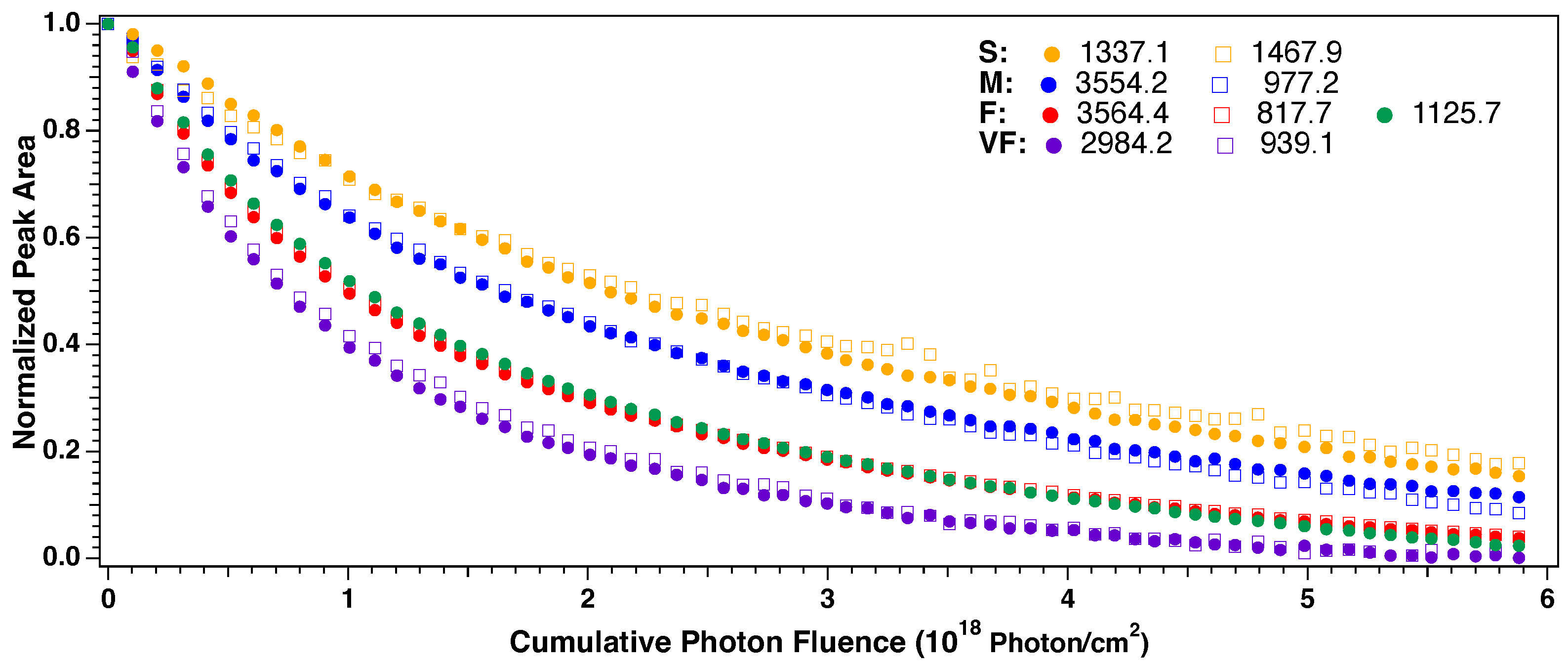
Photolysis kinetics of representative infrared absorption bands of valine isolated in solid parahydrogen during irradiation at 213 nm. The normalized peak areas are plotted as a function of cumulative photon fluence and scaled such that the initial intensity and the intensity extrapolated to infinite photon fluence are 1 and 0, respectively. The representative bands are grouped into four reproducible photolysis-rate categories: slow (S; 1337.1 and 1467.9 cm−1), medium (M; 3554.2 and 977.2 cm−1), fast (F; 3564.4, 817.7, and 1125.7 cm−1), and very fast (VF; 2984.2 and 939.1 cm−1). The conformer assignments corresponding to these decay-rate categories are discussed in Section 3.4. Integrated absorbances were determined by direct numerical integration over fixed wavenumber intervals for each peak. Therefore, fitting-derived error bars are not available for the individual data points.
The photon fluence was calculated from the measured laser pulse energy immediately before the vacuum chamber. Owing to geometrical constraints of the irradiation optics and scattering within the matrix, the entire sample was not exposed uniformly to the incident ultraviolet beam. Consequently, the actual photon fluence experienced by the molecules is smaller than the measured incident fluence, and the reported photolysis rate constants should be regarded as effective values. However, this does not affect the comparison of relative photolysis rates among different absorption bands, which forms the basis of the conformer assignments.
Figure 6 shows that different infrared absorption bands exhibit markedly different photolysis rates. The representative decay curves can be classified into four reproducible photolysis-rate categories, denoted as slow (S), medium (M), fast (F), and very fast (VF). Since all of these absorption bands are present in the as-deposited spectrum and originate from the parent valine molecule rather than photoproducts, the observed differences in photolysis behavior demonstrate that the infrared spectrum contains contributions from multiple valine conformers with different photochemical stabilities.
Conformer-dependent photochemical behavior has also been reported for the closely related amino acid proline. Hadidi et al. observed pronounced conformer dependence in the VUV photoionization and subsequent fragmentation of gas-phase proline [36]. Although that study involved ionization at higher photon energies, whereas the present work examines neutral photolysis of valine at 213 nm in solid parahydrogen, both studies demonstrate that the initial molecular conformation and intramolecular hydrogen-bonding arrangement can strongly influence amino-acid photodynamics.
The measured photolysis rate constants span a wide range, from approximately 2.3 to 7.7 × 10⁻¹⁹ cm2 photon−1. The four decay-rate categories have mean rate constants of approximately 6.7, 5.3, 4.1, and 2.9 × 10⁻¹⁹ cm2 photon−1 for the VF, F, M, and S groups, respectively. The same four kinetic categories were independently observed in both photolysis experiments, demonstrating that the classification is highly reproducible. Since absorption bands arising from the same conformer are expected to exhibit similar photolysis behavior, the observation of four reproducible decay-rate categories indicates that at least four conformers of valine coexist in the solid parahydrogen matrix. These kinetic categories therefore provide an independent experimental constraint for the conformer assignments discussed in the following section.
3.4. Conformer Assignments
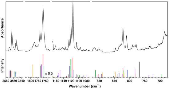
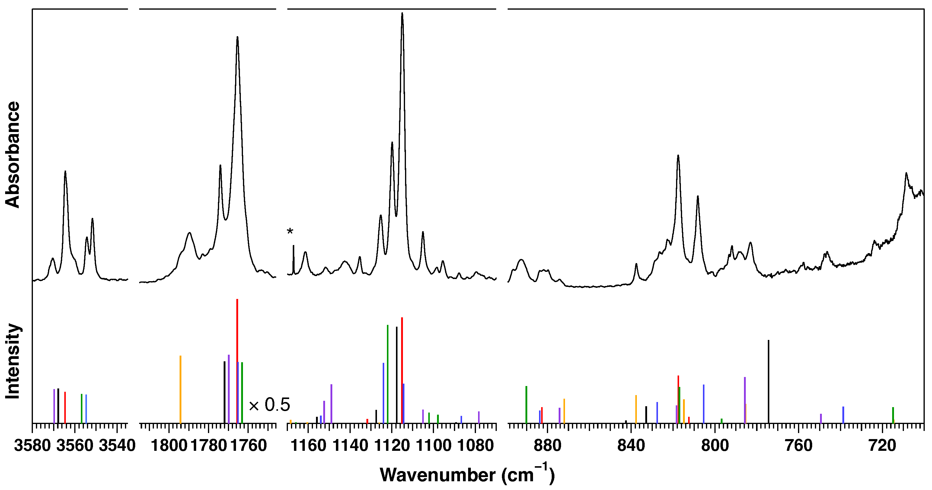
Having established experimentally that at least four conformers of valine coexist in the solid parahydrogen matrix, the remaining task is to identify the conformer responsible for each group of absorption bands. Figure 7 compares the experimental spectrum with the calculated spectra of the six low-energy conformers (Ia, Ib, Ic, II, III, and IV), while the complete assignments are summarized in Table S1 and representative assignments are listed in Table 2.
Figure 7.
Experimental infrared spectrum of valine isolated in solid parahydrogen (upper trace) compared with calculated harmonic vibrational frequencies and intensities of conformers Ia, Ib, Ic, II, III, and IV at the B97X-D/def2-TZVPP level of theory (lower sticks). Ia (red), Ib (blue), Ic (green), II (yellow), III (purple), and IV (black). The feature marked with an asterisk (*) arises from solid parahydrogen and is not associated with valine.
Table 2.
Selected assigned valine conformer bands in pH2, grouped by vibrational region. Full list is provided in Table S1 of the SI.
The assignments were established by combining the experimental infrared spectra with density functional theory (DFT) calculations. Four complementary criteria were considered simultaneously: (i) agreement between the observed and calculated vibrational frequencies, (ii) consistency between the observed and calculated relative infrared intensities, (iii) agreement with the characteristic vibrational modes predicted for each conformer, and (iv) consistency with the experimentally determined photolysis decay-rate categories. In addition, the calculated conformer populations predict that Ia and II are the two most abundant species. Therefore, the strongest experimental bands are expected to originate primarily from these conformers and provide the initial framework for the assignments. The remaining bands were then assigned by simultaneously considering all four criteria to obtain the most self-consistent interpretation of the experimental spectrum.
The assignments of the major absorption bands are discussed below according to the four most diagnostic spectral regions: the O–H stretching (3420–3580 cm−1), carbonyl stretching (1740–1810 cm−1), mixed skeletal stretching (1080–1180 cm−1), and low-frequency fingerprint (700–900 cm−1) regions.
3.4.1. O–H Stretching Region (3420–3580 cm−1)
The O–H stretching region provides the most diagnostic starting point for the conformer assignments because each conformer exhibits a distinct stretching frequency with relatively little overlap. The strongest observed band at 3564.4 cm−1 belongs to the fast (F) decay category and is assigned to the O–H stretching vibration of conformer Ia. The neighboring band at 3551.6 cm−1 is assigned to conformer Ib, while the band at 3554.2 cm−1, which belongs to the medium (M) decay category, is assigned to conformer Ic. A particularly distinctive feature is observed for conformer II. Owing to intramolecular hydrogen bonding between the hydroxyl group and the amino group, its O–H stretching vibration is strongly red-shifted to 3420.2 cm−1 with substantially lower intensity and belongs to the slow (S) decay category, providing a unique spectroscopic signature for this conformer. Additional weaker absorptions in the 3530–3580 cm−1 region are attributed mainly to conformers III and IV. These assignments establish that the fast (F) and slow (S) decay categories correspond to the Type I conformers and conformer II, respectively, providing the basis for interpreting the remaining spectral regions.
3.4.2. Carbonyl Stretching Region (1740–1810 cm−1)
The carbonyl stretching region provides an independent confirmation of the assignments established from the O–H stretching region. The isolated band at 1789.5 cm−1 belongs to the slow (S) decay category and coincides with the strongest calculated band of conformer II. The intense band at 1765.3 cm−1 contains unresolved contributions from conformers Ia, Ib, and Ic. Its fitted rate constant therefore represents the effective decay of the overlapping absorption rather than the rate constant of an individual conformer. The observed medium (M) decay of this band is therefore consistent with overlapping contributions from the fast-decaying Type I conformers Ia and Ib together with the medium-decaying conformer Ic. The weaker band at 1774.0 cm−1 agrees with the calculated bands of conformers III and IV and is therefore assigned to overlapping contributions from these two conformers. The apparent medium (M) decay of this band is consistent with the overlap of contributions from conformers with different photolysis rates.
3.4.3. Mixed Skeletal Stretching Region (1080–1180 cm−1)
The mixed skeletal stretching region contains the strongest absorptions of the Type I conformers and provides further confirmation of the assignments. The very intense band at 1115.1 cm−1 belongs to the fast (F) decay category and is assigned to the vibration of conformer Ia. The neighboring fast-decaying bands at 1119.9 and 1125.7 cm−1 are assigned to the and vibrations of conformer Ib, while the fast-decaying band at 1135.4 cm−1 is assigned to the vibration of conformer Ia. Medium-decay bands at 1098.5, 1142.4, and 1152.1 cm−1 are assigned to conformer IV, conformer Ic, and conformer IV, respectively. The agreement between the observed frequencies, relative intensities, and decay-rate categories provides strong support for these assignments despite partial overlap of several bands.
3.4.4. Low-Frequency Fingerprint Region (700–900 cm−1)
The low-frequency fingerprint region provides the final confirmation of the conformer assignments. The strong fast-decaying bands at 808.2 and 817.7 cm−1 are assigned to the and vibrations of conformer Ia, respectively. The medium-intensity, slow-decaying band at 788.1 cm−1 confirms the assignment of conformer II, whereas the weak very-fast-decaying bands at 746.1 and 837.8 cm−1 establish conformer III as the very fast (VF) decay component. The remaining bands are assigned by simultaneously considering the photolysis decay category together with agreement between the calculated and observed frequencies and relative infrared intensities. Although several absorptions remain partially overlapped, the combined analysis of frequencies, intensities, characteristic vibrational modes, and photolysis kinetics provides a consistent assignment of all six calculated valine conformers.
The combined analysis of vibrational frequencies, relative infrared intensities, characteristic vibrational modes, and photolysis kinetics provides a consistent assignment of the observed infrared spectrum to the six lowest-energy conformers of valine. The photolysis kinetics establish four reproducible decay-rate categories, which serve as an independent experimental constraint for the assignments. The strongest observed absorptions are consistently assigned to conformers Ia and II, whereas conformers Ib, Ic, III, and IV contribute primarily weaker bands. The observed conformer distribution is inconsistent with thermal equilibrium at the 3.5 K matrix temperature, where conformer II would be expected to dominate. Instead, it remains much closer to the distribution established prior to deposition, indicating that only limited conformational relaxation occurs during matrix isolation. This behavior contrasts with conventional rare-gas matrix isolation, where stronger collisional relaxation generally drives the conformer populations toward the lowest-energy structure. The conformer assignments and their corresponding photolysis-rate categories are summarized in Table 3.
Table 3.
Summary of conformer assignments and photolysis-rate categories.
3.5. Photochemistry
Because six low-energy conformers are predicted theoretically, the four kinetic categories imply that at least some conformers must share similar photolysis rates. Having established the conformer assignments of the parent valine bands, the newly appearing absorptions produced during ultraviolet irradiation can now be assigned to the photoproducts. These product bands are clearly visible in the difference spectra (Figure 5) and increase progressively with irradiation time. The observed frequencies were compared with previously reported spectra in solid parahydrogen and quantum-chemical calculations to identify the photoproducts.
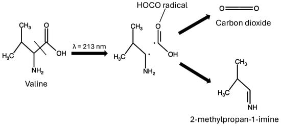
The HOCO radical is identified from characteristic absorptions at 1210.3, 1845.1, and 3612.1 cm−1, corresponding to the H–O–C bending, C=O stretching, and O–H stretching modes, respectively. These frequencies agree well with previously reported spectra of HOCO isolated in solid parahydrogen [37]. The simultaneous observation of these three characteristic absorptions provides unambiguous evidence for the formation of HOCO radicals. Their formation is consistent with -carbonyl C–C bond cleavage, the dominant photodissociation pathway previously established for glycine, alanine, leucine, serine, and proline isolated in solid parahydrogen [24,25].
Additional absorptions assigned to CO2 (2343 cm−1), 13CO2 (2278.8 cm−1), and CO (2143 cm−1) are consistent with secondary decomposition of HOCO within the matrix. A weak absorption at 1768.6 cm−1 is assigned to trans-formic acid, likely formed through the reaction of HOCO with residual hydrogen atoms in the matrix.
Additional product bands at 1244.8, 1315.7, and 1645.7 cm−1 are tentatively assigned to 2-methylpropan-1-imine and correspond to the C–H bending, CH3 bending, and C=N stretching vibrations, respectively. The observed frequencies are in reasonable agreement with the calculated spectrum (Tables S9–S12 in the SI), particularly for the characteristic C=N stretching vibration. Further support for this tentative assignment is provided by the formation profiles shown in Figure S4 in the SI. The HOCO band at 1845.0 cm−1 and the bands tentatively assigned to 2-methylpropan-1-imine exhibit simultaneous growth during the initial stage of irradiation, consistent with their formation as coproducts from the same primary photodissociation pathway. With increasing cumulative photon fluence, however, the HOCO band reaches a maximum and subsequently decreases, whereas the imine bands continue to grow. This difference is attributed to secondary photolysis of HOCO to produce CO2+ H, as previously observed in the photolysis of leucine in solid parahydrogen [24,25]. Together with the concurrent formation of HOCO, these observations support the tentative assignment of 2-methylpropan-1-imine as the coproduct formed by -carbonyl C–C bond cleavage. This interpretation is also consistent with recent theoretical studies, which predict low-barrier -C–C bond cleavage leading to HOCO and aminoalkyl radicals following ultraviolet excitation of amino acids [29].
The assigned photoproduct bands are summarized in Table 4. All observed photoproduct bands, including unassigned features, are listed in Table S13 in the SI. Weak features at 2025.1 and 3628.9 cm−1 may be attributable to HNC formed through secondary photochemistry, although this assignment remains tentative.
Table 4.
Infrared band assignments of photoproducts generated by 213 nm photolysis of valine in solid parahydrogen.
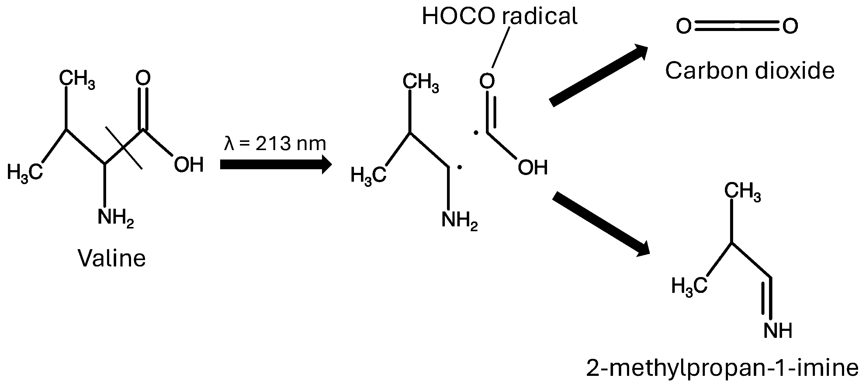
The proposed photodissociation pathway is summarized in Figure 8. The simultaneous formation of HOCO and 2-methylpropan-1-imine provides compelling evidence that -carbonyl C–C bond cleavage is the dominant primary photodissociation channel of valine at 213 nm. This mechanism is fully consistent with the photochemistry previously reported for glycine, alanine, leucine, serine, and proline isolated in solid parahydrogen [24,25], suggesting that -carbonyl C–C bond cleavage is a common ultraviolet photodissociation pathway for proteinogenic amino acids under these conditions. The aminoalkyl radical intermediate was not directly detected, as no experimental absorptions corresponding to its calculated vibrational spectrum (Tables S14–S16) were identified. The present experiments indicate that it undergoes rapid H-atom elimination to form 2-methylpropan-1-imine.
Figure 8.
Proposed photodissociation pathway of valine following 213 nm irradiation in solid parahydrogen, producing HOCO and 2-methylpropan-1-imine via -carbonyl C–C bond cleavage.
4. Conclusions
Non-zwitterionic neutral valine isolated in solid parahydrogen was investigated by high-resolution infrared spectroscopy, quantum-chemical calculations, and ultraviolet photolysis at 213 nm. The infrared spectrum was assigned by combining calculated vibrational frequencies, relative infrared intensities, characteristic vibrational modes, and conformer-specific photolysis kinetics. The photolysis experiments revealed four reproducible decay-rate categories, providing direct experimental evidence for the coexistence of at least four valine conformers in the solid parahydrogen matrix. The combined spectroscopic and kinetic analysis enabled assignment of the observed infrared bands to the six lowest-energy conformers predicted by the DFT calculations.
The assignments demonstrate that the photolysis rate depends strongly on molecular conformation. Hydrogen-bonded conformer II exhibits significantly slower photodepletion than the Type I conformers, suggesting that intramolecular O–H⋯NH2 hydrogen bonding enhances ultraviolet photostability of valine. Together with our previous results for leucine, these findings indicate that intramolecular hydrogen bonding is an important factor governing the conformer-dependent photochemistry of amino acids isolated in solid parahydrogen.
Ultraviolet irradiation at 213 nm produces HOCO radicals and bands tentatively assigned to 2-methylpropan-1-imine as the major photoproducts, confirming that -carbonyl C–C bond cleavage is the dominant photodissociation pathway of valine. These results extend previous observations for other proteinogenic amino acids and further demonstrate that solid parahydrogen provides a unique platform for investigating conformer-specific spectroscopy and photochemistry of isolated biomolecules.
Supplementary Materials
The following supporting information can be downloaded at: https://www.mdpi.com/article/10.3390/photochem6030028/s1, Table S1: Observed bands, photolysis rate constants, and conformer assignments; Tables S2–S7: Calculated vibrational wavenumbers and assigned pH2 bands for conformers Ia, Ib, Ic, II, III and IV; Table S8: Optimized geometries of the six valine conformers; Figure S1: Atom numbering scheme of valine used for calculations; Figure S2: The entire as-deposited spectrum of valine in solid parahydrogen; Figure S3: Concentration dependence of the infrared spectrum of valine in solid parahydrogen; Figure S4: Formation profiles of the principal photoproduct bands during irradiation at 213 nm; Figure S5: Relaxed potential-energy curves for conformational rotation in 2-methylpropan-1-imine; Tables S9–S12: Calculated wavenumbers for the conformers cis-120, cis-0, trans-120 and trans-0 of 2-methylpropan-1-imine; Table S13: Observed photoproduct bands; Figure S6: Relaxed potential-energy curves for conformational rotation in aminoalkyl radical; Tables S14–S16: Calculated wavenumbers of conformers A, B and C of aminoalkyl radical.
Author Contributions
Conceptualization, T.M. and B.M.; methodology, B.M., T.M. and P.D.; investigation, L.Z., C.Z., K.K. and C.N.; formal analysis, L.Z., C.Z., C.N., B.M., K.K. and T.M.; validation, B.M. and T.M.; writing—original draft preparation, L.Z.; writing—review and editing, L.Z., B.M., K.K. and T.M.; supervision, T.M.; funding acquisition, T.M. All authors have read and agreed to the published version of the manuscript.
Funding
This research was funded by the Natural Sciences and Engineering Research Council (NSERC) Discovery Grant of Canada (RGPIN-2020-05441, RGPIN-2025-06881), Canada Foundation for Innovation (Centre for Research on Ultracold Systems at UBC, CRUCS, 21591, and Chirality Research on Origins and Separation at UBC, CHIROS, 36414).
Institutional Review Board Statement
Not applicable.
Informed Consent Statement
Not applicable.
Data Availability Statement
The original contributions presented in this study are included in the article. Further inquiries can be directed to the corresponding author.
Conflicts of Interest
The authors declare no conflicts of interest.
References
- Evans, A.C.; Meinert, C.; Giri, C.; Goesmann, F.; Meierhenrich, U.J. Chirality, photochemistry and the detection of amino acids in interstellar ice analogues and comets. Chem. Soc. Rev. 2012, 41, 5447–5458. [Google Scholar] [CrossRef] [Scilit] [PubMed]
- Ruf, A.; D’Hendecourt, L.L.; Schmitt-Kopplin, P. Data-Driven Astrochemistry: One Step Further within the Origin of Life Puzzle. Life 2018, 8, 18. [Google Scholar] [CrossRef] [Scilit] [PubMed]
- Ehrenfreund, P.; Glavin, D.P.; Botta, O.; Cooper, G.; Bada, J.L. Extraterrestrial amino acids in Orgueil and Ivuna: Tracing the parent body of CI-type carbonaceous chondrites. Proc. Natl. Acad. Sci. USA 2001, 98, 2138–2141. [Google Scholar] [CrossRef] [Scilit] [PubMed]
- Kvenvolden, K.; Lawless, J.; Pering, K.; Peterson, E.; Flores, J.; Ponnamperuma, C.; Kaplan, I.R.; Moore, C. Evidence for Extraterrestrial Amino-acids and Hydrocarbons in the Murchison Meteorite. Nature 1970, 228, 923–926. [Google Scholar] [CrossRef] [Scilit] [PubMed]
- Price, W.D.; Jockusch, R.A.; Williams, E.R. Is Arginine a Zwitterion in the Gas Phase? J. Am. Chem. Soc. 1997, 119, 11988–11989. [Google Scholar] [CrossRef] [Scilit] [PubMed]
- Bernstein, M.P.; Dworkin, J.P.; Sandford, S.A.; Cooper, G.W.; Allamandola, L.J. Racemic amino acids from the ultraviolet photolysis of interstellar ice analogues. Nature 2002, 416, 401–403. [Google Scholar] [CrossRef] [Scilit] [PubMed]
- Schäfer, L.; Kulp-Newton, S.Q.; Siam, K.; Klimkowski, V.J.; Van Alsenoy, C. Ab initio studies of structural features not easily amenable to experiment: Part 71. Conformational analysis and structural study of valine and threonine. J. Mol. Struct. THEOCHEM 1990, 209, 373–385. [Google Scholar] [CrossRef] [Scilit]
- Dokmaisrijan, S.; Lee, V.S.; Nimmanpipug, P. The gas phase conformers and vibrational spectra of valine, leucine and isoleucine: An ab initio study. J. Mol. Struct. THEOCHEM 2010, 953, 28–38. [Google Scholar] [CrossRef] [Scilit]
- Torii, K.; Iitaka, Y. The crystal structure of L-valine. Acta Cryst. 1970, B26, 1317–1326. [Google Scholar] [CrossRef] [Scilit] [PubMed]
- Paiva, F.M.; Batista, J.C.; Rêgo, F.S.C.; Lima, J.A., Jr.; Freire, P.T.C.; Melo, F.E.A.; Mendes Filho, J.; de Menezes, A.S.; Nogueira, C.E.S. Infrared and Raman spectroscopy and DFT calculations of DL amino acids: Valine and lysine hydrochloride. J. Mol. Struct. 2017, 1127, 419–426. [Google Scholar] [CrossRef] [Scilit]
- da Silva, J.H.; Lima, J.A., Jr.; Freire, P.T.C.; Lemos, V.; Mendes Filho, J.; Melo, F.E.A.; Pizani, P.S.; Fischer, J.; Klemke, B.; Kemner, E.; et al. Raman spectroscopy and inelastic neutron scattering study of crystalline L-valine. J. Phys. Condens. Matter 2009, 21, 415404. [Google Scholar] [CrossRef] [Scilit]
- Lesarri, A.; Cocinero, E.J.; López, J.C.; Alonso, J.L. The Shape of Neutral Valine. Angew. Chem. Int. Ed. 2004, 43, 605–610. [Google Scholar] [CrossRef] [Scilit] [PubMed]
- Tiefenthaler, L.; Ončák, M.; Kollotzek, S.; Kočišek, J.; Scheier, P. Dissociation of Valine Cluster Cations. J. Phys. Chem. A 2020, 124, 8439–8445. [Google Scholar] [CrossRef] [Scilit] [PubMed]
- Papp, P.; Shchukin, P.; Kočíšek, J.; Matejčík, S. Electron ionization and dissociation of aliphatic amino acids. J. Chem. Phys. 2012, 137, 105101. [Google Scholar] [CrossRef] [Scilit] [PubMed]
- Lago, A.F.; Coutinho, L.H.; Marinho, R.R.; Brito, A.N.D.; Souza, G.G.D. Ionic dissociation of glycine, alanine, valine and proline as induced by VUV (21.21 eV) photons. Chem. Phys. 2004, 307, 9–14. [Google Scholar] [CrossRef] [Scilit]
- Stepanian, S.G.; Reva, I.D.; Radchenko, E.D.; Adamowicz, L. Conformers of Nonionized Proline. Matrix-Isolation Infrared and Post-Hartree-Fock ab Initio Study. J. Phys. Chem. A 2001, 105, 10664–10672. [Google Scholar] [CrossRef] [Scilit]
- Stepanian, S.G.; Ivanov, A.Y.; Adamowicz, L. Conformational composition of neutral leucine. Matrix isolation infrared and ab initio study. Chem. Phys. 2013, 423, 20–29. [Google Scholar] [CrossRef] [Scilit]
- Momose, T.; Shida, T. Matrix-Isolation Spectroscopy Using Solid Parahydrogen as the Matrix: Application to High-Resolution Spectroscopy, Photochemistry, and Cryochemistry. Bull. Chem. Soc. Jpn. 1998, 71, 1–15. [Google Scholar] [CrossRef] [Scilit]
- Momose, T.; Hoshina, H.; Fushitani, M.; Katsuki, H. High-resolution spectroscopy and the analysis of ro-vibrational transitions of molecules in solid parahydrogen. Vib. Spectrosc. 2004, 34, 95–108. [Google Scholar] [CrossRef] [Scilit]
- Weber, I.; Joshi, P.R.; Anderson, D.T.; Lee, Y.P. Unique Applications of para-Hydrogen Matrix Isolation to Spectroscopy and Astrochemistry. J. Phys. Chem. Lett. 2024, 15, 11361–11373. [Google Scholar] [CrossRef] [Scilit] [PubMed]
- Brus, L.E.; Bondybey, V.E. Cage effects and steric hindrance in van der Waals solids, with application to alkyl iodide photolysis in rare gas hosts. J. Chem. Phys. 1976, 65, 71–76. [Google Scholar] [CrossRef] [Scilit]
- Momose, T.; Fushitani, M.; Hoshina, H. Chemical reactions in quantum crystals. Int. Rev. Phys. Chem. 2005, 24, 533–552. [Google Scholar] [CrossRef] [Scilit]
- Momose, T.; Uchida, M.; Sogoshi, N.; Miki, M.; Masuda, S.; Shida, T. Infrared spectroscopic studies of photoinduced reactions of methyl radical in solid parahydrogen. Chem. Phys. Lett. 1995, 246, 583–586. [Google Scholar] [CrossRef] [Scilit]
- Moore, B.; Mahoney, K.; Zeng, M.F.; Djuricanin, P.; Momose, T. Ultraviolet Photodissociation of Proteinogenic Amino Acids. J. Am. Chem. Soc. 2023, 145, 11045–11055. [Google Scholar] [CrossRef] [Scilit] [PubMed]
- Moore, B.; Toh, S.Y.; Wong, Y.T.; Bashiri, T.; McKinnon, A.; Wai, Y.; Lee, K.W.A.; Ovchinnikov, P.; Chiang, C.Y.; Djuricanin, P.; et al. Hydrocarboxyl Radical as a Product of α-Alanine Ultraviolet Photolysis. J. Phys. Chem. Lett. 2021, 12, 11992–11997. [Google Scholar] [CrossRef] [Scilit] [PubMed]
- Francisco, J.S.; Muckerman, J.T.; Yu, H.G. HOCO Radical Chemistry. Acc. Chem. Res. 2010, 43, 1519–1526. [Google Scholar] [CrossRef] [Scilit] [PubMed]
- Molpeceres, G.; Enrique-Romero, J.; Ishibashi, A.; Oba, Y.; Hidaka, H.; Lamberts, T.; Aikawa, Y.; Watanabe, N. Hydrogenation of HOCO and formation of interstellar CO2: A not so straightforward relation. Mon. Not. R. Astron. Soc. 2025, 538, 1565–1575. [Google Scholar] [CrossRef] [Scilit]
- Ishibashi, A.; Molpeceres, G.; Hidaka, H.; Oba, Y.; Lamberts, T.; Watanabe, N. Proposed Importance of HOCO Chemistry: Inefficient Formation of CO2 from CO and OH Reactions on Ice Dust. Astrophys. J. 2024, 976, 162. [Google Scholar] [CrossRef] [Scilit]
- Guerra, C.; Rodríguez-Núñez, Y.A.; Ensuncho, A.E. Role of Triplet States in the Photolysis of Proteogenic Amino Acids. ChemPhysChem 2024, 25, e202300655. [Google Scholar] [CrossRef] [Scilit] [PubMed]
- Moore, B.; Zeng, L.; Djuricanin, P.; Cooke, I.R.; Madison, K.W.; Momose, T. Significant Chiral Asymmetry Observed in Neutral Amino Acid Ultraviolet Photolysis. J. Am. Chem. Soc. 2024, 146, 34333–34340. [Google Scholar] [CrossRef] [Scilit] [PubMed]
- Tom, B.A.; Bhasker, S.; Miyamoto, Y.; Momose, T.; McCall, B.J. Producing and quantifying enriched para-H2. Rev. Sci. Instrum. 2009, 80, 016108. [Google Scholar] [CrossRef] [Scilit] [PubMed]
- Wong, Y.T.A.; Toh, S.Y.; Djuricanin, P.; Momose, T. Conformational composition and population analysis of β-alanine isolated in solid parahydrogen. J. Mol. Spectrosc. 2015, 310, 23–31. [Google Scholar] [CrossRef] [Scilit]
- Fajardo, M.E.; Tam, S. Rapid vapor deposition of millimeters thick optically transparent parahydrogen solids for matrix isolation spectroscopy. J. Chem. Phys. 1998, 108, 4237–4241. [Google Scholar] [CrossRef] [Scilit]
- Frisch, M.J. Gaussian 09, Revision E.01; Gaussian Inc.: Wallingford, CT, USA, 2009. [Google Scholar]
- National Institute of Standards and Technology. NIST Computational Chemistry Comparison and Benchmark Database. NIST Standard Reference Database Number 101, Release 22, 2022. Available online: https://cccbdb.nist.gov/ (accessed on 29 July 2026).
- Hadidi, R.; Božanić, D.K.; Ganjitabar, H.; Garcia, G.A.; Powis, I.; Nahon, L. Conformer-dependent vacuum ultraviolet photodynamics and chiral asymmetries in pure enantiomers of gas phase proline. Comm. Chem. 2021, 4, 72. [Google Scholar] [CrossRef] [Scilit] [PubMed]
- Paulson, L.O.; Mutunga, F.M.; Follett, S.E.; Anderson, D.T. Reactions of Atomic Hydrogen with Formic Acid and Carbon Monoxide in Solid Parahydrogen I: Anomalous Effect of Temperature. J. Phys. Chem. A 2014, 118, 7640–7652. [Google Scholar] [CrossRef] [Scilit] [PubMed]
- Anderson, D.T.; Paulson, L.O. High-Resolution Vibrational Spectroscopy of trans-Formic Acid in Solid Parahydrogen. J. Phys. Chem. A 2009, 113, 1770–1778. [Google Scholar] [CrossRef] [Scilit] [PubMed]
- Ruzi, M.; Anderson, D.T. Photodissociation of N-methylformamide isolated in solid parahydrogen. J. Chem. Phys. 2012, 137, 194313. [Google Scholar] [CrossRef] [Scilit] [PubMed]
- Fajardo, M.E.; Tam, S.; DeRose, M.E. Matrix isolation spectroscopy of H2O, D2O, and HDO in solid parahydrogen. J. Mol. Struct. 2004, 695–696, 111–127. [Google Scholar] [CrossRef]
- Tam, S.; Fajardo, M.E. Observation of the high-resolution infrared absorption spectrum of CO2 molecules isolated in solid parahydrogen. Low Temp. Phys. 2000, 26, 653–660. [Google Scholar] [CrossRef] [Scilit]

Disclaimer/Publisher’s Note: The statements, opinions and data contained in all publications are solely those of the individual author(s) and contributor(s) and not of MDPI and/or the editor(s). MDPI and/or the editor(s) disclaim responsibility for any injury to people or property resulting from any ideas, methods, instructions or products referred to in the content. |
© 2026 by the authors. Licensee MDPI, Basel, Switzerland. This article is an open access article distributed under the terms and conditions of the Creative Commons Attribution (CC BY) license.