


Abstract
Three types of meso-substituted manganese(III) porphyrin complexes with phenyl, propyl, and isopropyl substituents were synthesized to examine their photochemical and electrochemical properties. The distortion of the porphyrin rings and redox potentials depended on the substituents. The fluorescence quantum yields of these porphyrins were significantly low, indicating the rapid deactivation of their singlet excited states. Although weak emissions attributed to higher singlet excited states and charge-transfer states were observed, their quantum yields remained low. Redox potential measurements showed the relatively strong photooxidative ability of these porphyrins from a thermodynamic standpoint. However, photosensitized protein oxidation was barely observed. The rapid deactivation of photoexcited states decreases the probability of photochemical reactions including biomolecule oxidation. These porphyrins suppressed the self-oxidation of photo-irradiated 1-benzyl-1,4-dihydronicotinamide and catalyzed the decomposition of hydrogen peroxide. In conclusion, these manganese(III) porphyrin complexes barely showed photooxidation activity toward biomolecules and demonstrated protective action against phototoxic reactions.
1. Introduction
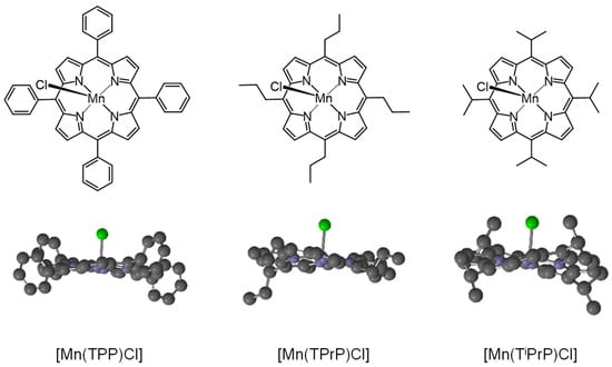
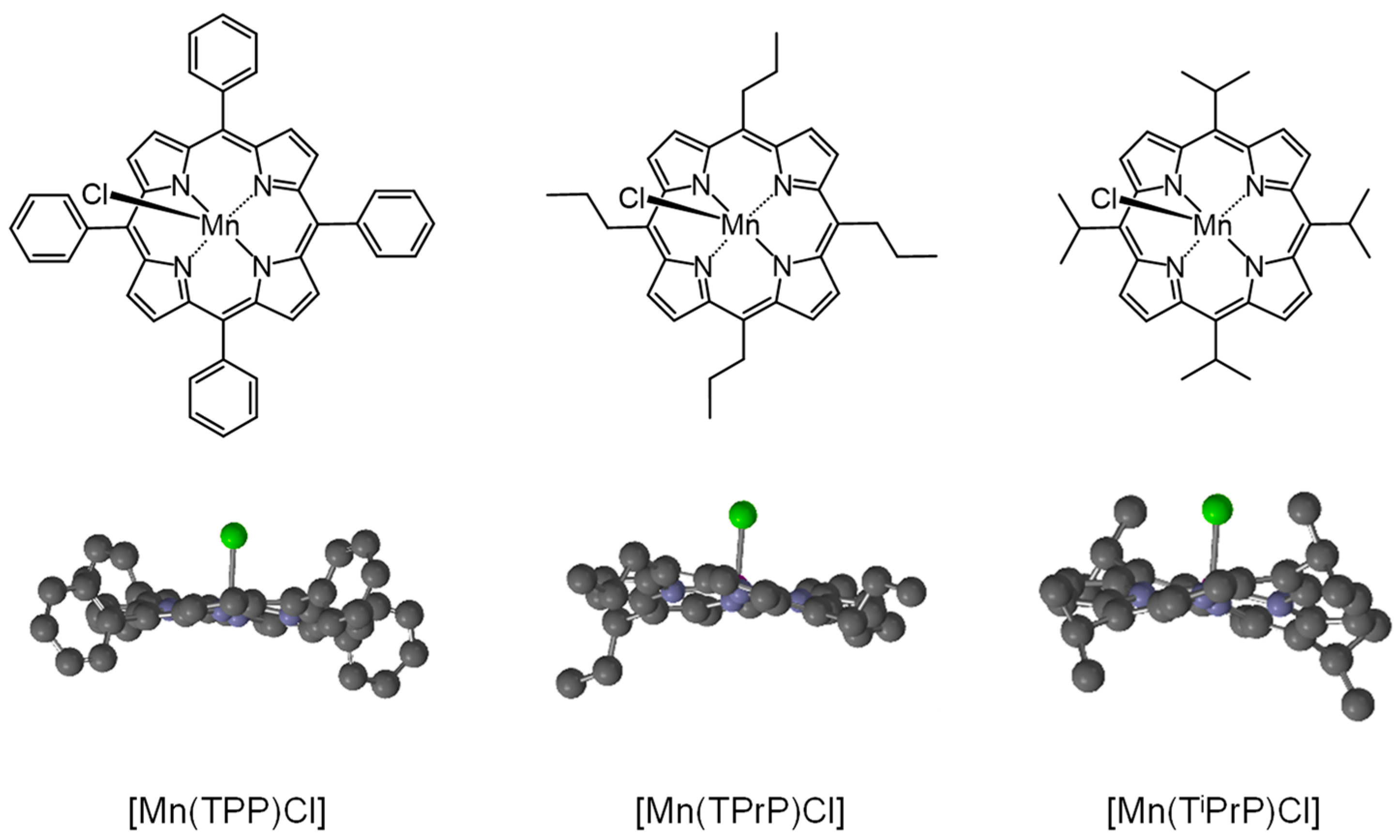
Porphyrins are important compounds in photochemistry, photobiology, and photomedicine [1,2]. In natural photosynthesis, porphyrin derivatives serve as essential photosensitizers [3]. Photosensitized damage to biomolecules by porphyrins has been utilized in cancer therapies, such as photodynamic therapy (PDT) [4,5] and antimicrobial photodynamic treatment [6]. Furthermore, free-base and metal porphyrin complexes exhibit specific catalytic and photocatalytic activities, such as the reductive defluorination of perfluorooctanesulfonate [7] and photo-redox reactions [8]. Superoxide dismutase (SOD), which effectively dismutates superoxide (O2•−) to hydrogen peroxide (H2O2), is an important example of a biocatalyst composed of metal porphyrin complexes [9]. Manganese(III) porphyrin complexes have been studied as catalysts [10] including model catalyst of SOD [11]. Furthermore, the photosensitized singlet oxygen (1O2) production activity of water-soluble manganese(III) porphyrin complexes has been reported [12]. In general, meso-substituted porphyrins, aromatic substituents, such as phenyl groups, are commonly used, whereas alkyl substituents are relatively uncommon. Because alkyl groups can induce different steric and electronic environments around the porphyrin core compared to aromatic groups, metal meso-alkylporphyrin complexes may exhibit distinct and potentially interesting photochemical, photobiological, and photomedical properties [13,14,15]. In this study, three types of meso-substituted manganese(III) porphyrin complexes, manganese(III) tetraphenylporphyrin chloride ([Mn(TPP)Cl]), manganese(III) tetrapropylporphyrin chloride ([Mn(TPrP)Cl]), and manganese(III) tetraisopropylporphyrin chloride ([Mn(TiPrP)Cl]) (Figure 1) [16,17], with phenyl, propyl, and isopropyl substituents, respectively, were synthesized to examine their photochemical and electrochemical properties and photodamaging abilities toward biomolecules. This study aimed to compare the photochemical properties of propyl-substituted porphyrin ([Mn(TPrP)Cl]) and phenyl-substituted porphyrin ([Mn(TPP)Cl]). The steric hindrance of the isopropyl substituent on the porphyrin ring is similar to that of the phenyl group. Thus, isopropyl-substituted porphyrin ([Mn(TiPrP)Cl]) was used to examine the effects of sterically hindered alky substituents. Their catalytic activities toward H2O2 decomposition were also evaluated.
Figure 1.
Structures of manganese(III) porphyrin complexes. The structures of these porphyrins were optimized using DFT calculations at the ωB97X-D/6-311+G(2df,2p) level.
2. Materials and Methods
[Mn(TPP)Cl], [Mn(TPrP)Cl], and [Mn(TiPrP)Cl] were synthesized according to previous reports [16,17] and studies [18,19,20]. Human serum albumin (HSA) and catalase were purchased from Sigma-Aldrich. LLC. (St. Louis, MO, USA). Copper(II) chloride dihydrate, ethanol, H2O2 solution, folic acid, methylhydroquinone (MeHQ), and tetrabutylammonium hexafluorophosphate were purchased from FUJIFILM Wako Pure Chemical Co., Ltd. (Osaka, Japan). 1-Benzyl-1,4-dihydronicotinamide (BNADH) was purchased from Tokyo Chemical Industry. (Tokyo, Japan). Spectroscopic-grade chloroform and water were purchased from Dojin Chemical Co. (Kumamoto, Japan). Sodium phosphate buffer (0.1 M, pH 7.6) was purchased from Nacalai-Tesque Inc. (Kyoto, Japan). All chemical reagents were used as received, without further purification.
The absorption and fluorescence spectra of the samples were measured using a UV-Vis spectrophotometer UV-1650PC (Shimadzu, Kyoto, Japan) and an F-4500 fluorescence spectrophotometer (Hitachi, Tokyo, Japan), respectively. The fluorescence lifetimes (τf) of porphyrins and HSA were measured using a Fluorescence Lifetime System TemPro (HORIBA, Kyoto, Japan). The excitation wavelengths for the τf measurements of porphyrins and HSA were 402 and 294 nm, respectively. The redox potentials of the porphyrins in dry acetonitrile, including 0.1 M tetrabutylammonium hexafluorophosphate (supporting electrolyte), were measured using a potentiostat/galvanostat (HA-301, Hokuto Denko Co., Tokyo, Japan), a digital function generator (DF1906, NF Co., Yokohama, Japan), and a midi logger (GL900-4, Graphtec Co., Yokohama, Japan), using a platinum working electrode (ALS Co., Ltd., Tokyo, Japan), a platinum counter electrode (ALS Co., Ltd.), and a saturated calomel electrode (SCE, ALS Co., Ltd.). The 1O2 production activity of these porphyrins in chloroform was evaluated by measuring near-infrared luminescence at approximately 1270 nm, as reported previously [21]. Circular dichroism (CD) spectra were measured with a J-820 spectrodichrometer (JASCO, Tokyo, Japan) over the spectral range of 420–520 nm and the CD magnitude was expressed in terms of molar ellipticity ([θ] in deg∙M−1·cm−1).
To evaluate HSA damage, a sample solution containing 5 μM porphyrins and 10 μM HSA was prepared in a 10 mM sodium phosphate buffer (pH 7.6) containing 1% ethanol. The sample solution was irradiated with a light-emitting diode (LED, λmax = 585 nm, full width at half maximum: 22 nm, 2.0 mW cm−2, CCS Inc., Kyoto, Japan). The intensity of the LED was measured using an 8230E optical power meter (ADC Corporation, Tokyo, Japan). The fluorescence intensity of HSA at 350 nm was measured using a Hitachi 650-60 fluorescence photometer. The excitation wavelength for this assay was 298 nm. The amount of damaged HSA was estimated from the results of fluorometry based on the decrease in the intrinsic fluorescence of the tryptophan residue of HSA [22] by photooxidation, as reported previously [23,24].
The photooxidation of BNADH was examined by measuring the absorbance change, as reported previously [25]. The sample solution containing 5 μM porphyrins and 100 μM BNADH in ethanol was irradiated with a blue LED (λmax: 468 nm, full width at half maximum: 22 nm, 1.0 mW cm−2; CCS Inc.). After photoirradiation, the absorption spectrum was measured using a UV-vis spectrophotometer (UV-1650PC).
The production of H2O2 through the oxidation of MeHQ was measured using a previously reported method with folic acid [26]. This assay is based on the fluorescence enhancement of less fluorescent folic acid via oxidative decomposition by H2O2 into strongly fluorescent 2-amino-4-oxo-3H-pterine-6-carboxylic acid. The concentration of H2O2 was determined using a calibration curve. The sample solution (1 mL) containing 10 µM folic acid, 10 µM copper(II) chloride, and 100 μM MeHQ with or without 5 μM porphyrin or 50 unit mL−1 catalase in a 10 mM sodium phosphate buffer (pH 7.6) including 1% ethanol was incubated in a microtube (1.5 mL Eppendorf) at 37 °C. After incubation, the fluorescence intensity of the solution at 450 nm was measured using a 650-60 fluorescence spectrophotometer with a 350 nm excitation.
The optimized structures of manganese(III) porphyrin complexes were calculated using density functional theory (DFT) at the ωB97X-D/6-311+G(2df,2p) level utilizing Spartan’20 (Wavefunction Inc., Irvine, CA, USA).
3. Results and Discussion
3.1. Structures and Electrochemical Properties of Manganese(III) Porphyrin Complexes
The optimized structures of these porphyrins are shown in Figure 1. To evaluate the effect of the meso-substituent on the macrocycle distortion of porphyrin rings, DFT calculations were performed. The porphyrin ring is nearly planar; however, the meso-substituents distort the ring. In an ideal planar structure, the angle between the pyrrole nitrogen and the central manganese atom (∠N-Mn-N) and between the meso-carbons and the central manganese atom (∠C-Mn-C) are expected to be 180 °, and smaller angles indicate greater distortion of the porphyrin ring. The shorter distance between the central manganese atom and the pyrrole nitrogen (dMn-N) also reflects increased distortion. The parameters calculated using DFT methods are listed in Table 1. The calculated structural data in the manganese(III) porphyrin complexes were corresponded to crystal structural data in those complexes. The mean angles ∠N-Mn-N and ∠C-Mn-C from the crystal structure of [Mn(TiPrP)Cl] are 163.4° and 160.0°, respectively [16]. The distances dMn-N from crystal structures of [Mn(TPP)Cl] and [Mn(TiPrP)Cl] are 2.008 (7) Å and 1.981 (2) Å, respectively [16,27]. These results show that the distortion effect of the meso-substituents increases in the following order: [Mn(TiPrP)Cl] > [Mn(TPrP)Cl] > [Mn(TPP)Cl].
Table 1.
Structure parameters and electrochemical property of manganese(III) porphyrin complexes.
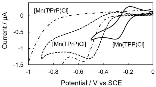
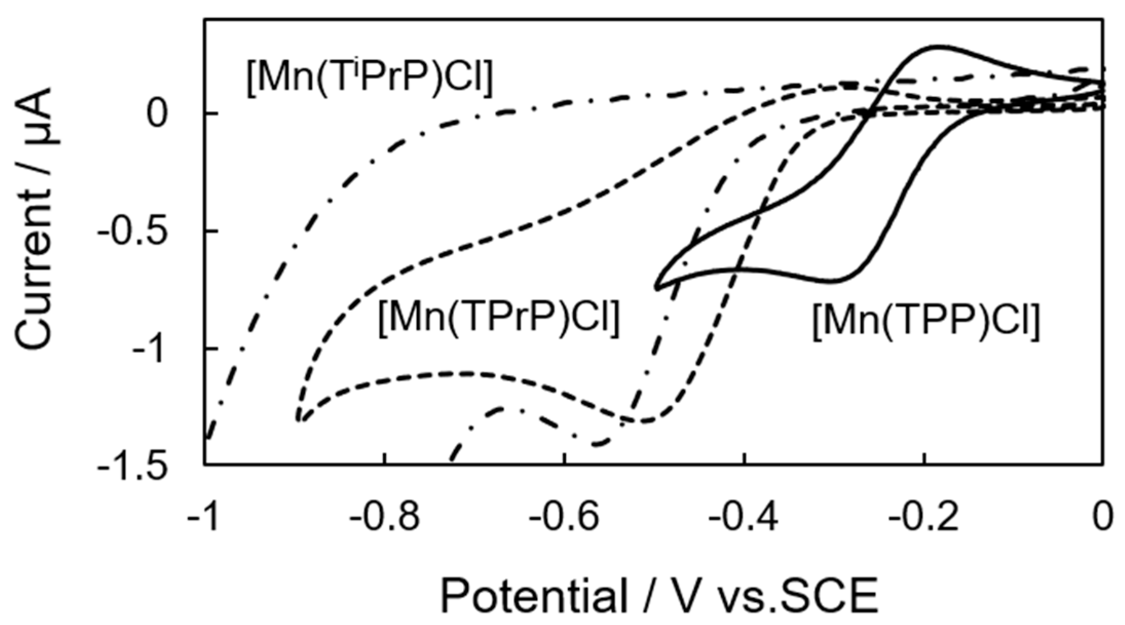
The cathode peak potentials (CP) and redox potentials of one-electron reduction (Ered) of these porphyrins are listed in Table 1. In cyclic voltammetry, the reduction current of [Mn(TiPrP)Cl] was found to be irreversible (Figure 2 and Supplementary Materials, Figure S1). The observed peaks were assigned to the redox processes of the central manganese ion (Mn3+/Mn2+) [11,28]. The differences in observed values should be explained by the effect of interaction between π-orbital of porphyrin ring and the central manganese. The electron-donating abilities of these porphyrins, influenced by their meso-substituents, were compared using their CP values. The electron-donating abilities of these porphyrins were as follows: [Mn(TiPrP)Cl] > [Mn(TPrP)Cl] > [Mn(TPP)Cl].
Figure 2.
Cyclic voltammograms of [Mn(TPP)Cl], [Mn(TPrP)Cl], and [Mn(TiPrP)Cl] in acetonitrile. The sample solution contained 1 mg of manganese(III) porphyrin complexes and 0.1 M tetrabutylammonium hexafluorophosphate (supporting electrolyte) in 1 mL of dry acetonitrile.
3.2. Photochemical Property of Manganese(III) Porphyrin Complexes
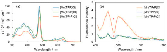
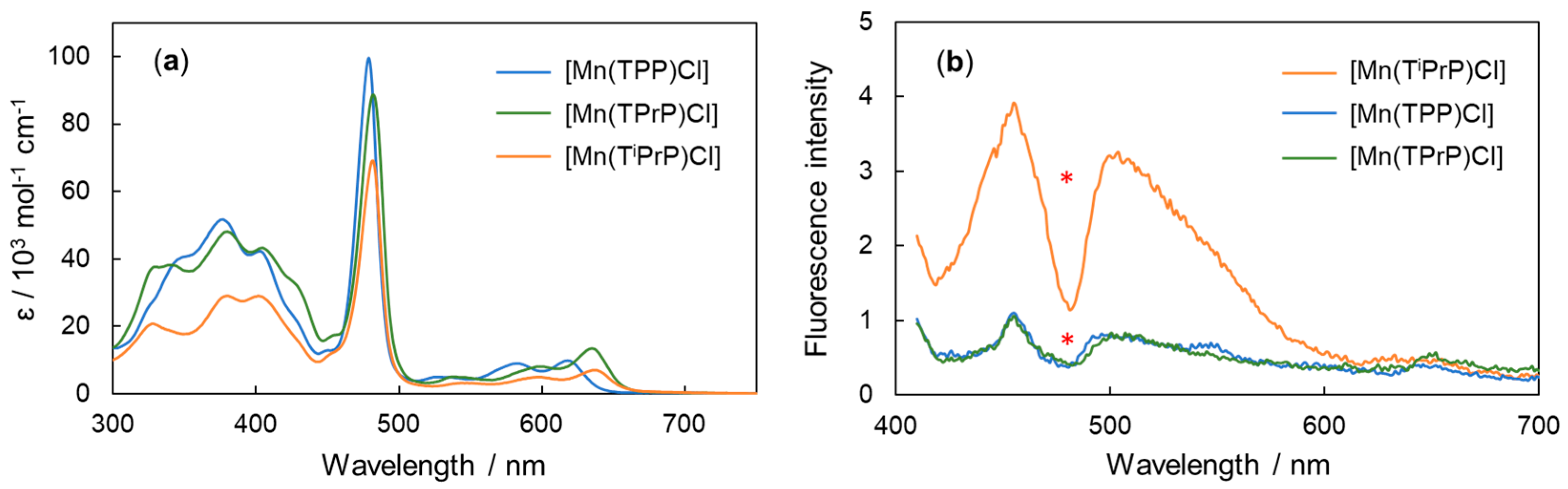
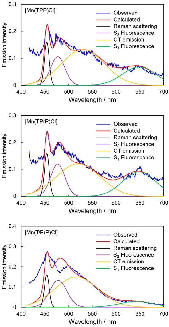
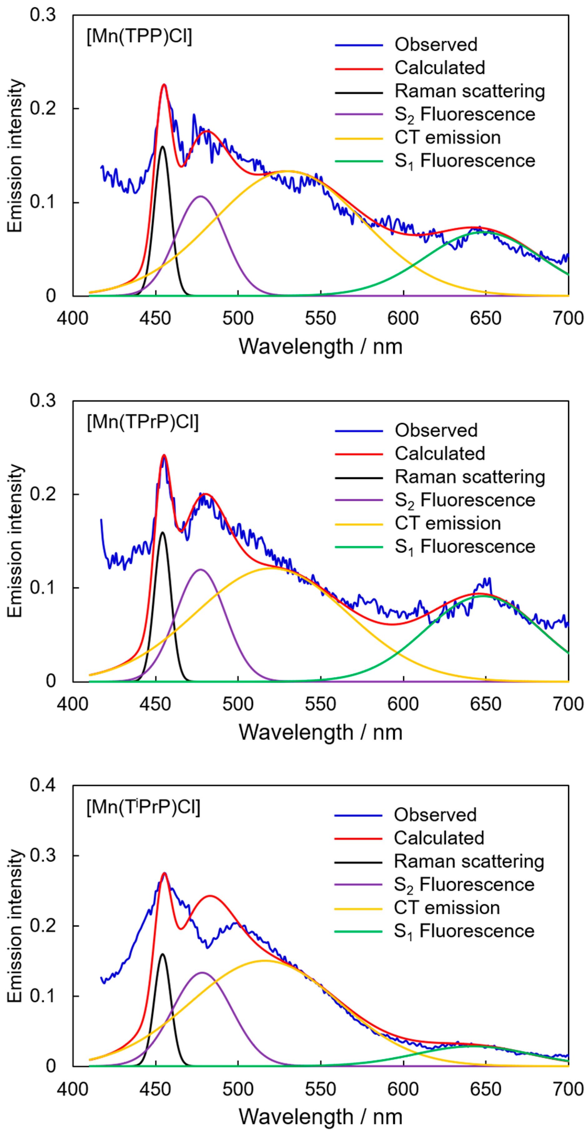
The absorption spectra of porphyrins are shown in Figure 3a. Typical Soret bands were observed at approximately 480 nm, while Q-bands appeared in the range of 530–640 nm. The absorption bands around 320–420 nm can be explained by a mixture of π-π transitions within the porphyrin ring (ligand-to-ligand electronic transition) and ligand-to-metal transitions (electronic transition from the porphyrin ring to the d-orbital of manganese ion) [29]. The fluorescence spectra of these porphyrins were barely observed upon Q-band excitation, indicating that the singlet excited state (S1) of these porphyrins was rapidly deactivated. Excitation at shorter wavelengths resulted in emission from higher excited states, although the quantum yields were very low (Figure 3b). In general, paramagnetic metalloporphyrin complexes, including manganese(III) porphyrin complexes, exhibit negligible fluorescence. However, under certain conditions, such as manganese(III) porphyrin complexes, emission is demonstrated in a silica matrix [30]. In the present study, these emissions were observed reproducibly. The observed emissions were considered to be fluorescence from the second excited (S2) state and charge-transfer (CT) emission, and the very weak emission at approximately 650 nm was assigned to S1 fluorescence. The observed emissions were analyzed according to the assumption that the spectrum was composed of the abovementioned emissions using a previously reported procedure [31] (Figure 4, Supplementary Materials, Section S2), and the corresponding emission quantum yields are listed in Table 2. [Mn(TiPrP)Cl] exhibited a relatively strong emission spectrum. The distortion of the porphyrin ring and increased electron-donating ability may have contributed to the enhanced emission from the higher excited state. The time profiles were analyzed using triple-exponential functions (Table 2) (Supplementary Materials, Figure S2). The obtained lifetimes were consistent across the detection wavelength range of 480–650 nm. These results suggest that the photosensitized reactions of these porphyrins in higher excited states may be feasible within the lifetimes of these excited states.
Figure 3.
Absorption (a) and fluorescence (b) spectra of manganese(III) porphyrin complexes in chloroform. The excitation wavelength was set to 402 nm. * Filter effect of the Soret band.
Figure 4.
Representative analysis of the emission spectra for [Mn(TPP)Cl] (Top), [Mn(TPrP)Cl] (middle), and [Mn(TiPrP)Cl] (bottom) using Gaussian functions. The observed spectrum (blue) was corrected for the filter effect caused by the Soret band and deconvoluted into Raman scattering, S2 fluorescence, CT emission, and S1 fluorescence components (Supplementary Materials, Section S2). The fitted spectra (red) and the individual spectral components are also shown.
Table 2.
Emission quantum yields and their lifetimes of manganese(III) porphyrin complexes.
3.3. Photochemical Protein Damaging Activities of Manganese(III) Porphyrin Complexes
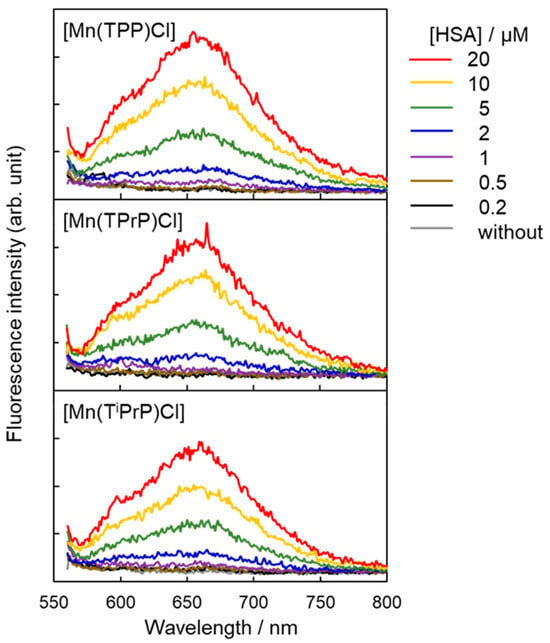
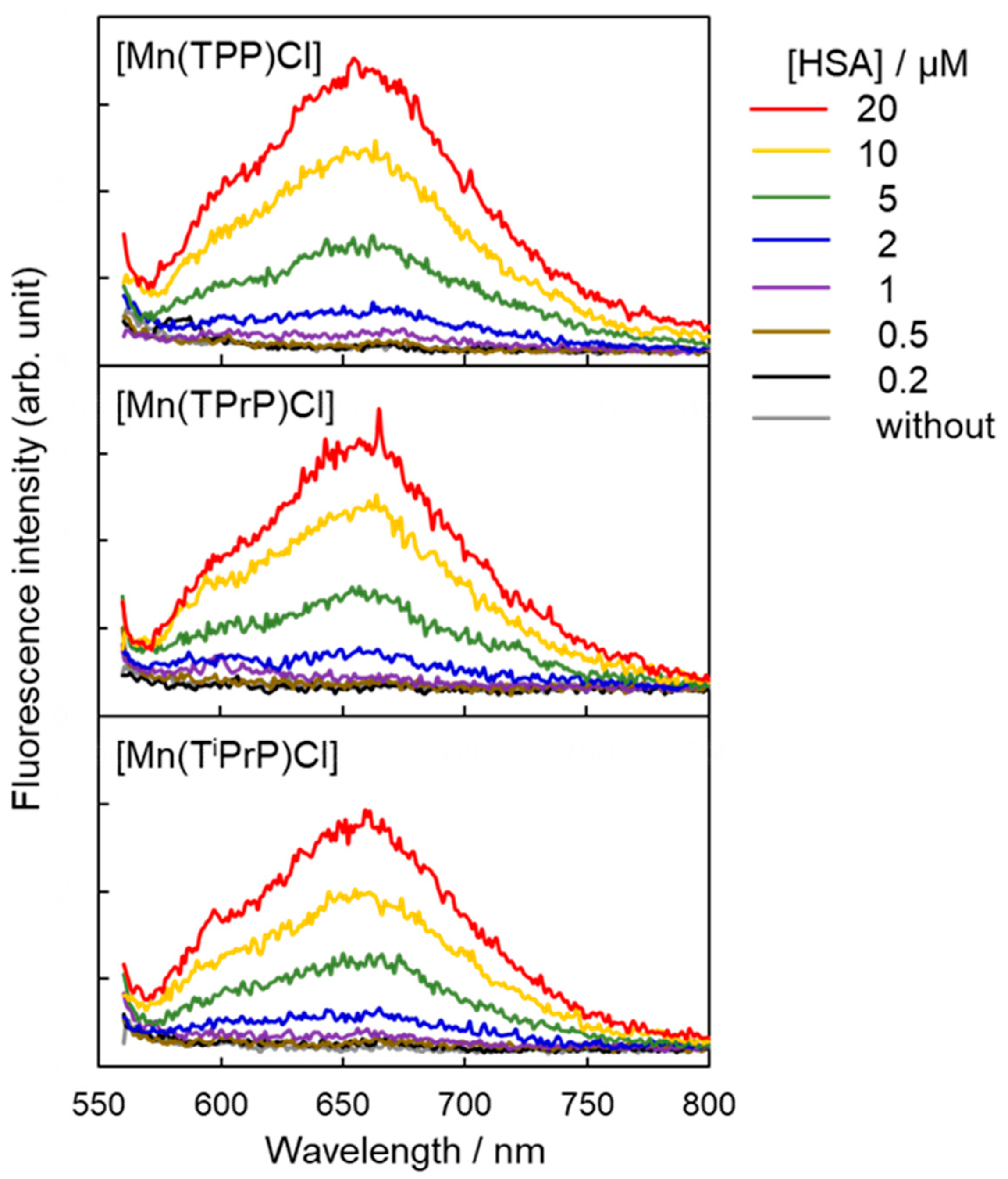
The photochemical properties of these porphyrins in the S1 state were examined. These porphyrins did not fluoresce in aqueous solutions (sodium phosphate buffer (pH 7.6) containing 1% ethanol). Due to their hydrophobic nature, these porphyrins likely form aggregates that suppress fluorescence and rapidly deactivate their S1 states. However, the fluorescence intensity of these porphyrins slightly increased upon interaction with HSA (Figure 5), possibly because HSA disrupts porphyrin aggregation. The interaction between porphyrin molecules and HSA was also analyzed based on the changes in the absorption spectra of porphyrins, according to previous reports [23,24]. The binding constant (Kbind) between porphyrins and HSA was analyzed under the assumption that one porphyrin molecule binds to HSA (1:1 complex model). The obtained Kbind values for [Mn(TPP)Cl], [Mn(TPrP)Cl], and [Mn(TiPrP)Cl] were 1.1 × 10⁶ M−1, 4.9 × 10⁵ M−1, and 1.7 × 10⁵ M−1, respectively, suggesting that distortion decreases the binding interaction. Furthermore, the phenyl substituent of [Mn(TPP)Cl] may increase the hydrophobic interaction with HSA. The CD spectra of these porphyrins and HSA did not exhibit any Cotton effect (Supplementary Materials, Figure S3), suggesting random binding. The binding positions of these porphyrins on HSA were roughly estimated using the Förster resonance energy-transfer (FRET) method based on the energy-transfer theory [32] with a previously reported procedure [33]. The estimated distances between the tryptophan residues in HSA and the porphyrins were 33–64 Å ([Mn(TPP)Cl]), 35–62 Å ([Mn(TPrP)Cl]), and 39–56 Å ([Mn(TiPrP)Cl]). The average diameter of HSA is approximately 80 Å [34], with the tryptophan residue located near the center of the molecule [22]. These results suggest that porphyrin molecules bind randomly to the hydrophobic microenvironment near the HSA surface.
Figure 5.
Fluorescence spectra of manganese(III) porphyrin complexes with and without HSA. The sample solution contained 5 μM porphyrins and the indicated concentration of HSA in a 10 mM sodium phosphate buffer (pH 7.6) containing 1% ethanol. The excitation wavelength was set to 550 nm.
The photosensitized 1O2 production ability of these porphyrins could not be confirmed by near-infrared emission measurements. However, the relatively high redox potentials of these porphyrins suggest that electron-transfer-mediated oxidation of HSA is thermodynamically possible (Supplementary Materials, Section S5). To examine the photosensitizer activity of these porphyrins, sample solutions containing these porphyrins and HSA were photo-irradiated according to previous reports [23,24]. A photosensitizer with a similar redox potential can oxidize HSA via photoinduced electron transfer [35]. However, HSA photodamage was barely observed under these experimental conditions. These findings indicate that manganese(III) porphyrin complexes cannot induce protein photooxidation, possibly because of the shorter lifetimes of their photoexcited states. Unlike conventional porphyrin photosensitizers, these manganese(III) porphyrin complexes showed a different photochemical behavior. Their short excited-state lifetimes together with catalytic decomposition of reactive oxygen species (ROS) (described in later) may be advantageous for suppressing phototoxic reactions.
3.4. Protective Action of Manganese(III) Porphyrin Complexes on BNADH Photodecomposition
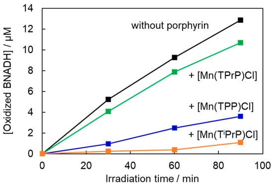
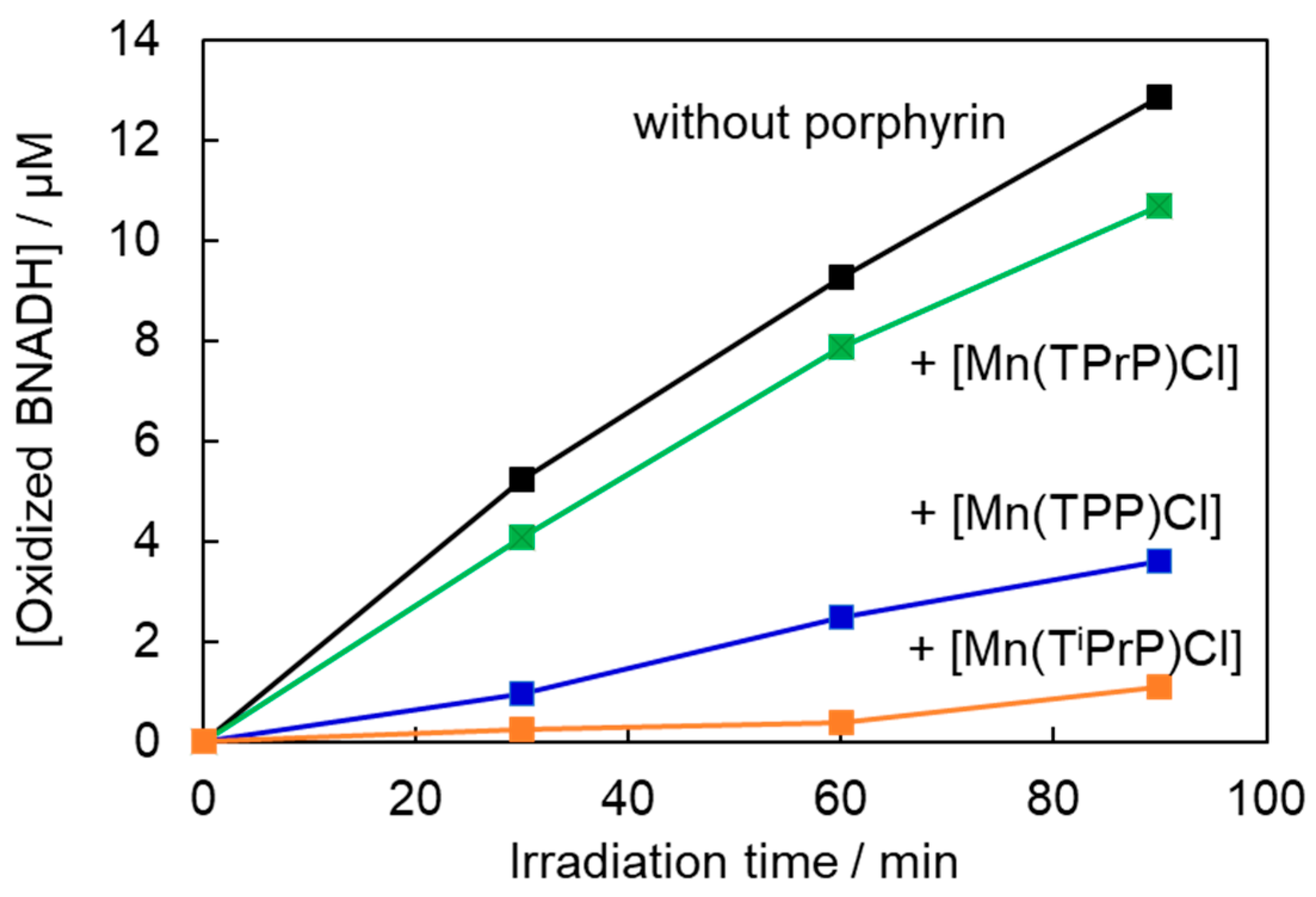
The photosensitized oxidation of BNADH by these porphyrins was examined (Figure 6). Photo-irradiated BNADH decomposed in the absence of porphyrins. Because the emission of the blue LED light source (λmax: 468 nm, full width at half maximum: 22 nm) overlapped with the absorption spectrum of BNADH (Supplementary Materials, Figure S4), the oxidation of BNADH can be explained by self-photooxidation. Since BNADH is a reductive compound, it is speculated that photoexcited BNADH reduces molecular oxygen to produce O2•−, and the resulting ROS subsequently induce the self-oxidation of BNADH. These porphyrins suppressed BNADH photodecomposition. The short lifetime of the photoexcited state of these porphyrins is insufficient to induce BNADH photooxidation. Because the absorption spectra of these porphyrins overlapped with that of BNADH, a filter effect contributed to the suppression of BNADH photooxidation. Therefore, the effect of these porphyrins on the photon fluence absorbed by BNADH was estimated (Supplementary Materials, Section S6). The estimated effect was relatively small in this study. Furthermore, the dependence of the suppression of BNADH photodecomposition on the compounds could not be explained by the filter effect. Another possibility is the removal of ROS by these porphyrins, suggesting that manganese(III) porphyrin complexes decompose ROS to protect against BNADH oxidation. Together with the negligible protein photooxidation described above, these findings suggest that these porphyrin complexes function predominantly as photoprotective compounds rather than conventional photosensitizers.
Figure 6.
Time profile of BNADH photooxidation with or without manganese(III) porphyrin complexes. The sample solution containing 100 μM BNADH and 5 μM manganese(III) porphyrin complex was irradiated with an LED (λmax = 470 nm, 2.0 mW cm−2). BNADH oxidation was monitored by measuring the absorbance at 340 nm.
3.5. H2O2 Decomposition Activity of Manganese(III) Porphyrin Complexes
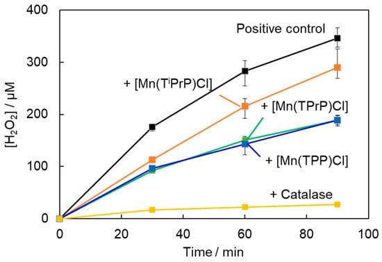
These manganese(III) porphyrin complexes demonstrated protective effects against BNADH self-photooxidation. H2O2 is an important ROS that is produced from O2•−. Furthermore, manganese(III) porphyrin complexes exhibit SOD activity to dismutate O2•− to H2O2 [9]. The SOD effect may contribute to the suppression of BNADH photodecomposition. H2O2 and O2•− can produce hydroxyl radicals (OH•). The lifetime of H2O2 is relatively long and contributes to various redox reactions. Thus, the catalytic H2O2 decomposition activity of these porphyrins was examined. In this assay system, MeHQ produces H2O2 via autooxidation promoted by copper ions, similar to the reaction reported previously [36]. The H2O2 generated in the presence of copper ions decomposes folic acid, leading to the formation of a strongly fluorescent pterin derivative [26]. The H2O2 removal activity of the porphyrins was evaluated by measuring the fluorescence intensity of the sample solution. These porphyrins decreased the amount of H2O2 produced (Figure 7). Catalase (50 units ml−1) also effectively decomposed H2O2. The catalytic H2O2 removal activities of these porphyrins were compared with those of catalase. The activities were as follows: [Mn(TPrP)Cl] (5.2 units mL−1 μM−1) > [Mn(TPP)Cl] (5.0 units mL−1 μM−1) > [Mn(TiPrP)Cl] (4.0 units mL−1 μM−1). These results suggest that a relatively large distortion of the porphyrin ring and high redox potential of [Mn(TiPrP)Cl] are not favorable for the catalytic H2O2 decomposition. The present results suggest that the combination of rapid excited-state deactivation and catalytic ROS decomposition distinguishes these manganese(III) porphyrin complexes from conventional porphyrin photosensitizers. Such characteristics may be useful for designing porphyrin-based photofunctional molecules with reduced phototoxicity.
Figure 7.
Time profile of H2O2 formation through autooxidation of MeHQ with or without manganese(III) porphyrin complex. The sample solution contained 100 μM MeHQ, 10 μM folic acid, and 10 μM copper chloride with or without 5 μM manganese(III) porphyrin complex or 50 unit mL−1 catalase in a 10 mM sodium phosphate buffer (pH 7.6) containing 1% ethanol. H2O2 formation was measured by the fluorescence intensity of the decomposed folic acid. The excitation and detection wavelengths for this assay were 350 nm and 450 nm, respectively. The concentration of H2O2 was estimated using a calibration curve.
4. Conclusions
The photochemical and electrochemical properties of three types of meso-substituted manganese(III) porphyrin complexes were investigated. The meso-substituents affected the distortion of the porphyrin ring and redox potential. These porphyrins exhibited weak emissions from higher excited states, such as fluorescence from the S2 and CT emissions; however, their quantum yields were markedly low. The S1 states of these porphyrins were also rapidly deactivated. Although these porphyrins exhibited relatively high redox potentials, photosensitized protein oxidation was barely observed. This is explained by the fact that the rapid deactivation of the photoexcited states of these porphyrins decreases the probability of photosensitized reactions, including biomolecule oxidation. In addition, these porphyrins could not photo-oxidize BNADH. The self-oxidation of photo-irradiated BNADH was inhibited by these porphyrins, and their catalytic activity toward H2O2 decomposition was confirmed. H2O2 is an important ROS that is produced through photochemical reactions. This study proposes manganese(III) porphyrin complexes as potential photoprotective agents via catalytic ROS decomposition. This photoprotective concept is different from the conventional use of porphyrins as photosensitizers and may provide an alternative direction for the development of porphyrin-based photofunctional materials.
Supplementary Materials
The following supporting information can be downloaded at https://www.mdpi.com/article/10.3390/photochem6030027/s1 [31,37], Figure S1: Cyclic voltammograms of [Mn(TPP)Cl] (A), [Mn(TPrP)Cl] (B), and [Mn(TiPrP)Cl] (C) in acetonitrile. The sample solution contained 1 mg of manganese(III) porphyrin complex and 0.1 M tetrabutylammonium hexafluorophosphate (supporting electrolyte) in 1 mL of dry acetonitrile; Figure S2: Time profile of the emission intensity of the manganese(III) porphyrin complexes. The sample solution contained 5 μM porphyrin in chloroform. The excitation wavelength was 402 nm. The instrument response function (IRF) was measured using a scatterer; Figure S3: CD and absorption spectra of [Mn(TPP)Cl] (A), [Mn(TPrP)Cl] (B), and [Mn(TiPrP)Cl] (C). The sample solution contained 5 μM manganese(III) porphyrin complexes with or without 20 μM HSA in a 10 mM sodium phosphate buffer (pH 7.6) containing 1% ethanol; Figure S4: Absorption spectrum of BNADH and emission spectrum of blue LED. The sample solution contained 100 μM BNADH in ethanol. The emission maximum wavelength of the blue LED was 468 nm and the full width at half maximum was 22 nm.
Author Contributions
Conceptualization, K.H.; methodology, K.H. and A.I.; validation, K.H. and K.M.; formal analysis, K.H.; investigation, K.H., K.M. and A.M.; resources, A.I.; data curation, K.H.; writing—original draft preparation, K.H.; writing—review and editing, A.M.; visualization, K.H.; supervision, K.H.; project administration, K.H.; funding acquisition, K.H. All authors have read and agreed to the published version of the manuscript.
Funding
This research was funded by Grant-in-Aid for Scientific Research (C) from the Japan Society for the Promotion of Science, JSPS KAKENHI 24K08600.
Data Availability Statement
The data presented in this study are available upon request.
Acknowledgments
The authors are grateful to Shigetoshi Okazaki for the near-infrared emission measurements used to evaluate the 1O2 production ability of manganese(III) porphyrin complexes.
Conflicts of Interest
The authors declare no conflicts of interest.
Abbreviations
The following abbreviations are used in this manuscript:
| BNADH | 1-Benzyl-1,4-dihydronicotinamide |
| CD | Circular dichroism |
| DFT | Density functional theory |
| FRET | Förster resonance energy transfer |
| HSA | Human serum albumin |
| MeHQ | Methylhydroquinone |
| [Mn(TiPrP)Cl] | Manganese(III) tetraisopropylporphyrin chloride |
| [Mn(TPP)Cl] | Manganese(III) tetraphenylporphyrin chloride |
| [Mn(TPrP)Cl] | Manganese(III) tetrapropylporphyrin chloride |
| PDT | Photodynamic therapy |
| SOD | Superoxide dismutase |
References
- Chen, J.; Zhu, Y.; Kaskel, S. Porphyrin-based Metal-organic Frameworks for Biomedical Applications. Angew. Chem. Int. Ed. Engl. 2021, 60, 5010–5035. [Google Scholar] [PubMed]
- Lindner, A.; Lesniewicz, A.; Kolman, A.; Larowska-Zarych, D.; Marciniak, B.; Lewandowska-Andralojc, A. When Porphyrins Meet 2D Materials: Spectroscopic and Photocatalytic Properties. J. Mater. Chem. C 2024, 12, 9012–9067. [Google Scholar] [CrossRef] [Scilit]
- Senge, M.O.; Sergeeva, N.N.; Hale, K.J. Classic Highlights in Porphyrin and Porphyrinoid Total Synthesis and Biosynthesis. Chem. Soc. Rev. 2021, 50, 4730–4789. [Google Scholar] [CrossRef] [Scilit] [PubMed]
- Akbar, A.; Khan, S.; Chatterjee, T.; Ghosh, M. Unleashing the Power of Porphyrin Photosensitizers: Illuminating Breakthroughs in Photodynamic Therapy. J. Photochem. Photobiol. B 2023, 248, 112796. [Google Scholar] [CrossRef] [Scilit] [PubMed]
- Yin, W.H.; Li, P.Y.; Huang, H.H.; Feng, L.; Liu, S.H.; Liu, X.; Bai, F.Q. Porphyrin Photosensitizer Molecules as Effective Medicine Candidates for Photodynamic Therapy: Electronic Structure Information Aided Design. RSC Adv. 2024, 14, 29368–29383. [Google Scholar] [CrossRef] [Scilit] [PubMed]
- Chaves, I.; Morais, F.M.P.; Vieira, C.; Bartolomeu, M.; Faustino, M.A.F.; Neves, M.G.P.M.S.; Almeida, A.; Moura, N.M.M. Can Porphyrin–triphenylphosphonium Conjugates Enhance the Photosensitizer Performance toward Bacterial Strains? ACS Appl. Bio Mater. 2024, 7, 5541–5552. [Google Scholar] [CrossRef] [Scilit] [PubMed]
- Sun, J.; Jennepalli, S.; Lee, M.; Jones, A.; O’Carroll, D.M.; Manefield, M.J.; Bhadbhade, M.; Åkermark, B.; Das, B.; Kumar, N. Efficient Reductive Defluorination of Branched PFOS by Metal–porphyrin Complexes. Environ. Sci. Technol. 2022, 56, 7830–7839. [Google Scholar] [CrossRef] [Scilit] [PubMed]
- Rybicka-Jasińska, K.; Wdowik, T.; Łuczak, K.; Wierzba, A.J.; Drapała, O.; Gryko, D. Porphyrins as Promising Photocatalysts for Red-light-induced Functionalizations of Biomolecules. ACS Org. Inorg. Au 2022, 2, 422–426. [Google Scholar] [CrossRef] [Scilit] [PubMed]
- Batinic-Haberle, I.; Tovmasyan, A.; Spasojevic, I. An Educational Overview of the Chemistry, Biochemistry and Therapeutic Aspects of Mn Porphyrins—From Superoxide Dismutation to H2O2-driven pathways. Redox Biol. 2015, 5, 43–65. [Google Scholar] [CrossRef] [Scilit] [PubMed]
- Vinhado, F.S.; Prado-Manso, C.M.C.; Sacco, H.C.; Iamamoto, Y. Cationic Manganese(III) Porphyrins Bound to a Novel Bis-functionalised Silica as Catalysts for Hydrocarbons Oxygenation by Iodosylbenzene and Hydrogen peroxide. J. Mol. Catal. A 2001, 174, 279–288. [Google Scholar] [CrossRef] [Scilit]
- Batinic-Haberle, I.; Tovmasyan, A.; Spasojevic, I. Mn Porphyrin-based Redox-active Drugs: Differential Effects as Cancer Therapeutics and Protectors of Normal Tissue against Oxidative Injury. Antioxid. Redox Signal. 2018, 29, 1691–1724. [Google Scholar] [CrossRef] [Scilit] [PubMed]
- Nemeth, T.; Pallier, A.; Çelik, Ç.; Garda, Z.; Yoshizawa-Sugata, N.; Masai, H.; Tóth, É.; Yamakoshi, Y. Water-soluble Mn(III)-Porphyrins with High Relaxivity and Photosensitization. Chem. Biomed. Imaging 2025, 3, 5–14. [Google Scholar] [PubMed]
- Peng, X.; Gao, C.; Huang, Y.; Peng, J.; Aratani, N.; Osuka, A. Benzylic Modifications of meso-Alkyl-substituted Porphyrins via Oxidation with DDQ. Tetrahedron Lett. 2012, 53, 1156–1159. [Google Scholar] [CrossRef] [Scilit]
- Copello, R.C.; Rodrigues, B.M.; Victória, H.F.V.; Krambrock, K.; Chaves, O.A.; Piquini, P.C.; Iglesias, B.A. Using Multi-spectroscopic Techniques to Evaluate Photophysical, Photobiological and Bio-interactive Properties of Meso-5,15-bis(substituted)porphyrins. J. Mol. Struct. 2025, 1337, 142175. [Google Scholar] [CrossRef] [Scilit]
- Jalilian, H.; Sardivand-chegini, I.; Qodrati-nasrabadi, F.; Zakavi, S. Photocatalytic Activity of Meso-tetra(alkyl)- vs. Meso-tetra(aryl)porphyrins: Comparison of the Free Base and Diprotonated porphyrins. J. Mol. Struct. 2026, 1349, 143623. [Google Scholar] [CrossRef] [Scilit]
- Ikezaki, A.; Nakamura, M. Effects of Porphyrin Deformation on the 13C and 1H NMR Chemical Shifts in High-spin Five- and Six-coordinate Manganese(III) Porphyrin Complexes. J. Porphyr. Phthalocyanines 2016, 20, 318–330. [Google Scholar] [CrossRef] [Scilit]
- Ikezaki, A.; Nakamura, M. Importance of the C-H N Weak Hydrogen Bonding on the Coordination Structures of Manganese(III) Porphyrin Complexes. Inorg. Chem. 2003, 42, 2301–2310. [Google Scholar] [CrossRef] [Scilit] [PubMed]
- Adler, A.D.; Longo, F.R.; Finarelli, J.D.; Goldmacher, J.; Assour, J.; Korsakoff, L. A Simplified Synthesis for meso-Tetraphenylporphine. J. Org. Chem. 1967, 32, 476. [Google Scholar] [CrossRef] [Scilit]
- Lindsey, J.S.; Wagner, R.W. Investigation of the Synthesis of ortho-Substituted Tetraphenylporphyrins. J. Org. Chem. 1989, 54, 828–836. [Google Scholar] [CrossRef] [Scilit]
- Adler, A.D.; Longo, F.R.; Nelson, N.Y.; Kim, F. On the Preparation of Metalloporphyrins. J. Inorg. Nucl. Chem. 1970, 32, 2443–2445. [Google Scholar] [CrossRef] [Scilit]
- Hirakawa, K.; Nishimura, Y.; Arai, T.; Okazaki, S. Singlet Oxygen Generating Activity of an Electron Donor-connecting Porphyrin Photosensitizer Can Be Controlled by DNA. J. Phys. Chem. B 2013, 117, 13490–13496. [Google Scholar] [CrossRef] [Scilit] [PubMed]
- He, X.M.; Carter, D.C. Atomic Structure and Chemistry of Human Serum Albumin. Nature 1992, 358, 209–215. [Google Scholar] [CrossRef] [Scilit] [PubMed]
- Hirakawa, K.; Ouyang, D.; Ibuki, Y.; Hirohara, S.; Okazaki, S.; Kono, E.; Kanayama, N.; Nakazaki, J.; Segawa, H. Photosensitized Protein-damaging Activity, Cytotoxicity, and Antitumor Effects of P(V)porphyrins Using Long-wavelength Visible Light through Electron Transfer. Chem. Res. Toxicol. 2018, 31, 371–379. [Google Scholar] [CrossRef] [Scilit] [PubMed]
- Hirakawa, K.; Suzuki, A.; Ouyang, D.; Okazaki, S.; Ibuki, Y.; Nakazaki, J.; Segawa, H. Controlled Photodynamic Action of Axial Fluorinated DiethoxyP(V)tetrakis(p-methoxyphenyl)porphyrin through Self-aggregation. Chem. Res. Toxicol. 2019, 32, 1638–1645. [Google Scholar] [CrossRef] [Scilit] [PubMed]
- Hasegawa, H.; Hirakawa, K. Evaluation of the Photosensitizer Activity of Hydrophobic Phosphorus(V) Porphyrins Using the Absorption Spectral Change of 1-Benzyl-1,4-dihydronicotinamide. Spectrochim. Acta A Mol. Biomol. Spectrosc. 2024, 308, 123765. [Google Scholar] [CrossRef] [Scilit] [PubMed]
- Hirakawa, K. Fluorometry of Hydrogen Peroxide Using Oxidative Decomposition of Folic Acid. Anal. Bioanal. Chem. 2006, 386, 244–248. [Google Scholar] [CrossRef] [Scilit] [PubMed]
- Tulinsky, A.; Chen, B.M.L. The crystal and Molecular Structure of Chloro-α,β,γ,δ,-tetraphenylporphinatomanganese(III). J. Am. Chem. Soc. 1977, 99, 3647–3651. [Google Scholar] [PubMed]
- Kadish, K.M.; Caemelbecke, E.V. Electrochemistry of Porphyrins and Related Macrocycles. J. Solid State Electrochem. 2003, 7, 254–258. [Google Scholar] [CrossRef] [Scilit]
- Fodor, M.A.; Szabó, P.; Lendvay, G.; Horváth, O. Characterization of the UV-Visible Absorption Spectra of Manganese(III)porphyrins with Time-dependent Density Functional Theory Calculations. Z. Phys. Chem. 2021, 236, 27–61. [Google Scholar] [CrossRef] [Scilit]
- Fagadar-Cosma, E.; Mirica, M.C.; Balcu, I.; Bucovicean, C.; Cretu, C.; Armeanu, I.; Fagadar-Cosma, G. Syntheses, Spectroscopic and AFM Characterization of Some Manganese Porphyrins and Their Hybrid Silica Nanomaterials. Molecules 2009, 14, 1370–1388. [Google Scholar] [CrossRef] [Scilit] [PubMed]
- Hirakawa, K.; Hosokawa, Y.; Nishimura, Y.; Okazaki, S. Relaxation Process of S2 Excited Zinc Porphyrin through Interaction with a Directly Connected Phenanthryl Group. Chem. Phys. Lett. 2019, 732, 136652. [Google Scholar] [CrossRef] [Scilit]
- Förster, T. Zwischenmolekulare Energiewanderung und Fluoreszenz. Ann. Phys. 1948, 437, 55–75. [Google Scholar] [CrossRef] [Scilit]
- Abou-Zied, O.K.; Al-Shihi, O.I.K. Characterization of Subdomain IIA Binding Site of Human Serum Albumin in Its Native, Unfolded, and Refolded States Using Small Molecular Probes. J. Am. Chem. Soc. 2008, 130, 10793–10801. [Google Scholar] [CrossRef] [Scilit] [PubMed]
- Ferrer, M.L.; Duchowicz, R.; Carrasco, B.; de la Torre, J.G.; Acuña, A.U. The Conformation of Serum Albumin in Solution: A Combined Phosphorescence Depolarization-hydrodynamic Modeling Study. Biophys. J. 2001, 80, 2422–2430. [Google Scholar] [CrossRef] [Scilit] [PubMed]
- Yamaoka, S.; Okazaki, S.; Hirakawa, K. Activity Control of pH-responsive Photosensitizer Bis(6-quinolinoxy)P(V)tetrakis(4-chlorophenyl)porphyrin through Intramolecular Electron Transfer. Chem. Phys. Lett. 2022, 788, 139285. [Google Scholar] [CrossRef] [Scilit]
- Hirakawa, K.; Sano, S. Platinum Nanoparticle Catalyst Scavenges Hydrogen Peroxide Generated from Hydroquinone. Bull. Chem. Soc. Jpn. 2009, 82, 1299–1303. [Google Scholar] [CrossRef] [Scilit]
- Sakura, S. Chemiluminescence of Tryptophan Enhanced by Electrochemical Energy. Electrochim. Acta 1992, 37, 2731–2735. [Google Scholar] [CrossRef] [Scilit]

Disclaimer/Publisher’s Note: The statements, opinions and data contained in all publications are solely those of the individual author(s) and contributor(s) and not of MDPI and/or the editor(s). MDPI and/or the editor(s) disclaim responsibility for any injury to people or property resulting from any ideas, methods, instructions or products referred to in the content. |
© 2026 by the authors. Licensee MDPI, Basel, Switzerland. This article is an open access article distributed under the terms and conditions of the Creative Commons Attribution (CC BY) license.