


Abstract
Low-molecular-weight lychee polyphenol (Lyp), a standardized oligomeric polyphenol derived from Litchi chinensis fruit, possesses potent antioxidant activity but is susceptible to physicochemical instability, which may limit its practical application. In this study, freeze-dried systems of Lyp with α-, β-, and γ-cyclodextrins (CDs) were prepared to investigate the effects of cyclodextrins on the physicochemical properties and antioxidant activity of Lyp. Powder X-ray diffraction, thermogravimetric analysis, Fourier transform infrared spectroscopy, near-infrared spectroscopy, and solution-state NMR were employed to characterize the prepared systems. Freeze-drying produced amorphous Lyp/CD systems accompanied by enhanced thermal stability and changes in the hydrogen-bonding environment compared with the corresponding physical mixtures. Solution-state 1H NMR and NOESY analyses suggested molecular association between Lyp constituents and cyclodextrins, with the βCD system exhibiting the most pronounced spectral changes. Consistent with these physicochemical findings, the freeze-dried βCD system showed the greatest enhancement of DPPH radical scavenging activity among the three cyclodextrins examined. These results suggest that βCD provides the most favorable molecular environment for Lyp, leading to improved physicochemical characteristics and antioxidant performance. The present findings demonstrate that freeze-drying combined with β-cyclodextrin is an effective strategy for improving the physicochemical performance and antioxidant functionality of low-molecular-weight lychee polyphenol.
1. Introduction
Low-molecular-weight lychee polyphenol (Lyp; OPLFE) is a standardized oligomeric polyphenol derived from Litchi chinensis fruit and is enriched in monomeric and oligomeric flavan-3-ols, including catechins and proanthocyanidins, compared with conventional lychee polyphenol preparations [1,2]. Owing to its potent antioxidant activity together with anti-inflammatory, renoprotective, stress-relieving, and microcirculation-improving effects, Lyp has attracted considerable attention as a functional ingredient for food, nutraceutical, cosmetic, and pharmaceutical applications [3,4,5]. Despite these promising biological properties, the practical utilization of Lyp may be limited by the inherent physicochemical instability of polyphenolic compounds, which are susceptible to thermal stress, oxidation, and environmental conditions during processing and storage. Therefore, improving the physicochemical stability of Lyp is an important step toward expanding its practical applications. Cyclodextrin-based formulation is considered a promising strategy to improve the physicochemical properties of natural polyphenols.
Cyclodextrins (CDs) are cyclic oligosaccharides composed of α-(1→4)-linked glucopyranose units and are widely used as pharmaceutical and food excipients because of their unique molecular architecture, featuring a hydrophobic inner cavity and a hydrophilic outer surface [6]. Through non-covalent molecular association with guest molecules, CDs can modify physicochemical properties such as aqueous solubility, thermal stability, dissolution behavior, and chemical stability without altering the chemical structure of the guest compounds [7,8,9,10]. Among the naturally occurring cyclodextrins, αCD, βCD, and γCD possess different cavity sizes, which may result in different interaction behaviors depending on the molecular size and structural characteristics of the guest molecules [11]. Consequently, cyclodextrins have been extensively investigated as functional carriers for natural products, including polyphenols, to improve their physicochemical performance and functional properties [12,13].
Although cyclodextrin complexation has been extensively investigated for a variety of natural polyphenols and bioactive compounds [14], little information is available regarding its application to low-molecular-weight lychee polyphenol (Lyp). Previous studies have demonstrated that cyclodextrins interact with representative natural polyphenols, including catechin, quercetin, and curcumin, through non-covalent host–guest interactions, resulting in improved physicochemical properties such as aqueous solubility and stability. These findings support the application of cyclodextrins as multifunctional excipients for natural polyphenols. Because Lyp consists of a mixture of monomeric and oligomeric polyphenols with different molecular sizes and structures [6], interactions with cyclodextrins are expected to influence its physicochemical characteristics rather than those of a single molecular species. However, the effects of αCD, βCD, and γCD on the physicochemical properties and antioxidant activity of Lyp have not been systematically compared. A better understanding of these interactions would facilitate the rational selection of cyclodextrins for the stabilization and formulation of Lyp-containing functional products.
Therefore, the present study aimed to investigate the effects of α-, β-, and γ-cyclodextrins on the physicochemical properties and antioxidant activity of low-molecular-weight lychee polyphenol (Lyp) using freeze-dried systems. The prepared samples were characterized by powder X-ray diffraction (PXRD), thermogravimetric analysis (TG), Fourier transform infrared (FT-IR) spectroscopy, near-infrared (NIR) spectroscopy, and solution-state NMR analyses, together with evaluation of DPPH radical scavenging activity. Particular attention was paid to clarifying how different cyclodextrins influence the physicochemical behavior and functional performance of Lyp.
2. Materials and Methods
2.1. Materials
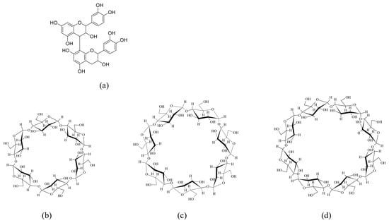
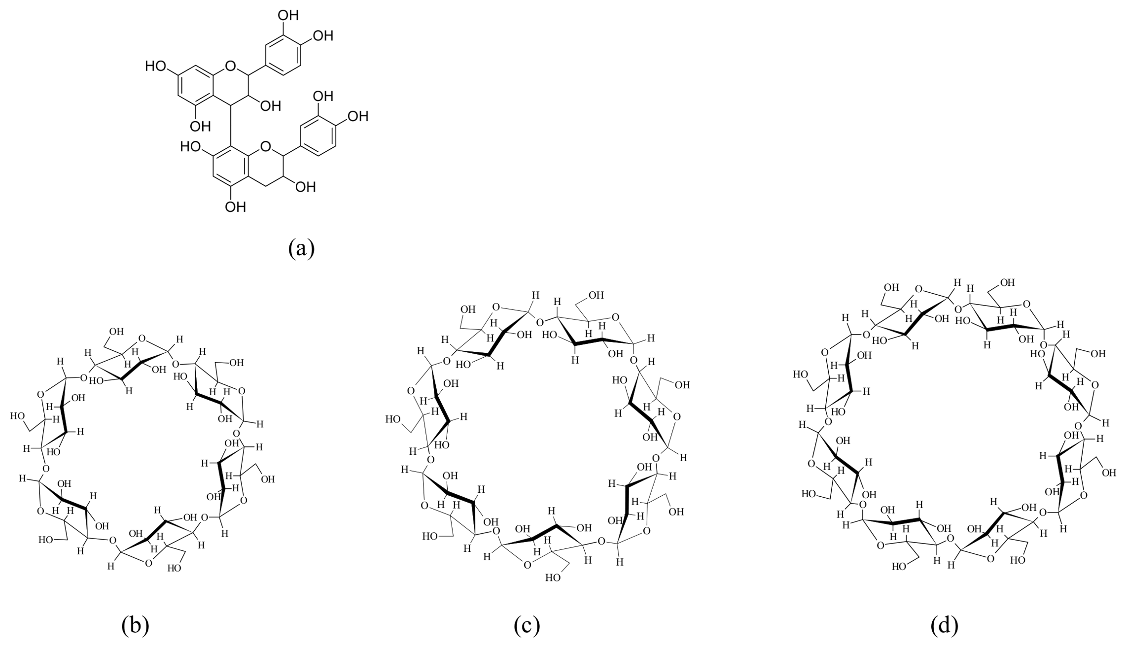
Low-molecular-weight lychee polyphenol (Lyp; OPLFE, Oligonol®, Sapporo, Japan), a standardized low-molecular-weight polyphenol product derived from Litchi chinensis fruit extract, was generously supplied by Amino Up Co., Ltd. (Sapporo, Japan). As previously reported [1,2], Lyp is enriched in low-molecular-weight flavan-3-ols, including catechin monomers and oligomeric proanthocyanidins. αCD, βCD, and γCD were kindly provided by CycloChem Co., Ltd. (Kobe, Japan) (Figure 1). Deuterium oxide (D2O) used for NMR measurements was purchased from FUJIFILM Wako Pure Chemical Corporation (Osaka, Japan). All other reagents and solvents were of analytical grade and used as received. Ultrapure water was used throughout the study.
Figure 1.
Chemical structures: (a) Representative flavan-3-ol structure of low-molecular-weight lychee polyphenol (Lyp), (b) α-cyclodextrin (αCD), (c) β-cyclodextrin (βCD), (d) γ-cyclodextrin (γCD).
2.2. Preparation of Lyp/CD Complexes
Physical mixtures (PMs) and freeze-dried products (FDs) of Lyp with αCD, βCD, and γCD were prepared at a weight ratio of 1:1 (w/w). PMs were prepared by mixing Lyp and the corresponding CD using a vortex mixer for 3 min. For the preparation of FDs, Lyp and each CD were dispersed in 10 mL of distilled water and mechanically stirred until a homogeneous dispersion was obtained. The resulting dispersions were pre-frozen at −30 °C and subsequently lyophilized using FDU-2110 (EYELA, Tokyo Rikakikai Co., Ltd., Tokyo, Japan). The freeze-dried powders were collected and stored in a desiccator at room temperature until further characterization.
2.3. Powder X-Ray Diffraction (PXRD)
Powder X-ray diffraction (PXRD) measurements were conducted using an X-ray diffractometer (Miniflex II, Rigaku Corp., Tokyo, Japan) equipped with a NaI scintillation detector. Diffraction patterns were recorded using Cu-Kα radiation operated at 30 kV and 15 mA. Data were collected over a 2θ range of 5–40° with a scanning rate of 4°/min and a step size of 0.02°. Powder samples were gently packed into a glass sample holder and leveled to obtain a smooth surface prior to measurement. The diffraction profiles were used to evaluate changes in the solid-state properties of Lyp and its complexes with CDs.
2.4. Thermogravimetric and Differential Thermal Analysis (TG-DTA)
Thermal behavior of Lyp, CDs, PMs, and FDs was investigated by thermogravimetric and differential thermal analysis (TG-DTA) using a Thermo Plus EVO system (Rigaku Corp., Tokyo, Japan). Approximately 5 mg of each sample was accurately weighed into an aluminum pan and heated from 30 to 390 °C at a rate of 10 °C/min under a nitrogen atmosphere with a flow rate of 200 mL/min. The obtained thermograms were used to evaluate the thermal stability and physicochemical changes associated with the interaction between Lyp and CDs.
2.5. Fourier Transform Infrared Spectroscopy (FT-IR)
Fourier transform infrared (FT-IR) spectra of Lyp, CDs, PMs, and FDs were recorded using an FT/IR-4600 spectrometer (JASCO Corp., Tokyo, Japan) equipped with an attenuated total reflection (ATR) accessory. Spectral measurements were carried out over the range of 4000–400 cm−1 with 64 accumulated scans. The obtained spectra were analyzed to identify spectral changes and potential intermolecular interactions associated with the formation of Lyp/CD complexes.
2.6. Near-Infrared Spectroscopy (NIR)
Near-infrared (NIR) spectra were obtained using a V-770 EX UV–visible–NIR spectrophotometer (JASCO Corp., Tokyo, Japan). Measurements were carried out at 25 °C over the spectral range of 10,000–4000 cm−1. Powder samples were placed in dedicated sample holders and scanned at 1 nm intervals with an acquisition time of 8 s. To enhance spectral resolution and facilitate interpretation of band variations, the recorded spectra were converted to second-derivative spectra. The resulting NIR profiles were compared to assess alterations in the molecular environment and intermolecular interactions arising from complexation between Lyp and CDs.
2.7. 1H-NMR and Two-Dimensional NOESY Measurements
1H-NMR and two-dimensional Nuclear Overhauser Effect spectroscopy (NOESY) measurements were performed using an AVANCE NEO 600 NMR spectrometer (Bruker, Billerica, MA, USA). Samples were dissolved in D2O, and spectra were acquired at 25 °C with a resonance frequency of 600.13 MHz. NOESY spectra were recorded using a pulse width of 7.850 μs, 32 scans, a relaxation delay of 2.0 s, and a mixing time of 1.5 s. The obtained spectra were analyzed to evaluate spatial correlations between Lyp constituents and CD protons, thereby providing information on host–guest interactions and molecular interactions in solution.
2.8. DPPH Radical Scavenging Assay
The antioxidant activity of Lyp, CDs, PMs, and FDs was evaluated using a 2,2-diphenyl-1-picrylhydrazyl (DPPH) radical scavenging assay. Ascorbic acid (ASC) was included as a positive control in the assay. Each sample was dissolved in a methanol/distilled water mixture (1:1, v/v) and serially diluted to the desired concentrations. A DPPH solution prepared in methanol was mixed with the sample solution at an equal volume ratio in a 96-well microplate. The reaction mixtures were incubated at 37 °C under light-protected conditions, and the absorbance of DPPH at 517 nm was monitored using a SpectraMax 190 microplate reader (Molecular Devices Japan Co., Ltd., Tokyo, Japan). Radical scavenging activity was determined relative to the control, and the concentration required to inhibit 50% of DPPH radicals (IC50) was calculated [15]. The assay was conducted to evaluate the effect of cyclodextrin complexation on the antioxidant activity of Lyp.
2.9. Statistical Analysis
All experiments were performed in triplicate, and the results are expressed as the mean ± standard deviation (SD). Statistical analyses were conducted using Excel Statistics 4.0 (Esumi Co., Ltd., Tokyo, Japan). Dunnett’s multiple comparison test was applied using Lyp as the control group. Differences were considered statistically significant when p < 0.05.
3. Results and Discussion
3.1. Powder X-Ray Diffraction Analysis
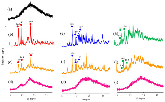
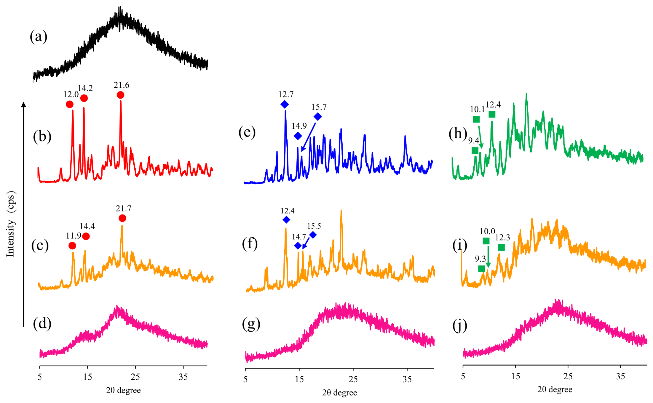
Powder X-ray diffraction (PXRD) analysis was performed to investigate changes in the crystalline state and solid-state organization of the Lyp/CD systems following physical mixing and freeze-drying. PXRD is widely employed to evaluate crystallinity and to detect structural changes associated with the cyclodextrin-based host–guest systems and amorphization processes [16]. The PXRD patterns of Lyp, CDs, PMs, and FDs are shown in Figure 2. Lyp exhibited a broad halo pattern without distinct diffraction peaks, indicating its predominantly amorphous nature. In contrast, αCD, βCD, and γCD displayed characteristic diffraction peaks corresponding to their crystalline structures. The PXRD patterns of the PMs were essentially a superposition of the diffraction peaks of the respective CDs and the amorphous halo pattern of Lyp, suggesting that simple physical mixing did not substantially alter the solid-state properties of the individual components. Characteristic diffraction peaks of αCD were observed at 12.0°, 14.2°, and 21.6°, while βCD exhibited prominent reflections at 12.7°, 14.9°, and 15.7°. Similarly, γCD showed characteristic peaks at 9.4°, 10.1°, and 12.4°. These diffraction peaks remained clearly detectable in the corresponding PMs, indicating that the crystalline structures of the CDs were largely preserved after physical mixing with Lyp.
Figure 2.
Powder X-ray diffraction (PXRD) patterns of Lyp, cyclodextrins (CDs), physical mixtures (PMs), and freeze-dried samples (FDs). (a) Lyp; (b) αCD; (c) PM (Lyp/αCD = 1/1); (d) FD (Lyp/αCD = 1/1); (e) βCD; (f) PM (Lyp/βCD = 1/1); (g) FD (Lyp/βCD = 1/1); (h) γCD; (i) PM (Lyp/γCD = 1/1); (j) FD (Lyp/γCD = 1/1).
In contrast, the characteristic diffraction peaks of αCD, βCD, and γCD disappeared in all FD samples, resulting in broad halo patterns. The marked reduction in crystallinity suggests that freeze-drying disrupted the ordered crystal lattices of the CDs and generated amorphous solid systems. Similar PXRD behavior has been reported for several CD complexes prepared by freeze-drying, including curcumin/γCD and gedunin/hydroxypropyl-βCD systems, in which the disappearance of characteristic diffraction peaks was attributed to the formation of amorphous molecularly dispersed host–guest assemblies [17]. Furthermore, these amorphous systems were associated with enhanced molecular-level interactions between guest molecules and cyclodextrins [18]. Because Lyp itself exhibited an amorphous halo pattern, the PXRD results alone cannot conclusively demonstrate molecular association formation. Nevertheless, the complete disappearance of the characteristic diffraction peaks of the CDs in the FD samples indicates that the molecular organization of the Lyp/CD systems differed substantially from that of the corresponding PMs. Freeze-drying is known to reduce the crystallinity of cyclodextrins by disrupting their ordered crystal lattices during dehydration. Therefore, the observed amorphization cannot be attributed solely to interactions between Lyp and cyclodextrins. Nevertheless, the PXRD profiles of the freeze-dried samples clearly differed from those of the corresponding physical mixtures, indicating substantial changes in the solid-state organization of the Lyp/CD systems during the freeze-drying process. These changes are consistent with increased molecular association between Lyp and cyclodextrins, although PXRD alone does not allow the contribution of freeze-drying to be distinguished from that of Lyp–CD interactions. Therefore, PXRD provides evidence that freeze-drying altered the solid-state properties of the Lyp/CD systems, while complementary spectroscopic and solution-state NMR analyses were required to further evaluate molecular interactions between Lyp and cyclodextrins.
3.2. Thermogravimetric Analysis
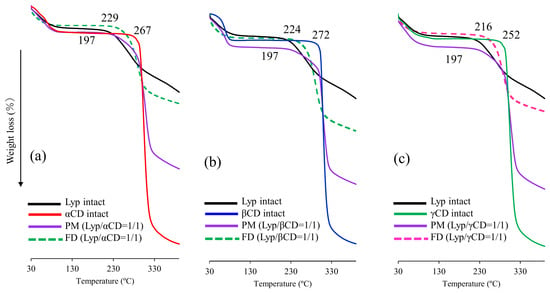
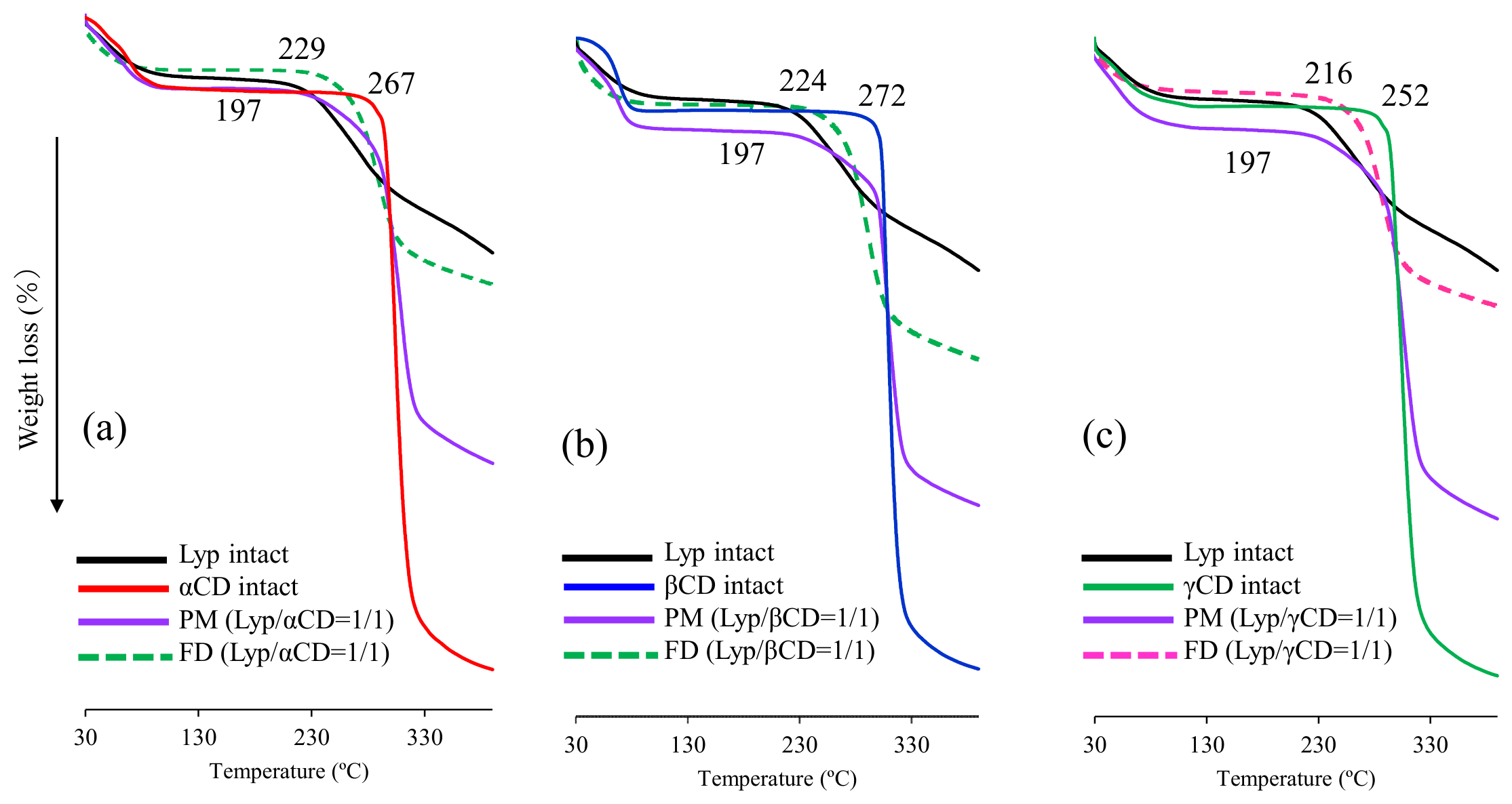
Thermogravimetric (TG) analysis was performed to evaluate the thermal stability of the Lyp/CD systems and to investigate whether interactions with CDs influenced the thermal degradation behavior of Lyp. Thermal analysis is widely used to characterize cyclodextrin-based host–guest systems because changes in the decomposition behavior can provide useful information regarding molecular interactions and stabilization effects associated with complex formation [19]. The TG curves of Lyp, CDs, PMs, and FDs are shown in Figure 3. Lyp exhibited the onset of thermal decomposition at approximately 197 °C. In contrast, αCD, βCD, and γCD showed decomposition at higher temperatures, reflecting their greater intrinsic thermal stability. The TG profiles of the PMs were generally consistent with the combined thermal behaviors of Lyp and the corresponding CDs, indicating that simple physical mixing did not markedly alter the thermal properties of the individual components.
Figure 3.
Thermogravimetric (TG) curves of the Lyp/CD systems. TG curves of the (a) αCD, (b) βCD, and (c) γCD systems. Each panel includes Lyp, the corresponding cyclodextrin, PM (Lyp/CD = 1/1), and FD (Lyp/CD = 1/1).
In contrast, all FD samples exhibited delayed thermal degradation compared with Lyp alone. The decomposition onset temperatures of FD(Lyp/αCD), FD(Lyp/βCD), and FD(Lyp/γCD) were approximately 229, 224, and 216 °C, respectively, indicating improved thermal stability of the freeze-dried systems.
The improved thermal stability observed in the freeze-dried samples suggests that the physicochemical environment surrounding Lyp was altered during preparation. Because freeze-drying itself can modify the solid-state properties of cyclodextrins through dehydration and amorphization, the observed thermal stabilization cannot be attributed solely to interactions between Lyp and cyclodextrins. Nevertheless, the consistent increase in decomposition temperature observed for all freeze-dried Lyp/CD systems, together with the PXRD results showing marked changes in solid-state properties, suggests that freeze-drying in the presence of cyclodextrins produced a different physicochemical environment from that of the corresponding physical mixtures.
These findings are consistent with increased molecular association between Lyp and cyclodextrins, which may contribute to the improved thermal stability of Lyp. However, TG analysis alone cannot distinguish the individual contributions of freeze-drying and Lyp–CD interactions. Therefore, the thermal results should be interpreted together with the spectroscopic and NMR analyses presented below.
3.3. Fourier Transform Infrared Spectroscopy Analysis
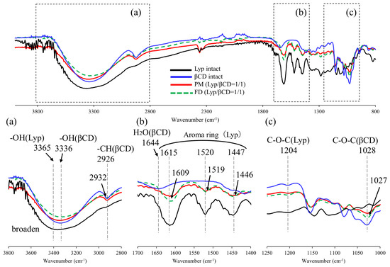
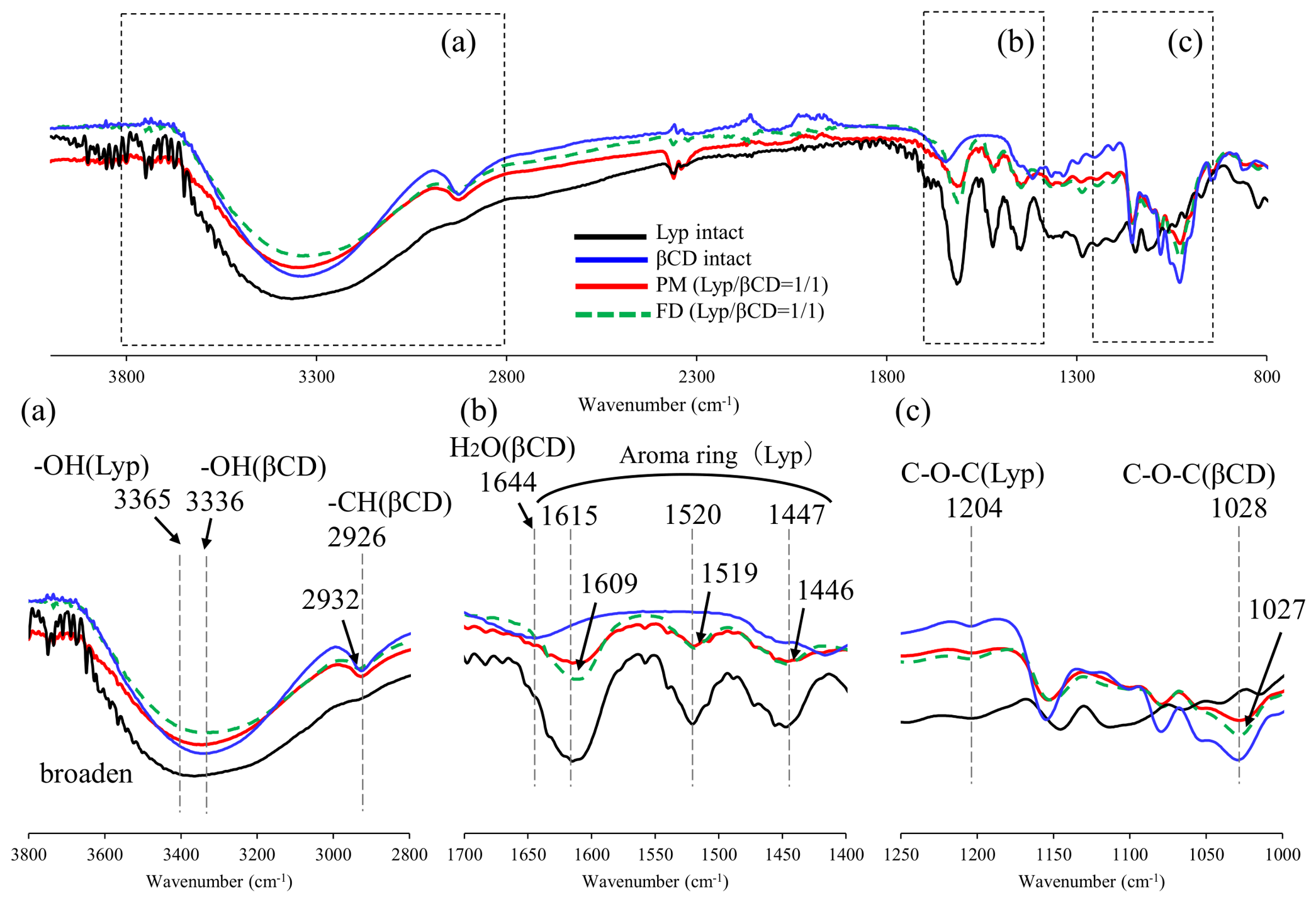
FT-IR spectroscopy was employed to investigate intermolecular interactions between Lyp and CDs in the solid state. The FT-IR spectra of the αCD, βCD, and γCD systems are shown in Figure 4, Figures S1 and S2. Lyp exhibited a broad O–H stretching band centered at approximately 3365 cm−1, reflecting the abundance of hydroxyl groups in its polyphenolic constituents. The corresponding O–H stretching bands of αCD, βCD, and γCD were observed at approximately 3320, 3336, and 3334 cm−1, respectively. In all PM and FD samples, broadening of the O–H stretching region (3700–3000 cm−1) was observed relative to the individual components. In the aromatic ring vibration region (1700–1400 cm−1), Lyp showed characteristic absorption bands at 1615, 1520, and 1447 cm−1. These bands became broader and less distinct in the PM and FD samples. Slight shifts in peak positions were also observed, particularly in the βCD and γCD systems, where the aromatic band at 1615 cm−1 shifted to approximately 1609 and 1614 cm−1, respectively. Similar changes were observed for the bands at 1520 and 1447 cm−1. Additional spectral modifications were detected in the C–O–C stretching region (1250–1000 cm−1). Characteristic absorption bands assigned to Lyp (1204–1206 cm−1) and CDs (1021–1028 cm−1) exhibited changes in band shape and intensity after preparation of the PM and FD systems. Although no new absorption bands were detected in any of the CD systems, broadening and slight peak shifts were consistently observed throughout the spectra. The broadening of the O–H stretching region indicates that the hydrogen-bonding environment was altered after preparation of the freeze-dried Lyp/CD systems. Because both Lyp and cyclodextrins contain numerous hydroxyl groups capable of intermolecular hydrogen bonding, these spectral changes are consistent with modifications in the hydrogen-bonding network within the freeze-dried systems [20,21,22]. In addition, slight changes in the aromatic ring vibration and C–O–C stretching regions suggest alterations in the local molecular environment surrounding the polyphenolic constituents of Lyp. Similar spectral changes have been reported for freeze-dried cyclodextrin systems containing natural compounds and are generally associated with changes in the solid-state organization and molecular environment rather than direct evidence of molecular association formation [23].
Figure 4.
Fourier transform infrared (FT-IR) spectra of the Lyp/βCD system. (a) 3800–2800 cm−1, (b) 1700–1400 cm−1, (c) 1250–1000 cm−1.
However, these spectral changes should be interpreted with caution because freeze-drying itself is known to modify the hydrogen-bonding network and solid-state structure of cyclodextrins. Therefore, FT-IR spectroscopy alone cannot distinguish the contribution of freeze-drying from that of interactions between Lyp and cyclodextrins. Nevertheless, when considered together with the PXRD and TG results, the FT-IR findings indicate that the freeze-dried Lyp/CD systems possessed physicochemical characteristics different from those of the corresponding physical mixtures. These observations are consistent with increased molecular association between Lyp and cyclodextrins and were further examined by NIR spectroscopy and solution-state NMR analyses. These observations were further investigated by NIR spectroscopy and solution-state NMR analyses to obtain complementary information on the molecular environment and interactions within the freeze-dried Lyp/CD systems.
3.4. Near-Infrared Spectroscopy Analysis
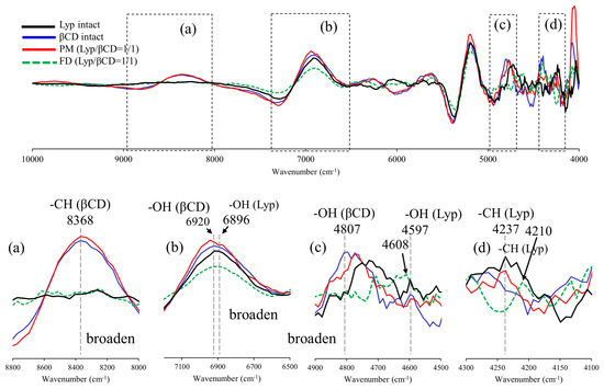
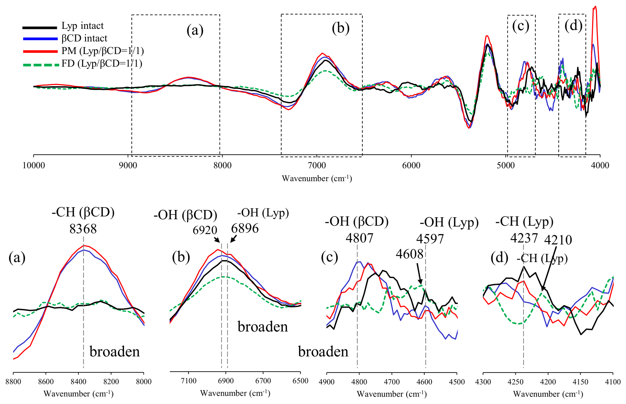
To further investigate molecular interactions between Lyp and CDs, near-infrared (NIR) spectroscopy was performed because NIR bands are highly sensitive to variations in hydrogen-bonding environments and molecular associations involving hydroxyl and aliphatic groups. The second-derivative NIR spectra of the αCD, βCD, and γCD systems are shown in Figure 5, Figures S3 and S4.
Figure 5.
NIR spectra of the Lyp/βCD system. (a) 8800–8000 cm−1, (b) 7200–6500 cm−1, (c) 4900–4500 cm−1, (d) 4300–4100 cm−1.
For all CD systems, spectral changes were observed in four characteristic regions. The bands around 6896–6920 cm−1 were assigned to the first overtone of O–H stretching vibrations, while the bands around 4773–4807 cm−1 corresponded to combination bands associated with hydroxyl groups. In addition, bands at approximately 8333–8368 cm−1 and 4210–4237 cm−1 were attributed to CH-related vibrations of CDs and Lyp, respectively.
In the αCD system (Figure S3), broadening of the O–H-related bands near 6896 and 4773 cm−1 was observed in both PM and FD samples compared with the individual components. Similar changes were detected in the CH-related regions around 8333 and 4237 cm−1. In the βCD system (Figure S4), broadening and intensity changes were observed in the O–H bands at 6920 and 4807 cm−1, together with alterations in the CH-related bands at 8368 and 4210–4237 cm−1. Comparable spectral modifications were also observed in the γCD system (Figure 5c), particularly in the O–H combination band region (4683–4773 cm−1) and the CH-related region around 4237 cm−1. These spectral changes were found to be more pronounced in the FD samples than in the corresponding PMs, suggesting that freeze-drying promoted closer molecular association between Lyp constituents and CDs. The broadening and intensity changes observed in the O–H-related bands indicate that the hydrogen-bonding environment was modified after freeze-drying, whereas the changes in the CH-related regions suggest alterations in the local molecular environment surrounding the polyphenolic constituents of Lyp. These observations are consistent with the FT-IR results, indicating that preparation of the freeze-dried systems altered the physicochemical environment of Lyp. Because NIR spectroscopy is highly sensitive to subtle changes in hydrogen bonding and molecular organization, the observed spectral variations provide complementary information regarding changes in the solid-state properties of the freeze-dried Lyp/CD systems [22].
Previous studies have reported similar changes in O–H- and CH-related NIR bands for cyclodextrin systems containing natural compounds, where such spectral variations were associated with changes in molecular organization and the hydrogen-bonding environment [24]. However, these spectral changes should be interpreted with caution because freeze-drying itself can modify the molecular organization and hydrogen-bonding network of cyclodextrins. Therefore, NIR spectroscopy alone cannot distinguish the contribution of freeze-drying from that of interactions between Lyp and cyclodextrins.
Nevertheless, the consistent spectral changes observed in both the O–H- and CH-related regions, together with the PXRD, TG, and FT-IR results, indicate that the freeze-dried Lyp/CD systems possessed physicochemical characteristics different from those of the corresponding physical mixtures. Although NIR spectroscopy cannot identify the specific Lyp constituents responsible for the observed spectral changes, it provides complementary information on the altered molecular environment within the freeze-dried Lyp/CD systems. These findings provided the rationale for subsequent solution-state 1H NMR and NOESY analyses to further characterize molecular associations between Lyp constituents and cyclodextrins in solution.
3.5. 1H NMR Analysis
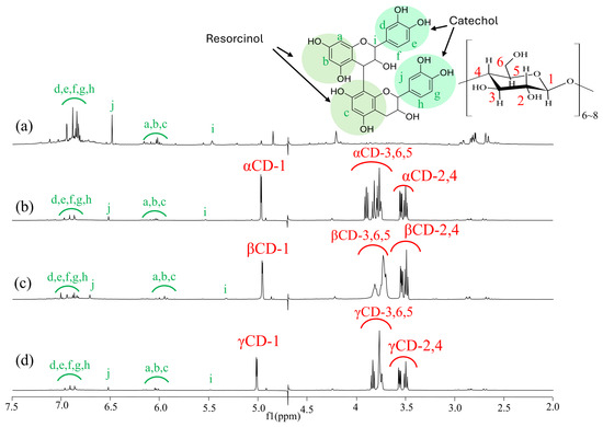
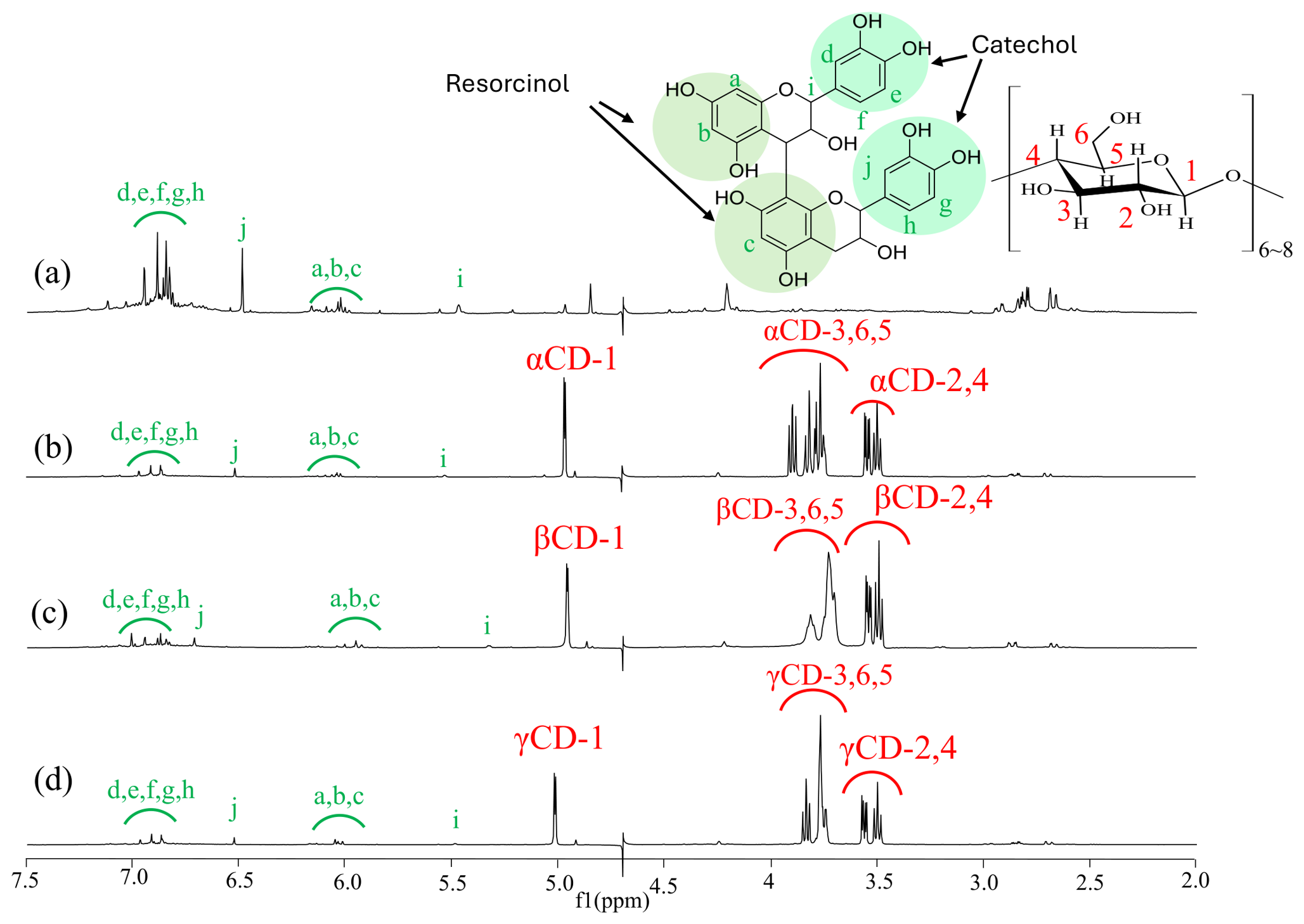
1H NMR measurements were performed to evaluate the solution-state behavior of Lyp and the FD(Lyp/CD) systems in D2O. The 1H NMR spectra of Lyp and FD(Lyp/αCD), FD(Lyp/βCD), and FD(Lyp/γCD) are shown in Figure 6. Lyp showed characteristic signals in the aromatic proton region. Signals observed at approximately 6.8–7.0 ppm were assigned to catechol-type aromatic protons, while signals around 6.0 ppm were attributed to resorcinol-type aromatic protons. Additional Lyp-derived signals were observed at approximately 6.5 and 5.5 ppm. These signals were also detected in the FD(Lyp/CD) spectra, indicating that the polyphenolic constituents of Lyp remained present after freeze-drying with CDs. In the FD(Lyp/CD) systems, characteristic CD proton signals were observed at approximately 5.0 ppm, corresponding to H-1, and in the region of 3.4–3.9 ppm, corresponding to H-2–H-6. The aromatic signals derived from Lyp were retained in the presence of αCD, βCD, and γCD; however, slight changes in signal intensity, peak shape, and broadening were observed in comparison with Lyp alone. These changes suggest that the local magnetic environment of the Lyp constituents was altered by the presence of CDs.
Figure 6.
Assignment of 1H-NMR signals of Lyp and cyclodextrins (CDs). (a) Lyp intact, (b) FD (Lyp/αCD), (c) FD (Lyp/βCD), (d) FD (Lyp/γCD).
Importantly, no new proton signals were observed in any of the FD(Lyp/CD) systems, suggesting that the preparation process did not induce covalent modification or degradation of Lyp. Therefore, the observed spectral changes are likely associated with non-covalent interactions between Lyp constituents and CDs. Similar changes in proton signals have been reported for cyclodextrin complexes of catechin-related compounds and other natural polyphenols, where they were attributed to host–guest association and changes in the local molecular environment [22]. Although the 1H NMR spectra provided evidence of solution-state interactions between Lyp and CDs, the one-dimensional spectra alone were insufficient to determine whether Lyp constituents were located inside the cyclodextrin cavities or associated with the outer surface of CDs. Therefore, two-dimensional NOESY measurements were subsequently performed to clarify spatial proximity and interaction between Lyp-derived protons and CD cavity protons.
3.6. NOESY Analysis
To identify which polyphenolic constituents of Lyp participated in cyclodextrin recognition and to obtain direct evidence of host–guest interactions in solution, two-dimensional NOESY spectroscopy was performed. Although PXRD, TG, FT-IR, NIR, and one-dimensional 1H NMR analyses suggested intermolecular interactions between Lyp constituents and CDs, these techniques could not directly determine the spatial relationship between guest molecules and cyclodextrin cavities. Therefore, two-dimensional 1H–1H NOESY measurements were performed to investigate host–guest interactions in aqueous solution. In cyclodextrins, the H3 and H5 protons are located inside the hydrophobic cavity, whereas H2 and H4 are positioned on the outer surface of the molecule. The H6 proton is located near the primary hydroxyl side at the narrower rim of the cavity. Therefore, NOE correlations involving H3, H5, and H6 provide valuable information regarding the spatial arrangement of guest molecules relative to the cyclodextrin cavity [25].
3.6.1. NOESY Analysis of the Lyp/αCD System
To obtain direct evidence of spatial proximity between Lyp constituents and αCD, two-dimensional 1H–1H NOESY measurements were performed in D2O. The NOESY spectrum of FD(Lyp/αCD) is shown in Figure S5. Several characteristic proton regions derived from Lyp were examined for possible NOE correlations with αCD protons. Cross-peaks were not observed between the catechol-derived aromatic proton signals (d, e, f, g, and h; approximately 6.8–7.0 ppm) and αCD proton signals (Figure S5α-1). Likewise, no NOE correlations were detected between signal i (approximately 5.5 ppm) and αCD protons (Figure S5α-3). In contrast, a distinct cross-peak was observed between the resorcinol-derived aromatic proton signals (a, b, and c; approximately 5.9–6.0 ppm) and αCD proton signals in the H3/H5/H6 region (Figure S5α-2). Because NOESY cross-peaks arise from dipolar interactions between nuclei located within approximately 5 Å of each other, this observation indicates close spatial proximity between the aromatic protons of Lyp and the αCD cavity region. The selective appearance of NOE correlations for the a–c proton region suggests that interactions between Lyp constituents and αCD were not random but occurred preferentially through specific aromatic moieties. Considering that Lyp is composed of structurally diverse polyphenolic constituents, including catechin monomers and proanthocyanidin oligomers, the observed NOE correlation may reflect preferential association of certain polyphenolic structures with the αCD cavity. These findings indicate that at least a portion of the polyphenolic constituents of Lyp was located in close proximity to the αCD cavity in aqueous solution. Similar NOE correlations between aromatic guest molecules and cyclodextrin cavity protons have been reported for polyphenol–cyclodextrin systems and are generally interpreted as evidence of host–guest association and partial host–guest interaction [25].
3.6.2. NOESY Analysis of the Lyp/βCD System
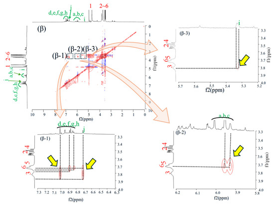
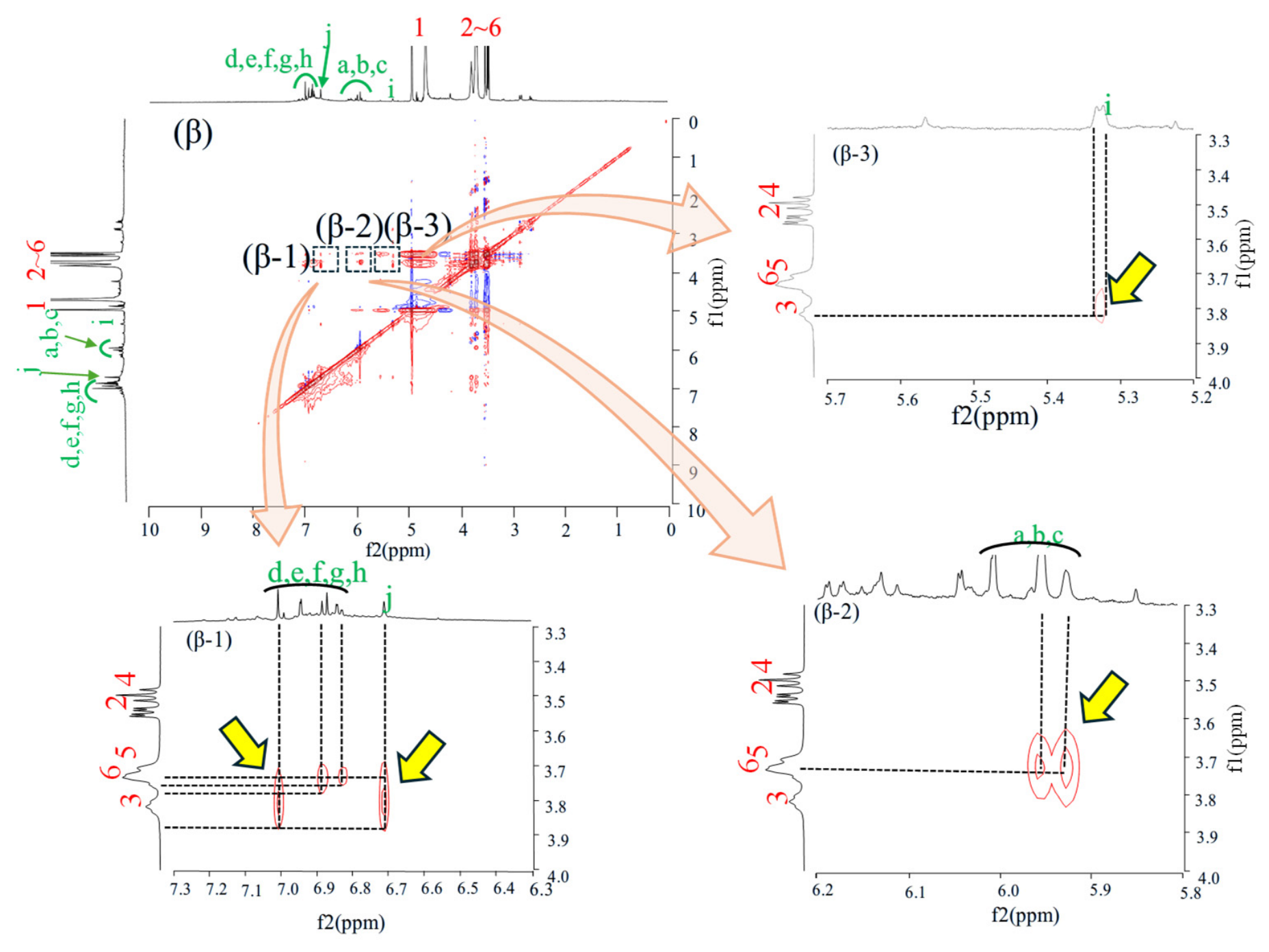
To further investigate host–guest interactions, two-dimensional 1H–1H NOESY measurements were performed for FD(Lyp/βCD) in D2O. The resulting spectrum is shown in Figure 7. In contrast to the αCD system, multiple NOE correlations were observed between Lyp-derived aromatic proton signals and βCD protons. Distinct cross-peaks were detected between the catechol-derived aromatic proton signals (d, e, f, g, and h; approximately 6.8–7.0 ppm) and βCD proton signals in the H3/H5/H6 region (Figure 7β-1). In addition, cross-peaks were observed between the resorcinol-derived aromatic proton signals (a, b, and c; approximately 5.9–6.0 ppm) and βCD cavity protons (Figure 7β-2). Interestingly, a NOE correlation was also detected between signal i (approximately 5.3 ppm) and βCD proton signals (Figure 7β-3), whereas no corresponding cross-peak was observed in the αCD system. The appearance of NOE correlations in all examined regions suggests that βCD interacted with a broader range of polyphenolic constituents than αCD. The observed correlations indicate that several aromatic moieties of Lyp were positioned within, or in close proximity to, the βCD cavity [26].
Figure 7.
1H−1H Nuclear Overhauser Effect spectroscopy (NOESY) NMR measurement of Lyp/βCD systems. (β) 0~10.0 ppm, (β-1) f1:3.3–4.0, f2:6.3–7.3 ppm, (β-2) f1:3.3–4.0, f2:5.8–6.3 ppm, (β-3) f1:3.3–4.0, f2:5.2–5.7 ppm.
3.6.3. NOESY Analysis of the Lyp/γCD System
To evaluate the spatial relationship between Lyp constituents and γCD in aqueous solution, two-dimensional 1H–1H NOESY measurements were performed for FD(Lyp/γCD). The resulting spectrum is shown in Figure S6. Distinct NOE cross-peaks were observed between the catechol-derived aromatic proton signals (d, e, f, g, and h; approximately 6.8–7.0 ppm) and γCD proton signals in the H3/H5/H6 region (Figure S6γ-1). In addition, a clear cross-peak was detected between the resorcinol-derived aromatic proton signals (a, b, and c; approximately 6.0 ppm) and γCD cavity proton signals (Figure S6γ-2). In contrast, no NOE correlation was detected between signal i and γCD protons (Figure S6γ-3). This behavior differs from that observed in the βCD system, where a distinct cross-peak was detected for signal i, suggesting differences in the interaction mode between Lyp constituents and the two cyclodextrins. The observed NOE correlations indicate that at least some aromatic components of Lyp were positioned within, or in close proximity to, the γCD cavity.
3.6.4. Comparison of NOESY Correlations Among αCD, βCD, and γCD
The NOESY results indicate close spatial proximity between proton signals of Lyp constituents and the cavity protons (H3/H5) of cyclodextrins, suggesting molecular association in solution. Clear differences were observed among the three cyclodextrins. In the αCD system, NOE correlations were observed only for the resorcinol-derived aromatic proton signals (a, b, and c), suggesting relatively limited association. In contrast, the βCD system exhibited NOE correlations for all examined Lyp-derived proton regions, including catechol-derived aromatic protons, resorcinol-derived aromatic protons, and signal i. The γCD system showed intermediate behavior, displaying correlations with both catechol- and resorcinol-derived aromatic proton signals but not with signal i. These findings indicate that the host–guest interactions of Lyp were strongly influenced by cyclodextrin cavity size. The relatively small cavity of αCD appears to restrict access of the bulky polyphenolic constituents, resulting in selective interactions. Conversely, the larger cavity of γCD may permit greater conformational freedom of the guest molecules, leading to less specific spatial arrangements. Among the three cyclodextrins examined, the βCD system exhibited the most extensive NOE correlations, consistent with stronger molecular associations under the present experimental conditions. Taken together, the NOESY results demonstrate differences in the extent of molecular association between Lyp constituents and the three cyclodextrins in aqueous solution. Because Lyp is a heterogeneous mixture of polyphenolic constituents, the present data do not allow identification of the individual components responsible for these associations or determination of inclusion stoichiometry. Nevertheless, the NOESY findings complement the physicochemical characterization and indicate that βCD provides the most favorable molecular environment for Lyp among the cyclodextrins investigated. The observed differences in molecular association are consistent with the physicochemical characteristics and antioxidant activity of the freeze-dried Lyp/CD systems. Interestingly, the extent of NOE correlations differed among the three cyclodextrins. The βCD system exhibited the largest number of cross peaks, whereas fewer correlations were observed for the αCD and γCD systems. This tendency suggests that the molecular recognition of Lyp constituents by CDs can be selectively influenced by cavity size. The cavity diameters of αCD, βCD, and γCD are approximately 4.7–5.3 Å, 6.0–6.5 Å, and 7.5–8.3 Å, respectively. The relatively small cavity of αCD may limit the accommodation of aromatic polyphenolic moieties present in Lyp, resulting in weaker host–guest interactions. In contrast, the cavity size of βCD appears to provide a more favorable steric environment for interaction with these constituents. Although γCD possesses a larger cavity capable of accommodating bulkier molecules, the larger internal space may reduce the efficiency of molecular recognition due to less optimal host–guest complementarity.
Therefore, the NOESY results suggest that βCD provides the most suitable cavity environment for the recognition of at least a portion of the polyphenolic constituents present in Lyp. This interpretation is consistent with the stronger molecular association observed in the βCD system and may explain the enhanced antioxidant activity observed for the corresponding complex. The observed differences in molecular association are consistent with the physicochemical characterization presented above and may contribute to the enhanced antioxidant activity observed for the βCD system.
3.7. Antioxidant Activity Assessed by the DPPH Radical Scavenging Assay
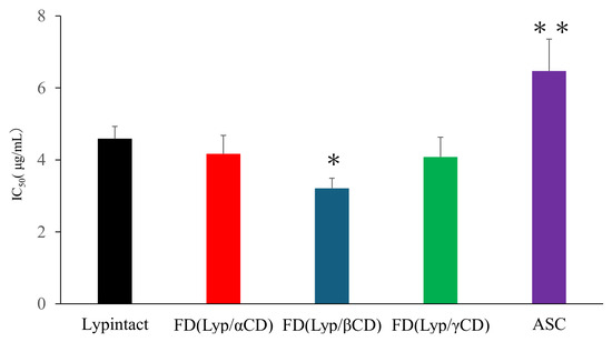
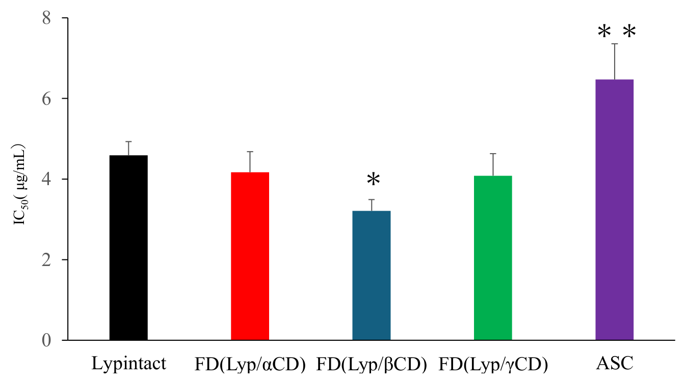
To investigate whether the physicochemical differences observed among the freeze-dried Lyp/CD systems influenced their functional performance, antioxidant activity was evaluated using the DPPH radical scavenging assay. The influence of cyclodextrin complexation on the antioxidant activity of Lyp was evaluated using the DPPH radical scavenging assay. The results are shown in Figure 8. Ascorbic acid (ASC) was included as a positive control to provide a reference for the antioxidant activity of the prepared Lyp/CD systems. The IC50 value of intact Lyp was 4.57 μg/mL. Among the Lyp/CD systems, FD(Lyp/βCD) exhibited the lowest IC50 value (3.20 μg/mL), indicating significantly higher radical scavenging activity than intact Lyp (p < 0.05). In contrast, FD(Lyp/αCD) and FD(Lyp/γCD) showed no significant improvement in antioxidant activity. The enhanced antioxidant activity observed for FD(Lyp/βCD) may be attributed to the stronger host–guest interactions demonstrated by the NOESY analyses. Cyclodextrin complexation has been reported to improve the dispersion and molecular accessibility of polyphenolic compounds by reducing intermolecular aggregation and increasing their effective interaction with radical species [27]. Furthermore, partial inclusion of aromatic moieties within the βCD cavity may stabilize the polyphenol structure while maintaining exposure of phenolic hydroxyl groups responsible for radical scavenging activity [28]. Notably, the βCD system exhibited the most extensive NOE correlations and the clearest co-diffusion behavior among the three CDs examined. These findings suggest that βCD provided the most favorable molecular environment for interaction with Lyp constituents, which may have facilitated more efficient radical scavenging reactions. Therefore, the enhanced antioxidant activity of FD(Lyp/βCD) appears to be closely related to the formation of stable host–guest associations between Lyp polyphenols and βCD. Taken together, the physicochemical characterization and NMR analyses indicate that βCD formed the most effective inclusion system with Lyp, resulting not only in altered molecular organization but also in improved antioxidant functionality. Considering that Lyp contains catechin monomers and low-molecular-weight proanthocyanidin oligomers, the dimensions and structural characteristics of these constituents may be particularly compatible with the βCD cavity. Further studies using purified marker compounds will be necessary to clarify the contribution of individual polyphenolic constituents to cyclodextrin recognition.
Figure 8.
DPPH radical scavenging activity (IC50) of Lyp, CDs, PMs, FDs, and ascorbic acid (ASC, positive control). IC50 values are expressed as mean ± SD (n = 3). Statistical significance: Dunnett’s multiple comparison test. * p < 0.05 versus LyP intact (FD(LyP/βCD)); ** p < 0.05 versus LyP intact (ASC). ASC: ascorbic acid.
4. Conclusions
In this study, freeze-dried systems of low-molecular-weight lychee polyphenol (Lyp) with α-, β-, and γ-cyclodextrins were successfully prepared and comprehensively characterized using PXRD, TG, FT-IR, NIR, 1H NMR, NOESY, and DPPH radical scavenging analyses. PXRD measurements revealed the disappearance of characteristic crystalline diffraction peaks of the CDs after freeze-drying, suggesting the formation of amorphous molecular assemblies. Thermal analysis demonstrated increased decomposition temperatures in the freeze-dried systems, indicating enhanced thermal stability resulting from intermolecular interactions. FT-IR and NIR analyses further suggested the involvement of hydroxyl and aromatic moieties in the interaction between Lyp constituents and CDs. NOESY measurements confirmed the host–guest association in aqueous solution. Among the three CDs examined, βCD exhibited the most extensive NOE correlations and the clearest co-diffusion behavior, suggesting the formation of the most favorable inclusion-type association with Lyp constituents. Consistent with these findings, the βCD system showed significantly enhanced DPPH radical scavenging activity compared with intact Lyp. These results demonstrate that cyclodextrin complexation, particularly with βCD, can effectively enhance the physicochemical properties and antioxidant functionality of Lyp. The present findings provide valuable insight into the molecular recognition behavior of lychee-derived polyphenols toward cyclodextrins and highlight the potential of βCD as a promising carrier for improving the performance of polyphenol-based functional ingredients.
5. Study Limitations
Several limitations of the present study should be acknowledged. First, Lyp is a complex mixture containing various catechin monomers and proanthocyanidin oligomers rather than a single purified compound. Therefore, the specific polyphenolic constituents responsible for the observed interactions with CDs could not be fully identified. Second, although NOESY analyses provided strong evidence of host–guest association, binding constants and inclusion stoichiometries were not determined. Third, the present study focused primarily on elucidating intermolecular interactions and their influence on antioxidant activity. Accordingly, quantitative evaluation of binding affinity and the contribution of individual polyphenolic components to the observed functional enhancement remain to be clarified. Furthermore, the inclusion of freeze-dried Lyp and freeze-dried cyclodextrins as control samples would provide further insight into the respective contributions of freeze-drying and Lyp–CD interactions to the observed physicochemical changes.
6. Future Perspectives
Future studies should focus on identifying the individual polyphenolic constituents involved in cyclodextrin complexation and clarifying their contributions to the observed antioxidant activity. Further investigation of binding affinity and storage stability will improve understanding of the interactions between lychee-derived polyphenols and cyclodextrins.
The enhanced antioxidant activity observed for the βCD system suggests that cyclodextrin complexation may be a useful strategy for improving the functionality of natural polyphenol-rich ingredients. Extending this approach to other lychee-derived polyphenols and plant-derived bioactive compounds may contribute to their effective biofunctionalization, as well as the development of value-added functional food ingredients and nutraceutical products. Furthermore, quantitative determination of the binding constants and inclusion stoichiometry will be important for achieving a more comprehensive understanding of the interactions between Lyp and cyclodextrin.
Supplementary Materials
The following supporting information can be downloaded at: https://www.mdpi.com/article/10.3390/physchem6030054/s1, Figure S1: Fourier transform infrared (FT-IR) spectra of the Lyp/αCD system; Figure S2: Fourier transform infrared (FT-IR) spectra of the Lyp/γCD system; Figure S3: NIR spectra of the Lyp/αCD system; Figure S4: NIR spectra of the Lyp/γCD system; Figure S5: 1H–1H Nuclear Overhauser Effect spectroscopy (NOESY) NMR measurement of Lyp/αCD systems; Figure S6: 1H–1H Nuclear Overhauser Effect spectroscopy (NOESY) NMR measurement of Lyp/γCD systems.
Author Contributions
K.K.: Investigation, Data curation, Formal analysis, Visualization, Writing—original draft preparation; N.K.: Investigation, Data curation, Formal analysis, Writing—review and editing; F.A.J.: Methodology, Validation, Writing—review and editing; G.L.S.: Methodology, Supervision, Writing—review and editing; Y.I.: Conceptualization, Methodology, Formal analysis, Supervision, Project administration, Funding acquisition, Writing—original draft preparation, Writing—review and editing. All authors have read and agreed to the published version of the manuscript.
Funding
This research received no external funding.
Institutional Review Board Statement
Not applicable.
Data Availability Statement
The data presented in this study are available from the corresponding author upon reasonable request.
Acknowledgments
The authors gratefully acknowledge Amino Up Co., Ltd. (Sapporo, Japan) for providing Lyp (a standardized oligomerized polyphenol derived from Litchi chinensis fruit extract; OPLFE) and CycloChem Co., Ltd. (Kobe, Japan) for supplying α-cyclodextrin, β-cyclodextrin, and γ-cyclodextrin used in this study. The authors also thank the staff of the Instrumental Analysis Center, Josai University, for their valuable technical assistance with NMR and spectroscopic analyses.
Conflicts of Interest
The authors declare no conflicts of interest. The companies (Amino Up Co., Ltd. and CycloChem Co., Ltd.) acknowledged in the manuscript had no role in the design of the study; in the collection, analyses, or interpretation of data; in the writing of the manuscript; or in the decision to publish the results.
Abbreviations
| CD | Cyclodextrin |
| αCD | α-Cyclodextrin |
| βCD | β-Cyclodextrin |
| γCD | γ-Cyclodextrin |
| Lyp | Low-molecular-weight lychee polyphenol |
| PM | Physical mixture |
| FD | Freeze-dried |
| PXRD | Powder X-ray diffraction |
| TG-DTA | Thermogravimetric-differential thermal analysis |
| FT-IR | Fourier transform infrared spectroscopy |
| NIR | Near-infrared spectroscopy |
| NMR | Nuclear magnetic resonance |
| NOESY | Nuclear Overhauser effect spectroscopy |
| DPPH | 2,2-Diphenyl-1-picrylhydrazyl |
| DMSO-d6 | Deuterated dimethyl sulfoxide |
References
- Yang, Z.; Zhang, L.; Wu, Y.H.; Li, D.P.; Li, W. Evaluation of chemical constituents of litchi pericarp extracts and its antioxidant activity in mice. Foods 2022, 11, 3837. [Google Scholar] [CrossRef] [Scilit] [PubMed]
- Sakurai, T.; Nishioka, H.; Fujii, H.; Nakano, N.; Kizaki, T.; Radak, Z.; Izawa, T.; Haga, S.; Ohno, H. Antioxidative effects of a new lychee fruit-derived polyphenol mixture, Oligonol, converted into a low-molecular form in adipocytes. Biosci. Biotechnol. Biochem. 2008, 72, 463–476. [Google Scholar] [CrossRef] [Scilit] [PubMed]
- Waki, H.; Suzuki, T.; Uebaba, K.; Hisajima, T. Effects of a standardized oligomerized-polyphenol from Litchi chinensis fruit extract and mixed plant extract supplementation on peripheral circulation and cold sensitivity. J. Nutr. Sci. Vitaminol. 2020, 66, 255–260. [Google Scholar] [CrossRef] [Scilit] [PubMed]
- Fujii, H.; Nishioka, H.; Wakame, K.; Magnuson, B.A.; Roberts, A. Acute, subchronic and genotoxicity studies conducted with Oligonol, an oligomerized polyphenol formulated from lychee and green tea extracts. Food Chem. Toxicol. 2008, 46, 3553–3562. [Google Scholar] [CrossRef] [Scilit] [PubMed]
- Ohno, H.; Sakurai, T.; Hisajima, T.; Abe, S.; Kizaki, T.; Ogasawara, J.E.; Ishibashi, Y.; Imaizumi, K.; Takemasa, T.; Haga, S.; et al. The supplementation of oligonol, the new lychee fruit-derived polyphenol converting into a low-molecular form, has a positive effect on fatigue during regular track-and-field training in young atheletes. Adv. Exerc. Sports Physiol. 2008, 14, 93–99. [Google Scholar]
- Prior, R.L.; Gu, L. Occurrence and biological significance of proanthocyanidins in the American diet. Phytochemistry 2005, 66, 2264–2280. [Google Scholar] [CrossRef] [Scilit] [PubMed]
- Crozier, A.; Jaganath, I.B.; Clifford, M.N. Dietary phenolics: Chemistry, bioavailability and effects on health. Nat. Prod. Rep. 2009, 26, 1001–1043. [Google Scholar] [CrossRef] [Scilit] [PubMed]
- de Freitas, V.; Mateus, N. Protein/Polyphenol Interactions: Past and Present Contributions. Mechanisms of Astringency Perception. Curr. Org. Chem. 2012, 16, 724–746. [Google Scholar] [CrossRef] [Scilit]
- Cid-Samamed, A.; Rakmai, J.; Mejuto, J.C.; Simal-Gandara, J.; Astray, G. Cyclodextrins inclusion complex: Preparation methods, analytical techniques and food industry applications. Food Chem. 2022, 384, 132467. [Google Scholar] [CrossRef] [Scilit] [PubMed]
- Del Valle, E.M.M. Cyclodextrins and their uses: A review. Process. Biochem. 2004, 39, 1033–1046. [Google Scholar] [CrossRef] [Scilit]
- Loftsson, T.; Duchêne, D. Cyclodextrins and their pharmaceutical applications. Int. J. Pharm. 2007, 329, 1–11. [Google Scholar] [CrossRef] [Scilit] [PubMed]
- Christaki, S.; Spanidi, E.; Panagiotidou, E.; Athanasopoulou, S.; Kyriakoudi, A.; Mourtzinos, I.; Gardikis, K. Cyclodextrins for the Delivery of Bioactive Compounds from Natural Sources: Medicinal, Food and Cosmetics Applications. Pharmaceuticals 2023, 16, 1274. [Google Scholar] [CrossRef] [Scilit] [PubMed]
- Adeoye, O.; Cabral-Marques, H. Cyclodextrin Nanosystems in Oral Drug Delivery: A Mini Review. Int. J. Pharm. 2017, 531, 521–531. [Google Scholar] [CrossRef] [Scilit] [PubMed]
- Munin, A.; Edwards-Lévy, F. Encapsulation of natural polyphenolic compounds: A review. Pharmaceutics 2011, 3, 793–829. [Google Scholar] [CrossRef] [Scilit] [PubMed]
- Chao, J.; Wang, H.; Zhao, W.; Zhang, M.; Zhang, L. Investigation of the Inclusion Behavior of Chlorogenic Acid with Hydroxypropyl-β-Cyclodextrin. Int. J. Biol. Macromol. 2012, 50, 277–282. [Google Scholar] [CrossRef] [Scilit] [PubMed]
- Ansari, M.J.; Kohli, K.; Ali, J. Preparation, characterization and dissolution behaviour of freeze-dried complexes of curcumin-γ-cyclodextrin. J. Pharm. Res. Int. 2021, 33, 17–26. [Google Scholar] [CrossRef] [Scilit]
- Ologe, M.O.; Tella, A.C.; Atolani, O.; Adegoke, O.A.; Ademowo, O.G. Inclusion complex of gedunin-2-hydroxypropyl-β-cyclodextrin prepared by kneading and freeze-drying methods: Synthesis and structural characterization. Acta Biol. Marisiensis 2021, 4, 83–97. [Google Scholar] [CrossRef] [Scilit]
- Jarrells, T.W.; Munson, E.J. Comparison of differential scanning calorimetry, powder X-ray diffraction, and solid-state nuclear magnetic resonance spectroscopy for measuring crystallinity in amorphous solid dispersions. J. Pharm. Sci. 2022, 111, 2305–2315. [Google Scholar] [CrossRef] [Scilit] [PubMed]
- Hădărugă, N.G.; Bandur, G.N.; David, I.; Hădărugă, D.I. A review on thermal analyses of cyclodextrins and cyclodextrin complexes. Environ. Chem. Lett. 2019, 17, 349–373. [Google Scholar] [CrossRef] [Scilit]
- Hunt, L.E.; Bourne, S.A.; Caira, M.R. Inclusion of hydroxycinnamic acids in methylated cyclodextrins: Host–guest interactions and effects on guest thermal stability. Biomolecules 2021, 11, 45. [Google Scholar] [CrossRef] [Scilit] [PubMed]
- de Freitas, C.A.B.; Costa, C.H.S.; da Costa, K.S.; da Paz, S.P.A.; Silva, J.R.A.; Alves, C.N.; Lameira, J. Assessment of host–guest molecular encapsulation of eugenol using β-cyclodextrin. Front. Chem. 2023, 10, 1061624. [Google Scholar] [CrossRef] [Scilit] [PubMed]
- Mura, P. Analytical techniques for characterization of cyclodextrin complexes in the solid state: A review. J. Pharm. Biomed. Anal. 2015, 113, 226–238. [Google Scholar] [CrossRef] [Scilit] [PubMed]
- Inoue, Y.; Onishi, M.; Tomita, J.; Kodama, N.; Suzuki, K.; Itakura, S.; Nishikawa, M.; Yukutake, S.; Ishida, Y.; Nakata, D.; et al. Comprehensive physicochemical and functional characterization of mechanochemically prepared α-mangostin/cyclodextrin inclusion complexes. Next Mater. 2026, 12, 102030. [Google Scholar] [CrossRef] [Scilit]
- Schneider, H.J.; Hackett, F.; Rüdiger, V.; Ikeda, H. NMR studies of cyclodextrins and cyclodextrin complexes. Chem. Rev. 1998, 98, 1755–1785. [Google Scholar] [CrossRef] [Scilit] [PubMed]
- He, J.; Zheng, Z.P.; Zhu, Q.; Guo, F.; Chen, J. Encapsulation mechanism of oxyresveratrol by β-cyclodextrin and hydroxypropyl-β-cyclodextrin and computational analysis. Molecules 2017, 22, 1801. [Google Scholar] [CrossRef] [Scilit] [PubMed]
- Zhao, R.; Sandström, C.; Zhang, H.; Tan, T. NMR study on the inclusion complexes of β-cyclodextrin with isoflavones. Molecules 2016, 21, 372. [Google Scholar] [CrossRef] [Scilit] [PubMed]
- López-Nicolás, J.M.; Rodriguez-Bonilla, P.; García-Carmona, F. Cyclodextrins and antioxidants. Crit. Rev. Food Sci. Nutr. 2014, 118, 1081–1090. [Google Scholar] [CrossRef] [Scilit] [PubMed]
- Ntuli, S.; Leuschner, M.; Bester, M.J.; Serem, J.C. Stability, morphology, and effects of in vitro digestion on the antioxidant properties of polyphenol inclusion complexes with β-cyclodextrin. Molecules 2022, 27, 3808. [Google Scholar] [CrossRef] [Scilit] [PubMed]
Disclaimer/Publisher’s Note: The statements, opinions and data contained in all publications are solely those of the individual author(s) and contributor(s) and not of MDPI and/or the editor(s). MDPI and/or the editor(s) disclaim responsibility for any injury to people or property resulting from any ideas, methods, instructions or products referred to in the content. |
© 2026 by the authors. Licensee MDPI, Basel, Switzerland. This article is an open access article distributed under the terms and conditions of the Creative Commons Attribution (CC BY) license.