


Abstract
This study uses a bismuth ferrite (BiFeO3) and MXene (Ti3C2Tx) to design a surface plasmon resonance (SPR) biosensor for the sensitivity enhancement at a 633 nm wavelength. Here, MXene serves as a biorecognition element (BRE) layer to ensure stable and reliable biomolecule adsorption. The MXene is a family of two-dimensional (2D) materials with metallic-like conductivity, a large surface area that can attach biomolecules, and improve biocompatibility. The addition of a conductive 2D MXene layer and a high-index BiFeO3 dielectric layer greatly improves light–matter interaction and evanescent field penetration at the sensing interface. Strong plasmonic coupling is indicated by the reflectance analysis, which shows a distinct and consistent shift in the resonance angle as analyte RI increases. This study examined the sensitivity at optimized Ag and BiFeO3 layer thickness. At an Ag of 39 nm and BiFeO3 of 3 nm thickness, the maximal sensitivity of 340.68°/RIU with a remarkable figure of merit (FoM) of 47.38/RIU is obtained. The overall detection accuracy (DA) and FoM are significantly improved by the large sensitivity enhancement, despite a slight increase in full width at half maximum (FWHM). Furthermore, the penetration depth (PD) of 198.50 nm (at RI:1.330) and 199.52 nm (at RI:1.335) is attained with the proposed structure. Due to its high sensitivity, reusability, and reproducibility, the SPR biosensor has the potential to be used in biochemical, environmental, and medical detection.
1. Introduction
The antigens, antibodies, or virus proteins have been detected using the SPR techniques [1]. SPR sensors are widely used in biosensing, medical diagnostics, environmental monitoring, food safety, and drug–protein interaction studies because of their high sensitivity, real-time response, and capacity for quantitative analysis without fluorescent or radioactive labels [2,3]. The sensitivity, DA, and applicability of SPR-based sensors have been further improved by recent developments involving nanomaterials, 2D materials, and multilayer structures [4,5]. A highly sensitive, label-free optical sensing platform called a SPR sensor is used to find minute variations in the refractive index (RI) at a metal–dielectric interface. The sensing principle is based on the polarized light, under certain resonance conditions, which excites surface plasmons (SPs), which are coherent oscillations of free electrons, at the surface of a thin noble metal layer (usually gold (Au), silver (Ag), or copper (Cu)) [6]. A detectable change in the resonance angle results from any interaction of target analytes (such as biomolecules, chemicals, or gases) with the sensing surface, which modifies the analyte RI. The Kretschmann configuration [7], in which light is coupled through a high-RI prism to excite SPs in the metal layer, is most frequently used to implement SPR sensors. A metal layer is struck by polarized light in SPR at the interface of media with different RI [8]. It stimulates free electrons and detects their collective motions in which light is directed onto a metal layer via a glass crystal (also referred to as SPs). Light is absorbed at an incident angle (also known as the resonance angle) because the plasmons are positioned to resonate with the light at that angle.
Real-time quantitative measurements of the kinetics, thermodynamic parameters, and binding affinities of interacting molecules can be made using SPR, a label-free technique. Compared to other methods, this sensor uses relatively little sample and produces data of the highest quality. It also has a moderately high throughput. Utilizing the Kretschmann configuration to examine the SPP mode, in this setup, the metal layer’s thickness is adjusted to a few tens of nanometers, the evanescent wave passes through the metal and is coupled to a SP mode. The structure of the prism-coupled SPR sensor is typical, with a plasmonic metal such as Au and Ag layer applied to a prism’s base [6]. An evanescent wave is generated at the prism/metal layers interface when transverse magnetic (TM) polarized incident light undergoes total internal reflection. This wave then passes through the thin metal film. At the junction of the metal layer and sensing medium (SM), it then stimulates a SP at the outer edge of the metal layer. When the SPs are excited, the reflected light’s angular spectrum exhibits a resonant dip at a fixed wavelength. A small change in the analyte’s RI will result in a change in the resonance position, since the excitation of the SP depends on the RI of the SM. The sensitive detection of RI change can be achieved by using this resonance angle variation. In a sensing operation, an effective detection is essential. The biological binding event’s impact on the RI alters the SPP waves’ propagation constant. The resonance peak is the most frequently utilized feature among them. The location of the resonance peak in the SPR curve is noted both prior to and following the biomolecules’ binding to the sensor surface using this method. The effective RI is altered by the binding of the target biomolecules, which also alters the SPP waves’ propagation constant and causes the resonance to occur at a different incidence angle. For this reason, the resonance angle shift is measured. A perovskite-type multiferroic oxide, BiFeO3, has a relatively high RI, good thermal and chemical stability, and a combination of ferroelectric and magnetic properties that improve electromagnetic interactions at interfaces. It is a useful dielectric spacer layer in SPR sensors because of these properties, which enhance evanescent field confinement and raise sensitivity. Furthermore, BiFeO3 has a moderate band gap (≈2 eV), good chemical, mechanical, and thermal stability, and resistance to corrosion, which allows it to endure harsh operating conditions and repeated sensing cycles without deteriorating [9]. When employed as an intermediate spacer layer in SPR sensors, these combined dielectric and magneto-electric qualities aid in strengthening the local field and electromagnetic interaction, which in turn sharpens the resonance dip and raises the sensitivity to minute changes in the analyte’s RI.
The use of 2D materials has expanded the scope of the research in the fields of optical sensors, optoelectronics, and energy conversion devices. Numerous 2D materials, including MXene, black phosphorus, and transition-metal dichalcogenides (TMDCs), have been discovered since 2004 [10,11,12] that possess distinct optical, chemical, mechanical, and electrical properties. After being discovered roughly ten years ago, MXene has recently gained attention. Due to its unique properties—including a large surface area, chemical–mechanical stability, strong carrier confinement, and high biomolecule binding energies—MXene demonstrates promising application as a sensing molecule when utilized as an interaction layer in the SPR configuration. Surface terminations determine MXene’s favorable plasmonic material properties [6]. SPR sensors effectively use MXenes, which were created by surface termination, to detect molecules. The MXenes are a 2D family of transition metal carbides and nitrides that possess a large surface area, metallic-like conductivity, and numerous tunable surface functional groups that can attach biomolecules and enhance biocompatibility. Additionally, their strong light–matter interaction and high electrical conductivity improve plasmonic coupling and signal stability. BiFeO3 and MXene layers work in concert to improve field localization, provide a stable interface, and increase analyte adsorption for extremely sensitive detection in multilayer SPR structures. In order to improve SPR sensor performance, a number of recent studies have investigated sophisticated material combinations. For instance, optimized multilayer plasmonic structures were examined in a study of references [13] to enhance sensitivity and electric field confinement. Similarly, studies [14] showed that adding functional nanomaterials to metallic layers can greatly enhance plasmon–analyte interactions and boost sensing efficiency. In a related study [15], the optical response of multilayer plasmonic systems that were engineered for high-precision sensing applications was evaluated. In order to achieve better plasmonic coupling, stronger field localization, and improved biosensing capabilities, these studies collectively demonstrate the growing interest in combining dielectric layers with advanced materials.
This study proposes an SPR biosensor based on a BiFeO3 and MXene layer that can significantly increase the sensitivity within a 1.33–1.335 biologically relevant RI range. The suggested structure synergistically combines a high-RI ferroelectric BiFeO3 spacer with a conductive MXene functional layer to concurrently enhance electromagnetic field confinement, plasmonic coupling, and biomolecule adsorption efficiency, in contrast to conventional SPR designs that use a single dielectric or 2D material layer. At optimized thickness of Ag and BiFeO3, the performance parameters are calculated like sensitivity, DA, FoM and PD. Furthermore, the PD and electric field intensity have been thoroughly analyzed to evaluate the extent of evanescent field confinement and its interaction with the SM. Additionally, an electric field distribution investigation, PD analysis, and an evaluation of the fabrication tolerance of ±5 percent are included in the study to demonstrate robustness and practical feasibility. These combined structural, theoretical, and performance improvements make the proposed sensor a high-performance and fabrication-resilient platform for precise biomedical and biochemical detection. In contrast to previously reported MXene-based and dielectric-assisted SPR sensors, the novelty of this work is the synergistic integration of a high-index multiferroic BiFeO3 dielectric layer with MXene functional material to enhance plasmonic coupling, electromagnetic field confinement, and biomolecule adsorption simultaneously, leading to significantly higher sensitivity and improved robustness. The suggested structure offers a fundamental advantage through the synergistic integration of high-index BiFeO3 and MXene, which simultaneously enhances electromagnetic confinement, plasmonic coupling, analyte adsorption, and structural stability. This results in higher sensitivity, improved robustness, and better practical feasibility compared to existing designs, even though several SPR sensors use 2D materials and dielectric spacers.
2. Proposed Structure, Fabrication Feasibility, Refractive Indices and Modeling
2.1. Proposed Structure
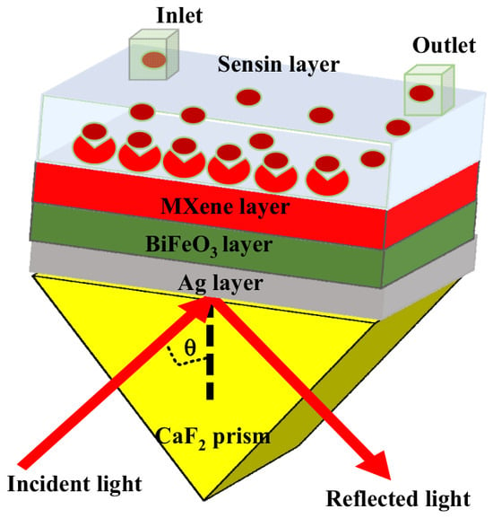
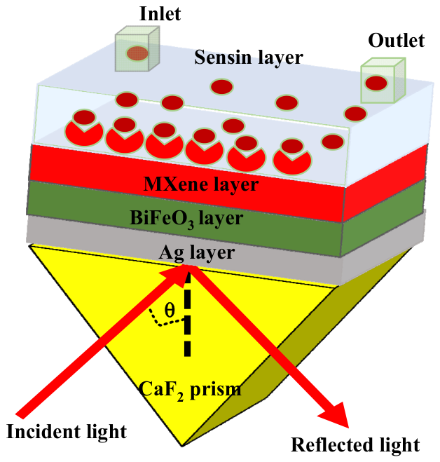
A Kretschmann-configured SPR biosensor based on a CaF2 prism is depicted in Figure 1. A thin layer of Ag reflects p-polarized light incident at an angle (θ) through the prism, causing SPs to be excited at the metal–dielectric interface. A BiFeO3 layer is used on the Ag layer due to its high RI. It can tune the resonance and improve optical confinement and adhesion. The high real part of the RI of the BiFeO3 layer is a key factor in improving SPR sensor performance. BiFeO3’s strong electronic polarization, perovskite crystal structure, and multiferroic nature—which combines ferroelectric and antiferromagnetic properties—are the sources of its high real RI. Despite the fact that BiFeO3 is a well-known perovskite-type multiferroic oxide with coupled ferroelectric and magnetic ordering, polarization switching, magneto-optic coupling, and external field tuning are not specifically utilized in this SPR analysis. The main reasons BiFeO3 is used in this work are its high real RI, large dielectric permittivity, and superior thermal and chemical stability—all of which have a significant impact on plasmonic behavior in the Kretschmann configuration. Stronger electromagnetic field confinement at the metal–dielectric interface is the result of improved phase matching between the incident p-polarized light and the surface plasmon polariton (SPP) mode due to BiFeO3’s high permittivity. By increasing the overlap between the evanescent field and the sensing medium, this improved confinement amplifies resonance angle shifts for slight changes in refractive index. Consequently, even though BiFeO3 has ferroelectric and magnetic properties by nature, the sensitivity improvement described in this work is solely due to its thickness optimization and dielectric properties. Although it is not explicitly used in the current theoretical modeling, BiFeO3 multiferroic nature is noted as an inherent material property and as a possible benefit for upcoming actively tunable SPR devices.
Figure 1.
Proposed SPR sensor.
By improving light–matter interaction close to the sensing surface, this improved field localization increases the sensor’s sensitivity to changes in the analyte region’s RI. Furthermore, minimal optical absorption is ensured by the dominant real part of the RI, which has a relatively low imaginary loss. This leads to a sharper resonance dip, better FoM, and increased DA. The BiFeO3 layer functions as an effective functional dielectric medium that simultaneously supports strong plasmon coupling and stable biosensing operation in the biologically relevant RI range because of its ferroelectric polarization, chemical stability, and high dielectric constant. After that, a thin layer of MXene is applied to enhance biomolecule immobilization, produce a large number of surface functional groups, and strengthen the local electric field. The analyte solution that passes through a microfluidic cell’s inlet and outlet channels is contained in the upper sensing layer. The resonance angle of the reflected light is changed when target biomolecules attach to the sensing surface because they change the local RI close to the metal interface. This change serves as the basis for detection, enabling label-free, real-time biological interaction monitoring. This multilayer (CaF2/Ag/BiFeO3/MXene) structure improves signal stability, sensitivity, and field confinement overall, making it ideal for accurate biosensing applications.
2.2. Fabrication Feasibility
In order to achieve high selectivity, stability, and repeatability, the CaF2 prism must be thoroughly cleaned before the SPR sensor is fabricated, as shown in Figure 2. After rinsing the prism surface with deionized water to get rid of any loose particles, organic contaminants are removed by ultrasonic cleaning in acetone and isopropyl alcohol. UV–ozone or oxygen plasma treatment can be used after nitrogen gas drying to eliminate remaining contaminants and enhance surface wettability. Strong adhesion of subsequent layers, decreased surface roughness, and steady excitation of SPs result from this cleaning procedure’s assurance of a smooth, chemically uniform surface. After the prism has been cleaned, a layer of Ag is applied using physical vapor deposition (PVD) [16,17]. Stable SPR with minimal damping losses is supported by the superior and homogeneous Ag layer. The enhanced adhesion brought about by prism cleaning reduces the effects of oxidation and metal delamination, improving the SPR signal’s long-term stability. Chemical vapor deposition (CVD) is then used to deposit a BiFeO3 dielectric layer [18]. This layer further contributes to signal stability by strengthening electromagnetic field confinement at the metal–dielectric interface and shielding the Ag layer from environmental deterioration. The BiFeO3 layer is then deposited with an MXene layer by the CVD techniques [18].
Figure 2.
Fabrication process.
The thin-film deposition methods, such as magnetron sputtering or e-beam evaporation for Ag and BiFeO3 layers, can be used to fabricate the suggested structure. For MXene deposition, solution-based coating techniques, such as spin coating, dip coating, or spray coating, can be used [19]. These techniques, which are more affordable and appropriate for large-scale manufacturing, are frequently employed in the creation of SPR sensors. Advanced techniques such as CVD may also be used if required, but they are not mandatory. MXene offers a biocompatible, high-surface-area platform with a large number of surface functional groups (–OH, –O, –F) that help immobilize particular bioreceptors [20]. The selectivity of the SPR sensor is achieved through the immobilized bioreceptors (such as antibodies, aptamers, or enzymes), while MXene enhances biomolecule adsorption and plasmon–analyte interaction [6]. A monolayer or near-monolayer nanosheet with an MXene layer thickness of 0.993 nm can be produced experimentally using solution-based deposition methods like spin coating, spray coating, dip coating, and layer-by-layer assembly. According to published research, single-layer MXene nanosheets usually have thicknesses between 0.8 and 1.2 nm, which makes the suggested thickness practically achievable. Because of the strong plasmon–surface interaction, nanoscale tolerance has little effect on the SPR response, despite the possibility of slight thickness variation during fabrication. Characterization methods like AFM, SEM, and ellipsometry can be used to confirm the thickness and homogeneity of MXene layers, ensuring dependable experimental execution of the suggested structure [21]. By encouraging particular analyte–surface interactions and inhibiting nonspecific adsorption, this functionalized surface greatly increases the sensor’s selectivity. Consistent sensing performance across repeated measurements is ensured by the uniform growth of the MXene layer made possible by the prism surface’s cleanliness and preparation. The active sensing region is generated when target biomolecules are immobilized on the MXene surface. Measurable changes in the SPR angle result from selective biomolecular interactions that cause minute changes in the RI. Because nearly identical resonance curves are obtained in subsequent sensing cycles, the combination of appropriate prism cleaning, uniform multilayer deposition, and chemically selective MXene functionalization yields excellent repeatability. Long-term sensor stability may be impacted by MXene materials’ known sensitivity to oxidation and environmental deterioration, especially in the presence of moisture and oxygen. According to earlier research, MXene layers may progressively oxidize in ambient conditions, changing their optical and electrical properties. However, this issue can be mitigated by using protective coatings, surface functionalization, encapsulation with dielectric layers, and controlled fabrication environments. By using MXene as an ultra-thin sensing layer supported by BiFeO3 and Ag layers, the suggested structure improves stability and minimizes direct environmental exposure. The long-term operational stability of the SPR sensor can be further enhanced by appropriate device packaging and passivation techniques. This analysis acknowledges the limitations of material stability while highlighting the practical viability of MXene-based SPR sensors. The SPR sensor is appropriate for accurate and long-term biosensing applications because the suggested multilayer configuration ensures high selectivity, strong stability, and dependable repeatability. The surface roughness is another significant factor that can affect SPR sensor performance in real-world thin-film fabrication, in addition to thickness tolerance. During deposition, thin metal and dielectric layers like Ag, BiFeO3, and MXene may show nanoscale surface roughness, which can cause scattering losses and a slight resonance curve broadening. Surface roughness in the 1–3 nm range usually has little effect on SPR sensitivity, but it may marginally lower resonance sharpness and detection accuracy. Atomic layer deposition, controlled solution-based coating, and magnetron sputtering are examples of advanced deposition methods that can reduce surface roughness and enhance film uniformity.
2.3. Refractive Index of Materials
In this work, the coupling is achieved using a CsF2 prism and having RI of 1.4329 and semi-infinite thickness [22]. The SP is generated by the Ag layer thickness variation of 30–60 nm and an RI of 0.0803 + 4.2347i [6]. Since Ag layers are prone to oxidation, a BiFeO3 and MXene layer was employed to boost sensitivity. The BiFeO3 and MXene layers’ thicknesses of 1–3 nm and 0.993 nm [23] are used in order to maximize the sensitivity and have RI of 2.9680 [24] and 2.38 + 1.33i [6], respectively. The transfer matrix method (TMM) is used to analyze the suggested SPR sensor, considering the thickness and RI of each layer (Ag, BIFeO3, and MXene). By calculating the reflectance as a function of incident angle, this modeling technique forecasts the resonance shift in response to variations in the RI of the SM [10,25,26]. Better interaction with the analyte and stronger plasmonic coupling at the interfaces are achieved by the sensor by optimizing the thicknesses of the Ag and BiFeO3, with a fixed MXene layer thickness. This improves performance and sensitivity for RI-based biosensing. Aqueous biological fluids fall within the analyte RI range ns = 1.33–1.335 [6], and slight variations within this small window are closely linked to pathological alterations in the human body. Pure water is represented by an RI close to 1.33, whereas elevated concentrations of proteins, glucose, lipids, cells, or pathogens cause increases toward 1.335. For instance, blood plasma from healthy people usually shows an RI between 1.333 and 1.334, but diseases like diabetes mellitus raise glucose levels, which results in a detectable increase in RI. Similar changes in hemoglobin content and red blood cell morphology caused by infectious diseases like malaria result in observable RI shifts within this range. The RI shifts toward higher values when inflammatory and cardiovascular conditions raise plasma protein and fibrinogen levels. Minute RI changes that stay within 1.33–1.335 are also caused by early-stage cancer and liver dysfunction, which alter serum protein composition. A theoretical platform for the detection of disease biomarkers in biological fluids like blood serum, plasma, urine, and saliva is offered by the suggested SPR sensor. However, in order to improve selectivity and stability and prevent nonspecific protein adsorption, surface functionalization with particular bioreceptors and antifouling coatings is necessary for direct analysis of complex biological samples. As a result, the current work concentrates on the design and theoretical assessment of the sensing platform, whereas further surface modification and experimental validation will be necessary for practical biofluid analysis. The optical constants of Ag, BiFeO3, and MXene are examined in this work at a fixed operating wavelength of 633 nm, which is frequently utilized in angular interrogation SPR sensors. The current study concentrates on single-wavelength angular interrogation for performance evaluation, despite the fact that MXene displays wavelength-dependent dispersive optical behavior.
2.4. Theoretical Modeling
This study uses the transfer matrix method (TMM) in conjunction with the Fresnel equations as the fundamental theory to investigate the SPR sensor. The Fresnel equations, which describe the reflection of light at the boundary between two media with different RIs, are useful in modeling the behavior of incident p-polarized light as it interacts with the prism, metal, and 2D material layers. The TM technique applies this idea to a designed sensor configuration by considering the continuity of the electric and magnetic field components at every boundary. Maxwell’s equations can be solved by applying the tangential components of the field at the interfaces. Equation (1) is represented in the mathematical equation by U1, V1 (at the first interface Z = Z1 = 0), and UN−1, VN−1 (at the final interface Z = ZN−1) [27].
Here, M represents the characteristics matrix of the proposed structure.
According to Equation (2), the matrices (Mk) of each internal layer situated between the incident medium (k = 1) and the SM (k = N) are multiplied to produce the TMM [28].
The kth layer’s propagation properties for p-polarized light are characterized by Equation (3), where βk is the phase shift, and qk is the admittance. The off-diagonal terms represent the interaction between the magnetic fields as light passes through the layer.
With thickness (dk), internal propagation angle (θ1), and RI (nk), Equation (4) measures the phase shift that light experiences after passing through the kth layer. Permittivity (εk) and wavelength (λ) have an impact on this. Equation (5) gives the ratio of magnetic to field components in the kth layer, which shows how well the layer transmits or reflects electromagnetic waves. The symbol for magnetic permeability is μk. The kth layer is represented by Zk−1, where k can range from 0 to N − 1. The wavelength is 633 nm (λ). The incident light is denoted by θ1.
rp is defined as the Fresnel reflection coefficient for the p-polarized (TM) light, achieved using the TMM. R is defined as the reflectance, measured as the squared magnitude of the reflection coefficient. The optical reflectance of incident light reflected by the multilayer stack is shown by Equation (7).
The TMM implemented in MATLAB R2025b was used in theoretical simulations to examine the SPR structure’s optical response. Using the Fresnel equations for the p-polarized light, the model determines the reflectivity vs. incident angle. The RI of the analyte ranges from 1.33 to 1.335 in order to observe the sensing capability of the proposed sensor. The performance parameters, including sensitivity, full width at half maximum, DA, and FoM, are defined in order to assess the performance of the suggested sensor [10,25,26]. All performance parameters have been measured from Equations (8)–(10), respectively. The sensor is thoroughly studied in order to assess the aforementioned parameters.
The sensor structure shows that the evanescent field is increased under different conditions by the electric field normalized of p-polarized light within each layer. P-polarized light in the structure is described and explained by the reflection, which also establishes the field’s distribution Equations (11)–(14) [29].
where measured as:
The electric and magnetic field distribution within layers j ≥ 2 is determined by the following expression.
Here, measured as:
3. Results and Discussion
3.1. Sensitivity, Minimum Reflectance Analysis
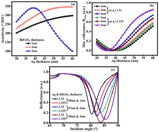
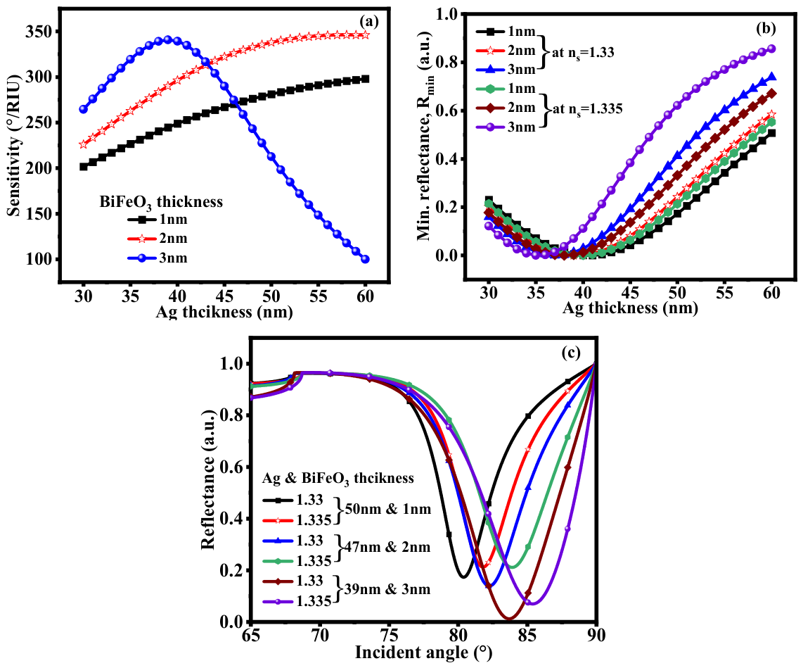
The sensitivity variation with Ag layer thickness for BiFeO3 thicknesses of 1 nm, 2 nm, and 3 nm is displayed in Figure 3a. With increasing Ag thickness, the sensitivity first rises, peaks, and then falls. This pattern suggests that the strongest plasmon excitation occurs at an ideal Ag thickness of 38–40 nm. After this, excessive reflection from a thicker Ag layer weakens the field’s penetration into the SM, lowering sensitivity. The sensitivity variation is from 201.68 to 298.05°/RIU, 225.97 to 345.95°/RIU and 263.47 to 340.68°/RIU with 1 nm, 2 nm, and 3 nm of BiFeO3 thickness. At 3 nm of BiFeO3 thickness, first, the sensitivity is increased at a certain thickness of Ag layer and then decreases up to 100.03°/RIU. Furthermore, the BiFeO3 layer thickness of 3 nm yields the highest sensitivity (340.68°/RIU), followed by 2 nm and 1 nm. At the sensing interface, the thicker BiFeO3 layer increases optical confinement and field intensity, which strengthens the interaction with the analyte and raises sensitivity. For various BiFeO3 thicknesses and refractive indices (ns = 1.33 and 1.335), the minimum reflectance (Rmin) is plotted against Ag thickness in Figure 3b. As Ag thickness increases, the Rmin value first falls and then rises at a certain thickness of Ag layer, exhibiting the usual SPR behavior. The optimal coupling conditions are confirmed by the lowest Rmin value occurring close to the same Ag thickness of 35–40 nm, where maximum sensitivity is observed. Rmin shifts slightly as the SM’s RI rises from 1.33 to 1.335; this shift reflects the resonance caused by the RI change. Stronger field coupling and improved plasmonic excitation are also indicated by deeper resonance dips in thicker BiFeO3 layers, particularly those at 3 nm. Reflectance versus incident angle curves for various Ag and BiFeO3 thickness combinations at RIs of 1.33 and 1.335 are shown in Figure 3c. The SPR angle is represented by a clear dip in each curve. The resonance dip moves toward higher angles as the RI rises or as the layer thicknesses vary. This shift is the primary sensing mechanism; a greater angular shift suggests that the device is more sensitive to changes in the analyte’s RI. It is clear from the reflectance curve that deeper and sharper resonance dips are produced when thinner Ag layers are combined with slightly thicker BiFeO3 layers. The resolution of the sensor is enhanced because of the stronger confinement of the electromagnetic field at the interface. The sensitivity and dependability of the sensor are directly improved by this metal–dielectric thickness optimization, which makes it useful for the accurate detection of minute biomolecular changes in the SM. The performance parameters of the suggested SPR biosensor are shown in Table 1 for various Ag and BiFeO3 layer thickness combinations, evaluated at a Rmin value of 0.30 a.u. As can be seen, when the BiFeO3 layer thickness increases from 1 nm to 3 nm while the Ag thickness decreases from 50 nm to 39 nm, the sensor sensitivity rises from 281.20°/RIU to 340.68°/RIU. The thicker BiFeO3 layer’s improved optical field confinement at the sensing interface and stronger plasmonic coupling are attributed to this improvement. As the thickness of BiFeO3 increases, the Rmin values at ns = 1.33 and 1.335 decrease, suggesting a stronger light–plasmon interaction and a deeper resonance dip. Higher sensitivity causes the FWHM to slightly broaden, indicating that a wider reflectance curve is the result of increased coupling. As a result, the FoM shows the trade-off between sensitivity and sharpness of resonance, while the DA = 1/FWHM slightly drops. Despite having the highest sensitivity, the configuration with BiFeO3 = 3 nm and Ag = 39 nm has a lower FoM due to its wider FWHM. A balanced performance is thus provided by the optimized structure with BiFeO3 = 2 nm and Ag = 47 nm, which produces the best overall FoM (66.60/RIU), high sensitivity (329.68°/RIU), and good DA.
Figure 3.
(a) Sensitivity, (b) Rmin value at ns = 1.33 and 1.335 at optimized Ag thickness, (c) reflectance curve at various thicknesses of Ag and BiFeO3.
Table 1.
Measured the performance parameter at Rmin value of 0.30 a.u.
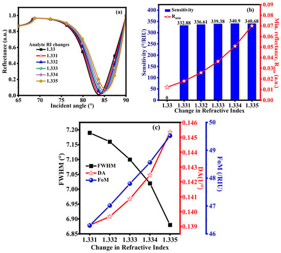
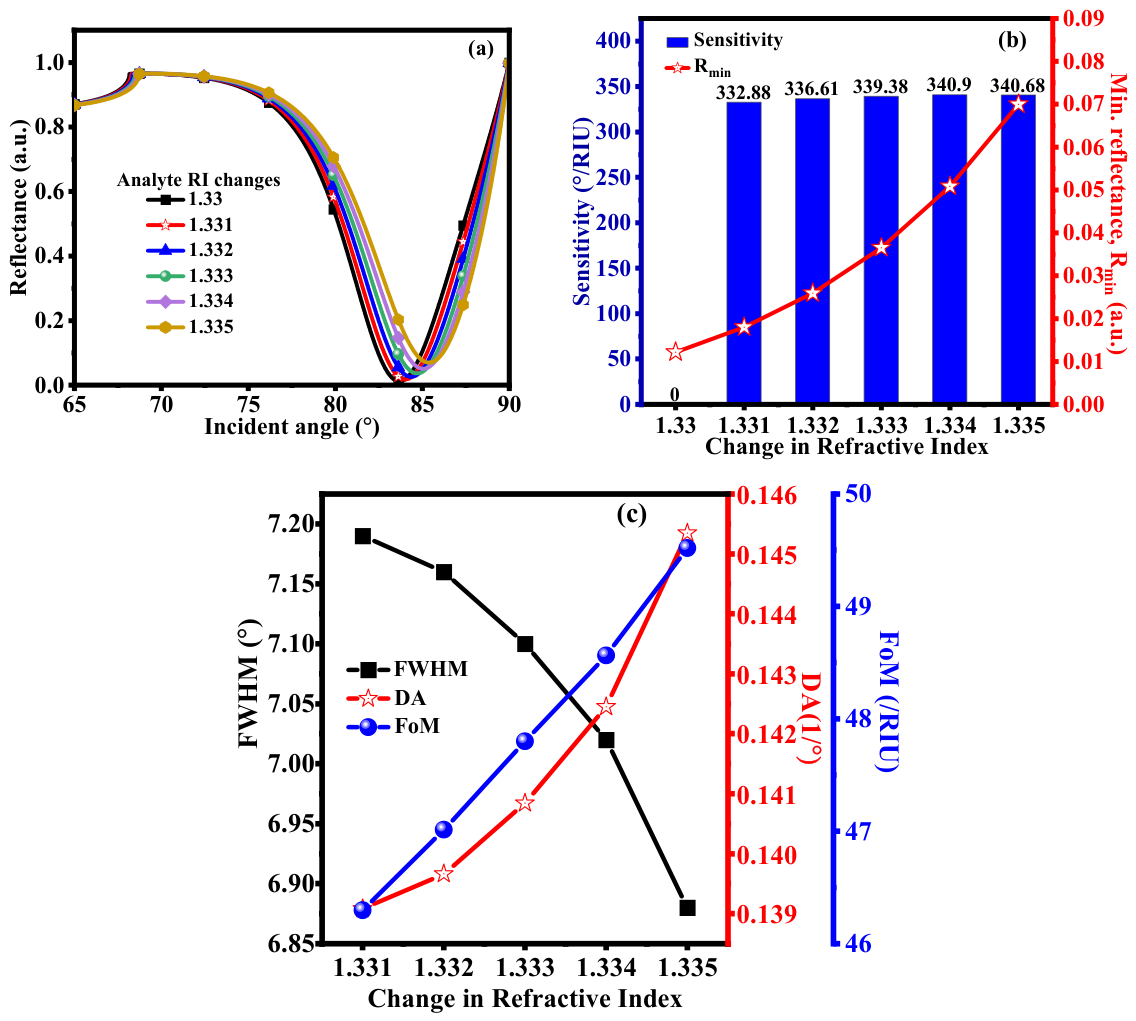
The sensing performance of the suggested SPR sensor for RI changes from 1.33 to 1.335 is shown in Figure 4. Strong light–plasmon coupling and high sensitivity to minute RI changes are observed as depicted in Figure 4a, where a distinct and sharp SPR dip is seen in the reflectance curves, and the resonance angle systematically shifts toward higher incident angles with increasing analyte RI. The resonance angles of 83.68°, 84.01°, 84.35°, 84.70°, 84.04° and 85.38° are obtained with RI of 1.330, 1.331, 1.332, 1.333, 1.334, and 1.335 respectively. The enhanced electromagnetic field confinement at the sensing interface caused by the combined effect of the conductive MXene layer and the high-RI BiFeO3 layer, which greatly enhances evanescent field interaction with the analyte, is the primary cause of this noticeable angular shift. It shows the sensitivity and Rmin value versus the change in RI of the analyte, as shown in Figure 4b. The sensitivity variation is from 332.88 to 340.68°/RIU with a change in the RI of the analyte. Figure 4c shows a slight increase in FWHM with RI, which causes a slight decrease in DA; however, the FoM increases noticeably due to the significant improvement in sensitivity. Overall, these findings show that the suggested SPR sensor provides excellent overall performance, high sensitivity, and remarkable DA, making it ideal for precise biosensing applications involving small RI variations.
Figure 4.
(a) Reflectance curve vs. incident angle; (b) sensitivity and Rmin; (c) FWHM, DA and FoM vs. change in RI.
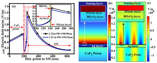
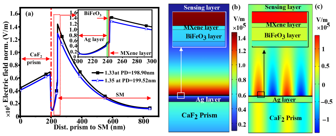
The normalized electric field distribution across the proposed structure for RIs of 1.33 and 1.35 is depicted in Figure 5a accordingly. Surface plasmon excitation results in a sharp electric field peak that emerges close to the Ag–BiFeO3 interface, indicating strong evanescent field localization. While the MXene layer improves surface interaction and stability, resulting in a higher field intensity at the sensing layer, the presence of BiFeO3 with a high refractive index strengthens this confinement. The exponential decay of the field amplitude into the SM (analyte) beyond the interface, where PD is the distance at which the field falls to 1/e (37%) of its maximum value, is defined as the evanescent wave penetration depth. As the evanescent field extends slightly deeper into the SM at higher RI, the plot displays PD of roughly 198.9 nm for RI 1.330 and 199.5 nm for RI 1.335. The ideal 1/e PD and high local field intensity at the interface maximize overlap with analyte molecules, which strengthens resonance shifts and increases the SPR sensor’s sensitivity. Surface plasmons excited by p-polarized light produce an evanescent plasmonic field, which is represented by the sharp peak seen close to the Ag/BiFeO3 interface. The evanescent wave and the analyte molecules in the SM interact more effectively because of the strong localization of the electric field at the metal–dielectric boundary. By improving confinement and raising the local RI contrast, the BiFeO3 layer raises the field intensity in comparison to a bare metal layer. The MXene layer enhances and stabilizes the evanescent field, enabling more sensitive detection because of its large surface area and conductivity. The overlap between the evanescent field and the analyte molecules is increased when a high field intensity at the metal–dielectric interface is paired with an ideal PD. This directly increases the SPR sensor’s sensitivity by producing greater shifts in the resonance condition for tiny RI changes. While the MXene layer offers stability and improves adsorption, the optimal thickness of Ag and BiFeO3 strikes a balance between field confinement (sharp peaks) and field penetration (long enough decay length), both of which are essential for attaining high detection performance. The electric field distribution and SPP modes are excited in the proposed structure that is made up of various layers, as shown in Figure 5b,c. A typical feature of SPPs is the electric field’s strong localization at the Ag/dielectric interface in Figure 5b, which decays exponentially into the metal and the sensing region. In the Ag layer, the field’s confinement at the interface shows that the incident light and the collective oscillations of conduction electrons are efficiently coupled. A more intense and regular distribution of the electric field is seen inside the dielectric layers in Figure 5c, with noticeable peaks that reach into the SM. This demonstrates that the interference effects further improve field localization in the proposed structure, highlighting the hybrid nature of plasmon–photon interactions. In order to tune and enhance the plasmon resonance and achieve deeper field penetration into the sensing region, the conductive MXene layer and the BiFeO3 ferroelectric layer are essential. The structure exhibits the potential for high-performance plasmonic sensing applications due to its extreme sensitivity to changes in the SM’s RI due to the intense localization of the electric field at the interface.
Figure 5.
(a) Normalized electric field; (b) electric field distribution; (c) SPP mode with proposed structure.
3.2. Potential Challenges, Limitations and Error Feasibility of the Suggested Sensor During Fabrication Process
The fabrication of the suggested SPR biosensor presents a number of difficulties, such as attaining consistent layer deposition, ensuring robust adhesion between various materials, and scaling up the procedure for mass production [30,31,32]. It is important to maintain uniformity and precise thickness control for the Ag, BiFeO3, and MXene layers, as any variations can adversely affect the sensor’s DA. Sputtering and evaporation are traditional PVD methods that may result in non-uniform films, especially when applied over large areas. For BiFeO3 and MXene, CVD can be used to achieve precise thickness control at the atomic level [33,34]. By modifying variables like gas pressure, target power, and deposition time, sputtering uniformity can also be enhanced. Spin coating guarantees a consistent layer for the electron-beam lithography resist prior to patterning. Another challenge is maintaining strong adherence between the different sensor layers, as poor adherence can cause delamination and reduce sensor life. By sandwiching the layers with a thin layer that encourages adhesion, such as titanium (Ti) or chromium (Cr), it is possible to improve the weak adhesion of metallic layers, such as Ag, to dielectric substrates, such as CaF2 prism [35]. Appropriate surface treatment methods, such as oxygen plasma or UV-ozone cleaning, are used prior to the deposition of the BiFeO3 and MXene layers in order to eliminate organic contaminants and increase surface energy, which improves interfacial adhesion and film uniformity. Because of its complex oxide nature and chemical stability, the deposition and patterning of the BiFeO3 layer require careful process control. To achieve well-defined thin films, optimized deposition parameters are usually followed by reactive ion etching (RIE) with appropriate chemistries to ensure precise thickness control and smooth interfaces. Similarly, MXene layers are difficult to pattern and etch using traditional wet processes due to their high conductivity and strong interlayer bonding. In order to achieve consistent and precisely defined MXene nanolayers without compromising their surface functionality, RIE-based dry etching methods—which frequently use regulated plasma conditions—are favored. All things considered, these surface treatment and fabrication techniques guarantee high-quality BiFeO3 and MXene layers, which are essential for attaining stable plasmonic coupling and improved sensing performance in the suggested SPR sensor.
Future research will concentrate on three main areas to address the shortcomings of the existing models and simulations: broader environmental testing, simulation model improvement, and experimental validation. In order to experimentally confirm resonance shifts, sensitivity, DA, and FoM derived from simulations, the proposed SPR sensor will be fabricated using controlled thin-film deposition techniques. This will be followed by angular interrogation-based SPR measurements. Realistic material parameters like wavelength-dependent complex RIs, surface roughness, interfacial diffusion, layer thickness nonuniformity, and potential oxidation or degradation effects of functional layers like BiFeO3 and MXene will be incorporated into simulation models as they are refined. To improve the models’ predictive power, multiphysics effects such as temperature-dependent optical constants and noise sources will also be considered. To ensure dependable operation in realistic biological and chemical sensing environments, broader environmental testing will evaluate sensor performance under various temperature, pH, humidity, and background RI conditions, as well as long-term stability and repeatability studies.
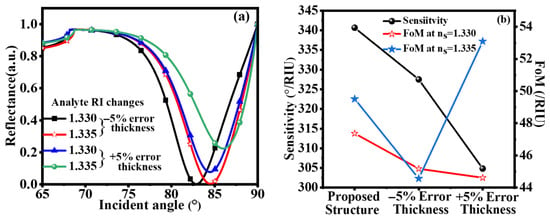
The robustness of the suggested SPR sensor against ±5% thickness error, which could occur during the actual fabrication of the multilayer structure, is examined in Figure 6. Figure 6a plots the reflectance curves at an analyte RI of 1.33 for both the ideal thickness and a variation in layer thickness of ±5%. All curves show a distinct and noticeable SPR dip, indicating that surface plasmon excitation is maintained despite thickness variations. For a ±5% error, there is a slight shift in the resonance angle and a slight change in dip depth, but the resonance profile is still clear and sharp. This behavior shows that the suggested structure is resilient to reasonable fabrication errors and maintains stable plasmonic coupling. The corresponding variation in sensitivity and FoM under ±5% thickness error is shown in Figure 6b. Thickness variations do not significantly deteriorate the sensor’s RI response, as evidenced by the fact that, despite a slight fluctuation in sensitivity, the values stay relatively high in comparison to the ideal structure. Due to the combined effects of sensitivity variation and a small broadening or narrowing of the resonance curve, the FoM exhibits a moderate change; however, it is still within an acceptable range for dependable sensing. The optimized design of the proposed structure is demonstrated by its superior overall performance when compared to the −5% thinner and +5% thicker cases. In general, Figure 6 verifies that the suggested SPR sensor exhibits outstanding resistance to fabrication-induced thickness errors, guaranteeing consistent sensitivity and FoM—a crucial component for useful and consistent biosensing applications. Despite fabrication-induced variations in the Ag, BiFeO3, and MXene layers, the proposed SPR sensor maintains stable performance, as shown by the ±5% thickness error analysis in Table 2. The sensitivity is still high (>300 °/RIU) in both thinner and thicker cases, but there is a slight shift in resonance angle and a moderate decrease in sensitivity when compared to the ideal structure. While DA and FoM are impacted by small changes in Rmin and FWHM, overall sensing performance is not substantially reduced.
Figure 6.
With ±5% error in thickness. (a) Reflectance curve. (b) Sensitivity and FoM.
Table 2.
Variation in performance parameter after ±5 error thickness.
In terms of sensitivity, DA, and FoM, Table 3 contrasts the performance of the suggested SPR biosensor with that of previously published sensors. The proposed sensor’s sensitivity of 340.68°/RIU is noticeably higher than that of previous studies, which had sensitivity values ranging from 235°/RIU to 273°/RIU. The improved plasmon–analyte interaction and electric field confinement are primarily attributable to the optimized proposed structure that includes Ag, BiFeO3, and MXene. Even so, the FoM (47.48/RIU) is marginally less than that of Kumar et al. It continues to perform well overall in 2025, exhibiting competitive accuracy and superior sensitivity. The suggested configuration is therefore a promising option for high-sensitivity biosensing applications since it exhibits a noticeable improvement over the current design. Due primarily to narrower resonance curves and greater DA, it has been noted that some previously reported SPR sensors exhibit higher FoM despite having lower sensitivity. The goal of the suggested MXene–BiFeO3-based SPR sensor is to improve plasmon–analyte interaction and electromagnetic field confinement, which broadens the resonance curve slightly while increasing sensitivity. In comparison to some current designs, this yields a moderate FoM. In SPR sensors, where greater field confinement and PD frequently result in increased FWHM, this trade-off between sensitivity and FoM is typical. As a result, the suggested structure offers a balanced performance that is appropriate for biosensing applications and has a high sensitivity and acceptable FoM.
Table 3.
Comparison of existing and proposed sensors.
4. Conclusions
The suggested SPR sensor involves optimization of Ag and BiFeO3 layer thicknesses for the sensitivity enhancement. The sensitivity increases dramatically as the Ag thickness increases from 30 to 60 nm and the BiFeO3 thickness increases from 1 nm to 3 nm. This indicates a stronger plasmon–analyte interaction because of improved electromagnetic field confinement at the sensing interface. The maximum sensitivity of 340.68°/RIU is achieved. The best performance is shown by the 39 nm of Ag and BiFeO3 of 3 nm structure-based SPR sensor. The PD of 198.9 nm for RI 1.330 and 199.5 nm for RI 1.335 as the evanescent field extends a little further into the SM at higher RI. Thus, the proposed configuration shows a significant improvement over the existing design, making it a promising choice for high-sensitivity biosensing applications.
Author Contributions
Conceptualization, R.K., L.G. and M.S.; methodology, R.K. and L.G., software, L.G. and C.-W.Y.; validation, R.K. and M.S.; formal analysis, R.K.; investigation, L.G. and C.-W.Y.; resources, C.-W.Y. and M.S.; data curation, visualization, R.K. and L.G.; writing—original draft preparation, R.K. and L.G.; writing—review and editing, M.S. and C.-W.Y.; supervision M.S., project administration, C.-W.Y. and M.S.; funding acquisition, C.-W.Y. and M.S. All authors have read and agreed to the published version of the manuscript.
Funding
This work was supported by Dongseo University’s “Dongseo Frontier Project” Research Fund of 2025.
Institutional Review Board Statement
Not applicable.
Informed Consent Statement
Not applicable.
Data Availability Statement
The original contributions presented in this study are included in the article/ Further inquiries can be directed to the corresponding authors.
Conflicts of Interest
The authors declare that they have no known competing financial or non-financial interests that could have appeared to influence the work reported in this paper. The authors further confirm that there are no commercial affiliations, patents, or financial relationships associated with this study that could be construed as a potential conflict of interest.
References
- Sobhanparast, S.; Shahbazi-Derakhshi, P.; Soleymani, J.; Amiri-Sadeghan, A.; Herischi, A.; Chaparzadeh, N.; Aftabi, Y. A Novel Electrochemical Immunosensor Based on Biomaterials for Detecting Carcinoembryonic Antigen Biomarker in Serum Samples. Sci. Rep. 2025, 15, 25396. [Google Scholar] [CrossRef] [Scilit] [PubMed]
- Homola, J.; Yee, S.S.; Gauglitz, G. Surface Plasmon Resonance Sensors: Review. Sens. Actuators B Chem. 1999, 54, 3–15. [Google Scholar] [CrossRef] [Scilit]
- Homola, J. Surface Plasmon Resonance Sensors for Detection of Chemical and Biological Species. Chem. Rev. 2008, 108, 462–493. [Google Scholar] [CrossRef] [Scilit] [PubMed]
- Shivangani; Sahu, A.; Kumar, D.; Verma, S.; Chauhan, S.S.; Kumar, S. Sensitivity Enhancement of SPR Sensor with Si/Perovskite Heterostructure for Early Detection of Malaria. Plasmonics 2025, 20, 7795–7802. [Google Scholar] [CrossRef] [Scilit]
- Zhang, S.; Yang, H.; Tang, C.; Yi, Z.; Zhang, J.; Wang, J.; Li, B. Multiple Tunable Six-Peak Graphene Absorber for High-Performance Refractive Index Sensing. Phys. B Condens. Matter 2025, 708, 417225. [Google Scholar] [CrossRef] [Scilit]
- Kumar, R.; Pal, S.; Prajapati, Y.K.; Kumar, S.; Saini, J.P. Sensitivity Improvement of a MXene- Immobilized SPR Sensor with Ga-Doped-ZnO for Biomolecules Detection. IEEE Sens. J. 2022, 22, 6536–6543. [Google Scholar] [CrossRef] [Scilit]
- Kretschmann, E.; Raether, H. Radiative Decay of Non Radiative Surface Plasmons Excited by Light. Z. Fur Naturforsch. Sect. A J. Phys. Sci. 1968, 23, 2135–2136. [Google Scholar] [CrossRef] [Scilit]
- Vibisha, G.A.; Daher, M.G.; Rahman, S.H.; Jaroszewicz, Z.; Rajesh, K.B.; Jha, R. Sensitivity Enhancement of Surface Plasmon Resonance-Based Biosensor Using Aluminium-Cobalt-Tungsten Disulfide-Graphene Heterostructure. J. Environ. Nanotechnol. 2022, 11, 5–13. [Google Scholar] [CrossRef] [Scilit]
- Bouferrache, K.; Habila, M.A.; Ghebouli, M.A.; Alomairy, S.; Alanazi, F.K.; Fatmi, M.; Ghebouli, B. Ferromagnetic and Half Metallic Properties of BaFeO3 and CaFeO3 for Spintronic Applications. Sci. Rep. 2025, 15, 37093. [Google Scholar] [CrossRef] [Scilit]
- Shivangani; Sahu, A.; Kumar, D.; Verma, S.; Chauhan, S.S.; Tiwari, R.K.; Kumar, S. Design and Analysis of Surface Plasmon Resonance Sensor Utilizing Silver, Nickel, and Black Phosphorous for Various Cancer Detection. Plasmonics 2025, 20, 6623–6639. [Google Scholar] [CrossRef] [Scilit]
- Khodaie, A.; Heidarzadeh, H. Ultra-Sensitive Surface Plasmon Resonance Sensor Integrating MXene (Ti3C2TX) and Graphene for Advanced Carcinoembryonic Antigen Detection. Sci. Rep. 2025, 15, 13571. [Google Scholar] [CrossRef] [Scilit] [PubMed]
- Mahalakshmi, R.; Wekalao, J.; Raja, M.R.; Jothi, S.A. Ultra-Sensitive Graphene-Metal Hybrid Metasurface for Non-Invasive Glucose Detection with Convolutional Neural Network Integration. Plasmonics 2025, 21, 549–574. [Google Scholar] [CrossRef] [Scilit]
- Zhao, D.; Wang, X.; Chen, Y.; Rao, X.; Gao, J.; Qi, Y.; Yang, H.; Zheng, X. Quasi-BIC Mode and Refractive Index Sensing by Asymmetric Silicon Nanodisk Arrays. Opt. Commun. 2025, 596, 132421. [Google Scholar] [CrossRef] [Scilit]
- Gu, P.; Yang, H.; Li, D.; Zhu, H.; Chen, J.; Zhang, Z.; Yan, Z.; Tang, C.; Liu, F.; Chen, Z. High-Q and Intense Lattice Plasmon Resonance in Hexagonal Nonclose Packed Thin Silver Nanoshells Array. J. Phys. Chem. C 2024, 128, 6431–6437. [Google Scholar] [CrossRef] [Scilit]
- Gao, J.; Wang, X.; Chen, Y.; Zhao, D.; Qi, Y.; Yang, H. Triple Fano Resonances Induced by a Square-Elliptical Cavity Coupled with an MIM Waveguide for High-Sensitivity Gas Refractive Index Sensing. J. Opt. Soc. Am. B 2025, 42, 742. [Google Scholar] [CrossRef] [Scilit]
- Nur-e-alam, M.; Basher, M.K.; Vasiliev, M.; Das, N. Physical Vapor-deposited Silver (Ag)-based Metal-dielectric Nanocomposites for Thin-film and Coating Applications. Appl. Sci. 2021, 11, 6746. [Google Scholar] [CrossRef] [Scilit]
- Abbas, N.; Shad, M.R.; Hussain, M.; Mehdi, S.M.Z.; Sajjad, U. Fabrication and Characterization of Silver Thin Films Using Physical Vapor Deposition, and the Investigation of Annealing Effects on Their Structures. Mater. Res. Express 2019, 6, 116437. [Google Scholar] [CrossRef] [Scilit]
- Yuan, H. Defect Engineering of BiFeO3 Photocathodes for Photoelectrochemical Water Splitting. Ph.D. Thesis, Queen Mary University of London, London, UK, 2024. [Google Scholar]
- Li, X.; Ran, F.; Yang, F.; Long, J.; Shao, L. Advances in MXene Films: Synthesis, Assembly, and Applications. Trans. Tianjin Univ. 2021, 27, 217–247. [Google Scholar] [CrossRef] [Scilit]
- Tang, R.; Xiong, S.; Gong, D.; Deng, Y.; Wang, Y.; Su, L.; Ding, C.; Yang, L.; Liao, C. Ti3C22D MXene: Recent Progress and Perspectives in Photocatalysis. ACS Appl. Mater. Interfaces 2020, 12, 56663–56680. [Google Scholar] [CrossRef] [Scilit]
- Wu, Q.; Li, N.; Wang, Y.; Xu, Y.; Wei, S.; Wu, J.; Jia, G.; Fang, X.; Chen, F.; Cui, X. A 2D Transition Metal Carbide MXene-Based SPR Biosensor for Ultrasensitive Carcinoembryonic Antigen Detection. Biosens. Bioelectron. 2019, 144, 111697. [Google Scholar] [CrossRef] [Scilit]
- Moore, H.H. Refractive Index of Alkaline Earth Halides and Its Wavelength and Temperature Derivatives. J. Phys. Chem. Ref. Data 1982, 9, 161–290. [Google Scholar] [CrossRef] [Scilit]
- Srivastava, S.; Singh, S.; Mishra, A.C.; Lohia, P.; Dwivedi, D.K. Numerical Study of Titanium Dioxide and MXene Nanomaterial-Based Surface Plasmon Resonance Biosensor for Virus SARS-CoV-2 Detection. Plasmonics 2023, 18, 1477–1488. [Google Scholar] [CrossRef] [Scilit] [PubMed]
- Polyanskiy, M.N. Refractiveindex.Info Database of Optical Constants. Sci. Data 2024, 11, 94. [Google Scholar] [CrossRef] [Scilit] [PubMed]
- Kumar, R.; Garia, S.; Joshi, S.; Pal, A.; Singh, I.; Sahu, U.K. A High-Performance SPR Biosensor for Female Reproductive Hormone Detection. IEEE Access 2025, 13, 86057–86068. [Google Scholar] [CrossRef] [Scilit]
- Mondal, T.P.; Mahmud, R.R.; Rafi, S.A.; Islam, M.S.; Barua, B. Design and Optimization of a Highly Sensitive Surface Plasmon Resonance Biosensor for Accurate Detection of Mycobacterium Tuberculosis. IEEE Sens. J. 2025, 25, 9216–9223. [Google Scholar] [CrossRef] [Scilit]
- Karki, B.; Sharma, S.; Singh, Y.; Pal, A. Sensitivity Enhancement of Surface Plasmon Resonance Biosensor with 2-D Franckeite Nanosheets. Plasmonics 2022, 17, 71–78. [Google Scholar] [CrossRef] [Scilit]
- Wu, L.; Guo, J.; Wang, Q.; Lu, S.; Dai, X.; Xiang, Y.; Fan, D. Sensitivity Enhancement by Using Few-Layer Black Phosphorus-Graphene/TMDCs Heterostructure in Surface Plasmon Resonance Biochemical Sensor. Sens. Actuators B Chem. 2017, 249, 542–548. [Google Scholar] [CrossRef] [Scilit]
- Shalabney, A.; Abdulhalim, I. Electromagnetic Fields Distribution in Multilayer Thin Film Structures and the Origin of Sensitivity Enhancement in Surface Plasmon Resonance Sensors. Sens. Actuators A Phys. 2010, 159, 24–32. [Google Scholar] [CrossRef] [Scilit]
- Wang, Q.; Ren, Z.H.; Zhao, W.M.; Wang, L.; Yan, X.; Zhu, A.S.; Qiu, F.M.; Zhang, K.K. Research Advances on Surface Plasmon Resonance Biosensors. Nanoscale 2022, 14, 564–591. [Google Scholar] [CrossRef] [Scilit]
- Gonzalez-Macia, L.; Morrin, A.; Smyth, M.R.; Killard, A.J. Advanced Printing and Deposition Methodologies for the Fabrication of Biosensors and Biodevices. Analyst 2010, 135, 845–867. [Google Scholar] [CrossRef] [Scilit]
- Singh, P. SPR Biosensors: Historical Perspectives and Current Challenges. Sens. Actuators B Chem. 2016, 229, 110–130. [Google Scholar] [CrossRef] [Scilit]
- Hu, Y.; Lu, J.; Feng, H. Surface Modification and Functionalization of Powder Materials by Atomic Layer Deposition: A Review. RSC Adv. 2021, 11, 11918–11942. [Google Scholar] [CrossRef] [Scilit]
- Chiappim, W.; Neto, B.B.; Shiotani, M.; Karnopp, J.; Gonçalves, L.; Chaves, J.P.; Sobrinho, A.D.S.; Leitão, J.P.; Fraga, M.; Pessoa, R. Plasma-Assisted Nanofabrication: The Potential and Challenges in Atomic Layer Deposition and Etching. Nanomaterials 2022, 12, 3497. [Google Scholar] [CrossRef] [Scilit]
- Wang, Z. Fabrication of Biomedical Composite Coatings by Electrophoretic Deposition and Dip Coating Methods. Ph.D. Thesis, McMaster University, Hamilton, ON, Canada, 2024. [Google Scholar]
- Uniyal, A.; Kumba, K.; Dhiman, G.; Ahmed, M.Z.; Pal, A.; Pal, D.; Muduli, A.; Srivastava, V. Advanced SPR Sensor for Human Sperm Analysis: Leveraging Silver and Nanomaterials for Enhanced Performance. Plasmonics 2024, 20, 3939–3950. [Google Scholar] [CrossRef] [Scilit]
- Karki, B.; Pal, A.; Sarkar, P.; Uniyal, A.; Yadav, R.B. Gold, MXene, and Graphene Nanofilm-Based Surface Plasmon Resonance Sensor for Malaria Detection. J. Opt. 2024, 53, 4805–4816. [Google Scholar] [CrossRef] [Scilit]
- Chowdhury, A.S.; Islam, M.A.; Islam, M.S.; Dey, B.; Park, J. Design and Analysis of PtSe2 and Blue Phosphorus/MoS2 Heterostructure-Based SPR Biosensor. ACS Appl. Opt. Mater. 2024, 2, 1046–1059. [Google Scholar] [CrossRef] [Scilit]
- Kumar, R.; Agarwal, S.; Pal, N.; Pal, S.; Prajapati, Y.K. Platinum Diselenide (PtSe2) Mediated Heterostructure Based SPR Sensor for the Detection of Formalin: A Theoretical Analysis. Phys. Scr. 2025, 100, 15546. [Google Scholar] [CrossRef] [Scilit]
- Pillai, A.M.; Nair, N.; Das, M.K.; Ram, S.K. Strategic Approaches to Enhance Efficiency and Commercial Feasibility of Copper-Based Surface Plasmon Resonance Sensing. Next Mater. 2025, 7, 100377. [Google Scholar] [CrossRef] [Scilit]



Disclaimer/Publisher’s Note: The statements, opinions and data contained in all publications are solely those of the individual author(s) and contributor(s) and not of MDPI and/or the editor(s). MDPI and/or the editor(s) disclaim responsibility for any injury to people or property resulting from any ideas, methods, instructions or products referred to in the content. |
© 2026 by the authors. Licensee MDPI, Basel, Switzerland. This article is an open access article distributed under the terms and conditions of the Creative Commons Attribution (CC BY) license.