


Abstract
Oxidative stress poses significant challenges for fungi inhabiting extreme environments. Elevated salinity frequently induces the excessive production of reactive oxygen species (ROS), which can damage cellular components and impair growth. In response, fungi—common inhabitants of extreme environments—activate coordinated adaptive mechanisms, including antioxidant defense systems and other stress-related pathways. In the present study, a newly isolated strain, Trichoderma afroharzianum B2.2, obtained from the poorly studied saline habitat of Atanasovsko Lake (Bulgaria), was investigated. The cellular response of this moderately halotolerant strain to increased salinity was characterized. Biomarkers of oxidative stress were evaluated, and the involvement of key enzymes from glycolysis and the pentose phosphate pathway in the strain’s adaptation to elevated salinity was examined. Understanding adaptations to salt environments is crucial not only for elucidating fungal survival mechanisms under extreme conditions but also for their potential applications in biotechnology, ecology, and food safety, particularly in the context of increasing ecosystem salinization and climate change.
1. Introduction
Hypersaline environments are among the most extreme habitats in which extremophilic microorganisms have been identified. These include not only seas, salt lakes, saline soils, salt deserts, salterns, and brines but also high-salt food products [1,2,3]. Such environments are inhabited by halophiles capable of surviving under high-salinity conditions and, in some cases, requiring salt for growth. Halophilic microorganisms are widely distributed and predominantly comprise by bacteria, archaea, algae, and fungi [4]. These organisms produce a diverse range of biomolecules with unique properties that have potential applications in various industrial processes.
Nevertheless, the osmotic and ionic stresses associated with high salinity significantly limit the survival and proliferation of most microorganisms. Osmotic stress results in cellular dehydration, disruption of osmotic balance, reduced turgor pressure, and inhibition of growth, whereas ionic stress interferes with enzyme activity and membrane function. Together, these stresses suppress both the growth and physiological activities of filamentous fungi. In addition, hypersaline environments are characterized by low water activity, frequently alkaline conditions, and limited nutrient availability [5]. A widely accepted classification of halophilic microorganisms was proposed by Kushner and Kamekura in 1988 [6]. This classification divides microorganisms into four groups according to their optimal salt concentration for growth. The first group comprises non-halophiles, which require less than 0.2 M NaCl for optimal growth; however, organisms capable of growing under higher salinity conditions without requiring salt are classified as halotolerant. The second group includes slight halophiles, which exhibit optimal growth at NaCl concentrations ranging from 0.2 to 0.5 M, typical of marine environments. The third group consists of moderate halophiles, whose optimal growth occurs at salt concentrations between 0.5 and 2.5 M NaCl, whereas the fourth group encompasses extreme halophiles, which grow best at NaCl concentrations exceeding 2.5 M [6]. It should be noted that, although halophiles are most commonly defined based on their tolerance to sodium chloride, the term may also encompass organisms adapted to elevated concentrations of other inorganic salts and soluble minerals [7,8,9].
The mycobiota of natural saline environments comprises both halotolerant and halophilic fungi, including not only well-characterized species but also newly identified and rare taxa [10]. With the exception of a few obligate halophilic species, such as Wallemia ichthyophaga, most halophilic fungi do not require sodium chloride for growth and are capable of adapting to a wide range of salinities, from concentrations typical of marine environments to nearly saturated NaCl solutions [11]. Numerous studies have examined the relationship between salt stress and oxidative stress. In plants, high salinity induces severe oxidative stress through the excessive production of reactive oxygen species (ROS), including hydrogen peroxide (H2O2) and hydroxyl radicals. The accumulation of these reactive molecules results in ion toxicity, osmotic imbalance, and damage to cellular membranes. Furthermore, elevated salt concentrations disrupt cellular redox homeostasis, leading to lipid peroxidation, impaired metabolic activity, and reduced growth. In many plant species, roots are more severely affected than shoots due to their direct exposure to saline conditions. To counteract these detrimental effects, plants activate both enzymatic and non-enzymatic antioxidant defense mechanisms, including superoxide dismutase (SOD), catalase (CAT), and glutathione (GSH), which collectively contribute to the maintenance of cellular redox balance and stress tolerance [12,13,14].
Similarly, fungi exposed to elevated NaCl concentrations experience severe osmotic and ionic stress, which promotes the excessive generation of ROS and consequently induces oxidative stress [15]. To survive and proliferate under such extreme environmental conditions, microorganisms have evolved a diverse array of physiological, biochemical, and molecular adaptation mechanisms that enable them to maintain cellular homeostasis and mitigate the detrimental effects of salt-induced stress.
Adaptations to high salinity involve morphological changes, including growth inhibition, structural alterations, and modifications in development. Structural adjustments occur in the plasma membrane and cell wall and are often accompanied by the accumulation of pigments and/or hydrophobins [16,17,18]. Increased salinity leads to a thickened cell wall and alterations in lipid composition, including changes in sterol content, fatty acyl chain profiles, and the nature of polar phospholipid head groups [19]. Pigments such as carotenoids and melanins further influence the properties of the cytoplasmic membrane, providing protection against UV radiation and other harmful effects of sunlight [20,21].
Biochemical adaptations include the activation of antioxidant defense enzymes, as well as other key enzymes involved in the regulation of cellular stress responses. These mechanisms act in concert with both enzymatic and non-enzymatic antioxidant systems and play a crucial role in maintaining cellular homeostasis and ensuring fungal survival under adverse environmental conditions [15].
The study of salt and oxidative stress in fungi is crucial for understanding the mechanisms underlying the adaptation and survival of eukaryotic microorganisms under extreme environmental conditions. High salinity is frequently associated with increased production of ROS, thereby necessitating a coordinated cellular response. Elucidation of these adaptive strategies not only advances fundamental knowledge of fungal stress biology but also has important practical implications for biotechnology, ecology, and food safety, particularly in the context of ongoing ecosystem salinization and climate change.
The present study provides new insights into the relationship between oxidative and salt stress in the moderately halotolerant fungus Trichoderma afroharzianum B2.2, isolated from a saltern in the Bulgarian Black Sea region. The adaptive mechanisms enabling the strain to survive under elevated salinity conditions were investigated. While most previous studies have focused on halophilic fungi, this work specifically addresses the cellular response of a moderately halotolerant species originating from a hypersaline environment. Moreover, information on Trichoderma species from saline habitats remains limited, underscoring the novelty of the present study.
2. Results
2.1. Morphological and Physiological Characteristics of the Strain Trichoderma afroharzianum B 2.2
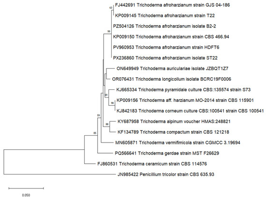
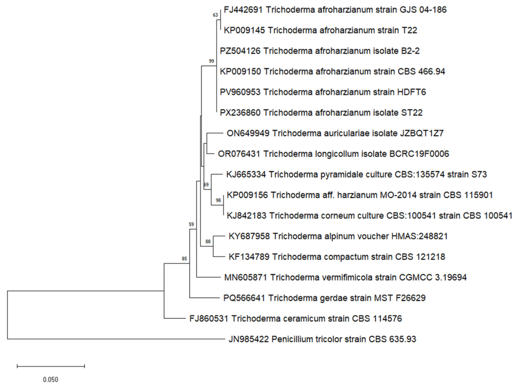
The model strain selected for this study was isolated from a high-salinity environment (salt pans), yet it is moderately halotolerant. It was identified as Trichoderma afroharzianum isolate B2-2 (PZ485173). Molecular identification of isolate B2.2 was performed by analysis of three DNA barcode markers—ITS, rpb2, tub2 and tef1. BLAST analysis of the ITS sequence showed 98% similarity to representatives of the genus Trichoderma from type material. In line with the recommendations of Cai and Druzhinina [22], the ITS result alone is not sufficient for definitive species identification. The sequence of the rpb2 gene demonstrated 99% similarity to Trichoderma afroharzianum strain GJS 04-186 (type material) and the sequence of the tef1 gene, and 98% similarity to Trichoderma afroharzianum isolate CBS124620 (type material). According to the criteria for molecular identification of Trichoderma proposed [22], the similarity values for rpb2 (≥99%) and tef1 (≥97%) are sufficient for reliable species identification. Therefore, isolate B2.2 was unambiguously identified as Trichoderma afroharzianum (Figure 1). GenBank accession numbers of deposited sequences are PZ485173, PZ504126, PZ504127, and PZ504128.
Figure 1.
A phylogenetic tree indicates the relationship between Trichoderma afroharzianum isolate B2.2 and closely related Trichoderma strains from GenBank, based on a partial sequence of the RNA polymerase II subunit (rpb2) gene. Evolutionary relationships of taxa were inferred using the Neighbor-Joining method. The percentage of replicate trees in which the associated taxa clustered together in the bootstrap test (1000 replicates) is shown above the branches. The tree is drawn to scale, with branch lengths in the same units as those of the evolutionary distances used to infer the phylogenetic tree. The evolutionary distances were computed using the Tamura 3-parameter method and are in the units of the number of base substitutions per site. This analysis involved 17 nucleotide sequences. All positions containing gaps and missing data were eliminated (complete deletion option). Evolutionary analyses were conducted in MEGA11.
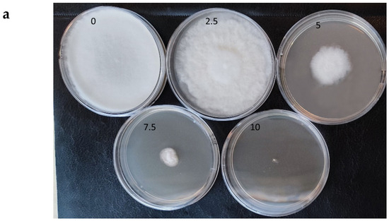
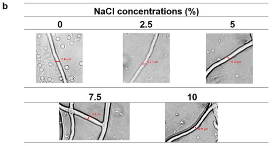
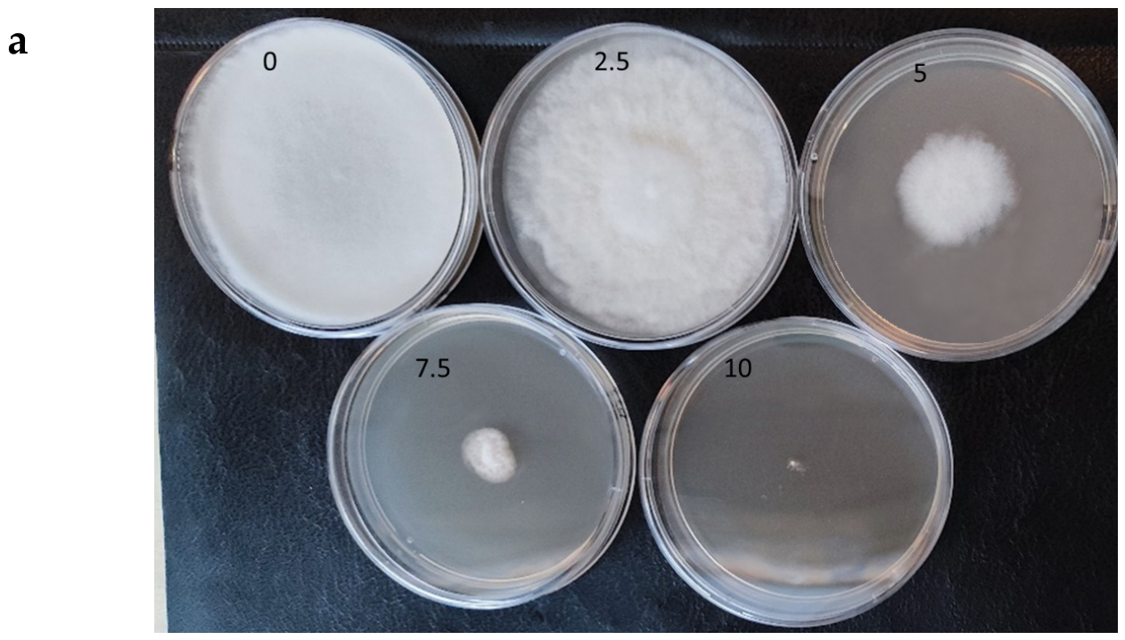
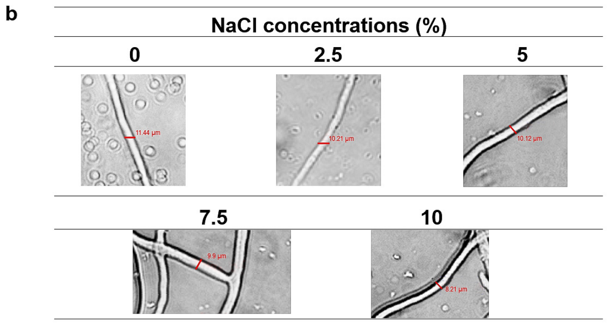
As shown in Figure 2, the strain exhibits optimal growth at 0% sodium chloride, with a growth range extending from 0 to 10%. Figure 2a,b illustrate its growth at different salt concentrations and highlight several of its morphological characteristics.
Figure 2.
Macro—(a) and micro—(b) morphology of the model strain after exposure to different concentrations of sodium chloride after 7 days of cultivation. The colonies were grown on PDA at 25 °C for 7 days in 90 mm Petri dishes in the dark. Representative bright-field micrographs showing filament morphology under different experimental conditions. Variations in branching, filament thickness, and structural organization are observed between groups. The dimensions are estimated based on micrometric measurements and optical system parameters (25×).
A decrease in the diameter of the model strain colonies with increasing salt concentration is clearly evident. The colony surface is velvety and mossy, and the mycelium appears hyaline to white, lacking conidia or exudates; however, growth is markedly reduced at the highest concentration tested. The colonies’ diameter decreases (3.5–1 cm) with increasing NaCl concentration (5–10%) (Figure 2a). An unpigmented reverse side and filamentous margins of the colonies were observed. It can be concluded that the model strain shows optimal growth both in the absence of sodium chloride and in the presence of 2.5% NaCl.
Figure 2b presents microscopic images of the strain’s hyphae under the studied conditions. As can be seen, filament diameter ranged approximately from 11.44 to 8.21 μm depending on the experimental condition. The thickest and most structurally heterogeneous filaments were observed in variant 5 (10%NaCl). In addition to a decrease in hyphal thickness, increasing sodium chloride concentrations also lead to darkening of the hyphae, i.e., possible melanization. The melanin is well known as a powerful antioxidant, and a common property of biological pigments is their ability to scavenge and neutralize exogenous free radicals.
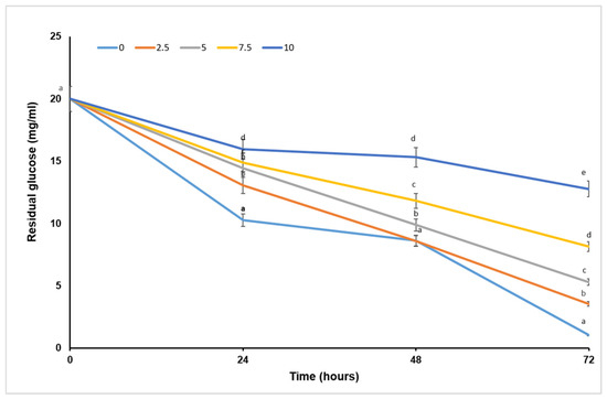
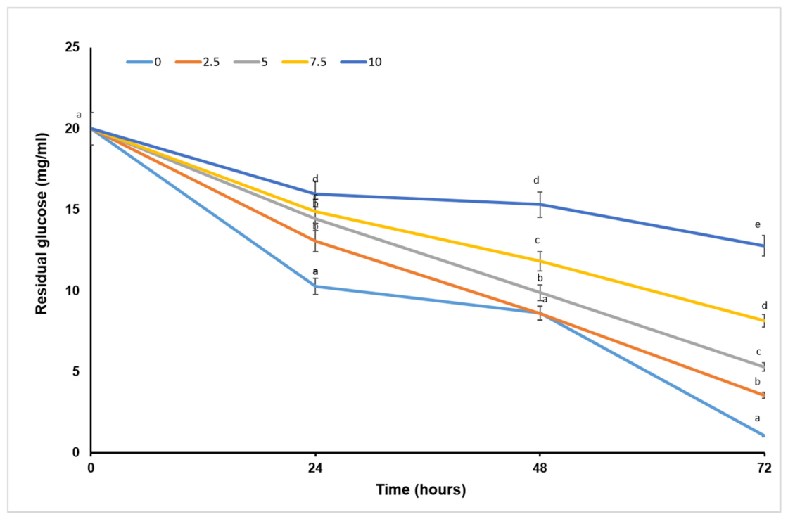
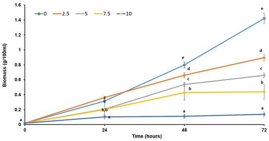
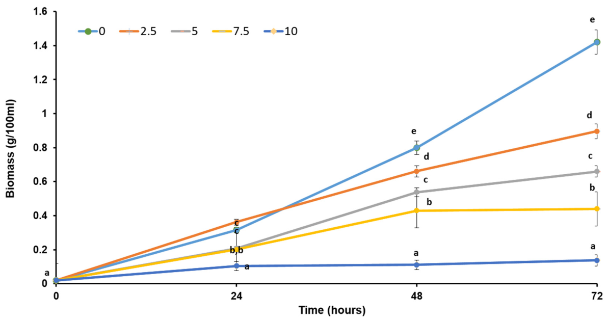
Monitoring of cellular uptake of the carbon source from the nutrient medium in the presence of different salt concentrations (Figure 3) shows a dose-dependent trend. Uptake decreases with increasing salt concentration, which corresponds to the reduced growth observed under the same conditions (Figure 4).
Figure 3.
Effect of sodium chloride concentration on glucose uptake during the cultivation of Trichoderma afroharzianum B2.2. Different lowercase letters indicate significant differences (p < 0.05) between treated and control variants at the same cultivation period.
Figure 4.
Biomass content during the cultivation of Trichoderma afroharzianum B2.2 under different salinity conditions. Different lowercase letters indicate significant differences (p < 0.05) between treated and control variants at the same cultivation period.
2.2. Relationship Between High Salinity and Oxidative Stress
2.2.1. EPR Analysis of Paramagnetic Centers in Mycelium Under Salt Stress
To investigate the relationship between elevated salt concentrations and oxidative stress signals, spectral changes in mycelia exposed to different NaCl concentrations were analyzed. Mycelial samples of the model strain treated with 0, 5, and 10% sodium chloride were subjected to electron paramagnetic resonance (EPR) spectroscopy, and the recorded spectra exhibited a complex profile.
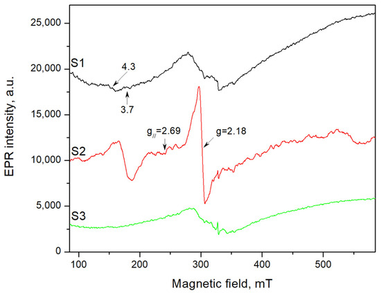
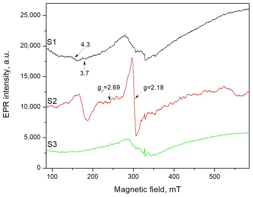
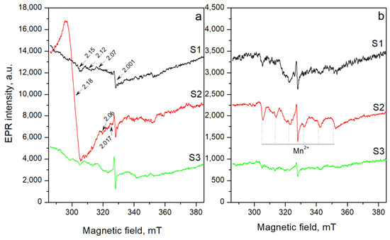
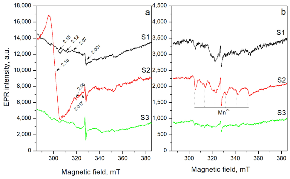
Figure 5 presents the EPR spectra recorded over the full magnetic field sweep range (500 mT), while Figure 6 shows an enlarged view of the 100 mT spectral region centered at 336 mT. Owing to the higher spectral resolution achieved in the narrower sweep range, additional paramagnetic species can be resolved in the studied samples.
Figure 5.
EPR spectra of the 24-h cells treated with (S1) 0% NaCl; (S2) 5% NaCl; (S3) 10% NaCl.
Figure 6.
EPR spectra of the cells treated with S1—0% NaCl, S2—5% NaCl, and S3—10% NaCl, recorded at (a) 24 h of treatment; (b) 48 h of treatment.
In Figure 5, two signals at g ≈ 4.3 and g ≈ 3.7 are observed. These signals are also present in the sample treated with 0% salt (S1), but their intensity reaches a maximum at 24 h in sample S2, corresponding to cells treated with 5% NaCl.
Figure 5 also shows a strong signal with g⊥ = 2.18 and g‖ = 2.69, suggesting the presence of a Cu2+ center. The highest intensity is again observed in cells of variant S2, treated with 5% NaCl, after 24 h.
A signal attributed to Mn2+ ions (3d5) is observed (Figure 6). Its EPR spectrum consists of six weak hyperfine lines arising from the interaction between the unpaired electron and the 55Mn nucleus (nuclear spin I = 5/2) [23]. EPR signals are recorded at g = 2.001, 2.07, 2.12, and 2.153.
Cells treated with 0% (S1), 5% (S2), and 10% (S3) sodium chloride at 48 h exhibit EPR spectra similar to those recorded at 24 h; however, the intensities of the detected signals are significantly lower. The intensities of the signals at g = 2.001 for the different samples are presented in Table 1.
Table 1.
EPR signal intensities of the line with g value 2.001 at the different samples.
2.2.2. Biomarkers of Oxidative Stress Under High-Salinity Growth Conditions
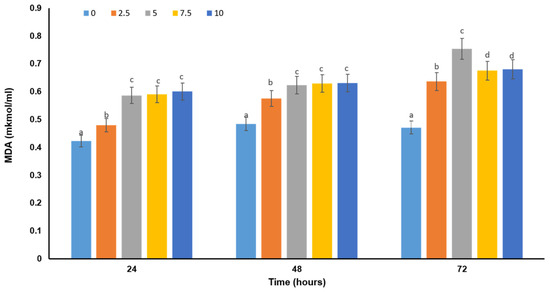
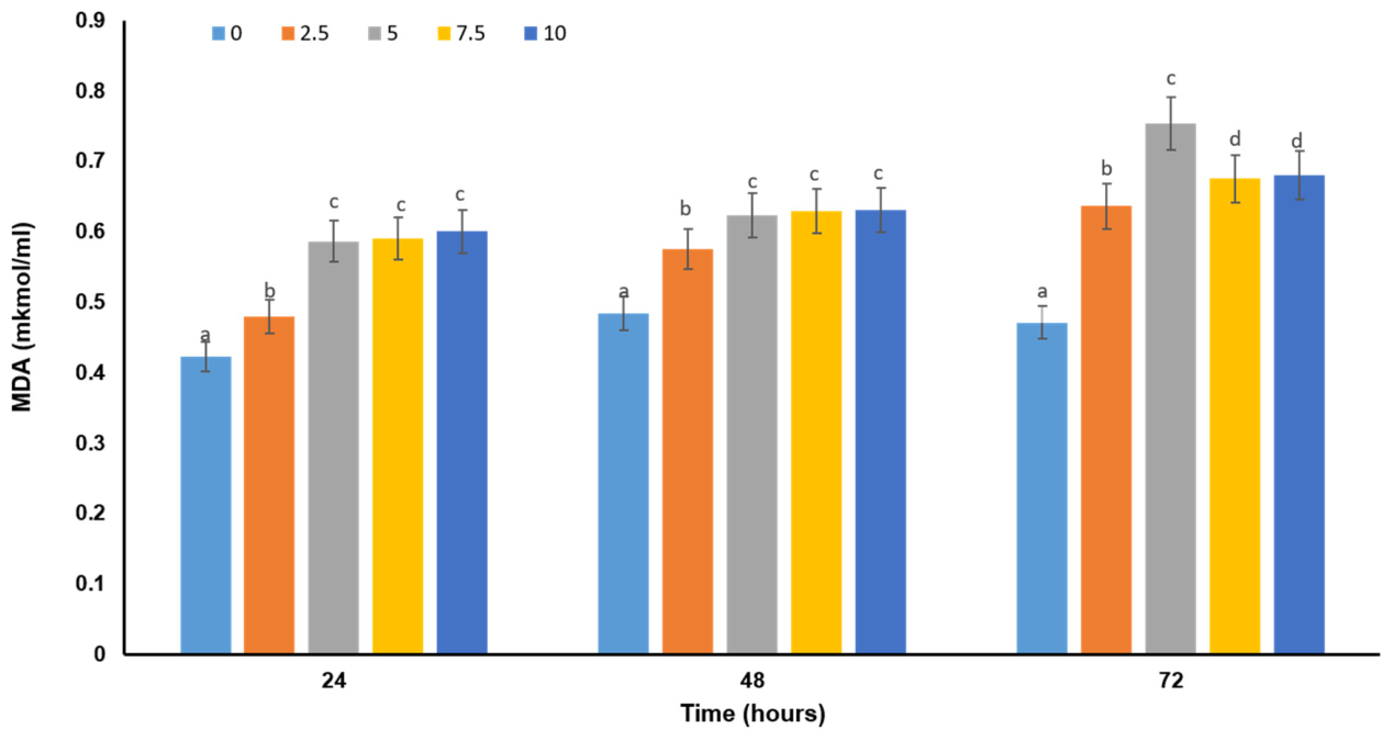
The cellular response of the studied fungal strain to elevated salinity was assessed by monitoring changes in selected key biomarkers of oxidative stress. Malondialdehyde (MDA) levels were used as an indicator of lipid peroxidation and cellular damage. As shown in Figure 7, MDA levels increase with rising sodium chloride concentrations up to 5%. However, further increases in salinity do not result in a corresponding rise in MDA levels. Instead, MDA values remain comparable to those measured at 5% NaCl across all examined time intervals.
Figure 7.
Levels of lipid peroxidation in Trichoderma afroharzianum B2.2 cells exposed to different salinity conditions. Different lowercase letters indicate significant differences (p < 0.05) between treated and control variants at the same cultivation period.
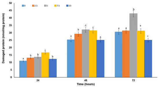
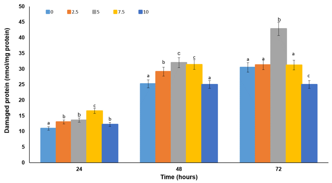
A similar trend was observed for protein carbonyl content, a marker of oxidative protein damage in the cells (Figure 8).
Figure 8.
Levels of carbonyl content as marker of protein damage in fungal cells exposed to increased salt concentrations. Different lowercase letters indicate significant differences (p < 0.05) between treated and control variants at the same cultivation period.
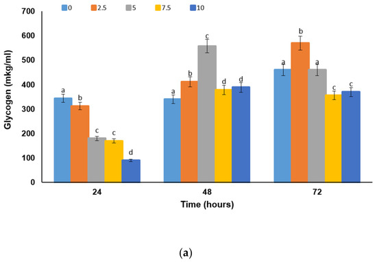
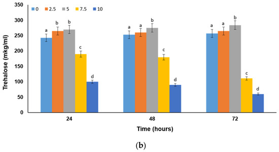
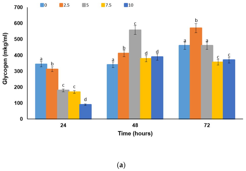
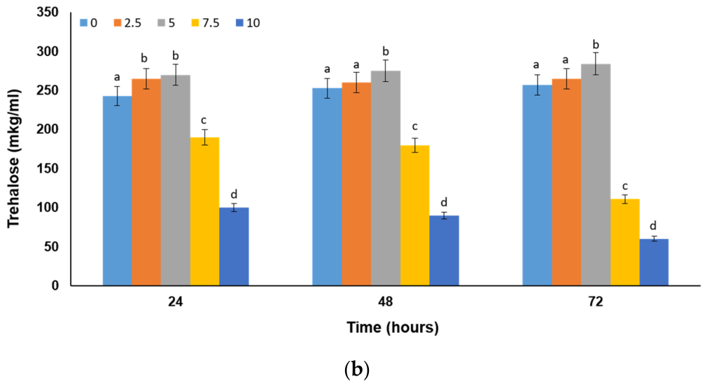
Another major stress biomarker is the level of reserve carbohydrates in the cell. Under physiological stress conditions, cells typically respond by accumulating the storage carbohydrates glycogen and trehalose. Our results show stimulation of reserve carbohydrate accumulation in response to increasing salinity of the medium up to 5%. However, further increases in salt concentration do not lead to higher levels of these biomarkers. On the contrary, a significant decrease is observed at higher concentrations compared to the other tested conditions (Figure 9a,b).
Figure 9.
Levels of reserve carbohydrates in cells subjected to different salinity conditions: (a) glycogen content; (b) trehalose content. Different lowercase letters indicate significant differences (p < 0.05) between treated and control variants at the same cultivation period.
2.3. Metabolic Adaptation
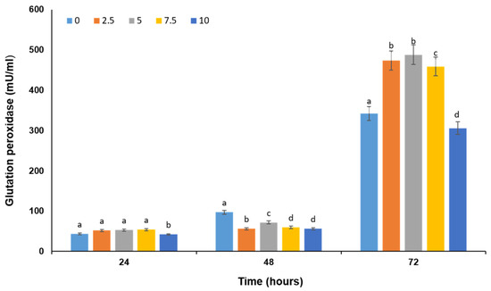
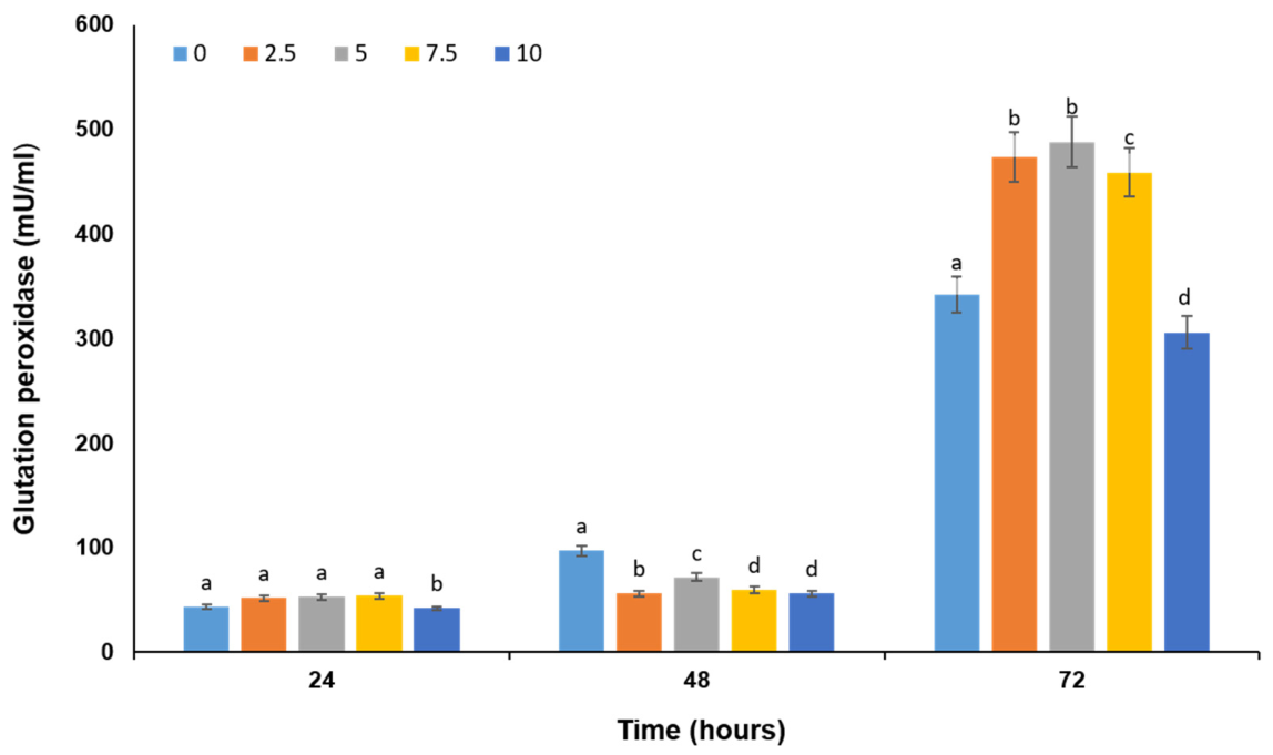
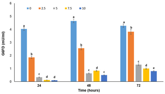
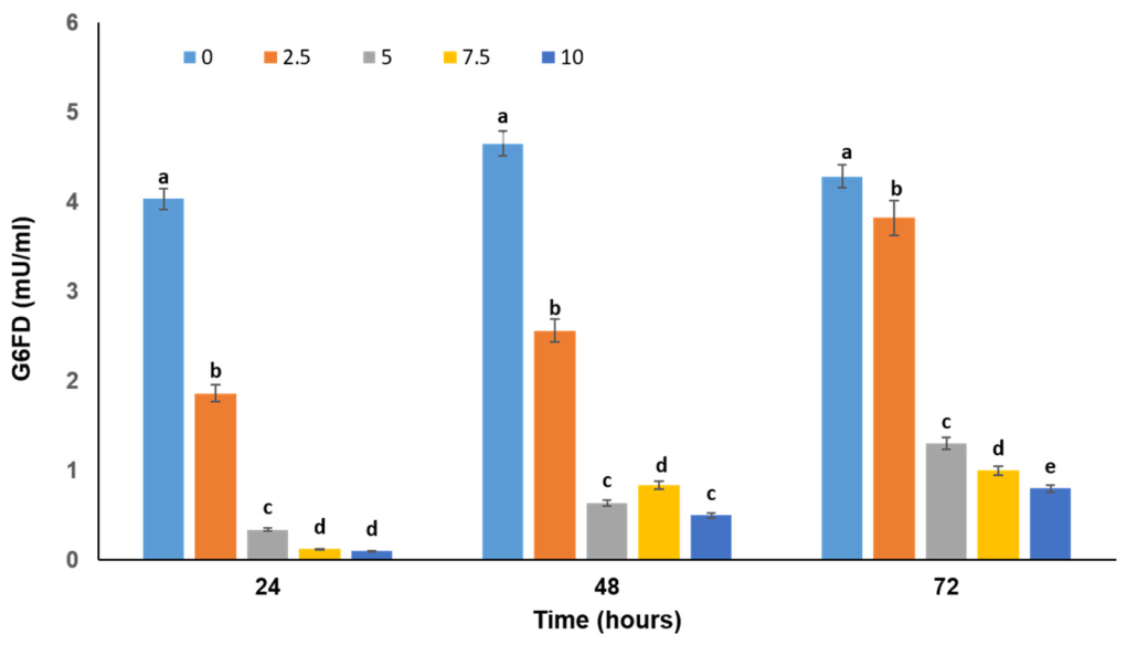
Cells utilize the glutathione antioxidant system as a primary mechanism for the neutralization of reactive oxygen species. In this study, glutathione peroxidase activity was measured (Figure 10), as it plays a direct role in regulating cellular glutathione levels and maintaining redox balance. The regeneration of reduced glutathione (GSH) requires NADPH, which is primarily supplied by the pentose phosphate pathway. Therefore, the activity of glucose-6-phosphate dehydrogenase (G6PD), a key enzyme in this pathway, was also analyzed (Figure 11).
Figure 10.
Glutathione peroxidase activity in cells exposed to different salinity conditions. Different lowercase letters indicate significant differences (p < 0.05) between treated and control variants at the same cultivation period.
Figure 11.
Glucose-6-phosphate dehydrogenase activity in cells of the model strain exposed to different sodium chloride concentrations. Different lowercase letters indicate significant differences (p < 0.05) between treated and control variants at the same cultivation period.
An increase in G6PD activity over time was observed under all tested conditions; however, this increase was not dose-dependent. At 72 h, when enzyme activity reached its maximum, G6PD activity in the highest-salinity variant remained lower than in the other variants.
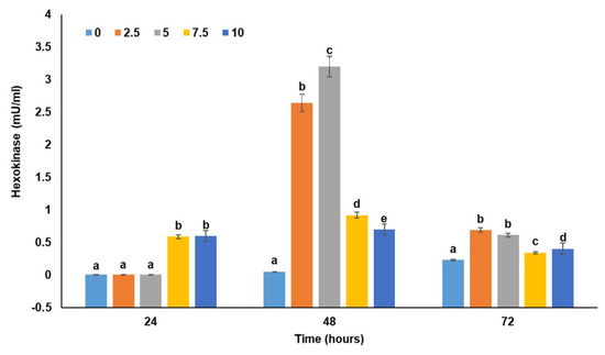
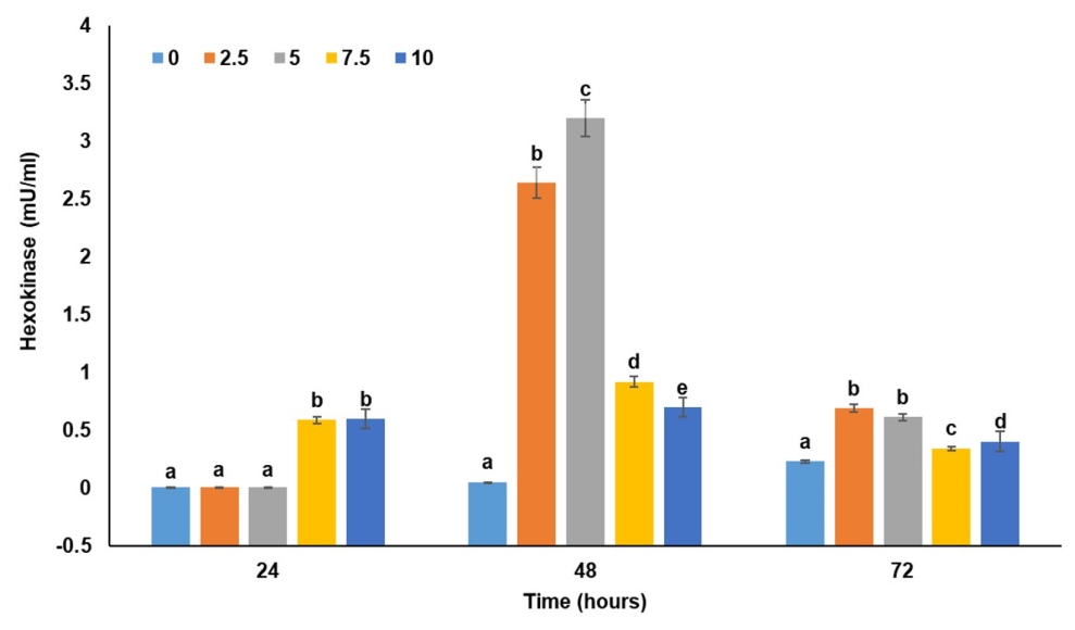
Hexokinase catalyzes the first reaction of glycolysis. Under oxidative stress, cells require additional ATP to support antioxidant systems, such as the glutathione system. Hexokinase activity ensures a continuous flux of glucose through glycolysis. As shown in Figure 12, no dose-dependent increase in hexokinase activity was observed, likely reflecting impaired glucose metabolism under conditions of elevated salinity.
Figure 12.
Hexokinase activity in cells of the model strain exposed to different sodium chloride concentrations. Different lowercase letters indicate significant differences (p < 0.05) between treated and control variants at the same cultivation period.
2.4. Role of Antioxidant Defense in the Cellular Response to High Salinity
Antioxidant enzyme defense is essential for the survival and protection of cells exposed to oxidative stress. In this study, we investigated changes in the activity of antioxidant enzymes representing both the first and second lines of antioxidant defense.
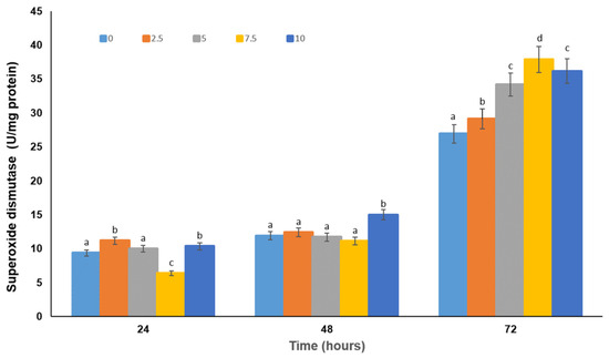
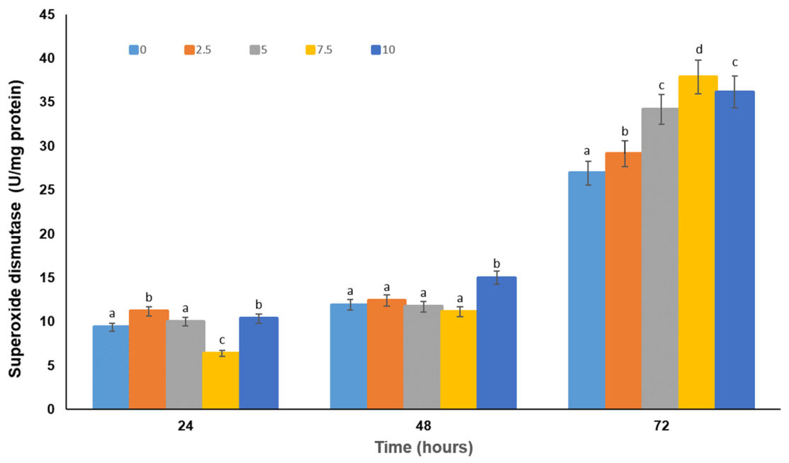
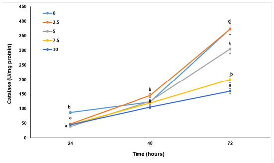
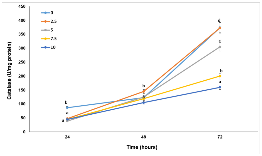
Figure 13 and Figure 14 show an increase in the activity of the main antioxidant enzymes, superoxide dismutase and catalase, with a maximum observed at 72 h. The increase in SOD activity is dose-dependent, with the highest level detected in cells treated with 10% sodium chloride.
Figure 13.
Superoxide dismutase activity in the cells of the studied variants. Different lowercase letters indicate significant differences (p < 0.05) between treated and control variants at the same cultivation period.
Figure 14.
Catalase activity in cells of the different studied variants. Different lowercase letters indicate significant differences (p < 0.05) between treated and control variants at the same cultivation period.
The observed increase in CAT and GPx activity (Figure 10 and Figure 14) also reached a maximum at 72 h. A dose-dependent increase was evident in cells exposed to 0–5% sodium chloride. However, further increases in salt concentration did not result in a corresponding rise in enzyme activity.
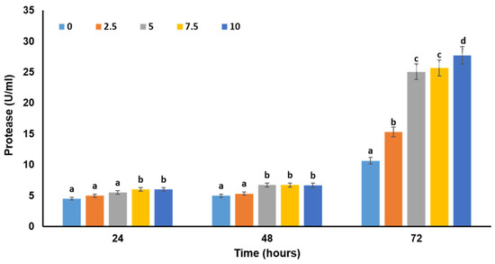
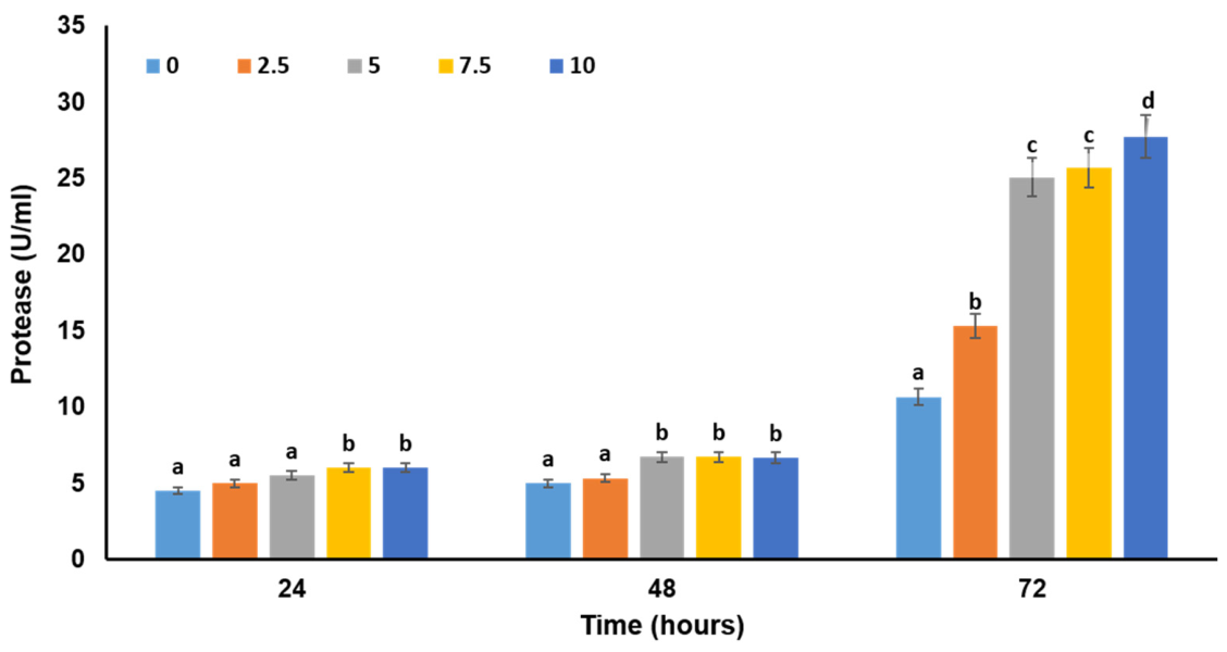
Proteases belong to the second line of antioxidant defense, and their primary function is the degradation of proteinaceous cellular components damaged by free radicals. Our results, shown in Figure 15, indicate protease activation that shows both time- and dose-dependent trends, correlating with the levels of oxidatively modified proteins.
Figure 15.
Changes in protease activity in response to exposure to different salt concentrations. Different lowercase letters indicate significant differences (p < 0.05) between treated and control variants at the same cultivation period.
3. Discussion
The investigation of salt and oxidative stress responses in fungi is essential for understanding the mechanisms that enable eukaryotic microorganisms to adapt and survive under extreme environmental conditions. Elevated salinity promotes the generation of ROS, thereby requiring a coordinated cellular defense response. Elucidating these adaptive mechanisms not only advances our understanding of fungal stress physiology but also has important implications for biotechnology, ecology, and food safety, particularly in light of the increasing salinization of natural ecosystems associated with climate change.
In the present study, a newly isolated fungal strain originating from the poorly explored hypersaline ecosystem of Atanasovsko Lake near Burgas, Bulgaria, was investigated. This coastal hypersaline lake is characterized by salt concentrations ranging from 20 to 27.5% (200–275‰) in the salt production ponds. Based on its growth characteristics and morphological features under different NaCl concentrations, the isolate was classified as moderately halotolerant, exhibiting optimal growth at 0–2.5% NaCl and sustained growth within a range of 0–10% NaCl (Figure 2a,b). These findings indicate an ability to tolerate moderate salinity, while confirming that the strain is not adapted to highly saline conditions.
Based on the analysis of the ITS, rpb2, tub2, and tef1 genetic markers, the isolate was identified as Trichoderma afroharzianum B2.2. In contrast to the genera Aspergillus, Penicillium, and Cladosporium, which are frequently reported from hypersaline environments [24,25,26], representatives of the genus Trichoderma have rarely been described in such habitats, and their physiological responses to salinity-induced stress remain insufficiently characterized.
The growth and hyphal morphology of T. afroharzianum B2.2 were markedly influenced by increasing NaCl concentrations in the culture medium. Hyphae exhibited a dose-dependent reduction in diameter accompanied by pronounced morphological alterations (Figure 2b). In addition, hyphal darkening observed at elevated NaCl concentrations suggested enhanced melanization. Comparable morphological responses have been reported in Aspergillus sydowii following exposure to osmotic stress induced by saturated NaCl concentrations [27]. Similar changes in colony morphology and growth have also been described in the halotolerant black yeast Hortaea werneckii and species of the halophilic genus Wallemia [28]. Furthermore, the observed reduction in hyphal diameter under saline conditions is consistent with previous reports describing the effects of salt stress on filamentous fungi [29,30].
The production of protective pigments, such as melanin and carotenoids, represents an important adaptive strategy that enables halophilic fungi to tolerate ultraviolet (UV) radiation and other deleterious effects of solar exposure [31,32,33]. For example, black yeasts, which comprise a group of halophilic fungi, synthesize melanin as a protective response to environmental stress [34]. In hypersaline environments, melanin deposited within the cell wall reduces water loss, prevents the leakage of intracellular compatible solutes, and contributes to the maintenance of membrane integrity and fluidity, thereby enhancing fungal survival under osmotic stress [35,36].
Morphological adaptations to salinity have also been described in other halotolerant fungi. For example, Aspergillus montevidensis ZYD4 exhibits pronounced changes in colony morphology under salt stress, including reduced pigmentation and extensive development of conidiophores, which are rarely observed under non-saline conditions [29].
To investigate the physiological response of the model strain to salinity-induced oxidative stress, several key biochemical stress biomarkers were analyzed. The results demonstrate that osmotic stress caused by elevated NaCl concentrations markedly affects the physiological state of the fungus. As shown in Figure 4, biomass accumulation decreased progressively with increasing salinity, indicating impaired fungal growth. This reduction was accompanied by delayed glucose utilization (Figure 3), suggesting substantial alterations in central carbon metabolism.
Under saline conditions, decreased glucose consumption is associated with the redistribution of metabolic flux toward the pentose phosphate pathway (PPP), thereby promoting the generation of NADPH required to maintain cellular redox homeostasis. Consistent with this metabolic adjustment, significant changes in the activities of glucose-6-phosphate dehydrogenase (G6PD) and hexokinase (HK) were observed (Figure 11 and Figure 12).
G6PD, the rate-limiting enzyme of the oxidative branch of the PPP, catalyzes the first step of the pathway and represents the major source of cellular NADPH. In contrast, HK catalyzes the first committed step of glycolysis by phosphorylating glucose, thereby regulating carbon flux into central metabolic pathways. During oxidative stress, increased NADPH availability is essential for maintaining antioxidant defense systems, including the glutathione-dependent detoxification pathway, while sustained glycolytic activity provides the ATP required to support energy-demanding stress responses.
Moderate levels of ROS have been shown to stimulate hexokinase expression and glycolytic activity. However, excessive ROS accumulation under severe salt stress can oxidize critical sulfhydryl (-SH) groups within the enzyme, leading to reduced catalytic activity and disruption of cellular metabolism.
In fungi, HK, and G6PD play complementary roles in the response to oxidative stress by coordinating metabolic adaptation with the maintenance of cellular redox homeostasis. A possible explanation for the reduced G6PD activity observed under saline conditions (Figure 11) is the enhanced production of SOD. Excessive SOD activity may promote a pro-oxidant state by generating hydrogen peroxide at a rate that exceeds the detoxification capacity of downstream antioxidant enzymes, thereby impairing the overall antioxidant defense system [37]. It has also been suggested that increased SOD synthesis may divert cellular resources toward its own production, limiting the expression of other stress-responsive proteins, including G6PDH [37].
ROS generation was evaluated by electron paramagnetic resonance (EPR) spectroscopy in cultures exposed to 0%, 5%, and 10% NaCl after 24 and 48 h of cultivation (Figure 5 and Figure 6). The EPR signals observed at g ≈ 4.3 and g ≈ 3.7 (Figure 5) are typically attributed to high-spin Fe3+ centers or specific iron-containing organometallic clusters, including certain redox states of nitrogenase FeMo cofactors. Similar signals associated with the MoFe protein have previously been reported by Lukoyanov et al. [38]. In addition, the pronounced signal with g⊥ = 2.18, and g‖ = 2.69 is characteristic of a Cu2+ center, indicating the presence of copper-containing paramagnetic species (Figure 5).
Several signals become more distinct within the narrower magnetic field range (Figure 6). The first is an EPR signal at g = 2.001, observed in all studied samples, which is attributed to semiquinone radicals. The typical g-value for semiquinone radicals ranges from 2.0038 to 2.0047; however, the shift to a lower value in this case is likely due to the formation of semiquinone–metal complexes. Such interactions were reported by Czechowski et al. (2004) [39], who demonstrated that an EPR line at g = 1.999 originates from the interaction of the unpaired electron with Pb(II) coordinated in a chelate ring system derived from lignin.
Another signal, detected at g = 2.07, may be attributed to hydroxyl radicals (•OH) [40]. The line observed at g = 2.12 is usually associated with spin–spin interactions between surface-bound paramagnetic ions and is often attributed to Fe3+ ions complexed with semiquinones (i.e., semiquinone-bridged Fe centers) [41,42]. The signal detected at g = 2.153 is likely due to Cu2+ centers, as g-values of approximately g⊥ ≈ 2.15 and g‖ ≈ 2.22 are characteristic of copper-containing enzymes or copper-bound complexes [43].
The signal intensity at g = 2.001 increases markedly in sample S3, corresponding to the 10% sodium chloride treatment, suggesting the formation or stabilization of semiquinone radicals under elevated oxidative stress. The recorded semiquinone signal exhibits an asymmetric shape (Figure 6), likely due to the overlap of two signals with very similar g-factors. One component corresponds to the semiquinone radical, while the other is most likely associated with melanin. Melanin is a pigment known for its antioxidant properties and its ability to scavenge and neutralize exogenous free radicals. In fungi, melanin contributes to virulence by interfering with host defense mechanisms, including the neutralization of the oxidative burst in phagocytic cells [44]. Typically, the stable melanin-derived free radical produces an EPR signal in the range g = 2.004–2.006 [45,46]. However, in the present case, the exact g-value cannot be determined due to signal overlap. Nevertheless, the presence of a melanin radical is further supported by the visible color change of the samples following salt stress.
Semiquinone radicals can exhibit exceptionally long half-lives (up to several days at 37 °C) and are generally neither highly reactive nor highly toxic. However, they can donate their excess electron to molecular oxygen, thereby generating superoxide (O2−) and hydrogen peroxide (H2O2).
Indeed, a new EPR signal was recorded in the stressed samples at g = 2.017 (Figure 6a), which is likely associated with oxygen-derived radicals, as such species typically exhibit g-values between 2.00 and 2.03 [40]. Another new signal was observed in the stressed samples at g = 2.06. A resonance at this g-value may originate from O2− species formed due to the presence of trace oxygen in the system, as suggested by Bruckner et al. [47], or it may be related to adsorbed water [48,49]. The intensities of the signals attributed to OH• radicals and O2− species increase noticeably in sample S3 at 24 h, indicating enhanced oxidative processes under salt stress.
Exposure to severe stress initially triggered the generation of large amounts of free radicals and the activation of metal-containing enzymes, which was reflected in the high EPR signal intensity recorded after 24 h. The highest signal intensity was observed in the most severely stressed sample (S3). After 48 h, the number of unpaired spins decreased markedly, resulting in a pronounced decline in EPR signal intensity.
Several factors may contribute to this decrease: free radicals can react with each other or with antioxidants, forming diamagnetic products; metal centers may undergo changes in oxidation state (Fe3+ → Fe2+, Cu2+ → Cu+), rendering them EPR-silent. In addition, metal ions may be exported from the cells or sequestered, while semiquinones and reactive oxygen species gradually decrease over time due to reduction or decay. The activation of antioxidant systems (e.g., catalase, superoxide dismutase, and melanin) further enhances the neutralization of free radicals. Metal centers (Cu, Fe, Mn) are likely reduced to diamagnetic states or released into the medium, resulting in decreased signal intensity or loss of EPR detectability. In many biological systems, ROS production and free radical formation peak at the early stages of stress and subsequently decline as the organism begins to adapt. Thus, 24 h likely represents the peak phase of the stress response, whereas 48 h corresponds to the adaptation phase. The decrease in all EPR signals at 48 h may also be exacerbated by cellular damage resulting from prolonged stress, leading to reduced metabolic activity and altered relaxation parameters.
Spectral analysis of stressed and unstressed mycelia confirmed that exposure to elevated salinity induces severe oxidative stress in the fungal cells. The resulting overproduction of ROS contributes to oxidative damage while exacerbating the detrimental effects of ionic toxicity and osmotic stress associated with high NaCl concentrations [15]. Structural adaptations to saline conditions include cell wall thickening and modifications in membrane lipid composition, which enhance cellular stability under osmotic stress [50].
In the present study, malondialdehyde (MDA) levels increased progressively with increasing salinity (Figure 7), indicating enhanced lipid peroxidation and membrane damage. As one of the major end products of lipid peroxidation, MDA is widely recognized as a reliable biomarker of oxidative membrane injury. Lipid peroxidation generates lipid hydroperoxides as primary oxidation products, which are subsequently detoxified by glutathione peroxidases. Consistent with this mechanism, glutathione peroxidase (GPx) activity increased in the cultures exposed to elevated NaCl concentrations (Figure 10). Similar increases in MDA accumulation under saline conditions have also been reported for the halotolerant fungus Aspergillus sydowii EXF-12860 [29].
Protein carbonylation, a well-established indicator of oxidative protein damage, increased in the cultures grown in the presence of 0–5% NaCl (Figure 8). However, no further increase was observed at higher salinity levels. This response may reflect the activation of antioxidant defense mechanisms together with enhanced proteolytic degradation of oxidatively damaged proteins, thereby limiting the accumulation of carbonylated protein species.
The levels of protein oxidation products were found to be comparable at both NaCl concentrations in studies on salt stress in Aspergillus sydowii EXF-12860 [29], suggesting the activation of both enzymatic and non-enzymatic cellular defense mechanisms aimed at mitigating oxidative damage. Water deficit induced by NaCl represents a major environmental stressor for eukaryotic organisms [15,51]. A well-established adaptive response to oxidative and osmotic stress is the accumulation of reserve carbohydrates [52,53].
Trehalose, a non-reducing disaccharide, is widely recognized as a key cellular protectant under stress conditions. Its role in stress tolerance has been demonstrated in various systems, including Anoectochilus species exposed to salinity stress [54], where trehalose accumulation contributed to improved physiological performance. Enhanced salt tolerance associated with increased trehalose levels has also been reported in tomato plants under saline conditions [55]. In addition, glycogen has been identified as an important storage carbohydrate with a protective role during stress adaptation [56,57].
The analysis of the two major stress-related carbohydrates, glycogen and trehalose, revealed a distinct response pattern. As shown in Figure 9a,b, increasing salinity within the lower concentration range was associated with a concentration-dependent accumulation of both compounds. However, further increases in NaCl concentration did not result in additional elevation of their intracellular levels. A plausible explanation for these observations is that enhanced oxidative stress promotes the mobilization of storage carbohydrates. Under severe stress conditions, the increased energetic demand associated with cellular maintenance and survival may drive the catabolism of carbohydrate reserves. A portion of the glucose released through this process may be redirected toward glycerol biosynthesis, which plays a central role as a compatible solute during salt stress, whereas trehalose accumulation is often associated with conditions of low or absent NaCl exposure.
In Aspergillus sydowii, cells grown in the absence of NaCl appear to experience hypoosmotic conditions that induce trehalose production [58]. In this organism, the expression of genes involved in trehalose biosynthesis is differentially regulated: α-trehalose-6-phosphate synthase is expressed under both hypo- and hyperosmotic stress, whereas trehalose phosphorylase expression is restricted to hypoosmotic conditions (i.e., in the absence of NaCl) [16]. By contrast, in Aspergillus nidulans, trehalose accumulation has been reported in response to oxidative and heat stress, as well as during conidial maturation, but not under osmotic stress conditions [58].
Fungal responses to osmotic stress involve the activation of both non-enzymatic antioxidant systems (e.g., glutathione) and enzymatic defense mechanisms, including catalases, superoxide dismutases, and glutathione peroxidases [15].
The antioxidant enzymes GPx, CAT, and SOD were evaluated in the present study. As shown in Figure 10, Figure 13 and Figure 14, the activities of these enzymes increased under salinity-induced oxidative stress. SOD activity exhibited a clear dose-dependent elevation. CAT and GPx displayed a similar trend in cultures exposed to 0–5% NaCl; however, at higher salinity levels, no further increase was observed. This plateau may reflect the limited capacity of the antioxidant system under conditions of excessive reactive oxygen species production and/or partial impairment of enzymatic function.
These enzymes represent key components of the cellular antioxidant defense network. SOD and CAT directly detoxify ROS, whereas HK and G6PDH contribute indirectly by supplying NADPH through the pentose phosphate pathway, thereby maintaining reduced glutathione levels and sustaining GPx activity.
The model strain exhibits moderate halotolerance, indicating effective adaptation to low-salinity conditions but restricted growth and limited physiological plasticity under high-salinity stress. Consistent with these findings, increased activities of SOD, CAT, and GPx have also been reported in Aspergillus sydowii EXF-12860 exposed to 5.13 M NaCl [59]. The activation of enzymatic antioxidant defenses contributes to the maintenance of intracellular redox homeostasis by limiting oxidative damage induced by ROS.
Proteases constitute an important component of the secondary line of cellular defense against oxidative stress. Intracellular proteases are primarily responsible for the degradation of oxidatively damaged and stress-induced misfolded proteins, as well as for protein turnover during cellular aging. In addition, extracellular proteases have been implicated in fungal stress responses [60,61,62].
Protein carbonylation results from the oxidation of specific amino acid residues—particularly arginine, histidine, lysine, proline, threonine, and tryptophan—leading to the formation of carbonyl groups. These modifications impair protein structure and function and increase susceptibility to proteolytic degradation [63]. Under stress conditions, proteolytic systems are activated to remove damaged proteins, recycle amino acids, and contribute to overall protein quality control. In coordination with antioxidant defenses, proteases play a key role in maintaining cellular proteostasis and supporting fungal survival. The observed increase in protease activity in the present study correlates with the extent of protein damage (Figure 8 and Figure 15). This suggests a stress-responsive activation of protein degradation pathways in response to salinity-induced oxidative damage.
Biochemical adaptation to stress involves the coordinated activation of antioxidant enzymes and other components of cellular stress-response systems. The enzymatic activity profiles should be interpreted as an integrated component of the cellular stress-response network rather than as isolated biochemical endpoints, as they collectively reflect the coordinated regulation of redox homeostasis and central carbon metabolism under salinity stress. Together, these mechanisms contribute to the maintenance of cellular homeostasis and are essential for survival under adverse environmental conditions [15]. Trichoderma afroharzianum B2.2, investigated in the present study, is classified as moderately halotolerant and therefore appears to possess limited adaptive capacity for survival under high salinity, despite its isolation from a saline environment. An adaptive response was observed at NaCl concentrations up to 5%, whereas concentrations of 7.5% and 10% exerted pronounced toxic effects on the cells. Although growth was still detectable at 10% NaCl, it was markedly reduced, indicating that the strain experiences severe physiological stress under these conditions. The observed responses to increasing salinity are largely determined by the intrinsic physiological characteristics of the organism. Overall, its cellular behavior differs from that reported for filamentous fungi isolated from highly saline habitats [13,29].
In contrast, representatives of the genera Aspergillus, Hortaea, and Wallemia have developed specialized genetic and physiological adaptations that enable effective regulation of osmotic balance, ion transport, and cellular structural integrity. By comparison, T. afroharzianum B2.2 appears to rely predominantly on more general stress-response mechanisms to cope with osmotic stress, including morphological remodeling, accumulation of compatible solutes, metabolic adjustments, and activation of antioxidant defense systems. The observed changes in redox-related parameters likely reflect a combination of time-dependent physiological processes and salinity-induced stress responses, with the latter contributing to the consistent trends observed across all analyzed oxidative stress biomarkers.
Overall, the investigation of salt and oxidative stress responses in fungi is essential for elucidating the mechanisms that enable eukaryotic microorganisms to adapt to extreme environmental conditions. Elevated salinity promotes the overproduction of reactive oxygen species, thereby requiring tightly coordinated cellular responses. Elucidation of these adaptive strategies contributes to a deeper understanding of fungal stress biology and provides broader insight into microbial survival under adverse environmental conditions.
4. Materials and Methods
4.1. Fungal Strains and Culture Conditions
In this study, a filamentous fungus isolated from a salt pan in Atanasovsko Lake near the town of Burgas (Bulgaria) (42°34′55″ N, 27°28′12″ E) and maintained in the mycological collection of the Institute of Microbiology, Bulgarian Academy of Sciences, was used. The lake is a coastal hypersaline ecosystem and, due to its high salinity ranging from 20 to 27.5%, is utilized for salt production.
4.1.1. Molecular Genetic Identification of the Fungal Strain
The identification of the fungal strain was performed according to Cai and Druzhinina [22] and the Molecular identification protocol for Trichoderma. The analysis included the following steps:
DNA isolation: Chromosomal DNA was isolated from 100 mg of 48-h mycelium with the GeneMATRIX Plant & Fungi DNA Purification Kit (EURx Ltd., Gdańsk, Poland) according to the manufacturer’s instructions. PCR was performed on a T100 Thermal Cycler (Bio-Rad Laboratories, Hercules, CA, USA) using PCR master mix (GenetBio, Daejeon, Republic of Korea). PCR products were visualized by 1% agarose gel electrophoresis and purified with the Gene JET PCR Purification Kit (Thermo Fisher Scientific Inc., Waltham, MA, USA).
PCR and phylogenetic markers: We used PCR to amplify and sequence three phylogenetic markers (DNA barcode loci): (1) ITS 1 and 2 of the rRNA gene cluster (ITS, including the 5.8S rRNA); (2) the fragment of RNA polymerase II subunit B gene (rpb2); (3) the translation elongation factor 1-α (tef1); (4) beta-tubulin (tub2) gene [22]. Primer synthesis and sequencing of PCR fragments were performed at Macrogen Europe (Amsterdam, The Netherlands). The ITS region (internal transcribed spacer) is a universal primary DNA barcode for identification and the most widely sequenced DNA region in the molecular ecology of fungi. The remaining DNA barcodes (tef1, rpb2, tub2) are secondary and are used for more accurate species identification and comparison between closely related strains.
Bioinformatics programs and analyses: Molecular identification (DNA barcoding) is identification based on the similarity of given DNA fragments. Nucleotide sequences were processed and analyzed with the Chromas and CAP3 programs. The BLAST application(BLAST + 2.17.0) of the NCBI (National Center for Biotechnology Information, Bethesda, MD, USA) was used to compare the sequences with the available sequences in the GenBank database (http://www.ncbi.nlm.nih.gov/BLAST:02.07.2026, accessed on 4 June 2026). All data were also compared with Index Fungorum (https://www.indexfungorum.org/names/names.asp, accessed on 4 June 2026) and Mycobank (https://www.mycobank.org/, accessed on 4 June 2026). According to the “Molecular identification protocol for Trichoderma”, Trichoderma species can be identified if their ITS sequence reaches a similarity value of ≥76% with the sequences in the dataset attached to the protocol in Cai and Druzhinina [22] and the three other DNA barcodes are highly similar to the corresponding sequences of the reference strain of a species such as rpb2 ≥ 99%, tub2 ≥ 97%, and tef1 ≥ 97%. These conditions can be abbreviated as the following sequence similarity standard: Trichoderma [ITS76]~ sp ∃! (rpb299 ≅ tef197). We extracted the sequences of the corresponding reference strains for the species of interest from the databases [63].
4.1.2. Morphological Studies
To investigate morphological changes in the studied strain, cultivation was performed on solid nutrient media. The fungus was grown on potato dextrose agar in Petri dishes, either without or with varying concentrations of sodium chloride (0, 2.5, 5, 7.5, and 10%). Cultures were incubated at 25 °C for 10 days.
For microscopic observation, a Docuval light microscope (Carl Zeiss, Jena, Germany) was used.
4.1.3. A Cultivation of the Strain
To study the cellular response to salt stress, submerged cultivation was carried out under the following conditions: potato dextrose broth as the nutrient medium, 220 rpm on a rotary shaker, 25 °C, and a spore suspension of 1 × 10⁶ conidia/mL.
To determine cellular changes resulting from salt stress, submerged cultivation was performed in 500 mL Erlenmeyer flasks. Five experimental variants were prepared using potato dextrose broth supplemented with different concentrations of sodium chloride (0, 2.5, 5, 7.5, and 10%). Cultivation was carried out on a shaker at 25 °C, and samples were collected at 24, 48, and 72 h after the start of incubation.
4.2. Biochemical Studies
4.2.1. EPR Analyses
EPR spectra in the X-band (9.4 GHz) were recorded as the first derivative of the absorption signal using a JEOL JES-FA100 EPR spectrometer (JEOL Ltd., Tokyo, Japan). The JEOL spectrometer, equipped with 100 kHz magnetic field modulation, was fitted with a standard TE011 cylindrical resonator. Measurements for the identification of paramagnetic species were performed at room temperature using a magnetic field centered at 336.00 mT, a microwave frequency of 9176.530 MHz, a modulation amplitude of 0.2 mT, and a microwave power of 2.0 mW. Previously dried biomass from the model strain treated with 0, 5, and 10% sodium chloride was used for EPR analysis. Solid-state measurements were performed directly on 12 mg samples without prior preparation, using identical EPR spectrometer gain settings for all samples.
All measurements were standardized with respect to gain settings and sample mass.
The g-factor, which represents the effective Zeeman splitting factor of a paramagnetic center, was determined from the resonance condition.
where (h) is the constant of Planck, (ν) is the microwave frequency, (β) is the Bohr magneton, and (H) is the resonance magnetic field determined from the measured spectra. While isolated paramagnetic species are typically characterized by a specific g-value, real materials often exhibit a distribution of g-values due to variations in their local structural and magnetic environments.
g = hν/βH,
Three parallel samples corresponding to three variants of NaCl-treated fungal cells—0% (S1), 5% (S2), and 10% (S3)—were analyzed. For each sample, three independent EPR measurements were performed, including repeated insertion and removal of the sample from the resonator cavity between measurements. The obtained results were averaged, and the experimental uncertainty was estimated to be approximately 3%.
4.2.2. Biomass Measurement
The growth of the fungal culture was assessed by determining the dry mass of the produced biomass.
Mycelium collected at a specific stage of cultivation was filtered through a Whatman No. 4 filter, rinsed with distilled water, and then dried at 105 °C to constant weight.
4.2.3. Cell-Free Extract Preparation
The cell-free extract was prepared as described previously [64]. Biomarkers of oxidative stress were determined in the resulting cell-free extract.
4.3. Enzyme Activities Determination
The specific activity of superoxide dismutase (SOD) was determined according to the method of Beauchamp and Fridovich [65]. One unit of SOD activity (U/mg protein) was defined as the amount of enzyme required to inhibit the rate of nitro blue tetrazolium (NBT) reduction by 50%, measured at 560 nm under assay conditions of pH 7.8 and 30 °C.
Catalase (CAT) activity was determined following the method of Beers and Sizer [66]. One unit of catalase activity was defined as the amount of enzyme required to decompose 1 mmol of H2O2 per minute at 25 °C and pH 7.0.
The activities of glutathione peroxidase (GPx), hexokinase (HK), and glucose-6-phosphate dehydrogenase (G6PDH) were determined using the corresponding assay kits (Sigma-Aldrich, Burlington, MA, USA) according to the manufacturer’s instructions.
GPx activity was measured using a coupled enzymatic assay in which glutathione reductase (GR) regenerates reduced glutathione. Enzyme activity was quantified indirectly by monitoring the change in absorbance at 340 nm.
G6PDH activity was determined by measuring the rate of NADPH formation during the oxidation of glucose-6-phosphate in the pentose phosphate pathway. The increase in NADPH concentration was monitored spectrophotometrically at 340 nm.
Hexokinase activity was determined using a coupled enzymatic assay in which the formation of NADPH (or NADH) is proportional to enzyme activity. The generated NADPH/NADH was quantified by measuring absorbance at 340 nm using the UV method.
4.4. Soluble Reducing Sugars Determination
The content of soluble reducing sugars was measured using the Somogyi–Nelson method, with glucose used as the standard reference [67].
4.5. Total Protein Determination
Protein content was determined according to the Lowry method [68], with bovine serum albumin used as the standard.
4.6. Oxidatively Modified Proteins and Lipids Determination
Determination of the content of carbonyl groups in oxidatively modified proteins and lipid peroxidation levels was performed using the corresponding kits from Sigma-Aldrich (Burlington, MA, USA). The protein carbonyl content assay is based on the reaction of oxidized amino acid residues with 2,4-dinitrophenylhydrazine (DNPH). Carbonyl groups react with DNPH to form stable dinitrophenylhydrazone (DNP-hydrazone) adducts. The resulting DNP-hydrazones are measured spectrophotometrically at a wavelength of 375 nm, where absorbance is directly proportional to the concentration of carbonyl groups in the sample.
The principle of the Lipid Peroxidation Assay Kit, Sigma-Aldrich (Burlington, MA, USA) is based on the reaction between malondialdehyde (MDA) and thiobarbituric acid (TBA), resulting in the formation of a colored adduct with maximum absorbance at 532–535 nm. The intensity of the color is directly proportional to the concentration of MDA, which reflects the level of lipid peroxidation in the sample.
4.7. Determination of the Content of Reserve Carbohydrates
The levels of glycogen and trehalose were analyzed following the procedures described by Becker (1978) [69] and Vandercammen (1989) [70], with modifications introduced by Parrou et al. [71]. The glucose released during the process was subsequently quantified using the Somogyi–Nelson method [67].
All the experiments were performed in triplicate.
4.8. Statistical Evaluation of the Results
The results of this study were derived from at least three independent experiments, each performed in triplicate, and are presented as mean values. All experiments were performed using three independent biological replicates, with each measurement conducted in triplicate. Error bars represent the standard deviation (SD) of these measurements. Statistical analysis was carried out using one-way analysis of variance (ANOVA), followed by Tukey’s post hoc test. In the figure legends, different lower letters indicate significant differences (Tukey’s test, p < 0.05) between treated and control variants at the same period of cultivation.
5. Conclusions
The present study characterizes the cellular response of a newly isolated fungal strain from a poorly explored ecological niche to increasing NaCl concentrations. The strain was identified as Trichoderma afroharzianum B2.2 and classified as moderately halotolerant. To date, relatively few studies have investigated the responses and adaptive mechanisms of Trichoderma species under saline conditions.
The relationship between salt stress and oxidative stress in this filamentous fungus was established and experimentally validated. The adaptive capacity of the strain to grow and survive under different salinity levels was evaluated, demonstrating that, consistent with its classification as a moderately halotolerant organism, it exhibits limited tolerance to elevated NaCl concentrations. Moreover, the increased levels of the analyzed oxidative stress biomarkers at 5%, 7.5%, and 10% NaCl provide further evidence that saline conditions induce oxidative stress in the fungal cells.
Furthermore, the involvement of key enzymes of glycolysis and the pentose phosphate pathway in the adaptive response to increased salinity was investigated. The absence of a proportional enhancement of protective responses at higher NaCl concentrations suggests the existence of a physiological threshold beyond which cellular defense mechanisms become impaired or are no longer capable of maintaining effective stress adaptation.
Overall, this study expands the current understanding of the physiological and biochemical mechanisms underlying adaptation to salinity in moderately halotolerant fungi and provides a foundation for future investigations. Further research should focus on elucidating the molecular basis of salinity tolerance in Trichoderma spp., with particular emphasis on the regulation of antioxidant defense systems and central metabolic pathways. Comparative studies involving strongly halotolerant and halophilic fungi may help identify the key determinants governing salt adaptation. In addition, integrated transcriptomic and proteomic approaches could provide a more comprehensive understanding of stress-induced cellular reprogramming. From an applied perspective, these findings may support the development of fungal-based biotechnological applications, including the bioremediation of saline environments and the discovery of novel stress-associated bioactive compounds.
Author Contributions
Conceptualization, E.K.; methodology, E.K.; software, Y.G.; validation, Y.G.; formal analysis, L.Y.; investigation, L.Y., V.D., J.M.-S., R.A., B.S., G.S., Y.K., R.M.; resources, L.Y.; data curation, B.S.; writing—original draft preparation, E.K.; writing—review and editing, E.K. and M.A.; visualization, M.A. and Y.G.; supervision, E.K.; funding acquisition, E.K. All authors have read and agreed to the published version of the manuscript.
Funding
This research was funded by Scientific Research Fund at the Ministry of Education and Science, Bulgaria, grant number KП-06-H81/12.
Institutional Review Board Statement
Not applicable.
Informed Consent Statement
Not applicable.
Data Availability Statement
Data are contained within the article. The original contributions presented in this study are included in the article. Further inquiries can be directed to the corresponding authors.
Acknowledgments
This research was funded by the Scientific Research Fund of the Ministry of Education and Science, Bulgaria, grant number KП-06-H81/12 “Mechanisms of adaptation to halo tolerance of filamentous fungi from the Bulgarian Black Sea region”, for which the authors are greatly acknowledged.
Conflicts of Interest
The authors declare no conflicts of interest.
References
- Gunde-Cimerman, N.; Zalar, P.; de Hoog, S.; Plemenitaš, A. Hypersaline waters in salterns—Natural ecological niches for halophilic black yeasts. FEMS Microbiol. Ecol. 2000, 32, 235–240. [Google Scholar] [CrossRef] [Scilit]
- Chamekh, R.; Deniel, F.; Donot, C.; Jany, J.-L.; Nodet, P.; Belabid, L. Isolation, identification and enzymatic activity of halotolerant and halophilic fungi from the Great Sebkha of Oran in northwestern of Algeria. Mycobiology 2019, 47, 230–241. [Google Scholar] [CrossRef] [Scilit] [PubMed]
- Lee, H.-S. Diversity of halophilic archaea in fermented foods and human intestines and their application. J. Microbiol. Biotechnol. 2013, 23, 1645–1653. [Google Scholar] [CrossRef] [Scilit] [PubMed]
- Oren, A. Life in hypersaline environments. In Their World: A Diversity of Microbial Environments; Springer: Berlin/Heidelberg, Germany, 2016; pp. 301–339. [Google Scholar]
- Corral, P.; Amoozegar, M.A.; Ventosa, A. Halophiles and their biomolecules: Recent advances and future applications in biomedicine. Mar. Drugs 2020, 18, 33. [Google Scholar]
- Kushner, D.J.; Kamekura, M. Physiology of halophilic eubacteria. In Halophilic Bacteria; CRC Press: Boca Raton, FL, USA, 1988; Volume 1, pp. 109–138. [Google Scholar]
- Orwa, P.; Mugambi, G.; Wekesa, V.; Mwirichia, R. Isolation of haloalkaliphilic fungi from Lake Magadi in Kenya. Heliyon 2020, 6, e02823. [Google Scholar] [CrossRef] [Scilit] [PubMed]
- Ahumada-Rudolph, R.; Novoa, V.; Becerra, J. Morphological response to salinity, temperature, and pH changes by marine fungus Epicoccum nigrum. Environ. Monit. Assess. 2019, 191, 35. [Google Scholar]
- Grum-Grzhimaylo, A.A.; Falkoski, D.L.; van den Heuvel, J.; Valero-Jiménez, C.A.; Min, B.; Choi, I.-G.; Lipzen, A.; Daum, C.G.; Aanen, D.K.; Tsang, A.; et al. The obligate alkalophilic soda-lake fungus Sodiomyces alkalinus has shifted to a protein diet. Mol. Ecol. 2018, 27, 4808–4819. [Google Scholar] [CrossRef] [Scilit] [PubMed]
- Gunde-Cimerman, N.; Zalar, P. Extremely halotolerant and halophilic fungi inhabit brine in solar salterns around the globe. Food Technol. Biotechnol. 2014, 52, 170–179. [Google Scholar]
- Plemenitaš, A.; Lenassi, M.; Konte, T.; Kejžar, A.; Zajc, J.; Gostinčar, C.; Gunde-Cimerman, N. Adaptation to high salt concentrations in halotolerant/halophilic fungi: A molecular perspective. Front. Microbiol. 2014, 5, 199. [Google Scholar] [CrossRef] [Scilit] [PubMed]
- AbdElgawad, H.; Zinta, G.; Hegab, M.M.; Pandey, R.; Asard, H.; Abuelsoud, W. High salinity induces different oxidative stress and antioxidant responses in maize seedlings organs. Front. Plant Sci. 2016, 7, 276. [Google Scholar] [CrossRef] [Scilit] [PubMed]
- Yovchevska, L.; Miteva-Staleva, J.; Dishliyska, V.; Stoyancheva, G.; Gocheva, Y.; Abrashev, R.; Spasova, B.; Angelova, M.; Krumova, E. Response to salt stress of the halotolerant filamentous fungus Penicillium chrysogenum P13. Molecules 2025, 30, 1196. [Google Scholar] [CrossRef] [Scilit] [PubMed]
- Yaakoub, H.; Mina, S.; Calenda, A.; Bouchara, J.-P.; Papon, N. Oxidative stress response pathways in fungi. Cell. Mol. Life Sci. 2022, 79, 333. [Google Scholar] [CrossRef] [Scilit] [PubMed]
- Gostinčar, C.; Gunde-Cimerman, N. Overview of oxidative stress response genes in selected halophilic fungi. Genes 2018, 9, 143. [Google Scholar] [CrossRef] [Scilit] [PubMed]
- Pérez-Llano, Y.; Rodríguez-Pupo, E.C.; Druzhinina, I.S.; Chenthamara, K.; Cai, F.; Gunde-Cimerman, N.; Zalar, P.; Gostinčar, C.; Kostanjšek, R.; Folch-Mallol, J.L.; et al. Stress reshapes the physiological response of halophile fungi to salinity. Cells 2020, 9, 525. [Google Scholar] [CrossRef] [Scilit] [PubMed]
- Plemenitaš, A.; Vaupotič, T.; Lenassi, M.; Kogej, T.; Gunde-Cimerman, N. Adaptation of extremely halotolerant black yeast Hortaea werneckii to increased osmolarity: A molecular perspective at a glance. Stud. Mycol. 2008, 61, 67–75. [Google Scholar] [CrossRef] [Scilit] [PubMed]
- Gostinčar, C.; Lenassi, M.; Gunde-Cimerman, N.; Plemenitaš, A. Fungal adaptation to extremely high salt concentrations. In Advances in Applied Microbiology; Academic Press: Cambridge, MA, USA, 2011; pp. 71–96. [Google Scholar]
- Turk, M.; Méjanelle, L.; Šentjurc, M.; Grimalt, J.O.; Gunde-Cimerman, N.; Plemenitaš, A. Salt-induced changes in lipid composition and membrane fluidity of halophilic yeast-like melanized fungi. Extremophiles 2004, 8, 53–61. [Google Scholar] [PubMed]
- Kogej, T.; Gorbushina, A.A.; Gunde-Cimerman, N. Hypersaline conditions induce changes in cell-wall melanization and colony structure in a halophilic and a xerophilic black yeast species of the genus Trimmatostroma. Mycol. Res. 2006, 110, 713–724. [Google Scholar] [CrossRef] [Scilit] [PubMed]
- Galasso, C.; Corinaldesi, C.; Sansone, C. Carotenoids from marine organisms: Biological functions and industrial applications. Antioxidants 2017, 6, 96. [Google Scholar] [CrossRef] [Scilit] [PubMed]
- Cai, F.; Druzhinina, I.S. In honor of John Bissett: Authoritative guidelines on molecular identification of Trichoderma. Fungal Divers. 2021, 107, 1–69. [Google Scholar] [CrossRef] [Scilit]
- Chandra, S.; Ruchi. Synthesis, spectroscopic characterization, molecular modeling and antimicrobial activities of Mn(II), Co(II), Ni(II), Cu(II) complexes containing the tetradentate aza Schiff base ligand. Spectrochim. Acta Part A Mol. Biomol. Spectrosc. 2013, 103, 338–348. [Google Scholar] [CrossRef] [Scilit]
- Wingfield, L.K.; Jitprasitporn, N.; Che-Alee, N. Isolation and characterization of halophilic and halotolerant fungi from man-made solar salterns in Pattani Province, Thailand. PLoS ONE 2023, 18, e0281623. [Google Scholar] [CrossRef] [Scilit] [PubMed]
- Nazareth, S.; Gonsalves, V.; Nayak, S. A first record of obligate halophilic Aspergilli from the Dead Sea. Indian J. Microbiol. 2012, 52, 22–27. [Google Scholar] [PubMed]
- Yadav, A.N.; Kaur, T.; Kour, D.; Rana, K.L.; Yadav, N.; Rastegari, A.A.; Kumar, M.; Paul, D.; Sachan, S.G.; Saxena, A.K. Saline microbiome: Biodiversity, ecological significance, and potential role in amelioration of salt stress. In New and Future Developments in Microbial Biotechnology and Bioengineering; Elsevier: Amsterdam, The Netherlands, 2020; pp. 283–309. [Google Scholar]
- Jiménez-Gómez, I.; Valdés-Muñoz, G.; Moreno-Perlin, T.; Mouriño-Pérez, R.R.; Sánchez-Carbente, M.d.R.; Folch-Mallol, J.L.; Pérez-Llano, Y.; Gunde-Cimerman, N.; Sánchez, N.d.C.; Batista-García, R.A. Haloadaptative responses of Aspergillus sydowii to extreme water deprivation: Morphology, compatible solutes, and oxidative stress at NaCl saturation. J. Fungi 2020, 6, 316. [Google Scholar] [CrossRef] [Scilit]
- Kunčič, M.K.; Kogej, T.; Drobne, D.; Gunde-Cimerman, N. Morphological response of the halophilic fungal genus Wallemia to high salinity. Appl. Environ. Microbiol. 2010, 76, 329–337. [Google Scholar] [CrossRef] [Scilit]
- Liu, K.-H.; Ding, X.-W.; Narsing Rao, M.P.; Zhang, B.; Zhang, Y.-G.; Liu, F.-H.; Liu, B.-B.; Xiao, M.; Li, W.-J. Morphological and transcriptomic analysis reveals the osmoadaptive response of endophytic fungus Aspergillus montevidensis ZYD4 to high salt stress. Front. Microbiol. 2017, 8, 1789. [Google Scholar] [CrossRef] [Scilit] [PubMed]
- Matsuda, Y.; Sugiyama, F.; Nakanishi, K.; Ito, S.-I. Effects of sodium chloride on growth of ectomycorrhizal fungal isolates in culture. Mycoscience 2006, 47, 212–217. [Google Scholar] [CrossRef] [Scilit]
- Hernández, J.A.; Jiménez, A.; Mullineaux, P.; Sevilia, F. Tolerance of pea (Pisum sativum L.) to long-term salt stress is associated with induction of antioxidant defences. Plant Cell Environ. 2000, 23, 853–862. [Google Scholar] [CrossRef] [Scilit]
- Jain, S.; Choudhary, D.K.; Varma, A. Ecological perspectives of halophilic fungi and their role in bioremediation. In Soil Bioremediation; John Wiley & Sons, Ltd.: Hoboken, NJ, USA, 2021; pp. 175–192. [Google Scholar]
- Batista-García, R.A.; Sutton, T.; Jackson, S.A.; Tovar-Herrera, O.E.; Balcázar-López, E.; Sánchez-Carbente, M.D.R.; Sánchez-Reyes, A.; Dobson, A.D.W.; Folch-Mallol, J.L. Characterization of lignocellulolytic activities from fungi isolated from the deep-sea sponge Stelletta normani. PLoS ONE 2017, 12, e0173750. [Google Scholar] [CrossRef] [Scilit] [PubMed]
- Petrovič, U. Role of oxidative stress in the extremely salt-tolerant yeast Hortaea werneckii. FEMS Yeast Res. 2006, 6, 816–822. [Google Scholar] [CrossRef] [Scilit] [PubMed]
- Śliżewska, W.; Struszczyk-Świta, K.; Marchut-Mikołajczyk, O. Metabolic potential of halophilic filamentous fungi—Current perspective. Int. J. Mol. Sci. 2022, 23, 4189. [Google Scholar] [CrossRef] [Scilit] [PubMed]
- Ayangbenro, A.S.; Babalola, O.O. A new strategy for heavy metal polluted environments: A review of microbial biosorbents. Int. J. Environ. Res. Public Health 2017, 14, 94. [Google Scholar] [CrossRef] [Scilit] [PubMed]
- Blagov, A.V.; Summerhill, V.I.; Sukhorukov, V.N.; Zhigmitova, E.B.; Postnov, A.Y.; Orekhov, A.N. Potential use of antioxidants for the treatment of chronic inflammatory diseases. Front. Pharmacol. 2024, 15, 1378335. [Google Scholar] [CrossRef] [Scilit] [PubMed]
- Lukoyanov, D.; Yang, Z.-Y.; Duval, S.; Danyal, K.; Dean, D.R.; Seefeldt, L.C.; Hoffman, B.M. A confirmation of the quench-cryoannealing relaxation protocol for identifying reduction states of freeze-trapped nitrogenase intermediates. Inorg. Chem. 2014, 53, 3688–3693. [Google Scholar] [CrossRef] [Scilit] [PubMed]
- Czechowski, F.; Golonka, I.; Jezierski, A. Organic matter transformation in the environment investigated by quantitative electron paramagnetic resonance (EPR) spectroscopy: Studies on lignins. Spectrochim. Acta Part A Mol. Biomol. Spectrosc. 2004, 60, 1387–1394. [Google Scholar] [CrossRef] [Scilit]
- Micic, O.; Zhang, Y.; Cromack, K.R.; Trifunac, D.A.; Thurnauer, M. Photoinduced hole transfer from titanium dioxide to methanol molecules in aqueous solution studied by electron paramagnetic resonance. J. Phys. Chem. 1993, 97, 13284–13288. [Google Scholar] [CrossRef] [Scilit]
- Gonzalez-Perez, M.; Martin-Neto, L.; Colnago, L.A.; Milori, D.M.B.P.; Antonio de Camargo, O.; Berton, R.; Bettiol, W. Characterization of Humic Acids Extracted from Sewage Sludge-Amended Oxisols by Electron Paramagnetic Resonance. Soil Tillage Res. 2006, 91, 95–100. [Google Scholar] [CrossRef] [Scilit]
- Chakarawet, K.; Harris, T.D.; Long, J.R. Semiquinone Radical-Bridged M2 (M = Fe, Co, Ni) Complexes with Strong Magnetic Exchange Giving Rise to Slow Magnetic Relaxation. Chem. Sci. 2020, 11, 8196–8203. [Google Scholar] [CrossRef] [Scilit] [PubMed]
- Guerra, J.P.L.; Penas, D.; Tavares, P.; Pereira, A.S. Influence of Cupric (Cu2+) Ions on the Iron Oxidation Mechanism by DNA-Binding Protein from Starved Cells (Dps) from Marinobacter nauticus. Int. J. Mol. Sci. 2023, 24, 10256. [Google Scholar] [CrossRef] [Scilit] [PubMed]
- Cordero, R.J.B.; Casadevall, A. Functions of Fungal Melanin beyond Virulence. Fungal Biol. Rev. 2017, 31, 99–112. [Google Scholar] [CrossRef] [Scilit] [PubMed]
- Buszman, E.; Pilawa, B.; Zdybel, M.; Wilczyński, S.; Gondzik, A.; Witoszyńska, T.; Wilczok, T. EPR Examination of Zn2+ and Cu2+ Binding by Pigmented Soil Fungi Cladosporium cladosporioides. Sci. Total Environ. 2006, 363, 195–205. [Google Scholar] [CrossRef] [Scilit] [PubMed]
- Pal, A.K.; Gajjar, D.U.; Vasavada, A.R. DOPA and DHN Pathway Orchestrate Melanin Synthesis in Aspergillus Species. Med. Mycol. 2014, 52, 10–18. [Google Scholar] [PubMed]
- Brückner, A. In Situ Electron Paramagnetic Resonance: A Unique Tool for Analyzing Structure–Reactivity Relationships in Heterogeneous Catalysis. Chem. Soc. Rev. 2010, 39, 4673–4684. [Google Scholar] [CrossRef] [Scilit] [PubMed]
- Kumar, C.P.; Gopal, N.O.; Wang, T.C.; Wong, M.-S.; Ke, S.C. EPR investigation of TiO2 nanoparticles with temperature-dependent properties. J. Phys. Chem. B 2006, 110, 5223–5229. [Google Scholar] [CrossRef] [Scilit] [PubMed]
- Nakaoka, Y.; Nosaka, Y. ESR investigation into the effects of heat treatment and crystal structure on radicals produced over irradiated TiO2 powder. J. Photochem. Photobiol. A Chem. 1997, 110, 299–305. [Google Scholar] [CrossRef] [Scilit]
- Su, C.; Jiang, L.Q.; Zhang, W.J. A Review on Heavy Metal Contamination in the Soil Worldwide: Situation, Impact and Remediation Techniques. Environ. Skept. Crit. 2014, 3, 24–38. [Google Scholar]
- Osman, M.S.; Badawy, A.A.; Osman, A.I.; Abdel Latef, A.A.H. Ameliorative Impact of an Extract of the Halophyte Arthrocnemum macrostachyum on Growth and Biochemical Parameters of Soybean under Salinity Stress. J. Plant Growth Regul. 2020, 40, 1245–1256. [Google Scholar] [CrossRef] [Scilit]
- Feofilova, E.P.; Tereshina, V.M.; Khokhlova, N.S.; Memorskaya, A.S. Different Mechanisms of the Biochemical Adaptation of Mycelial Fungi to Temperature Stress: Changes in the Cytosol Carbohydrate Composition. Microbiology 2000, 69, 504–508. [Google Scholar] [CrossRef] [Scilit]
- Kanwal, S.; Saharan, R.K.; Mahmood, A.; Sharma, S.C. Effect of Reserve Carbohydrates on Oxidative Stress in Yeast Saccharomyces cerevisiae Y6210. Curr. Res. J. Biol. Sci. 2011, 3, 633–636. [Google Scholar] [CrossRef] [Scilit]
- Yang, L.; Dai, L.; Zhang, H.; Sun, F.; Tang, X.; Feng, W.; Yu, H.; Zhang, J. Molecular and Functional Analysis of Trehalose-6-Phosphate Synthase Genes Enhancing Salt Tolerance in Anoectochilus roxburghii (Wall.) Lindl. Molecules 2023, 28, 5139. [Google Scholar] [CrossRef] [Scilit] [PubMed]
- Yang, Y.; Xie, J.; Li, J.; Zhang, J.; Zhang, X.; Yao, Y.; Wang, C.; Niu, T.; Bakpa, E.P. Trehalose Alleviates Salt Tolerance by Improving Photosynthetic Performance and Maintaining Mineral Ion Homeostasis in Tomato Plants. Front. Plant Sci. 2022, 13, 974507. [Google Scholar] [CrossRef] [Scilit] [PubMed]
- Krumova, E.; Andreyinski, N.; Abrashev, R.; Stoyancheva, G.; Kostadinova, N.; Miteva-Staleva, J.; Dishlijska, V.; Spasova, B.; Angelova, M. Comparison of the Oxidative Stress Response of Two Aspergillus fumigatus Strains Isolated from Polluted Soils against Combined Heavy Metal Toxicity. Geomicrobiol. J. 2021, 38, 515–523. [Google Scholar] [CrossRef] [Scilit]
- Krumova, E.T.; Koeva, E.K.; Stoitsova, S.R.; Paunova-Krasteva, T.S.; Stoyancheva, G.D.; Angelova, M.B. Cell Response of Antarctic Strain Penicillium griseofulvum against Low Temperature Stress. Pol. J. Microbiol. 2022, 43, 125–143. [Google Scholar]
- Fillinger, S.; Chaveroche, M.-K.; van Dijck, P.; de Vries, R.; Ruijter, G.; Thevelein, J.; d’Enfert, C. Trehalose Is Required for the Acquisition of Tolerance to a Variety of Stresses in the Filamentous Fungus Aspergillus nidulans. Microbiology 2001, 147, 1851–1862. [Google Scholar] [CrossRef] [Scilit] [PubMed]
- Warris, A.; Ballou, E.R. Oxidative Responses and Fungal Infection Biology. Semin. Cell Dev. Biol. 2019, 89, 34–46. [Google Scholar] [CrossRef] [Scilit] [PubMed]
- Gibson, B.R.; Lawrence, S.J.; Leclaire, J.P.R.; Powell, C.D.; Smart, K.A. Yeast Responses to Stresses Associated with Industrial Brewery Handling. FEMS Microbiol. Rev. 2007, 31, 535–569. [Google Scholar] [CrossRef] [Scilit] [PubMed]
- Vardi, A.; Eisenstadt, D.; Murik, O.; Berman-Frank, I.; Zohary, T.; Levine, A.; Kaplan, A. Synchronization of Cell Death in a Dinoflagellate Population Is Mediated by an Excreted Thiol Protease. Environ. Microbiol. 2007, 9, 360–369. [Google Scholar] [PubMed]
- Slade, D.; Radman, M. Oxidative Stress Resistance in Deinococcus radiodurans. Microbiol. Mol. Biol. Rev. 2011, 75, 133–191. [Google Scholar] [CrossRef] [Scilit] [PubMed]
- Møller, I.M.; Jensen, P.E.; Hansson, A. Oxidative Modifications to Cellular Components in Plants. Annu. Rev. Plant Biol. 2007, 58, 459–481. [Google Scholar] [CrossRef] [Scilit]
- Angelova, M.B.; Genova, L.K.; Pashova, S.B.; Slokoska, L.S.; Dolashka, P.A. Effect of cultural conditions on the synthesis of superoxide dismutase by Humicola lutea 110. J. Ferment. Bioeng. 1996, 82, 464–468. [Google Scholar] [CrossRef] [Scilit]
- Beauchamp, C.; Fridovich, I. Superoxide dismutase: Improved assays and an assay applicable to acrylamide gels. Anal. Biochem. 1971, 44, 276–287. [Google Scholar] [CrossRef] [Scilit] [PubMed]
- Beers, R.F.; Sizer, I.W. A spectrophotometric method for measuring the breakdown of hydrogen peroxide by catalase. J. Biol. Chem. 1952, 195, 133–140. [Google Scholar] [CrossRef] [Scilit] [PubMed]
- Somogyi, M. Notes on sugar determination. J. Biol. Chem. 1952, 195, 19–23. [Google Scholar] [CrossRef] [Scilit]
- Lowry, O.H.; Rosebrough, N.J.; Farr, A.L.; Randall, R.J. Protein measurement with the Folin phenol reagent. J. Biol. Chem. 1951, 193, 265–275. [Google Scholar] [CrossRef] [Scilit] [PubMed]
- Becker, J.U. A method for glycogen determination in whole yeast cells. Anal. Biochem. 1978, 86, 56–64. [Google Scholar] [CrossRef] [Scilit] [PubMed]
- Vandercammen, A.; François, J.; Hers, H.-G. Characterization of trehalose-6-phosphate synthase and trehalose-6-phosphate phosphatase of Saccharomyces cerevisiae. Eur. J. Biochem. 1989, 182, 613–620. [Google Scholar] [CrossRef] [Scilit]
- Parrou, J.L.; Teste, M.-A.; François, J. Effects of various types of stress on the metabolism of reserve carbohydrates in Saccharomyces cerevisiae: Genetic evidence for a stress-induced recycling of glycogen and trehalose. Microbiology 1997, 143, 1891–1900. [Google Scholar] [CrossRef] [Scilit] [PubMed]

Disclaimer/Publisher’s Note: The statements, opinions and data contained in all publications are solely those of the individual author(s) and contributor(s) and not of MDPI and/or the editor(s). MDPI and/or the editor(s) disclaim responsibility for any injury to people or property resulting from any ideas, methods, instructions or products referred to in the content. |
© 2026 by the authors. Licensee MDPI, Basel, Switzerland. This article is an open access article distributed under the terms and conditions of the Creative Commons Attribution (CC BY) license.