


Abstract
Persistent inflammatory and metabolic stress contribute to impaired tissue repair, particularly under diabetic conditions. Agave potatorum is traditionally used in Mexico to treat inflammation and wounds; however, its safety profile and potential to modulate stress-associated biological responses remain poorly investigated. This study evaluated the safety, anti-inflammatory, and wound-healing activities of the hydroalcoholic extract of A. potatorum and its fractions. Safety was assessed using human keratinocytes and fibroblasts, as well as an acute oral toxicity assay (OECD Guideline 420) in female CD-1 mice. Anti-inflammatory activity was evaluated using a TPA-induced ear edema model, while wound-healing activity was assessed in normoglycemic and alloxan-induced diabetic male CD-1 mice. The hydroalcoholic extract exhibited a favorable safety profile, showing low cytotoxicity at therapeutically relevant concentrations and no signs of systemic toxicity at 2000 mg/kg. The hydroalcoholic extract and its EtOAc and n-BuOH fractions significantly reduced TPA-induced ear edema. The n-BuOH fraction also accelerated wound contraction in diabetic mice from day 6 onward, whereas only limited effects were observed in normoglycemic animals. A. potatorum exhibits a favorable preclinical safety profile and modulates biological responses associated with inflammatory stress, supporting its therapeutic potential for chronic diabetic wound healing.
1. Introduction
The skin is the largest organ of the human body and serves as the first barrier against environmental insults. Beyond its protective function, it plays a critical role in maintaining tissue homeostasis. Following injury, wound healing begins immediately through a sequence of overlapping events involving hemostasis, inflammation, proliferation, and tissue remodeling [1,2]. Although these stages are well defined, successful repair depends on the coordinated interaction of multiple cell types, including keratinocytes, fibroblasts, endothelial cells, and immune cells, which regulate extracellular matrix deposition, angiogenesis, and re-epithelialization [3]. Disruption of this coordinated response impairs tissue repair and may lead to chronic wounds or excessive scar formation [4].
In healthy conditions, inflammation is a critical but transient phase of wound healing that facilitates the removal of damaged cells and initiates tissue repair processes [4]. When inflammation becomes prolonged, as occurs in many chronic wounds, the wound-healing process is impaired. Excessive production of reactive oxygen species (ROS), together with sustained inflammation, leads to oxidative stress and alters the wound microenvironment, resulting in defective extracellular matrix remodeling and impaired re-epithelialization [5,6].
Diabetes mellitus is one of the major conditions associated with impaired wound healing. Chronic hyperglycemia disrupts the normal repair process by maintaining a prolonged inflammatory state, increasing oxidative stress, and reducing tissue oxygenation. Consequently, excessive ROS production damages cells and extracellular matrix components, enhances matrix metalloproteinase activity, and interferes with angiogenesis and collagen deposition. These alterations prevent the wound from progressing through the proliferative and remodeling phases, ultimately contributing to the development of chronic, non-healing lesions [7].
Thus, considerable attention has been devoted to the development of novel therapeutic approaches capable of simultaneously managing inflammation, oxidative stress, and infection. The use of functional biomaterials, particularly reactive oxygen species (ROS)-responsive antioxidant hydrogels, has shown promising outcomes in the treatment of chronic wounds by restoring redox balance, regulating inflammation, and promoting tissue regeneration. However, many of these systems involve complex preparation procedures, require external activation, and present other limitations that may hinder their practical application. Consequently, the search for safe, accessible, and effective therapeutic alternatives remains an important area of research [8,9,10,11].
Natural products have attracted increasing attention as complementary therapeutic agents, as many medicinal plants contain structurally diverse bioactive compounds capable of modulating multiple biological pathways involved in tissue repair. Polyphenols, terpenoids, alkaloids, and saponins have been reported to exhibit antioxidant, anti-inflammatory, antimicrobial, and pro-regenerative activities that collectively contribute to wound healing [12]. Owing to these multitarget properties, plant-derived preparations represent promising candidates for the management of chronic wounds associated with persistent inflammatory and oxidative stress [13].
Agave potatorum Zucc. [14] is traditionally used in several regions of Mexico for the treatment of inflammatory disorders, swelling, skin injuries, diabetic ulcers, and diabetes mellitus, in addition to its economic importance as one of the principal species used in mezcal production [15,16]. Despite its extensive ethnomedicinal use, members of the Agave genus have also been associated with adverse effects, including mucosal irritation and gastrointestinal discomfort [17,18], highlighting the importance of establishing a preclinical safety profile prior to therapeutic application. International recommendations therefore advocate a stepwise toxicological evaluation beginning with in vitro cytocompatibility studies using relevant skin cell models, followed by in vivo toxicity assessment before pharmacological testing [19,20,21,22].
In addition to safety assessment, experimental models that reproduce distinct pathological microenvironments are essential for characterizing the pharmacological potential of medicinal plants. TPA-induced ear edema is a well-established model of acute inflammation that rapidly activates protein kinase C-dependent signaling, promotes leukocyte infiltration, increases vascular permeability, and stimulates the production of inflammatory mediators, thereby allowing the evaluation of compounds capable of attenuating acute inflammatory responses [23]. In contrast, chronic diabetic wound models reproduce the prolonged inflammatory and oxidative conditions responsible for delayed tissue repair and therefore provide a more clinically relevant platform for evaluating wound-healing therapies [24,25].
Therefore, the present study aimed to evaluate the safety profile, anti-inflammatory activity, and wound-healing effects of the hydroalcoholic extract of A. potatorum leaves and its fractions through a sequential preclinical approach comprising cytocompatibility assays, in vivo acute toxicity testing, a TPA-induced ear edema model, and excision wound healing models in normoglycemic and alloxan-induced diabetic mice. This integrated strategy provides a more comprehensive assessment of the therapeutic potential of A. potatorum under distinct biological conditions relevant to tissue repair.
2. Results and Discussion
Consistent with the medicinal use of A. potatorum leaves in Mexico to treat inflammation and wounds, this work evaluated the hydroalcoholic extract and its fractions as potential therapeutic agents. Systematic toxicological studies were first conducted to establish a safety profile. The results of these evaluations are discussed herein, integrating the plant’s anti-inflammatory and wound-healing properties to support its clinical potential for skin repair.
2.1. Lethal Effects on Artemia salina
In accordance with the 3Rs principles of animal research, the A. salina lethality assay was employed as a preliminary screening model to assess the effect of the hydroalcoholic extract of A. potatorum on the modulation of acute cellular stress pathways [26].
Although A. salina lacks complex mammalian metabolic systems, its nauplii possess conserved molecular targets involved in cellular stress-response mechanisms and survival pathways. Therefore, it has been widely used as a rapid and cost-effective bioassay for the preliminary evaluation of biologically active natural products and has shown a reasonable correlation with the toxicity of plant extracts reported in higher organisms [27].
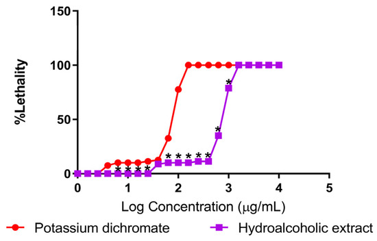
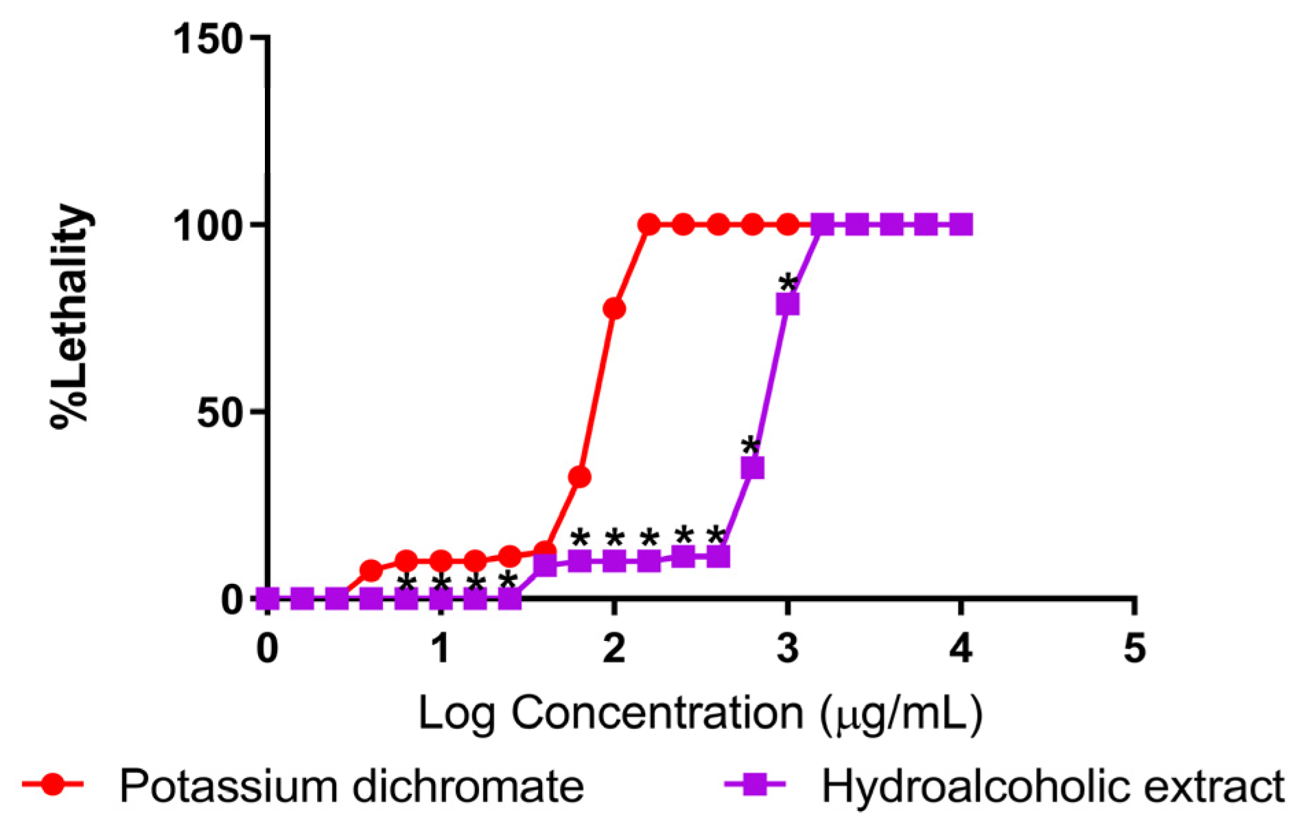
Lethality curves obtained in this assay (Figure 1) showed a concentration-dependent increase in mortality for both the positive control (potassium dichromate) and the hydroalcoholic extract of A. potatorum. As expected, potassium dichromate, a well-known inducer of severe oxidative stress and DNA damage, showed higher toxicity, reaching 50% lethality (LC50) at a much lower concentration (67.485 µg/mL). On the other hand, the hydroalcoholic extract showed a dose–response sigmoidal pattern with an LC50 of 399.369 ± 0.004 µg/mL, which, according to the criteria developed by Meyer et al. (1982) [28], shows a weak toxicity.
Figure 1.
Lethality of A. salina exposed to the hydroalcoholic extract and potassium dichromate. Percentage of lethality at different concentrations (0–10,000 µg/mL) for the hydroalcoholic extract of A. potatorum (LC50 = 399.369 ± 0.004 µg/mL) and potassium dichromate (LC50 = 67.485 ± 0.005 µg/mL). Each point represents the mean ± SEM of n = 5. * p < 0.05 vs. potassium dichromate, determined by two-way ANOVA followed by Bonferroni’s post hoc test.
The plateau observed at higher concentrations suggests a maximal lethal effect consistent with saturation kinetics of toxic stress responses. It is worth emphasizing that A. salina is directly exposed to the compounds in an aqueous environment and, due to its limited detoxification capacity, is particularly susceptible to xenobiotic-induced stress. Therefore, the low-to-moderate toxicity observed suggests that the extract presents a favorable safety profile. The results provide successful validation of A. salina as a preliminary chemical stress screening tool and support subsequent evaluation in mammalian systems to assess tissue-specific stress responses.
2.2. Oral Acute Toxicity
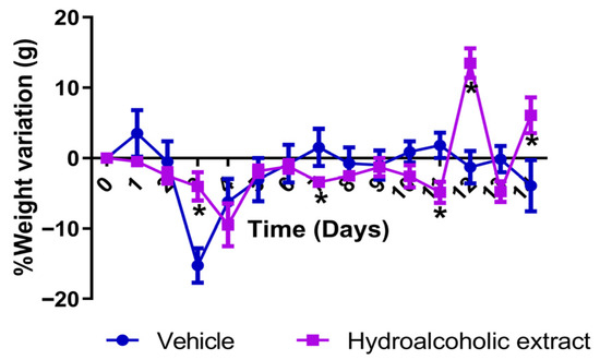
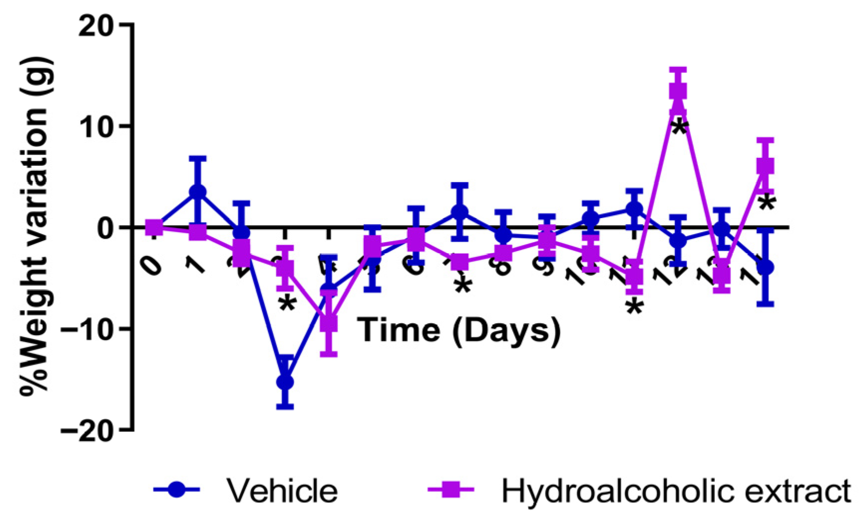
An acute oral toxicity study was conducted in vivo following the OECD 420 fixed-dose guideline [29] to evaluate the systematic safety profile and the capacity of mammalian organisms to endure acute chemical exposure without triggering harmful cellular stress. The hydroalcoholic extract of A. potatorum at a limited dose of 2000 mg/kg in the sighting and in the main studies did not cause mortality, behavioral changes, or clinical signs of systemic toxicity. Moreover, body weight, a sensitive physiological index of metabolic stress and general health status, was stable, and no significant deviation was observed compared with the control group during the 14-day observation period (Figure 2).
Figure 2.
Percentage of weight variation after a single oral administration of the hydroalcoholic extract of A. potatorum (2000 mg/kg) in CD-1 female mice. Each point represents the mean ± SEM of n = 5. * p < 0.05 vs. vehicle control, determined by two-way ANOVA followed by Bonferroni’s post hoc test.
Additionally, serum biochemical parameters were evaluated at the end of the experimental period to assess possible tissue-specific stress and organ damage. Serum transaminases, aspartate aminotransferase (AST) and alanine aminotransferase (ALT), were maintained at basal levels, indicating that no hepatocellular damage or acute hepatic stress occurred. In particular, the treated group showed a marked decrease in total cholesterol and triglyceride levels (Table A1). Hyperlipidemia is strongly related to metabolic stress at the systemic level, mitochondrial dysfunction, and overproduction of reactive oxygen species (ROS) in chronic pathologies. Therefore, the reduction in serum lipid levels is particularly noteworthy, given the traditional use of A. potatorum for diabetes management. Although lipid metabolism was not a primary endpoint of the present study, this observation is consistent with the possibility that the extract exerts broader metabolic effects that warrant future investigation.
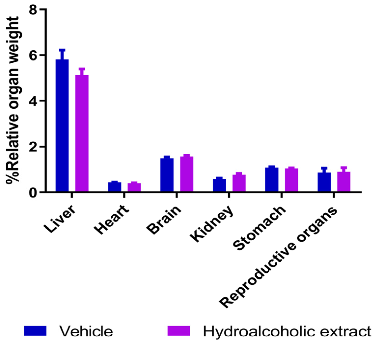
Finally, no morphological abnormalities, alterations, or lesions were observed in the macroscopic examination of vital organs (Figure A1). This was also confirmed by the analysis of the organ weights (Figure A2), where no significant differences were observed between the treated and the control groups. Together, these results indicate that A. potatorum has a high safety margin and does not induce systemic pathological stress responses, providing a strong safety baseline for topical use in compromised chronic microenvironments. Nevertheless, it should be noted that the present study was limited to an acute toxicity evaluation. Additional subacute and chronic toxicity studies will be necessary to further characterize the long-term safety of the extract.
2.3. Effects on Cell Viability
Keratinocytes (HaCaT) and human dermal fibroblasts (HDFn) are critical cellular components of the cutaneous healing cascade that lead to re-epithelialization and extracellular matrix synthesis [18]. It is therefore important to characterize the response of these cell lines to acute chemical exposure to assess whether topical agents induce deleterious cellular stress that could disrupt the physiological healing process.
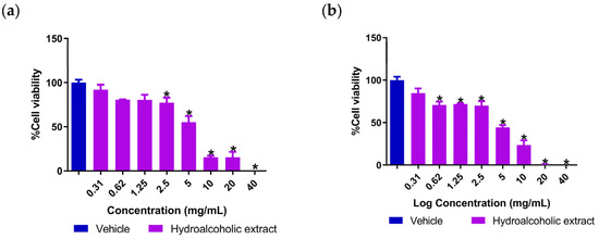
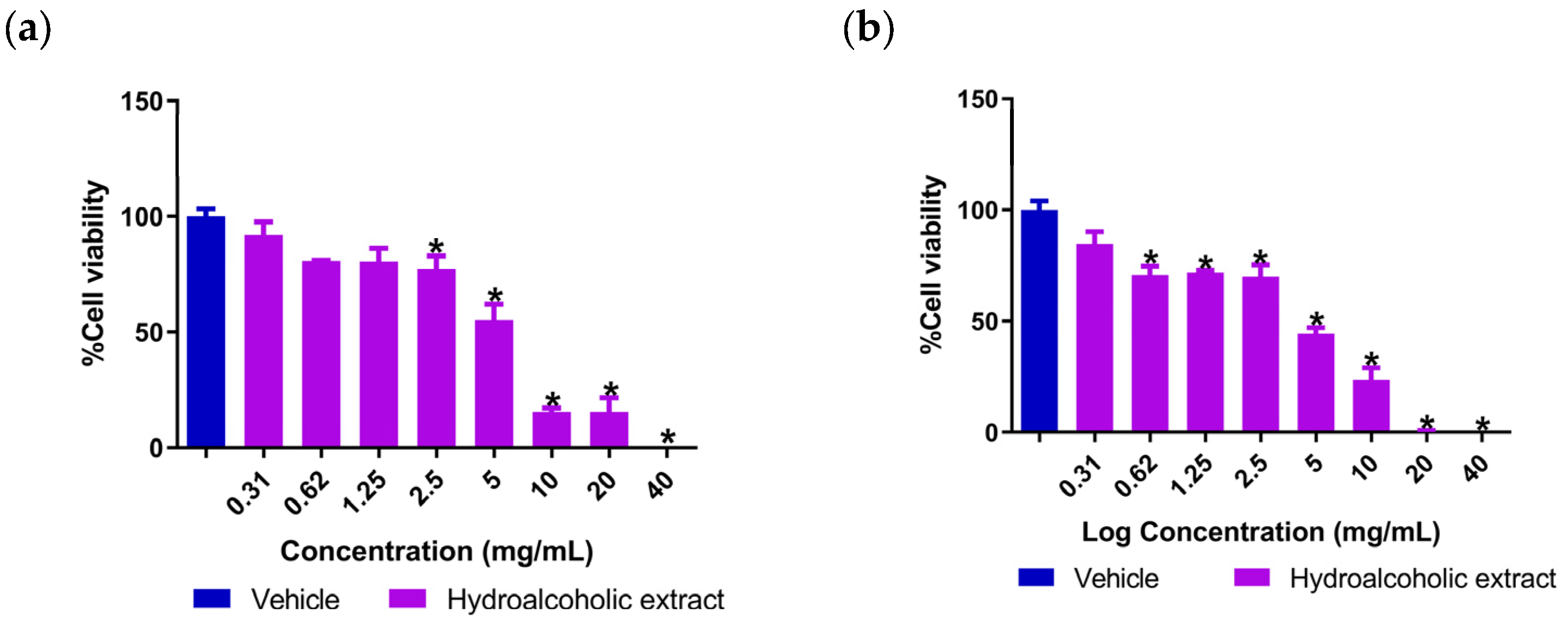
The hydroalcoholic extract of A. potatorum showed a dose-dependent reduction in cell viability in both cell lines (Figure 3). This trend was observed at high concentrations; however, the cell viability cut-off for both cell types was well above 70% at therapeutic concentrations (≤2.5 mg/mL). Cell viability above this 70% threshold, according to standardized toxicological criteria, is considered acceptable for cytocompatibility and suggests that the extract does not induce significant cytotoxic stress in the primary cellular components of the dermis and epidermis.
Figure 3.
Cytotoxicity of the hydroalcoholic extract of A. potatorum on (a) HaCaT cells. (b) HDFn cells. Data represent the mean ± SEM of n = 3. * p < 0.05 vs. vehicle control, determined by one-way ANOVA followed by Dunnett’s post hoc test.
In addition, in translating these in vitro observations to an in vivo model, the physical microenvironment of mammalian skin needs to be considered. Topical formulations in clinical or traditional use interface directly with the stratum corneum, a highly restrictive rate-limiting barrier to xenobiotic penetration [30]. This anatomical barrier prevents naturally living cells of the epidermis and dermis from being exposed to the high, direct concentrations measured in closed in vitro models. Thus, the observed cytocompatibility supports the continued preclinical evaluation of A. potatorum as a topical candidate for wound treatment. Nevertheless, additional studies using reconstructed human skin models, as well as skin irritation or sensitization assays, will be necessary to further characterize its topical safety profile before clinical application.
2.4. Evaluation of Anti-Inflammatory Activity
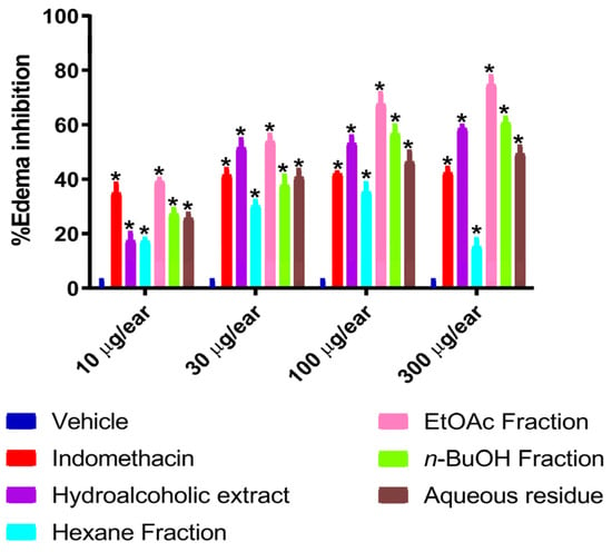
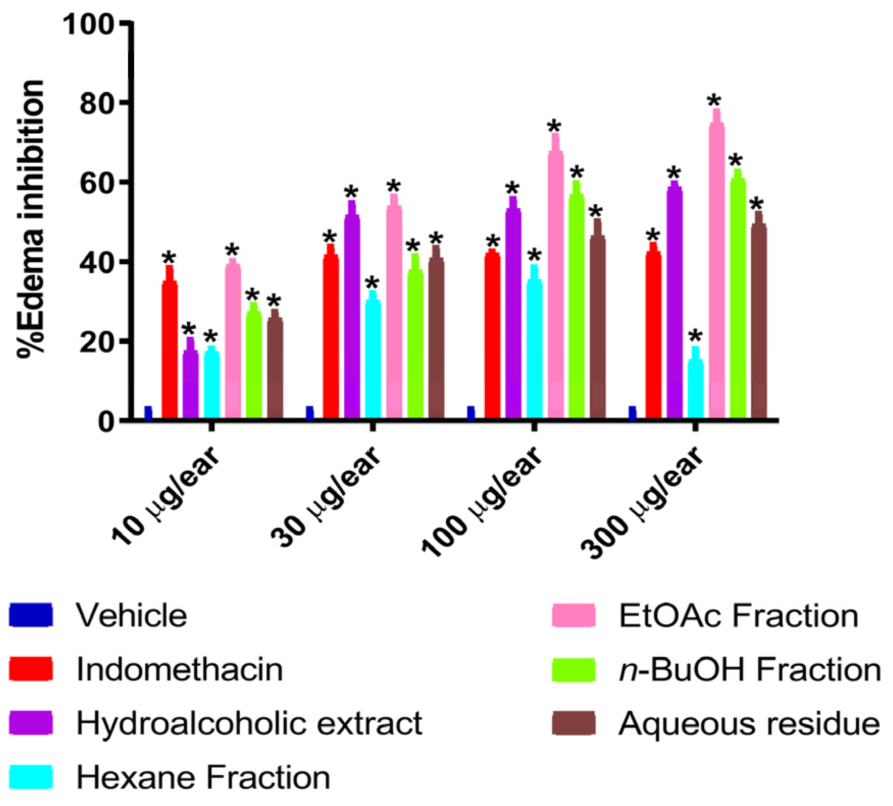
Topical TPA administration induces a strong acute tissue stress response, including rapid vascular permeability, leukocyte infiltration, and a massive burst of pro-inflammatory mediators through PKC/NF-κB signaling [23,31]. Thus, the capacity of exogenous compounds to suppress this chemically induced edema is a powerful approach to identify regulators of acute inflammatory stress pathways. As shown in Figure 4, the hydroalcoholic extract of A. potatorum and its fractions inhibited TPA-induced ear edema in a dose-dependent manner. Interestingly, the crude extract and the EtOAc and n-BuOH fractions showed the greatest protective effect against acute tissue inflammation, surpassing the effect shown by indomethacin, a standard non-steroidal anti-inflammatory drug. The pharmacological activity of these fractions may be explained by the presence of medium- and high-polarity secondary metabolites, such as phenolic compounds and steroidal saponins [32]. Previous studies have shown that these types of compounds possess anti-inflammatory and antioxidant properties through modulation of signaling pathways involved in inflammatory responses. Although the molecular targets responsible for the observed activity were not investigated in this study, these metabolites are likely to contribute, individually or synergistically, to the inhibition of acute inflammation [33,34,35,36].
Figure 4.
Percentage of edema inhibition by indomethacin, hydroalcoholic extract, hexane fraction, EtOAc fraction, n-BuOH fraction, and aqueous residue in the TPA-induced ear edema model in CD-1 male mice. All samples were tested at concentrations of 10, 30, 100, and 300 µg/ear. Data represent the mean ± SEM of n = 5. * p < 0.05 vs. vehicle control, determined by two-way ANOVA followed by Bonferroni’s post hoc test.
On the other hand, the relatively lower potency of the crude extract and weak anti-inflammatory effects of the hexane and aqueous residue fractions may be a consequence of a dilution effect or the presence of non-polar compounds and sugars interfering with the interactions of bioactive molecules and their cellular targets. In light of the present study, the observed anti-inflammatory properties are highly relevant to wound healing. Although acute inflammation is essential for initiating tissue regeneration, uncontrolled or prolonged inflammation can delay the wound-healing process, particularly under diabetic conditions, where sustained inflammatory signaling and oxidative stress impair tissue regeneration. Thus, the ability of A. potatorum to reduce acute inflammation provides pharmacological support for its evaluation in a diabetic wound-healing model and is consistent with its traditional use in the treatment of inflammatory disorders.
2.5. Assessment of Wound-Healing Activity
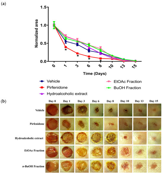
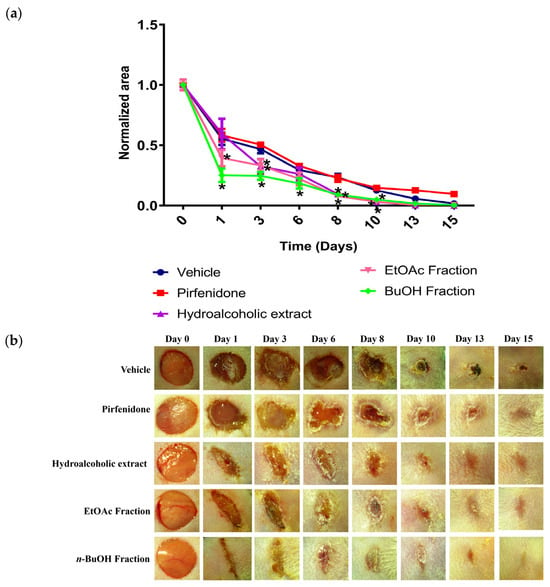
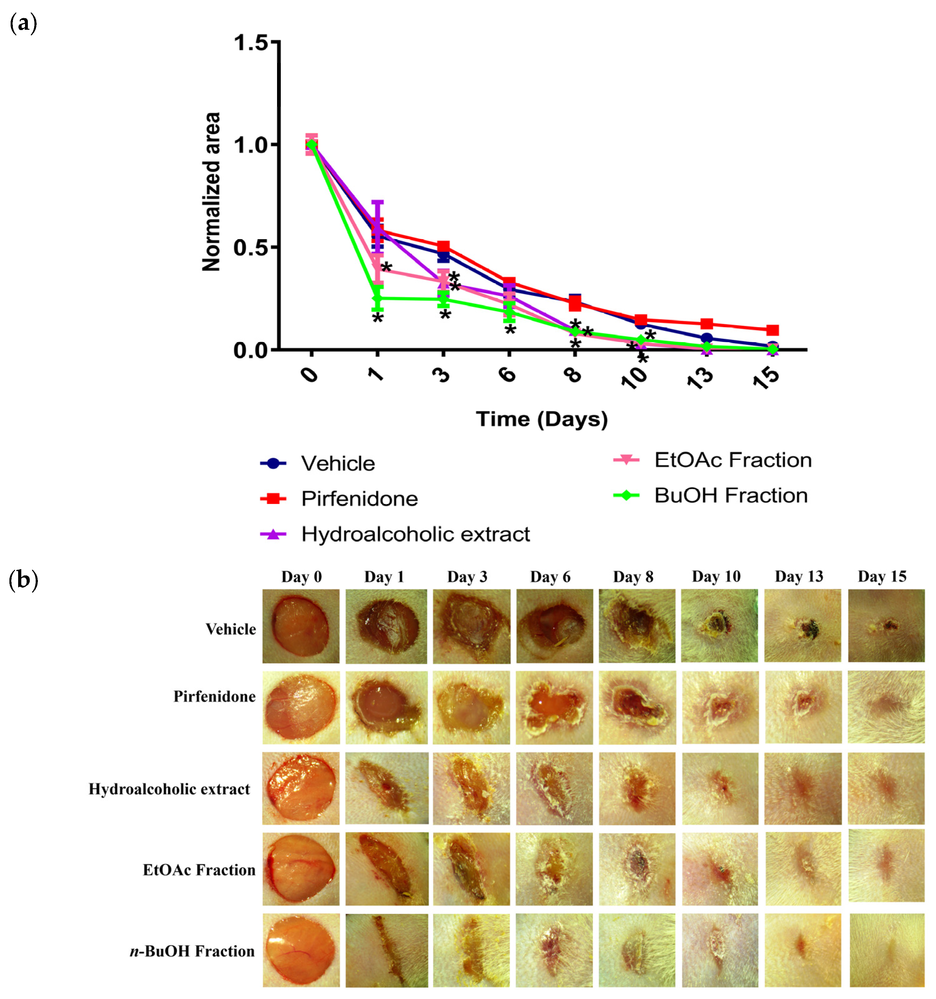
The excisional wound model provides a powerful experimental system to assess the response, adaptation, and orchestration of repair mechanisms by complex tissues to severe, localized mechanical and physiological stress. In normoglycemic mice, the topical treatment with the hydroalcoholic extract of A. potatorum and its fractions resulted in slightly larger normalized wound areas when compared to the vehicle group in the early stages of repair (Figure 5a,b). Although this finding may appear counterintuitive, it is important to consider that the early inflammatory phase is an essential component of normal wound healing. One possible explanation is that the anti-inflammatory activity observed in the TPA-induced edema model may have influenced this initial phase, potentially resulting in a transient delay in the onset of tissue repair. However, because inflammatory mediators, immune cell infiltration, and macrophage polarization were not evaluated in this study, this interpretation remains speculative and should be confirmed in future investigations.
Figure 5.
Wound-healing effect of pirfenidone, hydroalcoholic extract, EtOAc fraction, and n-BuOH fraction in normoglycemic male CD-1 mice. All treatments were tested at a concentration of 10 mg/mL. (a) Kinetic curve of wound closure. Data are presented as normalized areas, and each point represents the mean ± SEM of n = 5. * p< 0.05 vs. vehicle control, determined by two-way ANOVA followed by Bonferroni’s post hoc test. (b) Representative images of wounds taken on days 0, 1, 6, 8, 10, 13, and 15 following wound induction.
In contrast, the reference drug pirfenidone exhibited an early hastened contraction and re-epithelialization, resulting in smaller and cleaner wound beds with less exudate by days 8–10 and almost complete closure by day 15, confirming its role as an established regulator of balanced inflammatory signaling under healthy homeostatic conditions [37].
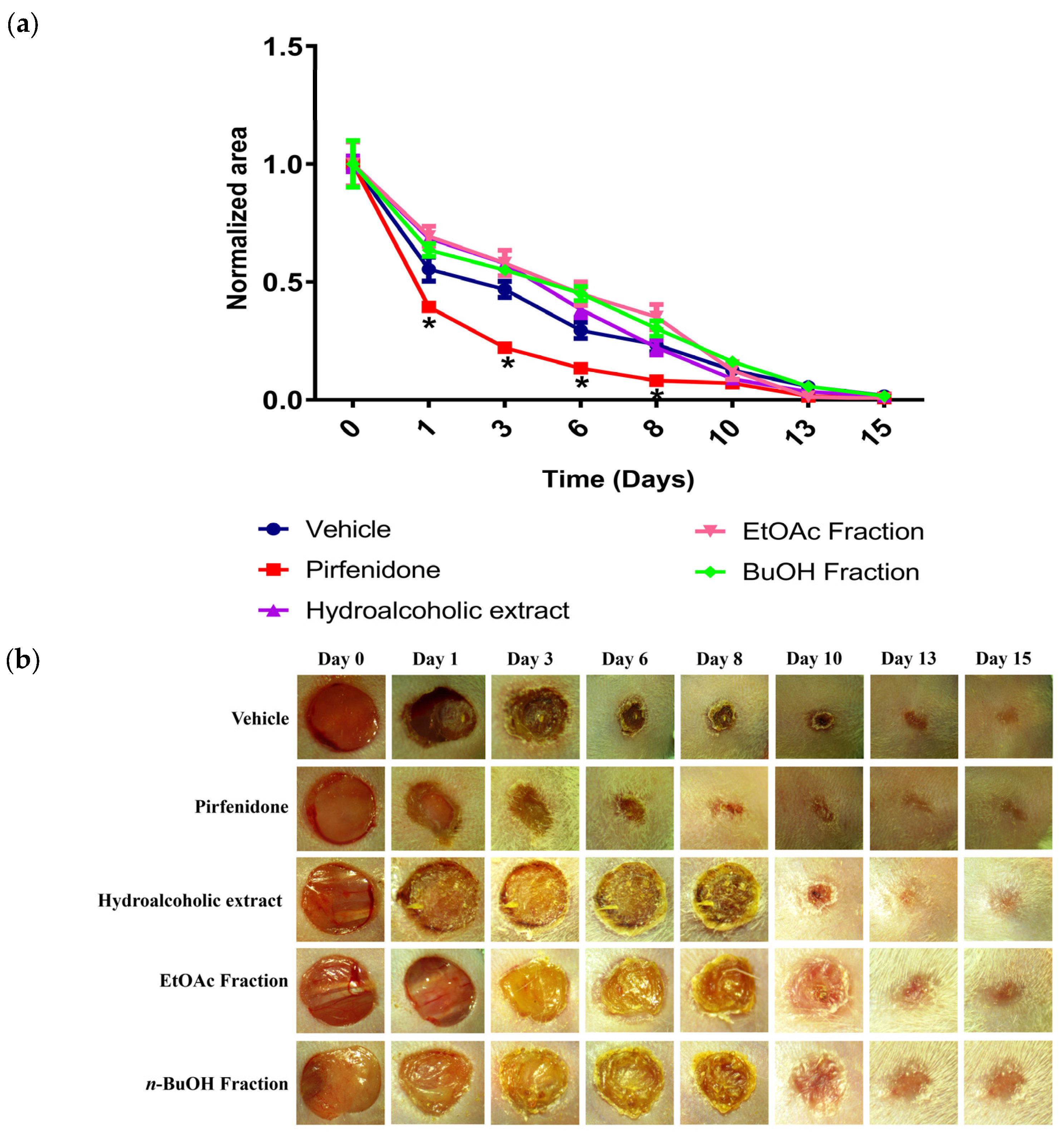
In diabetic mice, the response was clinically relevant and significantly different. The diabetic group treated with the vehicle showed severe delayed and incomplete wound closure, indicative of a microenvironment trapped in a state of persistent pathological inflammation. Interestingly, the A. potatorum hydroalcoholic extract significantly enhanced wound contraction relative to the vehicle and pirfenidone groups. From day 6 onwards, the treated lesions showed a significant reduction in fibrinous crust formation, significantly reduced wound areas, and a highly homogeneous wound bed, with near-complete closure by days 13–15 (Figure 6a,b). Although specialized sensitization assays were not conducted, no gross macroscopic signs of erythema or edema were observed during daily topical observations in both normoglycemic and diabetic mice.
Figure 6.
Wound-healing effect of pirfenidone, hydroalcoholic extract, EtOAc fraction, and n-BuOH fraction in alloxan-induced diabetic male CD-1 mice. All treatments were tested at a concentration of 10 mg/mL. (a) Kinetic curve of wound closure. Data are presented as normalized areas, and each point represents the mean ± SEM of n = 5. * p < 0.05 vs. vehicle control, determined by two-way ANOVA followed by Bonferroni’s post hoc test. (b) Representative images of wounds taken on days 0, 1, 6, 8, 10, 13, and 15 following wound induction.
The biological relevance of this accelerated repair is further emphasized by the chronicity of the experimental design. The mice remained in a sustained hyperglycemic state of more than 200 mg/dL for three months before the injury, allowing the establishment of a chronic hyperglycemic microenvironment that more closely resembles the pathophysiological conditions associated with diabetic wounds than short-term experimental models. Persistent hyperglycemia promotes oxidative stress, chronic inflammation, accumulation of advanced glycation end products, and impaired extracellular matrix remodeling, all of which contribute to delayed tissue repair. Importantly, the markedly different wound closure kinetics observed between normoglycemic and diabetic mice indicate that diabetic wounds do not simply heal more slowly. Rather, they become trapped in a pathological state characterized by persistent inflammatory and oxidative stress, which can ultimately lead to chronic non-healing lesions, increased susceptibility to infection, and tissue necrosis if left unresolved. Therefore, the ability of A. potatorum to improve wound closure under these conditions suggests that its pharmacological activity may be particularly relevant when healing occurs in a chronically altered inflammatory microenvironment.
This pro-healing effect was significantly improved by the EtOAc and n-BuOH fractions. The EtOAc fraction showed rapid early tissue contraction on day 8, while the n-BuOH fraction showed the most progressive, sustained, and better closure pattern throughout the study. The marked reduction in wound area after day 6 suggests a potential role in modulating the inflammatory microenvironment, which may favor the transition toward tissue repair stages, as reported for similar bioactive plant extracts. This phenotypic switch is critical to relieve cellular exhaustion and to reactivate the downstream proliferative mechanisms necessary for structural skin repair.
Surprisingly, pirfenidone did not improve wound closure in diabetic mice and was not statistically different from the vehicle group. Pirfenidone mainly inhibits TGF-β and other profibrotic cytokines. This mechanism prevents excessive scar formation in healthy tissues, but the downregulation of TGF-β signaling under conditions of chronic hyperglycemic stress may further impair the already depleted proliferative capacity of dermal fibroblasts and collagen deposition, which could partially explain its limited efficacy in this experimental model [38].
These differential results between healthy and diabetic mice suggest that A. potatorum may exert a context-dependent immunomodulatory activity. In healthy tissue with already optimized inflammation, aggressive external downregulation of NF-κB signaling transiently disturbs physiological adaptation. In stark contrast, in the chronically stressed diabetic microenvironment, the extract and its active fractions appear to improve tissue repair within the chronically stressed diabetic microenvironment. These effects may be associated with medium- and high-polarity constituents, and although the underlying mechanisms remain to be elucidated, their activity may involve modulation of inflammatory and oxidative stress pathways that are known to be dysregulated during diabetic wound healing. Previous studies have identified steroidal saponins and phenolic compounds in medium- and high-polarity fractions; thus, we hypothesize that these compounds may contribute, individually or synergistically, to the biological activity observed in this study [32,33,34,35,36]. However, future studies evaluating inflammatory mediators, macrophage polarization, oxidative stress biomarkers, and extracellular matrix remodeling will be necessary to clarify their contribution to wound repair. Additionally, it is important to acknowledge that all wound-healing evaluations in this study were conducted using a single fixed concentration of 10 mg/mL for the extract and its fractions. While this represents a validated, ethnopharmacologically supported effective dose for topical repair under chronic hyperglycemic conditions, the absence of a dose–response assessment is a limitation of this preliminary screening. Consequently, formal dose-range studies, multi-dose efficacy profiling, and comprehensive pharmacokinetic analyses will be required during subsequent pharmaceutical development to define optimal clinical dosing. Overall, the contrasting responses observed in normoglycemic and diabetic animals indicate that the biological activity of A. potatorum is highly dependent on the pathological context. Rather than promoting wound healing indiscriminately, the extract—particularly the EtOAc and n-BuOH fractions—was most effective under diabetic conditions, where chronic inflammation and oxidative stress impair normal repair processes. These findings are consistent with the traditional use of A. potatorum and identify this species as a promising source of bioactive compounds for the treatment of impaired wound healing associated with diabetes.
2.6. Limitations and Future Perspectives
The present study provides an integrated preclinical evaluation of the safety, anti-inflammatory activity, and wound-healing potential of A. potatorum under both physiological and diabetic conditions. Nevertheless, several limitations should be acknowledged. First, the wound-healing experiments were conducted using a single topical concentration selected for this initial pharmacological screening, based on previous studies reporting wound-healing effects of plant extracts. Although this approach allowed the identification of biologically active fractions, future studies should establish dose–response relationships and define the optimal therapeutic concentration for formulation development. Second, while the safety assessment included cytocompatibility, A. salina testing, and acute oral toxicity evaluation following OECD Guideline 420, additional investigations addressing subacute and chronic toxicity, as well as topical irritation and skin sensitization, will be required to further characterize the safety profile of the extract.
In addition, the mechanisms underlying the wound-healing activity remain to be elucidated. The present work was designed to evaluate pharmacological efficacy rather than molecular mechanisms; therefore, inflammatory cytokines, macrophage polarization, oxidative stress biomarkers, angiogenesis, collagen deposition, and extracellular matrix remodeling were not investigated. Future studies incorporating these endpoints, together with histological analyses, will help clarify the cellular and molecular pathways involved. Likewise, evaluation using splinted wound models or additional experimental approaches that distinguish wound contraction from re-epithelialization would provide further insight into the repair process. Finally, isolation of the active constituents present in the EtOAc and n-BuOH fractions, followed by pharmacokinetic studies and formulation development, will be essential to support the translational potential of A. potatorum as a therapeutic candidate for impaired wound healing associated with chronic inflammatory and metabolic stress.
3. Materials and Methods
3.1. Reagents
Human dermal neonatal fibroblasts and HaCaT keratinocytes were obtained from ATCC (Manassas, VA, USA). DMEM/F-12, antibiotic–antimycotic, L-glutamine, sodium pyruvate, and PBS were purchased from Gibco (Thermo Fisher Scientific, Waltham, MA, USA). Fetal bovine serum (FBS) was from Biowest (Nuaillé, France), while MTT was acquired from Biomatik (Kitchener, ON, Canada). TPA, indomethacin, alloxan, and standard chemical reagents were purchased from Sigma-Aldrich (St. Louis, MO, USA). Pirfenidone was obtained from Cell Pharmaceuticals (Coyoacán, Ciudad de México, México) and sodium pentobarbital were obtained from Laboratorios Aranda (Querétaro, México), and Artemia salina cysts were sourced from a local supplier (Cuernavaca, Morelos, México).
3.2. Plant Material
The fresh leaves of A. potatorum were collected in Chilapa de Díaz (17°36′21″ N and 97°38′26.8″ W), Oaxaca, Mexico, 1908 m above sea level (masl), on 2 July 2023, by M. Sci. Ramiro Ríos-Gómez. The plant (voucher FEZA-19734) was deposited in the Facultad de Estudios Superiores Zaragoza Herbarium (FEZA), UNAM, Ciudad de México, Mexico.
3.3. Animals
Female (20–30 g) and male (30–35 g) CD-1 mice were used for acute toxicity and pharmacological assays (anti-inflammatory and wound-healing), respectively. Animals were sourced from the Facultad de Medicina, UAEM. All protocols were approved by the local Ethics Committee (CCUAL-UAEM, 005-2018) following Mexican Official Standard NOM-062-ZOO-1999 [39] and international guidelines for animal care, equivalent to EU Directive 2010/63/EU.
Animals were distributed into cages of five animals (n = 5) in each group using simple randomization. The sample size in each experiment (n = 5) was determined based on previous literature and established protocols, also following the principle of the 3Rs.
3.4. Extraction
Fresh A. potatorum leaves (5.8 kg) were cut into small pieces (1 cm2) and extracted with EtOH/H2O (85:15, 10 L) for 72 h; this procedure was repeated three times. The solvent from three extractions was evaporated in vacuo until dryness to yield 186.9 g of a brown residue. An aliquot (180 g) of this residue was subjected to percolation with hexane, then EtOAc, n-BuOH, and water, obtaining ten eluates of 500 mL each solvent. In each case, the solvent was recovered by distillation under reduced pressure to afford 12.5, 2.0, and 13.4 g of residue, respectively.
3.5. A. Salina Lethality Assay
Preliminary toxicity was evaluated using the A. salina lethality assay. Cysts were incubated for 48 h in a 430 mM NaCl solution (pH 8.00 ± 0.003, adjusted with sodium bicarbonate) under continuous aeration. After hatching, 10 nauplii were transferred to each well and exposed to the extract or potassium dichromate (positive control) at concentrations ranging from 10 to 10,000 μg/mL (n = 5) following a logarithmic progression. After 24 h of incubation, the percentage of lethality was recorded and calculated according to Solis et al. (1993) [40].
3.6. Acute Oral Toxicity Assay
Acute oral toxicity was evaluated in female CD-1 mice following the OECD Guideline 420 (fixed-dose procedure) [29]. After a 4 h fast, a sighting study was conducted starting at 300 mg/kg, followed by 2000 mg/kg. As no mortality or clinical signs were observed, a main study was performed where groups (n = 5) received a single oral dose of 2000 mg/kg of the extract or vehicle (water, 10 mL/kg). Animals were monitored for 14 days to record behavioral, autonomic, and systemic clinical signs. Finally, mice were euthanized (sodium pentobarbital, 65 mg/kg, i.p.) for necropsy; vital organs were excised, rinsed, and weighed to assess gross pathological changes.
3.7. Cell Viability Assay
Human dermal neonatal fibroblasts (HDFn) were cultured in DMEM/F-12 supplemented with 10% FBS, 1% antibiotic–antimycotic, 2 mM L-glutamine, and 1% sodium pyruvate at 37 °C and 5% CO2. For the MTT assay, cells were seeded in 96-well plates (5 × 10³ cells per well) and allowed to attach for 24 h. Cells were then treated with the extract (1.56–100 mg/mL) or vehicle (DMSO) for 72 h. After treatment, cells were washed, incubated with 10 μL of MTT (5 mg/mL) for 4 h, and the resulting formazan crystals were dissolved in DMSO. Absorbance was obtained, and the percentage of cell viability was determined as previously described [41].
3.8. In Vivo Anti-Inflammatory Assay
The TPA-induced ear edema in mice was performed as described by García-Argáez et al. (2000) [42]. Mice (n = 5 per group) were sedated with sodium pentobarbital (i.p.) before topical application of TPA (2.5 μg/ear in ethanol) to the right ear. Ten minutes later, the extract or fractions (10, 30, 100, and 300 μg/ear), indomethacin (positive control) (10, 30, 100, and 300 μg/ear), or vehicle (10% DMSO) was applied topically. After 4 h, animals were euthanized, and 6 mm discs were weighed. Edema inhibition was calculated as the weight difference (ΔW) between the treated and control groups.
3.9. In Vivo Wound-Healing Model
Wound healing was assessed in normoglycemic and alloxan-induced diabetic mice using an excision model [32]. Diabetic mice were induced with a single dose of alloxan (200 mg/kg, i.p.). Animals were considered diabetic if basal blood glucose levels remained >200 mg/dL from the second week post-induction. Only subjects that reached inclusion criteria (basal blood glucose >200 mg/dL) for the diabetic group were included. To simulate a chronic diabetic state, these animals were maintained under this condition for three months before initiating the wound-healing experiment.
Under sedation, a 6 mm circular wound was created on the shaved dorsal area using a biopsy punch. Animals (n = 5) received daily topical applications (10 μL) of the extracts or fractions (10 mg/mL), pirfenidone (positive control; 10 mg/mL), or distilled water (vehicle). Digital photographs were captured periodically until wound closure using a stereomicroscope, and wound areas were quantified using ImageJ 1.54r software.
3.10. Statistical Analysis
Data were analyzed using GraphPad Prism 5.0. Differences between groups were determined by one-way ANOVA followed by Dunnett’s post hoc test (cell viability and organ weights) or two-way ANOVA followed by Bonferroni’s post hoc test (A. salina, anti-inflammatory and wound-healing assays). Results were considered statistically significant at p < 0.05.
4. Conclusions
The present study provides a comprehensive preclinical evaluation of the hydroalcoholic extract and fractions of A. potatorum, integrating safety assessment with experimental models that reproduce distinct biological stress conditions associated with tissue injury. The extract exhibited a favorable acute safety profile across the experimental models employed, including low toxicity in A. salina, absence of acute oral toxicity at the OECD Guideline 420 limit dose, and acceptable cytocompatibility in human keratinocytes and dermal fibroblasts.
The hydroalcoholic extract, and particularly the EtOAc and n-BuOH fractions, effectively attenuated acute inflammatory responses in the TPA-induced ear edema model and preferentially improved wound healing under chronic diabetic conditions, where persistent inflammatory and oxidative stress are known to impair tissue repair. These findings suggest that the biological activity of A. potatorum is closely associated with a pathological microenvironment characterized by dysregulated stress responses rather than with physiological wound healing.
Although the molecular mechanisms responsible for these effects remain to be elucidated, the present results support the traditional medicinal use of A. potatorum and identify this species as a promising source of bioactive compounds for the development of therapeutic strategies aimed at modulating inflammation and improving tissue repair under chronic stress conditions. Future studies should focus on elucidating the underlying molecular mechanisms, establishing dose–response relationships, and further characterizing the long-term and topical safety profile of the extract.
Author Contributions
Conceptualization, M.A.D.-R., M.Y.R. and A.B.A.-G.; methodology, M.A.D.-R.; software, M.A.D.-R.; validation, M.Y.R. and A.B.A.-G.; formal analysis, M.A.D.-R., M.Y.R. and A.B.A.-G.; investigation, M.A.D.-R., M.Y.R. and A.B.A.-G.; resources, R.R.-G., J.-J.A.-F., M.Y.R. and A.B.A.-G.; data curation, M.A.D.-R.; writing—original draft preparation, M.A.D.-R., M.Y.R. and A.B.A.-G.; writing—review and editing, M.A.D.-R., R.R.-G., J.-J.A.-F., M.Y.R. and A.B.A.-G.; visualization, M.Y.R. and A.B.A.-G.; supervision, M.Y.R. and A.B.A.-G.; project administration, M.Y.R. and A.B.A.-G. All authors have read and agreed to the published version of the manuscript.
Funding
This research did not receive any specific grant from funding agencies in the public, commercial, or not-for-profit sectors.
Institutional Review Board Statement
All protocols were approved by the local Ethics Committee (CCUAL-UAEM, 005-2018) following Mexican Official Standard NOM-062-ZOO-1999 [39] and international guidelines for animal care, EU Directive 2010/63/EU (date of approval: 29 May 2026).
Informed Consent Statement
Not applicable.
Data Availability Statement
All data generated and analyzed in the current study are included in the article and Appendix A. Further inquiries can be directed to the corresponding author.
Acknowledgments
The authors are thankful to Elizabeth Negrete, Elideth Mendez Tendero, and Eduardo Lira Díaz for their technical assistance.
Conflicts of Interest
The authors declare that they have no conflicts of interest.
Appendix A
Table A1.
Biochemical parameters of mice after a single oral administration of hydroalcoholic extract of A. potatorum (2000 mg/Kg) in female CD-1 mice.
Figure A1.
Macroscopic analysis of organs from mice treated with the hydroalcoholic extract and vehicle control.
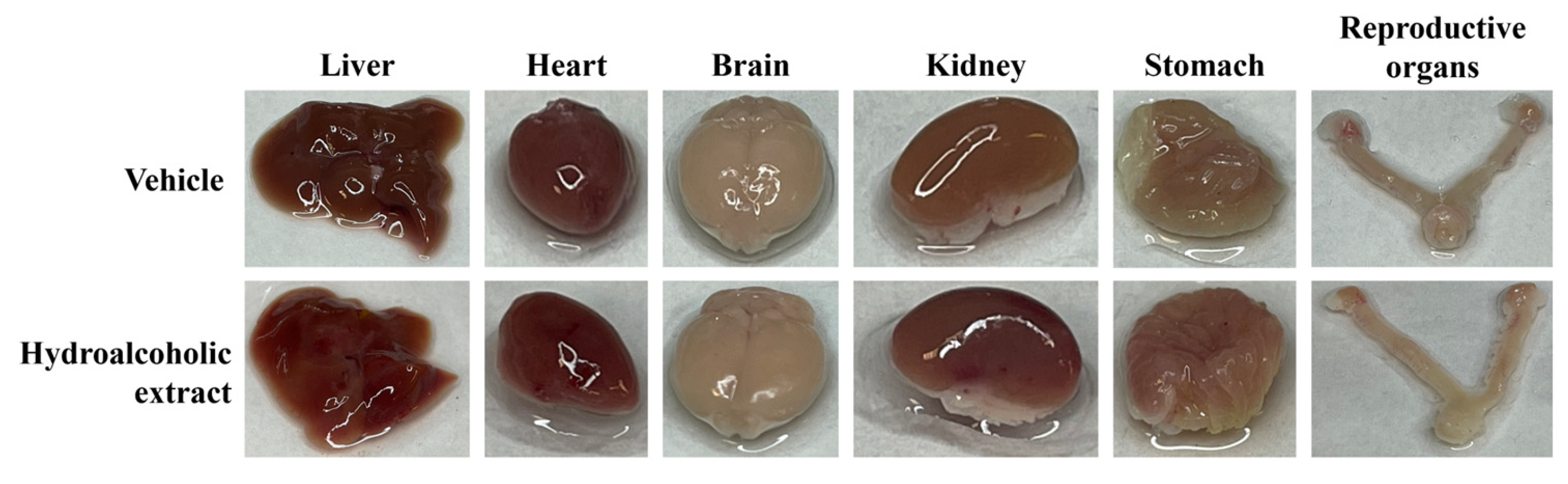
Figure A2.
Percentage of relative organ weight (liver, heart, brain, kidneys, stomach, and reproductive organs) after a single oral administration of hydroalcoholic extract at a dose of 2000 mg/kg in female CD-1 mice. Each group represents the mean ± SEM of n = 5. p< 0.05 vs. vehicle control, determined by two-way ANOVA followed by Bonferroni’s post hoc test.
References
- Peña, O.A.; Martin, P. Cellular and molecular mechanisms of skin wound healing. Nat. Rev. Mol. Cell Biol. 2024, 25, 599–616. [Google Scholar] [CrossRef] [Scilit] [PubMed]
- Esad, M.; Dimov, I.; Choneva, M.; Popova, M.; Kokova, V.; Apostolova, E.; Bivolarska, A. Molecular Mechanisms of Wound Healing: The Role of Medicinal Plants. Life 2025, 15, 1748. [Google Scholar] [CrossRef] [Scilit] [PubMed]
- Cheng, X.C.; Tong, W.Z.; Rui, W.; Feng, Z.; Shuai, H.; Zhe, W. Single-cell sequencing technology in skin wound healing. Burn. Trauma 2024, 12, tkae043. [Google Scholar] [CrossRef] [Scilit] [PubMed]
- Yuan, M.; Liu, Y.; Peng, X.; Li, Z.; Lei, M. Chronic Wound Healing: Research Advances from Pathological Mechanisms to Natural Herbal Active Ingredients and Material Delivery Systems. Molecules 2026, 31, 1024. [Google Scholar] [CrossRef] [Scilit] [PubMed]
- Gao, J.; Zhu, D.; Wang, J.; Khan, M.I.U.; Zhang, A.; Wen, X.; Li, Q.; Jin, P.; Shen, C. Macrophage efferocytosis promotes inflammation resolution and accelerates wound healing. Commun. Biol. 2026, 9, 613. [Google Scholar] [CrossRef] [Scilit] [PubMed]
- Chen, Y.; Li, H.; He, W. Mitochondria-mediated inflammation and diabetic wound healing: Mechanisms and therapeutic strategies. Front. Pharmacol. 2026, 17, 1786693. [Google Scholar] [CrossRef] [Scilit] [PubMed]
- Mirmiranpour, H.; Arden, C. The Interplay Between Reactive Oxygen Species, Glucose Metabolism and NF-κB in the Pathogenesis of Type 2 Diabetes. Diabetology 2026, 7, 53. [Google Scholar] [CrossRef] [Scilit]
- Pranantyo, D.; Yeo, C.K.; Wu, Y.; Fan, C.; Xu, X.; Yip, Y.S.; Vos, M.I.G.; Mahadevegowda, S.H.; Lim, P.L.K.; Yang, L.; et al. Hydrogel dressings with intrinsic antibiofilm and antioxidative dual functionalities accelerate infected diabetic wound healing. Nat. Commun. 2024, 15, 954, Correction in Nat. Commun. 2025, 16, 9047. https://doi.org/10.1038/s41467-025-64329-7. [Google Scholar] [CrossRef] [Scilit] [PubMed]
- Qi, X.; Li, Y.; Xiang, Y.; Chen, Y.; Shi, Y.; Ge, X.; Zeng, B.; Shen, J. Hyperthermia-enhanced immunoregulation hydrogel for oxygenation and ROS neutralization in diabetic foot ulcers. Cell Biomater. 2025, 1, 100020. [Google Scholar] [CrossRef] [Scilit]
- Qi, X.; Xiang, Y.; Li, Y.; Wang, J.; Chen, Y.; Lan, Y.; Liu, J.; Shen, J. An ATP-activated spatiotemporally controlled hydrogel prodrug system for treating multidrug-resistant bacteria-infected pressure ulcers. Bioact. Mater. 2024, 45, 301–321. [Google Scholar] [CrossRef] [Scilit] [PubMed]
- Zhang, W.; Hu, J.; Wu, H.; Lin, X.; Cai, L. Stimuli-responsive hydrogel dressing for wound healing. APL Mater. 2025, 13, 010601. [Google Scholar] [CrossRef] [Scilit]
- Criollo-Mendoza, M.S.; Contreras-Angulo, L.A.; Leyva-López, N.; Gutiérrez-Grijalva, E.P.; Jiménez-Ortega, L.A.; Heredia, J.B. Wound Healing Properties of Natural Products: Mechanisms of Action. Molecules 2023, 28, 598. [Google Scholar] [CrossRef] [Scilit] [PubMed]
- Herman, A.; Herman, A.P. Herbal Products in Postsurgical Wound Healing-Incision, Excision and Dead Space Wound Models. Planta Med. 2020, 86, 742–748. [Google Scholar] [CrossRef] [Scilit] [PubMed]
- World Flora Online Plant List. Agave potatorum. Zucc. 2025. Available online: https://wfoplantlist.org/taxon/wfo-0000754697-2025-12 (accessed on 15 April 2026).
- López-Serrano, P.M.; Hernández-Ramos, A.; Méndez-González, J.; Martínez-Salvador, M.; Aguirre-Calderón, O.; Vargas Larreta, B.; Corral Rivas, J.J. Mejores Prácticas de Manejo y Ecuaciones Alométricas de Biomasa Agave potatorum en el Estado de Oaxaca; Proyecto: 2017-4-292674; CONAFOR-CONACYT: Mexico City, Mexico, 2021.
- Galindo-Vargas, N.; García-Sánchez, E.; Juárez, E.; Santiago-Garcia, P.A.; Soto, D. First Approach to Unveiling the Antidiabetic Potențial of Agave potatorum: α-Glucosidase Inhibition and Phytochemistry. J. Mex. Chem. Soc. 2025, 69, 496–504. [Google Scholar] [CrossRef] [Scilit]
- Cherpelis, B.S.; Fenske, N.A. Purpuric irritant contact dermatitis induced by Agave americana. Cutis 2000, 66, 287–288. [Google Scholar] [PubMed]
- Salinas, M.L.; Ogura, T.; Soffchi, L. Irritant contact dermatitis caused by needle-like calcium oxalate crystals, raphides, in Agave tequilana among workers in tequila distilleries and Agave plantations. Contact Dermat. 2001, 44, 94–96. [Google Scholar] [CrossRef] [Scilit] [PubMed]
- Hardwick, R.N.; Betts, C.J.; Whritenour, J.; Sura, R.; Thamsen, M.; Kaufman, E.H.; Fabre, K. Drug-induced skin toxicity: Gaps in preclinical testing cascade as opportunities for complex in vitro models and assays. Lab Chip 2020, 20, 199–214. [Google Scholar] [CrossRef] [Scilit] [PubMed]
- Quinn, B. Preparation and Maintenance of Live Tissues and Primary Cultures for Toxicity Studies. In Biochemical Ecotoxicology Principles and Methods; Gagné, F., Ed.; Academic Press: Cambridge, MA, USA, 2014; pp. 33–47. [Google Scholar] [CrossRef] [Scilit]
- Lakshmanan, M. Preclinical Toxicity Studies. In Introduction to Basics of Pharmacology and Toxicology; Lakshmanan, M., Shewade, D.G., Raj, G.M., Eds.; Springer: Singapore, 2022; pp. 625–648. [Google Scholar] [CrossRef] [Scilit]
- Denny, K.H.; Stewart, C.W. Acute, Subacute, Subchronic, and Chronic General Toxicity Testing for Preclinical Drug Development. In A Comprehensive Guide to Toxicology in Nonclinical Drug Development; Faqi, A.S., Ed.; ASF Scientific Solutions: Portage, MI, USA, 2024; pp. 109–127. [Google Scholar] [CrossRef] [Scilit]
- Murakawa, M.; Yamaoka, K.; Tanaka, Y.; Fukuda, Y. Involvement of tumor necrosis factor (TNF)-alpha in phorbol ester 12-O-tetradecanoylphorbol-13-acetate (TPA)-induced skin edema in mice. Biochem. Pharmacol. 2006, 71, 1331–1336. [Google Scholar] [CrossRef] [Scilit] [PubMed]
- Karas, R.A.; Alexeree, S.; Elsayed, H.; Attia, Y.A. Assessment of wound healing activity in diabetic mice treated with a novel therapeutic combination of selenium nanoparticles and platelets rich plasma. Sci. Rep. 2024, 14, 5346. [Google Scholar] [CrossRef] [Scilit] [PubMed]
- Huynh, P.; Phie, J.; Krishna, S.M.; Golledge, J. Systematic review and meta-analysis of mouse models of diabetes-associated ulcers. BMJ Open Diabetes Res. Care 2020, 8, e000982. [Google Scholar] [CrossRef] [Scilit] [PubMed]
- Okumu, M.O.; Mbaria, J.M.; Gikunju, J.K.; Mbuthia, P.G.; Madadi, V.O.; Ochola, F.O.; Jepkorir, M.S. Artemia salina as an animal model for the preliminary evaluation of snake venom-induced toxicity. Toxicon X 2021, 12, 100082. [Google Scholar] [CrossRef] [Scilit] [PubMed]
- Olmedo, D.A.; Vasquez, Y.; Morán, J.A.; De León, E.G.; Caballero-George, C.; Solís, P.N. Understanding the Artemia salina (Brine Shrimp) Test: Pharmacological Significance and Global Impact. Comb. Chem. High Throughput Screen. 2024, 27, 545–554. [Google Scholar] [CrossRef] [Scilit] [PubMed]
- Meyer, B.N.; Ferrigni, N.R.; Putnam, J.E.; Jacobsen, L.B.; Nichols, D.E.; McLaughlin, J.L. Brine shrimp: A convenient general bioassay for active plant constituents. Planta Med. 1982, 45, 31–34. [Google Scholar] [CrossRef] [Scilit] [PubMed]
- OECD. Test No. 420: Acute Oral Toxicity—Fixed Dose Procedure; OECD: Paris, France, 2002. [Google Scholar] [CrossRef] [Scilit]
- Roberts, M.S.; Cheruvu, H.S.; Mangion, S.E.; Alinaghi, A.; Benson, H.A.E.; Mohammed, Y.; Holmes, A.; van der Hoek, J.; Pastore, M.; Grice, J.E. Topical drug delivery: History, percutaneous absorption, and product development. Adv. Drug Deliv. Rev. 2021, 177, 113929. [Google Scholar] [CrossRef] [Scilit] [PubMed]
- Sankhe, R.; Ranganath, S.; Pai, K.; Kishore, A. Tumor supression through modulation of neprilysin signaling: A comprehensive review. Eur. J. Pharmacol. 2021, 891, 173727. [Google Scholar] [CrossRef] [Scilit] [PubMed]
- Aguilar-Guadarrama, A.B.; Díaz-Román, M.A.; Osorio-García, M.; Déciga-Campos, M.; Rios, M.Y. Chemical Constituents from Agave applanata and Its Antihyperglycemic, Anti-inflammatory, and Antimicrobial Activities Associated with Its Tissue Repair Capability. Planta Med. 2024, 90, 397–410. [Google Scholar] [CrossRef] [Scilit] [PubMed]
- Misra, A.K.; Varma, S.K.; Kumar, R. Anti-inflammatory Effect of an Extract of Agave americana on Experimental Animals. Pharmacogn. Res. 2018, 10, 104–108. [Google Scholar] [CrossRef] [Scilit]
- Bermúdez-Bazán, M.; Castillo-Herrera, G.A.; Urias-Silvas, J.E.; Escobedo-Reyes, A.; Estarrón-Espinosa, M. Hunting Bioactive Molecules from the Agave Genus: An Update on Extraction and Biological Potential. Molecules 2021, 26, 6789. [Google Scholar] [CrossRef] [Scilit] [PubMed]
- Olán-Jiménez, K.A.; Cruz-Rodríguez, R.I.; Couder-García, B.D.C.; Jacobo-Herrera, N.; Ruiz-Lau, N.; Hernández-Cruz, M.d.C.; Ruíz-Valdiviezo, V.M. Antibacterial and Wound Healing Activity In Vitro of Individual and Combined Extracts of Tagetes nelsonii Greenm, Agave americana and Aloe vera. Sci. Pharm. 2024, 92, 41. [Google Scholar] [CrossRef] [Scilit]
- Xiong, B.; Wang, H.; Song, Y.X.; Lan, W.Y.; Li, J.; Wang, F. Natural saponins and macrophage polarization: Mechanistic insights and therapeutic perspectives in disease management. Front. Pharmacol. 2025, 16, 1584035. [Google Scholar] [CrossRef] [Scilit] [PubMed]
- Cho, M.E.; Kopp, J.B. Pirfenidone: An anti-fibrotic therapy for progressive kidney disease. Expert Opin. Investig. Drugs 2010, 19, 275–283. [Google Scholar] [CrossRef] [Scilit] [PubMed]
- Ballester, B.; Milara, J.; Cortijo, J. Pirfenidone anti-fibrotic effects are partially mediated by the inhibition of MUC1 bioactivation. Oncotarget 2020, 11, 1306–1320. [Google Scholar] [CrossRef] [Scilit] [PubMed]
- NOM-062-ZOO-1999; Especificaciones Técnicas para la Producción, Cuidado y uso de los Animales de Laboratorio. Diario Oficial de la Federación; SAGARPA: Ciudad de México, México, 2001.
- Solis, P.N.; Wright, C.W.; Anderson, M.M.; Gupta, M.P.; Phillipson, J.D. A Microwell Cytotoxicity Assay using Artemia salina (Brine Shrimp). Planta Med. 1993, 59, 250–252. [Google Scholar] [CrossRef] [Scilit] [PubMed]
- Lira-Díaz, E.; Cruz-Márquez, R.; González-Pedroza, M.G.; González-Pérez, O.; Morales-Luckie, R.A.; Acevedo-Fernández, J.J. Silver Nanoparticles Based on Annona muricata Peel Reduce Cell Viability in Medulloblastoma and Neuroblastoma Cell Lines. J. Nanotechnol. 2024, 2024, 2263514. [Google Scholar] [CrossRef] [Scilit]
- García-Argáez, A.; Ramírez Apan, T.; Delgado, H.; Velázquez, G.; Martínez-Vázquez, M. Anti-Inflammatory Activity of Coumarins from Decatropis bicolor on TPA Ear Mice Model. Planta Med. 2000, 66, 279–281. [Google Scholar] [CrossRef] [Scilit] [PubMed]
Disclaimer/Publisher’s Note: The statements, opinions and data contained in all publications are solely those of the individual author(s) and contributor(s) and not of MDPI and/or the editor(s). MDPI and/or the editor(s) disclaim responsibility for any injury to people or property resulting from any ideas, methods, instructions or products referred to in the content. |
© 2026 by the authors. Licensee MDPI, Basel, Switzerland. This article is an open access article distributed under the terms and conditions of the Creative Commons Attribution (CC BY) license.