


Abstract
Background: Injuries to the knee joint usually involve ligament or tendon structures. Most of these injuries are traumatic in origin. To better understand the structures and the associated failure mechanisms, a specific understanding of the biomechanical properties of the individual structures is necessary. Objectives: The aim of this study was to biomechanically analyze the tendon and ligament structures around the knee joint in order to acquire tissue-specific mechanical characteristics of these anatomical structures. Methods: Five knee joints (fresh frozen) from three donors were thawed over the course of three days. The respective ligaments and tendons of the knee were separated, removed and cut into a dogbone shape for uniaxial tensile tests. The specimens were clamped in the testing machine using 3D-printed specimen holders. Results: A total of 57 samples from 15 different ligaments and tendons were successfully tested. The tensile strengths were in the range of 0.67–67.99 MPa (patellar tendon vs. biceps femoris tendon) and the moduli of elasticity between 2.1 and 316.9 MPa. A range of 6.2–47.2% was determined for the failure strains. Similar fluctuation ranges were also found in intra-individual comparisons. Conclusions: The results show a clear tissue-specific variation in the strength and elastic modulus.
1. Introduction
Injuries to the knee joint usually affect ligament or tendon structures. The numerous ligaments and tendons of the lower limb fulfill several basic functions [1]. In addition to the tensile strapping effect of the iliotibial tract, for example, they have joint-stabilizing functions, such as the anterior and posterior cruciate ligaments. Additionally, tendons of muscles crossing the knee joint are essential for lower extremity movement, such as the semimembranosus and semitendinosus tendons. Biomechanically, the knee fulfills a central role as a hinge joint. It not only serves as the attachment point for some of the aforementioned ligamentous structures but also acts as a pulley for the quadriceps tendon and patellar tendon [2]. This highlights that a detailed examination of the associated ligamentous structures is of central importance for the biomechanical understanding of the knee joint. Uniaxial tensile testing, which has been used for many years for the determination of mechanical parameters of ligamentous tissue, may seem like the obvious choice [3,4,5]. However, there are many different protocols for tissue collection, storage and sample cutting, all of which have an influence on the parameters collected [6]. Testing of fixed soft tissue samples, for example, revealed a fundamental change in the elastic modulus [7,8,9]. There are different research results regarding the influence of freezing and thawing processes on the mechanical properties of soft tissue. According to a study from Moon et al. on rabbit tendons, single freezing is unproblematic for small cell-free tissues [10], while repeated freezing also causes a change in the mechanical properties [11]. The state of the art for uniaxial tensile testing of cell-poor ligamentous tissue is therefore the use of fresh or fresh-frozen tissue. Furthermore, studies are often focused on individual tissues, as, for example, in Wright et al. [12]. Since no cases were found in the literature in which complete knees were exposed and mechanical parameters were measured in as many knee-associated ligaments and tendons as possible, this was to be carried out as an example in this study. The main objective of this exploratory study was to determine how many knee ligaments could be successfully harvested during dissection of the complete human knees. At the same time, another objective was to characterize as many of these tissues as possible in terms of their biomechanical properties.
2. Materials and Methods
2.1. Specimen Preparation Technique
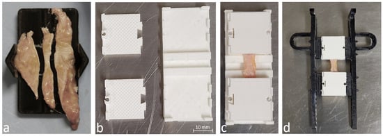
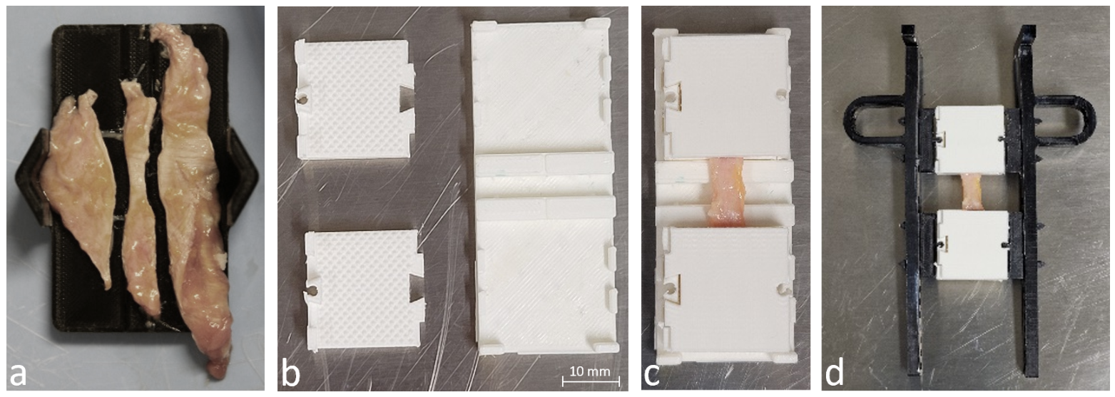
A total of 70 samples were prepared from 5 knees from 3 body donors (mean donor age was 88.3 years). The specimens were stored at −80 °C (fresh-frozen) and thawed to 5 °C in the refrigerator over a period of three days. On the day a knee was examined, all tendons were dissected and stored in a 0.9% saline solution between each step to prevent dehydration. After removal from the knee joint, the ligaments and tendons were prepared according to the protocol of Scholze et al. [13,14] (Figure 1).
Figure 1.
Preparation for mechanical testing. Panel (a): Sample cutting to dogbone shape. Panels (b,c): Placement of the cut specimens in the clamping device. Panel (d): Clamped sample with handles.
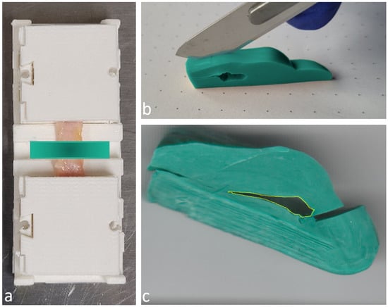
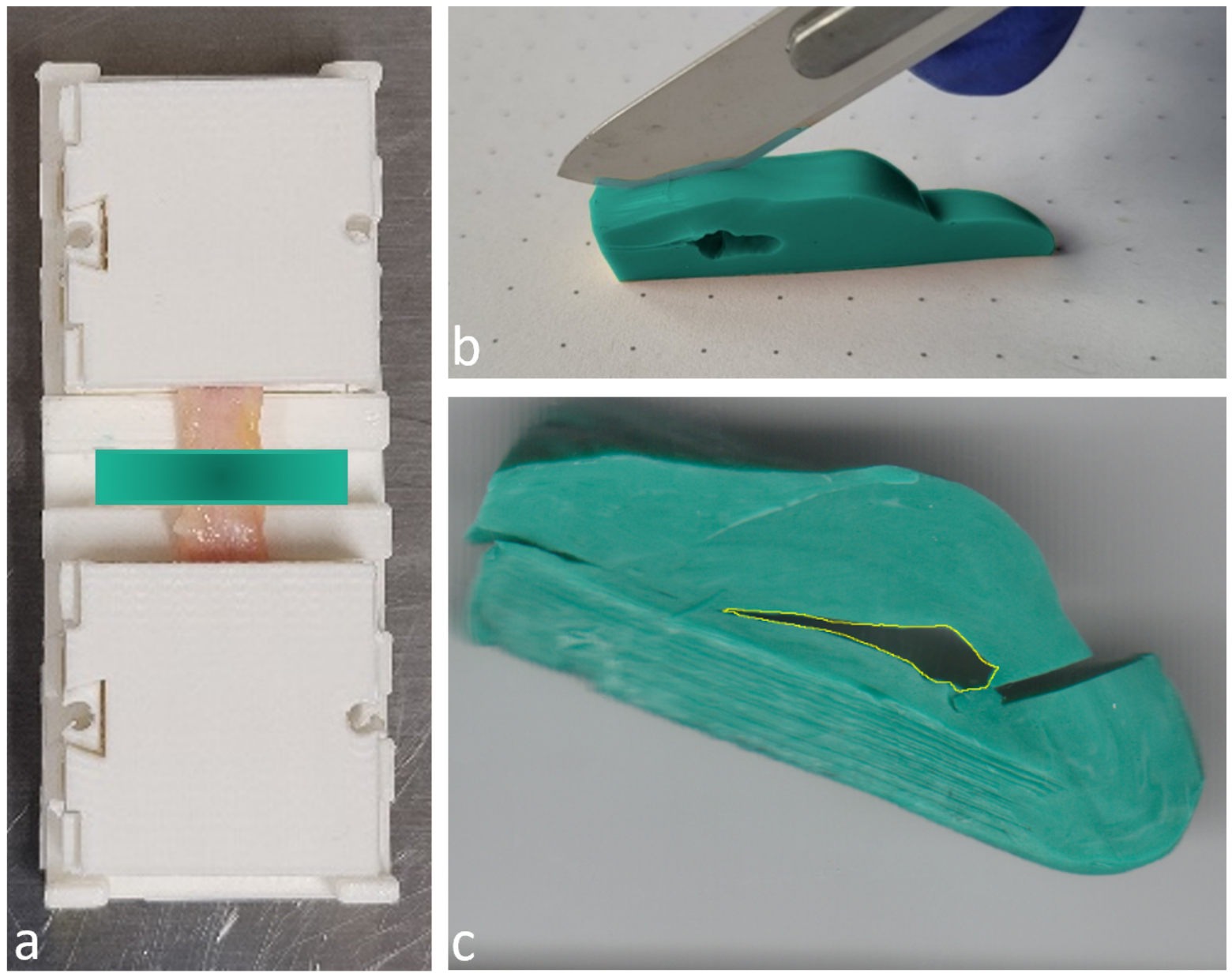
The tissue samples were spread flat on the provided cutting device, and the specified dogbone shape was cut using a scalpel. The cross-section itself was determined by molding the test area with a two-component silicone rubber (HS-A Silicone Light Body, VPS Hydro, regular set|Henry Schein Dental Deutschland GmbH, Langen, Germany) after clamping in the 3D-printed clamps. The molded cross-sections were digitized using a flatbed scanner (CanoScan LiDe 90|Canon Deutschland GmbH, Krefeld, Germany) and the cross-sectional area was then calculated using the dots per inch (DPI) value of the scanner and the number of pixels in the cross-sectional area (Figure 2).
Figure 2.
Measuring the cross-sectional area of tensile specimens using silicone impressions. Panel (a): Area of cross-sectional measurement. Panel (b): Trimming of the cross-sectional casts. Panel (c): Determination of the cross-sectional area.
All body donors provided informed written consent for the donation of their bodies for teaching and research purposes while alive. Details can be found in the Institutional Review Board Statement.
2.2. Mechanical Testing
The tensile tests were carried out on a servo-electric single-axis testing machine (Allroundline Z10|Zwick/Roell GmbH & Co. KG, Ulm, Germany) at a strain rate of 0.1 mm per 1 mm sample length %/min. This was based on our own experiences in the past and findings in the literature [15]. A 50% decrease in load in relation to the maximum load was selected as the termination criterion after specimen failure. The force was recorded by a force transducer (measuring range 2.5 kN) and the displacement by the traverse path of the testing machine with an internal correction of machine compliance. The data was analyzed with testXpert III (testXpert III V1.4|Zwick/Roell GmbH & Co. KG, Ulm, Germany) and Microsoft Excel 2016 (Microsoft Excel 2016|Microsoft Corporation, Redmond, WA, USA).
2.3. Statistical Analysis
Due to the small sample size, no significance tests could be carried out. Descriptive statistics were examined using Excel 2016.
3. Results
Out of 70 prepared samples, 57 samples from 15 different tissues were successfully tested. This included 10 tendons, 4 ligaments and an aponeurotic ligament structure. Only one sample from three of these structures was successfully tested, while at least two samples from all others were successfully tested. 16 different tissue types were planned; however, no usable mediopatellar ligament samples could be harvested (Table 1).
Table 1.
Harvested tendons and ligaments.
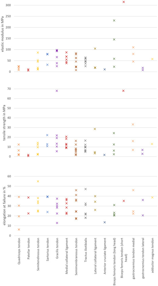
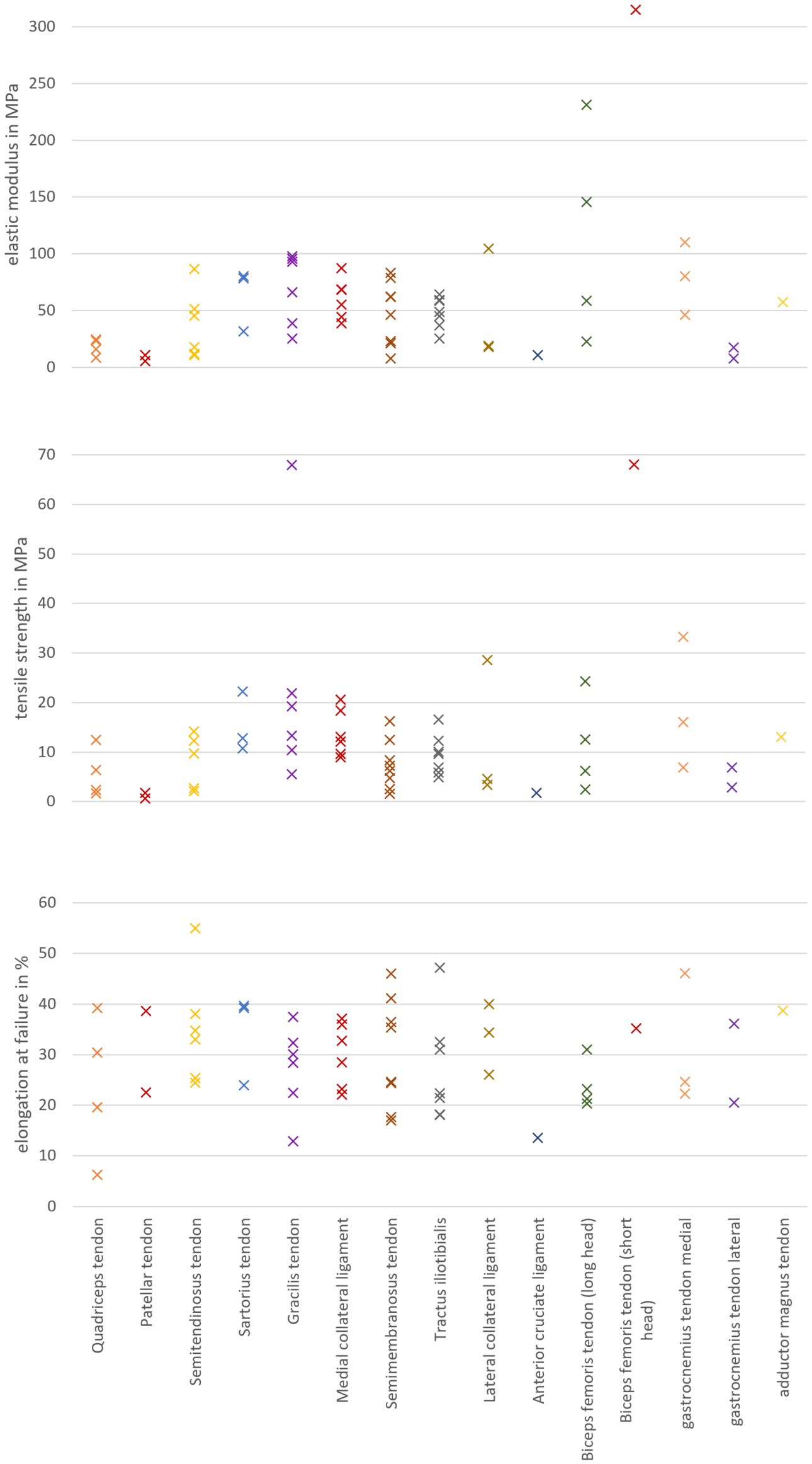
This resulted in a range of cross-sections across all structures of 1.9–69.0 mm2. The tensile strength was in the range of 0.5–68 MPa and the elastic modulus between 8.21 and 314.9 MPa (Figure 3). An overview presenting mean values and standard deviation for tensile strength, elastic modulus, elongation at failure, and cross-sectional area of tested samples can be found in Table 2.
Figure 3.
Determined elastic moduli, tensile strengths and elongation at failure.
Table 2.
Tensile strength, elastic modulus, elongation at failure and cross-sectional area of tested structures.
The elongation at failure was in a range of 6.2–47.2%. Similar fluctuation ranges were also found in intra-individual comparisons.
4. Discussion
Although the results of this study are very preliminary, they provide a useful overview of the harvestable knee ligaments and tendons and their mechanical parameters. A comprehensive examination of many ligamentous structures of the human knee and neighboring tissues in the fresh-frozen state has not yet been carried out before. Instead, previous studies have primarily focused on individual ligaments or tendons [16].
In the past, intact tendons with bony attachments were often tested when determining parameters on ligaments and tendons of the knee. This makes it difficult to compare them with the parameters we have determined because, on the one hand, no cross-sectional necking was achieved using the dogbone shape and, on the other hand, often only pure failure loads were determined, not tensile strength. This also results in sometimes significantly higher literature values for the failure loads, e.g., in the case of the anterior cruciate ligament in Woo et al. than in our testing [17]. For reasons of comparability, cut specimens without bony attachment were used. In addition, a clamping technique was chosen as the alternative; partial plastination was out of the question. This was due to the critical time factor of the plastination duration in combination with the large number of specimens. In addition, the slippage problem that often occurs in uniaxial tensile testing of ligamentous structures is also present in partial plastination [18].
New developments based on 3D-printed clamping systems currently represent a promising opportunity to realize fabric-specific, customized clamp surfaces and geometries [19]. Initially, it can be stated that the clamping systems presented by Scholz et al. were very well suited for thin samples [13]. Slippage was effectively inhibited there, clean specimen clamping was possible, and the design elements of the clamping system (engaging elements, pyramid pattern) functioned as intended.
The clamping method was not suitable for non-planar tendons, such as the quadriceps and patellar tendons, which could only be clamped inadequately due to their ellipsoidal cross-sectional structure. As a result, either the clamping elements did not close correctly or there were slippage effects even with high contact forces. In addition, the fixed sample geometry imposed restrictions on sample selection, meaning that most short samples, such as the anterior cruciate ligament, could not be tested because it was difficult to clamp them securely. It was not possible to clarify whether these were caused by the slipping of the samples themselves or by slippage or delamination of the collagenous base structure of the affected elements. At least some of the samples slipped, especially if the sample length in the clamping area was insufficient.
When evaluating the results, the small number of samples must always be considered. It should be emphasized at this point that, due to the sample sizes, the collected data were evaluated only descriptively. A more comprehensive study with a larger sample size would allow for the conduct of significance tests, which could yield more meaningful results. Non-fixed donor samples were used, as Thiel- or ethanol fixation has been shown to alter tissue properties [7,20]. In addition, the sample geometry was the same for all samples, but the analyzed structures differed considerably in size and shape. For some tissues (e.g., iliotibial tract), several samples could be taken without any problems. In the case of very short or irregularly shaped structures, such as the tendon of the short head of the biceps femoris muscle, the anterolateral ligament or the cruciate ligaments, it was difficult or impossible to obtain samples, as the samples were too short to ensure secure clamping in the jaws.
This study clearly shows the complexity of the ligamentous supporting apparatus and the diversity of the parameters of the individual ligaments and tendons of the knee. The measured and calculated values could be used, for example, to create more accurate finite-element models of the musculoskeletal system. Current research in the field of human knee ligaments has been carried out by Casp et al., for example, but their study was limited to the mechanical behavior of the knee as a whole and the movement of the knee ligaments in relation to each other [21]. Rachmat et al. also took tensile samples from the knee capsular apparatus and analyzed its parameters, but the results showed a considerable variation in parameters [22]. Zwirner et al. published a comprehensive study on the mechanical parameters of the iliotibial tract. They determined age-dependent values of 29 ± 17 MPa for tensile strength and 408 ± 232 MPa for the elastic modulus. Although the elastic modulus was several times higher, the determination of the linear elastic range for calculating the elastic modulus is a current field of research with considerable potential for discussion [19].
5. Conclusions
It should be emphasized at this point that this is purely an exploratory paper, which is intended to provide an initial overview of the various ligamentous structures of the human knee and their mechanical properties. Even though the focus was placed on an error-free and reproducible working method when collecting the parameters, it must of course always be borne in mind that the mechanical parameters of human soft tissue vary considerably [7,23]. In particular, these parameters vary considerably between individuals due to genetics, constitution, age and gender, among other things [24]. In addition, to understand the complex biomechanics of the soft tissue portion of the knee, other structures should be taken into account, like the capsular apparatus, for example, ref. [22]. A more extensive series of measurements, involving a significantly larger number of samples and a greater number of donor knees, would certainly be beneficial. This would allow for more specific conclusions to be drawn about the mechanical behavior of the knee’s numerous ligamentous structures, thanks to the improved statistical validity of the results.
Author Contributions
Conceptualization, B.F. and F.M.; methodology, B.F. and F.M.; validation, B.F. and S.S.; formal analysis, B.F.; investigation, B.F., F.M., F.W., L.Z. and S.S.; data curation, B.F.; writing—original draft, B.F.; writing—review and editing, B.F., F.M., S.L., J.-D.T., F.M. and S.S.; visualization, B.F.; project administration, S.S.; resources, S.L.; supervision, B.F. and S.S. All authors have read and agreed to the published version of the manuscript.
Funding
Supported by the Open Access Publishing Fund of Leipzig University.
Institutional Review Board Statement
All tissues originated from the Institute of Anatomy of Leipzig University. Being part of the body donor program regulated by the Saxonian Death and Funeral Act of 1994 (third section, paragraph 18, item 8), the Ethics Committee of the University of Leipzig Medical Center obtained institutional approval for the use of postmortem tissues from the Institute of Anatomy at the University of Leipzig (ethical approval no. 129/21-ck, approval date 1 March 2023). The authors declare that all experiments were conducted according to the principles of the Declaration of Helsinki (as revised in 2013).
Informed Consent Statement
While alive, all body donors provided informed and written consent to donate their post-mortem tissues for education and research purposes.
Data Availability Statement
Body donor-related data were anonymized to prevent inferences about their identity. Additional data are available from the corresponding author upon reasonable request.
Acknowledgments
We extend our utmost gratitude to the body donors and their families who made this research possible.
Conflicts of Interest
The authors declare no conflicts of interest.
References
- Wroble, R.R.; Brand, R.A. Function of Knee Ligaments: An Historical Review of Two Perspectives. Iowa Orthop. J. 1988, 8, 62–67. [Google Scholar]
- Loudon, J.K. Biomechanics and Pathomechanics of the Patellofemoral Joint. Int. J. Sports Phys. Ther. 2016, 11, 820–830. [Google Scholar]
- Stäubli, H.U.; Schatzmann, L.; Brunner, P.; Rincón, L.; Nolte, L.P. Mechanical tensile properties of the quadriceps tendon and patellar ligament in young adults. Am. J. Sports Med. 1999, 27, 27–34. [Google Scholar] [CrossRef] [Scilit] [PubMed]
- Schatzmann, L.; Brunner, P.; Stäubli, H.U. Effect of cyclic preconditioning on the tensile properties of human quadriceps tendons and patellar ligaments. Knee Surg. Sports Traumatol. Arthrosc. Off. J. ESSKA 1998, 6, S56–S61. [Google Scholar] [CrossRef] [Scilit] [PubMed]
- Mountney, J.; Senavongse, W.; Amis, A.A.; Thomas, N.P. Tensile strength of the medial patellofemoral ligament before and after repair or reconstruction. J. Bone Jt. Surg. Br. Vol. 2005, 87, 36–40. [Google Scholar] [CrossRef] [Scilit]
- Jung, H.J.; Fisher, M.B.; Woo, S.L.Y. Role of biomechanics in the understanding of normal, injured, and healing ligaments and tendons. Sport. Med. Arthrosc. Rehabil. Ther. Technol. SMARTT 2009, 1, 9. [Google Scholar] [CrossRef] [Scilit] [PubMed]
- Zwirner, J.; Scholze, M.; Waddell, J.N.; Ondruschka, B.; Hammer, N. Mechanical Properties of Human Dura Mater in Tension—An Analysis at an Age Range of 2 to 94 Years. Sci. Rep. 2019, 9, 16655. [Google Scholar] [CrossRef] [Scilit] [PubMed]
- Liao, X.; Kemp, S.; Corner, G.; Eisma, R.; Huang, Z. Elastic properties of Thiel-embalmed human ankle tendon and ligament. Clin. Anat. 2015, 28, 917–924. [Google Scholar] [CrossRef] [Scilit] [PubMed]
- Völlner, F.; Pilsl, U.; Craiovan, B.; Zeman, F.; Schneider, M.; Wörner, M.; Grifka, J.; Weber, M. Stability of knee ligament complex of Thiel-embalmed cadaver compared to in vivo knee. J. Mech. Behav. Biomed. Mater. 2017, 71, 392–396. [Google Scholar] [CrossRef] [Scilit] [PubMed]
- Moon, D.K.; Woo, S.L.Y.; Takakura, Y.; Gabriel, M.T.; Abramowitch, S.D. The effects of refreezing on the viscoelastic and tensile properties of ligaments. J. Biomech. 2006, 39, 1153–1157. [Google Scholar] [CrossRef] [Scilit] [PubMed]
- Ekiert, M.; Karbowniczek, J.; Stachewicz, U.; Mlyniec, A. The effect of multiple freeze-thaw cycles on the viscoelastic properties and microstructure of bovine superficial digital flexor tendon. J. Mech. Behav. Biomed. Mater. 2021, 120, 104582. [Google Scholar] [CrossRef] [Scilit] [PubMed]
- Wright, J.O.; Skelley, N.W.; Schur, R.P.; Castile, R.M.; Lake, S.P.; Brophy, R.H. Microstructural and Mechanical Properties of the Posterior Cruciate Ligament: A Comparison of the Anterolateral and Posteromedial Bundles. J. Bone Jt. Surgery. Am. Vol. 2016, 98, 1656–1664. [Google Scholar] [CrossRef] [Scilit] [PubMed]
- Scholze, M.; Singh, A.; Lozano, P.F.; Ondruschka, B.; Ramezani, M.; Werner, M.; Hammer, N. Utilization of 3D printing technology to facilitate and standardize soft tissue testing. Sci. Rep. 2018, 8, 11340. [Google Scholar] [CrossRef] [Scilit] [PubMed]
- Scholze, M.; Safavi, S.; Li, K.C.; Ondruschka, B.; Werner, M.; Zwirner, J.; Hammer, N. Standardized tensile testing of soft tissue using a 3D printed clamping system. HardwareX 2020, 8, e00159. [Google Scholar] [CrossRef] [Scilit] [PubMed]
- Jiang, M.; Lawson, Z.T.; Erel, V.; Pervere, S.; Nan, T.; Robbins, A.B.; Feed, A.D.; Moreno, M.R. Clamping soft biologic tissues for uniaxial tensile testing: A brief survey of current methods and development of a novel clamping mechanism. J. Mech. Behav. Biomed. Mater. 2020, 103, 103503. [Google Scholar] [CrossRef] [Scilit] [PubMed]
- Kranjec, M.; Trajkovski, A.; Krašna, S.; Hribernik, M.; Kunc, R. Material properties of human patellar-ligament grafts from the elderly population. J. Mech. Behav. Biomed. Mater. 2020, 110, 103994. [Google Scholar] [CrossRef] [Scilit] [PubMed]
- Woo, S.L.; Hollis, J.M.; Adams, D.J.; Lyon, R.M.; Takai, S. Tensile properties of the human femur-anterior cruciate ligament-tibia complex. The effects of specimen age and orientation. Am. J. Sports Med. 1991, 19, 217–225. [Google Scholar] [CrossRef] [Scilit] [PubMed]
- Sichting, F.; Steinke, H.; Wagner, M.F.X.; Fritsch, S.; Hädrich, C.; Hammer, N. Quantification of material slippage in the iliotibial tract when applying the partial plastination clamping technique. J. Mech. Behav. Biomed. Mater. 2015, 49, 112–117. [Google Scholar] [CrossRef] [Scilit] [PubMed]
- Zwirner, J.; Babian, C.; Ondruschka, B.; Schleifenbaum, S.; Scholze, M.; Waddell, N.J.; Hammer, N. Tensile properties of the human iliotibial tract depend on height and weight. Med. Eng. Phys. 2019, 69, 85–91. [Google Scholar] [CrossRef] [Scilit] [PubMed]
- Fessel, G.; Frey, K.; Schweizer, A.; Calcagni, M.; Ullrich, O.; Snedeker, J.G. Suitability of Thiel embalmed tendons for biomechanical investigation. Ann. Anat. Anat. Anz. Off. Organ Anat. Ges. 2011, 193, 237–241. [Google Scholar] [CrossRef] [Scilit] [PubMed]
- Casp, A.J.; Bryniarski, A.; Brady, A.W.; Fossum, B.W.; Godin, J.A. Reanalysis of the Posterior Oblique Ligament: Quantitative Anatomy, Radiographic Markers, and Biomechanical Properties. Orthop. J. Sports Med. 2023, 11, 23259671231174857. [Google Scholar] [CrossRef] [Scilit] [PubMed]
- Rachmat, H.H.; Janssen, D.; Verkerke, G.J.; Diercks, R.L.; Verdonschot, N. Material properties of the human posterior knee capsule. Bio-Med. Mater. Eng. 2015, 25, 177–187. [Google Scholar] [CrossRef] [Scilit] [PubMed]
- Zwirner, J.; Scholze, M.; Ondruschka, B.; Hammer, N. Tissue biomechanics of the human head are altered by Thiel embalming, restricting its use for biomechanical validation. Clin. Anat. 2019, 32, 903–913. [Google Scholar] [CrossRef] [Scilit] [PubMed]
- Akazawa, T.; Miyamoto, N.; Nishio, H.; Miyamoto-Mikami, E.; Kinoshita, M.; Kobayashi, Y.; Nagao, M.; Takazawa, Y. Age-related changes in mechanical properties of semitendinosus tendon used for anterior cruciate ligament reconstruction. J. Orthop. Surg. Res. 2022, 17, 501. [Google Scholar] [CrossRef] [Scilit] [PubMed]

Disclaimer/Publisher’s Note: The statements, opinions and data contained in all publications are solely those of the individual author(s) and contributor(s) and not of MDPI and/or the editor(s). MDPI and/or the editor(s) disclaim responsibility for any injury to people or property resulting from any ideas, methods, instructions or products referred to in the content. |
© 2026 by the authors. Licensee MDPI, Basel, Switzerland. This article is an open access article distributed under the terms and conditions of the Creative Commons Attribution (CC BY) license.