Article

A Human Shoulder Simulator for Cyclic Evaluation of Rotator Cuff Injury and Repair

1
Institute of Medical and Biological Engineering, University of Leeds, Leeds LS2 9JT, UK
2
School of Mechanical Engineering, University of Sheffield, Sheffield S1 3JD, UK
3
Leeds Teaching Hospitals NHS Trust, Leeds LS1 3EX, UK
*
Author to whom correspondence should be addressed.
Biomechanics 2026, 6(3), 69; https://doi.org/10.3390/biomechanics6030069
Submission received: 18 May 2026 / Revised: 29 June 2026 / Accepted: 14 July 2026 / Published: 16 July 2026
(This article belongs to the Section Injury Biomechanics and Rehabilitation)

Abstract

Background/Objectives: Surgical repair of the rotator cuff tendons can lead to unsatisfactory results and the requirement for further surgical treatment. Development of repair techniques is limited by a lack of appropriate functional pre-clinical testing, especially over extended motion cycles. The purpose of this study was to demonstrate the efficacy of a novel shoulder simulator by assessing changes in internal muscle forces following a rotator cuff tear and double-row surgical repair. Methods: The developed shoulder simulator used motors to apply controlled movements/displacements to tendons (supraspinatus, infraspinatus, subscapularis, teres minor, anterior deltoid and middle deltoid) of a cadaveric human shoulder to produce cyclic abduction motion representative of normal shoulder function. The required displacement for each muscle was determined using a musculoskeletal model. The resultant force applied to each tendon during the cycles was measured using a compression load cell. Results: The developed simulator in this proof-of-concept study enabled the contribution of the different muscles involved in the shoulder during abduction to be assessed for the intact shoulder. The shoulder was also tested with a 50% supraspinatus tear and a double row surgical repair of the supraspinatus to assess the change in internal muscle forces. Conclusions: The study indicated that successful cyclic testing of cadaveric samples could be achieved using the simulator and changes in the internal muscle forces of the shoulder were identified following a supraspinatus tear and double-row surgical repair.

1. Introduction

The shoulder joint is capable of the widest range of motion of any joint in the human body; however, to achieve this, it is also the most unstable joint [1]. The rotator cuff muscles surrounding the glenohumeral joint act to compress the humeral head into the glenoid fossa of the scapula and prevent dislocation [2]. Half of all major shoulder injuries are tears of the rotator cuff tendons, leading to over 62% of adults over the age of 80 years having a rotator cuff tear [3]. Tears of the rotator cuff are often initially managed conservatively; however, continuing pain often leads to the requirement for surgical intervention depending on the patient’s age. Tendon quality, size of tear and patient age affects the success of surgical repair with failure rates between 25 and 50% at 12 months post-surgery [4].
Experimental in vitro models enable surgical repair methods of the rotator cuff to be tested through clinically relevant loading cycles and ranges of motion. Static, uniaxial biomechanical testing of surgical rotator cuff tendon repairs is often used [5,6,7,8]. Whilst this technique allows for the strength of the repair methods to be determined, the natural biomechanics of the rotator cuff tendons and restoration of their function is not considered. For an experimental simulator to be representative of the shoulder environment, the muscles must be actively powered to produce motion. Previous experimental simulators using actively driven muscles to investigate joint biomechanics and muscle forces are limited. Most existing actively powered simulators report on a very low number of motion cycles (<3) which limits the suitability of assessing the mechanical efficacy of a rotator cuff repair over longer cycles of motion [9,10,11,12,13]. Another disadvantage of current simulators is the complexity and expense required; a benchtop and accessible simulator would allow for comparative screening of treatment options preclinically.
The aim of the study was to demonstrate the efficacy of the shoulder simulator by assessing changes in internal muscle forces between an intact rotator cuff, a supraspinatus tear and a double-row repair of the supraspinatus tendon.

2. Materials and Methods

2.1. Shoulder Simulator

The bespoke human shoulder simulator (Figure 1) actuated the rotator cuff tendons of a cadaveric human glenohumeral joint to generate cyclic abduction/adduction (AA) motions (tissue used with consent for research under HTA license 12279). The resultant force applied to each tendon during the motion cycles was measured.
In this study, cadaveric human tissue treated using the saturated salt solution method was used. The range of motion achievable at the preserved shoulder joint was comparable to that of the natural joint and surpasses the range of motion of a joint treated with formaldehyde [14]. A human cadaveric shoulder was dissected to isolate the glenohumeral joint with an intact joint capsule and approximately 15 cm of the humeral attachments of the four rotator cuff tendons (supraspinatus, infraspinatus, teres minor and subscapularis) and the anterior and middle deltoid tendons. These provided the attachment points for the artificial muscle actuation system. Braided polyethylene thread was secured to the tendon ends using a modified finger trap suture. Eyelet screws were attached to the scapula at the approximate insertion location of the muscles to act as a pulley to maintain the line of action of the muscles. The braided polyethylene thread was passed from the muscle attachment, through the pulley and secured to a stepper motor (NEMA 17) which produced actuation. The force applied by each stepper motor to the tendons was measured using a compression load cell and a custom load measuring platform. The simulator was displacement-driven rather than force-controlled, with the force in the muscles being the output of the simulator.
The simulator was used to generate cyclic motion of the shoulder by actuating the stepper motors attached to the tendons, to follow predefined motion trajectories. The required displacements for each tendon were determined using a bespoke musculoskeletal computational model of the shoulder joint (AnyBody v8.1.5). The outputs of the computational model were the force required in each muscle to produce motion; these were compared to values published in the literature and were shown to compare well to other computational models for the first 50° of abduction. The predicted ratios of forces required by each muscle were converted from the AnyBody model into ratios of displacements needed in each corresponding motor to produce the desired motion in the simulator. The ratio of displacement was tuned in order to obtain the most appropriate and smooth motion cycle based on the AnyBody model. The motion in the simulator was not restricted to just an abduction/adduction motion and so translation or rotation of the joint was possible but constrained by the natural soft tissues surrounding the joint.
A camera was used to record the human shoulder throughout the abduction/adduction motion cycles. The captured video was post processed using a Matlab (2024a) script to track the position of the markers, applied to the humerus, throughout the motion.

2.2. Test Scenarios

Following dissection of the samples as detailed in Section 2.1, the scapula was cemented into a custom fixture using polymethylmethacrylate (PMMA) bone cement. Prior to fixation, the tendon quality, pre-existing degeneration and age-related tissue changes were visually assessed. Samples which displayed pre-existing degeneration or poor-quality tissue were not used in the study.
Three independent left shoulder samples (2 male, 1 female and aged 91–94 years) were available for use in this study. They were biomechanically assessed in the described simulator sequentially in the following four test conditions:
  • Intact shoulder—The samples were tested immediately following dissection in the natural intact state;
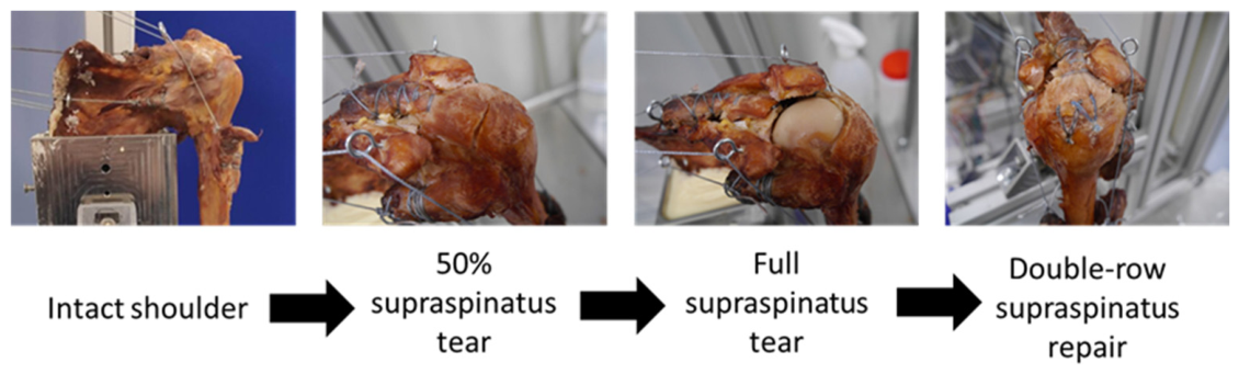
  • Partial 50% supraspinatus tear—A scalpel incision was made through the full thickness of the supraspinatus tendon close to the root to a length of 50% of the initial supraspinatus root length;
  • Full supraspinatus tear—The scalpel incision was continued to fully dissect the supraspinatus tendon from its attachment on the humeral head;
  • Double-row supraspinatus repair—A double-row repair of the supraspinatus tendon was carried out as deemed most appropriate by a consultant orthopedic surgeon.
Consultant orthopedic surgeons [PC/DH] carried out the dissection, incision and repair of the tendon in all cases. The progression of test conditions is shown in Figure 2.
At each test condition, the shoulder underwent motion sets of 20 cycles of 0–50° abduction/adduction in the human shoulder simulator at a speed of approximately 5 degrees/second. The scapula was held stationary throughout the abduction motion which limited the range of motion of the simulator to 0–50° of abduction. The force required in each rotator cuff muscle and the anterior and middle deltoid was recorded throughout all motions.

2.3. Data Analysis

The resultant forces from each muscle during the simulation process were recorded for every cycle and repeat of the motion. A uniform weight of 0.6 kg was applied axially to each load cell prior to the simulation commencing to enable calibration of each load cell. Each motion cycle was repeated three times, which resulted in three repeats of force data for each muscle through the 20 motion cycles. This enabled a mean of all the measurements at each point in the motion cycle to be found resulting in a mean motion cycle for the sample. To ensure that the cycles were in phase prior to the mean calculations, the first supraspinatus peak was aligned in all repeats. Due to the low sample size (n = 3), formal statistical comparison was not possible.

3. Results

3.1. Overview of Shoulder Simulator

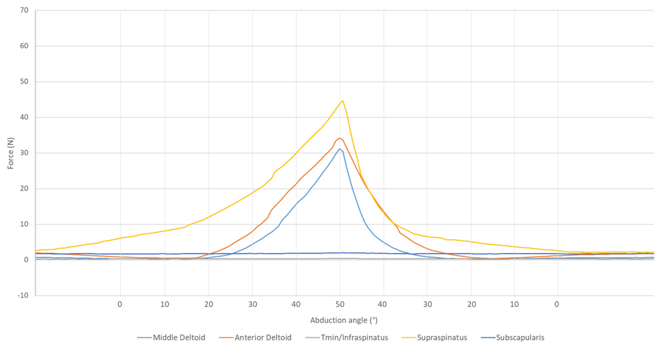
The shoulder simulator (shown in Figure 1) applied displacements to the tendons of the shoulder muscles to produce cyclic abduction/adduction motion. The resultant force in each muscle was recorded using a custom force measurement platform and compression load cells. The force measured in each muscle through a single cycle of abduction/adduction motion with an intact sample (sample 1) is shown in Figure 3.
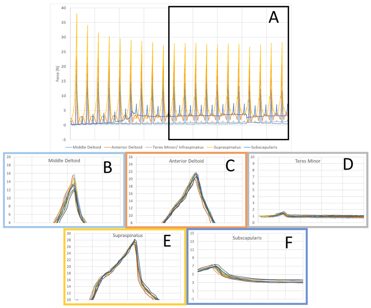
At each test condition, the shoulder underwent sets of 20 cycles of 0–50° abduction/adduction. The final 11 cycles of the motion were plotted on top of each other starting at 0° abduction to investigate the repeatability of the cyclic motion within the simulator. The graphs for each individual muscle are shown in Figure 4 along with the original force graph of a single repeat with the investigated section highlighted in blue.

3.2. Changes in Rotator Cuff Biomechanics

Three cadaveric samples, treated with the saturated salt solution preservation method, were tested with the novel human shoulder simulator. The samples were tested in an intact state, with a partial (50%) tear of the supraspinatus, a full (100%) tear and a double-row supraspinatus repair performed by a consultant shoulder surgeon. At each state the shoulders underwent cyclic (20 cycles) motion of 0–50° abduction/adduction in the simulator and the force required in each muscle was recorded.
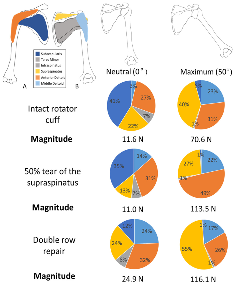
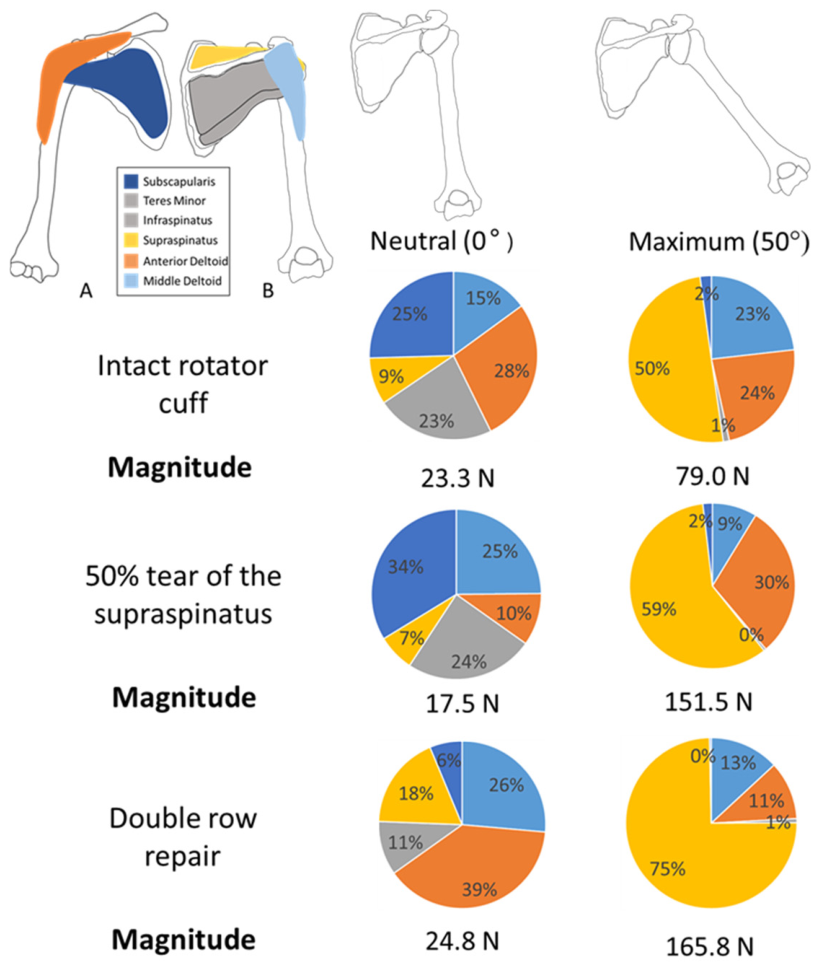
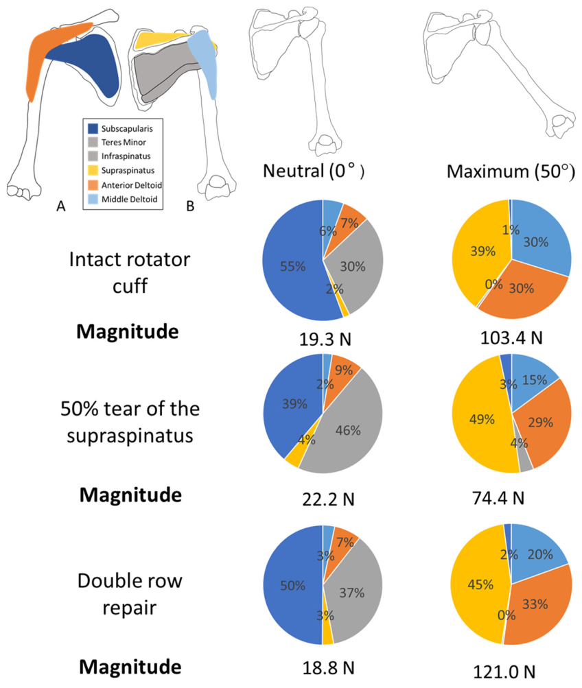
The contribution to the total force by each muscle for a mean abduction cycle (mean of cycles 11–20) at minimum (0) and maximum abduction (50) for samples 1, 2 and 3 at all test conditions is provided in Figure 5, Figure 6 and Figure 7, respectively. In all samples, the supraspinatus muscle provided the majority of total force at 50° abduction followed by the anterior and middle deltoid, when the rotator cuff was intact. Following a supraspinatus tear, the contribution of the supraspinatus decreased in sample 1, however, increased in samples 2 and 3. In all three samples the contribution of the supraspinatus to the total force increased following a double-row tendon repair and also surpassed the force required in the intact case.

4. Discussion

4.1. Overview of Shoulder Simulator

The developed simulator enabled the evaluation of the contribution of the different muscles involved in motion of the shoulder, we recognize that this is a pilot study with a small sample number, in the following sections we provide a descriptive comparison of the data. Considering the cycle of abduction/adduction motion shown in Figure 3, the supraspinatus muscle initiated the movement with the deltoid muscles then supporting the higher angle of abduction. This agrees with the literature such as Lam and Bordoni (2021) who stated that the primary muscle during the initiation of abduction was the supraspinatus muscle followed by the anterior and middle deltoid muscles [15].
The final 11 cycles of the motion were plotted (Figure 4) on top of each other starting at 0° abduction to investigate the repeatability of the cyclic motion within the simulator. The resultant force in all muscles was consistent across all repeats of the movement and cycles within a repeat, suggesting that the simulator delivered repeatable cyclic testing. In all cases, the magnitude of force in all muscles decreased during the initial six cycles to a plateau (shown in Figure 4A). The force in each muscle then remained similar throughout the remaining cycles. This was identified in all runs of the simulator and was considered to be a result of the initial bedding in of the humeral head into the glenoid fossa at the beginning of the motion cycle.

4.2. Changes in Rotator Cuff Biomechanics

Shoulder samples were tested in an intact state, with a partial (50%) tear of the supraspinatus, a full (100%) supraspinatus tear and a double-row tendon repair performed by a consultant shoulder surgeon. In each state, the force and percentage contribution of different rotator cuff tendons was assessed. The magnitude of force and the percentage contributions of different rotator cuff tendons are shown in Figure 5, Figure 6 and Figure 7 for samples 1–3 respectively. In all samples during the intact testing, the supraspinatus tendon provided the majority of the force at 50° abduction followed by the anterior and middle deltoid muscles. Following an artificial 50% tear of the supraspinatus tendon, the contribution of force provided by the supraspinatus decreased in sample 1 and was compensated for by an increase in force in the anterior deltoid muscle. As anterior tears of the supraspinatus progress, it has been reported in the literature that the force within the deltoid muscle increases to compensate for the decrease in force within the supraspinatus muscle [16,17,18]. This is in contrast to the results for samples 2 and 3 (Figure 6 and Figure 7), where the proportion of force increased in the supraspinatus after a 50% tear. In both cases, the force in the middle deltoid decreased whereas the force in the anterior deltoid either remained the same or increased, which was similar to the results found with sample 1. In two cases the magnitude of force at minimum and maximum abduction was higher in the torn state than when the supraspinatus was intact. This suggests that a greater quantity of force was required within the system to maintain the stability of the shoulder joint throughout motion.
The 50% tear was extended using a scalpel incision to the entire width of the supraspinatus tendon to form a 100% tear. When the same motion cycles were performed with the fully torn supraspinatus tendon, in all cases, the humeral head translated superiorly until there was impingement on the acromion of the scapula with very limited abduction. It was determined that it was impossible to initiate the abduction motion in the simulator without the supraspinatus tendon intact and that abduction could only begin if the humerus was able to start from an angle of 15°.
A double-row repair of the supraspinatus tendon was carried out on all samples by an orthopedic shoulder surgeon. In all specimens (Figure 5, Figure 6 and Figure 7), the proportion of force provided by the supraspinatus increased and surpassed that of the intact state. The double-row repair method involved the addition of bone anchors to the humeral head, from which suture tape was passed through the tendon to anchor it to the humerus. This increased the stiffness of the tendon–bone interface compared to the natural tendon–bone interface. The magnitude of force was also considerably higher in the repaired supraspinatus shoulder compared with the intact rotator cuff. In sample 1, the total magnitude of force increased by 64% between the intact and repaired state; for sample 2 this increase was 110% and for sample 3 it was 17%. This could suggest that the stiffening of the supraspinatus tendon led to an overcompensation in the compression of the humeral head into the glenoid fossa and therefore did not restore the initial joint biomechanics.

4.3. Overall Discussion

Some limitations exist for the shoulder simulator developed through this study. During dissection, the arm inferior to the elbow joint and the surrounding muscles, fat and skin were removed to allow for easy access to the glenohumeral joint and the rotator cuff muscles. No substitute was included to replicate the effect of the weight of the lower arm on the muscle forces required for motion of the shoulder. Due to the difference in mechanical properties of living tissue and saturated salt solution-treated cadavers, the magnitude of force in each joint was not an accurate output from the simulator. The comparisons between forces contributed by each muscle were a more appropriate measure to allow for comparisons between each repeat and the different test scenarios. As the samples were tested sequentially through different states, cumulative creep, mechanical degradation and tissue handling may have influenced later test scenarios. To mitigate this risk, hydration of the samples was maintained by spraying with water between every test and all tests were conducted within a 48 h period.
The age of the available cadavers was high and as a result the tendon and bone quality in these shoulders may differ substantially from those in younger patients. The potentially reduced bone quality may influence the mechanical behavior of the double-row repair, including anchor fixation in the humeral head. Testing was only carried out in cadavers which were deemed to have an intact, healthy rotator cuff by an orthopedic upper-limb surgeon. All testing was sequential to allow for comparisons between the test scenarios within a single sample.
Scapulothoracic motion was also not included in the simulator to prevent additional complexity of the design. As a result, the maximum abduction angle was limited due to the impingement of the humeral head onto the acromion of the scapula. Comparisons between forces contributed by each muscle during each damage scenario were a more appropriate measure than the magnitude of force due to the restriction of motion in the scapulothoracic joint.
Additional human tissue samples would be required to provide more conclusive results regarding the effect of rotator cuff tears and repairs on the biomechanics of the shoulder joint. However, this study has shown that the developed simulator could repeatably be used to measure the change in internal muscles forces between an intact sample, a supraspinatus tear and a double-row repair of the supraspinatus.
The ongoing work will focus on increasing the samples tested using the novel human shoulder simulator in order to provide more conclusive results regarding the efficacy of different rotator cuff repair methods. Further biomechanical studies are planned to use the simulator to assess the impact of augmentation devices on the biomechanical stability of the rotator cuff tendons compared to traditional surgical methods.

5. Conclusions

A novel actively controlled shoulder simulator for cyclic testing of the biomechanical behavior of the rotator cuff in different injury states was developed. The simulator has been shown to be capable of producing repeatable controlled movements over an extended number of motion cycles. The study showed that the novel human shoulder simulator was able to assess the change in internal muscle forces between an intact rotator cuff, a supraspinatus tear and a double-row repair of the tendon.

Author Contributions

Conceptualization, S.W. and C.B.; methodology, S.H.; software, S.H.; validation, S.H.; formal analysis, S.H.; investigation, S.H., P.C. (Paul Cowling) and D.H.; resources, S.W. and S.H.; data curation, S.H.; writing—original draft preparation, S.H.; writing—review and editing, S.W., P.C. (Peter Culmer), D.H. and P.C. (Paul Cowling); visualization, S.H. and P.C. (Peter Culmer); supervision, S.W., P.C. (Peter Culmer), C.B., D.H. and P.C. (Paul Cowling); project administration, S.W.; funding acquisition, S.W. and C.B. All authors have read and agreed to the published version of the manuscript.

Funding

This research was funded by EPSRC, grant number EP/T517860/1.

Institutional Review Board Statement

The study was conducted in accordance with the Declaration of Helsinki, and approved by the Institutional Review Board of the University of Leeds (LTMECH-10 approval date 13 December 2023).

Informed Consent Statement

Informed consent for participation in research was obtained from all subjects involved in the study.

Data Availability Statement

The data associated with this paper are openly available from the University of Leeds Data Repository [19].

Acknowledgments

Authors wish to acknowledge the donors and their families for allowing the use of cadaveric tissue in research, Sarah Wilson (University of Leeds) for facilitating this research and technical support from Phillip Wood and Matthew Broadbent (University of Leeds).

Conflicts of Interest

The authors declare no conflicts of interest.

References

  1. Rockwood, C.; Matsen, F. The Shoulder, 2nd ed.; W.B. Saunders Company: Philadelphia, PA, USA, 1998; Volume 1. [Google Scholar]
  2. Hess, S.A. Functional stability of the glenohumeral joint. Man. Ther. 2000, 5, 63–71. [Google Scholar] [CrossRef] [PubMed]
  3. May, T.; Garmel, G. Rotator Cuff Injury. 2020. Available online: https://www.ncbi.nlm.nih.gov/books/NBK547664/ (accessed on 6 February 2024).
  4. Greenall, G.; Carr, A.; Beard, D.; Rees, J.; Rangan, A.; Merritt, N.; Dritsaki, M.; Nagra, N.S.; Baldwin, M.; Hopewell, S.; et al. Systematic review of the surgical management of rotator cuff repair with an augmentative patch: A feasibility study protocol. Syst. Rev. 2018, 7, 187. [Google Scholar] [CrossRef] [PubMed]
  5. Oh, J.H.; Mcgarry, M.H.; Jun, B.J.; Gupta, A.; Chung, K.C.; Hwang, J.; Lee, T.Q. Restoration of Shoulder Biomechanics According to Degree of Repair Completion in a 178 Cadaveric Model of Massive Rotator Cuff Tear Importance of Margin Convergence and Posterior Cuff Fixation. Am. J. Sports Med. 2012, 40, 2448–2453. [Google Scholar] [CrossRef] [PubMed]
  6. Smith, C.; Alexander, S.; Hill, A.; Huijsmans, P.; Bull, A.M.J.; Amis, A.; De Beer, J.; Wallace, A. A Biomechanical Comparison of Single and Double-Row Fixation in Arthroscopic Rotator Cuff Repair. J. Bone Jt. Surg. 2006, 88, 2425–2431. [Google Scholar] [CrossRef]
  7. Waltrip, R.L.; Zheng, N.; Dugas, J.R.; Andrews, J.R. Rotator Cuff Repair: A Biomechanical Comparison of Three Techniques. Am. J. Sports Med. 2003, 31, 493–497. [Google Scholar] [CrossRef] [PubMed]
  8. Kim, D.H.; Elattrache, N.S.; Tibone, J.E.; Jun, B.; Delamora, S.N.; Kvitne, R.S.; Lee, T.Q. Biomechanical Comparison of a Single-Row Versus Double-Row Suture Anchor Technique for Rotator Cuff Repair. Am. J. Sports Med. 2006, 34, 407–414. [Google Scholar] [CrossRef] [PubMed]
  9. Verjans, M.; Siroros, N.; Eschweiler, J.; Radermacher, K. Technical concept and evaluation of a novel shoulder simulator with adaptive muscle force generation and free motion. Curr. Dir. Biomed. Eng. 2016, 2, 61–65. [Google Scholar] [CrossRef]
  10. Kedgley, A.E.; Mackenzie, G.A.; Ferreira, L.M.; Drosdowech, D.S.; King, G.J.W.; Faber, K.J.; Johnson, J.A. The effect of muscle loading on the kinematics of in vitro glenohumeral abduction. J. Biomech. 2007, 40, 2953–2960. [Google Scholar] [CrossRef] [PubMed]
  11. Baumgartner, D.; Tomas, D.; Gossweiler, L.; Siegl, W.; Osterhoff, G.; Heinlein, B. Towards the development of a novel experimental shoulder simulator with rotating scapula and individually controlled muscle forces simulating the rotator cuff. Med. Biol. Eng. Comput. 2014, 52, 293–299. [Google Scholar] [PubMed]
  12. Debski, R.E.; McMahon, P.J.; Thompson, W.O.; Woo, S.; Warner, J.; Fu, F.H. A new dynamic testing apparatus to study glenohumeral joint motion. J. Biomech. 1995, 28, 869–874. [Google Scholar] [CrossRef] [PubMed]
  13. Guo, R.; Ferle, M.; Nebel, D.; Hurschler, C. The development and evaluation of an invitro shoulder simulator with active muscle simulation. Sci. Rep. 2023, 13, 4423. [Google Scholar] [CrossRef] [PubMed]
  14. Burns, D.M.; Bell, I.; Katchky, R.; Dwyer, T.; Toor, J.; Whyne, C.; Safir, O. Saturated Salt Solution Cadaver-Embalming Method Improves Orthopaedic Surgical Skills Training. J. Bone Jt. Surg. 2018, 100, e104. [Google Scholar] [CrossRef]
  15. Lam, J.; Bordoni, B. Anatomy, Shoulder and Upper Limb, Arm Abductor Muscles; StatPearls Publishing: Treasure Island, FL, USA, 2021. Available online: https://www.ncbi.nlm.nih.gov/books/NBK537148/?report=reader#_NBK537148_pubdet (accessed on 6 February 2024).
  16. Oh, J.H.; Jun, B.J.; McGarry, M.H.; Lee, T.Q. Does a Critical Rotator Cuff Tear Stage Exist? J. Bone Jt. Surg. 2011, 93, 2100–2109. [Google Scholar] [CrossRef]
  17. Dyrna, F.; Kumar, N.S.; Obopilwe, E.; Scheiderer, B.; Comer, B.; Nowak, M.; Romeo, A.A.; Mazzocca, A.D.; Beitzel, K. Relationship Between Deltoid and Rotator Cuff Muscles During Dynamic Shoulder Abduction: A Biomechanical Study of Rotator Cuff Tear Progression. Am. J. Sports Med. 2018, 46, 1919–1926. [Google Scholar] [CrossRef] [PubMed]
  18. Mancuso, F.; Di Benedetto, P.; Tosolini, L.; Buttironi, M.M.; Causero, A.; Ortopedica, C.; Sanitaria, A.; Integrata, U.; Maria, P.S. Treatment options for massive rotator cuff tears: A narrative review. Acta Biomed. 2021, 92, e2021026. [Google Scholar]
  19. Hutchinson, S.; Culmer, P.; Brockett, C.; Henderson, D.; Cowling, P.; Williams, S. A Human Shoulder Simulator for Cyclic Evaluation of Rotator Cuff Injury and Repair. Ph.D. Thesis, University of Leeds, Leeds, UK, 2026. Available online: https://etheses.whiterose.ac.uk/id/eprint/35971/1/Hutchinson_S_Mechanical_Engineering_PhD_2024.pdf (accessed on 13 July 2026).
Figure 1. The novel human shoulder simulator. (A): A schematic of the simulator showing the key components of the design including the load cell and pivot platforms, stepper motors and eyelet screw pulleys. (B): A photograph of the resulting shoulder simulator in use with a human cadaveric shoulder joint.
Biomechanics 06 00069 g001
Figure 2. Progression of testing scenarios from the intact natural state, a partial (50%) anterior supraspinatus tear, a full (100%) supraspinatus tear and a double-row tendon repair.
Biomechanics 06 00069 g002
Figure 3. Force measured in each muscle during a single abduction/addutcion cycle for an intact cadaveric shoulder (sample 1).
Biomechanics 06 00069 g003
Figure 4. The repeatability of the abduction motion cycle for sample 2. Each individual cycle (10–20) is plotted on the same axis; the plotted cycles are indicated by the black box in (A). (A): Repeated cycles for all muscles in cycles 10–20. (B): Forces in the middle deltoid muscle for cycles 10–20. (C): Forces in the anterior deltoid for cycles 10–20. (D): Forces in the teres minor deltoid for cycles 10–20. (E): Forces in the supraspinatus for cycles 10–20. (F): Forces in the subscapularis for cycles 10–20.
Biomechanics 06 00069 g004
Figure 5. Magnitude of force at a neutral and maximum abduction angle for each rotator cuff muscle (shown as a percentage) for the intact rotator cuff, artifical 50% tear and double-row supraspinatus repair in sample 1. (A) Anterior view and (B) posterior view.
Biomechanics 06 00069 g005
Figure 6. Magnitude of force at a neutral and maximum abduction angle for each rotator cuff muscle (shown as a percentage) for the intact rotator cuff, artifical 50% tear and double-row supraspinatus repair in sample 2. (A) Anterior view and (B) posterior view.
Biomechanics 06 00069 g006
Figure 7. Magnitude of force at a neutral and maximum abduction angle for each rotator cuff muscle (shown as a percentage) for the intact rotator cuff, artifical 50% tear and double-row supraspinatus repair in sample 3. (A) Anterior view and (B) posterior view.
Biomechanics 06 00069 g007
Disclaimer/Publisher’s Note: The statements, opinions and data contained in all publications are solely those of the individual author(s) and contributor(s) and not of MDPI and/or the editor(s). MDPI and/or the editor(s) disclaim responsibility for any injury to people or property resulting from any ideas, methods, instructions or products referred to in the content.