


Abstract
Terpyridines are well-known ligands in coordination chemistry, are valued for their conformational flexibility and strong metal-binding properties, and are also of interest as fluorophores. This study focused on the synthesis and comprehensive investigation of a new class of bis-oxazolo[5,4-b]pyridine derivatives, designed based on their structural similarity to terpyridines. Four novel compounds, 4a–d, were synthesized by cyclization of amide derivatives of 3-aminopyridin-2(1H)-ones using pyridine-2,6-dicarboxylic acid and its dichloride as key acidic components. Their structures and purity were confirmed by melting point analysis, high-resolution mass spectrometry, and 1H, 13C NMR spectroscopy. Compounds 4a–c exhibit UV absorption at 323–357 nm and intense blue to deep-blue fluorescence (357–474 nm, цi ≈ 0.32–0.84) in chloroform, dichloromethane, and acetonitrile, attributed to p–p* transitions within the conjugated ring system. These findings suggest their potential as phosphors for organic electronics. Computational modeling of 4a–c molecules provided insight into their electronic structures, conformational stability, and predicted optical behavior. The most stable conformers (4a–II, 4b–II, 4c–II′) exhibited a progressive decrease in the HOMO–LUMO gap from 4a to 4c, correlated with the enhancement of photoactivity. Among them, compound 4a stands out as the most promising luminophore, displaying the most intense and narrow luminescence band, owing to its high molecular symmetry and stable emission characteristics. Overall, this study lays the foundation for future studies of bis(oxazolo[5,4-b]pyridine) derivatives in coordination chemistry and optoelectronic materials development.
1. Introduction
Creating advanced fluorophores and ligands that can be utilized in industrial processes, modern technologies, and daily life continues to be a key task in organic and coordination chemistry. Many organic fluorophores are successfully used as biosensors, biomarkers, and dyes for labeling cells [1,2,3,4,5]. They are also widely used in organic electronics, including LEDs, transistors, batteries, solar cells, and other devices [6,7,8]. Their effectiveness is evaluated by such parameters as fluorescence quantum yield, Stokes shift, molar extinction coefficient, and absorption/emission maxima [1]. Since only a limited set of fluorophores reach optimal values, their practical applicability remains restricted. Thus, the development and evaluation of new fluorophores with improved photophysical properties remains a challenging and time-consuming process.
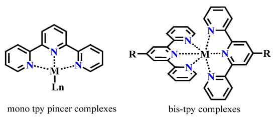
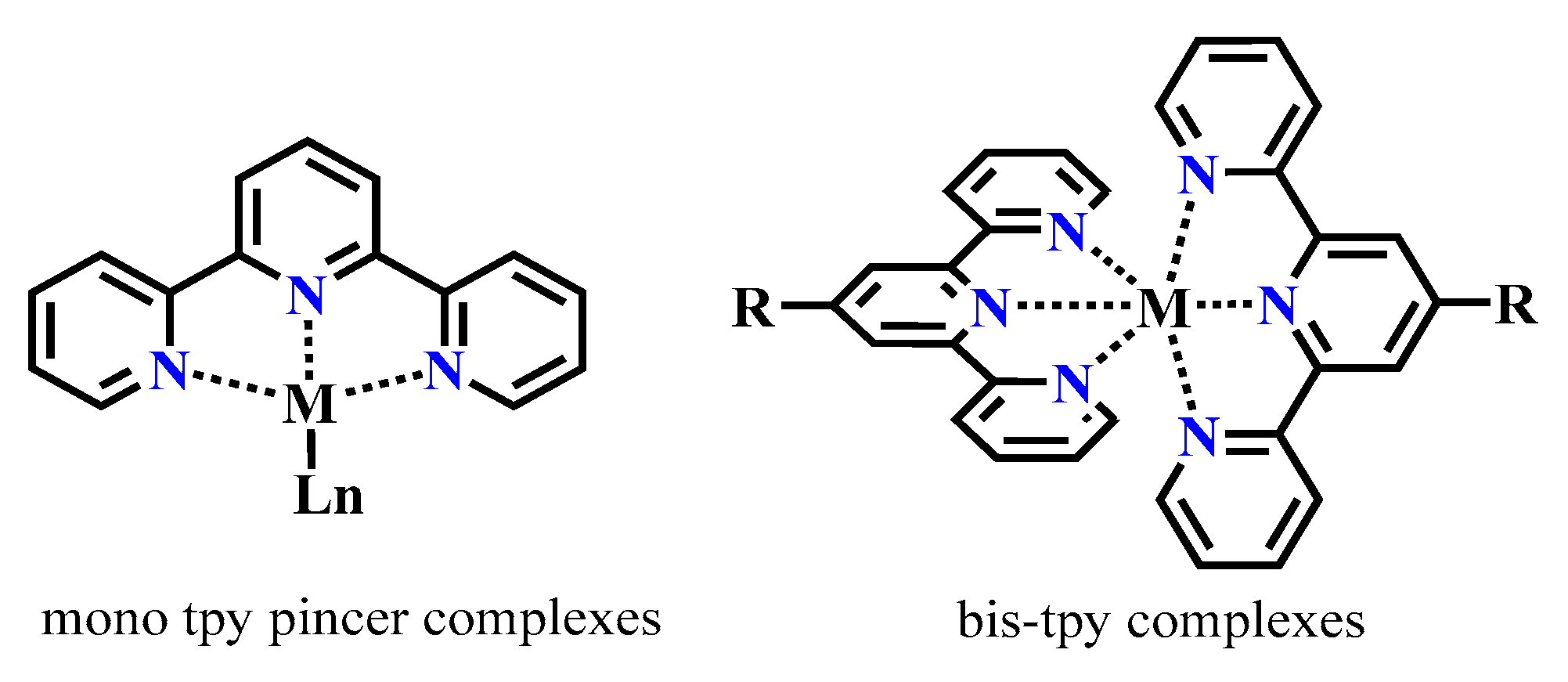
Organic fluorophores, often heterocyclic compounds, are widely used as ligands in coordination complexes with metal ions, where they play a crucial role in regulating metal ion reactivity [9]. Sometimes a single unique ligand can strongly determine the outcome of coordination processes by dictating the geometry, stability, and electronic properties of the resulting metal complexes [10,11,12,13]. Nitrogen-containing ligands are particularly important in coordination chemistry, varying from mono- to multidentate compounds depending on the number of coordination centers [14,15,16]. Among the nitrogen-based ligands, 2,2′:6′,2″-terpyridine 1 (tpy) has been extensively studied over the past 20 years [17,18,19] (Scheme 1).
Scheme 1.
Basic coordination mode of terpyridine ligands.
Terpyridine derivatives and their metal complexes [20,21,22] have found wide application in fluorescent sensors and biological labels, photocatalysis, solar cell sensitization, molecular logic devices, organic light-emitting diodes (OLEDs), and various catalytic processes [23,24,25].
Overall, 2,2′:6′,2″-terpyridine derivatives and their metal complexes possess broad potential and practical applications, but difficulties including low synthetic yields and inadequate solubility persist. Accordingly, the development of novel terpyridine derivatives with dual roles—as organic fluorophores and effective coordinating ligands—constitutes an important direction in current organic and coordination chemistry. This is especially important in light of the growing demand for functional materials in organic electronics, molecular science, medicine, and drug discovery [26,27,28].
The aim of this study was to identify and comprehensively examine a series of structurally unique and previously undescribed bis-derivatives of oxazolo[5,4-b]pyridines. These compounds were selected as close structural analogues of terpyridines, which are well known for their versatility and effectiveness as ligands in coordination chemistry.
2. Materials and Methods
2.1. Material and Instrumentation
1H and 13C NMR spectra were recorded on Bruker AVANCE 500 (500 and 126 MHz, respectively, Ettlingen, Germany), Bruker Avance NEO (400 and 101 MHz, respectively, Ettlingen, Germany) and Magritek spinsolve 80 carbon ultra (81 and 20 MHz, respectively, Aachen, Germany) instruments using DMSO-d6. Residual solvent signals (2.49 and 39.5 ppm for 1H and 13C nuclei) served as an internal standard.
Sample were analyzed by HPLC-MS on an Agilent 1260 Infinity II (Agilent Technologies, Santa Clara, CA, USA) chromatograph coupled to an Agilent 6545 LC/Q-TOF (Agilent Technologies, Santa Clara, CA, USA) high-resolution mass spectrometer with a Dual AJS ESI (Agilent Technologies, Santa Clara, CA, USA) ionization source operating in positive ion mode using the following parameters: capillary voltage: 4000 V; spray pressure: 20 (psi); drying gas: 10 L/min; gas temperature: 325 °C; sheathed gas flow: 12 L/min; shielding gas temperature: 400 °C; nozzle voltage: 0 V, fragmentation voltage: 180 V; skimmer voltage: 45 V; octopole RF: 750 V. Mass spectra with LC/MS accuracy were recorded in the range 100–1000 m/z, scan rate 1.5 spectrum/s.
Chromatographic separation was carried out on columns: ZORBAX RRHD Eclipse Plus C18 (2.1 × 50 mm, particle size 1.8 µm, Agilent Technologies, Santa Clara, CA, USA). The column temperature during the analysis was maintained at 35 °C. The mobile phase was formed by eluents A and B. In the positive ionization mode, 0.1% formic acid solution in deionized water was used as eluent A, and 0.1% formic acid solution in acetonitrile was used as eluent B. Chromatographic separation was performed with elution according to the following scheme: 0–10 min 95% A, 10–13 min 100% B, 13–15 min 95% A. The flow of the mobile phase was maintained at 400 μL/min throughout the analysis. In all experiments, the sample injection volume was 1 μL. The sample was prepared by dissolving the entire sample (in 1000 μL) in methanol (for HPLC). Sample dilution was carried out immediately before analysis.
Melting points were determined on a Stuart SMP10 apparatus (Stuart Equipment, Staffordshire, UK). Elemental analysis was performed on a Carlo Erba 1106 CHN instrument (Carlo Erba Strumentazione, Milan, Italy). The reaction progress and product purity were monitored by TLC on Sorbfil plates and visualized using iodine vapor or UV light.
UV-Vis absorption spectra were recorded on a Shimadzu UV-2600 spectrophotometer (Shimadzu Corporation, Kyoto, Japan) in the range of 200–600 nm. Measurements were carried out in quartz cuvettes with a 1 cm optical path length, and the spectral slit width was 2 nm. The concentrations of the studied compounds were 2 × 10⁻⁵ M (for compounds 4a, 4c, and 4d) and 1 × 10⁻⁵ M (for compound 4b).
Fluorescence spectra were recorded on a Shimadzu RF-5301 PC spectrofluorimeter (Shimadzu Corporation, Kyoto, Japan). Measurements were performed at room temperature in quartz cuvettes with a 1 cm optical path length. The spectral range was 300–700 nm, and the spectral slit width was 3 nm. The concentrations of the studied compounds were 2 × 10⁻⁵ M (for compounds 4a, 4c, and 4d) and 1 × 10⁻⁵ M (for compound 4b).
The relative quantum yield was calculated using the following formula:
where —is the fluorescence quantum yield of the studied compound (4a–d);
—is the fluorescence quantum yield of the standard used (quinine sulfate or tryptophan);
, —is the absorbances at excitation wavelength for the standard sample (st) and the studied compound (4a–d);
, —is the areas of fluorescence spectrum with spectral correction for the standard sample (st) and the studied compound (4a–d);
, —is the refractive index of the standard sample (st) and the studied compound (4a–d).
2.2. Synthesis and Spectral Analysis of Synthesized Compounds
3-Aminopyridine-2(1H)-ones 1a–d were obtained by a similar procedure described in [29].
Synthesis of Diacyl Derivatives of Pyridine-2,6-Dicarboxylic Acid
General Procedure for Compounds 3a–d
A mixture of 1.0 mmol of 3-aminopyridin-2(1H)-one (1a–d) and 2.0 mmol of triethylamine (TEA) was dissolved in 10 mL of dichloromethane. 0.5 mmol of pyridine-2,6-dicarboxylic acid dichloride dissolved in 3 mL of dichloromethane was added dropwise while stirring and cooling. The reaction mixture was stirred at room temperature for 20–25 h. After completion, the mixture was washed with distilled water and extracted with dichloromethane. The combined organic layers were dried over Na2SO4, and the solvent was removed. The residue was recrystallized from a hexane–isopropanol mixture (1:2).
N2,N6-Bis(4,6-dimethyl-2-oxo-1,2-dihydropyridin-3-yl)pyridine-2,6-dicarboxamide 3a Yield: 232 mg (57%), colorless microcrystalline powder, mp 306–307 °C. 1H NMR (500 MHz, DMSO-d6) δ (J, Hz): 2.02 (s, 6H, 2CH3), 2.16 (s, 6H, 2CH3), 5.97 (s, 2H, H-5,5′), 8.21–8.29 (m, 3H, H-3,4,5 Py), 10.43 (s, 2H, 2NHCO), 11.73 (s, 2H, 2NHCO). 13C NMR (125 MHz, DMSO-d6) δ: 18.2, 106.6, 122.1, 124.8, 139.6, 142.4, 147.6, 148.7, 160.4, 162.1. Found: C 61.72; H 5.02; N 17.70. Calculated for C21H21N5O4: C 61.91; H 5.20; N 17.19.
N2,N6-Bis(6-methyl-2-oxo-4-phenyl-1,2-dihydropyridin-3-yl)pyridine-2,6-dicarboxamide 3b Yield: 306 mg (62%), light gray microcrystalline powder, mp 238–239 °C. 1H NMR (400 MHz, DMSO-d6) δ: 2.25 (s, 6H), 6.10 (s, 2H), 7.28 (br. t, J = 3.1, 6H, Ph), 7.49–7.52 (m, 4H, Ph), 8.09 (br. s, 3H, H-3,4,5 Py), 10.34 (s, 2H), 11.96 (s, 2H). 13C NMR (100 MHz, DMSO-d6) δ: 18.4, 105.6, 120.9, 124.4, 127.6, 128.1, 128.3, 137.3, 139.6, 143.7, 148.4, 149.9, 160.9, 162.7. Found: C 70.28; H 4.50; N 13.48. Calculated for C31H25N5O4: C 70.05; H 4.74; N 13.18.
N2,N6-Bis(6-methyl-2-oxo-4-(thiophen-2-yl)-1,2-dihydropyridin-3-yl)pyridine-2,6-dicarboxamide 3c Yield: 315 mg (58%), light gray microcrystalline powder, mp 255–256 °C. 1H NMR (500 MHz, DMSO-d6) δ: 2.24 (s, 6H), 6.54 (s, 2H), 7.10 (dd, J = 4.9, 4.0, 2H), 7.67 (d, J = 4.9, 2H), 7.74 (d, J = 3.1, 2H), 8.23–8.29 (m, 3H, H-3,4,5 Py), 10.62 (s, 2H), 11.78 (s, 2H). 13C NMR (125 MHz, DMSO-d6) δ: 18.5, 102.3, 119.2, 124.6, 127.0, 128.9, 130.4, 139.6, 141.3, 143.2, 148.7, 160.8, 163.2. Found: C 59.80; H 3.67; N 13.03. Calculated for C27H21N5O4S2: C 59.66; H 3.89; N 12.88.
N2,N6-Bis(6-methyl-4-(5-methylfuran-2-yl)-2-oxo-1,2-dihydropyridin-3-yl)pyridine-2,6-dicarboxamide 3d Yield: 318 mg (59%), light gray microcrystalline powder, mp 258–260 °C. 1H NMR (80 MHz, DMSO-d6) δ: 2.25 (br. s, 12H), 6.54 (d, J = 3.1, 2H), 6.42 (s, 2H), 6.92 (d, J = 3.1, 2H), 8.28 (br. s, 3H, H-3,4,5 Py), 10.56 (br. s, 2H), 11.75 (br. s, 2H). 13C NMR (20 MHz, DMSO-d6) δ: 13.3, 18.6, 100.1, 109.0, 114.8, 117.5, 124.8, 137.2, 139.8, 143.7, 146.8, 148.9, 153.7, 160.8, 162.5. Found: C 64.89; H 4.94; N 13.23. Calculated for C29H25N5O6: C 64.56; H 4.67; N 12.98.
Synthesis of 2,5-Bis(oxazolo[5,4-b]pyridine) Derivatives
General Procedure for Compounds 4a–d
A mixture of 1.0 mmol of N2,N5-bis(2-oxo-1,2-dihydropyridin-3-yl)pyridine-2,5-dicarboxamide (3a–d) and 5.0 mmol of phosphorus oxychloride was heated at 90–100 °C under reflux with a calcium chloride drying tube for 5 h. Excess phosphorus oxychloride was removed under vacuum. The reaction mixture was treated with cold water, and the resulting precipitate was filtered, washed with distilled water, and air-dried. The product was recrystallized from a mixture of isopropanol and dichloromethane (2:3).
2-(6-(5,7-Dimethyl-3a,4-dihydrooxazolo[5,4-b]pyridin-2-yl)pyridin-2-yl)-5,7-dimethyloxazolo[5,4-b]pyridine 4a Yield: 304 mg (82%), colorless crystals, mp 259–260 °C. 1H NMR (500 MHz, DMSO-d6), δ, (J, Hz): 2.03 (s, 6H, 2 CH3), 2.16 (s, 6H, 2 CH3), 7.30 (s, 2H), 8.32 (t, J = 2.7, 1H, H-4 of Py), 8.53 (d, J = 7.8, 2H, H-3,5 of Py). High-resolution mass spectrum (ESI-QTOF): found m/z [M + H]+ 372.1453; calculated for C21H18N5O2+: 372.1455.
5-Methyl-2-(6-(5-methyl-7-phenyl-3a,4-dihydrooxazolo[5,4-b]pyridin-2-yl)pyridin-2-yl)-7-phenyloxazolo[5,4-b]pyridine 4b Yield: 342 mg (69%), white microcrystalline powder, mp 288–289 °C. 1H NMR (400 MHz, DMSO-d6), δ, (J, Hz): 2.70 (s, 6H, 2CH3), 7.57 (t, J = 7.1, 2H, H-4,4′ of Ph), 7.64 (t, J = 7.3, 4H, H-3,5,3′,5′ of Ph), 7.80 (s, 2H, H-6,6′), 8.30 (d, J = 7.8, 4H, H-2,6,2′,6′ of Ph), 8.34–8.36 (m, 1H, H-4 of Py), 8.60 (d, J = 7.8, 2H, H-3,5 of Py). Found: C 75.42; H 3.98; N 14.46. Calculated for C31H21N5O2: C 75.14; H 4.27; N 14.13.
2,6-Bis(5-methyl-7-(thiophen-2-yl)-3a,4-dihydrooxazolo[5,4-b]pyridin-2-yl)pyridine 4c Yield: 368 mg (72%), light gray microcrystalline powder, mp 363–365 °C. 1H NMR (80 MHz, DMSO-d6) δ (J, Hz): 2.65 (br. s, 6H, 2CH3), 7.35 (br. s, 2H, H-4′,4″ of thiophene), 7.74 (s, 2H, H-6,6′), 7.93 (d, J = 4.0, 2H, H-3′,3″ of thiophene), 8.25 (d, J = 4.5, 2H, H-5′,5″ of thiophene), 8.30–8.61 (m, 3H, H-3,4,5 Py). Found: C 63.75; H 4.38; N 13.47. Calculated for C27H21N5O2S2: C 63.39; H 4.14; N 13.69.
2,6-Bis(5-methyl-7-(5-methylfuran-2-yl)-3a,4-dihydrooxazolo[5,4-b]pyridin-2-yl)pyridine 4d Yield: 431 mg (85%), white microcrystalline powder, mp 296–298 °C. 1H NMR (80 MHz, DMSO-d6) δ (J, Hz): 2.41 (s, 6H, 2 CH3); 2.62 (s, 6H, 2 CH3); 6.26 (d, 2H, J = 2.5 Hz, H-4′,4″ Fu); 7.47 (s, 2H, H-6,6′); 7.63 (d, 2H, J = 2.9, H-3′,3″ Fu); 8.16 (t, 1H, J = 7.7, H-4 Py); 8.53 (d, J = 7.4, 2H, H-3,5 Py). Found: C 68.92; H 5.18; N 14.06. Calculated for C29H25N5O4: C 68.63; H 4.97; N 13.80.
2.3. Conformational Analysis and Electronic Structure of the New 2,6-Bis(oxazolo[5,4-b]pyridin-2-yl)pyridines
Geometry optimizations and frequency calculations (OPT + FREQ) were performed using DFT with the ωB97XD functional [30] and 6-311++G(d,p) basis set [31] in Gaussian 16 [32]. Molecular structures were built and visualized in GaussView 6.0 [33].
The ωB97XD functional was chosen for its reliable treatment of dispersion forces, conformational flexibility, and π-delocalization, which are key features in modeling heteroaromatic systems such as 2,6-bis(oxazolo[5,4-b]pyridin-2-yl)pyridines. Its long-range correction and empirical dispersion terms ensure accurate representation of noncovalent and stacking interactions [34].
Optimizations were first performed in vacuum to assess intrinsic conformational preferences. Solvent effects were then evaluated via geometry optimizations CPCM calculations [35] in dichloromethane and acetonitrile, representing specroscopic medium.
All optimized geometries were confirmed as true minima by the absence of imaginary frequencies [36]. Thermochemical data, dipole moments, and electronic descriptors were extracted from the output files [37].
HOMO and LUMO frontier molecular orbitals were visualized to assess electron distribution, energy gaps, and donor–acceptor potential. Orbital localization was analyzed to identify reactive centers and evaluate π-conjugation. Molecular electrostatic potential (MEP) maps were generated on the electron density isosurface (0.001 Hartree) to visualize charge distribution [38]. Color-coded gradients highlight nucleophilic and electrophilic regions, aiding interpretation of coordination and supramolecular behavior [39].
2.4. DFT-Based Simulation of Electronic Absorption and Luminescence Properties
UV–vis absorption and emission spectra were simulated using time-dependent density functional theory (TD-DFT) as implemented in Gaussian 16 [40]. All calculations were performed with the long-range corrected ωB97XD functional and the 6-311++G(d,p) basis set, incorporating solvent effects via the conductor-like polarizable continuum model (CPCM) with acetonitrile as the solvent.
Initial geometry optimizations of the ground state (S0) were carried out using DFT at the ωB97XD/6-311++G(d,p) level with CPCM (acetonitrile) solvation. Vertical excitation energies and oscillator strengths were then computed via TD-DFT using the keyword TD = (NStates = 30, Root = 1) to simulate the UV–vis absorption spectrum. The resulting transitions from S0 to excited singlet states (Sn) were analyzed to identify key absorption bands.
To simulate luminescence, the geometry of the first excited state (S1) was optimized using DFT at the ωB97XD/6-311++G(d,p) level with CPCM (acetonitrile) solvation. Following excited-state optimization, vertical transitions from the relaxed excited state back to the ground state (S1 → S0) were computed using TD-DFT. These transitions represent the emission processes and were used to construct theoretical luminescence spectra.
Excitation energies and oscillator strengths were extracted from Gaussian output files. Spectral profiles were visualized using MS Excel software.
3. Results and Discussion
3.1. Chemistry
Our previous studies demonstrated that 4-aryl(hetaryl)-substituted 3-aminopyridin-2(1H)-ones remain largely unexplored in the literature [26], but they represent promising synthons for the development of efficient luminophores [29]. The simplest derivative, 4-phenyl-3-aminopyridin-2(1H)-one, showed a notably high fluorescence quantum yield of 76% [41]. The amino acid-like fragment present in the 3-aminopyridone framework makes these compounds important precursors for designing peptidomimetics [42,43,44] and more complex biologically active heterocyclic systems [45,46,47].
The aim of this study was to synthesize structural analogues of terpyridines using our previously developed cyclization methods for amide derivatives of 3-aminopyridin-2(1H)-ones [48,49,50,51]. For this purpose, pyridine-2,6-dicarboxylic acid and its dichloride derivative were selected as the acid components.
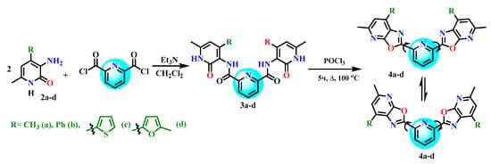
We have previously established that 4-aryl-substituted 3-aminopyridin-2(1H)-ones 2a–d undergo acylation with pyridine-2,6-dicarboxylic acid dichloride in the presence of triethylamine, forming the corresponding diamides 3a–d. Further heating of these diamides with phosphorus oxychloride leads to cyclization products of 2,6-bis(oxazolo[5,4-b]pyridin-2-yl)pyridines 4a–d with the yields of around 80% (Scheme 2).
Scheme 2.
Synthesis of 2,6-bis(oxazolo[5,4-b]pyridin-2-yl)pyridine derivatives 4a–d.
A comparative analysis of the synthesized 2,6-bis(oxazolo[5,4-b]pyridin-2-yl)pyridines 4a–d showed that they are structurally very similar to terpyridines. However, unlike terpyridine, these compounds contain three heteroatoms (one oxygen and two nitrogen atoms) in each peripheral condensed oxazolopyridine ring system. This structural feature may lead to improved coordination and luminescent properties due to the increased number of conjugated bonds, the presence of additional electron-accepting oxygen and nitrogen atoms, and the potential for new conformations resulting from rotation around the C(2) and C(6) bonds of the central pyridine core.
3.2. Photophysical Properties of Compounds
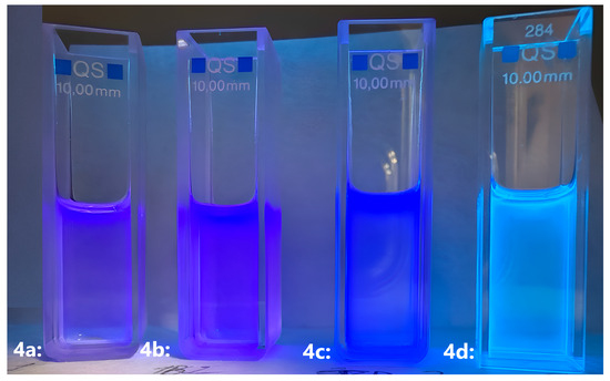
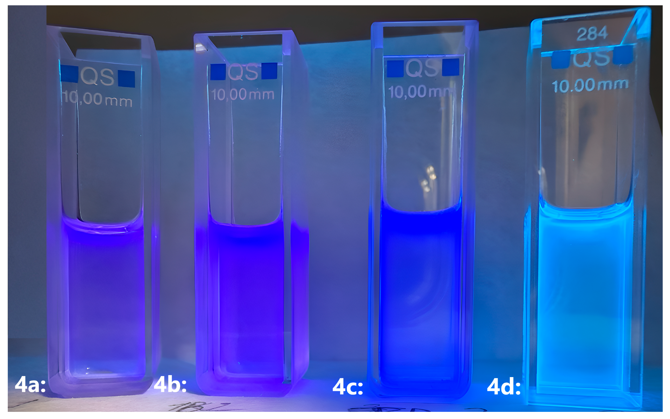
The synthesized 2,6-bis(oxazolo[5,4-b]pyridin-2-yl)pyridines (4a–d) show strong fluorescence in the blue and deep-blue region when exposed to UV light (Figure 1). This property is particularly important for potential applications in organic electronics, given the well-recognized shortage of organic luminophores that emit in the blue range. In addition, these compounds may serve as promising chelating agents and fluorescent markers for various metal cations, including rare-earth elements, with the potential to enhance luminescence upon complex formation.
Figure 1.
Observed luminescence of 2,6-bis(oxazolo[5,4-b]pyridin-2-yl)pyridine samples under UV irradiation.
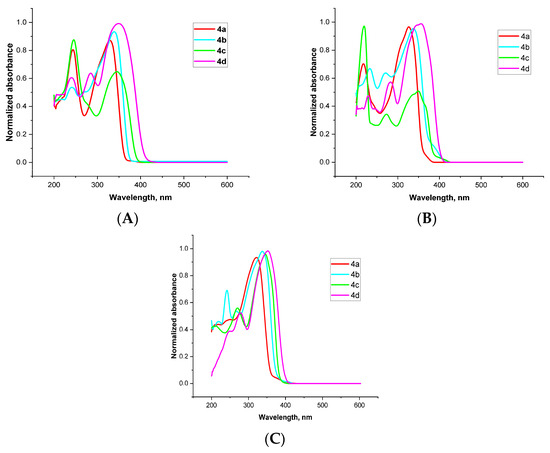
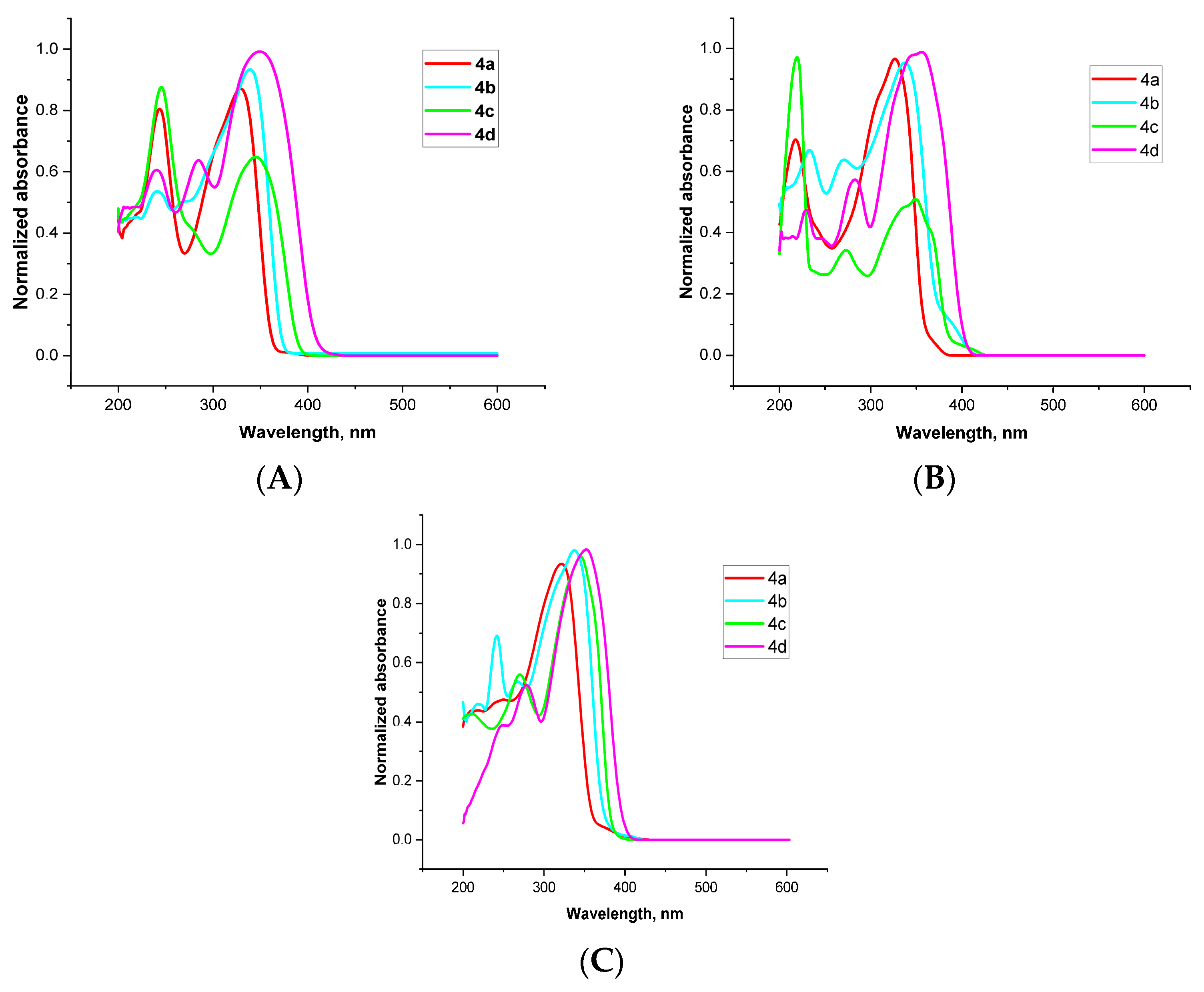
The spectral properties, including λmax, Stokes shift, and quantum yield of the synthesized compounds 4a–d in chloroform, dichloromethane and acetonitrile were measured, as they are of interest as potential luminophores. The initial step involved recording the UV–Vis absorption spectra of the bis(oxazolopyridine) derivatives 4a–d in chloroform, dichloromethane, and acetonitrile. The measurements were carried out at a concentration of 2 × 10⁻⁵ M for compounds 4a, 4c, and 4d, and 1 × 10⁻⁵ M for compound 4b. The spectral characteristics of the compounds 4a–d are summarized in Table 1. The absorption spectra of compounds 4a–d are shown in Figure 2.
Table 1.
Photophysical properties of compounds 4a–d.
Figure 2.
Normalized absorption spectra of bisoxazolo [5,4-b]pyridine derivatives 4a–d in chloroform (A), dichloromethane (B), acetonitrile (C).
It was found that all synthesized 2,2′-bis(oxazolo [5,4-b]pyridine) derivatives show absorption maxima in the range of 323–357 nm. This is attributed to π–π* electronic transitions within the conjugated bis(oxazolo [5,4-b]pyridine) ring system. According to spectral analysis, compounds 4c and 4d display pronounced absorption in the longer wavelength region. Compound 4c showed λabsmax = 351 nm in chloroform, 350 nm in dichloromethane, and 347 nm in acetonitrile, while compound 4d exhibited λabsmax = 343 nm in chloroform, 357 nm in dichloromethane, and 354 nm in acetonitrile. These values are shifted relative to compound 4a, which exhibits λabsmax = 329 nm in chloroform, 327 nm in dichloromethane, and 323 nm in acetonitrile. The methyl group at the 4-position of the pyridine ring in 4a does not participate in conjugation with the π-system, which explains the shorter wavelength absorption. The bathochromic shift observed in compounds 4c and 4d is may be due to the presence of heteroatoms (sulfur and oxygen) with lone electron pairs that engage in additional n–π conjugation with the pyridine ring. The difference in absorption maxima between the thiophene and furan rings is related to the higher polarizability of the sulfur atom compared to oxygen. Compound 4b also shows a bathochromic shift relative to 4a, which is caused by π–π conjugation between the phenyl group and the pyridine ring.
Since the synthesized compounds 4a–d contain two conjugated oxazolo [5,4-b]pyridine ring systems and are of considerable interest as potential luminophores, we aimed to determine their fluorescence quantum yields. This parameter, defined as the ratio of emitted photons to absorbed photons, indicates how efficiently a substance converts absorbed light energy into fluorescent emission.
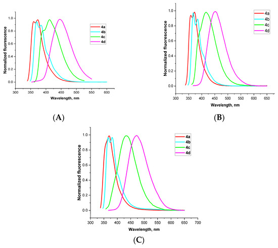
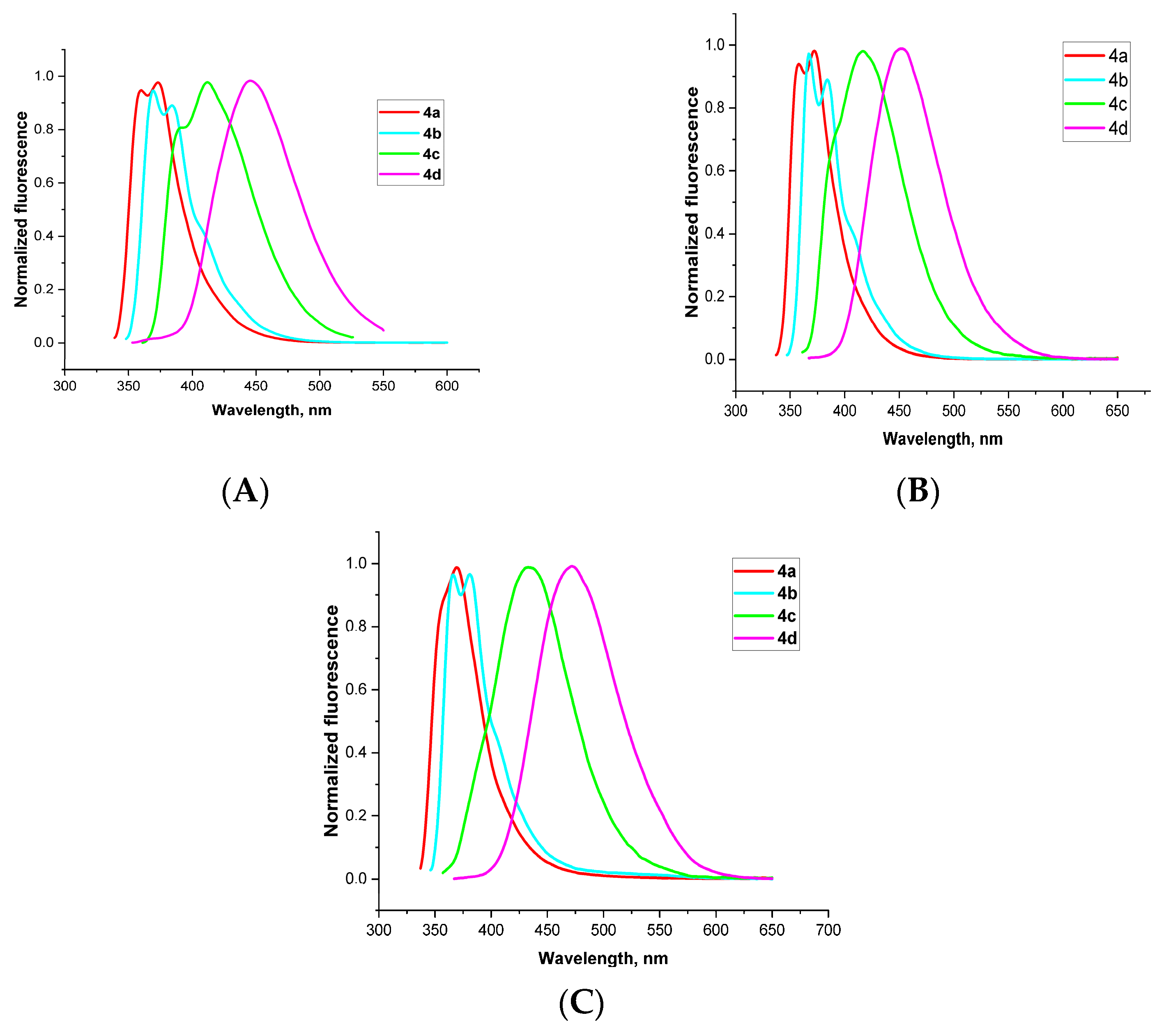
Quantum yield measurements for compounds 4a–d were carried out by recording fluorescence spectra in chloroform, dichloromethane, and acetonitrile, with solution concentrations of 2 × 10⁻⁵ M for 4a, 4c, and 4d, and 1 × 10⁻⁵ M for 4b. The excitation wavelengths were selected to match the absorption maxima of each compound (Figure 3). The emission maxima of the studied compounds fall within the range of 323 to 357 nm, corresponding to blue and deep-blue photoluminescence (Table 1).
Figure 3.
Normalized fluorescence spectra of bisoxazolo [5,4-b]pyridine derivatives 4a–d in chloroform (A), dichloromethane (B), acetonitrile (C).
Thus, we confirmed the photoluminescent properties of the newly synthesized 2,2′-bis(oxazolo [5,4-b]pyridine) derivatives 4a–d and found that they exhibit high fluorescence quantum yields (φi ≈ 0.32–0.84), surpassing well-known standards such as quinine sulfate (φi ≈ 0.55) and tryptophan (φi ≈ 0.15). These findings indicate that new compounds based on 2,6-bis(oxazolo [5,4-b]pyridin-2-yl)pyridines 4a–d are promising both as efficient luminophores and as potential ligands for metal complexation.
3.3. Conformational Analysis and Electronic Structure of the New 2,6-Bis(oxazolo[5,4-b]pyridin-2-yl)pyridines
The photophysical study of compounds 4a–d demonstrated that changes in the 2,2′-bis(oxazolo [5,4-b]pyridine) structure influence their spectral characteristics, fluorescence efficiency, and solvent sensitivity. Compounds 4a–c exhibited shorter-wavelength fluorescence (λemmax ≈ 358–414 nm) in the blue and deep-blue region, while compound 4d displayed a more red-shifted emission (λemmax ≈ 444–474 nm), extending into the blue-green region. Since our focus was on blue and deep-blue luminophores, we selected compounds 4a–c for further computational modeling in order to gain deeper insight into their electronic structure, assess the stability of possible conformations, and simulate their absorption and fluorescence spectra. The calculations were carried out using density functional theory (DFT) with the ωB97XD functional [30] and the 6-311++G(d,p) basis set [31] in Gaussian 16 [32]. Molecular structures were built and visualized using GaussView 6.0 [33].
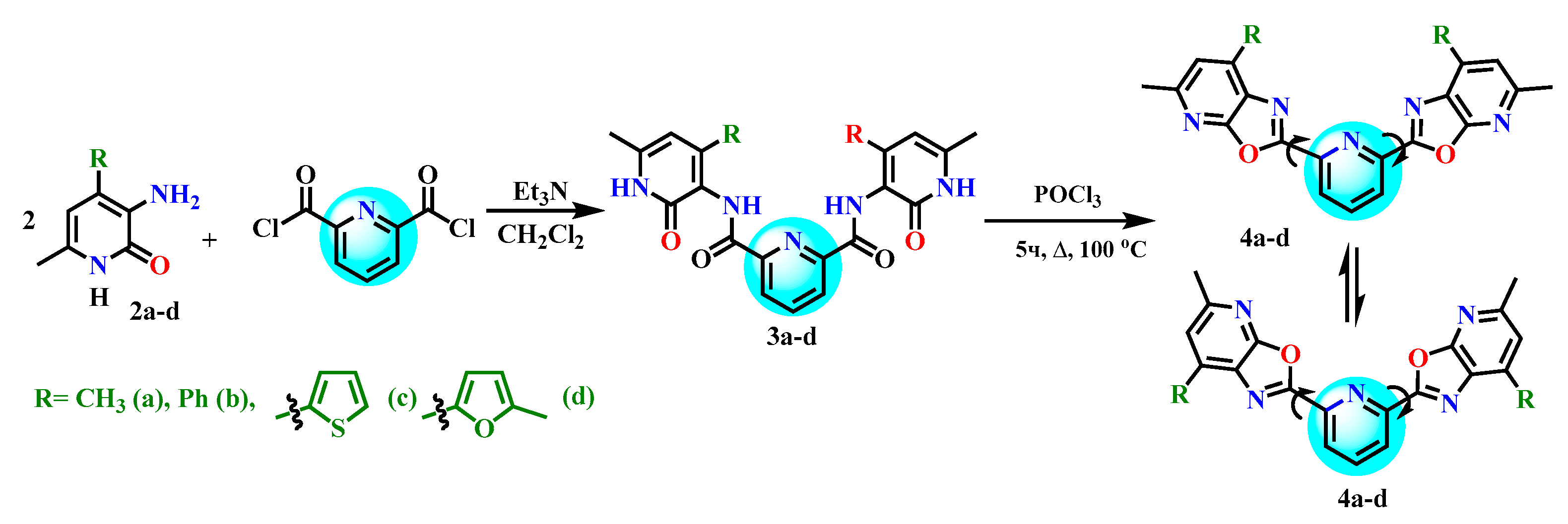
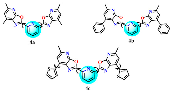
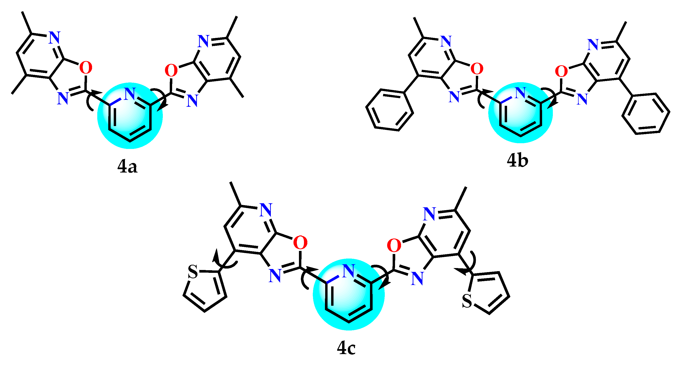
Scheme 3 illustrates the general conformational landscape of compounds 4a, 4b, and 4c, which was further used for conformer search and computational modeling.
Scheme 3.
Conformational landscape of compounds 4a, 4b and 4c.
As shown in Scheme 3, compounds 4a, 4b, and 4c share a terpyridine-like scaffold, where the central pyridine ring is symmetrically substituted at positions 2 and 6 with fused oxazolo[5,4-b]pyridine units carrying different substituents at positions 5 and 7 of the oxazole ring. Compound 4a (2,6-bis(5,7-dimethyloxazolo[5,4-b]pyridin-2-yl)pyridine) carries methyl substituents at both the 5- and 7-positions of each oxazolopyridine unit, which enhance the overall molecular symmetry and increase the electron-donating character. Compound 4b (2,6-bis(5-methyl-7-phenyloxazolo[5,4-b]pyridin-2-yl)pyridine) contains a phenyl substituent at position 7, introducing extended π-conjugation and potential for intermolecular interactions via aromatic stacking. Compound 4c (2,6-bis(5-methyl-7-(thiophen-2-yl)oxazolo[5,4-b]pyridin-2-yl)pyridine) incorporates a thiophene ring at position 7, which may influence electronic properties through sulfur-based heteroaromatic conjugation and increase polarizability. These structural variations are designed to modulate the electronic properties of the terpyridine analogues, with potential applications in luminescent materials and metal complexation.
Based on these structural features, detailed rotational transformation Schemes S1–S3 were developed to systematically explore the conformational landscape of compounds 4a–c. These schemes account for the torsional flexibility of the substituents at the 2 and 6 positions of the central pyridine ring, as well as the additional rotational freedom introduced by the thiophene groups in compound 4c. Transitions between forms occur due to rotation around the C(2)–C and C(6)–C bonds of the central pyridine ring.
The thermodynamic stability of the conformations of compounds 4a–c in different media was evaluated based on their total electronic energy (E) and Gibbs free energy (G), calculated using DFT level of theory with the ωB97XD functional and the 6-311++G(d,p) basis set in Gaussian 16 (Table S2). Table 2 presents the relative total energies (ΔE) and Gibbs free energies (ΔG) of the conformers of compounds 4a–c in vacuum and in solvents (CH2Cl2, CH3CN). In all cases, the most stable conformers have ΔE (ΔG) = 0 and serve as the reference point.
Table 2.
The differences in total energies (ΔE) and Gibbs free energies (ΔG) of the conformers of compounds 4a–c in vacuum and in solvents (CH2Cl2, CH3CN), calculated relative to the lowest-energy conformer.
The data presented in Table 2 show that each compound has a preferred conformation with the lowest Gibbs free energy in a given solvent. Specifically, the most stable conformer is 4a–II for compound 4a, 4b–II for compound 4b, and 4c–II′ for compound 4c.
The presence of solvents reduces the energy differences between conformers, making the less stable forms more accessible. For compounds 4a and 4b, the energy gaps between conformers are relatively small (2–6 kJ/mol in solvents), which suggests that several conformers may coexist. In contrast, compound 4c shows a broader range of conformers with similar energies, particularly in solvents, which may be due to the increased flexibility of the molecular framework and the presence of multiple local minima on the potential energy surface. This may account for the complexity of its spectral interpretation and indicate possible conformational dynamics.
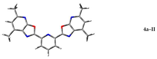
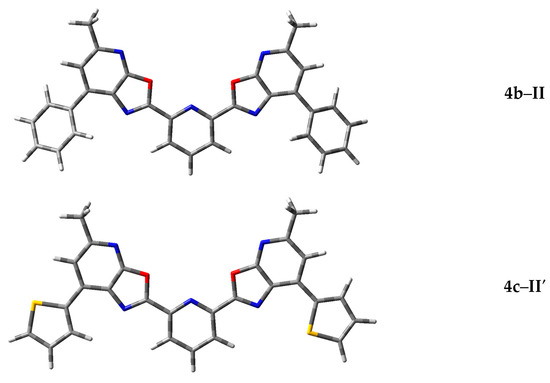
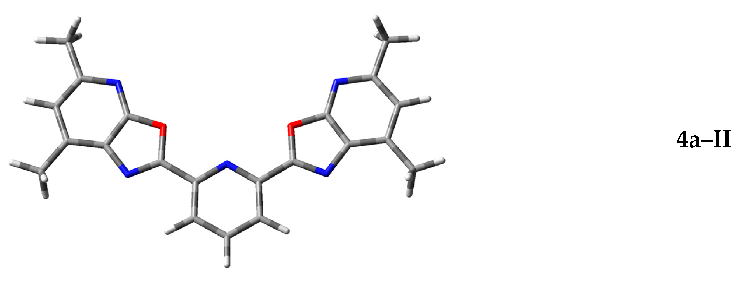
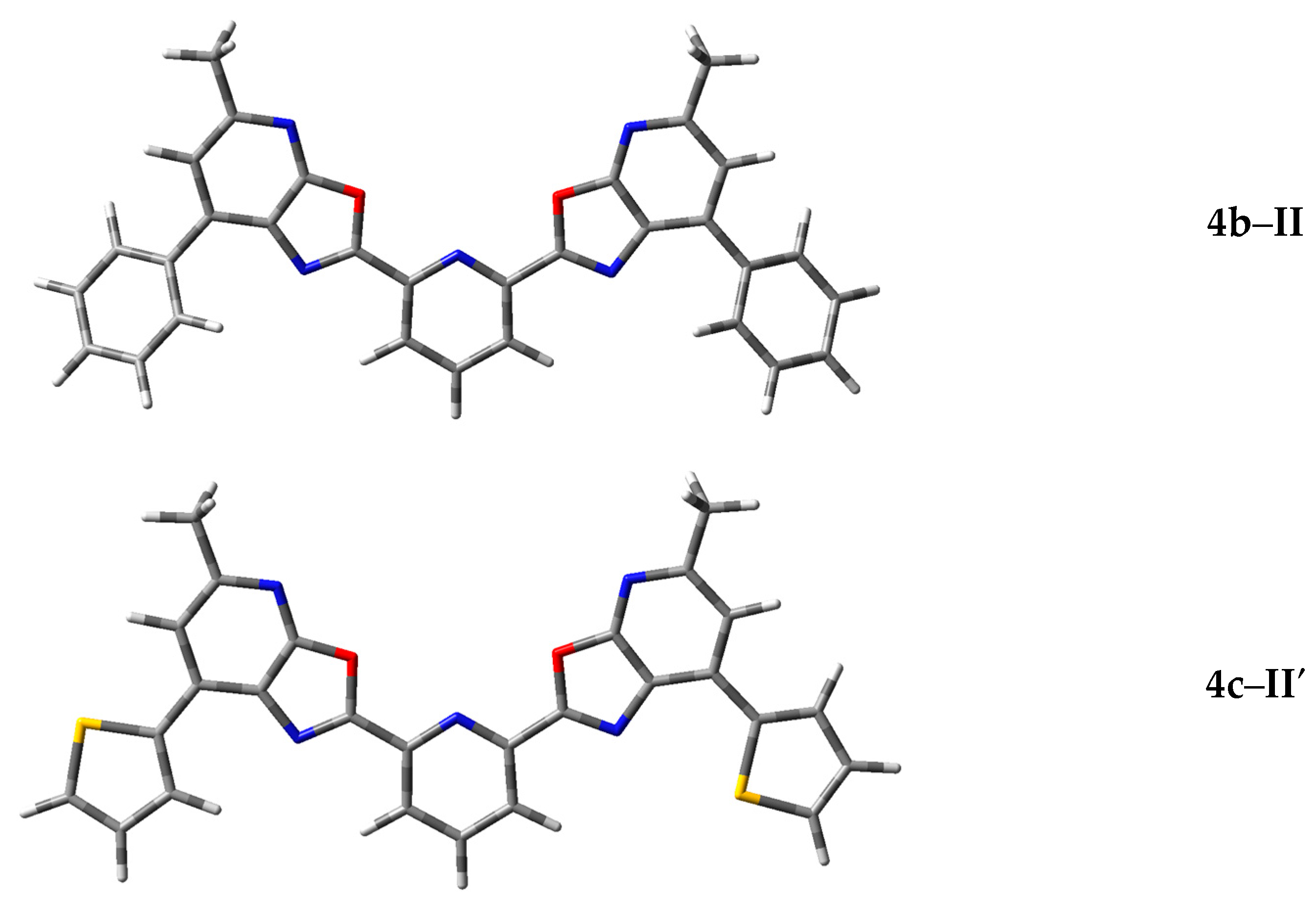
Figure 4 shows the optimized geometries of the most stable conformers 4a–II, 4b–II, and 4c–II′, obtained by DFT geometry optimization (ωB97XD/6-311++G(d,p)) in acetonitrile. Tables S2–S4 provide the atomic Cartesian coordinates of the optimized geometries of compounds 4a–II, 4b–II, and 4c–II′, together with their corresponding total energies in Hartree.
Figure 4.
Optimized geometries of the most stable conformations of compounds 4a–c calculated by DFT (ωB97XD/6-311++G(d,p)) in acetonitrile (CH3CN).
The structures shown in Figure 4 highlight key features of spatial organization that affect the photophysical properties and thermodynamic stability of the compounds. Conformation 4a–II is compact and symmetric, with a planar arrangement of oxazole fragments. This promotes effective conjugation and minimizes steric interactions between substituents, which explains its lowest Gibbs free energy among the 4a conformers. Conformation 4b–II has an extended molecular geometry with a well-developed π-conjugated system. The substituents are positioned to avoid steric clashes while maintaining electronic delocalization. This geometry supports high molar extinction and moderate fluorescence quantum yield. Conformation 4c–II′ is the most branched and spatially crowded structure. The presence of additional donor–acceptor fragments causes molecular bending and leads to an increased Stokes shift. All three conformers show a favorable balance between electronic conjugation and spatial stability, as confirmed by their minimal Gibbs energy values.
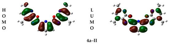
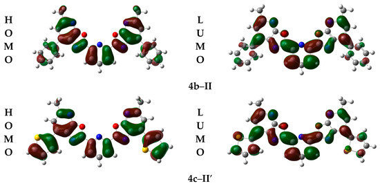
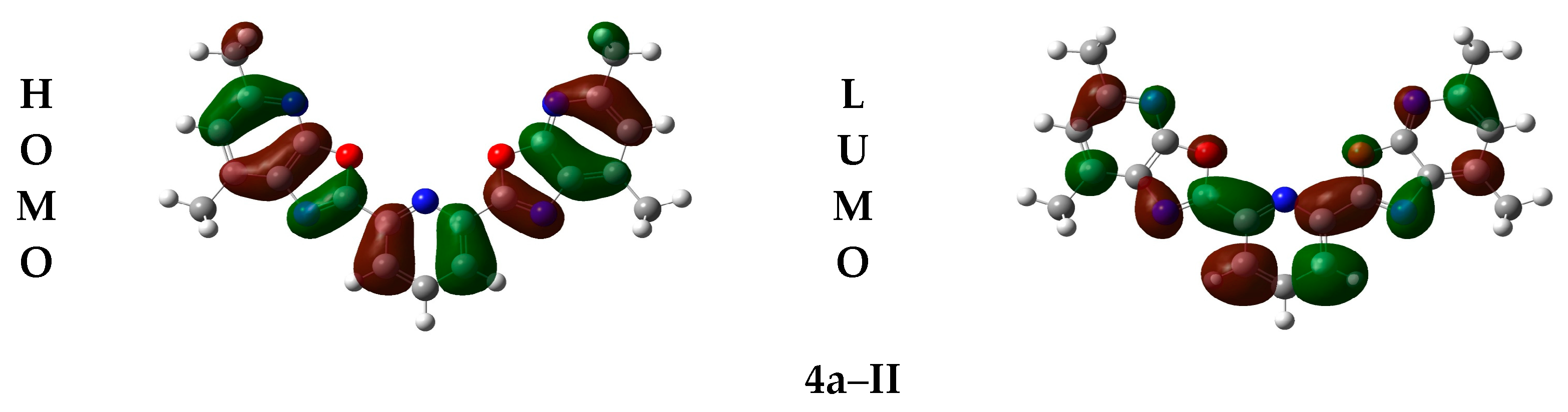
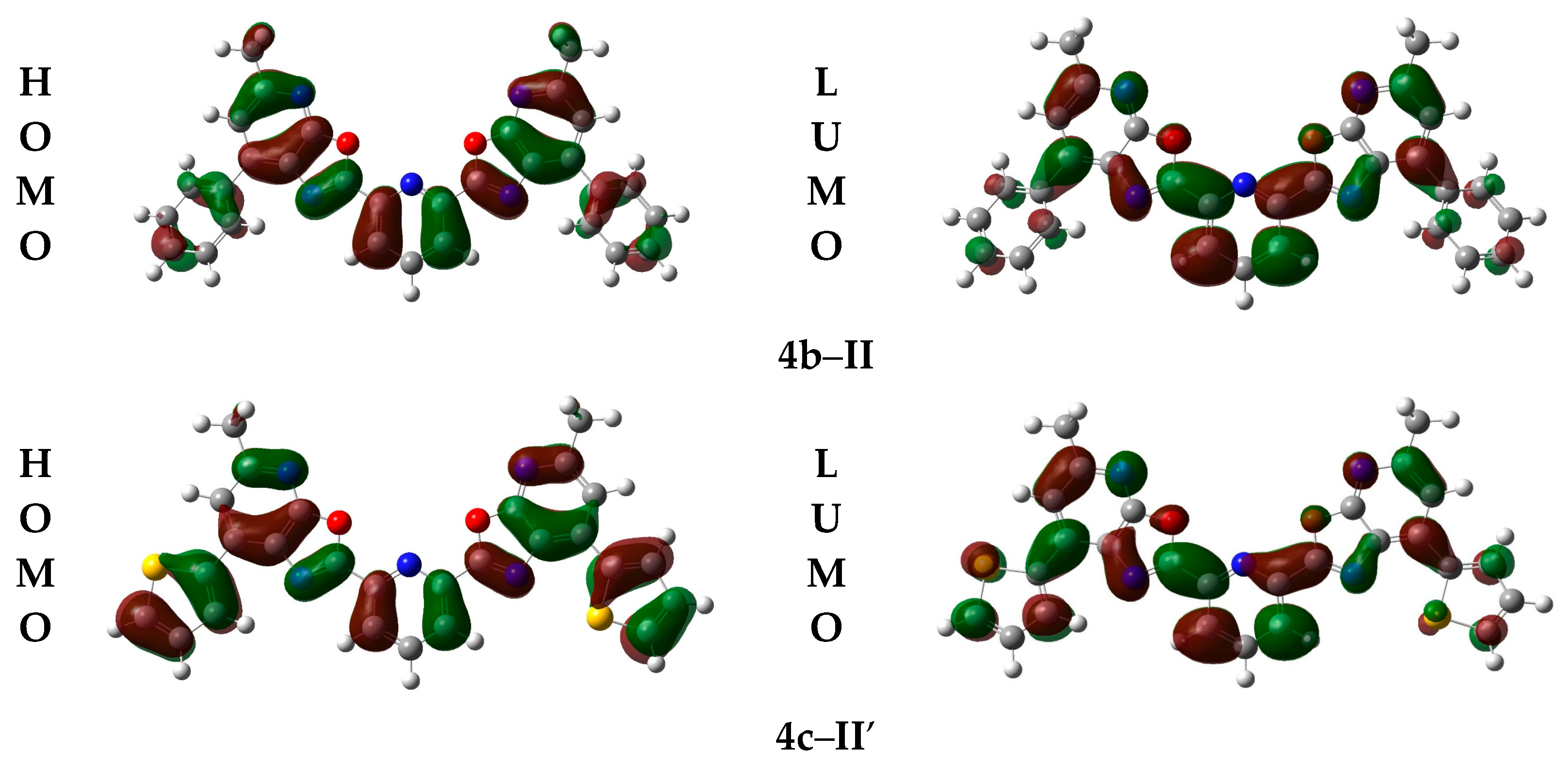
To better understand how spatial arrangement affects the electronic properties of these compounds, the next step was to compare the optimized geometries of conformers 4a–II, 4b–II, and 4c–II′ with the distribution of their HOMO–LUMO frontier molecular orbitals. These orbitals were calculated based on the optimized geometries using DFT (ωB97XD/6-311++G(d,p)) in Gaussian 16, and visualized in GaussView 6.0 (Figure 5).
Figure 5.
HOMO–LUMO frontier molecular orbitals visualization for the most stable conformations 4a–II, 4b–II and 4c–II′, calculated by DFT (ωB97XD/6-311++G(d,p)) in acetonitrile (CH3CN).
The diagrams shown in Figure 5 illustrate the spatial distribution of the frontier molecular orbitals, revealing characteristic electronic features of compounds 4a–c. In all structures, the HOMO is mainly localized on the aromatic and heterocyclic fragments, indicating their donor nature and involvement in electronic transitions. The LUMO is distributed over the p-conjugated system, including acceptor regions, which confirms the dominance of p → π* transitions. Conformation 4c–II′ is particularly notable for its more pronounced spatial separation between HOMO and LUMO, which may contribute to an increased Stokes shift and enhanced fluorescence efficiency.
Table 3 presents the calculated HOMO and LUMO energy values for the most stable conformations 4a–II, 4b–II and 4c–II′, along with their corresponding energy gaps.
Table 3.
Energy characteristics of HOMO and LUMO orbitals for the most stable conformations 4a–II, 4b–II and 4c–II′.
As shown in Table 3, the energy gap between the HOMO and LUMO orbitals decreases consistently from conformation 4a–II to 4c–II′, reflecting differences in the electronic structure and photophysical behavior of the compounds. The largest gap is observed for conformation 4a–II (ΔE = 7.70 eV), indicating high ground-state stability and a tendency toward shorter-wavelength absorption. Conformation 4b–II shows an intermediate gap (ΔE = 7.63 eV), which may suggest easier excitation and a shift toward longer wavelengths. The smallest gap is found in conformation 4c–II′ (ΔE = 7.32 eV), indicating a higher tendency for electronic excitation, which may enhance fluorescence and increase the Stokes shift.
Overall, the decreasing HOMO–LUMO gap from 4a–II to 4c–II′ indicates enhanced photoactivity. The narrowest gap observed for 4c–II′ is consistent with the previously noted spatial separation of the HOMO and LUMO orbitals, which may support more efficient charge transfer in the excited state and stronger fluorescence.
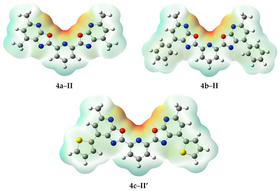
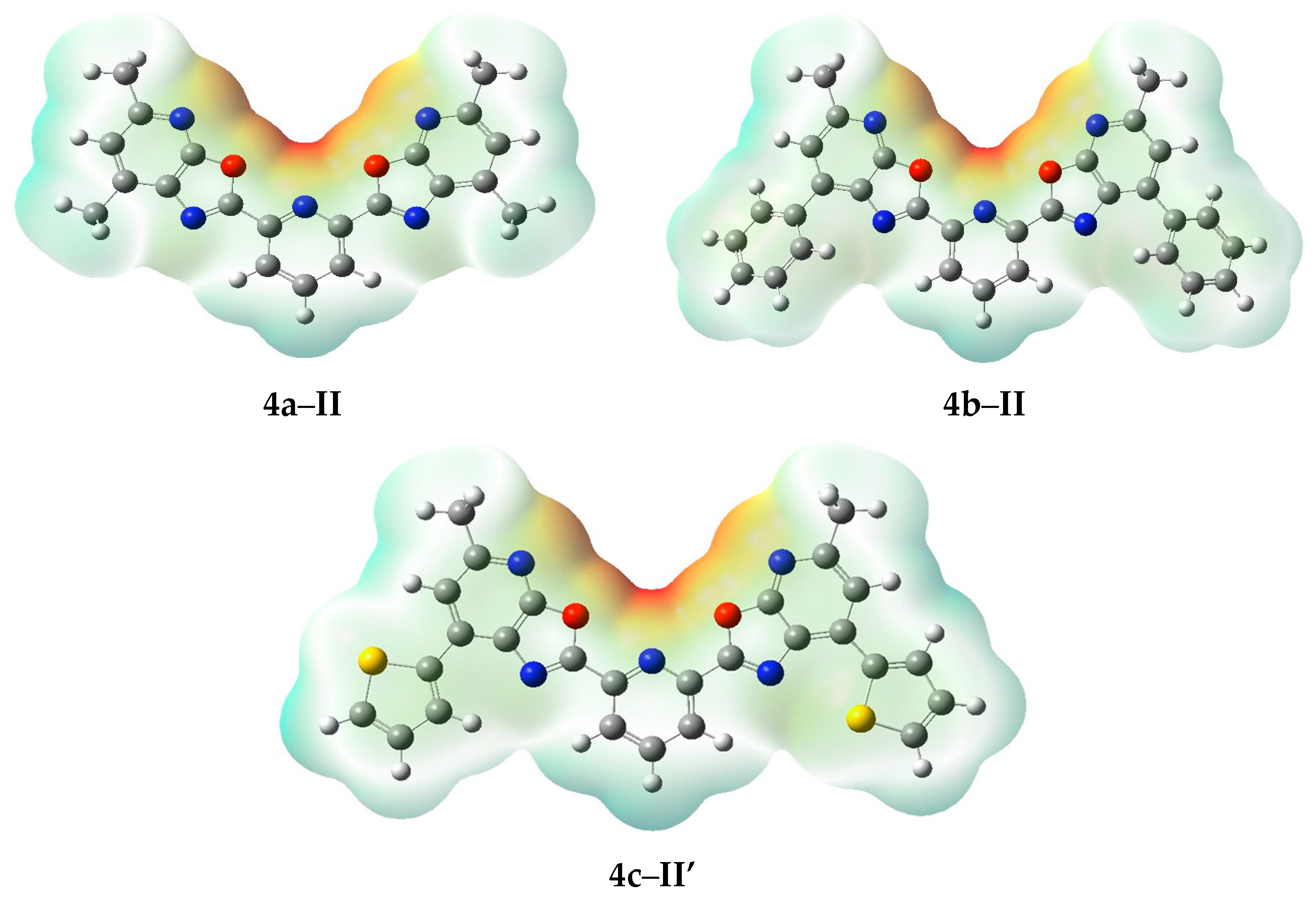
To further explore the distribution of electron density and identify potential reactive sites in the molecules, a molecular electrostatic potential (MEP) analysis was performed for the most stable conformations 4a–II, 4b–II and 4c–II′ (Figure 6). It was shown, that the molecular electrostatic potential (MEP) surfaces of the studied conformers exhibit comparable ranges. The potential ranges from −0.095 to +0.095 Hartree (−248.0 to +248.0 kJ/mol) for conformer 4a–II, from −0.092 to +0.092 Hartree (−240.7 to +240.7 kJ/mol) for conformer 4b–II and from −0.091 and +0.091 Hartree (−238.9 to +238.9 kJ/mol) for conformer 4c–II′. These values indicate that all three compounds possess similar charge distributions and polarity across their molecular surfaces.
Figure 6.
Molecular Electrostatic Potential (MEP) maps for the most stable conformations 4a–II, 4b–II and 4c–II′ calculated by DFT (ωB97XD/6-311++G(d,p)) in acetonitrile (CH3CN) MEP color scale: red areas indicate regions with the most negative potential; blue areas show regions with positive potential; green and yellow zones represent neutral charge distribution.
As can be seen in Figure 6, the regions of most negative potential (red zones) of conformations 4a–II, 4b–II and 4c–II′ are localized near electronegative atoms such as oxygen and nitrogen, indicating possible sites for nucleophilic attack and hydrogen bond acceptance. Conversely, the most positive potential areas (blue zones) are found around hydrogen atoms bound to heteroatoms, highlighting potential sites for electrophilic interactions and hydrogen bond donation. These distributions suggest similar polarity patterns across the conformers, with subtle differences that may influence their intermolecular recognition and reactivity. Among the studied structures, compound 4a shows the most pronounced spatial separation between donor and acceptor regions.
3.4. DFT-Based Simulation of Electronic Absorption and Luminescence Properties
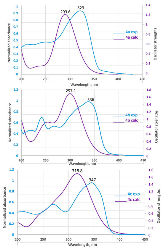
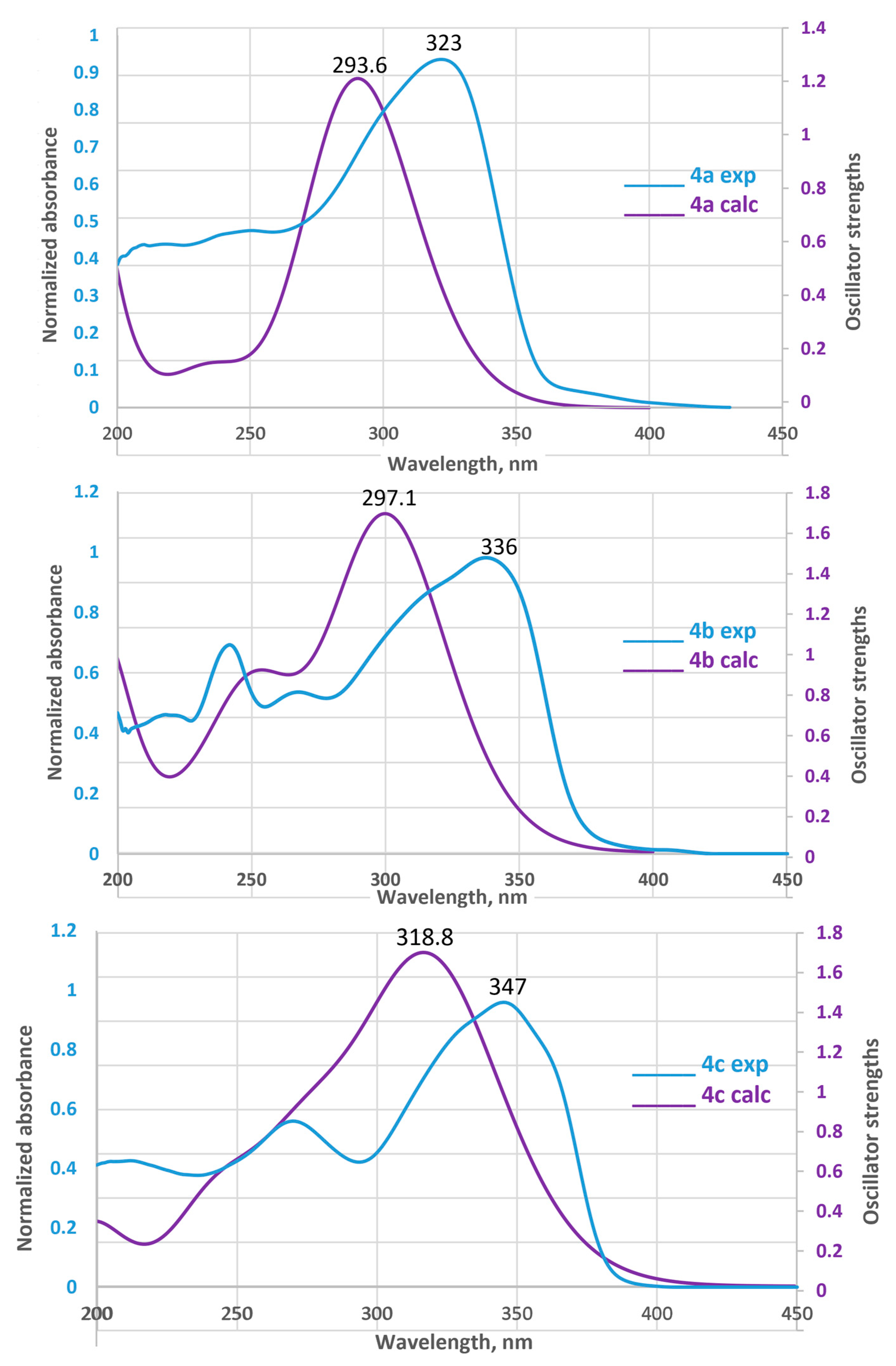
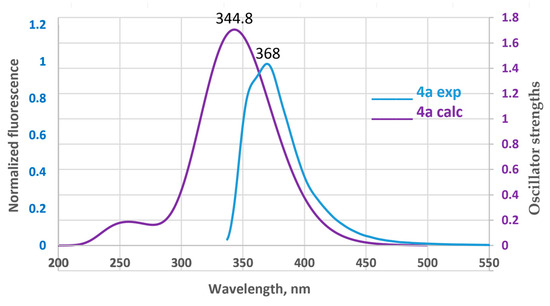
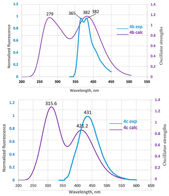
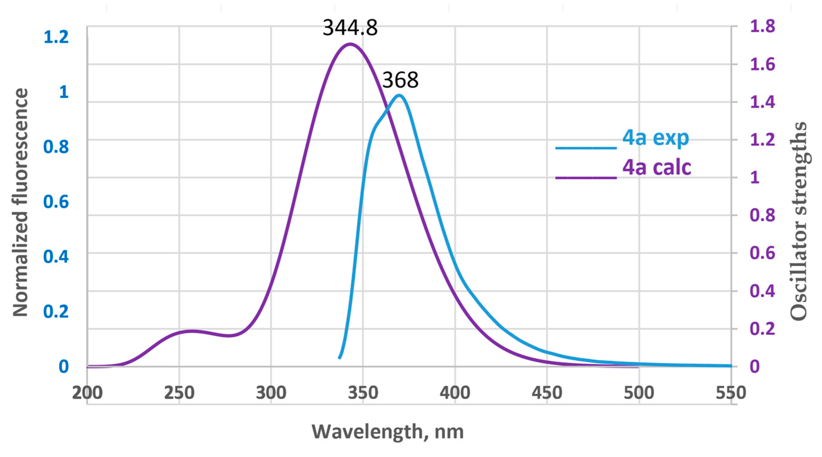
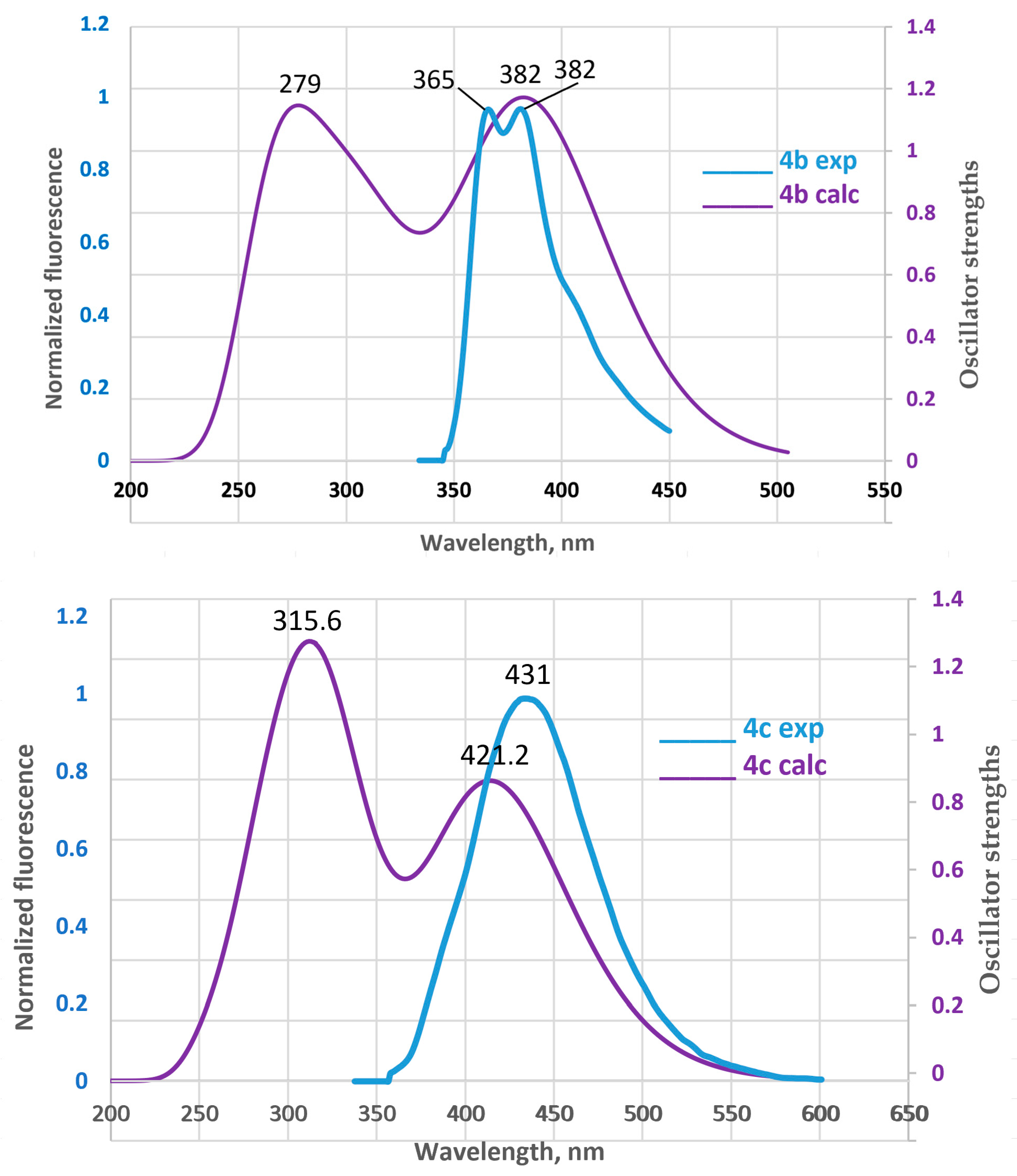
To elucidate the electronic transitions and photophysical behavior of the studied conformations, we performed TD-DFT calculations of UV-vis absorption spectra and emission luminescence spectra for 4a–II, 4b–II and 4c–II′ conformations, aiming to establish structure–property relationships relevant to optoelectronic applications. The calculated absorption and emission spectra are presented in Figure 7.
Figure 7.
TD-DFT calculated UV–vis absorption spectra in acetonitrile (CH3CN) for the 4a–II, 4b–II and 4c–II′ conformations.
The calculated UV–vis absorption spectra of the 4a–II, 4b–II and 4c–II′ conformations exhibit distinct profiles, reflecting differences in their electronic structures and transition energies (Figure 7). All spectra show pronounced absorption bands in the 290–320 nm range, characteristic of π→π* or n→π* electronic transitions and corresponding to experimental peaks in the range of 323–347 nm. The comparison between the experimental and calculated spectra shows a satisfactory level of agreement: the overall spectral pattern and the positions of the main peaks coincide qualitatively. At the same time, exact quantitative matching is not observed, as the calculated absorption maxima are shifted by 25–40 nm relative to the experimental values. This deviation is expected, since TD-DFT methods are well known to reliably reproduce spectral trends but typically exhibit absolute errors of 0.3–0.5 eV (approximately 25–40 nm), especially for π→π* transitions [52,53].
The 4a–II conformation displays the most blue-shifted absorption maximum, located near 293.6 nm, indicating the largest HOMO–LUMO energy gap among the three compounds. The 4b–II conformation peaks slightly later, around 297.1 nm, with comparable intensity and a narrower band shape, suggesting efficient electronic excitation with minimal structural distortion. In contrast, the 4c–II′ shows the most red-shifted and intense absorption band, centered near 318.8 nm, accompanied by a broader spectral profile. This behavior implies a reduced excitation energy and potentially greater charge delocalization or π-conjugation within the molecular framework. The observed bathochromic shifts and variations in peak intensity correlate with differences in molecular geometry, electronic distribution, and substituent effects.
The simulated emission spectra of the three compounds reveal distinct photophysical behaviors, as illustrated in Figure 8.
Figure 8.
TD-DFT calculated UV–vis emission spectra in acetonitrile (CH3CN) for the 4a–II, 4b–II and 4c–II′ conformations.
As can be seen in Figure 8, the 4a–II conformation exhibits the most intense and narrow luminescence band, with a maximum at approximately 344.8 nm, indicating the largest energy gap between the first excited singlet state (S1) and the ground state (S0) and corresponding to the experimental peak in the 368 nm range. In contrast, the 4b–II and 4c–II′ conformations display progressively broader and red-shifted emission profiles, with maxima near 382 (experimental peak 382 nm) and 421.2 nm (experimental peak 431 nm), respectively. These spectral shifts suggest a reduction in the S1–S0 energy gap, likely due to conformational relaxation, substituent effects, or enhanced charge delocalization. The observed trend 4a–II > 4b–II > 4c–II′ in both peak intensity and energy—correlates with experiment and reflect differences in electronic structure.
Overall, conformation 4a–II shows the most efficient luminescence, which is likely due to its high molecular symmetry and minimal energy loss from conformational changes. The weaker luminescence observed for conformations 4b–II and 4c–II′ may result from greater conformational flexibility, intermolecular interactions, or differences in electronic structure. The compound 4a can be considered a promising candidate for use as a fluorophore or in OLED materials, while 4b and 4c are of interest for studying how molecular structure affects spectral properties.
4. Conclusions
The aim of this study was to investigate a new group of structurally diverse bis-oxazolo[5,4-b]pyridine derivatives, selected for their close similarity to terpyridines, well-known ligands in coordination chemistry due to their flexible structure and strong binding capacity. Four new 2,6-bis(oxazolo[5,4-b]pyridin-2-yl)pyridine derivatives 4a–d were synthesized using our established approach to the cyclization of amide derivatives of 3-aminopyridin-2(1H)-ones, with pyridine-2,6-dicarboxylic acid and its dichloride serving as key acidic components. The newly synthesized compounds were characterized by melting point, HRMS, and 1H, 13C NMR spectroscopy, and were further examined for photophysical properties (λmax, Stokes shift, quantum yield) in chloroform, dichloromethane, and acetonitrile. It was found that compounds 4a–c exhibited strong blue or deep blue fluorescence emission in the region 357–474 nm under UV light attributed to π–π* electronic transitions within the conjugated bis(oxazolo[5,4-b]pyridine) ring system and high fluorescence quantum yields (φi ≈ 0.32–0.84), highlighting their potential as phosphors for use in organic electronics. Compounds 4a–c were further examined by DFT (ωB97XD/6-311++G(d,p)) computational modeling to assess their electronic structure, conformational stability, and photophysical behavior. The analysis revealed that the most stable conformations (4a–II, 4b–II and 4c–II′) exhibit a decreasing HOMO–LUMO gap and exhibit a progressive decrease in the HOMO–LUMO gap accompanied by enhanced photoactivity from 4a to 4c. Among the studied compounds, 4a can be identified as the most promising luminophore due to its intense and narrow luminescence band, which arises from its high molecular symmetry and stable emission behavior. Future research can focus on expanding the structural diversity of bis(oxazolo[5,4-b]pyridine) derivatives and studying their coordination behavior with metal ions to develop new functional materials for optoelectronic applications.
Supplementary Materials
The following supporting information can be downloaded at: https://www.mdpi.com/article/10.3390/compounds6010012/s1, Copies of 1H, 13C NMR spectra and Mass spectra; Scheme S1. Detailed conformational landscape of compound 4a; Scheme S2. Detailed conformational landscape of compound 4b; Scheme S3. Detailed conformational landscape of compound 4c; Table S1. Total Electronic Energy (Etotal) and Gibbs Free Energy (G) of various conformations of compounds 4a–c in vacuum and in solvents (CH2Cl2, CH3CN); Table S2. Atomic cartesian coordinates of optimized geometry of 4a–II; Table S3. Atomic cartesian coordinates of optimized geometry of 4b–II; Table S4. Atomic cartesian coordinates of optimized geometry of 4c–II′.
Author Contributions
Conceptualization, methodology, resources, supervision, I.V.K. and I.A.P.; software, I.A.P.; investigation, validation, formal analysis, data curation, I.V.P., S.S.V. and A.S.R.; writing—original draft preparation, writing—review and editing, visualization, I.V.P., I.V.K. and I.A.P.; writing—review and editing, visualization, A.S.N., project administration, funding acquisition, I.V.P. All authors have read and agreed to the published version of the manuscript.
Funding
The study was supported by a grant from the Russian Science Foundation (No. 24-23-00472, «Development of methods for the preparation and practical application of new derivatives of bisoxazolo[5,4-b]pyridine with potential luminescent, complexing and catalytic activity», https://rscf.ru/en/project/24-23-00472/ (accessed on 14 November 2025).
Data Availability Statement
The original contributions presented in this study are included in the article/Supplementary Material. Further inquiries can be directed to the corresponding authors.
Acknowledgments
Spectrophotometric studies were carried out using the equipment of the Center for Collective Use “Rational Nature Management and Physicochemical Research” of University of Tyumen. NMR studies were conducted using equipment purchased under the “Priority 2030” program.
Conflicts of Interest
The authors declare no conflict of interest.
References
- Wannas, F.A.; Gafel, R.A.; Jaffer, N.D.; Husien, N.S. A Literature Review on The Fluorescence and phosphorescent. Am. Int. J. Sci. Eng. Res. 2019, 2, 47–55. [Google Scholar] [CrossRef] [Scilit]
- Que, E.L.; Domaille, D.W.; Chang, C.J. Metals in Neurobiology: Probing Their Chemistry and Biology with Molecular Imaging. Chem. Rev. 2008, 108, 1517–1549. [Google Scholar] [CrossRef] [Scilit]
- Gonçalves, M.S.T. Fluorescent Labeling of Biomolecules with Organic Probes. Chem. Rev. 2009, 109, 190–212. [Google Scholar] [CrossRef] [Scilit]
- Urano, Y.; Asanuma, D.; Hama, Y.; Koyama, Y.; Barrett, T.; Kamiya, M.; Nagano, T.; Watanabe, T.; Hasegawa, A.; Choyke, P.L. Selective Molecular Imaging of Viable Cancer Cells with pH-Activatable Fluorescence Probes. Nat. Med. 2008, 15, 104–109. [Google Scholar] [CrossRef] [Scilit]
- Schäferling, M. The Art of Fluorescence Imaging with Chemical Sensors. Angew. Chem. Int. Ed. 2012, 51, 3532–3554. [Google Scholar] [CrossRef] [Scilit]
- Köhler, A.; Wilson, J.S.; Friend, R.H. Fluorescence and Phosphorescence in Organic Materials. Adv. Eng. Mater. 2002, 4, 453–459. [Google Scholar] [CrossRef] [Scilit]
- Shcheslavskiy, V.I.; Shirmanova, M.V.; Yashin, K.S.; Rück, A.C.; Skala, M.C.; Becker, W. Fluorescence Lifetime Imaging Techniques-A Review on Principles, Applications and Clinical Relevance. J. Biophotonics 2025, 18, e202400450. [Google Scholar] [CrossRef] [Scilit]
- Hagfeldt, A.; Boschloo, G.; Sun, L.; Kloo, L.; Pettersson, H. Dye-Sensitized Solar Cells. Chem. Rev. 2010, 110, 6595–6663. [Google Scholar] [CrossRef] [Scilit]
- Hartwig, J.F. Organotransition Metal Chemistry: From Bonding to Catalysis; University Science Books: Sausalito, CA, USA, 2010; 1128p. [Google Scholar]
- Liu, Z.; Sadler, P.J. Organoiridium complexes: Anticancer agents and catalysts. Acc. Chem. Res. 2014, 47, 1174–1185. [Google Scholar] [CrossRef] [Scilit]
- Luca, O.R.; Crabtree, R.H. Redox-active ligands in catalysis. Chem. Soc. Rev. 2013, 42, 1440–1459. [Google Scholar] [CrossRef] [Scilit]
- Peris, E.; Crabtree, R.H. Recent homogeneous catalytic applications of chelate and pincer N-heterocyclic carbenes. Coord. Chem. Rev. 2004, 248, 2239–2246. [Google Scholar] [CrossRef] [Scilit]
- Vougioukalakis, G.C.; Grubbs, R.H. Ruthenium-based heterocyclic carbene-coordinated olefin metathesis catalysts. Chem. Rev. 2010, 110, 1746–1787. [Google Scholar] [CrossRef] [Scilit]
- Chi, Y.; Chou, P.T. Transition-metal phosphors with cyclometalating ligands: Fundamentals and applications. Chem. Soc. Rev. 2010, 39, 638–655. [Google Scholar] [CrossRef] [Scilit]
- Deiters, A.; Martin, S.F. Synthesis of oxygen-and nitrogen-containing heterocycles by ring-closing metathesis. Chem. Rev. 2004, 104, 2199–2238. [Google Scholar] [CrossRef] [Scilit]
- Fache, F.; Schulz, E.; Tommasino, M.L.; Lemaire, M. Nitrogen-containing ligands for asymmetric homogeneous and heterogeneous catalysis. Chem. Rev. 2000, 100, 2159–2232. [Google Scholar] [CrossRef] [Scilit]
- Wei, C.; He, Y.; Shi, X.; Song, Z. Terpyridine-metal complexes: Applications in catalysis and supramolecular chemistry. Coord. Chem. Rev. 2019, 385, 1–19. [Google Scholar] [CrossRef] [Scilit]
- Chelucci, G.; Thummel, R.P. Chiral 2,2′-bipyridines, 1,10-phenanthrolines, and 2,2′:6′,2″-terpyridines: Syntheses and applications in asymmetric homogeneous catalysis. Chem. Rev. 2002, 102, 3129–3170. [Google Scholar] [CrossRef] [Scilit]
- Winter, A.; Newkome, G.R.; Schubert, U.S. Catalytic applications of terpyridines and their transition metal complexes. ChemCatChem 2011, 3, 1384–1406. [Google Scholar] [CrossRef] [Scilit]
- Shabunina, O.V.; Starnovskaya, E.S.; Shaitz, Y.K.; Kopchuk, D.S.; Sadieva, L.K.; Kim, G.A.; Taniya, O.S.; Nikonov, I.L.; Santra, S.; Zyryanov, G.V.; et al. Asymmetrically substituted 5,5″-diaryl-2,2′:6′,2″-terpyridines as efficient fluorescence “turn-on” probes for Zn2+ in food/cosmetic samples and human urine. J. Photochem. Photobiol. A Chem. 2021, 408, 113101. [Google Scholar] [CrossRef] [Scilit]
- Armspach, D.; Constable, E.C.; Housecroft, C.E.; Neuburger, M.; Zehnder, M. Carbaborane-functionalised 2,2′:6′,2″-terpyridine ligands for metallosupramolecular chemistry: Syntheses, complex formation, and the crystal and molecular structures of 4′-(ortho-carboranyl)-2,2′:6′,2″-terpyridine and 4′-(ortho-carboranylpropoxy)-2,2′:6′,2″-terpyridine. J. Organomet. Chem. 1998, 550, 193–206. [Google Scholar]
- Constable, E.C. 2,2′:6′,2″-Terpyridines: From chemical obscurity to common supramolecular motifs. Chem. Soc. Rev. 2007, 36, 246–253. [Google Scholar] [CrossRef] [Scilit]
- Kamata, K.; Suzuki, A.; Nakai, Y.; Nakazawa, H. Catalytic hydrosilylation of alkenes by iron complexes containing terpyridine derivatives as ancillary ligands. Organometallics 2012, 31, 3825–3828. [Google Scholar] [CrossRef] [Scilit]
- Tondreau, A.M.; Atienza, C.C.H.; Darmon, J.M.; Milsmann, C.; Hoyt, H.M.; Weller, K.J.; Nye, S.A.; Lewis, K.M.; Boyer, J.; Delis, J.G.P.; et al. Synthesis, electronic structure, and alkene hydrosilylation activity of terpyridine and bis(imino)pyridine iron dialkyl complexes. Organometallics 2012, 31, 4886–4893. [Google Scholar] [CrossRef] [Scilit]
- Hwang, S.H.; Moorefield, C.N.; Fronczek, F.R.; Lukoyanova, O.; Echegoyen, L.; Newkome, G.R. Construction of triangular metallomacrocycles:[M 3 (1,2-bis(2,2′:6′,2″-terpyridin-4-yl-ethynyl)benzene)3][M = Ru (ii), Fe (ii), 2Ru (ii) Fe (ii)]. Chem. Commun. 2005, 6, 713–715. [Google Scholar] [CrossRef] [Scilit]
- Kulakov, I.V.; Matsukevich, M.V.; Shulgau, Z.T.; Sergazy, S.; Seilkhanov, T.M.; Puzari, A.; Fisyuk, A.S. Synthesis and antiradical activity of 4-aryl(hetaryl)-substituted 3-aminopyridin-2(1H)-ones. Chem. Heterocycl. Compd. 2015, 51, 991–996. [Google Scholar] [CrossRef] [Scilit]
- Mohamed, A.A.; Nassr, A.A.; Sadeek, S.A.; Rashid, N.G.; Abd El-Hamid, S.M. First Report on Several NO-Donor Sets and Bidentate Schiff Base and Its Metal Complexes: Characterization and Antimicrobial Investigation. Compounds 2023, 3, 376–389. [Google Scholar] [CrossRef] [Scilit]
- Moiseev, R.V.; Morrison, P.W.J.; Steele, F.; Khutoryanskiy, V.V. Penetration Enhancers in Ocular Drug Delivery. Pharmaceutics 2019, 11, 321. [Google Scholar] [CrossRef] [Scilit]
- Shatsauskas, A.L.; Abramov, A.A.; Chernenko, S.A.; Kostyuchenko, A.S.; Fisyuk, A.S. Synthesis and Photophysical Properties of 3-Amino-4-arylpyridin-2 (1H)-ones. Synthesis 2020, 52, 227–238. [Google Scholar] [CrossRef] [Scilit]
- Chai, J.-D.; Head-Gordon, M. Long-range corrected hybrid density functionals with damped atom–atom dispersion corrections. Phys. Chem. Chem. Phys. 2008, 10, 6615–6620. [Google Scholar] [CrossRef] [Scilit]
- Krishnan, R.; Binkley, J.S.; Seeger, R.; Pople, J.A. Self-consistent molecular orbital methods. XX. A basis set for correlated wave functions. J. Chem. Phys. 1980, 72, 650–654. [Google Scholar] [CrossRef] [Scilit]
- Frisch, M.J.; Trucks, G.W.; Schlegel, H.B.; Scuseria, G.E.; Robb, M.A.; Cheeseman, J.R.; Scalmani, G.; Barone, V.; Petersson, G.A.; Nakatsuji, H.; et al. Gaussian 16, Revision A.03; Gaussian, Inc.: Wallingford, CT, USA, 2016. [Google Scholar]
- Dennington, R.; Keith, T.; Millam, J. GaussView, Version 6; Semichem Inc.: Shawnee Mission, KS, USA, 2016; Available online: http://gaussian.com (accessed on 1 September 2025).
- Huber, R.G.; Margreiter, M.A.; Fuchs, J.E.; von Grafenstein, S.; Tautermann, C.S.; Liedl, K.R.; Fox, T. Heteroaromatic π-Stacking Energy Landscapes. J. Chem. Inf. Model. 2014, 54, 1371–1379. [Google Scholar] [CrossRef] [Scilit]
- Barone, V.; Cossi, M. Quantum Calculation of Molecular Energies and Energy Gradients in Solution by a Conductor Solvent Model. J. Phys. Chem. A 1998, 102, 1995–2001. [Google Scholar] [CrossRef] [Scilit]
- Hehre, W.J.; Radom, L.; Schleyer, P.v.R.; Pople, J.A. Ab Initio Molecular Orbital Theory; Wiley: New York, NY, USA, 1986; pp. 235–240. [Google Scholar]
- Young, D.C. Computational Chemistry: A Practical Guide for Applying Techniques to Real-World Problems; Wiley-Interscience: New York, NY, USA, 2001; pp. 104–110. [Google Scholar]
- Tasi, G.; Pálinkó, I. Using molecular electrostatic potential maps for similarity studies. In Molecular Similarity II. Topics in Current Chemistry; Springer: Berlin/Heidelberg, Germany, 1995; Volume 174, pp. 45–71. [Google Scholar] [CrossRef] [Scilit]
- Rakhimzhanova, A.S.; Muzaparov, R.A.; Pustolaikina, I.A.; Kurmanova, A.F.; Nikolskiy, S.N.; Kapishnikova, D.D.; Stalinskaya, A.L.; Kulakov, I.V. Series of Novel Integrastatins Analogues: In Silico Study of Physicochemical and Bioactivity Parameters. Eurasian J. Chem. 2024, 29, 44–60. [Google Scholar] [CrossRef] [Scilit]
- Stratmann, R.E.; Scuseria, G.E.; Frisch, M.J. An efficient implementation of time-dependent density-functional theory for the calculation of excitation energies of large molecules. J. Chem. Phys. 1998, 109, 8218–8224. [Google Scholar] [CrossRef] [Scilit]
- Shatsauskas, A.L.; Shatalin, Y.V.; Shubina, V.S.; Chernenko, S.A.; Kostyuchenko, A.S.; Fisyuk, A.S. 5-Ethyl-5,6-dihydrobenzo[c][1,7]naphthyridin-4(3H)-ones—A new class of fluorescent dyes. Dyes Pigments 2022, 204, 110388. [Google Scholar] [CrossRef] [Scilit]
- Shatsauskas, A.; Shatalin, Y.; Shubina, V.; Zablodtskii, Y.; Chernenko, S.; Samsonenko, A.; Kostyuchenko, A.; Fisyuk, A. Synthesis and application of new 3-amino-2-pyridone based luminescent dyes for ELISA. Dyes Pigments 2021, 187, 109072. [Google Scholar] [CrossRef] [Scilit]
- Åberg, V.; Sellstedt, M.; Hedenström, M.; Pinkner, J.S.; Hultgren, S. Design, synthesis and evaluation of peptidomimetics based on substituted bicyclic 2-pyridones—Targeting virulence of uropathogenic E. coli. J. Bioorg. Med. Chem. 2006, 14, 7563–7581. [Google Scholar] [CrossRef] [Scilit]
- Verissimo, E.; Berry, N.; Gibbons, P.; Cristiano, M.L.S.; Rosenthal, P.J.; Gut, J.; Ward, S.A.; O’Neill, P.M. Design and synthesis of novel 2-pyridone peptidomimetic falcipain 2/3 inhibitors. Bioorg. Med. Chem. Lett. 2008, 18, 4210–4214. [Google Scholar] [CrossRef] [Scilit]
- Zhu, S.; Hudson, T.H.; Kyle, D.E.; Lin, A.J. Synthesis and in vitro studies of novel pyrimidinyl peptidomimetics as potential antimalarial therapeutic agents. J. Med. Chem. 2002, 45, 3491–3496. [Google Scholar] [CrossRef] [Scilit]
- Palamarchuk, I.V.; Shulgau, Z.T.; Kharitonova, M.A.; Kulakov, I.V. Synthesis and neurotropic activity of new 3-(arylmethyl)aminopyridine-2(1H)-one. Chem. Pap. 2021, 75, 4729–4739. [Google Scholar] [CrossRef] [Scilit]
- Kulakov, I.V.; Shatsauskas, A.L.; Matsukevich, M.V.; Palamarchuk, I.V.; Seilkhanov, T.M.; Gatilov, Y.V.; Fisyuk, A.S. A New Approach to the Synthesis of Benzo[c][1,7] naphthyridin-4(3H)-ones. Synthesis 2017, 49, 3700–3709. [Google Scholar]
- Kulakov, I.V.; Matsukevich, M.V.; Levin, M.L.; Palamarchuk, I.V.; Seilkhanov, T.M.; Fisyuk, A.S. Synthesis of the First Representatives of Thieno [3,2-c][1,7]naphthyridine Derivatives Based on 3-Amino-6-methyl-4-(2-thienyl)pyridin-2(1H)-one. Synlett 2018, 29, 1741–1744. [Google Scholar] [CrossRef] [Scilit]
- Palamarchuk, I.V.; Matsukevich, M.V.; Kulakov, I.V.; Seilkhanov, T.M.; Fisyuk, A.S. Synthesis of N-substituted 2-aminomethyl-5-methyl-7-phenyloxazolo [5,4-b]pyridines. Chem. Heterocycl. Compd. 2019, 55, 788–791. [Google Scholar] [CrossRef] [Scilit]
- Shatsauskas, A.L.; Abramov, A.A.; Saibulina, E.R.; Palamarchuk, I.V.; Kulakov, I.V.; Fisyuk, A.S. Synthesis of 3-amino-6-methyl-4-phenylpyridin-2(1H)-one and its derivatives. Chem. Heterocycl. Compd. 2017, 53, 186–191. [Google Scholar] [CrossRef] [Scilit]
- Palamarchuk, I.V.; Kulakov, I.V. A New Method for Obtaining Carboxylic Derivatives of Oxazolo [5,4-b]pyridine Based on 3-Aminopyridine-2(1H)-ones. Eurasian J. Chem. 2024, 29, 32–44. [Google Scholar] [CrossRef] [Scilit]
- Laurent, A.D.; Jacquemin, D. TD-DFT Benchmarks: A Review. Int. J. Quantum Chem. 2013, 113, 2019–2039. [Google Scholar] [CrossRef] [Scilit]
- Casida, M.E. Time-Dependent Density-Functional Theory for Molecules and Molecular Solids. J. Mol. Struct. THEOCHEM 2009, 914, 3–18. [Google Scholar] [CrossRef] [Scilit]

Disclaimer/Publisher’s Note: The statements, opinions and data contained in all publications are solely those of the individual author(s) and contributor(s) and not of MDPI and/or the editor(s). MDPI and/or the editor(s) disclaim responsibility for any injury to people or property resulting from any ideas, methods, instructions or products referred to in the content. |
© 2026 by the authors. Licensee MDPI, Basel, Switzerland. This article is an open access article distributed under the terms and conditions of the Creative Commons Attribution (CC BY) license.