


Abstract
Echinococcus multilocularis is a zoonotic cestode capable of causing disease in humans and animals. Its eggs are released into the environment via the feces of definitive hosts and are the direct infectious source of human alveolar echinococcosis. Therefore, sensitive and species-specific monitoring of E. multilocularis egg contamination is crucial for effective disease control. We used our previously published single-tube nested minor groove binder (MGB) probe real-time PCR method, which includes specific probes for E. multilocularis and E. granulosus, to detect E. multilocularis eggs. The assay combines nested pre-amplification for analytical sensitivity with MGB probe detection for specificity in a closed-tube format, reducing contamination risk compared with conventional two-step nested PCR. Compared with coproscopy, it enables species-specific identification, and compared with conventional PCR, it provides detectable amplification at lower tested concentrations. The results showed no cross-reactivity with tested non-target canine parasites and exhibited intra-assay repeatability with coefficients of variation below 3%. The assay reagents remained stable under various storage conditions, including long-term storage at 4 °C and multiple freeze–thaw cycles, when tested with DNA extracted from eggs. In a preliminary field evaluation of 101 canine fecal samples, the assay and conventional PCR identified the same 12 positive samples, and all 12 were confirmed as E. multilocularis by sequencing. This assay provides a practical tool for further evaluation in epidemiological surveillance. However, its diagnostic sensitivity and specificity require further evaluation in larger field studies.
1. Introduction
Echinococcus multilocularis is a zoonotic cestode, whose main definitive hosts are wild canids (such as foxes) and domestic dogs [1]. These animals release the eggs of E. multilocularis into the environment through their feces, leading to contamination of soil, water sources, and vegetation [2,3]. The adult worm is a 3–4 mm tapeworm that lives in the small intestine of canids and produces eggs that are immediately infective after excretion; because these eggs are light, they can also be dispersed through the air. After ingestion, the eggs release larvae that proliferate in the liver and form alveolar-like cysts, which can grow to several kilograms and often require lifelong treatment. Humans may accidentally ingest these parasite eggs, and develop a condition called alveolar echinococcosis [4]. According to data from the World Health Organization and related research, the 10-year mortality rate for patients with alveolar echinococcosis can be as high as 94% if left untreated [4]. However, because the incubation period of this disease is significantly long (up to 15 years), it is very difficult to establish a clear link between a case of echinococcosis and its specific source of infection or infectious agent [1]. This difficulty significantly complicates disease control and prevention efforts.
The key to controlling echinococcosis lies in effectively managing the source of infection and cutting off the transmission route [5]. The first initial step is to identify the pathogen and the source of transmission. Domestic dogs are particularly important because they are primary sources of environmental egg contamination. Dogs, in particular, play a key role in the transmission cycle to humans in endemic regions due to their close association with human populations. Alveolar echinococcosis is caused by the larval stage, and the source of infection is the egg [1,4]. Therefore, controlling canine infection and preventing environmental egg contamination are central and ensuring that these dogs are free of parasites and do not lay eggs [5,6].
Accurately detecting the contamination status of E. multilocularis eggs in the environment and diagnosing the infection status in dogs are crucial for understanding the epidemiology of AE and implementing effective control measures [7]. Developing and applying reliable analytical tools to detect E. multilocularis eggs in the environment will greatly facilitate epidemiological surveillance and risk assessment efforts, enabling the identification of high-risk areas and populations and the implementation of targeted interventions. At the same time, this knowledge helps to strengthen public health education and wildlife management in these high-risk areas, thereby reducing the spread of E. multilocularis in natural ecosystems [8].
Over the years, methods for detecting E. multilocularis infection in dogs have been gradually improved and standardized. Autopsy, as a traditional detection method, is complex and time-consuming, and therefore not suitable for large-scale screening [9]. Furthermore, this method can only detect infection at the adult stage; it cannot detect early, immature worms, nor can it assess contamination of worm eggs in the environment—a major route of human infection [10,11].
In contrast, fecal-based molecular analytical techniques have significant advantages because they are non-invasive and have high detection sensitivity [12]. Quantitative real-time polymerase chain reaction (qPCR) can detect extremely small amounts of DNA fragments in a sample, and therefore has been widely used for the detection of pathogenic microorganisms [13]. However, the complex composition of fecal samples, which contains a variety of substances such as proteases and bile salts, contains known potent polymerase chain reaction inhibitors [14]. The composition and concentration of these inhibitors are affected by the host’s diet, health status, and sample storage conditions, showing significant individual differences [15]. Therefore, this can lead to a decrease in polymerase activity, resulting in a drop in detection sensitivity and even false negative results [14,15,16].
It is crucial to develop a reaction system that can effectively resist interference from fecal matrix and has extremely high detection sensitivity [16]. TaqMan minor groove binder (MGB) quantitative real-time PCR (qPCR) technology, recognized for its rapidity, sensitivity, and high specificity, has become a widely used tool in pathogen detection [17]. The TaqMan-MGB probe technology adds an MGB structure to its 3′ end, which significantly improves the stability and specificity of probe binding to the target sequence. This improvement allows for the use of shorter probes, thereby enhancing the ability to distinguish highly homologous sequences [18]. In addition, its unique non-fluorescent quencher (NFQ) and stable probe structure can effectively reduce background fluorescence noise, thereby improving the signal-to-noise ratio [19]. When applying this technology to the direct detection of fecal samples, systematic optimization of primer-probe design, enzyme preparation, and reaction conditions is crucial [20]. This optimization process minimizes the impact of residual inhibitors, thereby ensuring accurate and reliable detection results even at extremely low concentrations of the target substance [21].
While several molecular methods have been developed for detecting E. multilocularis eggs, including conventional copro-PCR, loop-mediated isothermal amplification (LAMP) [22], and multiplex qPCR assays, these approaches often suffer from limitations such as insufficient sensitivity for low-level shedding, susceptibility to PCR inhibitors in fecal matrices, or lack of reagent stability under simulated storage conditions. Our one-tube nested MGB-qPCR assay addresses these challenges by combining nested pre-amplification (enhancing sensitivity) with MGB probe technology (improving specificity and performance with the tested fecal extracts), all in a single closed-tube format that minimizes contamination risk and maintains stability under field-relevant storage conditions.
In the previous study, we developed a one-tube nested MGB-qPCR assay for detecting Echinococcus multilocularis infection in human plasma cell-free DNA for early diagnosis of alveolar echinococcosis [23]. In this study, we applied this previously established platform to detect E. multilocularis eggs in canine feces, with the addition of an E. granulosus-specific probe. By cleverly combining the high sensitivity of nested PCR with the ease of operation of real-time fluorescence PCR, and adding MGB probes to enhance specific recognition, this method has demonstrated excellent detection performance for E.multilocularis infection in previous studies. The assay achieved a sensitivity of up to 1 copy per reaction for detecting cell-free DNA (cfDNA) in patient plasma, while concurrently exhibiting high specificity in differentiating between various parasite species, effectively avoiding cross-reactivity with the tested non-target parasites [23].
This study adapted and analytically evaluated a one-tube nested MGB-qPCR assay for the detection of E. multilocularis eggs in canine feces. Specifically, we evaluated its analytical specificity against common canine parasites and closely related Echinococcus species, its analytical sensitivity at the lowest tested plasmid concentration (nominal single copy), repeatability and stability under storage conditions relevant to field application (4 °C long-term and freeze-thaw cycles), and analytical performance using field-collected canine fecal samples from high-altitude endemic regions.
2. Materials and Methods
2.1. Stool Samples
All animal experimental procedures conducted in this study were performed in strict accordance with international guidelines for laboratory animal welfare and ethics, and were approved by the Research and Ethics Committee of Qinghai University Medical College (PJ-202302-03, 3 July 2023). Three male Beagle dogs (6–8 months of age) were obtained from Zhenjiang Wanwei Experimental Animal Breeding Co., Ltd. (Danyang, Zhenjiang, Jiangsu, China; license No. SCXK (Su) 2023-0006). According to the supplier, the dogs had undergone two rounds of deworming and were confirmed free of parasitic infection before delivery. The dogs were housed individually in the animal facility of Qinghai Institute for Endemic Disease Prevention and Control under controlled environmental conditions and were fed a commercial dry diet with free access to drinking water. A 7-day acclimatization period was allowed before the experiment. Throughout the study, the dogs had no access to raw meat or offal, minimizing the risk of infection with other taeniid species. Protoscolex used for infection were maintained in our laboratory. Each dog was orally administered 50 mL of protoscolex suspension (200,000/mL) [2]. Successful infection in the definitive hosts was confirmed by light microscopic examination of fecal smears, upon which the model was considered established. Fecal samples containing eggs were collected between 40 and 60 days post-infection [2].
The fecal samples were processed using a sieving method as previously described [7,10]. Briefly, approximately 2 g of feces were homogenized in 50 mL water. The supernatant was then passed sequentially through nylon sieves with mesh sizes of 150-mesh (100 μm) and 250-mesh (63 μm). After filtration, the filtrate was centrifuged at 3000 rpm for 5 min. The supernatant was discarded, and the resulting pellet containing taeniid eggs was resuspended in 200 μL of nuclease-free water for microscopic examination and subsequent DNA extraction. Egg recovery efficiency was not quantified. Sieving and centrifugation were applied to all field samples but not to the manually selected single-egg sensitivity experiment.
A 10-μL aliquot was examined under an Olympus BX53 light microscope (Olympus Corporation, Tokyo, Japan) on a glass slide for egg quantification. Counting was performed in triplicate by two experienced observers independently, and the mean value was used to calculate the concentration of the stock suspension.
Specimens of E. granulosus were maintained in our laboratory.
2.2. Genomic DNA Extraction
Genomic DNA was extracted using a widely adopted commercial kit, the TaKaRa MiniBEST Universal Genomic DNA Extraction Kit (version 5.0; Takara Bio Inc., Otsu, Japan), using a freeze–thaw combination method. The egg fluid was subjected to three freeze–thaw cycles. Each cycle consisted of incubation at 95 °C and −80 °C for 5 min each. The resulting lysates were then used for genomic DNA extraction and purification according to the manufacturer’s instructions for subsequent steps.
2.3. Real-Time PCR Reaction
The one-tube nested MGB-qPCR assay was performed using the primer/probe set listed in Table 1, following the reaction system established by Li et al. The modification was the addition of an E. granulosus-specific probe (Table 1) to enable duplex detection [23].
Table 1.
Primer and Probes of MGB-qPCR.
The real-time PCR assay was conducted with the 2 × Taqman Fast qPCR Master Mix (Low Rox) (Sangon Biotech, Shanghai, China), DNF buffer (Sangon Biotech, Shanghai, China). Sequence-specific probes and primers were also synthesized by the same supplier.
Each 20 μL reaction contained the following components: 10 μL of 2× Taqman Fast qPCR Master Mix (Low Rox), 2 μL of DNF buffer, 0.4 μL of E. multilocularis (10 μM) and 0.4 μL of E. granulosus probe (10 μM), 0.2 μL each of forward and reverse outer primers (10 μM), 0.4 μL each of forward and reverse inner primers (10 μM), 1 μL of template DNA, and 5.0 μL of sterile ddH2O. Amplification was carried out on an ABI 7500 real-time PCR instrument (Applied Biosystems, Foster City, CA, USA) under the following cycling protocol: 20 cycles of 95 °C for 5 s and 70 °C for 30 s, followed by 40 cycles of 95 °C for 5 s and 55 °C for 30 s, with fluorescence data collected during the second stage. Data analysis was performed using the accompanying ABI 7500 software v2.0.6.
Then, Genomic DNA was extracted from preserved E. multilocularis and E. granulosus specimens maintained at the Research Center for High Altitude Medicine of Qinghai University. The extracted DNA was amplified using a one-tube nested MGB-qPCR method to confirm the specific amplification of each target sequence.
A sample was considered positive if the amplification curve showed exponential growth with a Ct value ≤ 38 and the final fluorescence signal exceeded the threshold set by the ABI 7500 software. No-template controls consistently showed no amplification (Ct > 40 or undetermined). Samples with Ct between 36 and 38 were re-tested in duplicate; if at least two replicates showed Ct ≤ 38, they were considered weakly positive. Samples with Ct between 38 and 40 were considered negative, as their Ct values overlapped with the range occasionally observed in no-template controls. This interpretation was further supported by gel electrophoresis, which showed specific amplification products of the expected size.
2.4. Specificity
The MGB probes target a 17 bp analytical region within a 115 bp amplicon derived from a 1260 bp fragment of the E. multilocularis mitochondrial genome (Reference sequences were obtained from GenBank under accession numbers CBLO020001206.1 (E. multilocularis) and APAU002.1 (E. granulosus). The corresponding region in E. granulosus (GenBank accession APAU002.1) shares >99% sequence identity. Differentiation between the two species is achieved through two analytical single-nucleotide polymorphisms at positions 8 and 15 of the probe binding site.
The analytical specificity was evaluated using genomic DNA from the closely related cestode E. multilocularis, E. granulosus and two common non-echinococcal canine parasites, Toxocara canis and Dipylidium caninum. DNA was extracted using the TaKaRa MiniBEST kit(version 5.0; Takara Bio Inc., Otsu, Japan). The extracted DNA was subsequently amplified with the one-tube nested MGB-qPCR system to assess cross-reactivity.
Due to limited availability of reference DNA, Taenia hydatigena, Taenia multiceps and Mesocestoides spp. could not be tested. These taeniids are relevant differential diagnoses in canine feces, and the absence of cross-reactivity data for these species is a limitation of this study.
2.5. Sensitivity Analysis
Analytical sensitivity was evaluated using two complementary approaches. In this study, sensitivity refers to analytical sensitivity under controlled experimental conditions and does not represent diagnostic sensitivity.
The 115 bp inner amplicons of E. multilocularis and E. granulosus were cloned separately into the pGM-T vector (VT-302, TIANGEN, Beijing, China) and transformed into E. coli DH5α. Positive clones were confirmed by sequencing, and the resulting plasmids were designated PEm (3130 bp) and PEg (3130 bp). Plasmid DNA was extracted using a commercial miniprep kit (DP105, TIANGEN, Beijing, China), and the concentration was determined using a Nanodrop 2000 spectrophotometer (Thermo Fisher Scientific, Waltham, MA, USA). Copy number was calculated using the formula: (6.02 × 10²³) × (ng/μL × 10−9)/(plasmid length in bp × 660). The plasmids were serially diluted to 105–100 copies/μL and tested in triplicate by real-time PCR (equivalent to 105 to 1 copy per reaction, given 1 μL template per reaction). The plasmid-based analytical sensitivity was defined as the lowest copy number tested that yielded a Ct ≤ 38 in the tested replicates.
To verify the assay’s performance with intact biological material, the E. multilocularis egg stock suspension was serially diluted to obtain solutions containing 1, 5, 10, 50, and 100 eggs per 10 μL, based on the concentration determined by triplicate microscopic counting on a glass slide. For the single-egg condition, a 10-μL aliquot was placed on a glass slide and examined under a light microscope to visually confirm the presence of exactly one egg. The single egg was then aspirated with a micropipette under microscopic guidance and transferred to a PCR tube. Transfer was verified by re-examining the glass slide to ensure no egg remained. Genomic DNA was extracted from each dilution series using the freeze–thaw and kit-based method and analyzed by real-time PCR. A negative control was included in each experiment. The egg-based analytical sensitivity was defined as the lowest egg number tested that produced detectable amplification (Ct ≤ 38) in the tested replicates.
2.6. Repeatability
After determining the concentration of E. multilocularis eggs and performing subsequent serial dilutions, two suspension concentration levels were selected—10 and 50 eggs per 10 μL. Genomic DNA was extracted from these samples using a combination of freeze–thaw and kit-based methods, and subsequently amplified in a real-time PCR system. For each concentration level, this complete procedure was independently repeated ten times. The intra-assay repeatability of the method was assessed by calculating the coefficient of variation (CV).
2.7. Reagent Stability Testing Under Simulated Storage Conditions
The real-time PCR reagents were stored at 4 °C for periods of 30, 60, and 90 days. Following each storage interval, the reaction mixtures were prepared and assessed by amplifying genomic DNA from E. multilocularis eggs to evaluate the accelerated stability of the method.
Separately, the stability of the real-time PCR reagents against repeated freeze–thaw cycles was investigated. The reagents, stored at −20 °C, were subjected to 5, 10, and 20 cycles of freezing and thawing. After undergoing the specified number of cycles, the reagents were used to amplify the target E. multilocularis DNA to determine the method’s freeze–thaw stability.
2.8. Field Sample Detection
To assess the detection performance of this method in real-world endemic settings, a total of 101 canine fecal samples were collected from field environments on the Qinghai–Tibet Plateau between 2024 and 2025. Each sample weighed 5–10 g and was stored at −80 °C for at least 10 days post-collection to completely inactivate the infectivity of any parasite eggs, thereby minimizing biosafety risks during subsequent experimental procedures. After thawing, 2 g per sample was homogenized, and the homogenate was filtered sequentially through 150-mesh (100 μm) and then 250-mesh (63 μm) sieves to remove large debris, with eggs retained in the filtrate. The filtrate was centrifuged, and the pellet was resuspended in 200 μL water. The isolated eggs were observed and photographed under a light microscope (Supplementary Figure S2). DNA was extracted using the same freeze–thaw/kit method. Real-time PCR was performed with positive and negative controls included in each run. Fecal separation and egg enrichment procedures were performed at the Qinghai Provincial Institute of Endemic Diseases. All waste materials generated during processing were autoclaved at 121 °C for 30 min, and then collected by a licensed medical waste disposal company for harmless disposal.
Meanwhile, a multiplex PCR detection method was introduced for comparison to evaluate the feasibility of the proposed method for testing environmental samples (Table 2) [11]. The amplification reaction system (50 μL) consisted of: 25 μL of 2× Rapid Taq Master Mix (Sangon Biotech, Shanghai, China), 4 μL of primer mixture (containing primers Cest1, Cest2, and Cest4 each at 2 μM, and primer Cest5 at 8 μM, dissolved in ddH2O), 19 μL of ddH2O, and 2 μL of template DNA (250 ng of genomic DNA).The amplification program was: initial denaturation at 94 °C for 90 s; 40 cycles of 94 °C for 30 s, 58 °C for 90 s, and 72 °C for 10 s; and a final extension at 72 °C for 5 min.
Table 2.
Primers of PCR.
To confirm the specificity of both amplification steps, the outer primer (EM-O-FP/EM-O-RP) and inner primer (EM-I-FP/EM-I-RP) amplification products from all real-time PCR-positive samples were purified and sequenced by Sangon Biotech (Shanghai, China). The resulting sequences were confirmed to match the E. multilocularis reference mitochondrial genome.
3. Results
3.1. Specificity of Real-Time PCR
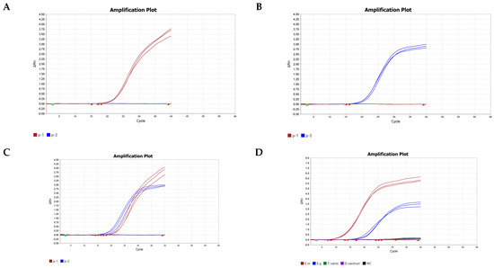
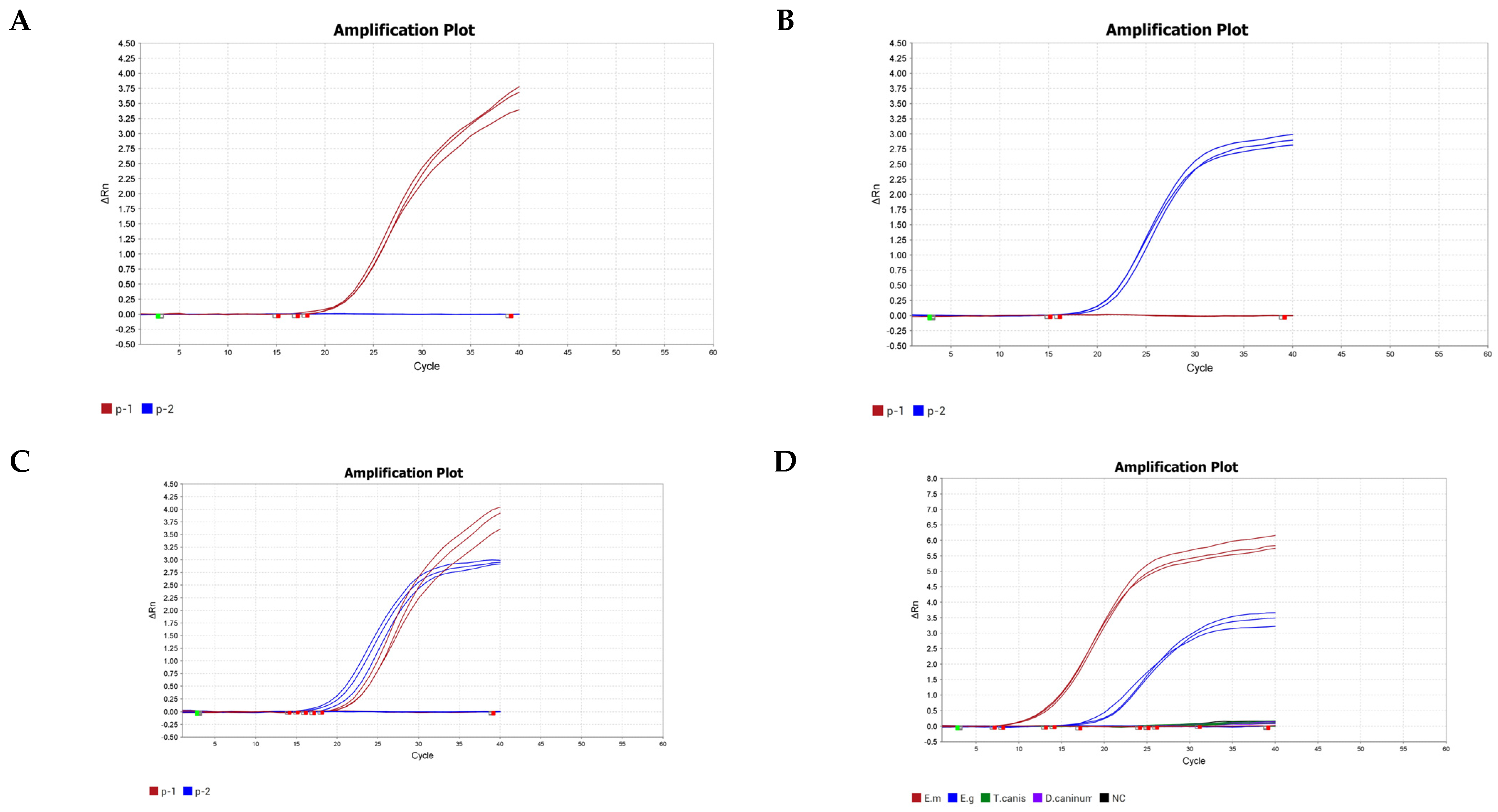
In Figure 1, the probe Em specifically detected DNA samples containing the E. multilocularis sequence with no cross-reaction from the probe Eg. Conversely, the probe Eg specifically identified DNA samples carrying the E. granulosus sequence, with no interference observed from the probe Em. When applied to mixed DNA samples containing both E. multilocularis and E. granulosus, amplification signals were observed from both probes.
Figure 1.
Analytical specificity of the one−tube nested MGB−qPCR assay. (A–C) Specific differentiation of E. multilocularis and E. granulosus using species−specific MGB probes. Fluorescence signals were detected exclusively with the matched probe−target combinations: (A) E. multilocularis genomic DNA detected with the probe Em; (B) E. granulosus genomic DNA detected with the probe Eg; (C) co−amplification of mixed Em and Eg DNA with both probes. No cross−fluorescence was observed between the heterologous probe–target pairs. (D) Cross−reactivity testing against common canine gastrointestinal parasites. Positive amplification (Ct ≤ 38) was observed only for E. multilocularis and E. granulosus; no signal was generated from Toxocara canis or Dipylidium caninum genomic DNA. The results represent three independent experiments. NC, no−template control. ΔRn represents the normalized reporter fluorescence signal after baseline correction.
Using the real-time PCR method applied in this study, genomic DNA extracted from target parasites (E. multilocularis) and non-target parasites (Toxocara canis, Dipylidium caninum) was amplified in a 20 μL reaction system. Specific amplification curves were generated only for the target parasites, consistent with the expected results, indicating a positive amplification result. In contrast, no specific amplification curves were generated for the non-target parasite DNA, resulting in a negative result. The results of three independent replicate experiments were completely consistent, demonstrating the high specificity of real-time PCR detection applied in this study, as shown in Figure 1D.
3.2. Sensitivity Analysis Results
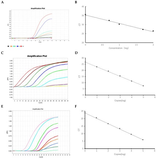
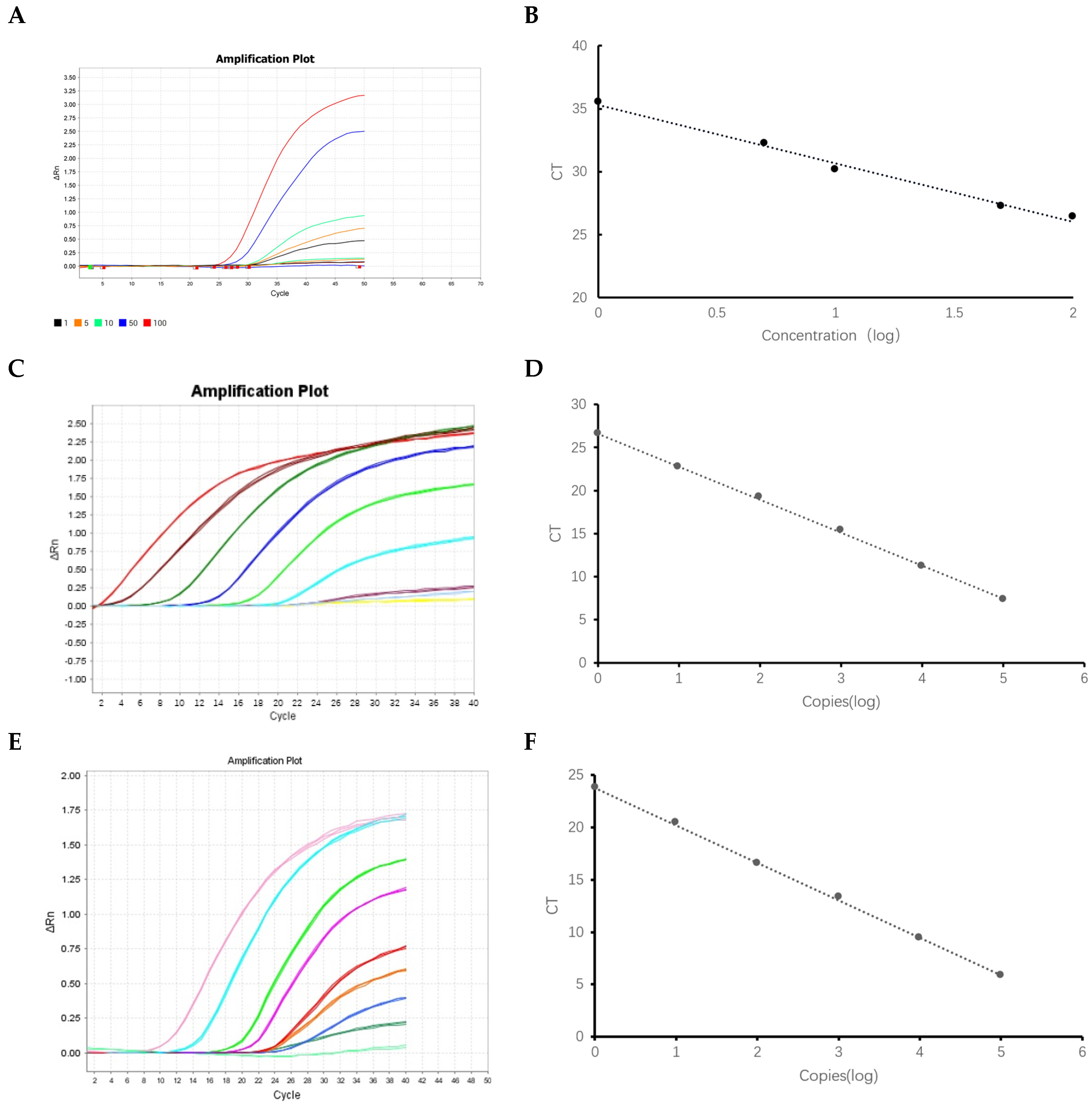
Plasmid-based sensitivity. The analytical sensitivity of the assay was evaluated using ten-fold serial dilutions of the PEm and PEg plasmid standards. Detectable amplification was observed at the lowest tested concentration (nominal single copy) for both plasmids in the tested replicates, with mean Ct values of 34.2 ± 0.85 for PEm and 33.8 ± 0.79 for PEg (Figure 2C,D). All replicates at the one-copy level produced positive amplification signals, indicating detection at a nominal single-copy level under the tested conditions. Standard curve analysis across 101 to 105 copies revealed a strong logarithmic correlation between Ct values and initial template copy number (Figure 2E,F). The regression equation for PEm was y = −3.322 log10(x) + 37.17, R2 = 0.9943, and for PEg was y = −3.224 log10(x) + 34.30, R2 = 0.9914. The amplification efficiency derived from the standard curve slightly higher than the value expected for a conventional single-stage qPCR assay, which is likely attributable to the nested pre-amplification phase that enriches target templates prior to real-time detection. As template copy number increased, Ct values decreased progressively, demonstrating clear discrimination between concentration levels.
Figure 2.
Analytical sensitivity of the one−tube nested MGB−qPCR assay. Analytical sensitivity determined using both intact eggs and plasmid standards. (A) Amplification curves obtained from 10-fold serial dilutions of E. multilocularis egg suspensions (1–100 eggs per reaction). Detectable exponential amplification was obtained from the single−egg dilution. (B) Standard curve derived from the egg dilution series, demonstrating strong linearity between Ct values and the logarithm (log10) of egg number (y = −4.629 log10(x) + 35.30, R2 = 0.9906). (C,D) Sensitivity verification using a 10−fold serial dilution of the PEm plasmid standard (107–100 copies/reaction). The assay reliably detected a single plasmid copy (Ct ≤ 38) in all replicates. (C) Representative amplification curves; (D) corresponding standard curve (y = −3.322 log10(x) + 37.17, R2 = 0.9943). (E,F) Parallel sensitivity assessment using the PEg plasmid standard (105–100 copies/reaction), showing detectable amplification at the lowest tested concentration (nominal single copy) and linearity. (E) Representative amplification curves; (F) corresponding standard curve (y = −3.224 log10(x) + 34.30, R2 = 0.9914). NC, negative control. ΔRn represents the normalized reporter fluorescence signal after baseline correction.
Egg-based sensitivity. Under controlled conditions, detectable amplification was observed at the lowest egg level tested, with a mean Ct value of 34.8 ± 0.92 and a signal-to-noise ratio exceeding the threshold (Figure 2A,B). The standard curve generated from egg dilution data showed a highly linear correlation between Ct values and egg number (log10 scale) across the range of 1 to 100 eggs, with a regression equation of y = −4.629 log10(x) + 35.3 (R2 = 0.9906).
These results suggest that the assay can be sensitive enough to detect low-abundance eggs in laboratory samples, although further analytical evaluation with naturally infected samples is needed to confirm field applicability.
3.3. Intra-Assay Repeatability Assay
To systematically evaluate the intra-assay repeatability of the method, two egg concentrations (10 and 50 eggs/10 μL) were each subjected to 10 independent replicate tests. As shown in Table 3, the low-concentration group exhibited a Ct value range of 30.19–31.75, with a mean of 30.968, a standard deviation (SD) of 0.781, and a coefficient of variation (CV) of 2.5%. The high-concentration group displayed Ct values ranging from 25.73 to 26.67, with a mean of 26.198, an SD of 0.467, and a reduced CV of 1.8%. Both egg concentrations showed CV values below 3%, with the higher concentration group exhibiting lower CV, consistent with the expected positive correlation between repeatability and template amount. No Ct values were detected in the negative control in all 20 replicate tests, and the background signal remained negative throughout, further confirming the system’s intra-assay CV below 3%. These results demonstrate that the method maintains stable quantification capability even under low loading conditions close to the detection limit.
Table 3.
Intra-assay repeatability results of E.multilocularis egg suspensions at different concentrations.
3.4. Reagent Stability Under Simulated Storage Conditions
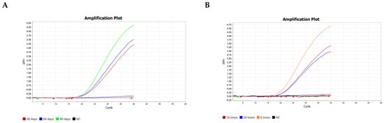
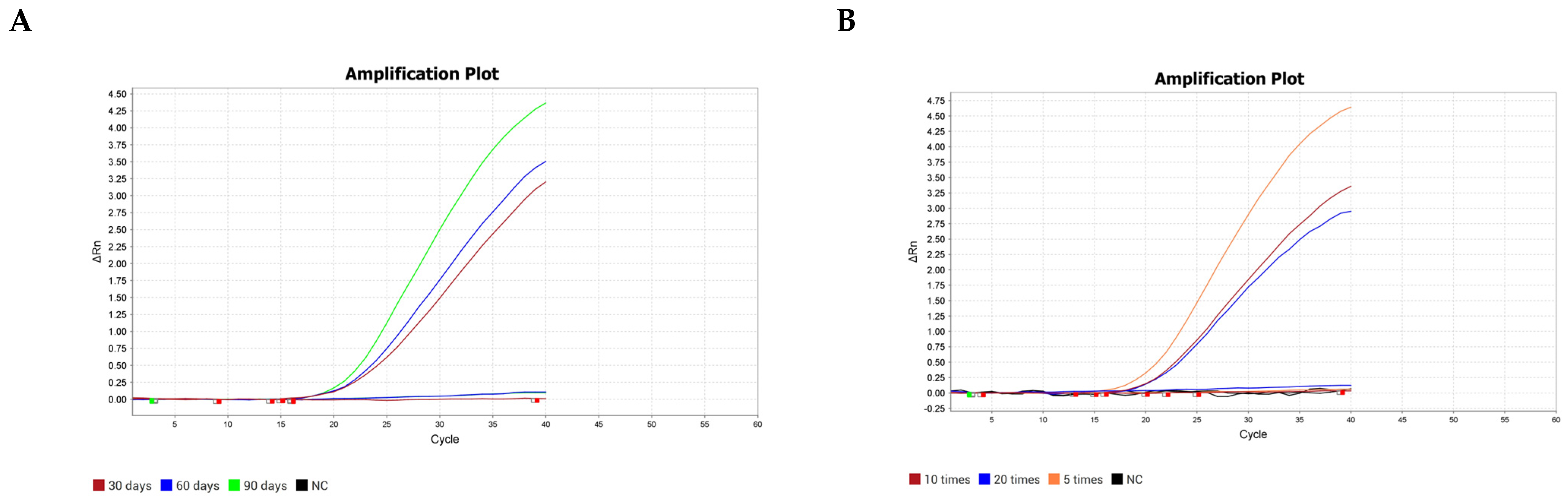
In the accelerated stability study, the pre-mixed PCR reaction buffer was stored at 4 °C in the dark. At storage days 30, 60, and 90, 50 ng of DNA extracted from E. multilocularis eggs were added for amplification and detection. The results showed (Figure 3A) that throughout the 90-day storage period, the amplification curve profiles at all time points remained highly consistent, with minimal signal intensity decay. No false negatives or delayed amplification attributable to reagent degradation were observed. These findings indicate that the formulation possesses remained stable after 90 days at 4 °C under conventional refrigeration conditions. This characteristic is primarily attributed to the stabilizers added to the reaction system and the 3′-end chemical modification of the MGB probe, which effectively mitigate probe hydrolysis and polymerase activity loss.
Figure 3.
Stability of the pre−mixed real−time PCR reagents under simulated storage and transport conditions. Stability was assessed by amplifying 50 ng of E. multilocularis DNA extracted from E. multilocularis eggs. (A) Thermal stability at 4 °C. Reagents stored continuously at 4 °C for 30, 60, and 90 days generated amplification curves that were highly consistent with fresh reagents, showing no significant loss of fluorescence intensity or delay in Ct values. (B) Freeze−thaw stability. Reagents subjected to 5, 10, or 20 complete freeze−thaw cycles (−20 °C to room temperature) retained full amplification efficiency, with no detectable decrease in positive signal. NC, negative control.
The freeze–thaw stability study further simulated the repeated freeze–thaw scenarios that might occur after sampling at the base level. The premixed reagent was stored at −20 °C and then subjected to 5, 10, and 20 complete forced freeze–thaw cycles, respectively. After each cycle group, 50 ng of DNA extracted from E. multilocularis eggs was added as template. The detection results demonstrated (Figure 3B) that even after the extreme treatment of 20 cycles, the amplification efficiency and detection signals of the system showed no discernible decay, and the positive interpretation results were fully repeatable under the same conditions. This robustness is credited to the heat-resistant DNA polymerase and the hypertonic buffer system employed in the TaqMan Fast qPCR Master Mix, which effectively preserve the spatial conformation of the enzyme and the integrity of the primers and probes during repeated phase transitions.
3.5. Preliminary Field Evaluation and Concordance Assessment
To assess the detection performance of this method in real-world endemic settings, a total of 101 canine fecal samples were collected from field environments in regions including Xining City, Qinghai Province (2300 m above sea level), Golmud City, Qinghai Province (2780 m above sea level), and Nagqu City, Tibet Autonomous Region (4500 m above sea level) between 2024 and 2025. Of these, 11 samples were from owned dogs and 90 from stray dogs. Fresh fecal samples were collected using sterile disposable tools, placed in individual sterile containers, and transported to the laboratory under cold conditions. The dog type (owned or stray) and collection location were recorded for each sample (Table S1). Each sample weighed 5–10 g and was stored at −80 °C for at least 10 days post-collection to completely inactivate the infectivity of any parasite eggs, thereby minimizing biosafety risks during subsequent experimental procedures. Following sample processing, the isolated eggs were examined and photographed under a light microscope, and representative images are shown in Figure S2. Parallel testing was performed using both the single-tube nested MGB-qPCR method applied in this study and conventional PCR.
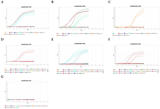
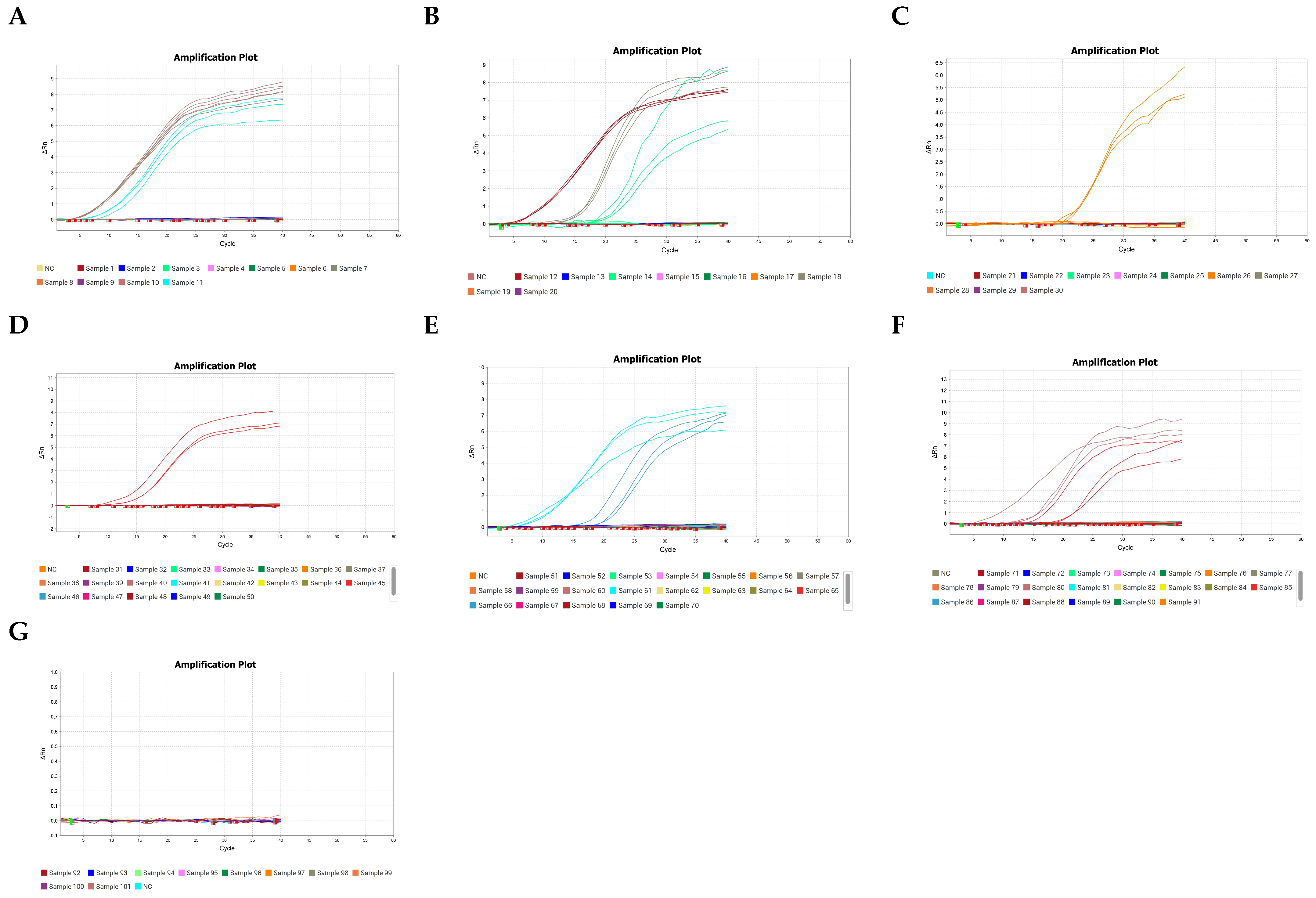
The results showed that the real-time PCR assay detected 12 positive samples for E. multilocularis. Conventional PCR concurrently identified the same 12 positive samples, indicating concordance between the two assays in this preliminary field evaluation (Table 4, Figure 4 and Figure S1). These results demonstrate that the assay performs consistently with field-collected samples after storage at −80 °C, supporting its potential utility for epidemiological surveillance in endemic regions. These results support the potential utility of this method for field epidemiological surveillance and canine infection screening.
Table 4.
Preliminary field evaluation in canine fecal samples.
Figure 4.
Application of the one−tube nested MGB−qPCR assay to field−collected canine fecal samples using dual−species detection. A total of 101 canine fecal samples collected from endemic areas in Qinghai Province and the Tibet Autonomous Region (2024–2025) were analyzed with both Probe Em and Probe Eg in the same closed−tube reaction. (A–G) Fluorescence amplification curves recorded in the FAM channel (Probe Em) for all 101 samples, shown in panels grouped by sample ID. Twelve samples (indicated in the respective panels) displayed typical sigmoidal amplification with Ct values ≤ 38, meeting the positivity threshold for E. multilocularis. No sample generated a positive signal in the VIC channel (Probe Eg), indicating the absence of E. granulosus DNA in this sample set. All 12 Em−positive samples were independently confirmed by conventional PCR and by sequencing of both the outer (484 bp) and inner (115 bp) amplicons, with consistent results. NC, no−template control.
Sequencing of both the outer primer (484 bp) and inner primer (115 bp) amplicons from all 12 positive samples confirmed that they contained E. multilocularis DNA, with all sequences matching the E. multilocularis mitochondrial reference genome (CBLO020001206.1).
4. Discussion
The life cycle of E.multilocularis depends on a cycle of transmission between hosts, with canids as the primary host and rodents as the intermediate host [1]. Adult parasites live in the small intestine of their definitive host and release thousands of eggs every day, which are then excreted in feces [2]. These eggs have a strong survival ability in the external environment and can remain infectious for months to a year [24]. Humans act as unintended accidental hosts; when these parasite eggs are ingested, the oncospheres hatch, penetrate the intestinal wall, enter the bloodstream, and eventually accumulate mainly in the liver, thus causing AE [4]. Due to the high pathogenicity and poor prognosis of AE, accurate monitoring of environmental egg contamination and infections in definitive hosts is of critical importance [6].
In traditional detection methods, although autopsy combined with optical microscopy has high reliability, its ability to distinguish between different types of samples is relatively limited (Hoberg, 2002) [25]. The eggs of various tapeworm species (e.g., Taenia solium, Taenia saginata) exhibit highly similar morphology, making accurate differentiation based solely on morphological characteristics challenging and prone to misidentification [25]. Immunological methods, such as enzyme-linked immunosorbent assay (ELISA), are suitable for large-scale screening, but they are prone to false positive results due to antigen cross-reactivity, and their insufficient specificity also limits their reliability in epidemiological surveys [26].
Molecular tools are increasingly used in epidemiological surveillance of canine and livestock parasites, including insect-borne pathogens in dogs [27], Coenurus cerebralis in sheep [28], and genetic characterization of Echinococcus granulosus sensu stricto and E. ortleppi [29].The development of molecular detection technology has provided a more accurate means for the identification of tapeworm species [1]. For instance, Knapp et al. established a highly sensitive detection system, MLX qPCR, using multiplex real-time quantitative PCR technology [30]. This system enables simultaneous detection of four pathogenic Echinococcus species and was successfully validated using 81 surgical specimens from patients with alveolar echinococcosis and cystic echinococcosis, achieving a specificity of 100%.Hifumi et al. employed RPA to establish a rapid method for detecting E. multilocularis in equine hepatic nodules [31]. The RPA assay showed 94.4% concordance with PCR results and was 100 times more sensitive than the PCR detection.
In this study, a previously established one-tube nested MGB-qPCR platform, with an added E. granulosus-specific probe, was applied to detect E. multilocularis eggs in canine feces, showing no cross-reactivity with the tested non-target parasites, intra-assay repeatability, and reagent stability. Utilizing the ability of MGB probes to distinguish highly homologous sequences and to reliably detect even with limited target material, this method did not exhibit cross-reactivity with non-target parasites (such as Toxocara canis), demonstrating its differentiated E. multilocularis from E. granulosus by two SNPs and ability to effectively prevent false positive results. The lowest concentration tested with detectable amplification was the final dilution in the series. In stability tests, the CT values did not show significant deviations after the premixed PCR reagents were stored at 4 °C for 90 days or after undergoing 20 freeze–thaw cycles at −20 °C, indicating that the reagent system-maintained amplification performance under the tested storage conditions. These results support further evaluation of reagent stability under field-relevant conditions.
The continued maturation of TaqMan MGB probe synthesis technology and the continuous reduction in cost have made the creation of cost-effective and efficient field-testing solutions increasingly feasible. At the same time, continuous technological and equipment innovation is accelerating the deployment of portable and intelligent molecular testing devices in grassroots public health systems. This may support further evaluation of the one-tube nested MGB probe real-time PCR method for detecting E. multilocularis eggs.
In this preliminary field evaluation, 11 samples were from owned dogs and 90 from stray dogs. No positive samples were detected in owned dogs, whereas all 12 positive samples were from stray dogs. This distribution may reflect the impact of national echinococcosis control programs, which have emphasized dog deworming, surveillance, and public health education, leading to reduced infection in owned dogs. However, the detection of positives exclusively in stray dogs suggests a potential gap in stray dog management. Strengthened control of free-roaming dogs, together with expanded surveillance, may further reduce environmental egg contamination and human exposure Several limitations should be noted. First, specificity was not experimentally assessed against a broader panel of canine taeniids (Taenia hydatigena, T. multiceps, Mesocestoides spp.) due to limited reference material. The preliminary field evaluation included only 101 canine fecal samples, and agreement with conventional PCR alone does not establish diagnostic sensitivity or specificity. Diagnostic sensitivity and specificity remain to be determined in a larger, clearly defined reference population. Second, no internal amplification control (IAC) was included. Therefore, true-negative results could not be distinguished from false-negative results caused by PCR inhibitors in fecal DNA extracts. Because fecal samples commonly contain PCR inhibitors, negative field results should be interpreted with caution. Future versions of this assay should include an exogenous IAC. Third, the single-copy and single-egg sensitivities were determined under controlled conditions using plasmid standards and manually selected eggs, respectively, and may not reflect detection of low egg numbers in a complex fecal matrix. Fourth, the preliminary field evaluation was restricted to canine feces, performance on environmental samples (soil, water) remains untested. In addition, egg recovery efficiency during sieving and centrifugation was not quantified. Sieving and centrifugation were applied to field samples but not to the single-egg sensitivity experiment, in which eggs were manually selected. Finally, no direct comparison with other emerging assays (e.g., LAMP) was performed. Despite these limitations, the assay’s analytical performance and single-tube format support further evaluation of its potential utility in field epidemiological surveillance.
In summary, the one-tube nested multiplex MGB probe real-time PCR method applied and analytically evaluated for detecting E. multilocularis eggs in canine feces. The detection assay combines the high sensitivity of nested PCR with the ease of operation of real-time fluorescence PCR, while utilizing MGB probes to enhance specificity. It showed no cross-reactivity with the tested non-target parasites and enabled species-specific identification of E. multilocularis. Under controlled conditions, detectable amplification was observed at the lowest tested plasmid concentration (nominal single copy) and at the lowest egg level tested (one egg) under controlled conditions. These findings support further evaluation of the assay in larger field studies; however, its diagnostic sensitivity and specificity remain to be determined.
In conclusion, this study applied and preliminarily evaluated a one-tube nested duplex MGB probe real-time PCR method for detecting E. multilocularis eggs in canine feces. The assay shows potential for canine fecal surveillance in endemic areas, but its application to environmental samples and its diagnostic sensitivity and specificity require further evaluation.
Supplementary Materials
The following supporting information can be downloaded at: https://www.mdpi.com/article/10.3390/parasitologia6050059/s1, Figure S1. PCR amplification of fecal samples. Figure S2. Taeniid eggs from positive samples. Figure S3. Sequence alignment of positive amplicons. Table S1. Positive samples by location and dog type. Including original images.
Author Contributions
Z.Y.: Investigation, Formal analysis, Writing Original draft. Z.-H.L.: Investigation. K.S.: Investigation. Y.Q.: Investigation. N.Z.: Resources. Y.W.: Investigation. S.C.: Investigation. X.Z.: Resources. Z.W.: Resources. F.T.: Methodology, Resources. R.L.: Methodology, Resources. All authors have read and agreed to the published version of the manuscript.
Funding
The author(s) declare financial support was received for the research and/or publication of this article. This research was supported by Qinghai Department of Science and Technology (Grant No. 2024-ZJ-715); the Natural Science Foundation of China (Grant No. 32360192), Open Research Projects of the National Health Commission Key Laboratory of Echinococcosis Prevention and Control (Grant No. 2023WZK1003), Qinghai University Research Ability Enhancement Project (2025KTSA05).
Institutional Review Board Statement
The animal study protocol was approved by the Research and Ethics Committee of Qinghai University Medical College (PJ-202302-03, 3 July 2023).
Informed Consent Statement
Not applicable.
Data Availability Statement
The original contributions presented in the study are included in the article/Supplementary Material. Further inquiries can be directed to the corresponding author.
Acknowledgments
We acknowledge the Research Center for High Altitude Medicine of Qinghai University for providing the experimental platform. We also thank the Qinghai Institute for Endemic Disease Prevention and Control for providing the experimental site. We would like to thank all the researchers in the laboratory for all the help in this work.
Conflicts of Interest
The authors declare that they have no known competing financial interests or personal relationships that could have appeared to influence the work reported in this paper.
References
- Romig, T.; Deplazes, P.; Jenkins, D.; Giraudoux, P.; Massolo, A.; Craig, P.S.; Wassermann, M.; Takahashi, K.; de la Rue, M. Ecology and Life Cycle Patterns of Echinococcus Species. Adv. Parasitol. 2017, 95, 213–314. [Google Scholar] [CrossRef] [Scilit] [PubMed]
- Kapel, C.M.O.; Torgerson, P.R.; Thompson, R.C.A.; Deplazes, P. Reproductive Potential of Echinococcus Multilocularis in Experimentally Infected Foxes, Dogs, Raccoon Dogs and Cats. Int. J. Parasitol. 2006, 36, 79–86. [Google Scholar] [CrossRef] [Scilit] [PubMed]
- Eckert, J.; Deplazes, P. Biological, Epidemiological, and Clinical Aspects of Echinococcosis, a Zoonosis of Increasing Concern. Clin. Microbiol. Rev. 2004, 17, 107–135. [Google Scholar] [CrossRef] [Scilit] [PubMed]
- Brunetti, E.; Kern, P.; Vuitton, D.A. Expert Consensus for the Diagnosis and Treatment of Cystic and Alveolar Echinococcosis in Humans. Acta Trop. 2010, 114, 1–16. [Google Scholar] [CrossRef] [Scilit] [PubMed]
- Mastin, A.; van Kesteren, F.; Torgerson, P.R.; Ziadinov, I.; Mytynova, B.; Rogan, M.T.; Tursunov, T.; Craig, P.S. Risk Factors for Echinococcus Coproantigen Positivity in Dogs from the Alay Valley, Kyrgyzstan. J. Helminthol. 2015, 89, 655–663. [Google Scholar] [CrossRef] [Scilit] [PubMed]
- Hegglin, D.; Deplazes, P. Control of Echinococcus Multilocularis: Strategies, Feasibility and Cost–Benefit Analyses. Int. J. Parasitol. 2013, 43, 327–337. [Google Scholar] [CrossRef] [Scilit] [PubMed]
- Bucher, B.J.; Muchaamba, G.; Kamber, T.; Kronenberg, P.A.; Abdykerimov, K.K.; Isaev, M.; Deplazes, P.; Alvarez Rojas, C.A. LAMP Assay for the Detection of Echinococcus Multilocularis Eggs Isolated from Canine Faeces by a Cost-Effective NaOH-Based DNA Extraction Method. Pathogens 2021, 10, 847. [Google Scholar] [CrossRef] [Scilit] [PubMed]
- Celva, R.; Crestanello, B.; Obber, F.; Dellamaria, D.; Trevisiol, K.; Bregoli, M.; Cenni, L.; Agreiter, A.; Danesi, P.; Hauffe, H.C.; et al. Assessing Red Fox (Vulpes Vulpes) Demographics to Monitor Wildlife Diseases: A Spotlight on Echinococcus Multilocularis. Pathogens 2022, 12, 60. [Google Scholar] [CrossRef] [Scilit] [PubMed]
- Dinkel, A.; von Nickisch-Rosenegk, M.; Bilger, B.; Merli, M.; Lucius, R.; Romig, T. Detection of Echinococcus Multilocularis in the Definitive Host: Coprodiagnosis by PCR as an Alternative to Necropsy. J. Clin. Microbiol. 1998, 36, 1871–1876. [Google Scholar] [CrossRef] [Scilit] [PubMed]
- Mathis, A.; Deplazes, P.; Eckert, J. An Improved Test System for PCR-Based Specific Detection of Echinococcus Multilocularis Eggs. J. Helminthol. 1996, 70, 219–222. [Google Scholar] [CrossRef] [Scilit] [PubMed]
- Trachsel, D.; Deplazes, P.; Mathis, A. Identification of Taeniid Eggs in the Faeces from Carnivores Based on Multiplex PCR Using Targets in Mitochondrial DNA. Parasitology 2007, 134, 911–920. [Google Scholar] [CrossRef] [Scilit] [PubMed]
- Jasuja, J.K.; Bub, F.; Veit, J.; Fofana, H.K.M.; Sacko, M.; Saye, R.; Chatigre, J.K.; N’Goran, E.K.; Yao, J.A.; Khanal, B.; et al. Multiplex PCR for Bacterial, Viral and Protozoal Pathogens in Persistent Diarrhoea or Persistent Abdominal Pain in Côte d’ivoire, Mali and Nepal. Sci. Rep. 2024, 14, 10926. [Google Scholar] [CrossRef] [Scilit] [PubMed]
- Xie, S.-T.; Ding, L.-J.; Huang, F.-Y.; Zhao, Y.; An, X.-L.; Su, J.-Q.; Sun, G.-X.; Song, Y.-Q.; Zhu, Y.-G. VFG-Chip: A High-Throughput qPCR Microarray for Profiling Virulence Factor Genes from the Environment. Environ. Int. 2023, 172, 107761. [Google Scholar] [CrossRef] [Scilit] [PubMed]
- Al-Soud, W.A.; Ouis, I.-S.; Li, D.-Q.; Ljungh, Ã.; Wadström, T. Characterization of the PCR Inhibitory Effect of Bile to Optimize Real-Time PCR Detection of Helicobacter Species. FEMS Immunol. Med. Microbiol. 2005, 44, 177–182. [Google Scholar] [CrossRef] [Scilit] [PubMed]
- Monteiro, L.; Bonnemaison, D.; Vekris, A.; Petry, K.G.; Bonnet, J.; Vidal, R.; Cabrita, J.; Mégraud, F. Complex Polysaccharides as PCR Inhibitors in Feces: Helicobacter Pylori Model. J. Clin. Microbiol. 1997, 35, 995–998. [Google Scholar] [CrossRef] [Scilit] [PubMed]
- Sidstedt, M.; Rådström, P.; Hedman, J. PCR Inhibition in qPCR, dPCR and MPS—Mechanisms and Solutions. Anal. Bioanal. Chem. 2020, 412, 2009–2023. [Google Scholar] [CrossRef] [Scilit] [PubMed]
- Xue, B.; Li, Y.; Wang, X.; Li, R.; Zeng, X.; Yang, M.; Xu, X.; Ye, T.; Bao, L.; Huang, Y. TaqMan-MGB Probe Quantitative PCR Assays to Genotype and Quantify Three mtDNA Mutations of Leber Hereditary Optic Neuropathy. Sci. Rep. 2020, 10, 12264. [Google Scholar] [CrossRef] [Scilit] [PubMed]
- Kutyavin, I.V.; Afonina, I.A.; Mills, A.; Gorn, V.V.; Lukhtanov, E.A.; Belousov, E.S.; Singer, M.J.; Walburger, D.K.; Lokhov, S.G.; Gall, A.A.; et al. 3′-Minor Groove Binder-DNA Probes Increase Sequence Specificity at PCR Extension Temperatures. Nucleic Acids Res. 2000, 28, 655–661. [Google Scholar] [CrossRef] [Scilit] [PubMed]
- Qi, J.; Li, P.; Yan, Y.; Li, G.; Kong, L. Development of a Detection Chip for Major Pathogenic Drug-Resistant Genes and Drug Targets in Bovine Respiratory System Diseases. Open Life Sci. 2024, 19, 20220778. [Google Scholar] [CrossRef] [Scilit] [PubMed]
- Liu, J.; Gratz, J.; Amour, C.; Kibiki, G.; Becker, S.; Janaki, L.; Verweij, J.J.; Taniuchi, M.; Sobuz, S.U.; Haque, R.; et al. A Laboratory-Developed TaqMan Array Card for Simultaneous Detection of 19 Enteropathogens. J. Clin. Microbiol. 2013, 51, 472–480. [Google Scholar] [CrossRef] [Scilit] [PubMed]
- Kurakawa, T.; Kubota, H.; Tsuji, H.; Matsuda, K.; Asahara, T.; Takahashi, T.; Ramamurthy, T.; Hamabata, T.; Takahashi, E.; Miyoshi, S.; et al. Development of a Sensitive rRNA-Targeted Reverse Transcription-Quantitative Polymerase Chain Reaction for Detection of Vibrio Cholerae/Mimicus, V. Parahaemolyticus/Alginolyticus and Campylobacter Jejuni/Coli. Microbiol. Immunol. 2012, 56, 10–20. [Google Scholar] [CrossRef] [Scilit] [PubMed]
- Feng, K.; Li, W.; Guo, Z.; Duo, H.; Fu, Y.; Shen, X.; Tie, C.; E, R.; Xiao, C.; Luo, Y.; et al. Development of LAMP Assays for the Molecular Detection of Taeniid Infection in Canine in Tibetan Rural Area. J. Vet. Med. Sci. 2017, 79, 1986–1993. [Google Scholar] [CrossRef] [Scilit] [PubMed]
- Li, R.; Bao, H.; Liu, C.; Zhao, L.; Kang, Y.; Ge, R.-L.; Fan, H.; Tang, F. One-Tube Nested MGB Probe Real-Time PCR Assay for Detection of Echinococcus Multilocularis Infection in Plasma Cell Free DNA. Acta Trop. 2022, 232, 106518. [Google Scholar] [CrossRef] [Scilit] [PubMed]
- Deplazes, P.; Eckert, J. Veterinary Aspects of Alveolar Echinococcosis—a Zoonosis of Public Health Significance. Vet. Parasitol. 2001, 98, 65–87. [Google Scholar] [CrossRef] [Scilit] [PubMed]
- Hoberg, E.P. Taenia Tapeworms: Their Biology, Evolution and Socioeconomic Significance. Microbes Infect. 2002, 4, 859–866. [Google Scholar] [CrossRef] [Scilit] [PubMed]
- Brehm, K.; Koziol, U. Echinococcus–Host Interactions at Cellular and Molecular Levels. In Advances in Parasitology; Elsevier: Amsterdam, The Netherlands, 2017; Volume 95, pp. 147–212. ISBN 978-0-12-811471-1. [Google Scholar]
- Aydin, N.; Ölmez, N.; Sari, B.; Vatansever, Z.; Tasci, G.; Kiziltepe, S.; Yïğït, M. Molecular Characterization of Insect-Borne (Mosquito-Sandfly) Parasitic Pathogens of Dogs in Selected Regions of Northeast Anatolia, Türkiye. Pak. Vet. J. 2025, 304–311. [Google Scholar] [CrossRef] [Scilit] [PubMed]
- Akkuş, E. Molecular Prevalence of Coenurus Cerebralis in Sheep with Neurological Symptoms in IğdırProvince, Türkiye. Pak. Vet. J. 2024. [Google Scholar] [CrossRef] [Scilit]
- Malik, M.; Subhani, M.; Alvi, A.; Wakid, M.; Alkhaldi, A.; Saqib, M.; Li, L.; Khan, A.; Liu, Y.-Y.; Fu, B.; et al. High Genetic Variability in Full-Length Cox2 and Nad6 Genes of Echinococcus Granulosus Sensu Stricto and Echinococcus Ortleppi Recovered from Cattle. Pak. Vet. J. 2024, 148–154. [Google Scholar] [CrossRef] [Scilit]
- Knapp, J.; Lallemand, S.; Monnien, F.; Felix, S.; Courquet, S.; Umhang, G.; Millon, L. Real-Time Multiplex PCR for Human Echinococcosis and Differential Diagnosis. Parasite 2023, 30, 3. [Google Scholar] [CrossRef] [Scilit] [PubMed]
- Hifumi, T.; Tanaka, T.; Sato, M.; Akioka, K.; Fujimata, C.; Miyoshi, N. Rapid Detection of Alveolar Echinococcosis in Hepatic Nodules of Horses by Recombinase Polymerase Amplification Assay. Vet. Anim. Sci. 2023, 20, 100291. [Google Scholar] [CrossRef] [Scilit] [PubMed]

Disclaimer/Publisher’s Note: The statements, opinions and data contained in all publications are solely those of the individual author(s) and contributor(s) and not of MDPI and/or the editor(s). MDPI and/or the editor(s) disclaim responsibility for any injury to people or property resulting from any ideas, methods, instructions or products referred to in the content. |
© 2026 by the authors. Licensee MDPI, Basel, Switzerland. This article is an open access article distributed under the terms and conditions of the Creative Commons Attribution (CC BY) license.