


Abstract
Fungal diversity has been well explored worldwide, but such knowledge in special environments remains limited. Here, we focus on the soil from Ngari Prefecture with high altitude in the northwestern part of Xizang, China. Among the 514 fungal strains isolated from the soil samples, two strains are newly described as Cymostachys pangongensis and Emericellopsis rutogensis in the Hypocreales on the basis of both morphological and phylogenetic evidence. Cymostachys pangongensis exhibits typical characteristics of Cymostachys, viz., irregularly cymosely branched conidiophores and olivaceous brown to dark brown, fabiform conidia. It represents the sixth known species of Cymostachys and the first species isolated from soils in this genus. Emericellopsis rutogensis has typical chlamydospores and conidia of Emericellopsis, although its sexual ascomata and ascospores were not observed. Notably, these two new species belong to interesting fungal genera, whose members are well known for rich medicinal natural products. In summary, this study enriches the knowledge of fungal diversity in special environments, and the isolated strains may provide important bioresources for future applications.
1. Introduction
Fungi have benefitted human beings in some ways and can be treated as important strategic bioresources [1]. For example, species of Penicillium and Streptomyces have been widely used for producing various antibiotics [2,3], while macrofungi, like Agaricus, Ganoderma, and Sanghuangporus, have been used as important sources of food and medicines in many countries [4,5]. Therefore, fungal diversity has been systematically explored for a long time all over the world [6]. However, there are still more than 90% of estimated species waiting to be known and described [7]. In particular, fungal diversity is extremely poorly known in special environments that are more suitable for microorganisms than other life forms.
Among various special environments, high-altitude mountains are affected by multiple climatic factors. Taking the Qinghai–Xizang Plateau as an example, which is the highest plateau on the Earth and recognized as the roof of the world, its ecosystems are shaped mainly by low temperatures, strong winds, and intense ultraviolet radiation in alpine meadows [8], and also by drought and high temperature resulting from the foehn effect in dry valleys [9]. These harsh conditions act as ecological filters that impose strong selective pressures on microbial communities in soils. Indeed, studies have revealed distinct elevational patterns in bacterial diversity [10], overall fungal communities [11], and ectomycorrhizal assemblages [12], which indicate that microorganisms in this region have developed unique adaptations. Consequently, soil fungal communities in high-altitude environments are often characterized by the dominance of stress-tolerant taxa, e.g., Alternaria, Mortierella, Preussia, and various members of the order Hypocreales [13]. More importantly, such adaptability may harbour special metabolic pathways in fungi and even new fungal species.
During the exploration of the Qinghai–Xizang Plateau, Xizang, China, in August 2024, soil samples were collected from Ngari Prefecture. Recently, investigations in this prefecture have revealed the depth-related patterns of soil microbial communities and identified moisture and salinity as key physicochemical drivers [14]. However, detailed taxonomic descriptions of culturable fungal species remain limited. When checking fungal strains isolated from these samples, two strains were identified as new species from morphological and phylogenetic perspectives. These two new species, belonging to Hypocreales, Ascomycota, are described here.
2. Materials and Methods
2.1. Sample Collection and Strain Isolation
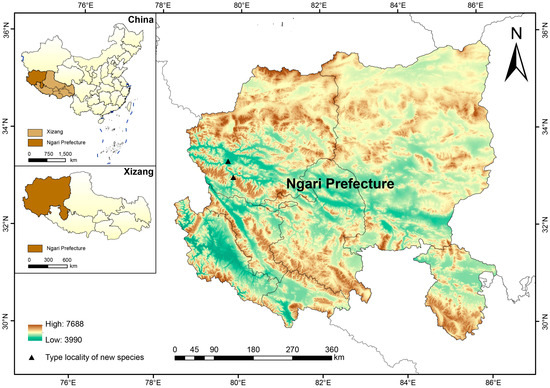
Ngari Prefecture, lying at a mean elevation of approximately 4500 m, is located in the northwestern part of Xizang, China (Figure 1). The region has an average annual precipitation of 50–100 mm, with the oxygen availability averaging less than 50% than that of lowland levels and a recorded minimum temperature of −44.6 °C. These factors collectively define a high-altitude, cold, hypoxic natural habitat. This special environment restricts large-scale field surveys on biodiversity, especially fungal diversity.
Figure 1.
Geographic location of Ngari Prefecture, Xizang, China, with type localities of two new species. The map was generated using ArcGIS version 10.8.2 (ESRI, Redlands, CA, USA). The administrative boundaries and Digital Elevation Model (DEM) data were obtained from the Geospatial Data Cloud (http://www.gscloud.cn; accessed on 15 September 2025).
Approximately 50 g of soil was collected from each of the 27 sampling sites in Ngari Prefecture with sterile tools and enclosed in sterile ziplocked plastic bags. The soil samples were then transported with dry ice to the lab at the Institute of Microbiology, Chinese Academy of Sciences. Upon arrival, 1 g of fresh soil was suspended in 9 mL of sterile distilled water, serially diluted to 10−1, 10−2, 10−3, and 10−4. Each dilution (400 µL) was spread on the potato dextrose agar (PDA) medium. All dishes were incubated at 28 °C for 1–2 weeks, from which the single colonies were picked up and inoculated onto new PDA dishes.
2.2. DNA Extraction, PCR, and Sequencing
Genomic DNA was extracted from fungal colonies grown on PDA at 28 °C for two weeks using the CTAB rapid plant genome extraction kit-DN14 (Aidlab Biotechnologies Co., Ltd., Beijing, China) according to the manufacturer’s instructions. The nuclear rDNA ITS1-5.8S-ITS2 (ITS) region was amplified with primers ITS5 and ITS4 [15], while the D1–D2 domains of the nuclear large subunit (nLSU) were amplified with LR0R/LR7 [16,17], the partial translation elongation factor 1-α (tef1-α) with 983F/1567R [18], the partial RNA polymerase II second largest subunit (rpb2) with fRPB2-5F/fRPB2-7cR [19], and the partial β-tubulin (tub2) with BT2a/BT2b [20,21].
The PCR procedures were as follows: for ITS and tef1-α, initial denaturation at 95 °C for 3 min, followed by 35 cycles at 94 °C for 40 s, 54 °C for 45 s, and 72 °C for 1 min, and a final extension at 72 °C for 10 min; for nLSU, initial denaturation at 94 °C for 1 min, followed by 34 cycles at 94 °C for 30 s, 50 °C for 1 min, and 72 °C for 1.5 min, and a final extension at 72 °C for 10 min; for rpb2, initial denaturation at 94 °C for 2 min, followed by 10 cycles at 94 °C for 45 s, 60 °C for 45 s (decreasing by 1 °C per cycle), and 72 °C for 1.5 min, followed by 36 cycles at 94 °C for 45 s, 53 °C for 1 min, and 72 °C for 1.5 min, and a final extension at 72 °C for 10 min; for tub2, initial denaturation at 94 °C for 3 min, 35 cycles at 94 °C for 30 s, 55 °C for 50 s, and 72 °C for 1 min, and a final extension at 72 °C for 10 min. PCR was performed in a 30 μL reaction mixture, containing 15 μL of 2 × EasyTaq® PCR SuperMix (TransGen Biotech Co., Ltd., Beijing, China), 12 μL of double-distilled water (ddH2O), 1 μL each of forward and reverse primers (10 μM), and 1 μL of genomic DNA template.
PCR products were sequenced with the same primers as those used in PCR by BGI Genomics Technology Co., Ltd. (Beijing, China). The newly generated sequences were submitted to GenBank (http://www.ncbi.nlm.nih.gov/genbank; accessed on 15 September 2025, see Table 1 and Table 2).
Table 1.
Information on taxa used in phylogenetic analyses of Cymostachys.
Table 2.
Information on taxa used in phylogenetic analyses of Emericellopsis.
2.3. Phylogenetic Analyses
Besides the newly generated sequences, additional reference sequences were downloaded from GenBank for phylogenetic analyses (Table 1 and Table 2). To explore the phylogenetic position of the newly sequenced fungal strain related to Cymostachys, a combined dataset of ITS, nLSU, rpb2, and tub2 regions adapted from Crous et al. [22] was employed. This updated dataset includes the main lineages of Stachybotryaceae as ingroup taxa with two strains of Fusarium sambucinum (Nectriaceae) as outgroup taxa. Another dataset with a combination of ITS, nLSU, and tef1-α regions modified from Zhao et al. [23] was used to explore the relationship of a newly sequenced strain within Emericellopsis. Besides species of Emericellopsis, other representative taxa of Bionectriaceae were also included, and Tilachlidium brachiatum (Tilachlidiaceae) was selected as the outgroup taxon.
Each region was separately aligned using MAFFT v7.110 [24]. The resulting alignments were trimmed with trimAl v1.4 [25] and then concatenated to two alignments corresponding to the datasets in PhyloSuite v1.2.3 [26,27]. The best-fit evolutionary model for each concatenated alignment was estimated with the Bayesian information criterion [28] using ModelFinder [29] implemented in IQ-TREE v2 [30]. Following these models, maximum likelihood (ML) and Bayesian inference (BI) algorithms were used to conduct phylogenetic analyses. The ML analysis was performed using IQ-TREE v2 with ultrafast bootstrap 5000 replicates (-bb 5000) and a bootstrap correlation cutoff of 0.90 (-bcor 0.90). IQ-TREE used automatic thread detection (-nt AUTO, two threads selected) and a fixed random seed (seed = 719,640). BI was conducted using MrBayes 3.2.7a [31]. Two independent MCMC runs were started in parallel from random starting topologies and run for 20,000,000 generations, sampling every 1000 generations. The first 25% of sampled trees were discarded as burn-in, while all remaining trees were used to construct a 50% majority-rule consensus tree and for calculating Bayesian posterior probabilities (BPPs). Chain convergence was assessed in Tracer v1.7 (ESS > 200; ASDSF ≤ 0.01) [32]. Trees were visualized in FigTree v1.4.3 [33].
2.4. Morphological Examination
For culture characteristics, the strains were separately inoculated on four media, viz., PDA, oatmeal agar (OA), malt extract agar (MEA), and Sabouraud dextrose agar (SDA), and then incubated at 28 °C in the dark. Colony diameters and macromorphological characteristics were documented after 20 days using a digital camera (Sony ZV-1, Sony Corporation, Tokyo, Japan). For micromorphological examination, the fresh colonies of strains cultivated on PDA, OA, MEA, and SDA were picked up with a sterile needle under a stereomicroscope and mounted in a drop of water. Then, the fungal microstructures were observed and photographed using an Axio Imager A2 light microscope (Zeiss, Oberkochen, Germany).
The dry cultures are preserved in the Fungarium of the Institute of Microbiology, Chinese Academy of Sciences (HMAS). The living cultures are preserved in Li-Wei Zhou’s Lab as the LWZ Culture Collection at the State Key Laboratory of Microbial Diversity and Innovative Utilization, Institute of Microbiology, Chinese Academy of Sciences.
3. Results
3.1. Molecular Phylogeny
In this study, a total of two ITS sequences, two nLSU sequences, one rpb2 sequence, one tef1-α sequence, and one tub2 sequence were newly generated from two strains (Table 1 and Table 2).
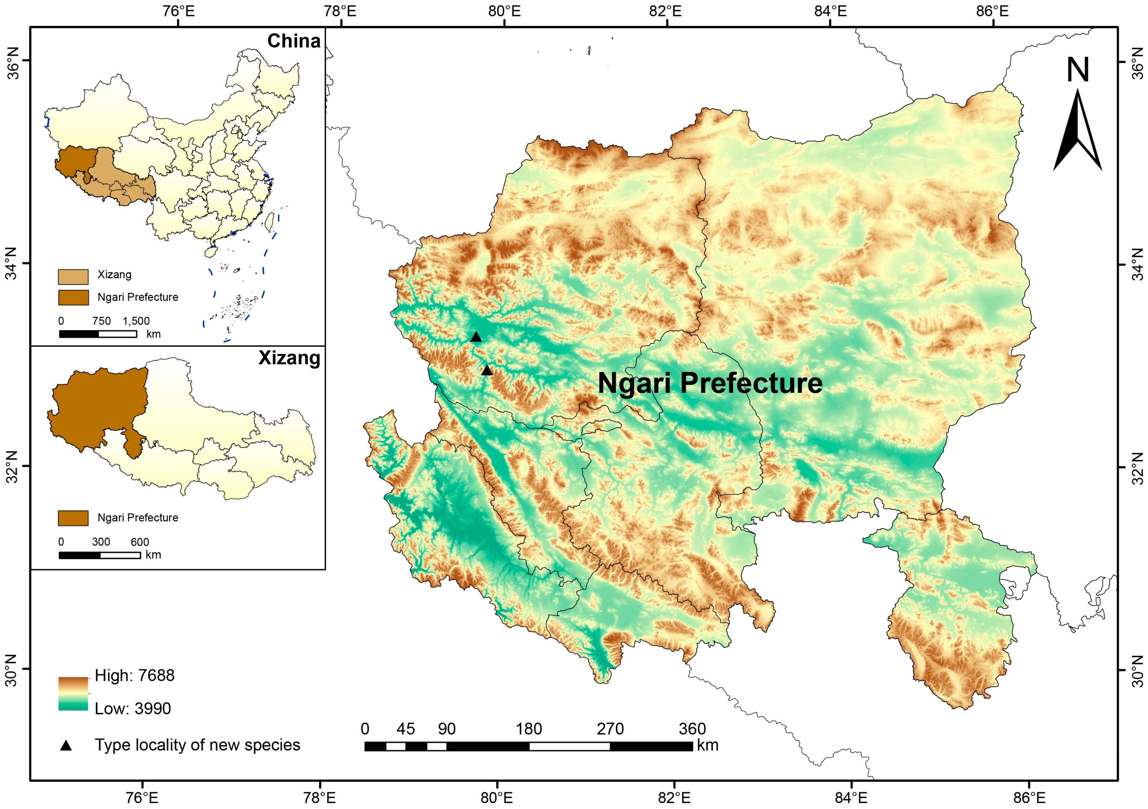
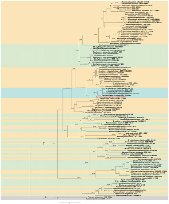
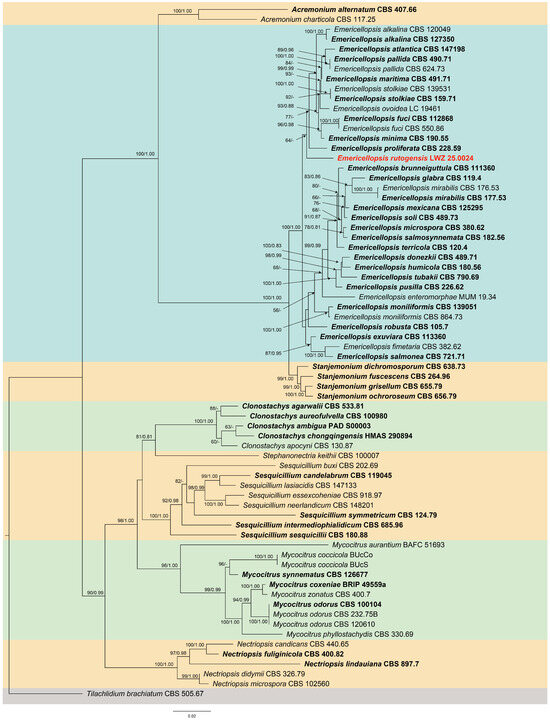
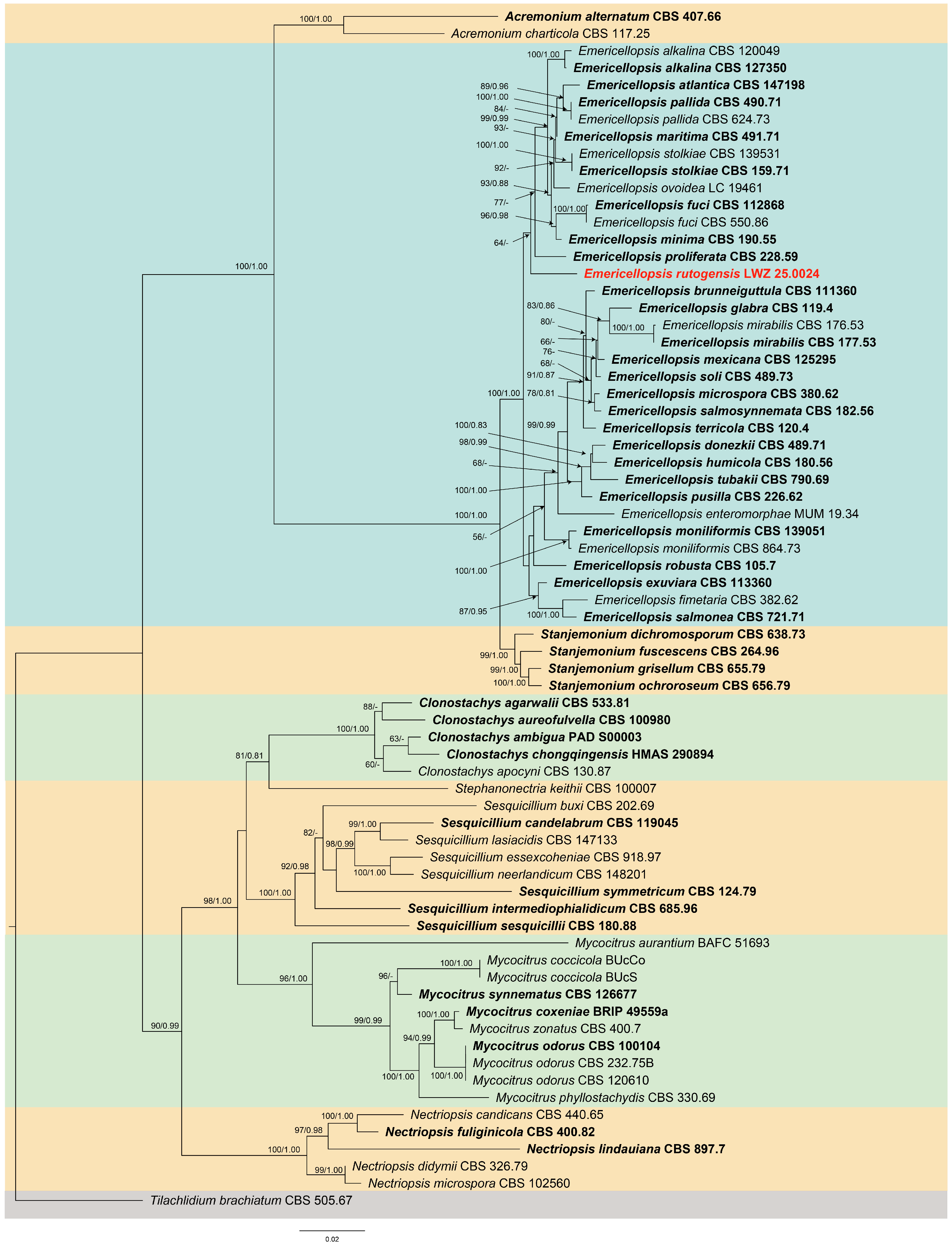
The combined dataset of ITS, nLSU, rpb2, and tub2 regions included 106 ITS, 96 nLSU, 88 rpb2, and 84 tub2 sequences from 114 taxa, resulting in an alignment of 2351 characters. The best-fit evolution model of this alignment for ML and BI analyses was estimated as GTR + I + G for each of the ITS, nLSU, rpb2, and tub2 partitions. The phylogeny inferred from this dataset recovered Cymostachys as a strongly supported (BS = 99%, BPP = 1.00) monophyletic genus within Stachybotryaceae, and the newly sequenced strain as a distinct lineage within Cymostachys (Figure 2). The combined dataset of ITS, nLSU, and tef1-α regions included 70 ITS, 65 nLSU, and 63 tef1-α sequences from 70 strains, resulting in an alignment of 2022 characters. The best-fit evolution model for ML and BI analyses was estimated as GTR + I + G for each of the ITS, nLSU, and tef1-α partitions. The phylogeny inferred from this dataset recovered the newly sequenced strain as a distinct lineage within the fully supported monophyletic genus Emericellopsis (BS = 100%, BPP = 1) within Bionectriaceae (Figure 3).
Figure 2.
Phylogenetic position of Cymostachys pangongensis inferred from the combined dataset of ITS, nLSU, rpb2, and tub2 regions. The topology was generated by the maximum likelihood algorithm, while bootstrap values and Bayesian posterior probabilities, respectively, not less than 50% and 0.80 are labelled at the nodes. Cymostachys pangongensis is highlighted in red, and the strains from type materials are in bold.
Figure 3.
Phylogenetic position of Emericellopsis rutogensis inferred from the combined dataset of ITS, nLSU, and tef1-α regions. The topology was generated by the maximum likelihood algorithm, while bootstrap values and Bayesian posterior probabilities, respectively, not less than 50% and 0.80 are labelled at the nodes. Emericellopsis rutogensis is highlighted in red, and the strains from type materials are in bold.
3.2. Taxonomy
Cymostachys pangongensis S.Y. Xia, P.W. Su & L.W. Zhou, sp. nov. (Figure 4).
Figure 4.
Cymostachys pangongensis (from ex-holotype LWZ 25.0023). Colonies on PDA (A), OA (B), MEA (C), SDA (D), and corresponding reverse sides (E–H) after the growth of 20 days at 28 °C in darkness. (I–M) Conidiophores from colonies on OA. (N) Conidia from colonies on OA. Scale bars: (I–N) = 10 μm.
Fungal Names: FN 573103.
Etymology: pangongensis (Latin), referring to Pangong Tso, the type locality.
Type: China, Xizang, Ngari Prefecture, Rutog County, Pangong Tso, 33°26′56″ N, 79°42′02″ E, elevation 4261 m, from sandy soil on the shore, 1 August 2024, leg. S.L. Liu & P.W. Su, HMAS 354212 (holotype), culture ex-type LWZ 25.0023.
Description: Culture characteristics (28 °C, 20 days, in the dark). Colonies on PDA reaching 25–27 mm diam, centrally slightly convex, floccose to felty, surface white to yellowish grey; the reverse side yellowish brown. Colonies on OA reaching 32–35 mm diam, effuse, floccose to thin-felty, centre grey to brownish grey; the reverse side light brown. Colonies on MEA reaching 26–28 mm diam, floccose, surface white; the reverse side brownish orange. Colonies on SDA reaching 14–16 mm diam, markedly folded, very slow-growing, centre greyish orange to greyish brown; the reverse side pale orange to dark brown.
Hyphae hyaline, septate, smooth, 3.0–5.0 μm diam (avg. = 5.5 µm, n = 30). Conidiophores macronematous, mononematous, solitary, erect, unbranched to sparingly branched, 1–2-septate, hyaline, smooth-walled, smooth to slightly verruculose, bearing a whorl of 3–6 phialides, 23.0–45.0 × 3.0–5.0 µm (avg. = 32.4 × 4.0 µm, n = 30). Conidiogenous cells clavate, phialidic, straight to slightly curved, with short collarettes, hyaline to dark brown, becoming paler towards base, 7.0–15.0 × 3.0–7.5 µm (avg. = 11.6 × 5.0 µm, n = 30). Conidia aseptate, ellipsoid–ovoid to short-cylindrical, smooth-walled, dark brown, aggregating in dark slimy masses, 7.5–14.5 × 5.0–9.0 µm (avg. = 10.4 × 6.0 µm, n = 40). Sexual morph not observed.
Notes: Cymostachys pangongensis differs from C. arthraeruae by its darker and apparently smoother conidia in mass, whereas the latter is characterized by an isabelline surface with continuously sigmoid striations [22]. Cymostachys pangongensis is clearly differentiated from C. coffeicola (7.5–10.0 × 5.5–7.0 µm), C. fabispora (6.5–7.5 × 4.0–5.0 µm) [34], C. garethjonesii (5–9 × 3.5–6.5 µm) [35], and C. thailandica (9.0–11.0 × 5.0–6.0 µm) [36] by its significantly larger conidia (7.5–14.5 × 5.0–9.0 µm). In addition, the obtuse and fabiform conidia of C. thailandica [36] also distinguish it from C. pangongensis producing broadly ellipsoidal conidia.
Emericellopsis rutogensis S.Y. Xia, P.W. Su & L.W. Zhou, sp. nov. (Figure 5).
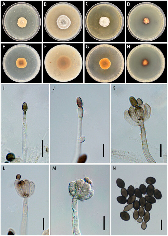
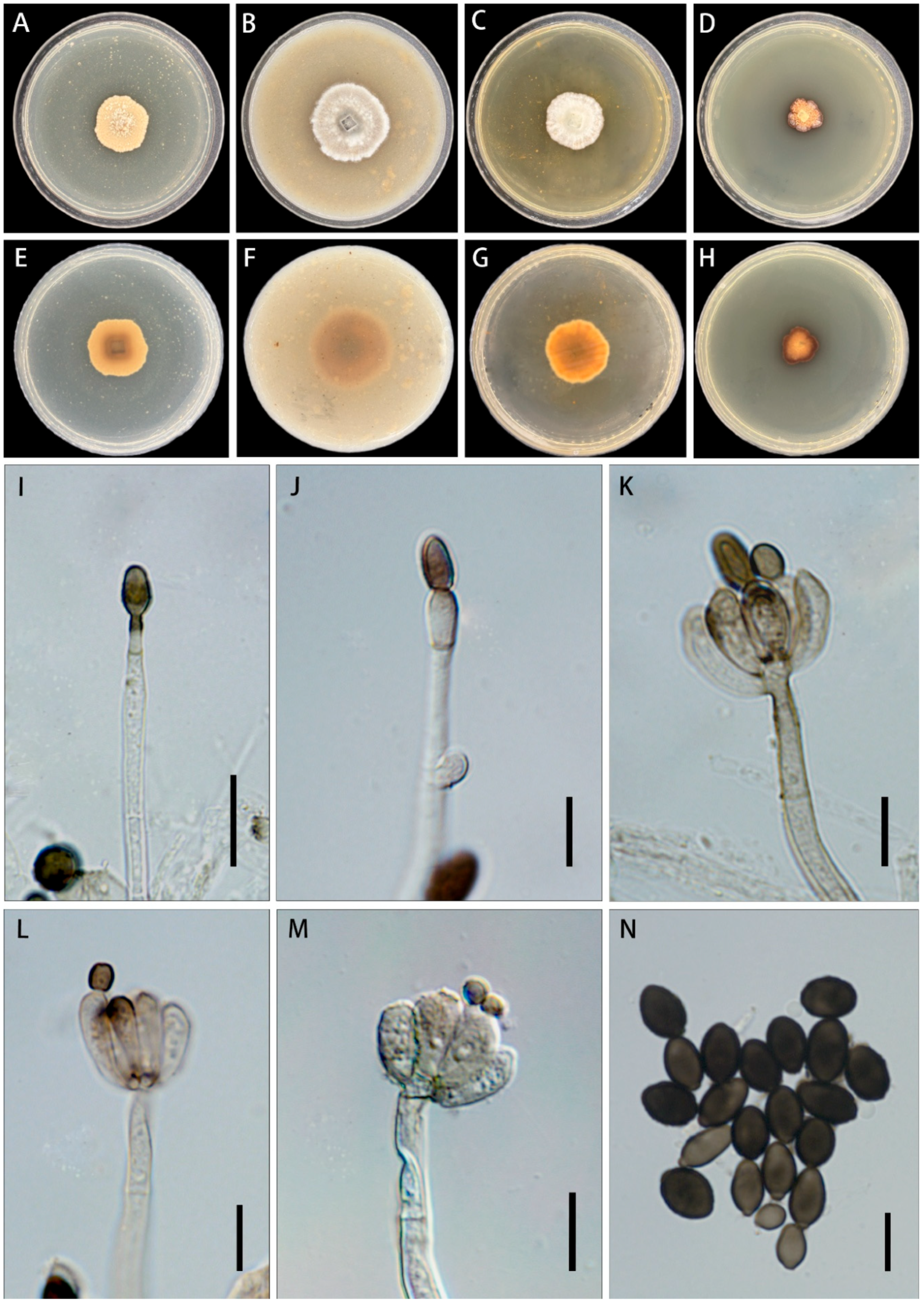
Figure 5.
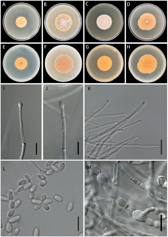
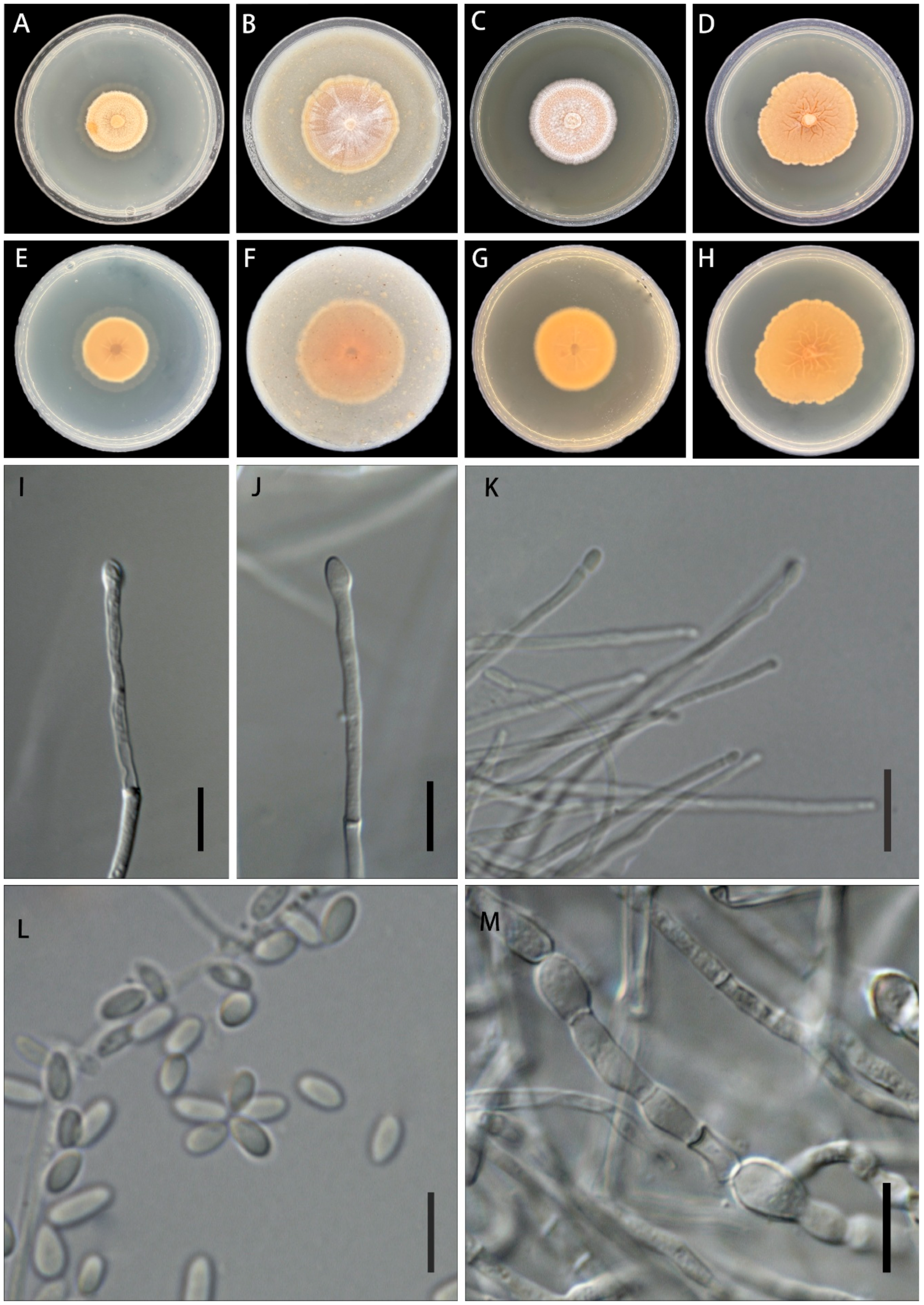
Emericellopsis rutogensis (from ex-holotype LWZ 25.0024). Colonies on PDA (A), OA (B), MEA (C), SDA (D), and corresponding reverse sides (E–H) after the growth of 20 days at 28 °C in darkness. (I–K) Conidiophores from colonies on OA. (L) Conidia from colonies on OA. (M) Chlamydospores from colonies on OA. Scale bars: (I–M) = 10 μm.
Fungal Names: FN 573104.
Etymology: rutogensis (Latin), referring to Rutog County, the type locality.
Type: China, Xizang, Ngari Prefecture, Rutog County, 33°26′56″ N, 79°42′02″ E, elevation 4261 m, from grassland soil, 1 August 2024, leg. S.L. Liu & P.W. Su, HMAS 354213 (holotype), culture ex-type LWZ 25.0024.
Description: Culture characteristics (28 °C, 20 days, in the dark). Colonies on PDA reaching 29–31 mm diam, floccose, centre pale orange; the reverse side light orange. On MEA reaching 44–45 mm diam, flat, thinly floccose to membranous, centre greyish orange, margin light orange; the reverse side pastel red. On MEA reaching 40 mm diam, plane, felty, centre pale red to white; the reverse side orange. On SDA reaching 43–46 mm diam, flat, greyish orange; the reverse side concolorous.
Hyphae hyaline, septate, branched, thin-walled and smooth, 1.5–3.5 μm wide (avg. = 2.5 μm, n = 20). Conidiophores arising from substrate or aerial hyphae, straight to slightly flexuous, usually 2–3-septate, percurrent proliferation occasionally observed, unbranched or sparsely branched, smooth-walled, 80.0–150.0 μm long (avg. = 120.0 μm, n = 20), basal width 1.0–2.5 μm (avg. = 1.90 μm, n = 20). Conidiogenous cells terminal or lateral, conical to subcylindrical, straight to slightly curved, collarettes short and inconspicuous, 3.0–6.0 μm (avg. = 4.9 μm, n = 30), basal width 1.0–2.5 μm (avg. = 1.7 μm, n = 30). Conidia hyaline, ellipsoid-cylindrical to bacilliform, aseptate, thin- and smooth-walled, 3.5–6.0 × 1.0–3.0 μm (avg. = 4.6 × 1.9 μm, n = 40). Chlamydospores present in ageing culture, terminal or intercalary, ellipsoidal, hyaline to pale brown, smooth-walled, 7.0–10.0 µm long, 3.0–7.0 μm diam. Sexual morph not observed.
Notes: Compared with Emericellopsis proliferata, E. rutogensis is primarily distinguished by its broadly ellipsoid conidia up to 1.0–3.0 μm wide, whereas the conidia of E. proliferata are slenderer (typically 1.4–2.0 μm wide) [23]. In addition, E. rutogensis differs by having longer conidiophores (80.0–150.0 μm) than those in E. proliferata (up to 135.0 μm) [23].
4. Discussion
Cymostachys was recently established to accommodate C. coffeicola as the generic type and C. fabispora within Stachybotriaceae, Hypocreales by Lombard et al. [34]. Later, Cymostachys garethjonesii, C. thailandica, and C. arthraeruae were continually described in this genus [22,35,36]. The most distinct morphological characteristics of Cymostachys are irregularly cymosely branched conidiophores and olivaceous brown to dark brown, fabiform conidia [34]. The newly described species C. pangongensis fits well with the morphological concept of this genus. Notably, within Cymostachys, the first five known species were all sampled from plants [22], while C. pangongensis is the first species isolated from soils.
Comparably, Emericellopsis is an older genus that was established by van Beyma (1940) with E. terricola as the generic type. According to the current taxonomic framework, this genus belongs to the Bionectriaceae, Hypocreales, and accommodates 24 species [23,37]. Emericellopsis is widely distributed in soils, freshwater, sediments, and plants [38]. Likely, the currently described species E. rutogensis was also isolated from soils, which makes its typical characteristics of ascomata and ascospores for Emericellopsis not observed. However, other morphological characteristics of E. rutogensis, like chlamydospores and conidia, fit well with Emericellopsis [37].
From the perspective of resource utilization, species of both Cymostachys and Emericellopsis have been subjected to the isolation of medicinal natural products [39,40,41,42]. In particular, Emericellopsis is well known for rich bioactive metabolites, possessing antibacterial, antifungal, antiviral, antitumor, and cytotoxic properties [38,43,44]. Therefore, it is worth performing in-depth studies of natural products on C. pangongensis and E. rutogensis in the future.
Besides C. pangongensis and E. rutogensis, we also isolated 514 fungal strains that were tentatively identified to 87 known species from Ngari Prefecture. Due to the special environment and lack of previous surveys in this region, more careful studies on these strains may result in the discovery of more new species. Moreover, these fungal strains, even representing previously known species, may serve as strategic bioresources for future utilization.
Author Contributions
Conceptualization, L.-W.Z.; methodology, S.-Y.X. and P.-W.S.; investigation, S.-Y.X.; resources, P.-W.S. and S.-L.L.; data curation, S.-Y.X. and P.-W.S.; formal analysis, S.-Y.X.; writing—original draft preparation, S.-Y.X.; writing—review and editing, S.-Y.X., P.-W.S., X.-J.W., S.-L.L., L.L. and L.-W.Z.; supervision, L.-W.Z.; funding acquisition, L.-W.Z. All authors have read and agreed to the published version of the manuscript.
Funding
This work was supported by the Survey of Wildlife Resources in Key Areas of Xizang (ZL202203601 & ZL202303601) and the Biological Resources Programme, Chinese Academy of Sciences (CAS-TAX-24-018).
Data Availability Statement
The original contributions presented in the study are included in the article, and further inquiries can be directed to the corresponding author.
Conflicts of Interest
The authors have declared that no competing interests exist.
References
- Bai, M.; Zhou, L.; Tong, Y.; Yin, F. Risk assessment and warning system for strategic biological resources in China. Innov. Life 2023, 1, 100004. [Google Scholar] [CrossRef] [Scilit]
- Shaaban, R.; Elnaggar, M.S.; Khalil, N.; Singab, A.N.B. A comprehensive review on the medicinally valuable endosymbiotic fungi Penicillium chrysogenum. Arch. Microbiol. 2023, 205, 240. [Google Scholar] [CrossRef] [Scilit]
- Zhgun, A.A. Industrial production of antibiotics in fungi: Current state, deciphering the molecular basis of classical strain improvement and increasing the production of high-yielding strains by the addition of low-molecular weight inducers. Fermentation 2023, 9, 1027. [Google Scholar] [CrossRef] [Scilit]
- Wu, F.; Zhou, L.W.; Yang, Z.L.; Torstein, B.; Li, T.H.; Dai, Y.C. Resource diversity of Chinese macrofungi: Edible, medicinal and poisonous species. Fungal Divers. 2019, 98, 1–76. [Google Scholar] [CrossRef] [Scilit]
- Zhou, L.W.; Ghobad-Nejhad, M.; Tian, X.M.; Wang, Y.F.; Wu, F. Current status of ‘Sanghuang’ as a group of medicinal mushrooms and their perspective in industry development. Food Rev. Int. 2022, 38, 589–607. [Google Scholar] [CrossRef] [Scilit]
- Zhou, L.W.; May, T.W. Fungal taxonomy: Current status and research agendas for the interdisciplinary and globalisation era. Mycology 2023, 14, 52–59. [Google Scholar] [CrossRef] [Scilit]
- Hawksworth, D.L.; Lücking, R. Fungal diversity revisited: 2.2 to 3.8 million species. Microbiol. Spectr. 2017, 5, FUNK-0052-2016. [Google Scholar] [CrossRef] [Scilit]
- Wang, F.Z.; Peng, C.H.; Zhang, J.X.; Jian, Z.J.; Torstein, B.; Yang, W.Q. Impacts of freezing and thawing dynamics on foliar litter carbon release in alpine/subalpine forests along an altitudinal gradient in the eastern Tibetan Plateau. Biogeosciences 2014, 11, 6471–6481. [Google Scholar] [CrossRef] [Scilit]
- Cao, R.; Yang, W.; Chang, C.; Wang, Z.; Wang, Q.; Li, H.; Tan, B. Differential seasonal changes in soil enzyme activity along an altitudinal gradient in an alpine-gorge region. Appl. Soil Ecol. 2021, 166, 104078. [Google Scholar] [CrossRef] [Scilit]
- Li, S.; Huang, X.; Shen, J.; Xu, F.; Su, J. Effects of plant diversity and soil properties on soil fungal community structure with secondary succession in the Pinus yunnanensis forest. Geoderma 2020, 379, 114646. [Google Scholar] [CrossRef] [Scilit]
- Cui, Y.; Bing, H.; Fang, L.; Wu, Y.; Yu, J.; Shen, G.; Jiang, M.; Wang, X.; Zhang, X. Diversity patterns of the rhizosphere and bulk soil microbial communities along an altitudinal gradient in an alpine ecosystem of the eastern Tibetan Plateau. Geoderma 2019, 338, 118–127. [Google Scholar] [CrossRef] [Scilit]
- Gong, S.; Feng, B.; Jian, S.P.; Wang, G.S.; Ge, Z.W.; Yang, Z.L. Elevation matters more than season in shaping the heterogeneity of soil and root-associated ectomycorrhizal fungal community. Microbiol. Spectr. 2022, 10, e01950-21. [Google Scholar] [CrossRef] [Scilit] [PubMed]
- Xing, R.; Gao, Q.B.; Zhang, F.Q.; Wang, J.L.; Chen, S.L. Environmental filtering affects fungal communities more than dispersal limitation in a high-elevation hyperarid basin on Qinghai-Tibet Plateau. FEMS Microbiol. Lett. 2021, 368, fnab033. [Google Scholar] [CrossRef] [Scilit]
- Wang, L.; Quzong, C.; Skal, S.G.; Mu, C.; Zhao, Y.; Fang, B.; Zhang, Y.; Yang, Z.; Hei, E.; Yuan, X.; et al. Depth-related patterns and physicochemical drivers of soil microbial communities in the alpine desert of Ngari, Xizang. Microorganisms 2025, 13, 2775. [Google Scholar] [CrossRef] [Scilit] [PubMed]
- White, T.J.; Bruns, T.; Lee, S.; Taylor, J. Amplification and direct sequencing of fungal ribosomal RNA genes for phylogenetics. In PCR Protocols: A Guide to Methods and Applications; Innis, M.A., Gelfand, D.H., Sninsky, J.J., White, T.J., Eds.; Academic Press: San Diego, CA, USA, 1990; pp. 315–322. ISBN 978-0-12-372180-8. [Google Scholar]
- Vilgalys, R.; Hester, M. Rapid genetic identification and mapping of enzymatically amplified ribosomal DNA from several Cryptococcus species. J. Bacteriol. 1990, 172, 4238–4246. [Google Scholar] [CrossRef] [Scilit]
- Rehner, S.A.; Samuels, G.J. Taxonomy and phylogeny of Gliocladium analysed from nuclear large subunit ribosomal DNA sequences. Mycol. Res. 1994, 98, 625–634. [Google Scholar] [CrossRef] [Scilit]
- Rehner, S.A.; Buckley, E. A Beauveria phylogeny inferred from nuclear ITS and EF1-alpha sequences: Evidence for cryptic diversification and links to Cordyceps teleomorphs. Mycologia 2005, 97, 84–98. [Google Scholar] [CrossRef] [Scilit]
- Liu, Y.J.; Whelen, S.; Hall, B.D. Phylogenetic relationships among ascomycetes: Evidence from an RNA polymerase II subunit. Mol. Biol. Evol. 1999, 16, 1799–1808. [Google Scholar] [CrossRef] [Scilit]
- Glass, N.L.; Donaldson, G.C. Development of primer sets designed for use with the PCR to amplify conserved genes from filamentous ascomycetes. Appl. Environ. Microbiol. 1995, 61, 1323–1330. [Google Scholar] [CrossRef] [Scilit]
- O’Donnell, K.; Cigelnik, E. Two divergent intragenomic rDNA ITS2 types within a monophyletic lineage of the fungus Fusarium are nonorthologous. Mol. Phylogenet. Evol. 1997, 7, 103–116. [Google Scholar] [CrossRef] [Scilit]
- Crous, P.W.; Cowan, D.A.; Maggs-Kölling, G.; Yilmaz, N.; Thangavel, R.; Wingfield, M.J.; Noordeloos, M.E.; Dima, B.; Brandrud, T.E.; Jansen, G.M.; et al. Fungal Planet description sheets: 1182–1283. Persoonia 2021, 46, 313–528. [Google Scholar] [CrossRef] [Scilit]
- Zhao, L.; Groenewald, J.Z.; Hou, L.W.; Summerbell, R.C.; Crous, P.W. Bionectriaceae: A poorly known family of hypocrealean fungi with major commercial potential. Stud. Mycol. 2025, 111, 115–198. [Google Scholar] [CrossRef] [Scilit]
- Katoh, K.; Standley, D.M. MAFFT multiple sequence alignment software version 7: Improvements in performance and usability. Mol. Biol. Evol. 2013, 30, 772–780. [Google Scholar] [CrossRef] [Scilit] [PubMed]
- Capella-Gutiérrez, S.; Silla-Martínez, J.M.; Gabaldón, T. trimAl: A tool for automated alignment trimming in large-scale phylogenetic analyses. Bioinformatics 2009, 25, 1972–1973. [Google Scholar] [CrossRef] [Scilit] [PubMed]
- Zhang, D.; Gao, F.; Jakovlić, I.; Zou, H.; Zhang, J.; Li, W.X.; Wang, G.T. PhyloSuite: An integrated and scalable desktop platform for streamlined molecular sequence data management and evolutionary phylogenetics studies. Mol. Ecol. Resour. 2020, 20, 348–355. [Google Scholar] [CrossRef] [Scilit] [PubMed]
- Xiang, C.Y.; Gao, F.; Jakovlić, I.; Lei, H.-P.; Hu, Y.; Zhang, H.; Zou, H.; Wang, G.-T.; Zhang, D. Using PhyloSuite for molecular phylogeny and tree-based analyses. iMeta 2023, 2, e87. [Google Scholar] [CrossRef] [Scilit]
- Schwarz, G. Estimating the dimension of a model. Ann. Stat. 1978, 6, 461–464. [Google Scholar] [CrossRef] [Scilit]
- Kalyaanamoorthy, S.; Minh, B.Q.; Wong, T.K.F.; von Haeseler, A.; Jermiin, L.S. ModelFinder: Fast model selection for accurate phylogenetic estimates. Nat. Methods 2017, 14, 587–589. [Google Scholar] [CrossRef] [Scilit]
- Minh, B.Q.; Schmidt, H.A.; Chernomor, O.; Schrempf, D.; Woodhams, M.D.; von Haeseler, A.; Lanfear, R. IQ-TREE 2: New models and efficient methods for phylogenetic inference in the genomic era. Mol. Biol. Evol. 2020, 37, 1530–1534. [Google Scholar] [CrossRef] [Scilit]
- Ronquist, F.; Teslenko, M.; van der Mark, P.; Ayres, D.L.; Darling, A.; Höhna, S.; Larget, B.; Liu, L.; Suchard, M.A.; Huelsenbeck, J.P. MrBayes 3.2: Efficient Bayesian phylogenetic inference and model choice across a large model space. Syst. Biol. 2012, 61, 539–542. [Google Scholar] [CrossRef] [Scilit]
- Rambaut, A.; Drummond, A.J.; Xie, D.; Baele, G.; Suchard, M.A. Posterior summarization in Bayesian phylogenetics using Tracer 1.7. Syst. Biol. 2018, 67, 901–904. [Google Scholar] [CrossRef] [Scilit] [PubMed]
- FigTree v1.4.3. Available online: http://tree.bio.ed.ac.uk/software/figtree/ (accessed on 12 November 2025).
- Lombard, L.; Houbraken, J.; Decock, C.; Samson, R.A.; Meijer, M.; Réblová, M.; Groenewald, J.Z.; Crous, P.W. Generic hyper-diversity in Stachybotriaceae. Persoonia 2016, 36, 156–246. [Google Scholar] [CrossRef] [Scilit] [PubMed]
- Lin, C. Stachybotrys-like taxa from karst areas and a checklist of Stachybotrys-like species from Thailand. Mycosphere 2016, 7, 1273–1291. [Google Scholar] [CrossRef] [Scilit]
- Crous, P.W.; Luangsa-Ard, J.J.; Wingfield, M.J.; Carnegie, A.J.; Hernández-Restrepo, M.; Lombard, L.; Roux, J.; Barreto, R.W.; Baseia, I.G.; Cano-Lira, J.F.; et al. Fungal Planet description sheets: 785–867. Persoonia 2018, 41, 238–417. [Google Scholar] [CrossRef] [Scilit]
- Hou, L.W.; Giraldo, A.; Groenewald, J.Z.; Rämä, T.; Summerbell, R.C.; Huang, G.Z.; Cai, L.; Crous, P.W. Redisposition of Acremonium-like fungi in Hypocreales. Stud. Mycol. 2023, 105, 23–203. [Google Scholar] [CrossRef] [Scilit]
- Gonçalves, M.F.M.; Hilário, S.; Van de Peer, Y.; Esteves, A.C.; Alves, A. Genomic and metabolomic analyses of the marine fungus Emericellopsis cladophorae: Insights into saltwater adaptability mechanisms and its biosynthetic potential. J. Fungi 2021, 8, 31. [Google Scholar] [CrossRef] [Scilit]
- Wang, T.; Zhou, J.; Zou, J.; Shi, Y.; Zhou, W.; Shao, P.; Yu, T.; Cui, W.; Li, X.; Wu, X.; et al. Discovery of cymopolyphenols A–F from a marine mesophotic zone Aaptos sponge-associated fungus Cymostachys sp. NBUF082. Front. Microbiol. 2021, 12, 638610. [Google Scholar] [CrossRef] [Scilit]
- Virués-Segovia, J.R.; Pinedo, C.; Zorrilla, D.; Sánchez-Márquez, J.; Sánchez, P.; Ramos, M.C.; de la Cruz, M.; Aleu, J.; Durán-Patrón, R. Discovery of new eremophilanes from the marine-derived fungus Emericellopsis maritima BC17 by culture conditions changes: Evaluation of cytotoxic and antimicrobial activities. Front. Mar. Sci. 2024, 11, 1386175. [Google Scholar] [CrossRef] [Scilit]
- Abraúl, M.; Alves, A.; Hilário, S.; Melo, T.; Conde, T.; Domingues, M.R.; Rey, F. Evaluation of lipid extracts from the marine fungi Emericellopsis cladophorae and Zalerion zaritima as a source of anti-inflammatory, antioxidant and antibacterial compounds. Mar. Drugs 2023, 21, 199. [Google Scholar] [CrossRef] [Scilit]
- Yuan, Y.; Li, T.; Wang, T.; Naman, C.B.; Ye, J.; Wu, X.; Lazaro, J.E.H.; Yan, X.; He, S. Targeted isolation of a cytotoxic cyclic hexadepsipeptide from the mesophotic zone sponge-associated fungus Cymostachys sp. NBUF082. Mar. Drugs 2021, 19, 565. [Google Scholar] [CrossRef] [Scilit]
- Agrawal, S.; Saha, S. The genus Simplicillium and Emericellopsis: A review of phytochemistry and pharmacology. Biotechnol. Appl. Biochem. 2022, 69, 2229–2239. [Google Scholar] [CrossRef] [Scilit]
- Kuvarina, A.E.; Gavryushina, I.A.; Kulko, A.B.; Ivanov, I.A.; Rogozhin, E.A.; Georgieva, M.L.; Sadykova, V.S. The emericellipsins A–E from an alkalophilic fungus Emericellopsis alkalina show potent activity against multidrug-resistant pathogenic fungi. J. Fungi 2021, 7, 153. [Google Scholar] [CrossRef] [Scilit]

Disclaimer/Publisher’s Note: The statements, opinions and data contained in all publications are solely those of the individual author(s) and contributor(s) and not of MDPI and/or the editor(s). MDPI and/or the editor(s) disclaim responsibility for any injury to people or property resulting from any ideas, methods, instructions or products referred to in the content. |
© 2026 by the authors. Licensee MDPI, Basel, Switzerland. This article is an open access article distributed under the terms and conditions of the Creative Commons Attribution (CC BY) license.