


Highlights
What are the main findings?
- Patients with primary Sjögren’s disease (pSjD) had significantly lower unstimulated whole saliva (UWS) and stimulated whole saliva (SWS) flow rates, a higher antinuclear antibody (ANA) prevalence, and higher frequencies of anti-SSA/Ro60, anti-SSA/Ro52, anti-SSB/La, and rheumatoid factor (RF) positivity compared with patients with sicca symptoms without Sjögren’s disease (SjD).
- Sialometry, anti-SSA antibodies, and minor labial salivary gland (MLSG) biopsy were identified as the most important diagnostic tools for distinguishing pSjD from non-Sjögren sicca patients.
What are the implications of the main findings?
- Early use of sialometry, anti-SSA antibody testing, and MLSG biopsy may improve diagnostic accuracy and timely recognition of pSjD in patients presenting with sicca symptoms.
- Distinct clinical and laboratory differences between pSjD and non-Sjögren sicca patients may support more precise patient stratification and facilitate earlier referral for rheumatological evaluation and management.
Abstract
Background/Objectives: Sjögren’s disease (SjD) is a chronic autoimmune disorder characterized by a wide range of clinical manifestations, often leading to delayed diagnosis. This study aimed to evaluate and compare the clinical and laboratory characteristics of patients with primary and secondary SjD and those with sicca symptoms without SjD. Methods: This comparative cross-sectional study included 84 participants enrolled between 2019 and 2024: 27 patients with primary Sjögren’s disease (pSjD), 4 with secondary Sjögren’s disease (sSjD), and 53 with sicca symptoms without SjD (non-SjD/NSjD). Primary SjD was diagnosed according to the 2016 American College of Rheumatology/European League Against Rheumatism (ACR/EULAR) classification criteria. Results: Compared with NSjD patients, those with pSjD and sSjD had significantly lower unstimulated whole saliva (UWS) and stimulated whole saliva (SWS) flow rates (p < 0.001), a higher prevalence of antinuclear antibodies (ANA) (twice as frequent) (p = 0.005), and higher frequencies of anti-SSA/Ro60, anti-SSA/Ro52, anti-SSB/La, and rheumatoid factor (RF) positivity (p = 0.002, p = 0.003, p = 0.005, and p = 0.019, respectively). In addition, SjD patients had higher EULAR Sjögren’s Syndrome Disease Activity Index (ESSDAI) scores (p = 0.026) and more frequently demonstrated a focus score (FS) ≥ 1 on minor labial salivary gland (MLSG) biopsy (p < 0.001). Serum vitamin D levels were lower in pSjD patients than in NSjD patients; however, the difference was not statistically significant (57.8 ± 20.4 vs. 70.6 ± 18.3; p = 0.259). Conclusions: Patients with pSjD exhibited distinct clinical and laboratory characteristics compared with those with sicca symptoms without SjD. Sialometry, anti-SSA antibodies, and MLSG biopsy were identified as the most important diagnostic tools for differentiating SjD from NSjD.
1. Introduction
Primary Sjögren’s disease (pSjD) is a chronic autoimmune disease characterized by a wide range of clinical features, ranging from exocrine gland involvement to extraglandular manifestations (EGMs) [1]. Histopathologically, the disease is marked by lymphocytic infiltration of the exocrine glands, resulting in dry mouth (xerostomia) and dry eyes (keratoconjunctivitis sicca). Primary SjD has a multifactorial and complex etiology, arising from the interaction of genetic, exogenous, and endogenous factors that trigger an abnormal autoimmune response mediated by B and T lymphocytes. The inflammation leads to tissue damage, causing progressive functional impairment of the affected organs and creating a chronic inflammatory environment [2].
A link has been established between certain human leukocyte antigen (HLA) alleles and Sjögren’s disease (SjD), specifically HLA-DR and HLA-DQ. It is difficult to determine which haplotypes contain the risk locus, as all haplotypes are unbalanced. Patients with HLA-DQ1 and HLA-DQ2 alleles exhibit more severe disease manifestations than those with other HLA-DQ allele combinations [2]. The presence of HLA-B8 and HLA-Dw3 also indicates an increased predisposition to the development of pSjD [3]. Exposure to environmental factors, especially viral infections, leads to dysfunction and activation of salivary gland epithelial cells (SGECs). The viruses mainly involved in the development of SjD are Epstein–Barr virus (EBV), which triggers hyperactivity of B lymphocytes, human T-lymphotropic virus type 1 (HTLV-1), hepatitis C virus (HCV), and Coxsackie virus [2]. Primary SjD is more common in menopausal women, indicating the role of sex hormones, particularly lower estrogen levels, as a risk factor for disease development. It is not known which of these factors is responsible for the onset of the autoimmune reaction, but viral infections are currently thought to be the main triggers [3].
Primary SjD occurs in approximately 0.1–4.8% of the general population, with a female-to-male ratio of 9:1. The prevalence of pSjD is higher in Europe than in Asia. The disease typically presents in middle age (30 to 60 years), with a mean age at diagnosis of 56.2 years [2]. SjD can also occur as part of another primary systemic autoimmune disease, such as rheumatoid arthritis (RA), systemic lupus erythematosus (SLE), or systemic sclerosis (SSc), and is then referred to as secondary Sjögren’s disease (sSjD). Secondary SjD has been reported as the most common cause of oral and ocular complications in patients with SLE [4,5,6]. The presence of HLA-DRw4 may indicate an increased predisposition to developing sSjD [3].
Assessing the cause of sicca symptoms presents a diagnostic challenge, even for experienced clinicians. Xerostomia and xerophthalmia may be side effects of medications that reduce body fluid secretion, such as antihypertensives, diuretics, and antihistamines [7]. As pSjD typically occurs in middle-aged women, dryness affecting the mouth, eyes, skin, and vagina can be mistaken for menopausal symptoms during the differential diagnosis. Xerostomia is the subjective sensation of a dry mouth and can result from various causes, including medication, smoking, environmental factors, and aging. Due to the nonspecific clinical presentation and symptoms, clinicians may take more than 10 years to reach an accurate diagnosis. Diagnosis is further complicated by the overlap of symptoms such as fatigue and musculoskeletal pain with signs of aging, anxiety, or perimenopause [2].
The aim of this study was to compare the clinical and laboratory characteristics of patients with pSjD, sSjD, and those presenting with sicca symptoms. The specific objectives were: (1) to evaluate the clinical and laboratory characteristics of patients with pSjD according to age and anti-SSA/Ro60 and anti-SSA/Ro52 antibody status; (2) to assess the distribution of patients with pSjD, sSjD, and sicca symptoms according to focus score (FS); and (3) to descriptively identify the autoimmune diseases associated with sSjD in our cohort, recognizing the limited size of the sSjD subgroup. We hypothesized that patients with SjD would exhibit distinct clinical and laboratory profiles compared with patients presenting with sicca symptoms, while analyses involving the sSjD subgroup were considered exploratory due to the limited sample size.
2. Materials and Methods
2.1. Study Design and Subjects
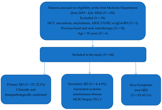
This comparative cross-sectional study was conducted at the Dental Polyclinic Split, a teaching base of the School of Medicine (Study of Dental Medicine), University of Split, Split, Croatia. A total of 84 patients (76 women and 8 men) who attended the Oral Medicine Department for their first specialist examination due to sicca symptoms or who were referred by a clinical immunologist or rheumatologist because of pathological immunological laboratory findings, were included in the study from June 2019 to July 2024. The diagnosis of pSjD was established according to the 2016 diagnostic criteria of the American College of Rheumatology (ACR) and the European League Against Rheumatism (EULAR) [8]. Patients were classified as having sSjD when sicca symptoms occurred in the presence of a previously diagnosed systemic autoimmune connective tissue disease. In all patients in the sSjD subgroup, a minor labial salivary gland (MLSG) biopsy was performed, and histopathological examination (HPE) demonstrated an FS ≥ 1, consistent with SjD. The distinction between pSjD and sSjD was based on the presence or absence of an associated systemic autoimmune disease. The study was approved by the Ethics Committee of the School of Medicine, University of Split, Split, Croatia (Class: 029-01/24-02/0001; Reg. No.: 2181-198-03-04-24-0045) on 29 April 2024. All subjects participated voluntarily and were fully informed, both verbally and in writing, about the protocol and purpose of the study before signing the informed consent form. Subjects who did not understand the nature or purpose of the study or the content of the informed consent form were excluded. The study was conducted in accordance with the principles of the Declaration of Helsinki (1964) and its subsequent amendments. No a priori sample size calculation was performed. The sample size was determined by the number of eligible patients who met the inclusion criteria during the study period.
The patients (N = 84, 100.0%) were divided into three groups:
1. Patients with a clinically and histopathologically confirmed diagnosis of pSjD (N = 27, 32.1%);
2. Patients with a clinically and histopathologically confirmed diagnosis of sSjD (N = 4, 4.8%);
3. Patients with sicca symptoms (N = 53, 63.1%).
The exclusion criteria were:
1. Patients with HCV, sarcoidosis, amyloidosis, acquired immunodeficiency syndrome (AIDS), graft-versus-host disease (GVHD), or immunoglobulin G4-related disease (IgG4-RD);
2. Patients who had been exposed to radiation in the head and neck area;
3. Subjects under 18 years old (Figure 1).
Figure 1.
Flow diagram of patient selection, inclusion criteria, exclusion criteria, and final allocation into study groups (pSjD, sSjD, and NSjD). Abbreviations: HCV, hepatitis C virus; AIDS, acquired immunodeficiency syndrome; GVHD, graft-versus-host disease; IgG4-RD, immunoglobulin G4-related disease; SjD, Sjögren’s disease; MLSG, minor labial salivary gland; FS, focus score; non-SjD, patients with sicca symptoms.
2.2. Clinical and Laboratory Parameters
The following data were collected from all subjects: medical history, demographic data, and duration of symptoms until the correct diagnosis (in months). Subjective eye dryness was assessed using the Schirmer test (ST) (measured in mm) and the tear break-up time test (TBUT) (measured in seconds). If the measurement results for the left and right eyes differed, the worse value was used as the reference for comparison among the three groups. ST and TBUT values of ≤5 mm or 5 s in at least one eye were considered pathological (positive). Sialometry test was used to determine the value of unstimulated whole saliva (UWS) (measured over 15 min and expressed in mL/min) and stimulated whole saliva (SWS) (measured over 5 min and expressed in mL/min after stimulation with a 1.0% vitamin C solution, i.e., 1 g of ascorbic acid in 1 dcl of water). A UWS value of ≤0.1 mL/min was considered pathological (positive). A biopsy of the MLSG was performed, and FS values were calculated. Tissue samples for pathohistological analysis of the MLSG were taken from clinically healthy mucosa of the lower lip. After application of a local anesthetic, the same oral medicine specialist (with more than five years of specialist experience) made a horizontal incision measuring 1.5–2 cm parallel to the vermilion of the lip. This method is most commonly used, as the incidence of complications is less than 1% [9,10,11]. Four to six lobes of the MLSG were excised. The samples were then placed in a container with 10% formaldehyde and sent to the Clinical Department of Pathology, Forensic Medicine and Cytology, Clinical Hospital Centre Split, Split, Croatia, to an experienced pathologist (with more than five years of specialist experience). The pathologist who evaluated the MLSG biopsy and calculated the FS was blinded to the clinical classification of the patients and their symptom history at the time of histopathological assessment. Laboratory findings included: erythrocyte sedimentation rate (ESR), C-reactive protein (CRP), rheumatoid factor (RF), antibodies to citrullinated proteins (anti-CCP), complement components (C3 and C4), antinuclear antibodies (ANA), antibodies to extractable nuclear antigens (ENA), and serum vitamin D (<50 nmol/L—deficit, 50–<75 nmol/L—deficiency, and ≥75 nmol/L—adequate level).
2.3. Instruments
EULAR Sjögren’s Syndrome Disease Activity Index (ESSDAI)
The ESSDAI is an objective measure of disease activity in patients with SjD. It includes 12 domains representing 12 organ systems: kidneys, muscles, peripheral nervous system (PNS), central nervous system (CNS), hematological, biological, constitutional, lymphadenopathy, glands, joints, skin, and lungs [12]. For each domain, clinical manifestations are classified according to the degree of activity (0—no activity, 1—low activity, 2—moderate activity, and 3—high activity) [13]. An experienced clinical immunologist with more than five years of specialist experience assessed the ESSDAI. The immunologist was blinded to patients’ group allocation during scoring.
2.4. Statistical Analysis
Data were entered and coded in a Microsoft Excel spreadsheet, version 16.0 (Microsoft Corporation, Redmond, WA, USA), and statistical analyses were performed using IBM SPSS Statistics software, version 23.0 (IBM Corp., Armonk, NY, USA). Demographic and clinical data were analyzed using descriptive statistics. The normality of data distribution was tested with the Kolmogorov–Smirnov test. Categorical variables are presented as frequencies and percentages, while continuous variables are presented as arithmetic means (AM) and standard deviations (SD), or medians (M) and interquartile ranges (IQR), depending on data distribution. Comparisons of continuous data between two groups was performed using the Mann–Whitney U test for nonparametric data or the Student t-test for parametric data. Comparisons of categorical variables between groups was performed using the chi-square test; when more than 20% of the expected frequencies in the cells were less than 5, the Fisher’s exact test was used. Statistical significance was set at p < 0.05.
3. Results
3.1. Study Subjects
This comparative cross-sectional study included 84 patients, of whom 31 (36.9%) were diagnosed with SjD [27 (87.1%) with pSjD and 4 (12.9%) with sSjD]. Fifty-three (63.1%) patients had sicca symptoms but did not meet the 2016 ACR/EULAR diagnostic criteria for SjD and were classified as non-SjD (NSjD). The median age was 69.0 years (IQR 55.0 to 76.0) for pSjD, 70.5 years (IQR 67.0 to 74.8) for sSjD, and 63.0 years (IQR 49.5 to 70.0) for NSjD. Of all subjects (N = 84), 76 (90.5%) were women and 8 (9.5%) were men. Women were the majority in all three groups: pSjD (N = 25, 92.6%), sSjD (N = 4, 100.0%), and NSjD (N = 47, 88.7%). There was no statistically significant difference in age or gender among patients with pSjD, sSjD, and NSjD (p = 0.104 and p = 0.710, respectively) (Table 1).
Table 1.
Comparison of demographic, clinical, and laboratory parameters in pSjD, sSjD, and NSjD patients.
3.2. Comparative Description of pSjD, sSjD, and NSjD Patients
The duration of symptoms until the correct diagnosis was longest in sSjD patients, averaging 36 months. Subjective dry mouth and dry eyes were equally prevalent among pSjD, sSjD, and NSjD patients (p = 0.408, p = 0.232). Therefore, these subjective diagnostic tools have limited ability to distinguish between SjD (pSjD, sSjD) and NSjD patients. Sialometric measurements (UWS and SWS) showed statistically significant differences between SjD and NSjD patients (p < 0.001 and p < 0.001, respectively). ANA was twice as common in SjD patients (pSjD, sSjD) as in NSjD patients (p = 0.005). The prevalence of autoantibodies (anti-SSA/Ro60, anti-SSA/Ro52, and anti-SSB/La) was significantly higher in pSjD patients compared to NSjD patients (p = 0.002, p = 0.003, and p = 0.005, respectively). Pathological values of ST and TBUT were equally high in pSjD, sSjD, and NSjD patients (p = 0.585, p = 1.000). Objective ocular diagnostic tools have limited ability to distinguish between SjD (pSjD, sSjD) and NSjD patients, unlike objective oral diagnostic tests (sialometry). The ESSDAI showed significantly higher scores in sSjD patients compared to pSjD patients (p = 0.026). The ESSDAI was positive in 20 (74.1%) pSjD patients. The most frequently affected organ systems/domains in pSjD patients were: joints (N = 11, 40.7%), CNS (N = 6, 22.2%), glands (N = 6, 22.2%), and biological (N = 4, 14.8%). The prevalence of RF was significantly higher in pSjD patients compared to NSjD patients (p = 0.019). The mean serum vitamin D concentration was lower in pSjD patients compared to NSjD patients (57.8 ± 20.4 vs. 70.6 ± 18.3, p = 0.259) (Table 1). Sialometric measurements and the prevalence of ANA, anti-SSA/Ro60, anti-SSA/Ro52, anti-SSB/La, and RF play important roles in differentiating SjD from NSjD patients.
Of the 4 (100.0%) sSjD patients, 3 (75.0%) had a diagnosis of RA and 1 (25.0%) had SSc. The prevalence of FS ≥ 1 was significantly lower in sSjD patients compared to pSjD patients [N = 2 (50.0%) vs. N = 20 (74.1%), p < 0.001] (Table 2).
Table 2.
Incidence of pSjD, sSjD, and NSjD based on FS according to the Chisholm–Mason FS criteria.
3.3. Focus Score
SjD patients (pSjD, sSjD) had a statistically significantly higher presence of FS ≥ 1 compared to NSjD patients [N = 20 (74.1%) vs. N = 2 (50.0%) vs. N = 3 (5.7%), p < 0.001]. FS < 1 was observed in 50 (94.3%) of NSjD patients. These findings indicate that biopsy of the MLSG and calculation of FS are valuable diagnostic tools for identifying SjD and distinguishing SjD from NSjD patients (Table 2).
3.4. The Influence of Age on Clinical and Laboratory Parameters in pSjD Patients
Given the very small sample size in the <40 years age group (N = 3), these analyses are exploratory and should be interpreted with caution. The pSjD patients were divided into three age groups: <40, 40–64, and ≥65 years. The duration of symptoms was longest in the 40–64 age group, at 36 months (p = 0.028). Subjective dry mouth and dry eyes were most frequent in the 40–64 age group (p = 0.266, p = 0.022). UWS and SWS scores were lower in the 40–64 and ≥65 age groups than in the <40 age group (p = 0.129, p = 0.851). ANA was equally common across all three age groups (p = 1.000). Autoantibodies (anti-SSA/Ro60, anti-SSA/Ro52, and anti-SSB/La) were more frequent in the <40 age group than in the other two groups (p = 0.635, p = 0.412, p = 1.000). Pathological ST values occurred equally across all three age groups (p = 1.000). Pathological TBUT values were most frequent in the <40 age group (p = 1.000). Pathological ST and TBUT values did not differentiate between subgroups. ESSDAI scores were highest in the 40–64 age group (p = 0.080). The <40 and 40–64 age groups had a higher prevalence of RF than the ≥65 age group (p = 0.400). Higher serum vitamin D concentrations were found in the <40 age group compared to the other two groups (Table 3).
Table 3.
Comparison of demographic, clinical, and laboratory characteristics of pSjD patients according to age.
The incidence of FS in pSjD patients was examined across three age groups (<40, 40–64, ≥65). The ≥65 age group showed a statistically significantly higher incidence of FS ≥ 1 compared to the other two groups (p = 0.047). Among all pSjD patients in the ≥65 age group, 12 (80.0%) had an FS of 1–<2 (Table 4).
Table 4.
Incidence of pSjD patients by age group according to the Chisholm–Mason FS criteria.
3.5. Correlation of Anti-SSA/Ro60 and Anti-SSA/Ro52 with Other Clinical and Laboratory Parameters
Anti-SSA/Ro60-positive pSjD patients were younger than anti-SSA/Ro60-negative pSjD patients. A statistically significantly higher number of anti-SSA/Ro60-positive pSjD patients were positive for anti-SSA/Ro52, anti-SSB/La, and RF compared to anti-SSA/Ro60-negative pSjD patients (p < 0.001, p = 0.004, p = 0.048). Anti-SSA/Ro60-positive pSjD patients had higher ESSDAI scores, indicating greater disease activity, than anti-SSA/Ro60-negative pSjD patients (p = 0.180). Anti-SSA/Ro60-positive pSjD patients had lower mean serum vitamin D concentrations (47.7 ± 22.8 vs. 64.5 ± 20.1) compared to anti-SSA/Ro60-negative pSjD patients (Table 5).
Table 5.
Comparison of demographic, clinical, and laboratory characteristics of pSjD patients according to anti-SSA/Ro60 positivity and negativity.
Anti-SSA/Ro52-positive pSjD patients were younger than anti-SSA/Ro52-negative pSjD patients. A statistically significantly higher number of anti-SSA/Ro52-positive pSjD patients were positive for ANA, anti-SSA/Ro60, anti-SSB/La, and RF compared to anti-SSA/Ro52-negative pSjD patients (p = 0.036, p < 0.001, p = 0.015 and p = 0.048, respectively). Anti-SSA/Ro52-positive pSjD patients had higher ESSDAI scores, indicating greater disease activity, than anti-SSA/Ro52-negative pSjD patients (p = 0.473). Anti-SSA/Ro52-positive pSjD patients had lower mean serum vitamin D concentrations (47.7 ± 22.8 vs. 64.5 ± 20.1) compared to anti-SSA/Ro52-negative pSjD patients (Table 6).
Table 6.
Comparison of demographic, clinical, and laboratory characteristics of pSjD patients according to anti-SSA/Ro52 positivity and negativity.
4. Discussion
This comparative cross-sectional study presents an overview of the clinical and laboratory characteristics of patients with SjD (pSjD and sSjD) and NSjD from a Croatian tertiary center. The aim was to improve understanding of disease characteristics in the Croatian population, given the absence of a national SjD registry, and to identify the most relevant diagnostic parameters for distinguishing SjD from NSjD. Management of SjD requires a multidisciplinary approach and the integration of subjective and objective diagnostic criteria to ensure an accurate diagnosis [14]. The findings showed significant differences in clinical and laboratory characteristics between patients with SjD and those with sicca symptoms, consistent with previous reports in the literature. These results provide additional evidence from a Croatian cohort and highlight the diagnostic value of key clinical, serological, and histopathological parameters in differentiating SjD from NSjD.
The findings of this study are consistent with those reported by de Oliveira et al., [15] who compared the demographic, clinical, serological, and histopathological characteristics of patients with pSjD (N = 198), sSjD (N = 149), and NSjD (N = 163) in a Brazilian cohort. Their results highlighted the diagnostic value of UWS, serological markers (anti-SSA, anti-SSB/La, RF, and ANA), and MLSG biopsy in distinguishing SjD from NSjD patients [15]. In addition to its diagnostic value in SjD, saliva plays a fundamental role in maintaining oral homeostasis and the stability of the oral ecosystem. Salivary secretions provide lubrication, buffering capacity, antimicrobial defense, remineralization of dental tissues, and regulation of the oral microbiota. Reduced salivary flow can lead to consequences beyond subjective oral dryness, increasing susceptibility to dental caries, periodontal disease, oral infections, and dysbiosis of the oral microbiome. Recent evidence has emphasized the importance of saliva in preserving the balance of the oral ecosystem and maintaining overall oral health, underscoring the clinical relevance of salivary assessment in patients with xerostomia and sicca symptoms [16]. Notably, anti-SSA antibodies were detected about twice as frequently as anti-SSB/La antibodies in both pSjD and sSjD patients, supporting their role as a key serological marker of SjD [15]. Our findings are consistent with these observations. By analyzing anti-SSA/Ro60 and anti-SSA/Ro52 antibodies separately, we further demonstrated their high prevalence among patients with SjD, reinforcing the diagnostic significance of anti-SSA autoantibodies in differentiating SjD from patients with sicca symptoms.
Fernández Castro M et al. conducted a prospective multicenter study of Spanish patients with pSjD and reported that 437 patients (95%) experienced subjective symptoms of dry mouth and dry eyes at diagnosis [1]. These findings are consistent with the results of the present study, in which subjective sicca symptoms were also highly prevalent among patients with SjD. However, although xerostomia and xerophthalmia are hallmark symptoms of SjD, their diagnostic specificity is limited because similar symptoms are frequently observed in the general population and may result from aging, medication use, or other medical conditions. Consistent with this, pathological ST and TBUT results were similarly prevalent among patients with SjD and NSjD in our cohort. This suggests that ocular dryness tests alone have limited discriminatory value and should be interpreted alongside serological and histopathological findings when differentiating SjD from NSjD patients.
ANA positivity was detected in 13 (25.5%) NSjD patients, highlighting the limited specificity of this marker for SjD. This finding is consistent with previous reports showing that ANA may also be present in some healthy individuals and in patients with various non-SjD conditions [17,18]. Therefore, ANA positivity alone is insufficient to establish a diagnosis of SjD. However, the concomitant presence of ANA and anti-SSA antibodies increases the likelihood of SjD and may support the diagnostic evaluation when interpreted in the appropriate clinical context, although ANA itself is considered a relatively weak predictor of pSjD [19,20].
SjD remains a diagnostic challenge, even for experienced clinical immunologists, particularly in patients presenting with EGMs without sicca symptoms [21,22]. In these cases, a comprehensive assessment of systemic disease involvement is essential. The ESSDAI is a validated tool for evaluating systemic disease activity and can help identify clinically relevant organ involvement in routine practice. The findings of this study support its clinical utility. Positive ESSDAI scores were observed in 20 (74.1%) patients with pSjD and were significantly higher in SjD patients than in NSjD patients. Overall disease activity was low, which may be explained by the outpatient-based study population, as patients with more severe disease manifestations are more likely to be managed in inpatient or tertiary referral settings. The most frequently involved ESSDAI domains in patients with pSjD were the articular, CNS, glandular, and biological domains.
Vitamin D deficiency has been implicated in the pathogenesis of several autoimmune diseases, including RA, SLE, multiple sclerosis (MS), and SjD. However, evidence regarding its role in SjD remains limited and inconsistent. Mehta V et al. reported lower vitamin D concentrations in patients with an FS of 1, although this association was not observed at higher FS values [23]. Other studies have suggested a relationship between vitamin D deficiency and specific disease manifestations, such as peripheral neuropathy and lymphoma in SjD patients [24]. The systematic review and meta-analysis by Radić M et al. demonstrated lower vitamin D concentrations in SjD patients than in healthy controls but found no significant association between vitamin D status and disease activity [25]. Our findings generally support these observations. Although mean serum vitamin D concentrations were lower in pSjD patients than in NSjD patients, the difference did not reach statistical significance (p = 0.259). Similarly, lower vitamin D concentrations were observed in the older age groups (40–64 and ≥65) than in the <40 age group, and in anti-SSA/Ro60- and anti-SSA/Ro52-positive patients compared with their seronegative counterparts. These differences are likely influenced by age-related confounding and the small number of patients within each subgroup, which limits the ability to draw definitive conclusions regarding their biological or disease-specific significance. These findings may suggest a trend toward lower vitamin D concentrations in patients with a more pronounced autoimmune phenotype. However, the absence of statistically significant differences precludes firm conclusions regarding a causal or clinically relevant association.
SjD is a clinically heterogeneous disease with considerable variability in its manifestations, course, and prognosis. Although current clinical, serological, and histopathological parameters are essential for diagnosis and assessment, they do not fully capture the complexity of the different phenotypic and biological subsets of SjD. Consequently, increasing attention has been given to multi-omics approaches, including genomics, proteomics, and metabolomics, which may improve patient stratification, provide insights into disease mechanisms, and facilitate the development of more personalized diagnostic and therapeutic strategies [26,27].
Non-SjD patients showed a low frequency of autoantibody positivity, including anti-SSA, anti-SSB/La, and RF. In contrast, sicca symptoms and pathological results on objective ocular tests (ST and TBUT) were common in this group. This finding is consistent with the high prevalence of ocular and oral dryness in the general population, where such symptoms may be related to medication use, menopause, smoking, environmental factors, or aging rather than to underlying autoimmune disease. The overlap in sicca manifestations between SjD and NSjD highlights the limited specificity of subjective symptoms and objective ocular dryness tests when used alone. In contrast, UWS measurements, anti-SSA antibodies, and MLSG biopsy with FS assessment provide greater diagnostic discrimination and remain essential components in the evaluation of patients with suspected SjD [8,28].
This study has several limitations that should be considered when interpreting the findings. First, the study population was recruited from a single outpatient center; therefore, selection bias cannot be excluded. Therefore, the findings may not fully represent the broader SjD population, especially patients with more severe disease who require inpatient care. Furthermore, the cross-sectional design precludes assessment of temporal relationships and causal inferences. It also does not allow evaluation of disease progression, prognostic factors, or long-term outcomes, such as lymphoma development. Not all laboratory parameters were available for all patients, and some tests were performed selectively based on clinical indication, resulting in non-uniform data availability and potential information bias. The relatively small sample size and unequal distribution of participants across the pSjD, sSjD, and NSjD groups may have limited the statistical power of some analyses. In particular, the very small number of patients in the sSjD group (N = 4) substantially limits the reliability of subgroup-specific analyses, which should be regarded as exploratory rather than confirmatory. Similarly, the very small number of patients in certain age-stratified subgroups within the pSjD cohort, particularly those younger than 40 years (N = 3), limits the interpretability of these analyses and renders statistical comparisons exploratory. In addition, no adjustment for multiple comparisons was performed, which may increase the risk of a type I error given the number of statistical tests conducted. Finally, only a small number of male patients were included, reflecting the known female predominance of SjD and limiting the ability to evaluate potential sex-related differences.
Future studies should include larger, multicenter cohorts with longitudinal follow-up to better characterize the evolution of clinical, serological, and histopathological features over time. Such studies would also enable assessment of temporal relationships, disease progression, and prognostic factors. Furthermore, they could provide valuable information on long-term outcomes, including the risk of lymphoma development, and contribute to a more comprehensive understanding of SjD in the Croatian population.
5. Conclusions
Patients with pSjD showed distinct clinical and laboratory profiles compared with those presenting with sicca symptoms, highlighting the heterogeneity of SjD. Among sSjD patients, RA was the most frequently associated autoimmune disease. Our findings support the diagnostic value of UWS, anti-SSA antibodies, and MLSG biopsy with FS assessment in distinguishing SjD from NSjD patients. Although lower serum vitamin D concentrations were observed in SjD patients and in older individuals, no statistically significant association was found. These results should be interpreted with caution due to potential confounding factors. Further studies are needed to clarify the relationship between vitamin D status, disease activity, and potential therapeutic implications in SjD.
Author Contributions
Conceptualization, A.G.; methodology, A.G., A.M.Z., M.K. and D.M.; software, D.M.; validation, A.G. and A.T.; formal analysis, A.M.Z. and M.K.; investigation, A.M.Z. and M.K.; resources, A.G.; data curation, D.M.; writing—original draft preparation, A.M.Z. and M.K.; writing—review and editing, A.G.; visualization, A.G. and A.T.; supervision, A.G.; project administration, A.G.; funding acquisition, A.G. All authors have read and agreed to the published version of the manuscript.
Funding
This research received no external funding.
Institutional Review Board Statement
The study was conducted in accordance with the Declaration of Helsinki and was approved by the Ethics Committee of the School of Medicine, University of Split, Split, Croatia (Class: 029-01/24-02/0001; Reg. No.: 2181-198-03-04-24-0045) on 29 April 2024.
Informed Consent Statement
Informed consent was obtained from all subjects involved in the study. Written informed consent has been obtained from the patients to publish this paper.
Data Availability Statement
The datasets generated and/or analyzed during the current study are available from the corresponding author upon reasonable request.
Conflicts of Interest
The authors declare no conflicts of interest.
Abbreviations
The following abbreviations are used in this manuscript:
| SjD | Sjögren’s disease |
| pSjD | Primary Sjögren’s disease |
| sSjD | Secondary Sjögren’s disease |
| non-SjD/NSjD | Patients with sicca symptoms |
| ACR | American College of Rheumatology |
| EULAR | European League Against Rheumatism |
| HPE | Histopathological examination |
| UWS | Unstimulated whole saliva |
| SWS | Stimulated whole saliva |
| ANA | Antinuclear antibodies |
| RF | Rheumatoid factor |
| ESSDAI | EULAR Sjögren’s Syndrome Disease Activity Index |
| FS | Focus score |
| MLSG | Minor labial salivary gland |
| EGM | Extraglandular manifestations |
| HLA | Human leukocyte antigen |
| SGEC | Salivary gland epithelial cells |
| EBV | Epstein–Barr virus |
| HTLV-1 | Human T-lymphotropic virus type 1 |
| HCV | Hepatitis C virus |
| RA | Rheumatoid arthritis |
| SLE | Systemic lupus erythematosus |
| SSc | Systemic sclerosis |
| AIDS | Acquired immunodeficiency syndrome |
| GVHD | Graft-versus-host disease |
| IgG4-RD | Immunoglobulin G4-related disease |
| ST | Schirmer’s test |
| TBUT | Tear break-up time test |
| ESR | Erythrocyte sedimentation rate |
| CRP | C-reactive protein |
| anti-CCP | Antibodies to citrullinated proteins |
| ENA | Extractable nuclear antigens |
| PNS | Peripheral nervous system |
| CNS | Central nervous system |
| AM | Arithmetic mean |
| SD | Standard deviation |
| M | Median |
| IQR | Interquartile range |
| N | Number |
| MS | Multiple sclerosis |
References
- Fernández Castro, M.; Andreu, J.L.; Sánchez-Piedra, C.; Martínez Taboada, V.; Olivé, A.; Rosas, J.; Sánchez-Alonso, F.; en Representación del Grupo de Trabajo en Enfermedades Autoinmunes Sistémicas de la Sociedad Española de Reumatología (EAS-SER) y de la Unidad de Investigación de la Sociedad Española de Reumatología (UI-SER). Sjögren SER: National registry of the Spanish Society of Rheumatology of patients with primary Sjögren syndrome: Objectives and methodology. Reumatol. Clin. 2016, 12, 184–189. (In Spanish) [Google Scholar] [CrossRef] [Scilit] [PubMed]
- Parisis, D.; Chivasso, C.; Perret, J.; Soyfoo, M.S.; Delporte, C. Current State of Knowledge on Primary Sjögren’s Syndrome, an Autoimmune Exocrinopathy. J. Clin. Med. 2020, 9, 2299. [Google Scholar] [CrossRef] [Scilit] [PubMed]
- Margaix-Muñoz, M.; Bagán, J.V.; Poveda, R.; Jiménez, Y.; Sarrión, G. Sjögren’s syndrome of the oral cavity. Review and update. Med. Oral Patol. Oral Cir. Bucal 2009, 14, E325–E330. [Google Scholar] [PubMed]
- Bjordal, O.; Norheim, K.B.; Rødahl, E.; Jonsson, R.; Omdal, R. Primary Sjögren’s syndrome and the eye. Surv. Ophthalmol. 2020, 65, 119–132. [Google Scholar] [CrossRef] [Scilit] [PubMed]
- Bagavant, H.; Michrowska, A.; Deshmukh, U.S. The NZB/W F1 mouse model for Sjögren’s syndrome: A historical perspective and lessons learned. Autoimmun. Rev. 2020, 19, 102686. [Google Scholar] [CrossRef] [Scilit] [PubMed]
- Pasoto, S.G.; Adriano de Oliveira Martins, V.; Bonfa, E. Sjögren’s syndrome and systemic lupus erythematosus: Links and risks. Open Access Rheumatol. 2019, 11, 33–45. [Google Scholar] [CrossRef] [Scilit] [PubMed]
- Mohamed, H.B.; Abd El-Hamid, B.N.; Fathalla, D.; Fouad, E.A. Current trends in pharmaceutical treatment of dry eye disease: A review. Eur. J. Pharm. Sci. 2022, 175, 106206. [Google Scholar] [CrossRef] [Scilit] [PubMed]
- Shiboski, C.H.; Shiboski, S.C.; Seror, R.; Criswell, L.A.; Labetoulle, M.; Lietman, T.M.; Rasmussen, A.; Scofield, H.; Vitali, C.; Bowman, S.J.; et al. 2016 American College of Rheumatology/European League Against Rheumatism classification criteria for primary Sjögren’s syndrome: A consensus and data-driven methodology involving three international patient cohorts. Ann. Rheum. Dis. 2017, 76, 9–16. [Google Scholar] [CrossRef] [Scilit] [PubMed]
- Fisher, B.A.; Jonsson, R.; Daniels, T.; Bombardieri, M.; Brown, R.M.; Morgan, P.; Bombardieri, S.; Ng, W.F.; Tzioufas, A.G.; Vitali, C.; et al. Standardisation of labial salivary gland histopathology in clinical trials in primary Sjögren’s syndrome. Ann. Rheum. Dis. 2017, 76, 1161–1168. [Google Scholar] [CrossRef] [Scilit] [PubMed]
- Guellec, D.; Cornec, D.; Jousse-Joulin, S.; Marhadour, T.; Marcorelles, P.; Pers, J.O.; Saraux, A.; Devauchelle-Pensec, V. Diagnostic value of labial minor salivary gland biopsy for Sjögren’s syndrome: A systematic review. Autoimmun. Rev. 2013, 12, 416–420. [Google Scholar] [CrossRef] [Scilit] [PubMed]
- Varela-Centelles, P.; Seoane-Romero, J.M.; Sánchez-Sánchez, M.; González-Mosquera, A.; Diz-Dios, P.; Seoane, J. Minor salivary gland biopsy in Sjögren’s syndrome: A review and introduction of a new tool to ease the procedure. Med. Oral Patol. Oral Cir. Bucal 2014, 19, e20–e23. [Google Scholar] [CrossRef] [Scilit] [PubMed]
- Seror, R.; Ravaud, P.; Bowman, S.J.; Baron, G.; Tzioufas, A.; Theander, E.; Gottenberg, J.E.; Bootsma, H.; Mariette, X.; Vitali, C.; et al. EULAR Sjogren’s syndrome disease activity index: Development of a consensus systemic disease activity index for primary Sjogren’s syndrome. Ann. Rheum. Dis. 2010, 69, 1103–1109. [Google Scholar] [CrossRef] [Scilit] [PubMed]
- Seror, R.; Bowman, S.J.; Brito-Zeron, P.; Theander, E.; Bootsma, H.; Tzioufas, A.; Gottenberg, J.E.; Ramos-Casals, M.; Dörner, T.; Ravaud, P.; et al. EULAR Sjögren’s syndrome disease activity index (ESSDAI): A user guide. RMD Open 2015, 1, e000022. [Google Scholar] [CrossRef] [Scilit] [PubMed]
- Alves, M.; Reinach, P.S.; Paula, J.S.; Vellasco e Cruz, A.A.; Bachette, L.; Faustino, J.; Aranha, F.P.; Vigorito, A.; de Souza, C.A.; Rocha, E.M. Comparison of diagnostic tests in distinct well-defined conditions related to dry eye disease. PLoS ONE 2014, 9, e97921. [Google Scholar] [CrossRef] [Scilit] [PubMed]
- de Oliveira, F.R.; Motta, A.C.F.; Módulo, C.M.; Garcia, D.M.; Chiorini, J.A.; Louzada-Junior, P.; Rocha, E.M. Clinical and laboratory evaluation of sicca complaints: Distinctive aspects of primary, secondary and non-Sjogren syndrome. Adv. Rheumatol. 2022, 62, 23. [Google Scholar] [CrossRef] [Scilit] [PubMed]
- Uchida, H.; Ovitt, C.E. Novel impacts of saliva with regard to oral health. J. Prosthet. Dent. 2022, 127, 383–391. [Google Scholar] [CrossRef] [Scilit] [PubMed]
- Restrepo-Jiménez, P.; Molano-González, N.; Anaya, J.M. Geoepidemiology of Sjögren’s syndrome in Latin America. Jt. Bone Spine 2019, 86, 620–626. [Google Scholar] [CrossRef] [Scilit] [PubMed]
- Maślińska, M.; Mańczak, M.; Wojciechowska, B.; Kwiatkowska, B. The prevalence of ANA antibodies, anticentromere antibodies, and anti-cyclic citrullinated peptide antibodies in patients with primary Sjögren’s syndrome compared to patients with dryness symptoms without primary Sjögren’s syndrome confirmation. Reumatologia 2017, 55, 113–119. [Google Scholar] [CrossRef] [Scilit] [PubMed]
- Huo, A.P.; Lin, K.C.; Chou, C.T. Predictive and prognostic value of antinuclear antibodies and rheumatoid factor in primary Sjogren’s syndrome. Int. J. Rheum. Dis. 2010, 13, 39–47. [Google Scholar] [CrossRef] [Scilit] [PubMed]
- Theander, E.; Jonsson, R.; Sjöström, B.; Brokstad, K.; Olsson, P.; Henriksson, G. Prediction of Sjögren’s Syndrome Years Before Diagnosis and Identification of Patients with Early Onset and Severe Disease Course by Autoantibody Profiling. Arthritis Rheumatol. 2015, 67, 2427–2436. [Google Scholar] [CrossRef] [Scilit] [PubMed]
- Bunya, V.Y.; Bhosai, S.J.; Heidenreich, A.M.; Kitagawa, K.; Larkin, G.B.; Lietman, T.M.; Gaynor, B.D.; Akpek, E.K.; Massaro-Giordano, M.; Srinivasan, M.; et al. Association of Dry Eye Tests with Extraocular Signs Among 3514 Participants in the Sjögren’s Syndrome International Registry. Am. J. Ophthalmol. 2016, 172, 87–93. [Google Scholar] [CrossRef] [Scilit] [PubMed]
- Haldorsen, K.; Moen, K.; Jacobsen, H.; Jonsson, R.; Brun, J.G. Exocrine function in primary Sjögren syndrome: Natural course and prognostic factors. Ann. Rheum. Dis. 2008, 67, 949–954. [Google Scholar] [CrossRef] [Scilit] [PubMed]
- Mehta, V.; Peredo-Wende, R. Association Between Vitamin D and Minor Salivary Gland Inflammation. Cureus 2022, 14, e32160. [Google Scholar] [CrossRef] [Scilit] [PubMed]
- Athanassiou, P.; Mavragani, C.; Athanassiou, L.; Kostoglou-Athanassiou, I.; Koutsilieris, M. Vitamin D Deficiency in Primary Sjögren’s Syndrome: Association with Clinical Manifestations and Immune Activation Markers. Mediterr. J. Rheumatol. 2022, 33, 106–108. [Google Scholar] [CrossRef] [Scilit] [PubMed]
- Radić, M.; Kolak, E.; Đogaš, H.; Gelemanović, A.; Bučan Nenadić, D.; Vučković, M.; Radić, J. Vitamin D and Sjögren’s Disease: Revealing the Connections-A Systematic Review and Meta-Analysis. Nutrients 2023, 15, 497. [Google Scholar] [CrossRef] [Scilit] [PubMed]
- Garcia, D.M.; Reis de Oliveira, F.; Módulo, C.M.; Faustino, J.; Barbosa, A.P.; Alves, M.; Rocha, E.M. Is Sjögren’s syndrome dry eye similar to dry eye caused by other etiologies? Discriminating different diseases by dry eye tests. PLoS ONE 2018, 13, e0208420. [Google Scholar] [CrossRef] [Scilit] [PubMed]
- Nezos, A.; Gravani, F.; Tassidou, A.; Kapsogeorgou, E.K.; Voulgarelis, M.; Koutsilieris, M.; Crow, M.K.; Mavragani, C.P. Type I and II interferon signatures in Sjogren’s syndrome pathogenesis: Contributions in distinct clinical phenotypes and Sjogren’s related lymphomagenesis. J. Autoimmun. 2015, 63, 47–58. [Google Scholar] [CrossRef] [Scilit] [PubMed]
- Vitali, C.; Bombardieri, S.; Jonsson, R.; Moutsopoulos, H.M.; Alexander, E.L.; Carsons, S.E.; Daniels, T.E.; Fox, P.C.; Fox, R.I.; Kassan, S.S.; et al. Classification criteria for Sjögren’s syndrome: A revised version of the European criteria proposed by the American-European Consensus Group. Ann. Rheum. Dis. 2002, 61, 554–558. [Google Scholar] [CrossRef] [Scilit] [PubMed]

Disclaimer/Publisher’s Note: The statements, opinions and data contained in all publications are solely those of the individual author(s) and contributor(s) and not of MDPI and/or the editor(s). MDPI and/or the editor(s) disclaim responsibility for any injury to people or property resulting from any ideas, methods, instructions or products referred to in the content. |
© 2026 by the authors. Licensee MDPI, Basel, Switzerland. This article is an open access article distributed under the terms and conditions of the Creative Commons Attribution (CC BY) license.