


Abstract
A 66-year-old female patient was referred to the oral surgery unit for the enucleation of a radicular cyst centered at the apex of the necrotic tooth 12. Initial panoramic and intraoral periapical radiographs suggested a persistent periapical cystic lesion despite previous root canal treatment. However, cone-beam computed tomography (CBCT) revealed the coexistence of two distinct lesions within the same anatomical region: a periapical lesion associated with tooth 12, radiographically suggestive of a radicular cyst, and a second well-defined lesion centered within the nasopalatine canal, consistent with a nasopalatine duct cyst and not detectable on conventional radiographs. This unusual association represented a significant diagnostic challenge, as the clinical and two-dimensional radiographic findings initially suggested a single persistent endodontic lesion. Surgical enucleation of both lesions was performed. Histopathological examination confirmed the diagnosis of a nasopalatine duct cyst for the lesion located within the incisive canal, whereas the periapical lesion was confirmed as a radicular cyst. Postoperative follow-up at 10 days, 3 months, 6 months, and 1 year demonstrated satisfactory healing without recurrence or complications. This case highlights the diagnostic value of CBCT in the identification of coexisting odontogenic and non-odontogenic lesions of the anterior maxilla, particularly when conventional radiography is insufficient to distinguish separate pathologies.
1. Introduction
Nasopalatine duct cysts (NPDCs) are the most common non-odontogenic cysts of the oral cavity and maxillofacial region. They are developmental epithelial cysts arising from remnants of the nasopalatine duct. According to the World Health Organization classification, NPDCs are benign developmental lesions of the maxilla [1,2,3,4]. First described by Meyer in 1914, they have been reported under various synonyms, including incisive duct cyst, median palatine cyst, and anterior midline cyst. Historically, they were grouped among fissural cysts [1,2,3].
NPDCs represent approximately 1% of jaw cysts and are most frequently diagnosed in the fourth to sixth decades of life, with a male predilection [5,6]. Anatomically, they are located within or adjacent to the incisive canal in the anterior maxilla and may occasionally extend into the palatal soft tissues [7,8,9,10]. Embryologically, the incisive canal results from the fusion of the palatal processes and may present anatomical variations, including multiple foramina [9,10].
Clinically, NPDCs are usually asymptomatic and are often discovered incidentally during routine radiographic examinations [1,8,11,12,13,14,15]. When symptomatic, they may present as anterior palatal swelling, pain, or less frequently a burning sensation, particularly in cases of secondary infection. Radiographically, they appear as well-defined unilocular radiolucencies, often ovoid or heart-shaped, located between or near the apices of the maxillary central incisors [7,8,16,17]. However, this appearance may overlap with inflammatory periapical lesions or a prominent incisive canal, making differential diagnosis challenging.
The main differential diagnoses include radicular cysts, periapical granulomas, and anatomical variations in the incisive canal [8,18,19]. In this context, cone-beam computed tomography (CBCT) has become essential for accurate three-dimensional assessment of anterior maxillary lesions, allowing precise evaluation of lesion extent and differentiation between developmental and inflammatory origins. Histopathological examination remains the diagnostic gold standard, demonstrating a cystic cavity lined by stratified squamous or respiratory epithelium with associated neurovascular bundles [19]. Surgical enucleation followed by histological analysis is the treatment of choice, with a low recurrence rate [19].
Although NPDCs are developmental in origin, the influence of local inflammatory conditions on their clinical presentation remains uncertain and poorly documented. Any association with chronic periodontal or endodontic pathology remains hypothetical.
In this report, we describe an incidental diagnosis of an NPDC identified on CBCT performed before removal of a radicular cyst associated with tooth 12. The case is further complicated by a history of severe periodontal disease affecting the anterior maxilla. This unusual combination of findings highlights the diagnostic challenges in this anatomical region and raises questions about potential interactions between inflammatory and developmental processes, although no causal relationship can be established.
2. Case Presentation
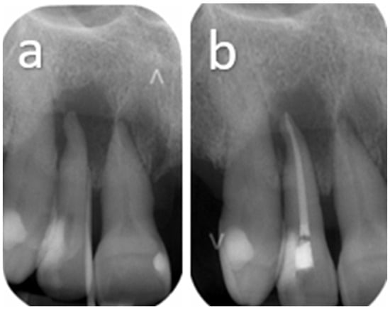
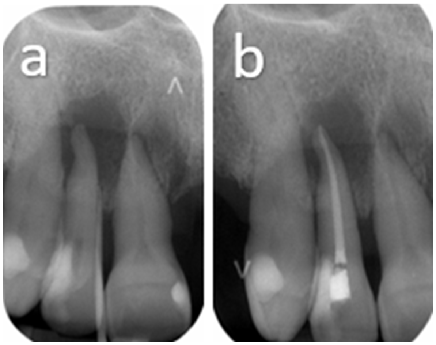
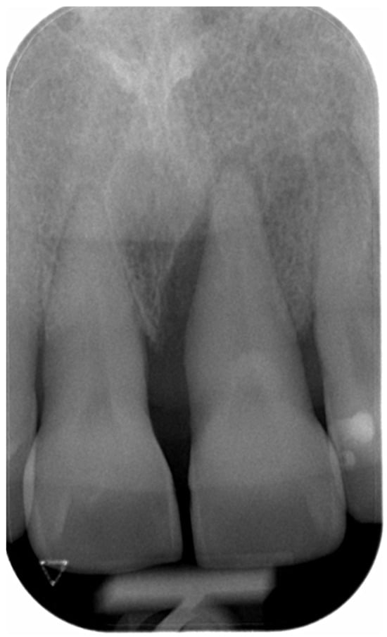
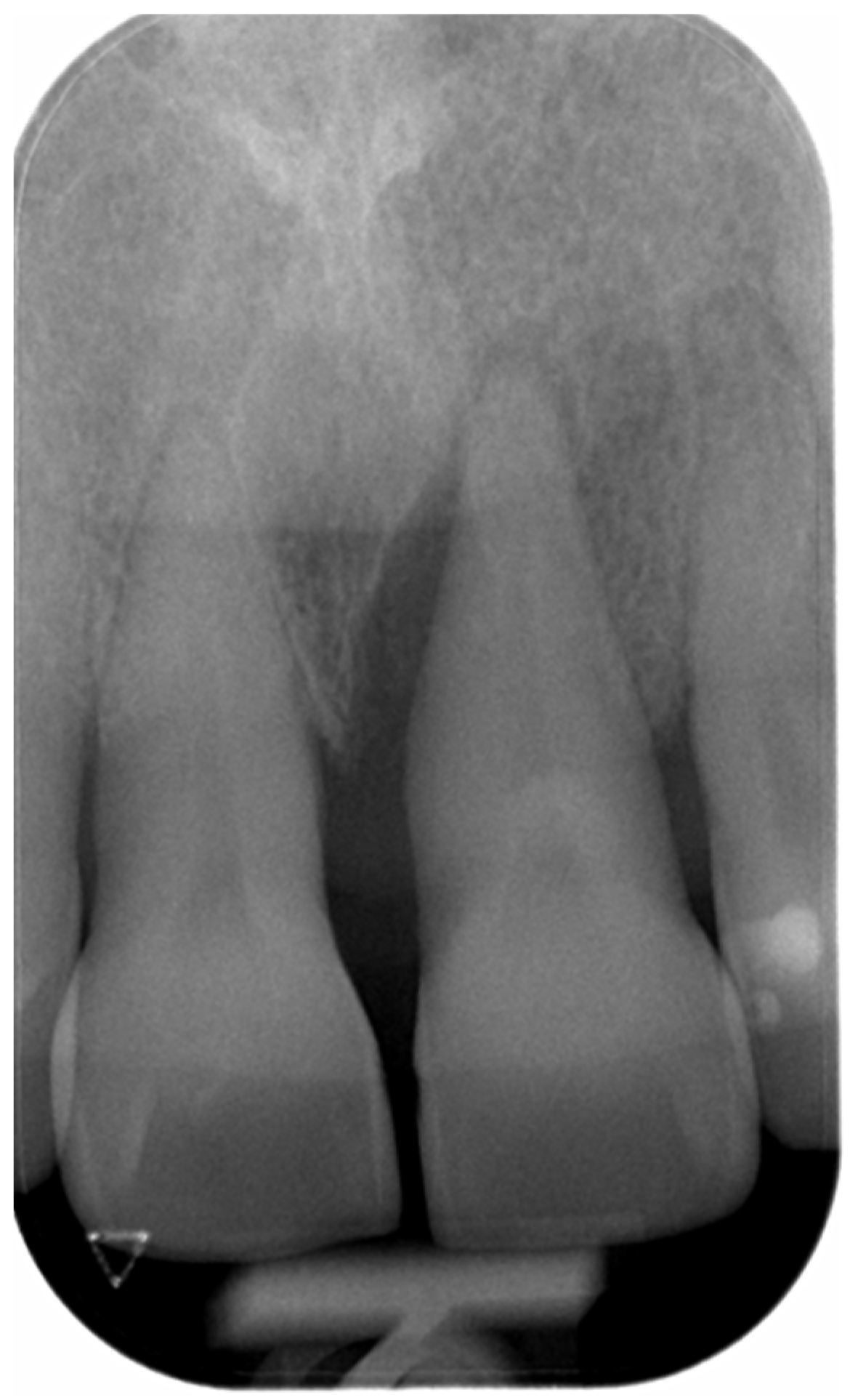
A 66-year-old female patient, under medical follow-up for hypertension and treated with escitalopram, candesartan, and lamotrigine, who also has a penicillin allergy, was referred to the Unit of Oral Surgery and Implantology for the enucleation of a radicular cyst centered at the apex of tooth 12 in the right anterior maxillary region, extending adjacent to teeth 11 and 13. Although teeth 11 and 13 responded positively to vitality testing and were not considered the primary source of the pathology, the lesion was in close proximity to their roots. In addition, their extraction had already been planned by the referring general dentist as part of a comprehensive prosthetic rehabilitation of the anterior maxilla in the context of a severely compromised dentition. The referring dentist had previously performed root canal treatment on tooth 12; however, no clinical or radiological improvement was observed during follow-up (Figure 1).
Figure 1.
(a) Initial periapical radiograph showing a well-defined periradicular radiolucency associated with tooth 12, extending toward the adjacent interradicular area. (b) Six-month follow-up periapical radiograph after root canal treatment of tooth 12, demonstrating persistence of the periradicular radiolucent lesion without significant radiographic resolution.
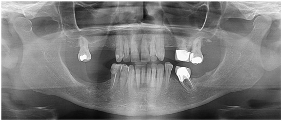
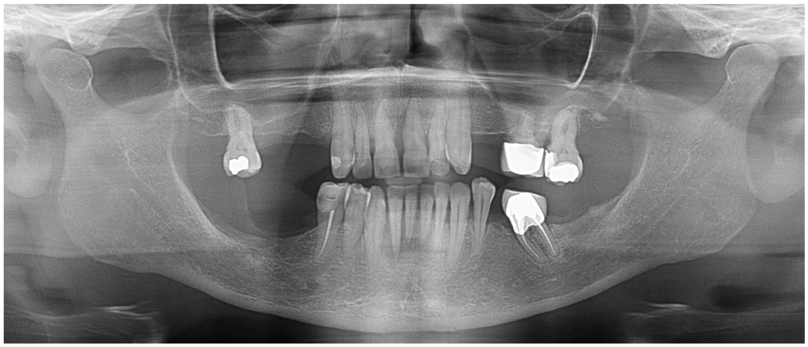
The patient’s dental history revealed a background of advanced periodontal disease with multiple tooth losses over the years, including teeth 14, 15, 16, 24, 25, 46, 47, 35, and 37 (Figure 2 and Figure 3). Tooth 21 had previously presented a severe endo-periodontal lesion. More recently, teeth 21 and 22 were extracted in this context.
Figure 2.
Panoramic radiograph obtained two years prior to referral, showing a partially dentate patient with teeth 21 and 22 still present, with a greater number of remaining teeth compared with the CBCT examination performed at the time of presentation.
Figure 3.
Apical radiograph showing a severe periodontal lesion is observed on the mesial aspect of tooth 21, characterized by marked alveolar bone loss.
Extraoral clinical examination revealed no facial swelling or asymmetry. No signs of systemic involvement were observed, including dysphagia, dyspnea, or sensorimotor deficits involving the trigeminal or facial nerve territories.
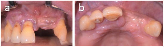
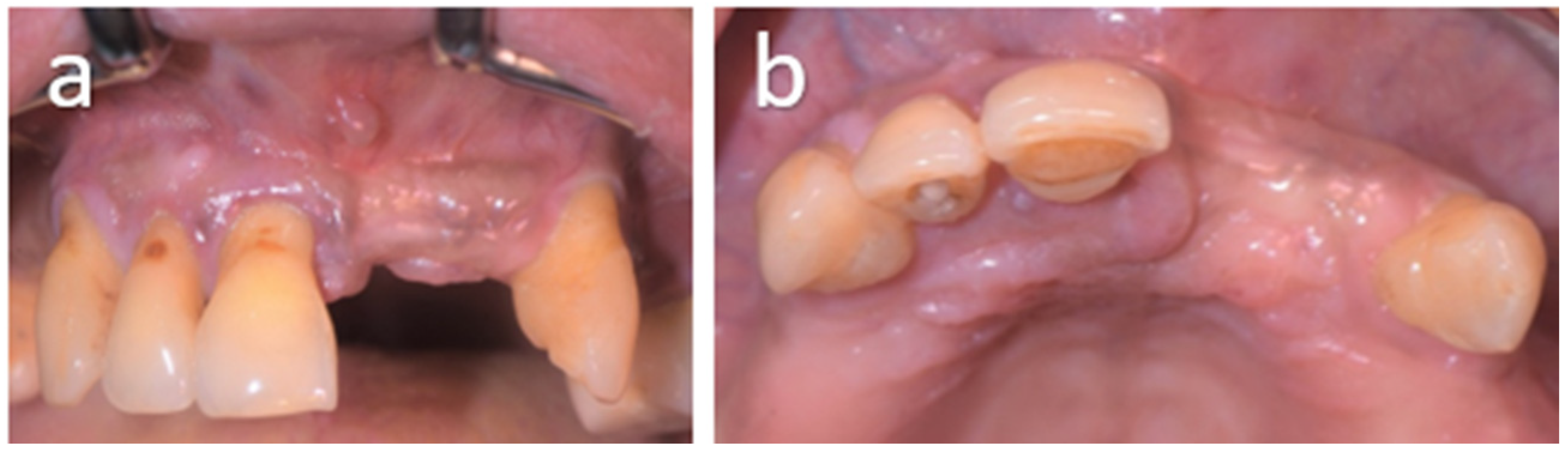
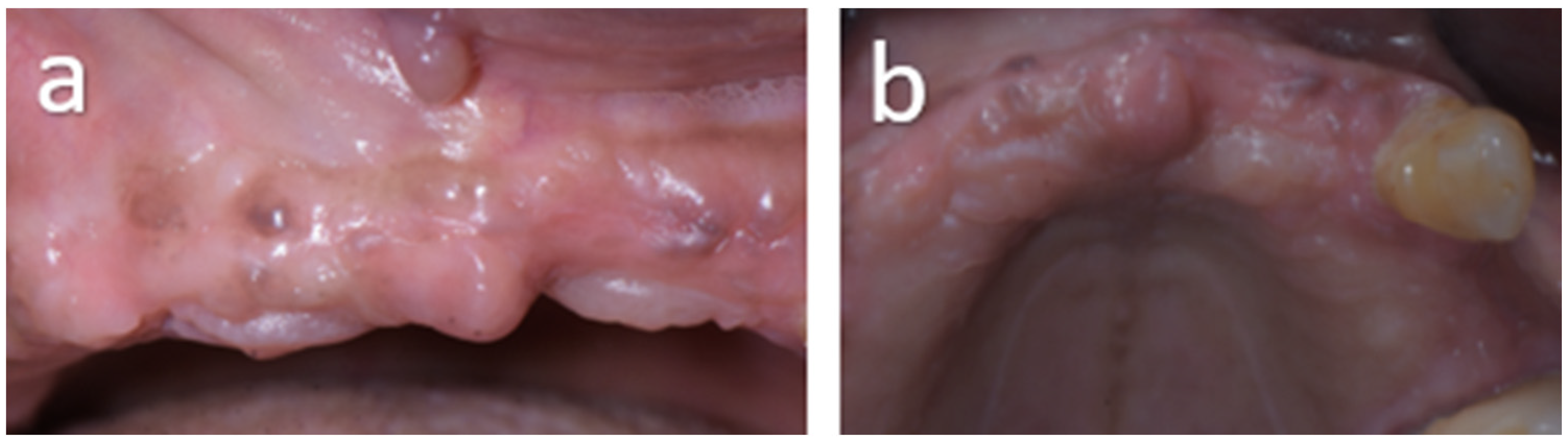
Intraoral examination revealed a partial maxillary anterior edentulous space corresponding to missing teeth 21 and 22, with the remaining anterior dentition extending from canine to canine (Figure 4). The mucosa appears healthy, with no signs of inflammation or pathology. Vestibular palpation of teeth 11, 12, and 13 is positive, with no swelling or collection. Vitality tests show a positive response for teeth 11 and 13 and a negative response for tooth 12. Percussion tenderness is noted on tooth 12, whereas adjacent teeth were asymptomatic.
Figure 4.
(a) Vestibular and (b) occlusal views of the initial clinical presentation showing the anterior maxilla. Clinical examination reveals absence of teeth 14, 21, 22, and 24, associated with periodontal involvement characterized by gingival recession affecting the remaining teeth. No swelling, mucosal bulging, or other soft tissue abnormalities are observed.
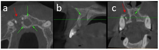
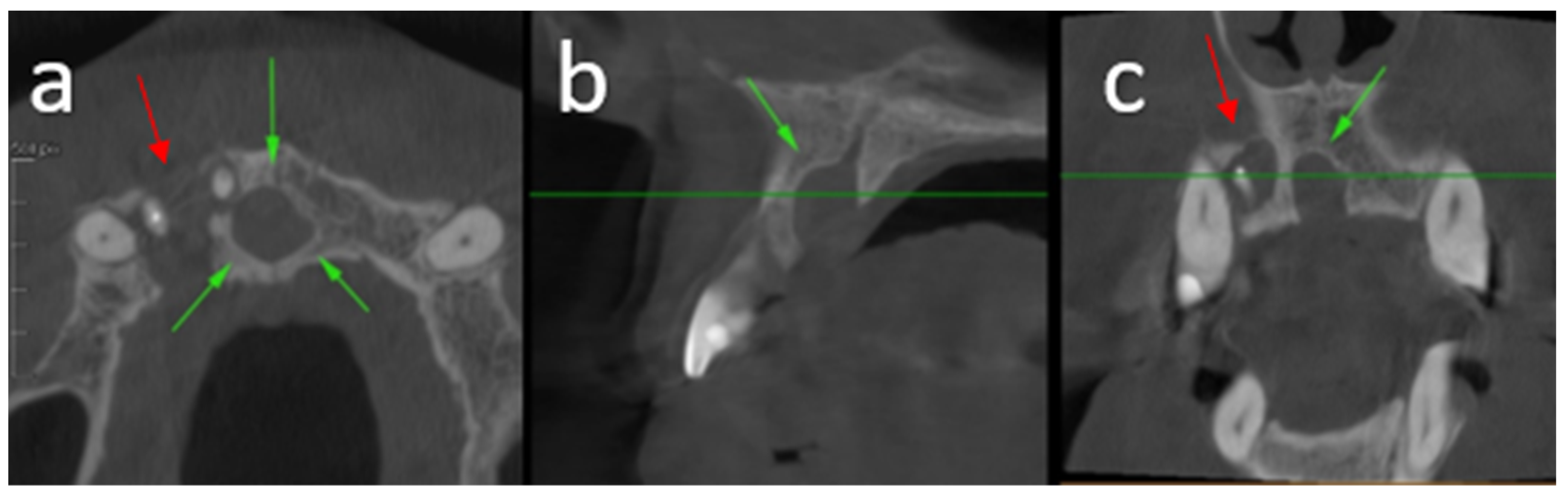
Radiographic examination with periapical radiography supplemented by an orthopantomogram (OPG) revealed a radiolucent area apical to teeth 11, 12, and 13. A supplementary cone beam computed tomography (CBCT) scan was performed to assess the extent of the lesion and its anatomical relationships (Figure 5). This revealed a radiolucent lesion in close relationship with the incisive canal and a second lesion with apices of teeth 11 and 12. Two lesions are visible on the CBCT. The first, centered on tooth 12 which had undergone root canal treatment, extends mesially to the apex of tooth 11 and distally toward tooth 13. It exhibits an expansive behavior, resulting in dehiscence of both the buccal and palatal cortical plates in the region of tooth 12, as well as palatal cortical dehiscence in the region of tooth 13. The second lesion is in regions 11 and 21, causing dehiscence of the mesial wall of the incisive canal. The nasopalatine canal is enlarged, measuring approximately 7 × 6 mm in the axial plane, with associated buccal and palatal cortical thinning and dehiscence. Its imaging characteristics are highly suggestive of a nasopalatine duct cyst.
Figure 5.
Cone beam computed tomography (CBCT) in axial (a), sagittal (b), and coronal (c) planes demonstrating a well-defined radiolucent lesion centered on the nasopalatine canal, consistent with a nasopalatine duct cyst, with associated palatal cortical dehiscence. An additional adjacent cystic lesion of radicular origin is observed, primarily centered at the apex of the non-vital tooth 12, associated with buccal and palatal cortical dehiscence, extending into contact with the apices of teeth 11 and 13. Green arrows indicate the nasopalatine duct cyst. Red arrows indicate the radicular cystic lesion.
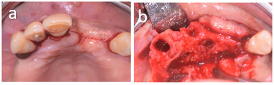
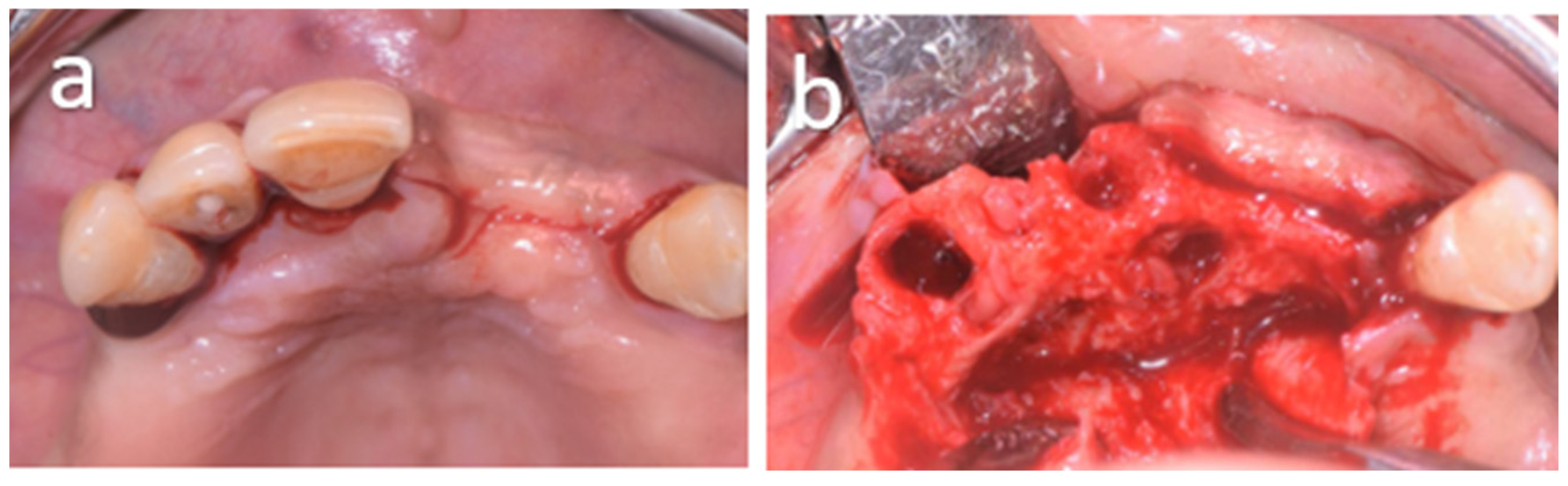
Based on clinical and radiological findings, a preoperative diagnosis of a nasopalatine cyst was established. An excisional biopsy was planned, and a decision was made to proceed with surgical management. The procedure was performed under local anesthesia in the operating room. It involved the extraction of teeth 11, 12, and 13, as well as the enucleation of the cystic lesion. Local vestibular anesthesia was administered and supplemented by a palatal block injection using 4% articaine with epinephrine 1:200,000. A crestal incision was made in the edentulous area corresponding to teeth 21–22 and extended by sulcular incisions around teeth 11, 12, and 13, together with a distal releasing incision at tooth 13, allowing the elevation of a combined vestibular and palatal mucoperiosteal flap. The incisive foramen was identified intraoperatively (Figure 6).
Figure 6.
(a) Occlusal intraoperative view showing the initial surgical access prior to flap elevation. A crestal incision was performed in the anterior maxilla, extended by sulcular incisions around teeth 11, 12, 13, and 23; (b) Occlusal intraoperative view after elevation of the mucoperiosteal flap and extraction of teeth 11, 12, and 13. A bony dehiscence is observed in the anterior maxilla, exposing part of the nasopalatine duct cyst. In addition, soft tissue consistent with the radicular lesion is visible within the extraction socket of tooth 12.
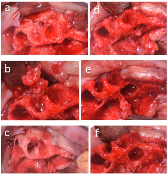
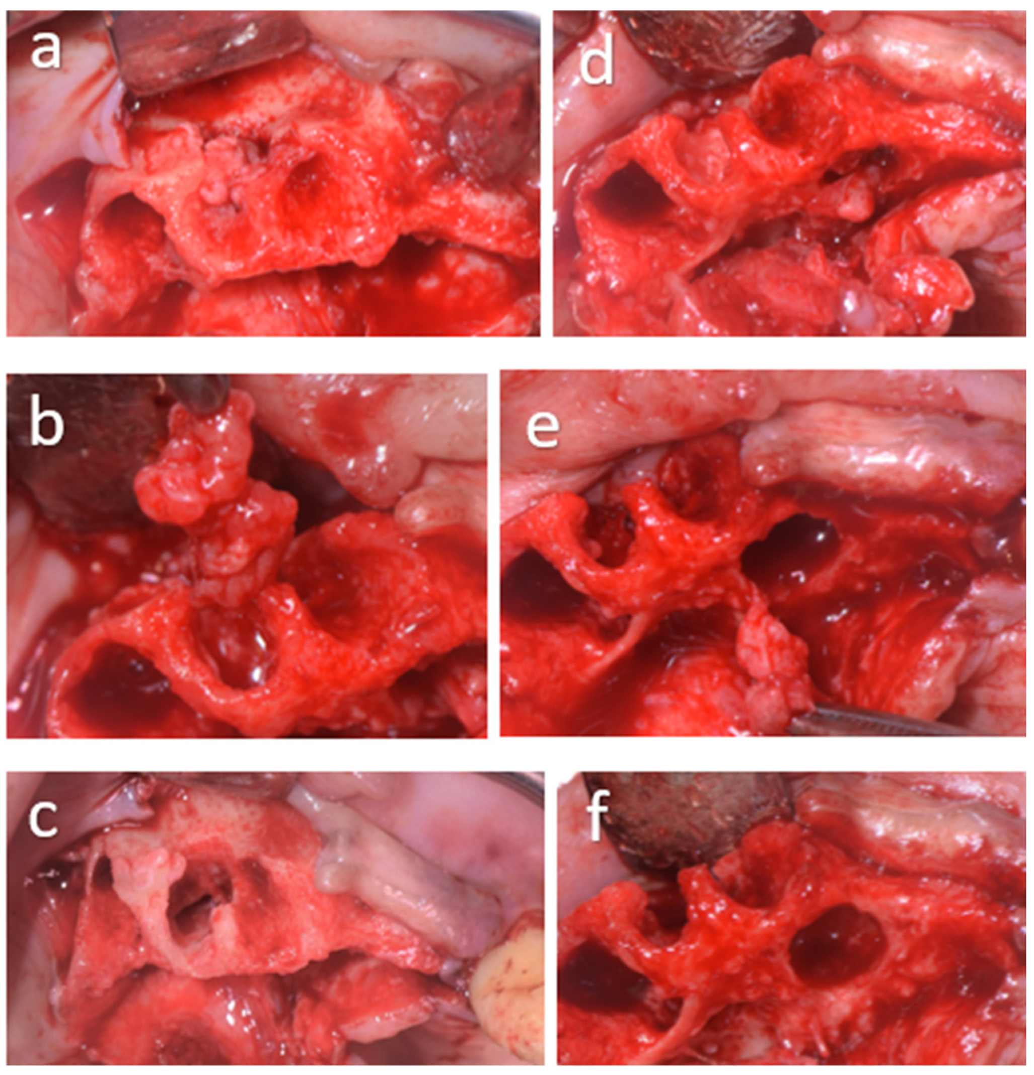
Meticulous enucleation and curettage of the two bony lesions were performed, combined with mechanical debridement using a diamond round bur mounted on a surgical handpiece (Figure 7).
Figure 7.
(a–f) Intraoperative views illustrating the surgical management of the lesions in the anterior maxilla. (a) Vestibular view showing the cystic lesion at site 12 and the alveolar region of teeth 11, 12, and 13. (b) Occlusal view during surgical enucleation of the radicular cystic lesion at site 12. (c) Vestibular view after complete surgical curettage and debridement of the enucleation site at position 12, demonstrating the residual bony cavity following removal of the cystic and inflammatory tissues. (d) Occlusal intraoperative view after partial detachment of the nasopalatine cystic lesion. (e) Occlusal view showing the final stage of enucleation of the nasopalatine lesion. (f) Occlusal view of the surgical cavity after complete enucleation and debridement of the nasopalatine lesion.
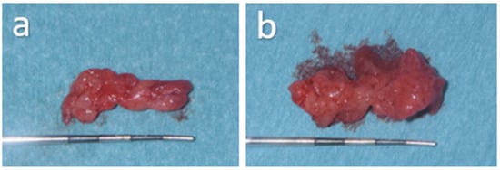
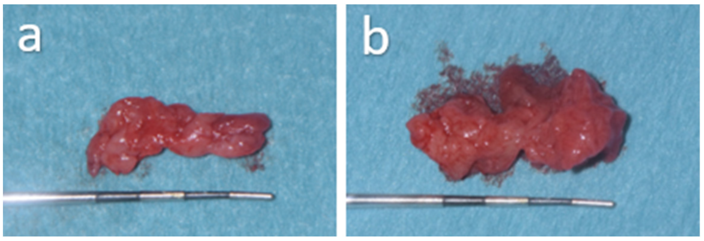
The radicular cystic lesion, measuring approximately 12 mm, and the nasopalatine lesion, measuring 14 mm, were immediately fixed in formalin for histopathological examination (Figure 8).
Figure 8.
(a,b) Macroscopic views of the enucleated lesions following surgical removal, measured using a periodontal probe. (a) Radicular cystic lesion measuring approximately 12 mm. (b) Nasopalatine lesion measuring approximately 14 mm.
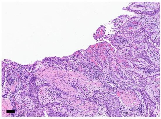
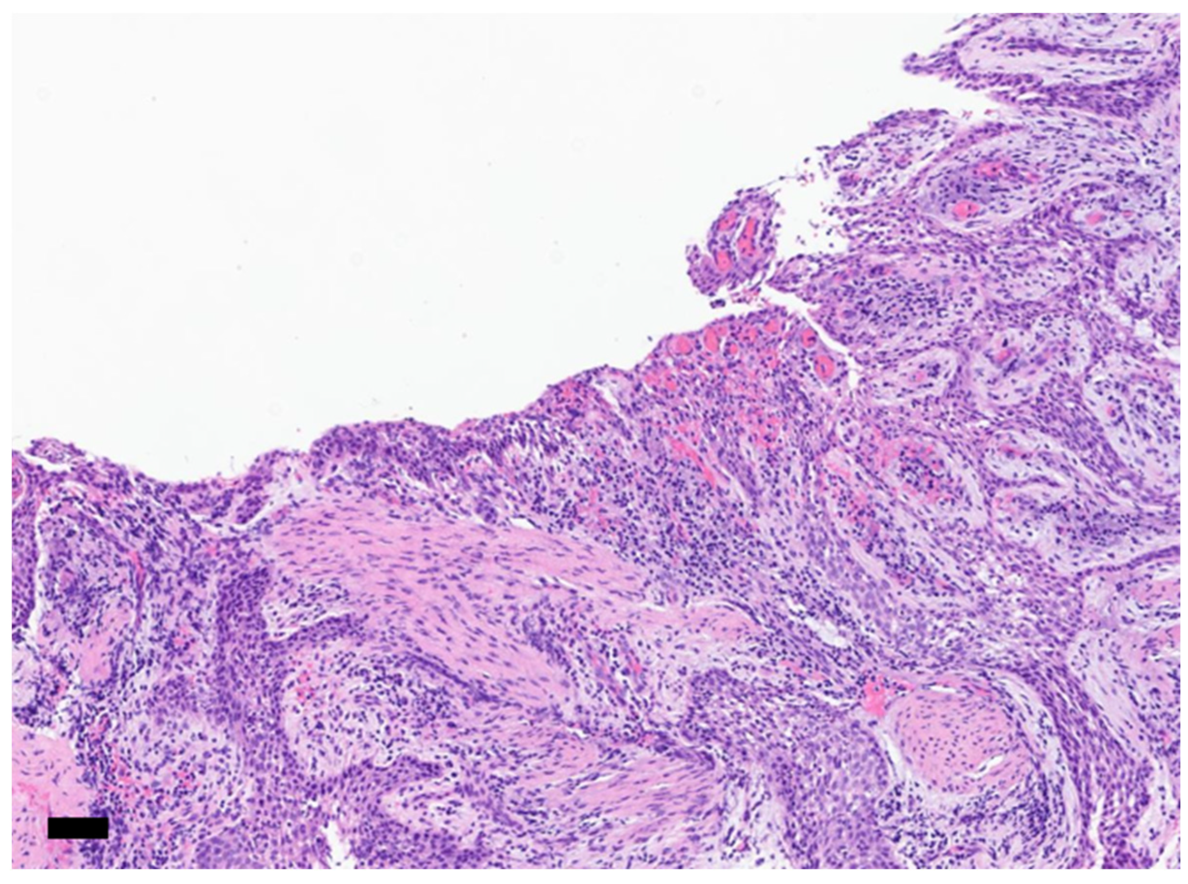
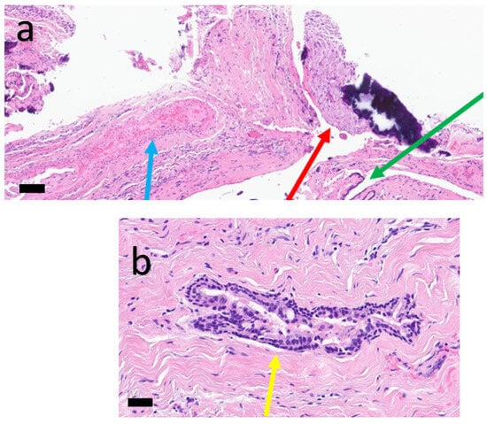
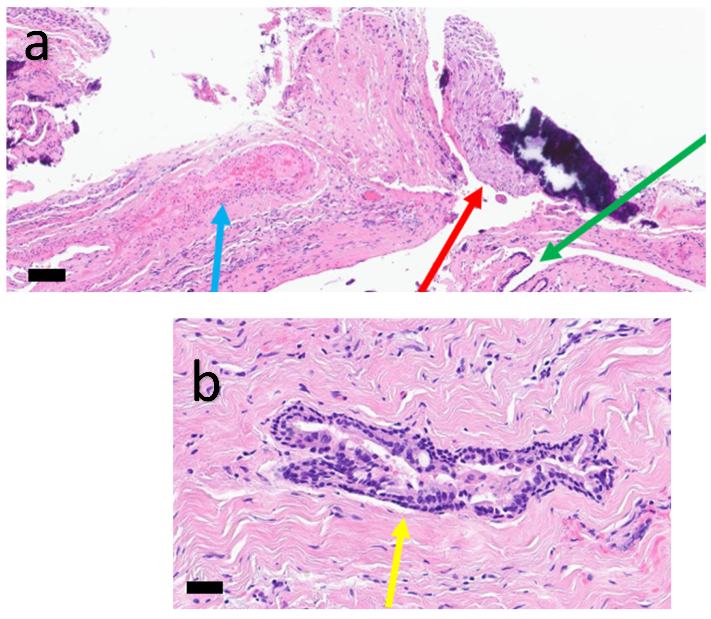
Postoperative management included routine analgesic medication (ibuprofen and paracetamol as needed) together with standard postoperative instructions. Histological analysis confirmed the diagnosis of a nasopalatine cyst. The lesion located adjacent to the apices of teeth 11 and 12 was consistent with a radicular cyst. Histological examination showed a lining of non-keratinized stratified squamous epithelium associated with a chronic inflammatory infiltrate (Figure 9). The lesion associated with the nasopalatine canal consisted of cuboidal epithelial lining in a fibrous tissue, findings consistent with a nasopalatine duct cyst (Figure 10).
Figure 9.
Hematoxylin and eosin-stained histological section at magnification ×40 showing a cyst wall lined by non-keratinizing squamous epithelium showing rete ridges. Presence of a rather dense chronic inflammatory infiltrate in the underlying connective tissue. Scale bar = 500 µm.
Figure 10.
Hematoxylin and eosin-stained histological section at low ×20 (a) and high ×40 magnification (b) showing artery (red arrow), nerve bundle (green arrow) and epithelial lining (blue arrow) and cuboidal epithelial lining in a fibrous tissue (yellow arrow). Scale bar = 500 µm (a,b).
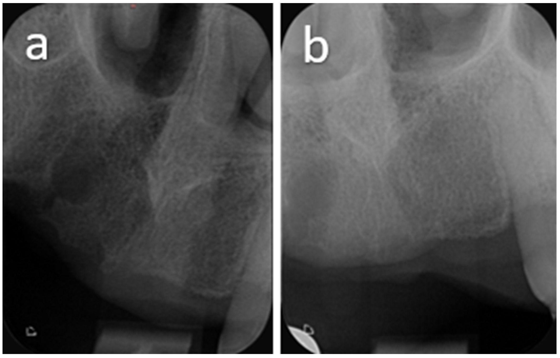
Postoperative follow-up was conducted at regular intervals. At the one-week review, the patient reported no postoperative complications, and clinical examination revealed uneventful early healing. The sutures were removed, and the patient was instructed to continue saline mouth rinses to promote optimal healing. At the one-month follow-up, the mucosa in the surgical area showed no signs of irritation or inflammation. At three months postoperatively, favorable soft tissue healing was observed, and periapical radiography demonstrated the onset of bone regeneration within the surgical site. Further follow-up examinations at six months showed progressive and favorable bone healing, with no clinical abnormalities. At the one-year follow-up, radiographic assessment revealed stable bone levels and an overall highly favorable evolution; however, a small residual radiolucent area persisted at the site of the previously treated periapical granuloma, without any clinical or radiographic signs suggestive of recurrence (Figure 11 and Figure 12).
Figure 11.
(a,b) One-year postoperative follow-up showing vestibular (a) and occlusal (b) views of the surgical site, demonstrating satisfactory healing and stable soft tissue conditions.
Figure 12.
One-year postoperative follow-up x-ray showing partial ossification at site 12 (a) and complete ossification at median site (b).
This finding was interpreted as consistent with delayed but ongoing bone remodeling. Follow-up at one year showed no recurrence. At follow-up visits, the patient reported no postoperative complaints and expressed satisfaction with the outcome of the treatment. The timeline of the patient’s clinical course is summarized in Table 1.
Table 1.
Timeline of clinical events.
3. Discussion
The present case illustrates the diagnostic challenge posed by the coexistence of a radicular cyst and a nasopalatine duct cyst in the anterior maxilla. Although both lesions are relatively common when considered separately, their simultaneous occurrence in close anatomical proximity may obscure the diagnosis and lead to the erroneous interpretation of a single pathological entity. In the present patient, the inflammatory lesion associated with tooth 12 initially masked the presence of an underlying nasopalatine duct cyst, highlighting the importance of comprehensive radiological assessment.
Nasopalatine duct cysts (NPDCs) are well recognized as the most common non-odontogenic cysts of the oral cavity, typically arising from epithelial remnants of the nasopalatine duct within the incisive canal [20,21]. Although their developmental origin is well established, their clinical presentation may overlap with several inflammatory and odontogenic entities, making diagnosis challenging, particularly in complex anatomical and pathological contexts of the anterior maxilla [21,22].
According to the 5th edition of the WHO Classification of Head and Neck Tumours, nasopalatine duct cysts are classified as developmental, non-odontogenic cysts arising from epithelial remnants of the nasopalatine duct located within the incisive canal [1]. The WHO classification also emphasizes the importance of correlating clinical, radiological, and histopathological findings to distinguish NPDCs from inflammatory periapical lesions and other radiolucent entities of the anterior maxilla [1]. These considerations are particularly relevant in the present case, where the coexistence of a radicular cyst and an NPDC created significant diagnostic overlap.
In the present case, the NPDC was incidentally identified on cone-beam computed tomography (CBCT) performed for the assessment of a separate endodontic lesion affecting tooth 12 [3]. This illustrates the increasing role of three-dimensional imaging in the evaluation of anterior maxillary radiolucencies, particularly when conventional radiographs are insufficient to accurately delineate lesion boundaries or to distinguish between developmental and inflammatory origins. CBCT allowed precise localization of the lesion in relation to the incisive canal and adjacent dental structures, providing strong radiological support for the diagnosis of NPDC [20,21,22].
A major finding in this case was the coexistence of two distinct pathological entities: a radicular cyst associated with a previously endodontically treated tooth (tooth 12), and a second lesion centered on the nasopalatine canal. Their close anatomical proximity in the anterior maxilla resulted in significant overlap on two-dimensional imaging, where both lesions initially appeared as a single radiolucent area. Only CBCT enabled clear separation of these entities, highlighting its essential role in modern diagnostic workflows in endodontics and oral surgery [3,4,21].
The limitations of conventional two-dimensional radiography in the present case were related to the complex superimposition of multiple radiolucent structures in the anterior maxilla. In particular, the periapical lesion associated with tooth 12 was radiographically underestimated, with incomplete visualization of its true mesiodistal extension and its relationship with adjacent teeth. In addition, assessment of cortical bone involvement, including both buccal and palatal cortical thinning and fenestration, was not possible on two-dimensional imaging. This resulted in a composite radiographic appearance in which the radicular cyst and the nasopalatine duct cyst could not be reliably differentiated. CBCT was therefore essential to overcome these limitations by providing three-dimensional visualization and accurate assessment of lesion extent, boundaries, and anatomical relationships.
Several reports in the literature have described the coexistence of nasopalatine duct cysts with odontogenic cystic lesions, although such associations remain exceptionally rare. Sadeghian et al. reported the simultaneous presence of a nasopalatine duct cyst and a dentigerous cyst in the anterior maxilla, where diagnostic difficulty resulted primarily from overlapping radiographic appearances, with definitive diagnosis achieved through histopathological examination [7]. Similarly, Alassaf et al. described the coexistence of a nasopalatine duct cyst and a radicular cyst, emphasizing the challenges in differentiating multiple radiolucent lesions in the anterior maxilla using conventional imaging and the need for combined radiological and histopathological assessment [8].
In contrast to these previously reported cases, the present report further illustrates that diagnostic complexity may be increased when multiple factors coexist, including lesion superimposition, masking of a developmental lesion by an adjacent inflammatory pathology, and incidental detection of an NPDC on CBCT performed for a separate endodontic indication. This highlights an additional layer of diagnostic difficulty beyond that described in prior reports.
Collectively, these observations reinforce the importance of advanced imaging and histopathological confirmation in cases of multiple radiolucent lesions in the anterior maxilla.
Clinically, the absence of symptoms attributable to the NPDC is consistent with its typically asymptomatic nature. The tenderness observed in the region of tooth 12 was most likely related to the radicular cyst rather than the nasopalatine lesion itself, further illustrating the potential for diagnostic confusion when multiple pathologies coexist in the same anatomical region [4,5,6,7,21].
A history of severe periodontal disease was noted in the anterior maxilla, particularly in the region of tooth 21. This finding is mentioned only to describe the local clinical history and anatomical context of the case. No causal relationship between the previous periodontal condition and the nasopalatine duct cyst can be inferred.
The differential diagnosis of NPDCs includes periapical inflammatory lesions such as radicular cysts and periapical granulomas, as well as anatomical variations such as an enlarged incisive canal [11,14,20]. In this case, the presence of a non-vital tooth (tooth 12) with periapical pathology further complicated radiological interpretation.
Another relevant diagnostic consideration in this case concerns the periapical lesion associated with tooth 12. Although its radiographic dimensions exceeded 10 mm, a value often associated with radicular cysts, histopathological examination confirmed a diagnosis of a radicular cyst [12]. This case highlights the overlap in imaging characteristics between radicular cysts and other periapical inflammatory lesions, underscoring that lesion size alone cannot be considered a reliable diagnostic criterion. It therefore reinforces the essential role of histopathological analysis in establishing a definitive diagnosis of periapical lesions [13], as well as the importance of integrating clinical vitality testing with imaging findings to avoid misdiagnosis and potential overtreatment [14].
The lesion measured approximately 12 mm after excision, which is within the range reported in the literature. Although physiological enlargement of the incisive canal is generally considered to remain below 8 mm [13,15], pathological conditions may exceed this threshold and mimic other radiolucent entities, illustrating the limitations of size-based diagnostic criteria on two-dimensional imaging.
Histopathological examination remains the gold standard for definitive diagnosis. In this case, the NPDC was confirmed by the presence of a cystic cavity lined by non-keratinized and cuboidal epithelium and the presence of chronic inflammatory infiltrate [16]. The associated lesion at tooth 12 was diagnosed as a radicular cyst, confirming the coexistence of two distinct pathological processes.
From a therapeutic standpoint, surgical enucleation remains the treatment of choice for NPDCs, with generally favorable outcomes and low recurrence rates [17,18,23,24]. In this case, complete surgical removal was achieved without complications, and postoperative healing was uneventful, with progressive bone regeneration observed during follow-up. These findings are consistent with the predictable healing pattern reported in the literature [19,20,21,22,23].
Overall, this case highlights several key clinical implications. First, anterior maxillary radiolucencies may conceal multiple coexisting pathologies, particularly when developmental and inflammatory lesions overlap anatomically. Second, CBCT plays a crucial role in accurate lesion characterization and in reducing the risk of diagnostic misinterpretation.
This case therefore underscores the importance of a comprehensive multimodal diagnostic approach combining clinical examination, vitality testing, advanced imaging, and histopathological analysis to ensure accurate diagnosis and appropriate management.
4. Conclusions
This case highlights the diagnostic challenges associated with radiolucent lesions of the anterior maxilla, particularly when developmental and inflammatory entities coexist or overlap in their radiographic presentation. It emphasizes the importance of a comprehensive diagnostic approach combining clinical assessment, vitality testing, and advanced imaging.
CBCT plays a key role in accurately characterizing lesion origin, extent, and anatomical relationships, thereby reducing the risk of misdiagnosis. In particular, CBCT evaluation of the anterior maxilla should be considered before extensive periapical surgery when conventional radiographs do not adequately explain the clinical findings or when lesion boundaries appear unclear. Such assessment may reveal additional or coexisting pathologies that could significantly influence treatment planning and surgical management. Histopathological examination remains essential for definitive diagnosis and appropriate management of maxillary radiolucencies. Overall, maintaining a broad differential diagnosis is crucial to avoid attributing all anterior maxillary radiolucencies to endodontic pathology.
Author Contributions
Conception, draft of the manuscript, preparation of images, and patient treatment, A.P.; draft of the manuscript and radiological interpretation and preparation of images, A.P., N.J. and L.R.; conception, drafting, and critical review of the manuscript, A.P. and T.L. All authors have read and agreed to the published version of the manuscript.
Funding
This research received no external funding.
Institutional Review Board Statement
The presented study adhered to the Helsinki Declaration of ethical principles by the World Medical Association. The study did not require the approval of the Ethics Commission on Human Research of Geneva (CCER-Geneva) according to the Federal Human Research Act (Art.3al.a), since the study involved fewer than five patients.
Informed Consent Statement
Written informed consent was obtained from the patient to publish this paper.
Data Availability Statement
The original contributions presented in the study are included in the article.
Conflicts of Interest
The authors declare no conflicts of interest.
References
- WHO Classification of Tumours Editorial Board. Head and Neck Tumours, 5th ed.; WHO Classification of Tumours Series; International Agency for Research on Cancer: Lyon, France, 2024; Volume 9. [Google Scholar]
- Escoda Francolí, J.; Almendros Marqués, N.; Berini Aytés, L.; Gay Escoda, C. Nasopalatine duct cyst: Report of 22 cases and review of the literature. Med. Oral Patol. Oral Cir. Bucal 2008, 13, E438–E443. [Google Scholar] [PubMed]
- Vasconcelos, R.; de Aguiar, M.F.; Castro, W.; de Araújo, V.C.; Mesquita, R. Retrospective analysis of 31 cases of nasopalatine duct cyst. Oral Dis. 1999, 5, 325–328. [Google Scholar] [CrossRef] [Scilit] [PubMed]
- Robertson, H.; Palacios, E. Nasopalatine duct cyst. Ear Nose Throat J. 2004, 83, 313. [Google Scholar] [CrossRef] [Scilit] [PubMed]
- Herráez-Vilas, J.M.; Gay-Escoda, C.; Berini-Aytés, L. Quiste del conducto nasopalatino. Revisión de la literatura y aportación de 14 casos. Rev. Eur. Odontoestomatol. 1994, 6, 231–236. [Google Scholar] [CrossRef] [Scilit]
- Gnanasekhar, J.D.; Walvekar, S.V.; Al-Kandari, A.M.; Al-Duwairi, Y. Misdiagnosis and mismanagement of a nasopalatine duct cyst and its corrective therapy: A case report. Oral Surg. Oral Med. Oral Pathol. Oral Radiol. Endod. 1995, 80, 465–470. [Google Scholar] [CrossRef] [Scilit] [PubMed]
- Elliott, K.A.; Franzese, C.B.; Pitman, K.T. Diagnosis and surgical management of nasopalatine duct cysts. Laryngoscope 2004, 114, 1336–1340. [Google Scholar] [CrossRef] [Scilit] [PubMed]
- Alassaf, M.S.; Abu Aof, M.M.; Othman, A.; Zaki, H.; Almutairi, A.G. Coexistence of a Nasopalatine Duct Cyst and Radicular Cyst: A Unique Clinical Presentation. Cureus 2023, 15, e46774. [Google Scholar] [CrossRef] [Scilit] [PubMed]
- Sadeghian, A.; Taheri, A.; Bakhshaei, P. Coincidence of Nasopalatine Duct Cyst and Dentigerous Cyst: A Case Report. J. Craniomaxillofac. Res. 2023, 10, 127–131. [Google Scholar]
- Allard, R.H.B.; van der Kwast, W.A.M.; van der Waal, I. Nasopalatine duct cyst: Review of the literature and report of 22 cases. Int. J. Oral Surg. 1981, 10, 447–461. [Google Scholar] [CrossRef] [Scilit] [PubMed]
- Swanson, K.S.; Kaugars, G.E.; Gunsolley, J.C. Nasopalatine duct cyst: An analysis of 334 cases. J. Oral Maxillofac. Surg. 1991, 49, 268–271. [Google Scholar] [CrossRef] [Scilit] [PubMed]
- Hilfer, P.B.; Bergeron, B.E.; Ozgul, E.S.; Wong, D.K. Misdiagnosis of a nasopalatine duct cyst: A case report. J. Endod. 2013, 39, 1185–1188. [Google Scholar] [CrossRef] [Scilit] [PubMed]
- Faitaroni, L.A.; Bueno, M.R.; Carvalhosa, A.A.; Mendonça, E.F.; Estrela, C. Differential diagnosis of apical periodontitis and nasopalatine duct cyst. J. Endod. 2011, 37, 403–410. [Google Scholar] [CrossRef] [Scilit] [PubMed]
- Rey Lescure, M.; Perez, A.; Lombardi, T. Une lésion ostéolytique mandibulaire inhabituelle. Ann. Pathol. 2021, 41, 334–338. [Google Scholar] [CrossRef] [Scilit] [PubMed]
- Cecchetti, F.; Ottria, L.; Bartuli, F.; Bramanti, N.E.; Arcuri, C. Prevalence, distribution, and differential diagnosis of nasopalatine duct cysts. Oral Implantol. 2012, 5, 47–53. [Google Scholar]
- Cavalcante, I.L.; Barros, C.C.; Cunha, J.L.; Cruz, V.M.; Pedrosa, G.A.; Santos, A.D.; Turatti, E.; Albuquerque-Júnior, R.L.; Cavalcante, R.B. Clinicopathologic features of nasopalatine duct cysts: A retrospective study in two Brazilian oral and maxillofacial pathology referral centers. Med. Oral Patol. Oral Cir. Bucal 2021, 26, e676–e683. [Google Scholar] [CrossRef] [Scilit] [PubMed]
- Guo, J.; Simon, J.H.; Sedghizadeh, P.; Soliman, O.N.; Chapman, T.; Enciso, R. Evaluation of the reliability and accuracy of using cone-beam computed tomography for diagnosing periapical cysts from granulomas. J. Endod. 2013, 39, 1485–1490. [Google Scholar] [CrossRef] [Scilit] [PubMed]
- Bornstein, M.M.; Bingisser, A.C.; Reichart, P.A.; Sendi, P.; Bosshardt, D.D.; von Arx, T. Comparison between radiographic 2-dimensional and 3-dimensional and histologic findings of periapical lesions treated with apical surgery. J. Endod. 2015, 41, 804–811. [Google Scholar] [CrossRef] [Scilit] [PubMed]
- Perez, A.; Lenoir, V.; Lombardi, T. Aggressive nasopalatine cyst with nasal involvement in an edentulous patient. Appl. Sci. 2022, 12, 11002. [Google Scholar] [CrossRef] [Scilit]
- Bodin, I.; Isacsson, G.; Julin, P. Cysts of the nasopalatine duct. Int. J. Oral Maxillofac. Surg. 1986, 15, 696–706. [Google Scholar] [CrossRef] [Scilit] [PubMed]
- Lee, Y.P.; Hwang, M.J.; Jin, Y.T.; Chiang, C.P. Nasopalatine duct cyst: Characteristic histopathological features. J. Dent. Sci. 2024, 19, 1216–1218. [Google Scholar] [CrossRef] [Scilit] [PubMed]
- Scolozzi, P.; Martinez, A.; Richter, M.; Lombardi, T. A nasopalatine duct cyst in a 7-year-old child. Pediatr. Dent. 2008, 30, 530–534. [Google Scholar] [PubMed]
- Suter, V.G.; Altermatt, H.J.; Voegelin, T.C.; Bornstein, M.M. Die nasopalatinale Zyste. Die nasopalatinale Zyste. Epidemiologie, Diagnostik und Therapie [The nasopalatine duct cyst—Epidemiology, diagnosis and therapy]. Schweiz. Monatsschr. Zahnmed. 2007, 8, 824–839. [Google Scholar]
- Oliveira, L.J.; Leão, P.L.R.; Alvarenga, R.R.; Horta, M.C.R.; Souza, P.E.A. Nasopalatine duct cyst: Diagnosis, treatment and postoperative complications: Report of two cases. J. Bras. Patol. Med. Lab. 2017, 53, 407–413. [Google Scholar] [CrossRef] [Scilit]

Disclaimer/Publisher’s Note: The statements, opinions and data contained in all publications are solely those of the individual author(s) and contributor(s) and not of MDPI and/or the editor(s). MDPI and/or the editor(s) disclaim responsibility for any injury to people or property resulting from any ideas, methods, instructions or products referred to in the content. |
© 2026 by the authors. Licensee MDPI, Basel, Switzerland. This article is an open access article distributed under the terms and conditions of the Creative Commons Attribution (CC BY) license.