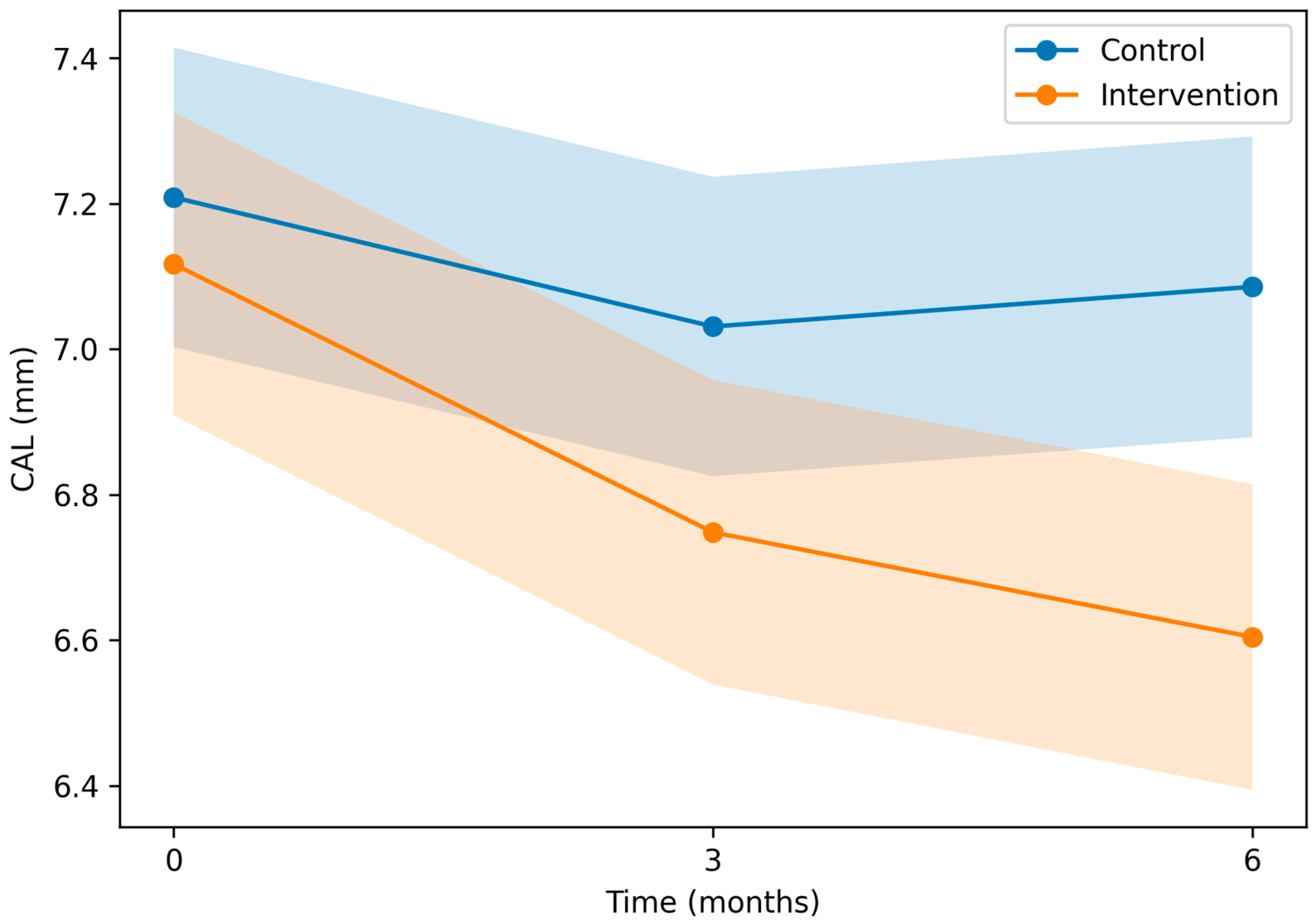
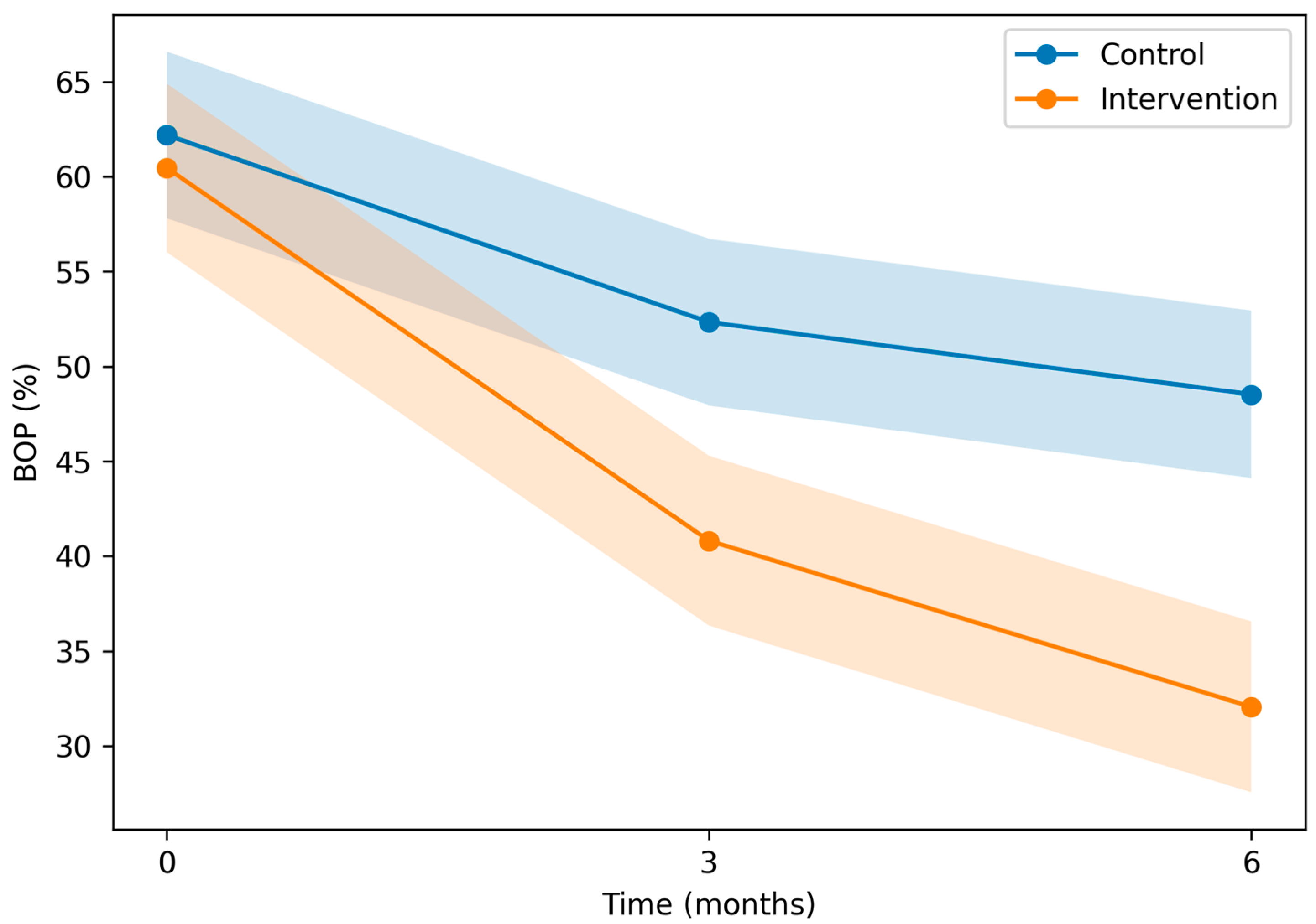
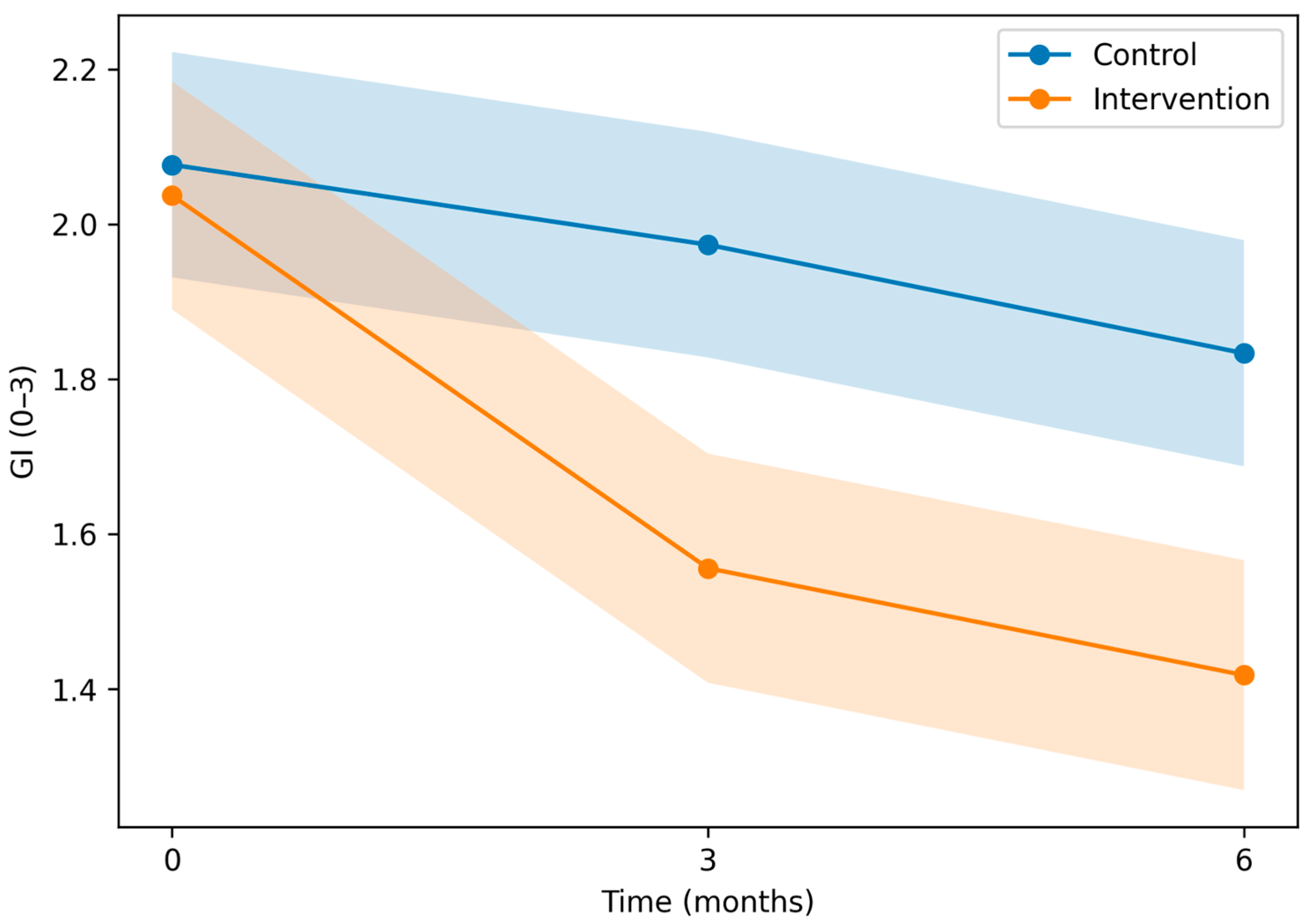
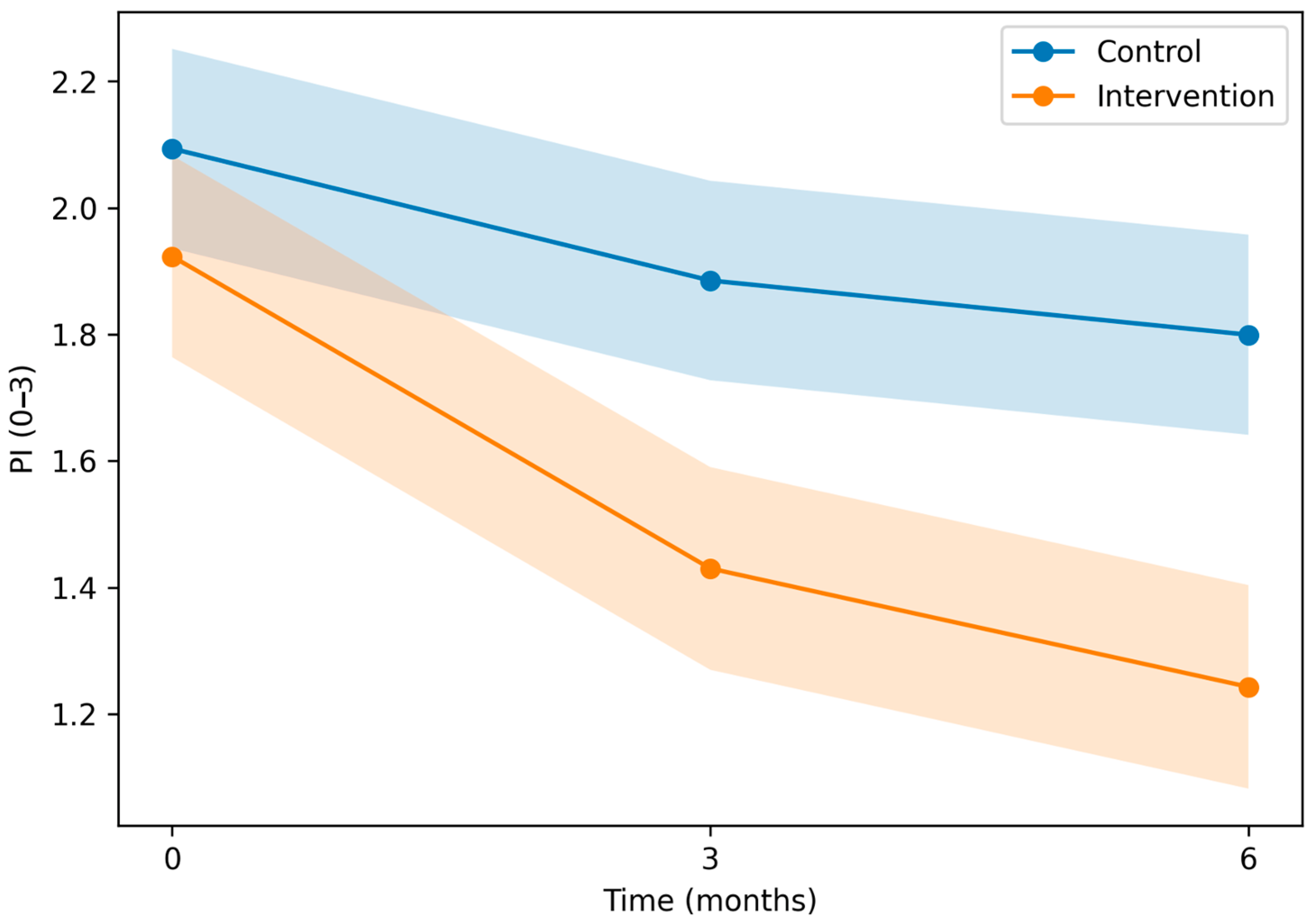
1. Introduction
Type 2 diabetes mellitus (T2DM) is a chronic condition characterized by low-grade systemic inflammation, which contributes to insulin resistance and cardiometabolic complications. High-sensitivity C-reactive protein (hsCRP) is a sensitive biomarker of systemic low-grade inflammation, produced predominantly in the liver and regulated by pro-inflammatory cytokines (notably interleukin-6 [IL-6], with contributions from interleukin-1β [IL-1β] and tumor necrosis factor-alpha [TNF-α]) [
1]. Unlike conventional CRP assays, hsCRP allows detection of subtle elevations in circulating CRP concentrations, making it particularly suitable for assessing chronic inflammatory burden. Elevated hsCRP levels have been consistently associated with increased cardiovascular morbidity and mortality and are considered a reliable marker of systemic inflammatory activity [
2,
3,
4].
Periodontitis is a chronic, biofilm-induced inflammatory disease characterized by persistent bacterial challenge and dysregulated host immune response. Periodontal pathogens and their virulence factors, including lipopolysaccharides (LPS), can enter the systemic circulation, promoting transient bacteremia and endotoxemia. This systemic dissemination stimulates hepatic acute-phase response pathways and amplifies cytokine production, contributing to elevated hsCRP levels. In patients with type 2 diabetes mellitus (T2DM), chronic hyperglycemia leads to the formation of advanced glycation end products (AGEs), oxidative stress, and activation of inflammatory signaling pathways such as NF-κB. These mechanisms exacerbate systemic inflammation and impair immune regulation. The bidirectional relationship between periodontitis and diabetes further intensifies the inflammatory burden, creating a pathogenic feedback loop characterized by increased IL-6, TNF-α, and CRP production [
5,
6,
7]. Consistent with this oral–systemic link, consensus reports and clinical guidance support periodontal care as part of comprehensive diabetes management [
6,
7]. Non-surgical periodontal therapy (NSPT), by reducing subgingival bacterial load and resolving local periodontal inflammation, may attenuate systemic cytokine levels and modulate the acute-phase response. Consequently, assessing changes in hsCRP following NSPT provides valuable insight into the systemic anti-inflammatory potential of periodontal treatment, particularly in patients with T2DM, who represent a high-risk group for both periodontal destruction and cardiovascular complications [
8,
9].
Previous systematic reviews, meta-analyses, and related randomized evidence have evaluated the effects of periodontal treatment on systemic outcomes in patients with periodontitis, including those with T2DM. Overall, this evidence suggests that non-surgical periodontal therapy may improve selected systemic outcomes, including glycaemic control, endothelial function, and inflammatory biomarkers; however, reported effects are modest and heterogeneous across studies [
10,
11,
12,
13,
14,
15,
16]. Differences in baseline glycaemic control, periodontal severity, systemic inflammatory status, treatment intensity, adjunctive therapies, and follow-up duration may partly explain these inconsistent findings [
10,
11,
12,
13,
14,
15,
16]. Evidence specifically addressing hsCRP responses after NSPT in patients with T2DM remains less consistent, and the timing of systemic inflammatory changes after periodontal treatment is still uncertain.
The aim of this study was to determine whether NSPT reduces systemic inflammation, as measured by hsCRP, in patients with T2DM and periodontitis compared with a control group. The primary objective was to evaluate the effect of NSPT on hsCRP levels at 6 months. Secondary objectives were to assess changes in hsCRP at 3 months and to examine the pattern of systemic inflammatory response over the study period.
2. Materials and Methods
2.1. Study Design and Participants
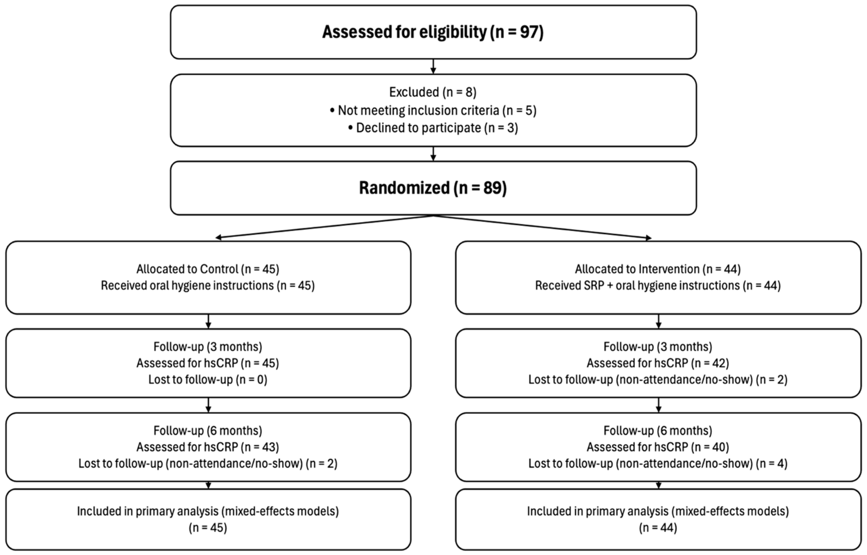
This study was a single-centre, parallel-group randomized controlled clinical trial (1:1 allocation) conducted to evaluate the effect of non-surgical periodontal therapy on systemic inflammation in patients with T2DM and periodontitis. It was approved by the Ethics Committee of the University of Medicine of Tirana, and all participants provided written informed consent before enrollment. The trial had a longitudinal design with assessments at baseline, 3 months, and 6 months. The study protocol followed SPIRIT recommendations, and reporting adheres to CONSORT guidance. The trial was registered in the ISRCTN registry (ISRCTN12954826; registered on 20 January 2026). A total of 89 participants meeting the eligibility criteria were enrolled and randomized to the intervention or control group; participant flow is presented in the CONSORT diagram (
Figure 1). The CONSORT checklist can be found in the
Supplementary Materials. Eligible participants were adults aged 18–70 years with a diagnosis of T2DM, HbA1c ≥ 7%, and periodontitis defined as ≥4 teeth with at least one site with probing depth (PD) ≥ 5 mm, full-mouth bleeding on probing (BOP) ≥ 10%, and ≥10 natural teeth. Exclusion criteria included periodontal treatment within the previous 6 months, systemic antibiotic use within the previous 3 months, systemic conditions affecting periodontal response, pregnancy or breastfeeding, and inability to attend follow-up visits.
2.2. Randomization, Masking, and Clinical Procedures
Participants were randomized after completion of baseline assessments using a computer-generated 1:1 allocation sequence prepared by an independent operator who was not involved in enrollment, treatment delivery, or outcome assessment. Group assignment was concealed using consecutively numbered sealed envelopes that were prepared in advance and opened only after enrollment. Periodontal examinations were performed by a single examiner. Because the intervention was procedural, neither participants nor clinicians could be masked to group allocation. Periodontal examinations were performed using a standardized protocol. Before the start of participant recruitment, intra-examiner calibration was performed in six individuals who were not included in the final randomized study sample. The same examiner who performed the trial assessments completed two full-mouth periodontal examinations for each calibration participant, with a 3-day interval between examinations and no periodontal treatment between sessions. Full-mouth periodontal charting was performed at six sites per tooth, excluding third molars, using a millimetric periodontal probe and dental mirror. Probing force was standardized at approximately 0.20–0.30 N (20–30 g) after prior practice using a scale.
Intra-examiner reliability was assessed for probing depth (PD), clinical attachment level (CAL), bleeding on probing (BOP), gingival index (GI), and plaque index (PI). Reliability was very good for PD and CAL, with intraclass correlation coefficients of 0.89 (95% CI: 0.83–0.94) and 0.87 (95% CI: 0.80–0.93), respectively. Agreement was good for BOP, with a kappa value of 0.76 (95% CI: 0.71–0.81) and 92% agreement between sessions. Reliability was also acceptable for GI and PI, with intraclass correlation coefficients of 0.82 (95% CI: 0.74–0.89) and 0.80 (95% CI: 0.71–0.88), respectively. The mean absolute differences for PD and CAL were 0.28 mm and 0.32 mm, and 95% of PD measurements and 93% of CAL measurements were within ±1 mm. PD and clinical attachment level (CAL) were recorded at six sites per tooth. BOP was recorded and expressed as the percentage of sites with bleeding. Gingival index (GI) and plaque index (PI) were recorded on a 0–3 scale. Fasting venous blood samples were collected at baseline, 3 months, and 6 months. hsCRP was measured using the Roche Cardiac C-Reactive Protein (Latex) High Sensitive assay (CRPHS) on a cobas c 503 analyzer (Roche Diagnostics, Rotkreuz, Switzerland). The assay is a latex particle-enhanced immunoturbidimetric assay, and results were reported in mg/L. Analyses were performed by an accredited external laboratory, and laboratory personnel were blinded to treatment allocation.
2.3. Interventions
Participants in the intervention group received full-mouth NSPT consisting of scaling and root planing (SRP) using manual and ultrasonic instruments, together with oral hygiene instructions. Treatment was delivered as full-mouth therapy in a single visit (one session). Supportive periodontal therapy was provided at 3 and 6 months and included reinstrumentation of residual sites as needed, professional polishing, and reinforcement of oral hygiene instructions. The control group received oral hygiene instructions only during the study period. For ethical reasons, full non-surgical periodontal therapy was offered to control group participants after completion of the 6-month follow-up.
2.4. Statistical Analysis
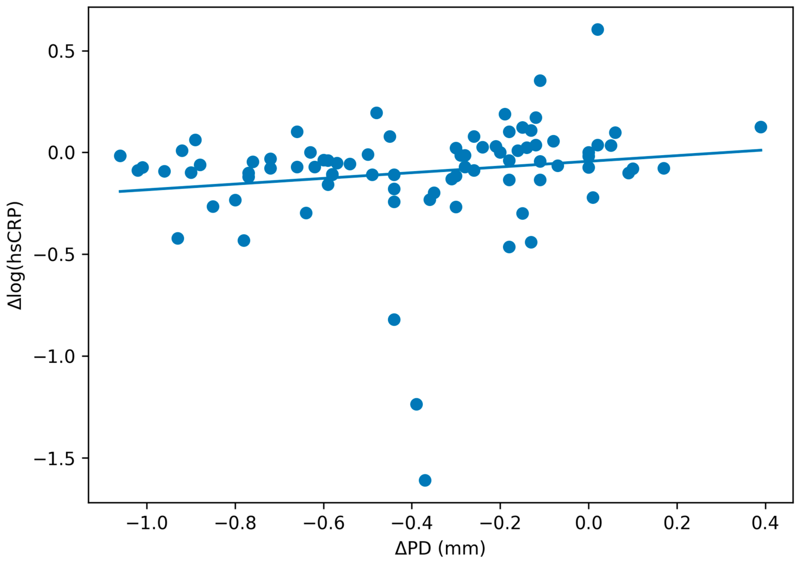
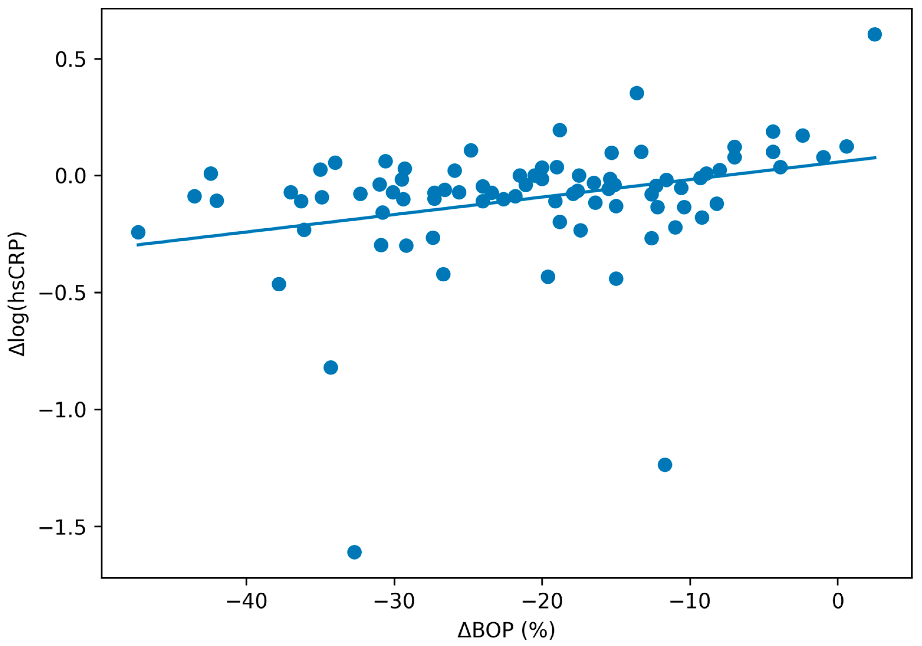
Analyses were conducted according to randomized treatment allocation. All available hsCRP measurements from randomized participants were analyzed using linear mixed-effects models (participant-level random intercept) with fixed effects for group, time (baseline, 3 months, 6 months), and group × time interaction. Given the skewed distribution of hsCRP, analyses were performed on the log scale; results are presented as exponentiated effects (ratios) with 95% confidence intervals. The primary contrast was the between-group difference in change from baseline at 6 months; the 3-month contrast was prespecified as secondary. Sensitivity analyses included baseline-adjusted analysis of covariance (ANCOVA) at each follow-up (log[hsCRP] at follow-up adjusted for baseline log[hsCRP]) and a covariate-adjusted mixed model including baseline age, sex, BMI, smoking status, statin use, baseline HbA1c, and diabetes duration. Values > 10 mg/L were prespecified to indicate possible acute inflammation. Periodontal outcomes (PD, CAL, BOP, GI, PI) were analyzed using analogous mixed-effects models. Exploratory analyses (restricted to participants with hsCRP at baseline and 6 months) assessed associations between Δlog(hsCRP) and changes in periodontal measures (ΔBOP, ΔPD, ΔCAL) using linear regression adjusted for treatment group. Mixed-effects models used all available observations and implicitly accommodated incomplete follow-up under a missing-at-random assumption; we did not impute missing hsCRP values. Analyses were performed using IBM SPSS Statistics v.22 (IBM Corp., Armonk, NY, USA). No formal sample-size calculation was performed specifically for hsCRP in the parent trial; therefore, hsCRP analyses, particularly exploratory associations between periodontal changes and hsCRP change, should be interpreted as hypothesis-generating.
4. Discussion
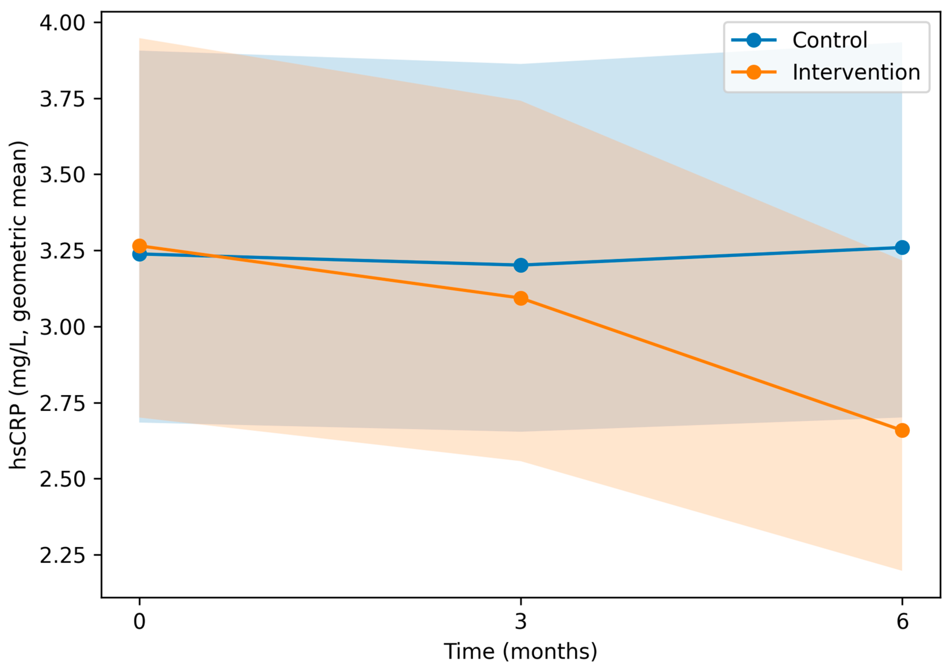
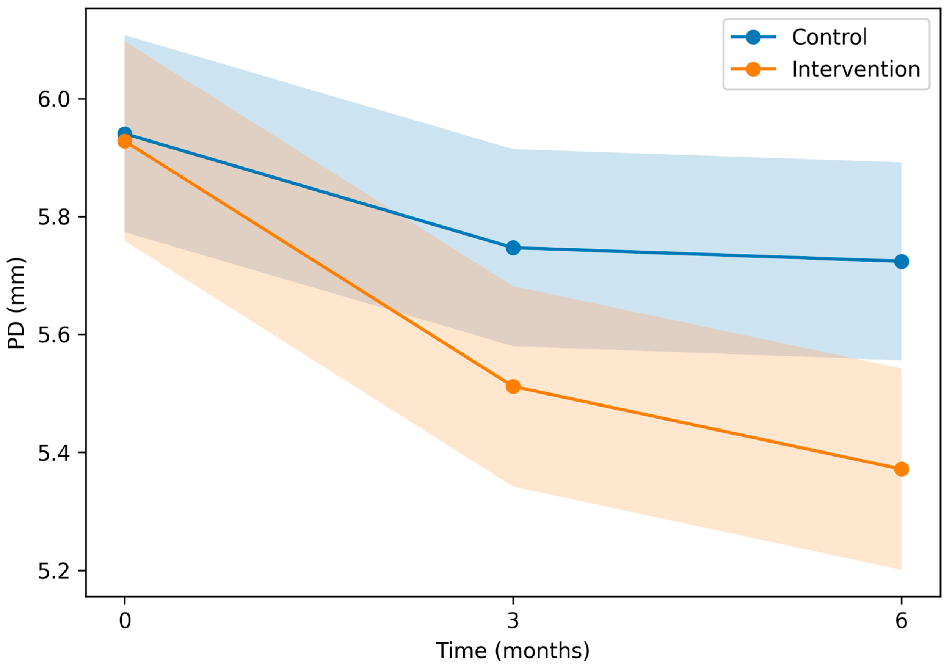
In this predefined secondary analysis of a randomized controlled trial in adults with T2DM and periodontitis, non-surgical periodontal therapy was associated with lower hsCRP at 6 months compared with oral hygiene instructions alone, whereas no clear between-group difference was observed at 3 months. Findings were consistent across sensitivity analyses. As expected, periodontal clinical measures improved more in the intervention group, confirming effective local disease control. Exploratory analyses did not demonstrate a clear association between the magnitude of periodontal improvement and hsCRP change after accounting for treatment assignment. This may reflect limited power to detect dose–response relationships and/or that hsCRP is influenced by factors not captured by individual clinical periodontal measures.
The absence of a detectable between-group difference at 3 months, followed by a clear separation at 6 months, is biologically plausible in a chronic inflammatory setting such as T2DM. Periodontal healing and stabilization may require sustained suppression of the subgingival inflammatory burden and maintenance before systemic inflammatory signaling decreases sufficiently to be detected by hsCRP, a biomarker with substantial within-person variability. In addition, supportive periodontal therapy and reinforcement of oral hygiene during follow-up may contribute to a more durable reduction in periodontal inflammation over time. Nonetheless, alternative explanations should be considered. The intervention group had more clinical contact than controls, which could influence oral-health behaviors and other health behaviors, and hsCRP can be affected by intercurrent conditions and other unmeasured factors. Therefore, the observed temporal pattern should be interpreted as consistent with a lagged systemic response, while acknowledging potential behavioral and non-specific influences.
The between-group difference corresponds to an approximate 19% relative reduction and an absolute separation of ~0.6 mg/L at 6 months. In clinical risk frameworks, hsCRP values are often interpreted as low (<1 mg/L), intermediate (1–3 mg/L), and high (>3 mg/L) inflammatory risk [
8], and hsCRP ≥ 2 mg/L is frequently used as a risk-enhancing threshold in cardiovascular risk assessment [
1]. In our cohort, baseline hsCRP values were in a range typically considered elevated, and the intervention group’s model-based geometric mean decreased from an elevated range toward lower values within commonly used risk categories. While this magnitude of change suggests a downward shift in systemic inflammatory burden, it does not by itself justify changes in cardiometabolic management, nor does it demonstrate reduced cardiovascular events. Rather, it supports hsCRP as a responsive systemic biomarker following periodontal treatment in this high-risk population.
Although periodontal clinical measures improved more in the intervention group, we did not detect an independent linear association between change in individual periodontal parameters and change in hsCRP after adjustment for treatment group. This finding cautions against a simple dose–response interpretation in which larger local clinical improvements necessarily translate into proportionally larger systemic hsCRP reductions. Several explanations are possible, including limited power for within-group dose–response analyses, measurement variability in both periodontal indices and hsCRP, and the likelihood that systemic inflammation in T2DM reflects multiple concurrent drivers (e.g., adiposity, glycaemic control, medication use, and intercurrent conditions). While our models adjusted for key covariates (including statin use), residual confounding cannot be excluded, and hsCRP should be interpreted as a non-specific biomarker rather than a direct proxy for periodontal inflammation.
Evidence supports a two-way association between diabetes and periodontitis, likely mediated by overlapping inflammatory and immune-metabolic pathways linking periodontal inflammation to systemic inflammation [
5,
6,
7]. CRP reflects hepatic acute-phase responses to inflammatory cytokine signaling and is sensitive to low-grade inflammation [
1]. Consistent with this oral–systemic interface, observational studies and meta-analyses report higher circulating CRP/hsCRP in periodontitis than in periodontal health [
8], and mechanistic reviews describe potential pathways linking periodontal inflammation to systemic inflammatory comorbidities [
10], supporting hsCRP as a relevant systemic outcome in periodontal intervention studies.
Meta-analyses of randomized trials in mixed populations suggest that non-surgical periodontal therapy can reduce circulating CRP/hsCRP, although effect sizes vary by population, comparator intensity, adjunctive measures, and follow-up duration [
9]. Evidence specific to T2DM cohorts is more limited and heterogeneous: systematic reviews report modest and variable effects on glycaemic outcomes [
14,
15,
16,
17,
18], and randomized trials have reported variable findings for inflammatory biomarkers such as CRP/hsCRP [
11,
17,
19]. Individual randomized trials in T2DM cohorts have reported variable changes in systemic inflammatory markers after periodontal treatment [
12,
18], and recent systematic reviews suggest that HbA1c improvements after periodontal therapy are modest and heterogeneous [
19]. Overall, systemic responses may depend on multiple factors such as baseline inflammation, adiposity, concomitant medications, periodontal severity, and maintenance intensity [
5,
6,
7]. Within this context, our findings add randomized evidence that hsCRP showed clearer between-group separation at 6 months than at 3 months, consistent with the possibility that sustained periodontal stabilization and maintenance are required before biomarker changes become detectable in patients with T2DM. Compared with mixed-population RCTs, trials in T2DM cohorts have reported more variable systemic responses after NSPT, likely reflecting differences in baseline inflammation and glycaemic control, concomitant cardiometabolic therapies (including statins), periodontal severity, and variation in treatment intensity and maintenance protocols [
11,
12,
17,
18]. In this context, our findings support hsCRP as a responsive systemic biomarker at 6 months in T2DM while underscoring that effect sizes may be context-dependent and heterogeneous across trials.
CRP is an acute-phase reactant synthesized in the liver, largely regulated by cytokine signaling (notably IL-6), and hsCRP assays capture lower concentrations relevant to chronic inflammation and cardiometabolic risk [
1,
2]. Periodontitis is a chronic biofilm-driven inflammatory disease with ulcerated pocket epithelium and potential for recurrent bacteremia/endotoxemia, which can increase systemic inflammatory signaling [
20]. In T2DM, hyperglycemia-related immune dysfunction, oxidative stress, and advanced glycation end-products may amplify periodontal inflammation and impair resolution, contributing to heightened systemic inflammation [
5,
6,
7]. Non-surgical periodontal therapy reduces subgingival bacterial load and local inflammation and may thereby attenuate systemic inflammatory responses [
9,
11,
12,
18]. Although hsCRP is non-specific, a reduction in this marker may be relevant because hsCRP is widely used to quantify low-grade inflammation and inform cardiometabolic risk assessment [
2,
3,
4]. Beyond inflammatory biomarkers, periodontal treatment has also been associated with improvements in endothelial function in systematic reviews and meta-analyses, supporting the plausibility of broader cardiometabolic benefits [
13]. However, the present results should be interpreted as biomarker improvement rather than evidence of reduced cardiovascular events or diabetes complications, and studies with clinical endpoints are required.
In our study, hsCRP was available for most participants, and mixed-effects models maximized the use of repeated measures while accounting for within-participant correlation. hsCRP analyses were performed by personnel blinded to treatment allocation, limiting outcome measurement bias. Nevertheless, several limitations should be considered. The trial was conducted at a single center, which may limit generalizability. Because NSPT is a procedural intervention, blinding of participants and treating clinicians was not feasible. In addition, periodontal examinations were performed by a single examiner who was not blinded to treatment allocation. Although a standardized examination protocol and prior intra-examiner calibration were used, the lack of examiner blinding may have introduced detection bias in the assessment of periodontal outcomes.
The intervention group also received greater clinical contact than the control group, including full-mouth scaling and root planing and supportive periodontal care, whereas the control group received oral hygiene instructions only during the study period. This imbalance may have contributed to performance bias or non-specific behavioral effects, including improved oral hygiene or other health-related behaviors. Therefore, the observed hsCRP difference should be interpreted cautiously and should not be attributed exclusively to the biological effect of periodontal debridement. hsCRP is a non-specific biomarker influenced by adiposity, smoking, intercurrent illness, and concomitant medications, such as statins, which are common in T2DM populations [
1,
2,
4]. Although no hsCRP values exceeded 10 mg/L, residual confounding and biological variability cannot be excluded.
Finally, no formal sample-size calculation was performed specifically for hsCRP in the parent trial. Although the mixed-effects models used all available repeated measurements, the modest sample size limits precision for secondary and exploratory analyses and reduces power to detect treatment-effect heterogeneity or modest dose–response relationships between periodontal improvement and hsCRP change. Therefore, the exploratory associations between periodontal changes and hsCRP should be interpreted as hypothesis-generating rather than causal. Future trials should include longer follow-up with clearly defined maintenance protocols and consider stratification by baseline inflammatory status and glycaemic control. Broader biomarker panels (e.g., IL-6, TNF-α) may help clarify underlying inflammatory pathways. These findings are hypothesis-generating for systemic inflammatory benefit and should not be interpreted as evidence of reduced cardiovascular events or diabetes-related complications; adequately powered multicenter trials with longer follow-up and clinical or validated surrogate endpoints are needed to determine whether improvements in inflammatory biomarkers translate into reduced cardiometabolic risk.





 ,
,