


Highlights
What are the main findings?
- Random EZ-Tn5 transposon mutagenesis of Aggregatibacter actinomycetemcomitans SA269 generated mutants exhibiting altered biofilm and aggregation-associated phenotypes.
- Twelve transposon mutants showed reduced biofilm biomass, and selected mutants demonstrated altered autoaggregation and coaggregation behavior with Fusobacterium nucleatum.
What are the implications of the main findings?
- The findings provide preliminary phenotypic insights into bacterial surface-associated properties involved in biofilm development and interspecies interactions.
- These observations establish a foundation for future transposon insertion-site identification and molecular characterization of genes associated with biofilm-related phenotypes.
Abstract
Background/Objectives: Aggregatibacter actinomycetemcomitans is an important periodontal pathogen whose biofilm formation and bacterial aggregation contribute to colonization and persistence within the oral cavity. This study aimed to generate random transposon mutants of A. actinomycetemcomitans and screen for alterations in biofilm- and aggregation-related phenotypes. Methods: A transposon mutagenesis library was created in the serotype d strain SA269 using the EZ-Tn5 transposon system. Transformants were screened in vitro for biofilm formation, autoaggregation, and coaggregation with Fusobacterium nucleatum. Biofilm biomass was quantified using a standardized microtiter plate assay, while aggregation assays were performed by optical density measurements. Results: Among 44 transformants, 12 exhibited a markedly reduced ability to form biofilms compared with the wild-type strain, indicating that disruption of certain loci compromises surface adherence and biofilm biomass accumulation. Five representative mutants were selected for aggregation studies. Three mutants showed reduced autoaggregation, suggesting impaired intraspecies cell–cell interactions, whereas four mutants demonstrated enhanced coaggregation with F. nucleatum, reaching approximately 50% coaggregation after 120 min. These findings indicate that insertional inactivation of genes in A. actinomycetemcomitans can differentially affect biofilm formation, autoaggregation, and interspecies coaggregation. Conclusions: This preliminary study demonstrates the utility of random transposon mutagenesis for identifying mutants with altered adhesion- and biofilm-related phenotypes. Although the disrupted genes were not identified, the mutant library provides a foundation for future molecular characterization of genetic determinants involved in biofilm development and microbial community interactions.
1. Introduction
The aggressive form of periodontitis, which is now classified as periodontitis stage III–IV, grade C, is a form of periodontal disease possessing multiple characteristics helping to distinguish it from chronic periodontitis. It is a severe disease with rapid rate of periodontal tissue destruction, often afflicting patients at a far earlier age than its chronic counterpart, as it commonly begins during or after the circumpubertal period, and less commonly prior to puberty. This disease is further characterized by a certain unique pattern of destruction, involving a number of teeth, without any other systemic diseases afflicting the periodontitis patient [1]. Although shown to have a relatively low prevalence, specific ethnic groups seem to be more frequently afflicted, thus having a certain geographic distribution [2]. It is often found in adolescents of African descent [3]. The reason for aggregation of this disease in certain individuals has been attributed to a number of factors, including a genetic predisposition (due to the disease having a familial nature of aggregation), as well as infection with specific micro-organisms [1].
For many years, Aggregatibacter actinomycetemcomitans has been largely accepted as the primary agent bearing responsibility for the etiological basis of aggressive periodontitis. It is known to be only a very small fraction of the normal flora found within the oral cavity, and has been found to act as an opportunistic pathogen in certain individuals. Despite it being a certain minor proportion of resident flora, the specific A. actinomycetemcomitans-JP2 clone (belonging to the cluster of serotype b strains) [4] has been discovered to likely play an important part in the pathogenesis of aggressive periodontitis in certain populations, with very minimal data available on the possible role of other microbes in the development of this disease [5]. This specific clone is prevalent in North and West Africa [6]. Not only is Aa implicated in the field of periodontology, but it is also involved in a variety of other medical pathologies such as infectious endocarditis, chest wall abscesses, and brain abscesses [6].
Research over many years has aimed to clearly elucidate the specific mechanisms by which this microbe contributes to such severe periodontal disease. A. actinomycetemcomitans has been found to be an early colonizer that colonizes at the same time as lactate-producing bacteria, after which it upregulates dspB in order to migrate to a relatively more protected subgingival domain, away from the stressful environment it was in. Despite a subgingival innate host immune response aiming to defend the patient from the subsequent arising pathology, A. actinomycetemcomitans functions to upregulate both complement resistance genes, as well as the production of leukotoxin. The undermined defense response of the host leads to the growth of an association of pathobionts, thus producing a myriad of inflammatory cytokines. It is these inflammatory cytokines that contribute to connective tissue loss, bone loss, and impairment of the periodontal attachment apparatus of the host [3].
The available data suggests that mutations in major genes or multiple small-effect genes and gene–gene–environment interaction effects have significant roles in the pathogenesis of aggressive periodontitis. Recent studies showed that an autosomal mode of transmission is more valid since the X-linked mode of inheritance is inconsistent with father-to-son transmissions of this disorder. Genetic markers are a significant diagnostic tool to detect individuals at risk and enhance the early detection of disease, thus improving treatment prognosis, as well as future approaches based on gene therapy [2].
Transposon mutagenesis is an important tool in discovering regions of the genome that are required for the survival of bacteria in different environments [7,8]. There is a lack of evidence regarding the set of genes that contributes in the virulence process of A. actinomycetemcomitans.
Aims
The aims of this study are to create random mutants of A. actinomycetemcomitans by using the transposon mutagenesis technique and investigate the effect of the mutagenesis on important phenotypic properties of the species. This may lead to identifying novel virulence factors.
2. Materials and Methods
A. actinomycetemcomitans SA269, a serotype-d strain which has a good transformation efficiency, was cultured on tryptic soy agar plates at 37 °C in 5% CO2 for 2 days.
2.1. Electroporation of Host Cells with EZ-Tn5 <KAN-2> Tnp Transposome and Selection of Transposition Clones
Electrocompetent A. actinomycetemcomitans cells were electroporated using 1 μL of the EZ-Tn5 <KAN-2> Tnp Transposome (Lucigen®; Lucigen, Middleton, WI, USA). The A. actinomycetemcomitans strain SA269 was expected to have a transformation efficiency of >107 cfu/μg of DNA. The complete electroporation procedure was performed according to the manufacturer’s recommendations (Eporator, Eppendorf AG, Hamburg, Germany).
The electroporated bacterial cells were recovered immediately after electroporation. SOC medium was added to the electroporation cuvette to a 1 mL final volume immediately after electroporation. The medium and the bacterial cells were gently mixed by pipetting in and out. The mixture was then transferred to a tube and incubated on a 37 °C shaker for 30–60 min to facilitate cell outgrowth.
The recovered cells were diluted (1:10 and 1:100) and plated (100 μL) alongside undiluted cells, with each cell dilution plated separately, on plates containing 50 μg/mL kanamycin. After 2 days of incubation as above, the plates were observed for transformant colonies.
2.2. Confirmation of the Transposon Insertion by PCR
2.2.1. DNA Purification
DNA was purified from the transformant bacterial colonies by using DNeasy kit (Qiagen Inc., Valencia, CA, USA) according to the manufacturer’s instructions. Briefly, bacterial suspensions were treated with lysis buffer and proteinase-K at 56 °C for 1 h. DNA from lysed samples was applied onto a spin column, washed, and eluted with an equal volume of elution buffer or sterile water for all samples. This DNA was used as template for PCR and RT-PCR.
2.2.2. PCR and Agarose Gel Electrophoresis
For all PCR reactions, Ready-To-Go beads (GE HealthCare Biosciences, Buckinghamshire, UK) were used. When each bead was reconstituted to a 25 µL final volume, the concentration of each dNTP was 200 µM in 10 mM Tris-HCl (pH 9), 50 mM KCl and 1.5 mM MgCl2.
The following are the primer pair used for detecting the KanR gene in the transposon inserted in the bacterial genome:
Forward Primer: 5′-ACGGATCGCGTATTTCGTCT-3′,
Reverse Primer: 5′-CGGGAAGATGCGTGATCTGA-3′.
The amount of template DNA used was 50–100 ng per reaction. PCR products were visualized by electrophoresis through a 2% agarose gel after adding the dye ethidium bromide to the PCR product. The gels were exposed to UV light on a trans illuminator and pictures taken by the attached imaging system.
2.2.3. Screening the Transformants for Novel Phenotypes
The transformant A. actinomycetemcomitans colonies were screened for novel phenotypes in biofilm formation, autoaggregation and coaggregation.
2.2.4. Biofilm Assay
Biofilm cultures were established using a modified version of a previously published method [9]. Initially, an inoculum of OD600 = 1 suspension for each test species was prepared. Bacterial colonies were harvested from agar plates using sterile plastic loops and suspended in sterile phosphate-buffered saline (PBS). Subsequently, the suspensions underwent two washes in sterile PBS by centrifugation at 5000× g for 5 min. After centrifugation, the cell pellet was suspended in 1 mL of brucella broth. Adjustments to a standard OD600 = 1 were made for each test species based on OD measurements of the suspensions. Biofilms were initiated by inoculating 24-well plates with 900 µL of brucella broth, to which 100 µL aliquots from the OD600 = 1 stock suspension of bacteria were added. The plates were then incubated in 5% CO2 at 37 °C for 2 days.
2.2.5. Aggregation Assay
Auto- and coaggregation were quantified by an optical density (OD) assay [10]. Microbial cell suspensions of OD600 = 1 from each of the test species and the partner species were mixed in equal amounts, and optical density was measured for 2 h with a 15 min time interval at 600 nm on a spectrophotometer (BioPhotometer, Eppendorf AG, Hamburg, Germany). For autoaggregation, cell suspension of single species was considered. A decline in the OD of the microbial suspensions with time was used as a measure of auto- or coaggregation and was calculated [11] as follows:
% Coaggregation or autoaggregation = (time zero value − sample value)/(time zero value) × 100.
2.3. Statistics
All biofilm and aggregation assays were performed in triplicate in at least three independent experiments. Data are presented as mean ± standard deviation (SD). Differences between the groups were compared by non-parametric Mann–Whitney U test. A p value less than 0.05 was considered statistically significant. SPSS vs. 22 for Windows was used for all analyses.
3. Results
3.1. Screening and Confirmation of the Transformants
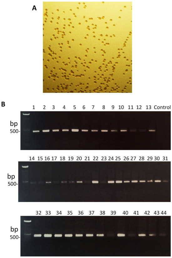
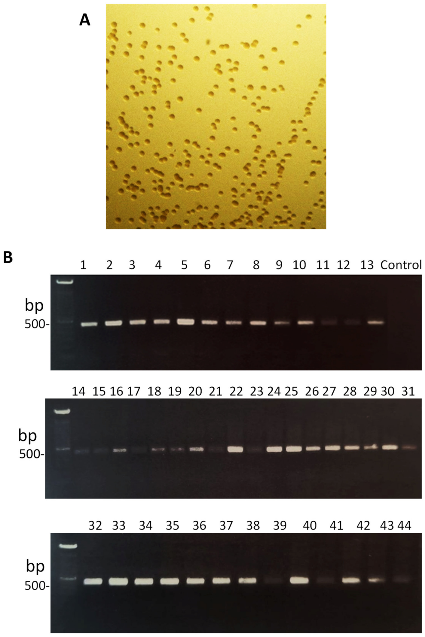
After electroporation and genetic transformation of A. actinomycetemcomitans SA269 with the EZ-Tn5 transposon, the colonies that appeared on the kanamycin-containing TSA medium (Figure 1, panel A) were screened for the insertion of the transposon by using the target KanR gene in Tn5 transposon. As seen in panel B of Figure 1, a 510 bp KanR sequence was amplified in all of the transformant colonies, confirming the insertion of the transposon sequence, which contained the kanamycin-resistance marker.
Figure 1.
Confirmation of the transformants. Following transposon mutagenesis of the A. actinomycetemcomitans SA269 strain, transformant colonies on kanamycin-containing media (panel (A)) were subject to confirmation by PCR using primers specific for KanR gene of the Tn5 transposon (panel (B)).
3.2. Biofilm Formation Ability of the Transformants
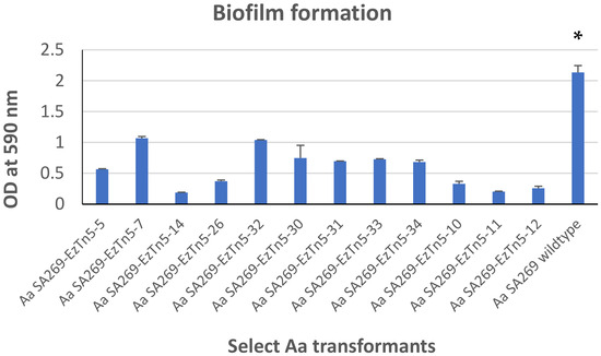
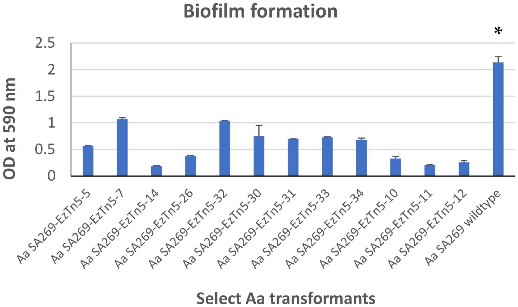
In order to study the phenotypic characteristics of the transformants, we chose biofilm forming ability as the first choice due to the significance of the remarkable ability of this species to tightly adhere to surfaces. Of the 44 transformants tested, 12 isolates displayed a significantly reduced ability to form biofilms (Figure 2) (p < 0.001). While the wild-type strain SA269 formed a strong biofilm, only two of the 12 transformants showed an OD value of ~1 following crystal violet staining. The rest of the transformants were able to form only poor biofilms.
Figure 2.
Biofilm formation by A. actinomycetemcomitans SA269 transformants. Twelve randomly selected transformants were tested for their biofilm formation ability. Standard monospecies biofilm protocol was followed. After the biofilms were grown, crystal violet staining was performed for crude estimation of the biofilm biomass. * p < 0.05.
3.3. Autoaggregation and Coaggregation
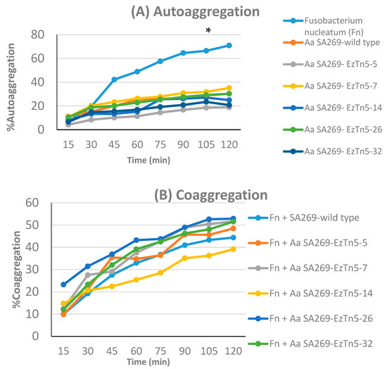
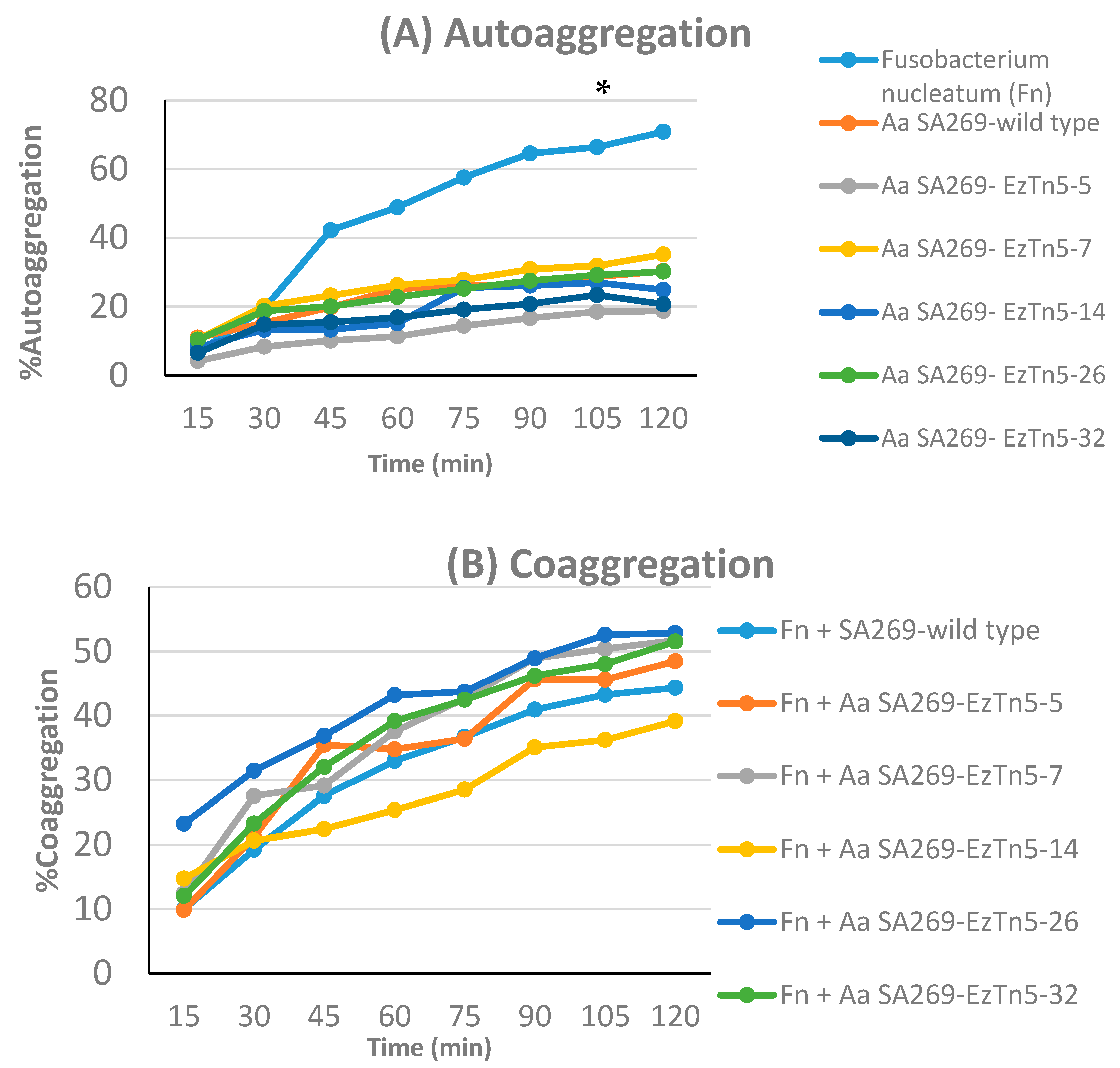
From the biofilm formation experiments, we chose five transformants to study their auto- and coaggregation behaviors. These five were selected based on representing the range of biofilm phenotypes observed: two mutants with near-complete loss of biofilm formation (OD < 0.2), two with intermediate reduction, and one with borderline reduction, to capture the spectrum of phenotypic alteration across the dataset. As depicted in panel A of Figure 3, four of five transformants showed lower autoaggregation than the SA269 wild-type strain. F. nucleatum showed the highest autoaggregation (p < 0.05), showing 20% autoaggregation at 30 min followed by a steady increase with 50% at 60 min and 70% at 120 min.
Figure 3.
Autoaggregation (A) and coaggregation (B) of the transformants. From the transformants that showed a marked reduction in the biofilm formation, 5 transformants were assessed for autoaggregation and co-aggregation ability. A standard optical density measurement protocol was used for the assay. * p < 0.05.
In the case of co-aggregation, four of the five transformants displayed a higher percent coaggregation with F. nucleatum than the wild-type SA269 strain. The percent coaggregation of these transformants was in the range of 20% at 30 min and reached ~50% at the end, i.e., 120 min.
4. Discussion
The present preliminary phenotypic screening identified A. actinomycetemcomitans transposon mutants exhibiting altered biofilm-related and aggregation-associated phenotypes. Among the 44 transformants analyzed, 12 displayed reduced biofilm biomass compared with the wild-type SA269 strain, indicating that transposon insertion altered bacterial surface-associated characteristics involved in biofilm development [12]. Random transposon mutagenesis remains a valuable strategy for generating mutant libraries and identifying genetic determinants associated with bacterial phenotypes [13,14]. However, because the transposon insertion sites were not identified in the present study, the molecular basis underlying these observed phenotypic alterations remains unknown and requires future genetic characterization.
Biofilm formation is an important colonization-associated characteristic of A. actinomycetemcomitans and contributes to its persistence within the periodontal environment [3]. Biofilms enable bacteria to adhere to surfaces and enhance their survival under environmental challenges [15]. The observed reduction in biofilm biomass among several transformants suggests alterations in mechanisms involved in bacterial surface attachment, extracellular matrix production, or other factors contributing to biofilm development.
The role of biofilm formation in bacterial colonization, persistence, and host interactions is well established. Biofilms provide protection from environmental stresses, antimicrobial agents, and host immune responses, thereby enhancing bacterial survival and persistence in the host [16]. In A. actinomycetemcomitans, known biofilm regulators include genes of the tad locus, the apaH gene, surface adhesins such as apiA and emaA, and the flp fimbriae [17,18,19]. Although the observed phenotypes may reflect alterations in pathways associated with biofilm formation and surface adhesion, the absence of transposon insertion-site identification prevents determination of whether these known determinants or previously uncharacterized loci were affected. These findings add to a growing body of work investigating genetic determinants of biofilm-associated phenotypes in A. actinomycetemcomitans [20].
Autoaggregation and coaggregation are important processes in bacterial adhesion and biofilm formation. Autoaggregation refers to the spontaneous aggregation of bacterial cells, while coaggregation involves the aggregation of different bacterial species. These interactions influence biofilm architecture and stability, as well as microbial community dynamics within biofilms [21].
In this study, the autoaggregation and coaggregation behaviors of selected transformants were investigated. Three out of the five transformants exhibited lower autoaggregation compared to the wild-type SA269 strain. This suggests that the genetic alterations introduced during transformation may have affected cell–cell interactions and aggregation properties. Conversely, in coaggregation assays with F. nucleatum, four out of the five transformants exhibited a higher percentage of coaggregation compared to the wild-type strain. This indicates that transposon-induced alterations may influence surface-associated molecules involved in interspecies interactions [22]. Interestingly, the observation that some transformants exhibited reduced biofilm formation while simultaneously demonstrating increased coaggregation with F. nucleatum suggests that distinct mechanisms may regulate attachment to abiotic surfaces and interactions with neighboring bacterial species. Alterations in surface structures could impair stable biofilm accumulation while simultaneously modifying the accessibility or spatial arrangement of adhesins involved in coaggregation. However, because the disrupted loci were not identified, these interpretations remain speculative and require further molecular characterization.
The observed alterations in autoaggregation and coaggregation behaviors among the transformants could have implications for microbial colonization and community dynamics in the oral cavity. Changes in these processes may influence the development and structural organization of polymicrobial oral biofilms and affect microbial community interactions within dental plaque. Further studies involving additional strains, bacterial partners, and more complex experimental models are required to determine the broader biological significance of these observations. Further genetic and molecular studies are required to identify the specific loci responsible for these phenotypic alterations and to determine their roles in bacterial adhesion and biofilm development.
Study Limitations
Several limitations of the present study deserve explicit acknowledgement. First, the transposon insertion sites were not sequenced; consequently, the specific genes responsible for the observed phenotypic changes remain unknown. Second, the experiments were conducted using a single serotype-d strain (SA269) and a single coaggregation partner (F. nucleatum), which limits the generalizability of the findings to other A. actinomycetemcomitans strains, including the clinically significant serotype-b JP2 clone associated with aggressive periodontitis. Third, all assays were performed in vitro; while in vitro biofilm formation and aggregation assays provide useful proxies for bacterial surface-associated behaviors, they do not fully recapitulate the complexity of the subgingival environment. Fourth, because only total biofilm biomass (crystal violet staining) was measured and baseline planktonic growth kinetics were not evaluated, it is not possible to distinguish whether reduced biomass resulted from impaired biofilm initiation, altered extracellular matrix production, or differences in overall bacterial growth or fitness. Finally, the criteria evaluated in the present study reflect adhesion- and aggregation-associated phenotypes rather than virulence per se, and extrapolation to clinical pathogenicity should, therefore, be made with caution. These limitations should be addressed in future studies through transposon insertion-site identification, growth kinetics analysis, and validation in additional strains and more physiologically relevant experimental models.
5. Conclusions
This preliminary, descriptive study demonstrates that random transposon mutagenesis in A. actinomycetemcomitans SA269 can produce mutants with reproducibly altered biofilm formation, autoaggregation, and coaggregation phenotypes. The study is limited by the absence of gene identification, the use of a single strain and a single coaggregation partner, and the strictly in vitro nature of all assays. These limitations preclude direct inference about clinical virulence, and the term “virulence” has been used cautiously throughout to refer to adhesion-related phenotypes rather than pathogenic capacity per se. Notwithstanding these limitations, the transposon mutant library established here provides a useful phenotypic platform for future molecular characterization.
Future studies should prioritize the molecular identification of disrupted loci through inverse PCR or whole-genome sequencing, growth-curve analysis to exclude general fitness defects as the cause of reduced biofilm, and extension of the phenotypic screen to additional strains, including the clinically relevant JP2 clone [4]. Such mechanistic follow-up is necessary to advance from descriptive phenotyping to a genuine understanding of the genetic determinants of biofilm architecture and interspecies interactions in A. actinomycetemcomitans, with potential long-term relevance for the development of targeted therapeutic strategies against periodontal disease.
Author Contributions
Conceptualization, M.K.; methodology, M.S., M.A.-S., H.D. and M.K.; investigation, M.S., M.A.-S. and H.D.; formal analysis, M.S. and M.K.; data curation, M.S.; writing—original draft preparation, M.S. and M.K.; writing—review and editing, M.K., M.A.-S. and H.D.; supervision, M.K.; project administration, M.K.; funding acquisition, M.S. All authors have read and agreed to the published version of the manuscript.
Funding
This research received no external funding.
Institutional Review Board Statement
Not applicable.
Informed Consent Statement
Not applicable.
Data Availability Statement
The data supporting the findings of this study are available from the corresponding author upon reasonable request. Certain original datasets generated during preliminary aggregation experiments are no longer available due to limitations in long-term data archiving; however, all available data and the summarized results presented in the manuscript have been retained.
Acknowledgments
Authors thank the Oral Microbiology Research Laboratory for Equipment and other resources (SRUL 01/14). Asma Haneef’s excellent technical help is gratefully acknowledged.
Conflicts of Interest
The authors declare no conflicts of interest.
References
- Albandar, J.M. Aggressive periodontitis: Case definition and diagnostic criteria. Periodontol. 2000 2014, 65, 13–26. [Google Scholar]
- Albandar, J.M. Aggressive and acute periodontal diseases. Periodontol. 2000 2014, 65, 7–12. [Google Scholar]
- Fine, D.H.; Patil, A.G.; Velusamy, S.K. Aggregatibacter actinomycetemcomitans (Aa) Under the Radar: Myths and Misunderstandings of Aa and Its Role in Aggressive Periodontitis. Front. Immunol. 2019, 10, 728. [Google Scholar] [CrossRef] [Scilit] [PubMed]
- Haubek, D.; Johansson, A. Pathogenicity of the highly leukotoxic JP2 clone of Aggregatibacter actinomycetemcomitans and its geographic dissemination and role in aggressive periodontitis. J. Oral Microbiol. 2014, 6, 23980. [Google Scholar] [CrossRef] [Scilit]
- Kononen, E.; Muller, H.P. Microbiology of aggressive periodontitis. Periodontol. 2000 2014, 65, 46–78. [Google Scholar]
- Kaplan, A.H.; Weber, D.J.; Oddone, E.Z.; Perfect, J.R. Infection due to Actinobacillus actinomycetemcomitans: 15 cases and review. Rev. Infect. Dis. 1989, 11, 46–63. [Google Scholar] [CrossRef] [Scilit] [PubMed]
- Hadjifrangiskou, M.; Gu, A.P.; Pinkner, J.S.; Kostakioti, M.; Zhang, E.W.; Greene, S.E.; Hultgren, S.J. Transposon mutagenesis identifies uropathogenic Escherichia coli biofilm factors. J. Bacteriol. 2012, 194, 6195–6205. [Google Scholar] [CrossRef] [Scilit] [PubMed]
- Yu, J.; Wang, Y.; Han, D.; Cao, W.; Zheng, L.; Xie, Z.; Liu, H. Identification of Streptococcus mutans genes involved in fluoride resistance by screening of a transposon mutant library. Mol. Oral Microbiol. 2020, 35, 260–270. [Google Scholar] [CrossRef] [Scilit] [PubMed]
- Karched, M.; Bhardwaj, R.G.; Inbamani, A.; Asikainen, S. Quantitation of biofilm and planktonic life forms of coexisting periodontal species. Anaerobe 2015, 35, 13–20. [Google Scholar] [CrossRef] [Scilit] [PubMed]
- Karched, M.; Bhardwaj, R.G.; Asikainen, S.E. Coaggregation and biofilm growth of Granulicatella spp. with Fusobacterium nucleatum and Aggregatibacter actinomycetemcomitans. BMC Microbiol. 2015, 15, 114. [Google Scholar] [CrossRef] [Scilit] [PubMed]
- Nagaoka, S.; Hojo, K.; Murata, S.; Mori, T.; Ohshima, T.; Maeda, N. Interactions between salivary Bifidobacterium adolescentis and other oral bacteria: In vitro coaggregation and coadhesion assays. FEMS Microbiol. Lett. 2008, 281, 183–189. [Google Scholar] [CrossRef] [Scilit] [PubMed]
- Senadheera, D.B.; Cordova, M.; Ayala, E.A.; Chavez de Paz, L.E.; Singh, K.; Downey, J.S.; Svensäter, G.; Goodman, S.D.; Cvitkovitch, D.G. Regulation of bacteriocin production and cell death by the VicRK signaling system in Streptococcus mutans. J. Bacteriol. 2012, 194, 1307–1316. [Google Scholar] [CrossRef] [Scilit] [PubMed]
- Voelker, L.L.; Dybvig, K. Transposon mutagenesis. Methods Mol. Biol. 1998, 104, 235–238. [Google Scholar] [CrossRef] [Scilit] [PubMed]
- Zhang, Y.; Li, Z.; Xu, X.; Peng, X. Transposon mutagenesis in oral Streptococcus. J. Oral Microbiol. 2022, 14, 2104951. [Google Scholar] [CrossRef] [Scilit] [PubMed]
- Koo, H.; Allan, R.N.; Howlin, R.P.; Stoodley, P.; Hall-Stoodley, L. Targeting microbial biofilms: Current and prospective therapeutic strategies. Nat. Rev. Microbiol. 2017, 15, 740–755. [Google Scholar] [CrossRef] [Scilit] [PubMed]
- Donlan, R.M. Biofilms: Microbial life on surfaces. Emerg. Infect. Dis. 2002, 8, 881–890. [Google Scholar] [CrossRef] [Scilit] [PubMed]
- Kram, K.E.; Hovel-Miner, G.A.; Tomich, M.; Figurski, D.H. Transcriptional regulation of the tad locus in Aggregatibacter actinomycetemcomitans: A termination cascade. J. Bacteriol. 2008, 190, 3859–3868. [Google Scholar] [CrossRef] [Scilit] [PubMed]
- Danforth, D.R.; Melloni, M.; Tristano, J.; Mintz, K.P. Contribution of adhesion proteins to Aggregatibacter actinomycetemcomitans biofilm formation. Mol. Oral Microbiol. 2021, 36, 243–253. [Google Scholar] [CrossRef] [Scilit] [PubMed]
- Umeda, J.E.; Longo, P.L.; Simionato, M.R.L.; Mayer, M.P.A. Differential transcription of virulence genes in Aggregatibacter actinomycetemcomitans serotypes. J. Oral Microbiol. 2013, 5, 21473. [Google Scholar] [CrossRef] [Scilit]
- Lemos, J.A.; Palmer, S.R.; Zeng, L.; Wen, Z.T.; Kajfasz, J.K.; Freires, I.A.; Abranches, J.; Brady, L.J. The Biology of Streptococcus mutans. Microbiol. Spectr. 2019, 7, 10.1128. [Google Scholar] [CrossRef] [Scilit]
- Rickard, A.H.; Gilbert, P.; High, N.J.; Kolenbrander, P.E.; Handley, P.S. Bacterial coaggregation: An integral process in the development of multi-species biofilms. Trends Microbiol. 2003, 11, 94–100. [Google Scholar] [CrossRef] [Scilit] [PubMed]
- Kolenbrander, P.E.; Palmer, R.J., Jr.; Periasamy, S.; Jakubovics, N.S. Oral multispecies biofilm development and the key role of cell-cell distance. Nat. Rev. Microbiol. 2010, 8, 471–480. [Google Scholar] [CrossRef] [Scilit] [PubMed]
Disclaimer/Publisher’s Note: The statements, opinions and data contained in all publications are solely those of the individual author(s) and contributor(s) and not of MDPI and/or the editor(s). MDPI and/or the editor(s) disclaim responsibility for any injury to people or property resulting from any ideas, methods, instructions or products referred to in the content. |
© 2026 by the authors. Licensee MDPI, Basel, Switzerland. This article is an open access article distributed under the terms and conditions of the Creative Commons Attribution (CC BY) license.