


Abstract
Objectives: This study aimed to evaluate and compare the effects of 38% silver diamine fluoride (SDF) and nano-hydroxyapatite (nHAp) toothpaste on dentinal tubule surface area, elemental composition, and microhardness of mechanically exposed dentin. Methods: Thirty extracted human molars with standardized exposed occlusal dentin were randomly assigned to control, SDF, or nHAp groups (n = 10 each). Surface morphology and dentinal tubule area were assessed using scanning electron microscopy, elemental composition by energy-dispersive X-ray spectroscopy, and microhardness by Vickers testing before and after application of SDF and nHAp. Data were analyzed using one-way ANOVA with Bonferroni post hoc tests and paired t-tests (α = 0.05). Results: Baseline dentinal tubule area and microhardness were comparable among groups. SDF showed the strongest effects, reducing dentinal tubule area by 84.5% and producing the greatest microhardness increase (p < 0.001), while nHAp reduced dentinal tubule area by 45.2% and increased microhardness within the group (p < 0.001). Calcium content was highest in the SDF group (p < 0.001), whereas phosphorus showed no significant difference (p = 0.195). The Ca/P ratio differed overall (p = 0.046), mainly between Control and SDF (p = 0.024). Scanning electron microscopy confirmed dense tubule occlusion with SDF, partial occlusion with nHAp, and open tubules in controls. Conclusions: SDF produced greater dentinal tubule occlusion and microhardness improvement than nHAp toothpaste in this short-term in vitro model using mechanically exposed dentin. nHAp toothpaste produced moderate tubule occlusion and a smaller improvement in microhardness under the tested conditions.
1. Introduction
Tooth wear refers to the cumulative surface loss of mineralized tooth substance caused by physical and chemo-physical processes unrelated to dental caries, resorption, or injury, and it has emerged as a growing clinical issue in older adults, primarily because of increased life expectancy and improved tooth retention in later years. Unlike tissue loss due to caries, non-carious tooth wear arises from mechanical and/or chemical processes not influenced by bacterial activity. Among these, attrition—characterized by the gradual loss of tooth structure from tooth-to-tooth contact—is especially prevalent in older adults and in individuals with parafunctional habits such as bruxism [1]. Over time, attrition can expose the underlying dentin, leading to weakened structural integrity, heightened dentin hypersensitivity, and aesthetic concerns.
Attrited dentin presents distinct clinical challenges. Removal of enamel protection exposes dentinal tubules, increasing fluid movement within the dentin and producing sensitivity in accordance with the hydrodynamic theory [2]. Exposed dentin also exhibits a modified mineral composition and altered surface morphology, often characterized by enlarged tubules, variable smear layers, and reduced microhardness, compared with intact enamel [3]. Consequently, minimally invasive approaches that enhance dentin remineralization, promote dentinal tubule occlusion, and maintain structural integrity have attracted increasing attention in contemporary restorative and preventive dentistry. Existing agents, including fluoride-based varnishes and gels, provide remineralization but offer only superficial, short-term effects and require frequent reapplication. Bioactive glass materials induce ion release and tubule occlusion, yet their efficacy depends on environmental pH. Resin-based desensitizing agents provide immediate tubule sealing but show limited durability due to mechanical wear and hydrolytic degradation [4]. These collective limitations underscore the need for more effective and durable alternatives for managing exposed dentin.
Silver diamine fluoride (SDF) is a topical alkaline solution composed principally of silver ions (Ag+), fluoride ions (F−), and ammonia as a stabilizer [5]. SDF has been extensively studied for its ability to arrest caries, improve dentin hardness, and exhibit antibacterial activity [6,7]. Multiple in vitro and clinical investigations have demonstrated that SDF markedly enhances dentin surface microhardness and reduces lesion progression [8]. Scanning electron microscopy (SEM) studies have additionally shown partial or complete occlusion of dentinal tubules following SDF application, suggesting its potential effectiveness in addressing dentin hypersensitivity [9]. Despite these functional benefits, SDF is associated with a significant aesthetic drawback: dark dentin staining arising from the formation of silver oxide and metallic silver deposits in demineralized dentin [10]. This staining may limit patient acceptance, particularly in visible areas, highlighting the need for complementary or alternative agents.
Nano-hydroxyapatite (nHAp) has emerged as a promising biomimetic material in preventive and restorative dentistry. Hydroxyapatite (Ca10(PO4)6(OH)2) is the primary inorganic constituent of enamel and dentin; nano-sized particles exhibit increased surface area and bioactivity, facilitating their incorporation into dental hard tissue structures [11]. Studies have demonstrated that nHAp enhances remineralization by supplying calcium and phosphate ions to demineralized regions, restoring mineral density, and increasing surface microhardness. Additionally, nHAp has been shown to occlude dentinal tubules through crystal formation, thereby reducing permeability and sensitivity [12]. Unlike SDF, nHAp possesses a white, biomimetic appearance that may improve surface smoothness and light reflection without causing dark staining [13]. However, its remineralizing effect is gradual and requires repeated application, with limited penetration depth and variable effectiveness depending on formulation properties [12].
Although SDF and nHAp have been individually assessed for their effects on dentin remineralization, microhardness, and tubule occlusion, comparative studies on exposed dentin surfaces are lacking. Most published investigations have been conducted on caries-affected or chemically demineralized dentin. Attrited dentin differs substantially from these substrates owing to chronic mechanical stress, which results in tubular sclerosis, obliteration, and alterations in organic and inorganic composition [14,15]. These structural modifications may influence the penetration, adhesion, and bioactivity of therapeutic agents such as SDF and nHAp [16]. Findings from caries or demineralization models may therefore not translate directly to mechanically worn surfaces, making dedicated evaluation of attrited dentin clinically important.
The increasing emphasis on conservative dentistry and biological preservation of tooth structure has accelerated interest in biomimetic approaches capable of restoring both function and structural stability [17,18,19]. Contemporary preventive dentistry aims not only to alleviate hypersensitivity symptoms but also to reinforce weakened dental tissues and reduce further progression of tooth wear. Comparative investigations between SDF and nHAp may provide meaningful evidence regarding their relative advantages, limitations, and potential indications in the management of attrition-related dentin exposure, supporting the development of evidence-based clinical protocols.
Accordingly, the present study aimed to evaluate and compare the effects of 38% silver diamine fluoride and nano-hydroxyapatite toothpaste on dentinal tubule surface area, elemental composition, and microhardness of ethylenediaminetetraacetic acid-conditioned mechanically exposed dentin under standardized in vitro conditions. This in vitro model allows controlled comparison of surface changes induced by the tested materials while acknowledging that it does not reproduce the sclerosis and structural adaptations characteristic of clinically attrited dentin.
The working hypothesis was that both 38% silver diamine fluoride and nano-hydroxyapatite toothpaste would reduce dentinal tubule surface area and increase dentin microhardness compared with untreated controls, with silver diamine fluoride producing greater surface changes under the tested protocol.
2. Materials and Methods
2.1. Ethical Approval and Specimen Collection
This research protocol was reviewed and approved by the Research and Ethics Committee of the Faculty of Dentistry, The British University in Egypt (Project No. FD BUE REC 25-061; 30 October 2025), and was conducted in accordance with the Declaration of Helsinki. This study used a standardized mechanically exposed dentin model. Thirty intact, non-carious human permanent molars extracted for periodontal reasons from adult patients aged 40–65 years were collected. Signed and witnessed informed consent was obtained from all participants prior to tooth extraction. All personal identifiers were removed before specimen processing, and teeth were handled in accordance with institutional biosafety guidelines.
Teeth were included only if they presented intact crowns with visible occlusal surfaces suitable for standardized dentin exposure. Exclusion criteria included dental caries, cracks, fractures, restorations, developmental defects, enamel hypoplasia, erosion, abrasion, cervical lesions, previous endodontic treatment, or any structural defect that could affect dentin surface morphology, elemental composition, or microhardness measurements. Following extraction, adherent soft tissue and debris were removed, and teeth were disinfected and stored in distilled water at room temperature until specimen preparation.
2.2. Sample Size Calculation
The sample size was calculated using G*Power software (version 3.1.9.7). Based on a one-way ANOVA design with three independent groups, an effect size of f = 0.6, α = 0.05, and power of 80%, the minimum required sample size was 27 specimens. Thirty specimens were therefore recruited, with ten specimens allocated to each group.
2.3. Specimen Preparation
To obtain standardized exposed dentin surfaces, the occlusal enamel was removed using a precision grinding machine equipped with a diamond disc under continuous water cooling. Grinding was performed parallel to the occlusal plane by the same trained operator until a uniform, flat dentin surface was achieved. Dentin exposure was confirmed by the characteristic yellowish appearance of dentin and by tactile examination with an explorer. Specimens were inspected under magnification to exclude samples with residual enamel islands, uneven surfaces, cracks, or preparation defects; any such specimen was replaced.
Following mechanical grinding, specimens were conditioned with 17% ethylenediaminetetraacetic acid for 1 min to remove the smear layer generated during specimen preparation and to standardize dentinal tubule exposure before treatment application and scanning electron microscopy analysis. This step was used to obtain comparable baseline tubule openness among specimens and to allow reproducible dentinal tubule area quantification.
Specimens were thoroughly rinsed with distilled water for 30 s and gently air-dried. Gypsum molds were prepared to embed the teeth, leaving only the occlusal dentin surface exposed, ensuring that the working surface remained horizontal during all material application and testing procedures. Specimens were stored in distilled water at room temperature for 24 h before random allocation to three groups (n = 10): Group I (Control), Group II (nHAp), and Group III (SDF).
2.4. Application of Nano-Hydroxyapatite (nHAp)
Boka Nano-Hydroxyapatite toothpaste Ela Mint (Boka LLC, Chicago, IL, USA) was used in this study. To standardize the brushing procedure, a custom brushing apparatus was fabricated in-house. The apparatus employed a soft-bristle toothbrush head mounted on a motorized reciprocating arm, set to 120 strokes/min with a 250 g load. Toothpaste was applied for 2 min twice daily for 7 days, simulating a clinically representative short-term home-care protocol. Following each brushing cycle, specimens were rinsed with distilled water for 30 s and stored in distilled water between sessions [20,21].
2.5. Application of Silver Diamine Fluoride (SDF)
Silver diamine fluoride (38% SDF; FAgamin®, Tedequim Company, Córdoba, Argentina) was applied by a single trained operator using a disposable microbrush, in accordance with the manufacturer’s instructions. SDF was allowed to remain in contact with the dentin surface for 3 min, followed by a 30 s rinse with distilled water. A second application was performed after 7 days, reflecting a clinically relevant, repeated professional-application protocol [22].
The difference in application frequency between the nHAp and SDF groups was intentional and based on the distinct clinical use patterns of the two materials. Nano-hydroxyapatite toothpaste is designed for repeated daily home use; accordingly, it was applied twice daily for 7 days. In contrast, 38% SDF is a professionally applied topical agent used intermittently rather than daily; it was therefore applied at baseline and reapplied at day 7. The study was designed to compare each material under its intended clinical application pattern rather than to compare equivalent numbers of applications.
2.6. Scanning Electron Microscopy (SEM) and Surface Morphology Assessment
Surface morphology was assessed using a ThermoFisher Quattro S Field Emission Gun Environmental SEM (FEG-ESEM; ThermoFisher Scientific, Waltham, MA, USA). For each specimen, three standardized, non-overlapping fields were imaged from the central region of the exposed occlusal dentin surface at magnifications of 5000×, 10,000×, and 20,000×. Image acquisition was performed by a trained operator blinded to group allocation. Fields containing cracks, debris, or preparation artifacts were excluded [23].
2.7. Elemental Composition Analysis (EDX)
Elemental composition was determined by energy-dispersive X-ray spectroscopy (EDX) attached to the SEM. For each specimen, EDX spectra were collected from three standardized, non-overlapping areas under the same magnification and accelerating voltage. Mean values for calcium weight percentage (Ca wt%), phosphorus weight percentage (P wt%), and the Ca/P weight ratio were calculated from the three readings per specimen and used for statistical analysis. The Ca/P weight ratio was calculated by dividing Ca wt% by P wt%. The operator performing EDX area selection was blinded to group allocation, and intra-operator repeatability was confirmed by repeating measurements on a random subset of specimens before final analysis [24].
2.8. Dentinal Tubule Surface Area Quantification
Dentinal tubule (DT) surface area was measured from SEM micrographs captured at 10,000× magnification. Three standardized, non-overlapping images per specimen were obtained from the central region of the exposed occlusal surface. Images were analyzed using ImageJ software (Version 1.54s) (National Institutes of Health, Bethesda, MD, USA) by a single examiner calibrated prior to analysis using the SEM scale bar for each image to convert pixel measurements to μm2. The examiner manually outlined visible dentinal tubule openings, and total tubule surface area was calculated per image. The mean of the three images per specimen was used as the representative DT area value [25].
Dentinal tubule area measurements were performed by an examiner blinded to group allocation. Each image was calibrated using the corresponding scanning electron microscopy scale bar before analysis. Visible dentinal tubule openings were manually outlined using standardized criteria, including a clearly identifiable tubule boundary and exclusion of cracks, debris, and preparation artifacts. Intra-examiner reliability was assessed by repeating measurements on a randomly selected subset of images before final analysis.
2.9. Microhardness Assessment
Vickers microhardness was assessed before and after material application using a Qness Micro Hardness Tester (QATM, Golling, Austria). For each specimen, three indentations were made on the exposed dentin surface using a 1 kg load applied for 10 s. Pre- and post-treatment indentations were placed in comparable but non-overlapping areas, with adequate spacing maintained between indentations and from specimen margins to avoid interaction between indentation fields. The 1 kg load was selected because the specimens had standardized flat dentin surfaces and this load produced clear, reproducible indentations suitable for comparison among groups. All indentations were measured using Qpix T12 hardness testing software at 40× magnification, and the mean of the three measurements was used as the representative Vickers hardness value for each specimen.
Post-treatment scanning electron microscopy, energy-dispersive X-ray spectroscopy, and Vickers microhardness testing were performed 24 h after completion of the final material application. All specimens were rinsed with distilled water before post-treatment assessment.
2.10. Statistical Analysis
Statistical analysis was performed using SPSS for Windows, version 26.0 (IBM Corp., Armonk, NY, USA). Data normality was assessed using the Shapiro–Wilk test and visual inspection of distributions. Descriptive statistics are expressed as mean ± standard deviation (SD). For elemental composition outcomes (Ca, P, Ca/P ratio), between-group differences were evaluated using one-way ANOVA followed by Bonferroni post hoc testing. For DT area and microhardness, baseline comparability among groups was first confirmed by one-way ANOVA. Post-treatment values were then compared among groups using one-way ANOVA with Bonferroni post hoc corrections. Within-group changes from baseline to post-treatment were assessed using paired-samples t-tests. The percentage change from baseline was calculated for both DT area and microhardness. The level of statistical significance was set at α = 0.05 for all analyses. Effect sizes for ANOVA results are reported as partial eta-squared (partial η2), with values of 0.01, 0.06, and 0.14 considered small, medium, and large, respectively.
3. Results
3.1. Scanning Electron Microscopy (SEM)
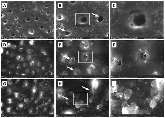
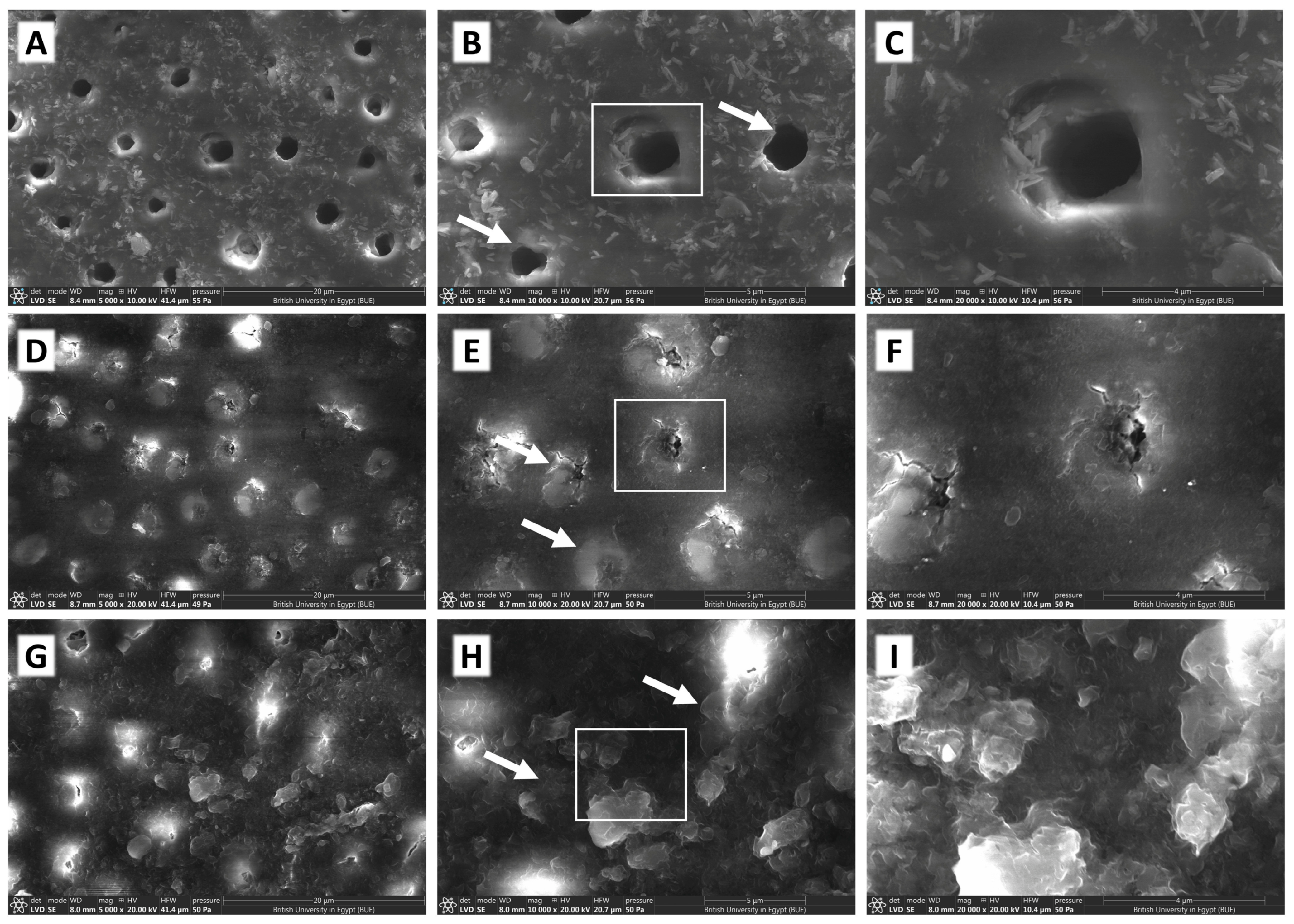
SEM analysis revealed distinct differences in dentinal tubule morphology and surface deposits among the three groups. In the control group, dentinal tubules appeared open with circular or oval profiles and minimal surface deposits, consistent with untreated mechanically exposed dentin after EDTA conditioning (Figure 1A–C). In the nHAp-treated group, partial tubule occlusion was observed at all magnifications, characterized by scattered mineral deposits within and around tubule openings; complete sealing was not evident, and some tubule lumina remained partially patent (Figure 1D–F). The SDF-treated group demonstrated the most pronounced surface changes, with extensive tubule occlusion by dense, heterogeneous precipitates covering the majority of the dentin surface; most tubule openings appeared completely or substantially sealed, with a surface texture markedly different from the control (Figure 1G–I).
Figure 1.
Scanning electron microscopy (SEM) micrographs of dentin surfaces at 5000× (left column), 10,000× (center column), and 20,000× (right column) magnification. (A–C) Control group: open dentinal tubules with minimal surface deposits, representing untreated mechanically exposed dentin following EDTA conditioning. (D–F) nHAp group: partial tubule occlusion with scattered mineral deposits within and around tubule openings; some tubule lumina remain partially patent. (G–I) SDF group: extensive tubule occlusion by dense, heterogeneous mineral-containing precipitates (silver and fluoride were not quantified by EDX); most tubule openings appear completely or substantially sealed. In the center-column panels (B,E,H), white arrows indicate the peritubular dentin and white squares delineate a representative dentinal tubule, shown at higher magnification in the corresponding right-column panel (B→C, E→F, H→I). These regions illustrate the group-specific changes in both compartments: an unobstructed tubule lumen with a distinct peritubular cuff in the control; a partially occluded lumen with mineral deposits extending over the peritubular dentin in the nHAp group; and an obliterated tubule orifice with confluent precipitate obscuring the peritubular margin in the SDF group.Scale bars are indicated on individual micrographs.
3.2. Elemental Composition (Ca, P, and Ca/P Ratio)
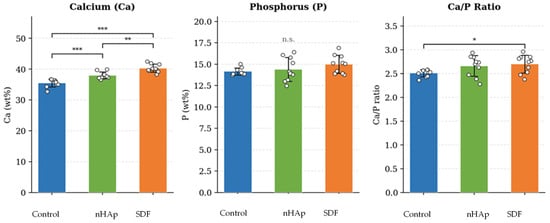
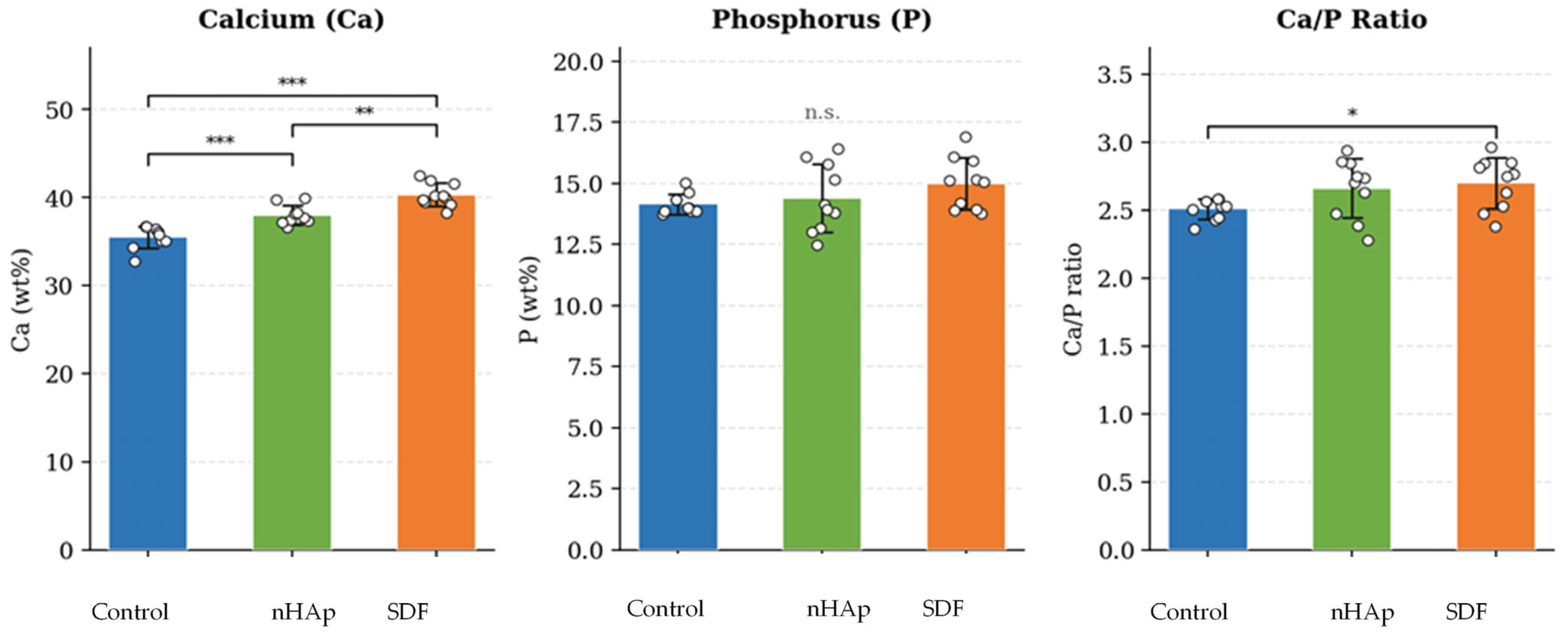
Calcium (Ca) content differed significantly across groups (F(2, 27) = 39.12, p < 0.001, partial η2 = 0.743). The SDF group recorded the highest mean Ca (40.31 ± 1.32 wt%; 95% CI: 39.36–41.26), followed by the nHAp group (38.00 ± 1.07 wt%; 95% CI: 37.24–38.77), with the Control group showing the lowest values (35.50 ± 1.25 wt%; 95% CI: 34.61–36.39). Bonferroni-corrected post hoc comparisons confirmed significant pairwise differences between all three groups (Control vs. nHAp: p < 0.001; Control vs. SDF: p < 0.001; nHAp vs. SDF: p = 0.001).
Phosphorus (P) content showed no significant between-group differences (F(2, 27) = 1.74, p = 0.195, partial η2 = 0.114), with means of 14.16 ± 0.42 wt% (Control), 14.39 ± 1.39 wt% (nHAp), and 15.00 ± 1.07 wt% (SDF). Notably, the Control group exhibited a markedly narrower distribution of P values (SD = 0.42) compared to the treatment groups (SD = 1.07–1.39), suggesting greater inter-specimen variability following treatment.
The Ca/P weight ratio differed significantly across groups (F(2, 27) = 3.45, p = 0.046, partial η2 = 0.203). Means were 2.508 ± 0.074 (Control), 2.659 ± 0.217 (nHAp), and 2.699 ± 0.188 (SDF). Post hoc testing with Bonferroni correction identified a significant difference between the Control and SDF groups (pa = 0.024), while Control vs. nHAp and nHAp vs. SDF comparisons were non-significant. It should be noted that silver (Ag) and fluoride (F) were not quantitatively assessed by EDX in the present study; their inclusion in future analyses would provide additional insight into the nature of SDF-derived deposits. These findings are summarised in Table 1, Table 2 and Table 3 and illustrated in Figure 2.
Table 1.
Descriptive statistics for elemental composition, dentinal tubule (DT) area, and microhardness by experimental group (n = 10 per group).
Table 2.
One-way ANOVA results with Bonferroni-corrected post hoc pairwise comparisons for all outcome variables. n.s. = not significant, * = p < 0.05, *** = p < 0.001.
Table 3.
Within-group before–after changes assessed by paired-samples t-test (df = 9 per group).
Figure 2.
Mean (± SD) elemental composition across experimental groups. Individual specimen values overlaid as dots. Significance brackets reflect Bonferroni-corrected post hoc comparisons (* p < 0.05; ** p ≤ 0.01; *** p ≤ 0.001; n.s. = not significant). Ca/P bracket spans Control–SDF only. Error bars = ±1 SD.
3.3. Dentinal Tubule Surface Area
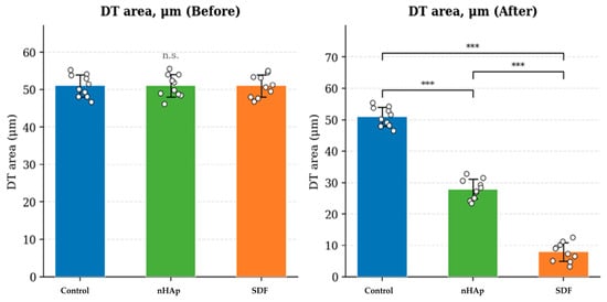
Before treatment, DT area measurements were virtually identical across all groups (Control: 51.03 ± 2.97 μm2; nHAp: 51.04 ± 3.05 μm2; SDF: 51.05 ± 2.98 μm2), with no significant between-group differences [F(2, 27) < 0.001, p = 0.999], confirming adequate baseline comparability.
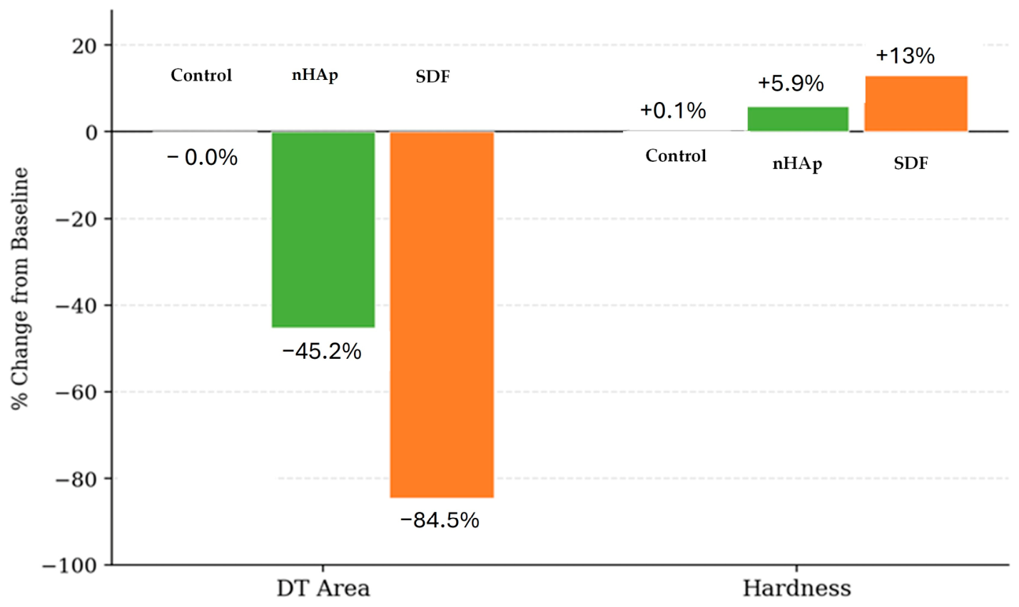
Following treatment, DT area values diverged markedly across groups [F(2, 27) = 493.387, p < 0.001, partial η2 = 0.973]. The control group exhibited no meaningful change (51.02 ± 3.03 μm2; −0.0%; paired t(9) = 0.057, p = 0.956). The nHAp group showed a significant reduction of 45.2% (from 51.04 ± 3.05 to 27.95 ± 3.17 μm2; paired t(9) = 12.11, p < 0.001). The SDF group demonstrated the most pronounced reduction, with an 84.5% decrease in DT area (from 51.05 ± 2.98 to 7.92 ± 3.01 μm2; paired t(9) = 27.34, p < 0.001). Bonferroni post hoc comparisons confirmed significant differences between all group pairs following treatment (all p < 0.001). These findings are summarized in Table 1, Table 2 and Table 3 and Figure 3.
Figure 3.
Mean (± SD) DT area before and after treatment. Groups were statistically equivalent at baseline (n.s.). All post-treatment pairwise differences were significant (*** p < 0.001). Error bars = ±1 SD.
3.4. Dentin Microhardness
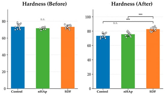
Baseline microhardness values were comparable across groups (Control: 73.60 ± 3.22 HV; nHAp: 71.70 ± 1.38 HV; SDF: 73.35 ± 2.17 HV), with no significant between-group differences [F(2, 27) = 1.888, p = 0.171].
Post-treatment hardness differed significantly among groups [F(2, 27) = 23.461, p < 0.001, partial η2 = 0.635]. The control group showed no significant change (73.70 ± 3.25 HV; +0.1%; paired t(9) = −0.42, p = 0.685). The nHAp group demonstrated a statistically significant within-group increase of 5.9% (from 71.70 ± 1.38 to 75.91 ± 3.16 HV; paired t(9) = −5.50, p < 0.001); however, post-treatment hardness in the nHAp group did not differ significantly from the control group on between-group analysis (Bonferroni: p = 0.421). The SDF group exhibited the greatest improvement, with hardness increasing by 13.0% (from 73.35 ± 2.17 to 82.89 ± 2.97 HV; paired t(9) = −7.34, p < 0.001). Bonferroni post hoc testing revealed significant differences between the control and SDF groups (p < 0.001) and between the nHAp and SDF groups (p < 0.001). These findings are summarized in Table 1, Table 2 and Table 3 and Figure 4 and Figure 5.
Figure 4.
Mean (± SD) dentin microhardness before and after treatment. Post-treatment: *** indicates p < 0.001 vs. SDF; n.s. = Control vs. nHAp. Error bars = ±1 SD.
Figure 5.
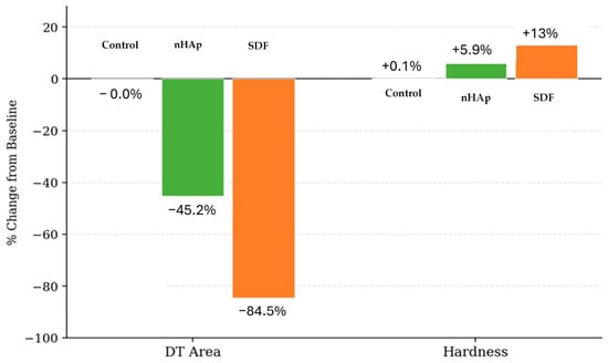
Percentage change from baseline in DT area and microhardness. Negative values indicate reduction; positive values indicate improvement in hardness. nHAp = nano-hydroxyapatite; SDF = silver diamine fluoride.
4. Discussion
The present study evaluated the effects of SDF and nHAp on mechanically exposed dentin, focusing on tubule occlusion, elemental composition, and microhardness. SDF demonstrated superior dentinal tubule occlusion and greater microhardness improvement, whereas nHAp produced moderate effects with a more favorable aesthetic outcome. These findings are discussed in the context of the existing literature and their clinical implications.
The significant increase in surface microhardness observed in the SDF group aligns with earlier research demonstrating that SDF improves dentin mechanical properties by modifying both mineral and organic matrices. This effect can be attributed to the formation of calcium fluoride (CaF2) and silver phosphate (Ag3PO4) precipitates, which contribute to the synthesis of fluorapatite (Ca10(PO4)6F2), a mineral highly resistant to degradation, thereby reinforcing the dentin surface [26]. Additionally, SDF plays a crucial role in protecting collagen within demineralized dentin, inhibiting matrix metalloproteinases (MMPs) and reducing the activity of cysteine cathepsins, enzymes involved in collagen degradation, thereby preserving the collagen matrix and enhancing structural integrity [6,8]. This dual mechanism of mineral reinforcement and collagen protection explains the superior mechanical performance of SDF compared with nHAp.
The nHAp group showed a statistically significant within-group increase in microhardness (+5.9%; p < 0.001); however, this improvement did not reach significance on between-group comparison with the control (Bonferroni: p = 0.421). This distinction is clinically important: while nHAp produced a measurable within-specimen change, the magnitude was insufficient to produce a group-level difference detectable against the natural variability of the control. The moderate effect is consistent with nHAp’s biomimetic mode of action, whereby nano-particles attract calcium and phosphate ions and promote gradual crystal growth on the dentin surface [12]. This remineralization is primarily surface-driven and may not penetrate deeply into the dentin substrate, and repeated application over an extended period may be required to achieve clinically meaningful hardness gains. The 7-day treatment window employed in the current study may represent an insufficient duration to fully demonstrate nHAp’s hardness improvement potential.
SEM analysis corroborated these findings. SDF-treated specimens exhibited extensive tubule occlusion by dense, heterogeneous precipitates, consistent with the silver-containing deposits described in earlier SEM-based studies of silver diamine fluoride; silver was not quantified by EDX here, so this correspondence rests on the published literature rather than on elemental data from the present study [9]. Such occlusion reduces dentin permeability and is directly relevant to the management of dentin hypersensitivity. The nHAp group showed partial tubule occlusion with discrete mineral deposits, in agreement with published studies demonstrating that nHAp forms apatite crystals that contribute to tubule sealing but achieve less uniform and complete occlusion than SDF [12]. The quantitative DT area data confirmed this gradient: SDF reduced tubule area by 84.5%, compared with 45.2% for nHAp, with both significantly exceeding the control (p < 0.001 for all comparisons).
The partial tubule occlusion observed after nHAp toothpaste application is also consistent with previous reports showing that nHAp particles can deposit on dentin surfaces and within dentinal tubules, promoting biomimetic mineral deposition and reducing tubule patency [27,28]. However, the smaller effect observed in the present study may be related to the short application period and to the structural characteristics of mechanically exposed dentin.
The superior dentinal tubule occlusion observed in the silver diamine fluoride group agrees with scanning electron microscopy studies demonstrating dense mineral-containing precipitates after silver diamine fluoride application; because silver and fluoride were not quantified by energy-dispersive X-ray spectroscopy in the present study, the silver-containing nature of these deposits is inferred from the established chemistry of silver diamine fluoride reported in earlier work [29]. In contrast, nHAp tends to produce a more gradual and surface-driven remineralization pattern, which may explain the moderate occlusion observed in the present study.
The EDX analysis revealed significantly higher calcium content in the SDF group relative to the nHAp and control groups, reflecting surface mineral deposition and chemical precipitation reactions involving silver, fluoride, calcium, and phosphate ions [7]. Phosphorus content, however, did not differ significantly among groups, suggesting that the treatment-induced surface changes were driven mainly by changes in calcium-rich deposits rather than proportional increases in phosphorus. The Ca/P ratio showed a small but statistically significant overall difference among groups, with post hoc analysis indicating that this difference was limited to the Control versus SDF comparison. Therefore, the elevated Ca/P ratio in the SDF group should be interpreted cautiously as evidence of altered surface chemistry and calcium-dominant precipitation rather than definitive hydroxyapatite-like remineralization [8].
A clinically relevant observation of the present study is the trade-off between therapeutic efficacy and aesthetic acceptability. Although SDF demonstrated superior improvements in microhardness and tubule occlusion, the characteristic dark discoloration caused by the oxidation of silver ions and the formation of metallic silver and silver oxide deposits is a well-recognized limitation that may reduce patient acceptance, particularly in aesthetically sensitive regions [30,31,32]. In contrast, nHAp’s white, biomimetic coloration, which improves surface smoothness and light reflection, represents a significant aesthetic advantage [13,33]. Although nHAp may provide less immediate structural reinforcement than SDF, its superior aesthetic profile may potentially make it a more acceptable option in visible areas and for patients with aesthetic priorities; because colour change and clinical outcomes were not evaluated here, this preventive role remains a potential implication requiring confirmation under clinically representative conditions.
The present findings also illustrate the importance of considering the unique characteristics of the dentin substrate when evaluating remineralizing agents. Although the current study employed a mechanically exposed dentin model rather than naturally attrited dentin, the use of EDTA-conditioned ground dentin was intended to approximate the open-tubule surface condition encountered clinically. Naturally attrited dentin undergoes chronic mechanical stress resulting in tubular sclerosis, altered mineral composition, and smear layer formation, all of which may influence ion diffusion, crystal deposition, and material penetration [14,15]. The relatively greater effectiveness of SDF in the current study may reflect its high chemical reactivity and capacity to form dense precipitates even on morphologically altered surfaces. Conversely, nHAp’s performance may have been limited by reduced ion diffusion through sclerotic tubules and the inherent dependence of biomimetic remineralization on gradual crystal growth. Outcomes observed in caries or demineralization models may therefore not translate directly to mechanically worn or attrited dentin surfaces, and the current study contributes to filling this evidence gap by evaluating both agents under standardized mechanically exposed conditions.
The findings support the growing shift toward preventive and regenerative approaches in restorative dentistry [34,35,36]. Because permeability, aging, abrasion resistance, colour change, and clinical hypersensitivity were not assessed in this in vitro study, the following clinical roles are presented as potential implications only and require clinical verification. On this basis, SDF may potentially be useful where rapid symptom control and immediate reinforcement of structurally compromised dentin are priorities, whereas nHAp may potentially be more suitable as a longer-term maintenance or adjunctive preventive option. The possibility of combining these materials or sequentially applying them may represent an important future direction: integrating biomimetic calcium-phosphate systems with fluoride-based agents may optimize remineralization while mitigating staining and enhancing biocompatibility.
Although attrited dentin represents the clinical motivation for this investigation, the present study was conducted using a standardized mechanically exposed dentin model. Consequently, the findings should be interpreted as short-term surface responses under controlled laboratory conditions rather than as direct evidence of material performance on naturally attrited or sclerotic dentin, which exhibits different tubular morphology and mineral characteristics.
This study has several limitations. First, it used a short-term in vitro model of ethylenediaminetetraacetic acid-conditioned mechanically exposed dentin, which does not fully reproduce naturally attrited or sclerotic dentin. Second, ethylenediaminetetraacetic acid conditioning removed the smear layer and increased tubule openness, which may have influenced material penetration, mineral deposition, and apparent tubule occlusion. Third, the tested agents were applied according to different clinically relevant protocols rather than equivalent application frequency or equivalent material exposure. Fourth, energy-dispersive X-ray spectroscopy analysis was limited to calcium and phosphorus; silver and fluoride were not quantitatively assessed. Finally, the study did not include artificial saliva, pH cycling, thermal aging, brushing abrasion after treatment, dentin permeability testing, quantitative color assessment, or clinical hypersensitivity outcomes. Further studies using clinically relevant attrited dentin and more dynamic oral simulation models are needed.
The present findings should be interpreted as short-term in vitro surface effects rather than direct evidence of clinical efficacy. Future studies should assess the durability, permeability reduction, color changes, and clinical hypersensitivity outcomes of these materials under more clinically representative conditions.
5. Conclusions
Within the limitations of this short-term in vitro model using ethylenediaminetetraacetic acid-conditioned mechanically exposed dentin, 38% silver diamine fluoride produced greater dentinal tubule occlusion and higher post-treatment microhardness than nano-hydroxyapatite toothpaste. Nano-hydroxyapatite toothpaste produced moderate tubule occlusion and a smaller improvement in microhardness under the tested conditions. These findings support material- and protocol-dependent surface effects, but further studies using clinically relevant attrited dentin, artificial saliva, pH cycling, aging protocols, permeability testing, quantitative color assessment, and clinical outcomes are needed before firm clinical recommendations can be made.
Author Contributions
Conceptualization, N.E. and M.S.; methodology, N.E., N.T., M.A.A. and M.S.; software, N.E., M.S. and M.A.A.; investigation, M.M.B., M.A.A., N.T. and M.S.; resources, M.M.B. and M.S.; data curation, N.E. and N.T.; validation, M.M.B. and M.A.A.; visualization, M.A.A. and M.S.; project administration, M.M.B. and M.A.A.; writing—original draft preparation, N.E., M.M.B. and M.S.; writing—review and editing, N.E., M.M.B., N.T., M.A.A. and M.S.; supervision, M.M.B. and M.S. All authors have read and agreed to the published version of the manuscript.
Funding
This research received no external funding.
Institutional Review Board Statement
This study was conducted in accordance with the Declaration of Helsinki and approved by the Research and Ethics Committee of the Faculty of Dentistry, The British University in Egypt (Project No. FD BUE REC 25-061; 30 October 2025).
Informed Consent Statement
Informed consent was obtained from all participants prior to tooth extraction. All teeth were anonymized prior to laboratory processing in accordance with institutional guidelines.
Data Availability Statement
All original contributions supporting this study are included in the article. Further inquiries may be directed to the corresponding author.
Conflicts of Interest
The authors declare no conflicts of interest.
Abbreviations
The following abbreviations are used in this manuscript:
| SEM | Scanning electron microscopy |
| EDX | Energy-dispersive X-ray spectroscopy |
| EDTA | Ethylenediaminetetraacetic acid |
| FEG-ESEM | Field Emission Gun Environmental Scanning Electron Microscope |
| Ca | Calcium |
| P | Phosphorus |
| Ca/P | Calcium-to-phosphorus ratio |
| wt% | Weight percentage |
| HV | Vickers hardness value |
| ANOVA | Analysis of variance |
| SPSS | Statistical Package for the Social Sciences |
| MMPs | Matrix metalloproteinases |
References
- Rajapaksa, R.D.W.; Wang, Y.-C.; Chin, Y.C.; Jang, K.; Abdal-hay, A.; Ivanovski, S.; Feroz, S. Dental Erosion Management: From Remineralization to Emerging Regenerative Approaches—A Narrative Review. Biomimetics 2026, 11, 107. [Google Scholar] [CrossRef] [Scilit] [PubMed]
- Paqué, F.; Luder, H.; Sener, B.; Zehnder, M. Tubular sclerosis rather than the smear layer impedes dye penetration into the dentine of endodontically instrumented root canals. Int. Endod. J. 2006, 39, 18–25. [Google Scholar] [CrossRef] [Scilit] [PubMed]
- Martín, N.; García, Á.; Vera, V.; Garrido, M.A.; Rodríguez, J. Mechanical characterization of sclerotic occlusal dentin by nanoindentation and nanoscratch. Am. J. Dent. 2010, 23, 108–112. [Google Scholar] [PubMed]
- Kelleher, M.G.; Bomfim, D.I.; Austin, R.S. Biologically based restorative management of tooth wear. Int. J. Dent. 2012, 2012, 742509. [Google Scholar] [CrossRef] [Scilit] [PubMed]
- Mei, M.L.; Lo, E.; Chu, C.-H. Clinical use of silver diamine fluoride in dental treatment. Compend. Contin. Educ. Dent. 2016, 37, 93–98. [Google Scholar] [PubMed]
- Horst, J.A.; Ellenikiotis, H.; Milgrom, P.L. UCSF protocol for caries arrest using silver diamine fluoride: Rationale, indications and consent. J. Calif. Dent. Assoc. 2016, 44, 17–28. [Google Scholar] [CrossRef] [Scilit]
- Knight, G.; McIntyre, J.; Craig, G.; Mulyani; Zilm, P.; Gully, N. An in vitro model to measure the effect of a silver fluoride and potassium iodide treatment on the permeability of demineralized dentine to Streptococcus mutans. Aust. Dent. J. 2005, 50, 242–245. [Google Scholar] [CrossRef] [Scilit] [PubMed]
- Zhao, I.S.; Gao, S.S.; Hiraishi, N.; Burrow, M.F.; Duangthip, D.; Mei, M.L.; Lo, E.C.-M.; Chu, C.-H. Mechanisms of silver diamine fluoride on arresting caries: A literature review. Int. Dent. J. 2018, 68, 67–76. [Google Scholar] [CrossRef] [Scilit] [PubMed]
- Nascimento, M. Potential uses of arginine in dentistry. Adv. Dent. Res. 2018, 29, 98–103. [Google Scholar] [CrossRef] [Scilit] [PubMed]
- Llodra, J.; Rodriguez, A.; Ferrer, B.; Menardia, V.; Ramos, T.; Morato, M. Efficacy of silver diamine fluoride for caries reduction in primary teeth and first permanent molars of schoolchildren: 36-month clinical trial. J. Dent. Res. 2005, 84, 721–724. [Google Scholar] [CrossRef] [Scilit] [PubMed]
- Vasquez, E.; Zegarra, G.; Chirinos, E.; Castillo, J.L.; Taves, D.R.; Watson, G.E.; Dills, R.; Mancl, L.L.; Milgrom, P. Short term serum pharmacokinetics of diammine silver fluoride after oral application. BMC Oral Health 2012, 12, 60. [Google Scholar] [CrossRef] [Scilit] [PubMed]
- Oliveira, S.; Slot, D.; Van der Weijden, G. What is the cause of palate lesions? A case report. Int. J. Dent. Hyg. 2013, 11, 306–309. [Google Scholar] [CrossRef] [Scilit] [PubMed]
- Limeback, H.; Meyer, F.; Enax, J. Tooth whitening with hydroxyapatite: A systematic review. Dent. J. 2023, 11, 50. [Google Scholar] [CrossRef] [Scilit] [PubMed]
- Nudel, I.; Pokhojaev, A.; Bitterman, Y.; Shpack, N.; Fiorenza, L.; Benazzi, S.; Sarig, R. Secondary dentin formation mechanism: The effect of attrition. Int. J. Environ. Res. Public Health 2021, 18, 9961. [Google Scholar] [CrossRef] [Scilit] [PubMed]
- Mokeem, L.S.; Garcia, I.M.; Melo, M.A. Degradation and failure phenomena at the dentin bonding interface. Biomedicines 2023, 11, 1256. [Google Scholar] [CrossRef] [Scilit] [PubMed]
- Kim, S.; Larnani, S.; Son, J.E.; Lappanakokiat, N.; Truong, V.M.; Jin, B.-H.; Park, Y.-S. Impact of demineralization, brushing, and remineralization sequences on dentin mineral retention and surface properties. Odontology 2025, 113, 1456–1466. [Google Scholar] [CrossRef] [Scilit] [PubMed]
- Zafar, M.S.; Amin, F.; Fareed, M.A.; Ghabbani, H.; Riaz, S.; Khurshid, Z.; Kumar, N. Biomimetic aspects of restorative dentistry biomaterials. Biomimetics 2020, 5, 34. [Google Scholar] [CrossRef] [Scilit] [PubMed]
- Singer, L.; Fouda, A.; Bourauel, C. Biomimetic approaches and materials in restorative and regenerative dentistry. BMC Oral Health 2023, 23, 105. [Google Scholar] [CrossRef] [Scilit] [PubMed]
- Alluhaidan, T.; Hung, B.; Qaw, M.; Garcia, I.M.; Melo, M.A.S. Biomimetic Design of Dental Restorative Materials: Conceptual Framework and Translational Challenges. Biomimetics 2026, 11, 256. [Google Scholar] [CrossRef] [Scilit] [PubMed]
- Shamel, M.; Al-Ankily, M.M.; Bakr, M.M. Influence of different types of whitening tooth pastes on the tooth color, enamel surface roughness and enamel morphology of human teeth. F1000Research 2019, 8, 1764. [Google Scholar] [CrossRef] [Scilit] [PubMed]
- Shamel, M.; El Banna, S. In vitro comparative SEM analysis of nano-hydroxyapatite toothpaste and diode laser: Evaluation of efficacy of dentinal tubular occlusion. J. Stomatol. 2022, 75, 1–6. [Google Scholar] [CrossRef] [Scilit]
- Romão, D.A.; Fernández, C.E.; de Melo Santos, L. Commercial silver diamine fluoride (SDF) products on caries lesion progression in primary enamel: An in vitro study. Oral Health Prev. Dent. 2020, 18, b871057. [Google Scholar] [CrossRef] [Scilit] [PubMed]
- Al Ankily, M.; Baz, S.; Mahmoud, H.; Bakr, M.M.; Shamel, M. Bovine teeth as substitutes for human teeth in dental research: Ultrastructural and radiographic analysis. Eur. J. Dent. 2025, 20, 457–465. [Google Scholar] [CrossRef] [Scilit] [PubMed]
- Al Ankily, M.; Makkeyah, F.; Bakr, M.M.; Shamel, M. Evaluating the effects of cigarette smoking and heated tobacco products on hard dental tissues: A comparative histological and colorimetric analysis. Clin. Exp. Dent. Res. 2024, 10, e941. [Google Scholar] [CrossRef] [Scilit] [PubMed]
- El-Tobgy, R.H.; Halawa, A.M.; Amin, R.M.; El-Messiry, H. The Effect of Black Charcoal Versus Calcium Silicate and Sodium Phosphate in Occluding Dentinal Tubules and Its Resistance to Acidic Soft Drinks: An In Vitro Study. J. Int. Oral Health 2024, 16, 236–245. [Google Scholar] [CrossRef] [Scilit] [PubMed]
- Mei, M.L.; Nudelman, F.; Marzec, B.; Walker, J.M.; Lo, E.C.M.; Walls, A.W.; Chu, C.H. Formation of Fluorohydroxyapatite with Silver Diamine Fluoride. J. Dent. Res. 2017, 96, 1122–1128. [Google Scholar] [CrossRef] [Scilit] [PubMed]
- Gupta, I.; Chauhan, S.; Amaranath, B.J.J.; Das, N.; Johnson, L.; Mehrotra, V. Effect of Commercially Available Nano-Hydroxyapatite Containing Desensitizing Toothpaste and Mouthwash on Dentinal Tubular Occlusion: A SEM Analysis. J. Pharm. Bioallied Sci. 2023, 15, S1027–S1029. [Google Scholar] [CrossRef] [Scilit] [PubMed]
- Bologa, E.; Stoleriu, S.; Iovan, G.; Ghiorghe, C.A.; Nica, I.; Andrian, S.; Amza, O.E. Effects of Dentifrices Containing Nanohydroxyapatite on Dentinal Tubule Occlusion—A Scanning Electron Microscopy and EDX Study. Appl. Sci. 2020, 10, 6513. [Google Scholar] [CrossRef] [Scilit]
- Kiesow, A.; Menzel, M.; Lippert, F.; Tanzer, J.M.; Milgrom, P. Dentin tubule occlusion by a 38% silver diamine fluoride gel: An in vitro investigation. BDJ Open 2022, 13, 1. [Google Scholar] [CrossRef] [Scilit] [PubMed]
- Magno, M.B.; Silva, L.P.d.; Ferreira, D.M.; Barja-Fidalgo, F.; Fonseca-Gonçalves, A. Aesthetic perception, acceptability and satisfaction in the treatment of caries lesions with silver diamine fluoride: A scoping review. Int. J. Paediatr. Dent. 2019, 29, 257–266. [Google Scholar] [CrossRef] [Scilit] [PubMed]
- Cappiello, M.C.; Durán, A.V.; Crystal, Y.O.; Bagattoni, S.; Jimeno, F.G. Comparison of aesthetic perception and acceptability of silver diamine fluoride staining between Spanish and Italian parents. Sci. Rep. 2025, 15, 3442. [Google Scholar] [CrossRef] [Scilit] [PubMed]
- AlSheikh, R.N. The use of silver diamine fluoride to prevent/treat enamel carious lesions: A narrative review. PeerJ 2024, 12, e17897. [Google Scholar] [CrossRef] [Scilit] [PubMed]
- Abedi, M.; Ghasemi, Y.; Nemati, M.M. Nanotechnology in toothpaste: Fundamentals, trends, and safety. Heliyon 2024, 10, e24949. [Google Scholar] [CrossRef] [Scilit] [PubMed]
- Thalakiriyawa, D.S.; Dissanayaka, W.L. Advances in regenerative dentistry approaches: An update. Int. Dent. J. 2024, 74, 25–34. [Google Scholar] [CrossRef] [Scilit] [PubMed]
- Umapathy, V.R.; Natarajan, P.M.; Swamikannu, B. Regenerative strategies in dentistry: Harnessing stem cells, biomaterials and bioactive materials for tissue repair. Biomolecules 2025, 15, 546. [Google Scholar] [CrossRef] [Scilit] [PubMed]
- Tattar, R. Regenerative approaches in dentistry: An evidence-based perspective. Evid. Based Dent. 2025, Epub ahead of print. [Google Scholar] [CrossRef] [Scilit] [PubMed]

Disclaimer/Publisher’s Note: The statements, opinions and data contained in all publications are solely those of the individual author(s) and contributor(s) and not of MDPI and/or the editor(s). MDPI and/or the editor(s) disclaim responsibility for any injury to people or property resulting from any ideas, methods, instructions or products referred to in the content. |
© 2026 by the authors. Licensee MDPI, Basel, Switzerland. This article is an open access article distributed under the terms and conditions of the Creative Commons Attribution (CC BY) license.