


Abstract
Prolidase deficiency is an ultra-rare autosomal recessive metabolic disorder caused by pathogenic variants in the PEPD gene, resulting in impaired turnover of proline-rich proteins, including collagen. It is characterized by chronic skin lesions, recurrent infections, hepatosplenomegaly, and immune dysregulation. Hematologic manifestations remain poorly defined so far. An 18-year-old male was referred for unexplained hyperferritinemia, splenomegaly, thrombocytopenia, and intermittent mild anemia. Since childhood, he had had recurrent respiratory and skin infections, chronic lower-limb ulcers, cutaneous thickening, and multiple hospitalizations without definitive diagnosis. Examination revealed dysmorphic facial features, telangiectasias, hidradenitis-like lesions, severe splenomegaly, mild hepatomegaly, and chronic skin ulcers. Laboratory investigations showed hyperferritinemia with normal transferrin saturation, thrombocytopenia, polyclonal hypergammaglobulinemia, and mild proteinuria. Magnetic resonance imaging excluded iron overload. Infectious and immunological investigations were inconclusive. Plasma amino acid analysis demonstrated markedly reduced hydroxyproline levels. Prolidase deficiency was confirmed by severely reduced erythrocyte prolidase activity due to a homozygous pathogenic PEPD variant (c.977G>A). During follow-up, an acute episode of immune thrombotic thrombocytopenic purpura (iTTP) with detectable anti-ADAMTS13 autoantibodies was observed. To the best of our knowledge this is the first reported case of iTTP associated with prolidase deficiency. Prolidase deficiency-related immune dysregulation may contribute to the development of anti-ADAMTS13 autoantibodies, while the proline-rich structure of ADAMTS13 invites further speculation on potential mechanistic links with iTTP. This case expands the spectrum of hematologic manifestations of prolidase deficiency by suggesting possible association with iTTP. Recognition of multisystemic clinical features may shorten the diagnostic delay and improve therapeutic management of these ultra-rare patients.
1. Introduction
Prolidase deficiency is an ultra-rare autosomal recessive disorder caused by pathogenic variants in the PEPD gene, resulting in impaired turnover of proline- and hydroxyproline-rich proteins, such as collagen [1]. Fewer than 200 cases are described in the literature [2]. Among the possible clinical manifestations, skin lesions, dysmorphic features, and neurological symptoms, including developmental delay and intellectual disability, are the most frequent, followed by recurrent infections and musculoskeletal anomalies. Among hematological abnormalities, splenomegaly is the most prevalent, with anemia and thrombocytopenia being observed in a considerable proportion of subjects. Additionally, approximately one fifth of patients are affected by autoimmune disorders including systemic lupus erythematosus [3]. Due to its highly heterogeneous and multisystemic clinical presentation, the diagnosis is often substantially delayed, and patients may undergo years of inconclusive investigations before the disease is recognized. In a patient with typical clinical features and elevated iminodipeptiduria, the diagnosis is confirmed by demonstrating reduced enzymatic activity (in erythrocytes, leukocytes, or fibroblasts) or by the molecular analysis of the PEPD gene [2]. A disease-modifying therapy is still lacking.
To the best of our knowledge, an association between prolidase deficiency and thrombotic microangiopathies has not been previously reported.
We report the case of a young man referred for evaluation of unexplained hyperferritinemia, in whom a complex syndromic phenotype led to the diagnosis of prolidase deficiency, later complicated by immune thrombotic thrombocytopenic purpura (iTTP).
2. Case Presentation
An 18-year-old Tunisian boy was referred to our EuroBloodNet tertiary center for Iron Disorders at the University Hospital of Verona because of an unexplained hyperferritinemia.
Since early childhood, the patient has undergone multiple hospitalizations and extensive investigations. Splenomegaly, thrombocytopenia, anemia, and hyperferritinemia were first detected during a hospitalization in a pediatric clinic when he was two. At that time, peripheral blood smear and bone marrow analysis were not diagnostic. In the following years, the patient suffered from recurrent upper respiratory tract infections with severe otitis (perforation of the eardrum in 2008 and subsequent tympanoplasty) and sinusitis. He also developed bilateral bronchopneumonia, which required hospitalization. Remarkably, he had recurrent hospital admissions for infected skin ulcers, especially of the lower limbs. Because of an elephantiasis-like scrotal swelling, he underwent bilateral hydrocele drainage in the first years of life without benefit. Skin thickening, initially interpreted as edema, involving the cheeks, lips, and hands, had also been noted since childhood. He underwent several investigations to exclude an atopic disorder or a hereditary angioedema, including the C1-INH assay, which was normal. Cycles of steroids and antihistaminic agents were ineffective. For eyelid redness, he had regular ophthalmologic examinations with recurrent findings of corneal opacity attributed to dry eye, which were treated with lubricant eye drops. He had normal growth and development for age and no bone or vertebral column disorder. He had psychological frailty but did not show significant intellectual disability; nonetheless, he refused a comprehensive behavioral and psychiatric assessment.
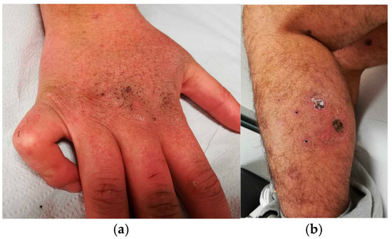
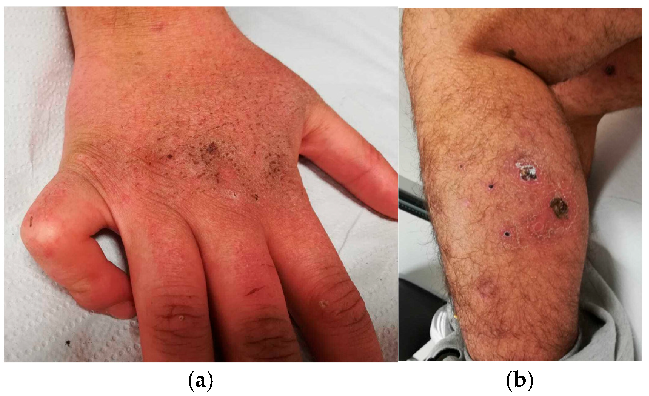
At our evaluation, we detected multiple dysmorphic features (hypertelorism, depressed nasal bridge, thin vermilion of the upper lip, camptodactyly), several telangiectasias, scrotal and skin thickening, axillary suppurative adenitis, and skin ulcers of the legs (Figure 1).
Figure 1.
Cutaneous manifestations: skin thickening (a) and leg ulcers (b).
Physical examination revealed marked splenomegaly, with the spleen palpable nearly to the left iliac fossa, mild hepatomegaly, and overweight (80 kg, 166 cm; BMI 29 kg/m2). Laboratory investigations revealed Hb 14.3 g/dL, MCV 78.1 fl, RBC 5,200,000/mmc, PLTs 130,000/mmc, polyclonal hypergammaglobulinemia (lgG 21 g/L, lgA 8 g/L), non-selective mild proteinuria (230 mg/L), and ferritin 1037 mcg/L with normal transferrin saturation. C-Reactive Protein, liver enzymes, and renal function were within the normal limits. Antinuclear antibodies were 1:160 speckled.
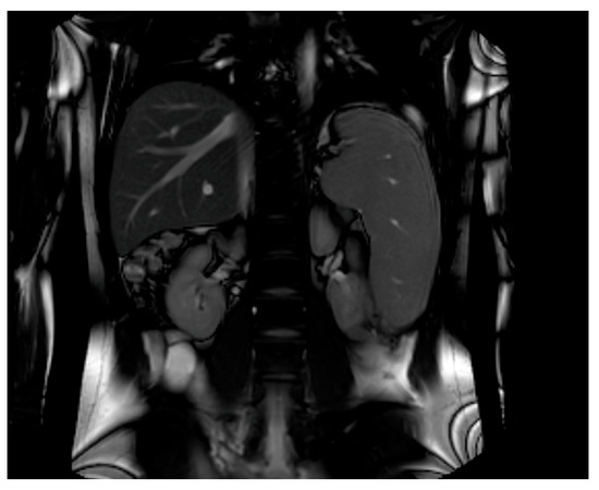
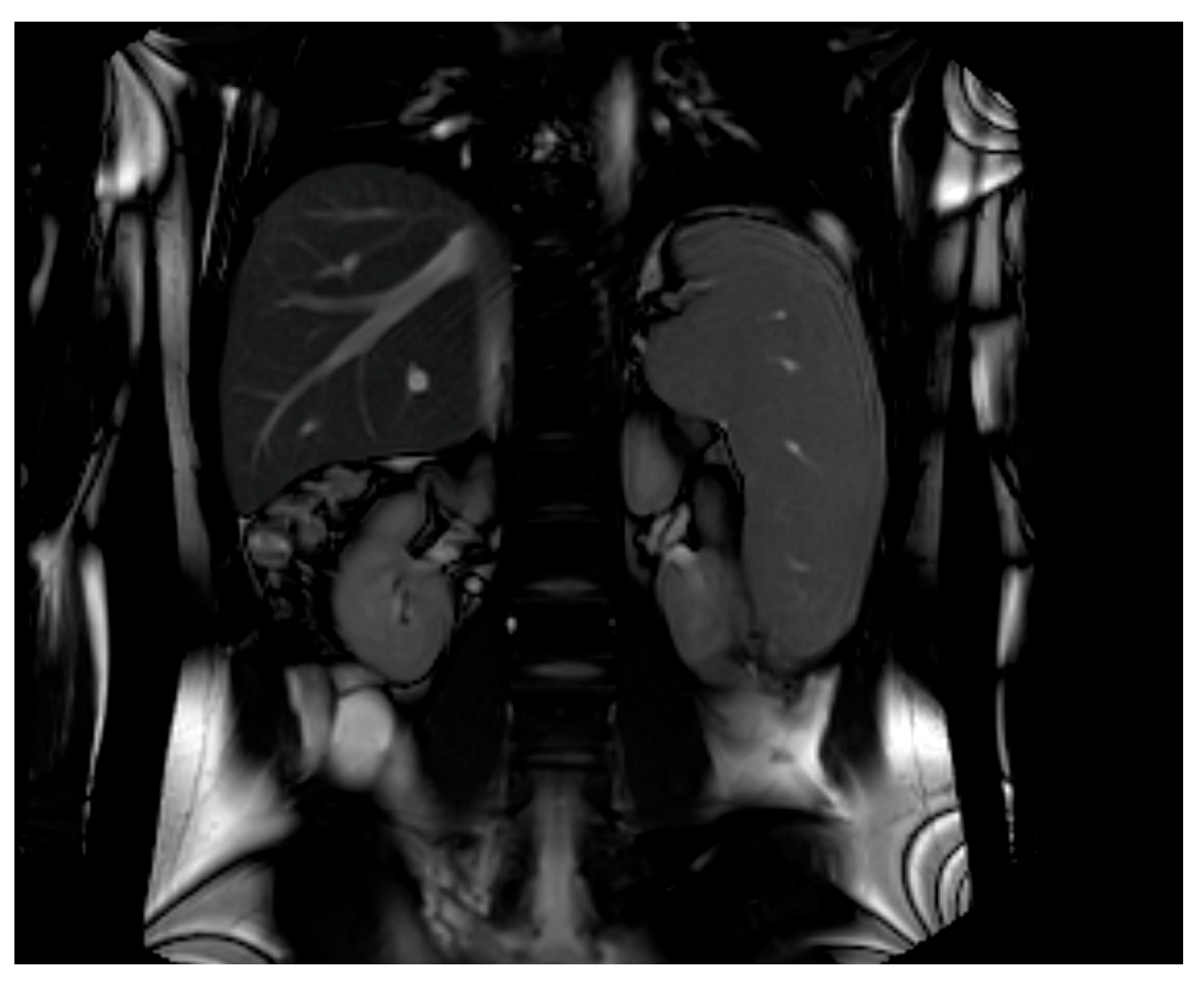
An abdominal magnetic resonance imaging (MRI) was prescribed to investigate the organomegaly. The three-axis calculated volumes were 1069 mL for spleen (Reference Range—RR < 480 mL) and 1476 mL for liver (RR < 1600 mL), with no evidence of nodular lesions (Figure 2); no significant iron accumulation in the liver or spleen was detected, ruling out a disorder of iron metabolism.
Figure 2.
Abdominal MRI shows liver volume at the upper limit of normal, with splenomegaly without nodular lesions.
Considering the syndromic dysmorphic features and the splenomegaly, we suspected an inherited metabolic disorder. Urinary mucopolysaccharide quantification and an enzymatic assay for mucopolysaccharidosis (MPS) type I, ll, Ill, IVa, IVb and VI, Gaucher disease, and acid sphingomyelinase deficiency were performed, all of which were negative. Furthermore, the presence of recurrent ulcerative skin lesions, the skin thickening, and the absence of neurological involvement were atypical for most of the lysosomal storage disorders.
Given the persistent suspicion of a metabolic disorder, we performed a plasma amino acid profile, which showed a marked reduction in hydroxyproline. After a literature review, we found that prolidase deficiency could explain the overall clinical scenario [2,3]. A very low prolidase activity in erythrocytes (2.6% compared to a control sample) confirmed the diagnosis. A genetic test identified a pathogenetic homozygous variant in PEPD gene: c.977G>A. At the familiar screening, the patient’s sister was’ unaffected. Since no specific treatment is available for prolidase deficiency, only supportive strategies to manage ulcers and infections were performed.
After 5 years of follow-up, the patient presented to the Emergency Department (ED) of our ERN center in Verona with asthenia, abdominal pain and vomiting for five days, associated with dark urine, scleral jaundice, and focal neurological signs, including facial paresthesia and asymmetry with transient deviation in the oral commissure. The patient reported neither regular medication uses nor any medication intake in the days preceding admission.
Emergency laboratory investigations revealed severe normocytic normochromic anemia (Hb 6 g/dL; RR 13.5–17.5), reticulocytosis (310,000/mmc; RR 15,000–98,000), profound thrombocytopenia (5000/mmc; RR 150,000–400,000), a normal white blood cell count (WBC 6420/mmc; RR 4500–11,000) and a mild elevation in inflammatory markers (CRP 13 mg/L; RR < 5). Laboratory findings were also consistent with hemolysis, including mixed hyperbilirubinemia (total bilirubin 4.0 mg/dL; RR < 1.05) predominantly due to indirect fraction (direct bilirubin 0.8 mg/dL; RR < 0.35), elevated lactate dehydrogenase (LDH) levels (953 U/L; RR 135–225), and haptoglobin at the lower limit of normal (0.4 g/L; RR 0.3–2.0). On a peripheral blood smear, schistocytosis was detected. The direct antiglobulin test (DAT) was negative, arguing against autoimmune hemolytic anemia. Severe vitamin B12 and folate deficiencies, potentially contributing to the clinical presentation, were also excluded. Mild acute kidney injury (creatinine 1.15 mg/dL compared to previous value 0.6 mg/dL; RR 0.59–1.29 mg/dL) and slight prolongation of coagulation times (PT ratio 1.37; RR 0.8–1.17 and aPTT ratio 0.96; RR 0.8–1.2) were also observed, whereas fibrinogen levels remained within normal limits (2.06 g/L; RR 2.0–4.0). Given the symptoms at presentation, brain and abdominal CT scans were performed in the ED, both showing no evidence of acute ischemic events.
Given the suspicion of TTP (PLASMIC score = 6), further laboratory investigations were performed, showing a severe decrease in ADAMTS13 activity (<1%) due to the presence of anti-ADAMTS13 antibodies above the upper limit of normal and thus defining the diagnosis of iTTP. The patient was treated with plasma exchange (six sessions in total), immunosuppressive-dose glucocorticoids (prednisone 1 mg/kg), and caplacizumab (10 mg/die), together with oral folate and vitamin B12 supplementation. However, caplacizumab was discontinued on the eighth day because of the development of otorrhagia arising from chronic ulcerative auricular lesions. Moreover, rituximab was not administered because of recurrent infections involving the chronic lower-limb ulcers and the associated risk of osteomyelitis. Given the splenomegaly, the severe acute cytopenia and the previous history of anemia and mild thrombocytopenia, a bone marrow biopsy was performed before corticosteroid therapy was initiated. Histological examination showed only mild disruption of the normal marrow architecture, with preserved myeloid and erythroid maturation and no other significant abnormalities. Screening for acute and chronic viral infections potentially contributing to the cytopenia, including EBV, CMV, parvovirus B19, HIV, HBV, and HCV, was also negative.
The platelet count recovered within 10 days of treatment initiation (134,000/mmc; RR 150,000–400,000), accompanied by a progressive normalization of ADAMTS13 activity (56%). Corticosteroid therapy was continued at an immunosuppressive dose for 4 weeks and was subsequently tapered gradually without evidence of disease recurrence. The patient subsequently achieved sustained clinical remission without further iTTP relapses with a persistent mild normochromic-normocytic anemia (Hb 10–11 g/dL) and thrombocytopenia (120–130.000/mmc) during the following two years of follow-up.
3. Discussion
Prolidase deficiency is an ultra-rare autosomal recessive disorder caused by pathogenic variants in the PEPD gene (19q13.11), encoding prolidase, a Xaa-Pro dipeptidase that plays a crucial role in the turnover of proline- and hydroxyproline-rich proteins, such as collagen. The pathophysiology of this inherited disorder is still not fully understood and appears to extend beyond a simple deficiency in proline availability, involving complex alterations in extracellular matrix remodeling, cellular signaling pathways, and immune regulation [4].
Fewer than 200 cases have been reported in the literature, most of them in pediatric patients. The largest dataset on natural history of the disease in 2021 reports 19 newly described patients and 161 patients from a literature systematic review [2]. This study highlights the early onset of clinical manifestations (mean 5.5 years) and significant diagnostic delay (11.7 years). Within the studied cohort, the survival rate was 90% at age 20, 88% at age 30, and 82% at age 40. The diagnosis of prolidase deficiency is particularly challenging because clinical manifestations are heterogeneous, nonspecific and often overlap with autoimmune, infectious, hematological, and lysosomal storage disorders. They include dermatological alterations (e.g., chronic skin ulcers, recurrent ulcer infections, scarring, rash, telangiectasias, and hyperkeratosis), dysmorphic facial features (e.g., hypertelorism, proptosis, and saddle nose), frequent infections, musculoskeletal anomalies, variable degrees of intellectual disability, hepatosplenomegaly, immune dysregulation, ocular and ENT/dental anomalies, and thoracic, gastrointestinal, renal and endocrine alterations. The most frequent manifestations are dermatological alterations (83.1%), dysmorphic features (65.7%), and neurological symptoms (60.7%), particularly developmental delay, intellectual disability, and learning difficulties (58.4%). These are followed by frequent infections (48.3%) and musculoskeletal anomalies (34.3%). Among hematological abnormalities, splenomegaly is the most prevalent (44.9%), followed by anemia (29.8%), thrombocytopenia (18%), and pancytopenia (5.1%). Additionally, 20.2% of patients are positive for autoantibodies, 12.4% have an autoimmune disorder and 5.6% are diagnosed with systemic lupus erythematosus. Hypergammaglobulinemia, hypocomplementemia, and hemophagocytic lymphohistiocytosis (HLH) are also described [5].
In our patient, recurrent infections, chronic skin lesions, dysmorphic features, splenomegaly, thrombocytopenia, and hyperferritinemia prompted extensive investigations before a metabolic disorder was eventually suspected. Furthermore, the combination of chronic ulcerative skin lesions, skin thickening, and the absence of significant neurological involvement was atypical for most lysosomal storage disorders, further complicating the diagnostic workup. Our adult patient reported many of the dermatological and dysmorphic clinical features described in the literature. Regarding hematological manifestations, he presented splenomegaly, mild fluctuant anemia, thrombocytopenia, and polyclonal hypergammaglobulinemia, which have already been described in the literature. To our knowledge, hyperferritinemia and iTTP have not previously been reported in prolidase deficiency case reports and series.
Regarding hyperferritinemia, iron overload was excluded by blood tests (normal transferrin saturation) and MRI. Elevated ferritin levels may reflect chronic inflammation, recurrent infections, and ongoing tissue damage and have a wide differential diagnosis [6]. Inherited metabolic disorders are to be considered in the diagnostic work-up of an apparently unexplained hyperferritinemia [7].
Considering prolidase deficiency and iTTP, a connection between the two disorders appears biologically plausible, since immune dysregulation is recognized as a central feature of prolidase deficiency. Autoimmune manifestations, including systemic lupus erythematosus-like disease, autoantibody production, and complement abnormalities, may contribute to the development of thrombotic microangiopathies [8]. Furthermore, ADAMTS13 is rich in proline residues. It is worth noting that 108 of the 118 total prolines reside in ADAMTS13 active chain [9,10]. Consequently, it appears biologically plausible that altered proline metabolism may affect its structure, stability, function, or antigenicity, thereby increasing the susceptibility to TTP in these patients.
The prognosis of prolidase deficiency is highly variable, although patients frequently experience a reduced quality of life and shortened life expectancy because of recurrent and sometimes life-threatening infections. No disease-modifying therapy is currently available, and management remains largely supportive. Treatments reported in the literature, like skin grafting, antibiotics, proline or glycine and proline ointments, immunosuppressive agents, blood transfusions, plasmapheresis, hyperbaric oxygen therapy or hematopoietic stem cell transplants, are mostly inconsistent. In our patient, topical 5% proline and 5% glycine ointment did not significantly reduce the severity or recurrence of skin ulcers.
4. Conclusions
This case highlights that prolidase deficiency, an ultra-rare inherited metabolic disorder, may present with complex and challenging hematological manifestations. Hyperferritinemia and iTTP may expand the hematological phenotype of this disease and raise intriguing questions regarding the potential role of altered proline metabolism in ADAMTS13 function and/or antigenicity. Increased awareness among hematologists may reduce the diagnostic delay and improve therapeutic management of patients with this ultra-rare disorder.
Author Contributions
Writing—original draft preparation, F.C., G.M. and M.F.; writing—review and editing, F.B., A.C., N.M. and D.G.; supervision, D.G. All authors have read and agreed to the published version of the manuscript.
Funding
This research received no external funding.
Informed Consent Statement
Written informed consent has been obtained from the patient to publish this paper.
Data Availability Statement
Data are contained within the article.
Conflicts of Interest
The authors declare no conflicts of interest.
Abbreviations
The following abbreviations are used in this manuscript:
| BMI | Body Mass Index |
| C1-INH | C1 inhibitor |
| Hb | Hemoglobin |
| MCV | Mean corpuscular volume |
| MRI | Magnetic resonance imaging |
| PEPD | Peptidase |
| PLT | Platelets |
| RBC | Red blood cells |
| TTP | Thrombotic thrombocytopenic purpura |
| iTTP | Immune thrombotic thrombocytopenic purpura |
References
- Spodenkiewicz, M.; Spodenkiewicz, M.; Cleary, M.; Massier, M.; Fitsialos, G.; Cottin, V.; Jouret, G.; Poirsier, C.; Doco-Fenzy, M.; Lèbre, A.S. Clinical Genetics of Prolidase Deficiency: An Updated Review. Biology 2020, 9, 108. [Google Scholar] [CrossRef] [Scilit]
- Rossignol, F.; Duarte Moreno, M.S.; Benoist, J.F.; Boehm, M.; Bourrat, E.; Cano, A.; Chabrol, B.; Cosson, C.; Díaz, J.L.D.; D’Harlingue, A.; et al. Quantitative analysis of the natural history of prolidase deficiency: Description of 17 families and systematic review of published cases. Genet Med. 2021, 23, 1604–1615. [Google Scholar] [CrossRef] [Scilit]
- Gungoren, E.Y.; Meric, Z.; Sefer, A.P.; Deveci Ozkan, A.; Can, S.; Babayeva, R.; Kasap, N.; Nain, E.; Ozek Yucel, E.; Kiykim, A.; et al. Beyond Dermatological Findings: Multisystem Involvement in Prolidase Deficiency. Turk. Arch. Pediatr. 2025, 60, 48–56. [Google Scholar] [CrossRef] [Scilit]
- Sainsbury, S.G.; Van den Broek, T.; Van Hasselt, P.M. Beyond proline shortage: New insights into the pathophysiology of prolidase deficiency. Mol. Genet Metab. 2025, 145, 109152. [Google Scholar] [CrossRef] [Scilit]
- Rossignol, F.; Wang, H.; Ferreira, C. Prolidase Deficiency. In GeneReviews® [Internet]; Adam, M.P., Bick, S., Mirzaa, G.M., Wallace, S.E., Amemiya, A., Eds.; 2015 June 25 [Updated 2022 July 7]; University of Washington: Seattle, WA, USA; pp. 1993–2026. Available online: https://www.ncbi.nlm.nih.gov/books/NBK299584/?utm (accessed on 27 June 2026).
- Kim, Y.; Landry, T.C.; Seifer, L.; LaVasseur, C.; Martens, K.; Shatzel, J. Hyperferritinemia: Pathophysiology, Etiologies, and Diagnostic Approach. Eur. J. Haematol. 2026, 1–19. [Google Scholar] [CrossRef] [Scilit]
- Marchi, G.; Nascimbeni, F.; Motta, I.; Busti, F.; Carubbi, F.; Cappellini, M.D.; Pietrangelo, A.; Corradini, E.; Piperno, A.; Girelli, D. Hyperferritinemia and diagnosis of type 1 Gaucher disease. Am. J. Hematol. 2020, 95, 570–576. [Google Scholar] [CrossRef] [Scilit]
- Hodgson, R.; Crockford, T.L.; Bhandari, A.; Kepple, J.D.; Back, J.; Cawthorne, E.; Abeler-Dörner, L.; Laing, A.G.; Clare, S.; Speak, A.; et al. Prolidase Deficiency Causes Spontaneous T Cell Activation and Lupus-like Autoimmunity. J. Immunol. 2023, 210, 547–557. [Google Scholar] [CrossRef] [Scilit]
- Hershko, K.; Simhadri, V.L.; Blaisdell, A.; Hunt, R.C.; Newell, J.; Tseng, S.C.; Hershko, A.Y.; Choi, J.W.; Sauna, Z.E.; Wu, A.; et al. Cyclosporin A impairs the secretion and activity of ADAMTS13 (a disintegrin and metalloprotease with thrombospondin type 1 repeat). J. Biol. Chem. 2012, 287, 44361–44371. [Google Scholar] [CrossRef] [Scilit]
- Jankowska, K.I.; Katneni, U.; Lin, B.C.; Amarasinghe, R.; Phue, J.-N.; Wu, W.W.; Hamasaki-Katagiri, N.; Jankowski, W.; Shen, R.-F.; Kimchi-Sarfaty, C. An Optimized Purification Design for Extracting Active ADAMTS13 from Conditioned Media. Processes 2022, 10, 322. [Google Scholar] [CrossRef] [Scilit]
Disclaimer/Publisher’s Note: The statements, opinions and data contained in all publications are solely those of the individual author(s) and contributor(s) and not of MDPI and/or the editor(s). MDPI and/or the editor(s) disclaim responsibility for any injury to people or property resulting from any ideas, methods, instructions or products referred to in the content. |
© 2026 by the authors. Licensee MDPI, Basel, Switzerland. This article is an open access article distributed under the terms and conditions of the Creative Commons Attribution (CC BY) license.