


Abstract
Emperipolesis, a cell-in-cell phenomenon, involves a viable cell being transiently internalized within another cell’s cytoplasm from which it can exit without damaging either cell. In this report, we discuss the rarity of the emperipolesis of erythrocytes and erythroblasts, instead of the more commonly observed neutrophils. We report the case of a 52-year-old Chinese male who presented with pancytopenia and 33% blasts. Multiple mutations were revealed, including DNMT3A K826R, RUNX1 F396fs159, BCOR Q1208fs8, BCORL1 S575*, and PHF6 H302R. The patient was diagnosed with acute myeloid leukemia, myelodysplasia-related changes (AML-MR), and was treated with azacitidine and venetoclax, followed by daunorubicin and cytarabine (DA 3+7). This case highlights the rare occurrence of emperipolesis involving erythroid cells in AML-MR. We conducted a literature review exploring emperipolesis using PubMed, with search terms consisting of “emperipolesis”, “megakaryocytes”, “erythrocyte”, “erythroblast”, “neutrophil” and “lymphocyte”. A total of 24 articles that observed erythroid emperipolesis were referenced in this review, including 7 relevant case reports/series.
1. Introduction
Emperipolesis is defined by Rastogi et al. as “an uncommon biological process, in which a cell penetrates another living cell [temporarily]” [1]. Both cells are not structurally or functionally altered in emperipolesis, and the absorbed cell remains viable rather than being broken down by lysosomal enzymes (phagocytosis) after it enters the other living cell. Goyal et al. explain that there are two main types of emperipolesis—megakaryocytic and histiocytic. The former involves the engulfment of a hematopoietic cell such as erythroblasts, myeloid cells, and neutrophils, with neutrophils being the most common by megakaryocytes. The latter involves the engulfment of inflammatory cells such as lymphocytes and plasma cells by histiocytes [2]. In this case presentation, we report the case of a 52-year-old Chinese male who presented with pancytopenia with 33% blasts. There was significant emperipolesis of erythrocytes and erythroblasts within the cytoplasm of dysplastic megakaryocytes. Numerous mutations were discovered, including DNMT3A K826R, RUNX1 F396fs159, BCOR Q1208fs8, BCORL1 S575*, and PHF6 H302R. The patient was diagnosed with acute myeloid leukemia, myelodysplasia-related changes (AML-MR). He was initially treated with azacitidine and venetoclax (AZA/VEN). Due to persistent disease, he was later treated with daunorubicin and cytarabine (DA 3+7). This case report illuminates the rare phenomenon of megakaryocytic emperipolesis of erythrocytes and erythroblasts rather than neutrophils.
2. Case Presentation
A previously healthy 52-year-old Chinese male presented to the Emergency Department with lower abdominal pain and a high fever for one week. Hepatosplenomegaly was present upon physical examination. His full blood count (FBC) revealed pancytopenia with a hemoglobin level of 7.3 g/dL, a white blood cell count of 1.9 × 10⁹/L, and a platelet count of 57 × 109/L. The patient had mild macrocytosis, MCV 100 fL, with an increased RDW of 19.6%, indicating anisocytosis. The absolute differential counts showed neutropenia, lymphopenia, and monocytopenia, with absolute counts of 0.71 × 10⁹/L, 1.03 × 10⁹/L, and 0.08 × 10⁹/L, respectively.
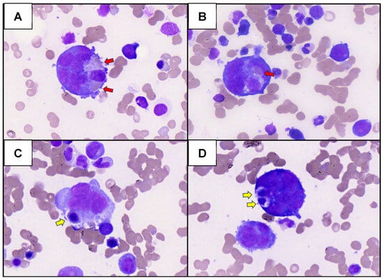
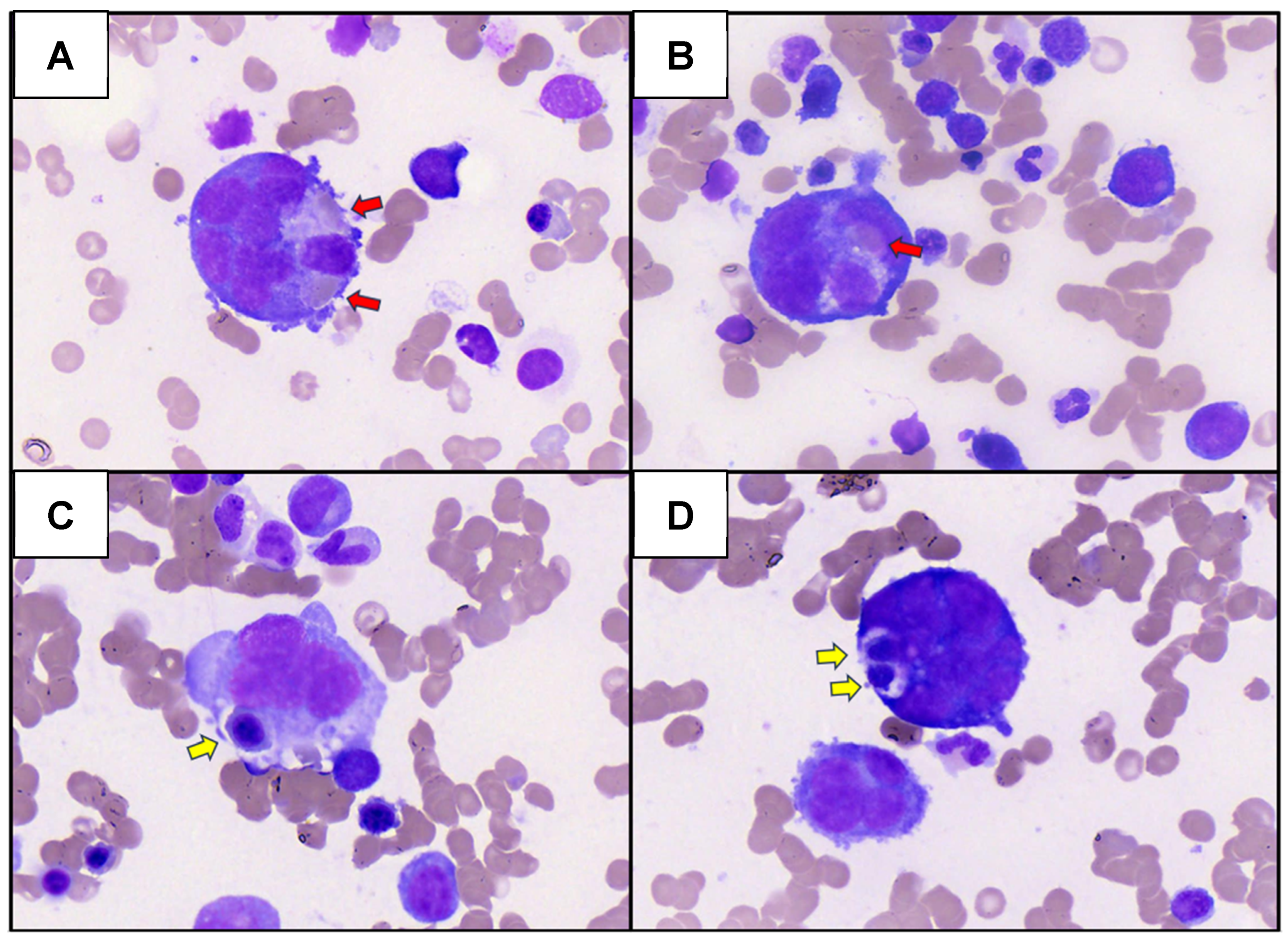
He was referred to our hematology department after his peripheral blood film (PBF) demonstrated 33% circulating blasts characterized by scant cytoplasm and occasional prominent nucleoli without Auer rods. His PBF also showed trilineage dysplasia, with red cell anisocytosis, elliptocytes, teardrop cells and occasional nucleated red cells. Neutrophils exhibited hyposegmentation and hypogranularity, while platelets were reduced in number with occasional large and giant hypogranular forms. Initial investigations revealed an elevated ferritin level (1588 µg/L), consistent with an inflammatory state, and vitamin B12 deficiency (119 pmol/L) was noted as a potential contributor to macrocytic pancytopenia but did not explain the circulating blasts. The initial bone marrow aspirate (BMA) was mildly hypercellular for age and showed significant trilineage dysplasia with 22% blasts. Erythroid dysplasia was characterized by megaloblastoid normoblasts, macronormoblasts, normoblasts with irregular nuclei, and a few binucleated normoblasts. Granulocytic dysplasia was evidenced by hypersegmented and hypogranular neutrophils. Megakaryopoiesis was increased, with dysplastic features including megakaryocytes with separated nuclei, micromegakaryocytes, hyperlobulated megakaryocytes, and megakaryoblasts. Morphologically, some blasts were identified as myeloblasts, while others exhibited cytoplasmic blebs consistent with megakaryoblasts, and a few had scanty cytoplasm. Iron stores were increased (grade 4/6), with no ring sideroblasts identified. Notably, there was significant emperipolesis of erythrocytes and erythroblasts within the cytoplasm of dysplastic megakaryocytes (Figure 1A–D) (May-Grünwald Giemsa Stain, 80× objective, Motic EasyScan Infinity 60). The trephine biopsy revealed a hypercellular marrow with increased blasts. Erythropoiesis appeared normal. However, the myeloid series demonstrated left-shifted maturation and increased large blasts. Megakaryocytes showed focal clustering and scattered hypolobated and multilobated forms. Reticulin staining was increased and graded 2–3+/4. Immunohistochemistry showed singly scattered and small clusters of large CD117+ blasts, while CD34 showed fewer singly scattered blasts. Terminal deoxynucleotidyl transferase (TDT) was negative. Flow cytometry identified two immunophenotypically related blast populations, CD34+ blasts accounting for 17.0% of total nucleated cells and CD34- blasts 5.1%. Both populations demonstrated a myeloblast phenotype characterized by CD45 dim/FSC-lo/SSC-lo/CD117+/CD13++/CD33het/HLA-DR+/CD38+. Critically, they expressed the megakaryocytic markers cyCD41/61+ with concurrent negativity for cyCD42b-, supporting immature megakaryoblastic differentiation. The blasts were uniformly negative for myeloperoxidase (cyMPO-), B-lineage markers (CD19-, cyCD79a-, CD22-), T-lineage markers (sCD3-, cyCD3-, nuTdT-), monocytic markers (CD11b-, CD16-, CD14-, CD64-, CD4-, CD56-) and other lineage markers including CD10, CD15, CD105, CD203c, and IREM2. Aberrant expression of CD7 (partial) and CD35 (partial) was observed, confirming the malignant nature of the blasts. Dim expression of CD123 was noted, representing a potential therapeutic target. NG2 negativity argued against KMT2A-rearranged AML. Erythroid dysplasia was supported by heterogeneous CD36 and CD71 expression on a significant erythroblast subset. Cytogenetic analysis revealed a complex karyotype 46,XY,+1,der(1;7)(q10;p10). Next-generation sequencing (NGS) was performed using a laboratory-developed 54-gene myeloid panel, which included FLT3 analysis for both internal tandem duplications (FLT3-ITD) and tyrosine kinase domain (FLT3-TKD) mutations. The assay had a validated limit of detection of 5% variant allele frequency (VAF). Variant interpretation was performed according to the laboratory’s validated clinical reporting criteria, incorporating available clinical evidence, published literature, and relevant databases. The analysis identified the following pathogenic and likely pathogenic variants: DNMT3A K826R with VAF 37%, which encodes a DNA methyltransferase and is associated with poorer prognosis in AML [3]. RUNX1 F396fs159 with VAF 12% encodes for core binding factors and confers adverse risk following 2017 European LeukemiaNet guidelines [4]. BCOR Q1208fs8 with VAF 18% encodes for a transcriptional corepressor and is associated with poorer survival and a higher rate of transformation from myelodysplastic syndrome (MDS) to AML [5]; BCORL1 S575* with VAF 6% encodes a transcriptional corepressor that interacts with class II histone deacetylases [6] and has been found to be mutated in AML [5]. PHF6 H302R with VAF 62% encodes a member of the plant homeodomain (PHD)-like finger family and plays a role in the transcriptional regulation of hematopoietic stem cells. It has been associated with myeloid neoplasms and mixed phenotype acute leukemia [7,8]. No FLT3-ITD mutations were detected.
Figure 1.
(A–D) Dysplastic megakaryocytes with emperipolesis (erythrocytes: red arrows; erythroblasts: yellow arrows) (May-Grünwald Giemsa Stain, 80× objective, Motic EasyScan Infinity 60).
According to the World Health Organization Classification of Hematolymphoid Tumors, 5th edition (WHO-HAEM5), the findings were consistent with AML-MR with megakaryoblastic differentiation [9]. This diagnosis was supported by ≥20% marrow blasts, multilineage dysplasia, a myelodysplasia-related cytogenetic abnormality [der(1;7)(q10;p10)], and an MDS-related gene mutation (BCOR variant).
The patient’s febrile episode was managed with empirical antibiotic therapy. Approximately one month after diagnosis, the patient commenced cycle 1 of AZA/VEN with 7 days of administered therapy. Antimicrobial prophylaxis with aciclovir and posaconazole was administered during therapy. An interval BMA was performed 2 weeks after completion of cycle 1, demonstrating persistent AML without morphological remission. The aspirate showed ongoing trilineage dysplasia. Trephine was suboptimal but showed left-shifted myelopoiesis and erythropoiesis. Cytogenetic analysis continued to demonstrate the abnormal karyotype 46,XY,+1,der(1;7)(q10;p10). Given persistent disease, AZA/VEN treatment was discontinued. The treatment regimen was switched to intensive induction chemotherapy with DA 3+7 a week later. A repeat BMA performed 5 weeks after treatment demonstrated morphological remission, with blast reduction to <5% and no morphological or flow cytometric evidence of residual AML-MR. The trephine biopsy showed a mildly hypercellular marrow with decreased myeloid elements, increased erythroid elements, reduced megakaryocytes, and mild reticulin fibrosis graded 1/4. Flow cytometry identified only a very small population of CD34-positive myeloblasts with a normal immunophenotype, and the previously identified megakaryoblastic immunophenotype was no longer detected. Cytogenetic analysis, however, showed persistent clonal abnormalities 46,XY,+1,der(1;7)(q10;p10), indicating evidence of diagnostic cytogenetic clones despite morphological remission. Unfortunately, the patient developed sepsis and died at the age of 53 before his planned allogeneic hematopoietic stem cell transplantation.
3. Methods
We conducted a literature review to identify studies exploring emperipolesis, including its definition, mechanistic framework, role in normal and pathological hematopoiesis, pathogenesis of bone marrow fibrosis, inflammatory drivers, and the rare finding of erythroid emperipolesis. The review was performed using the PubMed database with search terms, including “emperipolesis”, “megakaryocytes”, “erythroid”, “erythroblast”, “lymphocytes”, and “neutrophils”. Subsequently, manual screening of reference lists of included articles was performed to identify additional articles of interest. Non-English reports without available translations and preprint articles were excluded. A total of 24 articles were selected: 2 focused on introduction, 2 focused on definition, mechanistic framework, 3 focused on role in normal and pathological hematopoiesis, 3 focused on pathogenesis, 3 focused on inflammatory drivers, 3 focused on erythroid emperipolesis, and 7 original case reports and case series reporting megakaryocytic emperipolesis involving erythrocytes or erythroblasts. Screening of articles and data extraction were conducted by two independent reviewers (B.E.F. and L.C.), with discrepancies resolved during discussion and consultation with all other reviewers.
4. Emperipolesis: Definition and Mechanistic Framework
Emperipolesis refers to a cell-in-cell phenomenon in which one viable cell is transiently internalized within the cytoplasm of another cell, with the internalized cell retaining the capacity to exit intact. This distinguishes emperipolesis from phagocytosis, where the engulfed cell is typically degraded, and from entosis, which classically involves homotypic cell-in-cell invasion and may culminate in the death of the internalized cell. Emperipolesis is also distinct from hemophagocytosis, a process in which phagocytic cells, most commonly macrophages or histiocytes, engulf and digest hematopoietic cells, including erythrocytes, leukocytes, or platelets. Morphologically, hemophagocytosis is characterized by the presence of intact or degenerating blood cells and cellular debris within the cytoplasm of macrophages [10]. In the megakaryocytic context, emperipolesis has increasingly been recognized not as a passive histological curiosity but as a regulated and dynamic interaction between megakaryocytes and hematopoietic cells, particularly neutrophils. Cunin and Nigrovic described megakaryocyte emperipolesis as proceeding through sequential stages of integrin-mediated adhesion, active cytoskeletal remodeling by both the entering cell and the megakaryocyte, entry into a membrane-bound vacuolar compartment termed the “emperisome,” and subsequent egress of the internalized cell without overt injury [11].
This mechanistic model was further refined by Huang et al., who used live-cell spinning disk microscopy to demonstrate that neutrophil emperipolesis is not a uniform process but comprises at least two morphologically and temporally distinct forms. “Fast emperipolesis” is characterized by rapid neutrophil transit through the megakaryocyte, typically within fewer than 10 min, accompanied by an ameboid morphology [12]. In contrast, “slow emperipolesis” involves more prolonged residence, often lasting 60 min or more, during which neutrophils remain relatively sessile and localize near the megakaryocyte nucleus, demarcation membrane system, or perinuclear endoplasmic reticulum [12]. These observations support the concept that emperipolesis encompasses biologically heterogeneous interactions, potentially serving different physiological, inflammatory, or pathological functions depending on the cellular context.
5. Emperipolesis in Normal and Pathological Hematopoiesis
Under physiological conditions, mature neutrophils are the most frequently observed hematopoietic cells undergoing megakaryocyte emperipolesis [13]. In normal marrow, emperipolesis is generally uncommon, with reported baseline frequencies of fewer than 15% of megakaryocytes in experimental settings [14]. Although historically regarded as an unusual morphological phenomenon, contemporary work has suggested a functional role in thrombocytogenesis. Neutrophils entering the emperisome may interact with the demarcation membrane system, transferring membrane material to developing platelets and thereby contributing to platelet production. Following this interaction, neutrophils can egress intact while carrying megakaryocyte-derived membrane components on their surface [11].
In pathological states, particularly myeloproliferative and reactive conditions, emperipolesis may be markedly increased. Cashell and Buss reported megakaryocytic emperipolesis in 77% of primary thrombocythaemia, 100% of polycythemia vera, and 75% of reactive thrombocytosis cases, compared with 17% of chronic granulocytic leukemia specimens [15]. More recently, Collinson et al. found that 84% of 163 patients with myeloproliferative neoplasms exhibited megakaryocyte emperipolesis, with the highest frequencies observed in myelofibrosis and in megakaryocytes showing pyknotic or condensed nuclei [13]. Importantly, this study provided molecular evidence that emperipolesis is associated with bidirectional material transfer. CD15 immunohistochemistry confirmed that most internalized cells were neutrophils, while megakaryocytes demonstrated intracellular positivity for neutrophil granule proteins, including myeloperoxidase and lactoferrin. Proteomic and transcriptomic profiling of platelets from patients with myeloproliferative neoplasms further revealed enrichment of neutrophil-associated mRNA transcripts and granule proteins, including increased cathepsin G and lactoferrin expression [13]. These findings position emperipolesis as an active biological process with measurable consequences for megakaryocytes and their platelet progeny.
6. Emperipolesis and the Pathogenesis of Bone Marrow Fibrosis
The relationship between pathological emperipolesis and marrow fibrosis has been most clearly explored in myelofibrosis. Schmitt et al. demonstrated, using thrombopoietin-overexpressing murine models and marrow biopsies from patients with idiopathic myelofibrosis, that abnormal P-selectin distribution within the megakaryocyte demarcation membrane system may promote selective sequestration of polymorphonuclear cells through emperipolesis [16]. Subsequent work proposed that this abnormal megakaryocyte-neutrophil interaction contributes to rupture of alpha-granule membranes and release of pro-fibrotic mediators, including transforming growth factor-β, platelet-derived growth factor, von Willebrand factor, and fibrinogen, into the marrow microenvironment [16,17]. These mediators may then activate fibroblasts and contribute to reticulin and collagen deposition.
Tefferi further highlighted the potential contribution of neutrophil-derived elastase and related proteolytic mediators to the abnormal egress of myeloid progenitors observed in myelofibrosis with myeloid metaplasia [18]. Collectively, these observations suggest that pathological emperipolesis may contribute to marrow fibrosis through several convergent mechanisms: aberrant cellular adhesion, granule membrane disruption, release of pro-fibrotic proteins, inflammatory amplification, and altered trafficking of hematopoietic progenitors. In the present case, the coexistence of trilineage dysplasia, complex cytogenetic abnormalities, and adverse myeloid mutations may have created a permissive marrow environment for abnormal cell-in-cell interactions and their downstream microenvironmental effects.
7. Inflammatory Drivers of Emperipolesis
Inflammation appears to be an important amplifier of emperipolesis. Kolman et al. demonstrated that interleukin-1α, a cytokine implicated in emergency thrombopoiesis, promotes emperipolesis in murine bone marrow cultures [19]. Interleukin-1α exposure induced larger, higher-ploidy megakaryocytes and significantly increased emperipolesis of Ly6G-positive neutrophils. Proteomic analysis of interleukin-1α-cultured megakaryocytes showed enrichment of neutrophil-associated proteins, antimicrobial peptides, and inflammation-related proteins, a signature that persisted through late megakaryocyte maturation [19]. These data support a model in which inflammatory cytokines do not merely accompany emperipolesis but may actively promote the megakaryocyte phenotype that facilitates it.
This inflammatory axis is relevant to the present case, where high fever and sepsis may have intensified cytokine signaling in an already dysregulated leukemic marrow. Although the precise cytokine profile was not measured, sepsis-associated inflammatory activation could plausibly have augmented megakaryocyte-neutrophil or megakaryocyte-erythroid interactions. More broadly, megakaryocytes are increasingly recognized as immune-responsive cells capable of antigen handling, cytokine secretion, and modulation of the marrow niche [20]. In myeloproliferative neoplasms, Yang et al. showed that JAK2V617F-mutant megakaryocytes exhibit enhanced antigen uptake, MHC class I presentation, and secretion of inflammatory mediators including PF4, TGF-β, and IL-1β, contributing to T-cell dysregulation and immune remodeling within the hematopoietic niche [21]. Although the present case lacked a JAK2 mutation, these findings provide a broader conceptual framework in which abnormal megakaryocytes may act as active inflammatory and immunomodulatory participants rather than passive bystanders.
8. Emperipolesis of Erythroid Cells: A Rare and Distinctive Finding
The emperipolesis observed in the present case is unusual because the internalized cells were erythrocytes and erythroblasts rather than the more typical neutrophils. The predominance of neutrophil emperipolesis is explained by the β2 integrin/ICAM-1 adhesion pathway. In emperipolesis, neutrophil β2 integrins interact with ICAM-1 on megakaryocytes to facilitate entry [22]. Erythroid cells, lacking β2 integrin expression, cannot utilize this entry pathway, accounting for the rarity of erythroid emperipolesis. When erythroid emperipolesis does occur, it may reflect aberrant megakaryocyte adhesion properties in marrow dysplasia or stress. Thus, megakaryocytic emperipolesis involving erythroid-lineage cells is rare, with only limited prior descriptions in the literature, cases occurring in the context of AML-MR and severe marrow stress states (Table 1). Wiseman et al. reported AMKL with t(1;22)(p13;q13)/RBM15-MKL1 in an infant, showing extensive hemophagocytosis by leukemic megakaryoblasts [23]. This phenomenon is related but mechanistically distinct from true emperipolesis, as hemophagocytosis implies phagocytic degradation of the engulfed cells, whereas emperipolesis requires preservation and potential egress of the internalized cell.
This distinction is central to the interpretation of the present case. To differentiate true emperipolesis from other possibilities, we observed the following morphological features. The engulfed cells displayed preserved nuclear chromatin and intact cytoplasmic borders without degradation, excluding phagocytosis and erythrophagocytosis. They were confirmed to lie within the same focal plane as the host cell, excluding cell overlap. Film artifact was excluded by the absence of cellular distortion. Pseudo-inclusion was excluded by the presence of intact erythrocytes with distinct cellular membranes and visible nuclei, and no intranuclear cytoplasmic invaginations were observed. The engulfed erythrocytes and erythroblasts appeared morphologically intact within the megakaryocyte cytoplasm, aligning with emperipolesis. The involvement of erythroid cells also suggests that the cellular selectivity of megakaryocyte emperipolesis may be altered in dysplastic or leukemic marrow. In the present case, dysplastic megakaryocytes may have acquired abnormal surface receptor expression or adhesion properties that permitted atypical erythroid internalization.
The RUNX1 frameshift mutation identified in this patient is especially relevant in this regard, given the central role of RUNX1 in hematopoietic differentiation and megakaryopoiesis. Disruption of RUNX1 may alter megakaryocyte maturation, cytoskeletal organization, adhesion molecule expression, and interactions with neighboring marrow cells [24]. Similarly, der(1;7)(q10;p10), resulting in a gain of 1q and a loss of 7q material, is associated with myelodysplasia-related biology and disordered hematopoiesis [25]. Together, these abnormalities may have contributed to an intrinsically abnormal megakaryocyte phenotype favoring atypical cell-in-cell interactions involving erythroid elements.
Table 1.
Table summarizing megakaryocytic emperipolesis involving erythroid-lineage cells in acute myeloid leukemia, myelodysplasia-related (AML-MR), and severe bone marrow stress states. Abbreviations: CMML; chronic myelomonocytic leukemia, MDS; myelodysplastic syndrome, RBC; red blood cell, PRP; platelet-rich plasma, IVIG; intravenous immunoglobulin, AML; acute myeloid leukemia, AML-MR; acute myeloid leukemia, myelodysplasia-related, DA; daunorubicin and cytarabine.
9. Genetic Context and Prognostic Implications
To our knowledge, this case is the first to describe megakaryocytic emperipolesis of erythroid cells in AML-MR harboring der(1;7)(q10;p10) and a RUNX1 mutation. Both abnormalities are associated with adverse-risk disease biology and impaired normal hematopoietic differentiation. The co-occurrence of DNMT3A, BCOR, BCORL1, and PHF6 mutations further supports a high-risk molecular profile characterized by epigenetic dysregulation, transcriptional disruption, and treatment resistance. The patient’s refractory clinical course, with failure of both azacitidine-venetoclax and conventional daunorubicin-cytarabine induction chemotherapy, is consistent with this adverse genetic background.
The observation of erythroid emperipolesis may therefore represent more than an incidental morphological finding. It may be a visible manifestation of profoundly deranged megakaryopoiesis and marrow architecture. Experimental data support this interpretation. Bobik and Dabrowski showed that emperipolesis increases substantially under marrow stress, rising from fewer than 15% of megakaryocytes in baseline conditions to 34–43% after sublethal irradiation, with internalized cells including granulocytic, erythroid, and lymphoid lineages [14]. McGarry et al. further demonstrated in the gunmetal mouse mutant that the incidence of emperipolesis is influenced by megakaryocyte genotype rather than solely by systemic factors and that increased emperipolesis is associated with altered megakaryocyte ploidy [33]. These observations support the hypothesis that the genetic abnormalities in the present case, particularly the RUNX1 mutation and der(1;7), may have intrinsically altered megakaryocyte behavior and cell–cell selectivity.
10. Conclusions
This case adds to the very limited literature on megakaryocytic emperipolesis involving erythroid-lineage cells and appears to be the first reported example in AML-MR harboring der(1;7)(q10;p10) and RUNX1 mutation. The evolving understanding of emperipolesis has shifted the field away from viewing it as a passive morphological oddity and toward recognizing it as an active, integrin-mediated, cytoskeleton-dependent, and potentially bidirectional biological process. In physiological hematopoiesis, emperipolesis may contribute to thrombocytogenesis; in pathological states, it may reflect or amplify marrow inflammation, megakaryocyte dysregulation, and fibrotic remodeling.
In the present case, erythroid emperipolesis likely reflects a convergence of adverse genetic lesions, dysplastic megakaryopoiesis, inflammatory marrow activation, and systemic sepsis. While causality cannot be inferred from a single case, this observation raises the possibility that aberrant emperipolesis may serve as a morphological marker of disturbed megakaryocyte biology in genetically high-risk AML. Further studies are warranted to determine whether specific mutations, cytogenetic lesions, or inflammatory states alter megakaryocyte adhesion profiles and thereby influence the lineage selectivity, frequency, and biological consequences of emperipolesis.
Author Contributions
L.C. and B.E.F. wrote the initial draft, with all authors revising it critically for important intellectual content. All authors have read and agreed to the published version of the manuscript.
Funding
This research received no external funding.
Institutional Review Board Statement
Approval by the NHG DSRB was not required for a single case report.
Informed Consent Statement
Informed consent was obtained from the patient for publication of this case report and for the use of accompanying images.
Data Availability Statement
The data that support the findings of this study are available from the corresponding author upon reasonable request.
Conflicts of Interest
The authors declare no conflicts of interest.
References
- Rastogi, V.; Sharma, R.; Misra, S.R.; Yadav, L.; Sharma, V. Emperipolesis—A review. J. Clin. Diagn. Res. 2014, 8, ZM01–ZM02. [Google Scholar] [CrossRef] [Scilit] [PubMed]
- Goyal, M.; Thekkelakayil, S.T.; Gupta, A. Megakaryocytic Emperipolesis Associated with Thrombocytopenia: Causative or Coincidence? Turk. J. Haematol. 2017, 34, 370–371. [Google Scholar] [CrossRef] [Scilit] [PubMed]
- Roller, A.; Grossmann, V.; Bacher, U.; Poetzinger, F.; Weissmann, S.; Nadarajah, N.; Boeck, L.; Kern, W.; Haferlach, C.; Schnittger, S.; et al. Landmark analysis of DNMT3A mutations in hematological malignancies. Leukemia 2013, 27, 1573–1578. [Google Scholar] [CrossRef] [Scilit] [PubMed]
- Döhner, H.; Estey, E.; Grimwade, D.; Amadori, S.; Appelbaum, F.R.; Büchner, T.; Dombret, H.; Ebert, B.L.; Fenaux, P.; Larson, R.A.; et al. Diagnosis and management of AML in adults: 2017 ELN recommendations from an international expert panel. Blood 2017, 129, 424–447. [Google Scholar] [CrossRef] [Scilit] [PubMed]
- Damm, F.; Chesnais, V.; Nagata, Y.; Yoshida, K.; Scourzic, L.; Okuno, Y.; Itzykson, R.; Sanada, M.; Shiraishi, Y.; Gelsi-Boyer, V.; et al. BCOR and BCORL1 mutations in myelodysplastic syndromes and related disorders. Blood 2013, 122, 3169–3177. [Google Scholar] [CrossRef] [Scilit] [PubMed]
- Li, M.; Collins, R.; Jiao, Y.; Ouillette, P.; Bixby, D.; Erba, H.; Vogelstein, B.; Kinzler, K.W.; Papadopoulos, N.; Malek, S.N. Somatic mutations in the transcriptional corepressor gene BCORL1 in adult acute myelogenous leukemia. Blood 2011, 118, 5914–5917. [Google Scholar] [CrossRef] [Scilit] [PubMed]
- Van Vlierberghe, P.; Patel, J.; Abdel-Wahab, O.; Lobry, C.; Hedvat, C.V.; Balbin, M.; Nicolas, C.; Payer, A.R.; Fernandez, H.F.; Tallman, M.S.; et al. PHF6 mutations in adult acute myeloid leukemia. Leukemia 2011, 25, 130–134. [Google Scholar] [CrossRef] [Scilit] [PubMed]
- Alexander, T.B.; Gu, Z.; Iacobucci, I.; Dickerson, K.; Choi, J.K.; Xu, B.; Payne-Turner, D.; Yoshihara, H.; Loh, M.L.; Horan, J.; et al. The genetic basis and cell of origin of mixed phenotype acute leukaemia. Nature 2018, 562, 373–379. [Google Scholar] [CrossRef] [Scilit] [PubMed]
- WHO Classification of Tumours Editorial Board. WHO Classification of Tumours: Haematolymphoid Tumours, 5th ed.; International Agency for Research on Cancer: Lyon, France, 2024; Volume 11. [Google Scholar]
- Zini, G.; Viscovo, M. Cytomorphology of normal, reactive, dysmorphic, and dysplastic megakaryocytes in bone marrow aspirates. Int. J. Lab. Hematol. 2021, 43, 23–28. [Google Scholar] [CrossRef] [Scilit] [PubMed]
- Cunin, P.; Nigrovic, P.A. Megakaryocyte emperipolesis: A new frontier in cell-in-cell interaction. Platelets 2020, 31, 700–706. [Google Scholar] [CrossRef] [Scilit] [PubMed]
- Huang, F.Y.; Cunin, P.; Radtke, F.A.; Darbousset, R.; Grieshaber-Bouyer, R.; Nigrovic, P.A. Neutrophil transit time and localization within the megakaryocyte define morphologically distinct forms of emperipolesis. Blood Adv. 2022, 6, 2081–2091. [Google Scholar] [CrossRef] [Scilit] [PubMed]
- Collinson, R.J.; Linden, M.D.; A Fuller, K.; Wilson, L.; Mirzai, B.; Boey, D.; Ng, Z.Y.; Chuah, H.S.; Malherbe, J.A.J.; Howman, R.; et al. Megakaryocyte emperipolesis in myeloproliferative neoplasms: Are neutrophils friends or foes? J. Leukoc. Biol. 2025, 117, qiaf093. [Google Scholar] [CrossRef] [Scilit] [PubMed]
- Bobik, R.; Dabrowski, Z. Emperipolesis of marrow cells within megakaryocytes in the bone marrow of sublethally irradiated mice. Ann. Hematol. 1995, 70, 91–95. [Google Scholar] [CrossRef] [PubMed]
- Cashell, A.W.; Buss, D.H. The frequency and significance of megakaryocytic emperipolesis in myeloproliferative and reactive states. Ann. Hematol. 1992, 64, 273–276. [Google Scholar] [CrossRef] [Scilit] [PubMed]
- Schmitt, A.; Jouault, H.; Guichard, J.; Wendling, F.; Drouin, A.; Cramer, E.M. Pathologic interaction between megakaryocytes and polymorphonuclear leukocytes in myelofibrosis. Blood 2000, 96, 1342–1347. [Google Scholar] [CrossRef] [Scilit]
- Schmitt, A.; Drouin, A.; Massé, J.-M.; Guichard, J.; Shagraoui, H.; Cramer, E.M. Polymorphonuclear neutrophil and megakaryocyte mutual involvement in myelofibrosis pathogenesis. Leuk. Lymphoma 2002, 43, 719–724. [Google Scholar] [CrossRef] [Scilit] [PubMed]
- Tefferi, A. Pathogenesis of myelofibrosis with myeloid metaplasia. J. Clin. Oncol. 2005, 23, 8520–8530. [Google Scholar] [CrossRef] [Scilit] [PubMed]
- Kolman, R.; Voß, H.; Bertović, I.; Peradinović, J.; Munitić, I.; Engel, D.R.; Shevchuk, O.; Begonja, A.J. Interleukin-1α alters megakaryocyte maturation, promotes emperipolesis, and induces a distinct proteomic profile. J. Thromb. Haemost. 2026, 24, 303–317. [Google Scholar] [CrossRef] [Scilit] [PubMed]
- Cunin, P.; Nigrovic, P.A. Megakaryocytes as immune cells. J. Leukoc. Biol. 2019, 105, 1111–1121. [Google Scholar] [CrossRef] [Scilit] [PubMed]
- Yang, X.; Lee, S.; Masarik, K.; Ahmed, T.; Zheng, L.; Zhan, H. Immunomodulatory role of megakaryocytes in the hematopoietic niche of myeloproliferative neoplasms. Haematologica 2026, 111, 2403–2416. [Google Scholar] [CrossRef] [Scilit] [PubMed]
- Ley, K.; Laudanna, C.; Cybulsky, M.I.; Nourshargh, S. Getting to the site of inflammation: The leukocyte adhesion cascade updated. Nat. Rev. Immunol. 2007, 7, 678–689. [Google Scholar] [CrossRef] [Scilit] [PubMed]
- Wiseman, D.H.; Bonney, D.K.; Wynn, R. Hemophagocytosis by leukemic megakaryoblasts in acute myeloid leukemia (megakaryoblastic) with t(1;22)(p13;q13);RBM15-MKL1. J. Pediatr. Hematol. Oncol. 2012, 34, 576–580. [Google Scholar] [CrossRef] [Scilit] [PubMed]
- Wang, C.; Tu, Z.; Cai, X.; Wang, W.; Davis, A.K.; Nattamai, K.J.; Paranjpe, A.; Dexheimer, P.; Wu, J.; Huang, F.L.; et al. A critical role of RUNX1 in governing megakaryocyte-primed hematopoietic stem cell differentiation. Blood Adv. 2023, 7, 2590–2605. [Google Scholar] [CrossRef] [Scilit] [PubMed]
- Lang, W.; Luo, Y.; Wang, L.; Zhang, Y.; Hu, C.; Wang, H.; Tong, H. The der(1;7)(q10;p10) defining a distinct profile from -7/del(7q) in myelodysplastic syndromes: A systematic review and meta-analysis. Cancer Med. 2024, 13, e6890. [Google Scholar] [CrossRef] [Scilit] [PubMed]
- Wandersee, N.J.; Lee, J.C.; Kaysser, T.M.; Bronson, R.T.; Barker, J.E. Hematopoietic cells from -spectrin-deficient mice are sufficient to induce thrombotic events in hematopoietically ablated recipients. Blood 1998, 92, 4856–4863. [Google Scholar] [CrossRef] [Scilit]
- Samii, K.; Pasteur, E. Images in hematology emperipolesis. Am. J. Hematol. 1998, 59, 64. [Google Scholar] [CrossRef]
- Cooper, N.; Bain, B.J. Emperipolesis in a patient receiving romiplostim. Am. J. Hematol. 2016, 91, 166. [Google Scholar] [CrossRef] [Scilit] [PubMed][Green Version]
- Rai, S.; Sharma, M.; Muhary, M.; Naik, R.; Sinha, R. Increased emperipolesis in megakaryocytes in a case of idiopathic thrombocytopenic purpura. Indian J. Pathol. Microbiol. 2009, 52, 452–453. [Google Scholar] [CrossRef] [Scilit] [PubMed]
- Badyal, R.K.; Sharma, P.; Varma, N.; Dhir, V.; Prakash, G. Extensive Megakaryocytic Emperipolesis in an Adult Male with Immune Thrombocytopenia. Indian J. Hematol. Blood Transfus. 2018, 34, 158–160. [Google Scholar] [CrossRef] [Scilit] [PubMed]
- Knight, T.; Savaşan, S. Megakaryocytic Emperipolesis in an Adolescent With Hodgkin Lymphoma. J. Pediatr. Hematol./Oncol. 2018, 40, 306. [Google Scholar] [CrossRef] [Scilit] [PubMed]
- Wu, S.; Zhong, Z. Megakaryocytic Emperipolesis in Acute Myeloid Leukemia with Trisomy 8. Turk. J. Haematol. 2026, 43, 78–79. [Google Scholar] [CrossRef] [Scilit] [PubMed]
- McGarry, M.P.; Reddington, M.; Jackson, C.W.; Zhen, L.; Novak, E.K.; Swank, R.T. Increased incidence and analysis of emperipolesis in megakaryocytes of the mouse mutant gunmetal. Exp. Mol. Pathol. 1999, 66, 191–200. [Google Scholar] [CrossRef] [Scilit] [PubMed]
Disclaimer/Publisher’s Note: The statements, opinions and data contained in all publications are solely those of the individual author(s) and contributor(s) and not of MDPI and/or the editor(s). MDPI and/or the editor(s) disclaim responsibility for any injury to people or property resulting from any ideas, methods, instructions or products referred to in the content. |
© 2026 by the authors. Licensee MDPI, Basel, Switzerland. This article is an open access article distributed under the terms and conditions of the Creative Commons Attribution (CC BY) license.