


Abstract
Electrospraying has emerged as a promising technique for producing biopolymeric structures containing bioactive compounds. In this study, gelatin and gelatin–curcumin nanoparticles were produced by uniaxial electrospraying, and the effects of gelatin and curcumin extract concentrations on their rheological, morphological, and physicochemical characteristics were evaluated. Gelatin solutions (5–15% w/v) were first characterized by rheological analysis, which exhibited Newtonian flow behavior over the evaluated concentration range, favorable for the electrospraying process. Electrospraying was performed at fixed conditions of 15 kV voltage, 0.1 mL h−1 flow rate, and 10 cm tip-to-collector distance. Increasing gelatin concentrations influenced the resulting morphology, with structures ranging from pearled fibers to spherical particles and, at higher concentrations, fibers with few pearls. Gelatin structures exhibited mean hydrodynamic diameters of 420.63–762.66 nm, determined by dynamic light scattering (DLS), with PDI values of 0.49–0.89. At a fixed gelatin concentration of 8% (w/v), varying curcumin extract concentration from 1 to 5% (w/v) resulted in a non-linear change in particle size, with mean diameters ranging from 196.30 to 622.16 nm and PDI values of 0.60–0.79. The selected formulation containing 8% (w/v) gelatin and 5% (w/v) curcumin extract exhibited the highest absolute zeta potential (−28.56 ± 1.25 mV), suggesting a lower tendency toward particle aggregation. Fourier transform infrared spectroscopy (FTIR) supported molecular interactions between gelatin and curcumin. Overall, the results demonstrate that formulation composition influences the morphology, particle size distribution, and surface charge of gelatin–curcumin structures produced by uniaxial electrospraying. Further studies are required to evaluate encapsulation efficiency, curcumin storage stability, release behavior, and biological performance.
1. Introduction
Nanotechnology has emerged as a key strategy to enhance the stability, bioavailability, and functionality of bioactive compounds. In this context, the development of nanoparticle systems using efficient green techniques has gained significant attention in food, nutraceutical, and biomedical applications [1].
Among available fabrication techniques, electrospraying (electrohydrodynamic atomization) is widely recognized as a versatile and cost-effective method for producing micro- and nanoparticles. In this process, a high electric field (typically 1–30 kV) is applied to a polymer solution, inducing the formation of a Taylor cone. Subsequent breakup into fine droplets, driven by electrostatic repulsion, followed by rapid solvent evaporation, results in the formation of solid particles. Compared to conventional techniques, this approach minimizes thermal degradation and eliminates the need for post-processing steps [2,3,4].
The physicochemical properties of the precursor solution, particularly viscosity and rheological behavior, are crucial in determining jet stability and particle morphology. While low-viscosity systems favor jet stability, higher viscosities promote chain entanglement and fiber formation [3,5,6]. Therefore, controlling solution properties and processing parameters, such as flow rate and applied voltage, is essential to achieve uniform and stable nanoparticulate systems.
Biopolymers such as gelatin are widely used as encapsulation matrices due to their biocompatibility, biodegradability, and amphiphilic nature [7]. Gelatin, derived from collagen denaturation, possesses a flexible linear structure that can adopt a random coil conformation under appropriate conditions, facilitating processing via electrospraying [8,9]. Chemically, it consists of a heterogeneous mixture of polypeptide chains rich in glycine, proline, and hydroxyproline. Its characteristic Gly-X-Y sequence enables the formation of thermoreversible, three-dimensional networks in aqueous media [9,10,11]. Commercially, gelatin is classified into two types based on the collagen pretreatment method. Type A gelatin is produced via acid hydrolysis, typically from porcine skin, and exhibits an isoelectric point between pH 7.0 and 9.0. In contrast, Type B gelatin is obtained through the alkaline treatment of bovine skin and bones, resulting in a lower isoelectric point (pH 4.5–6.0) due to the deamidation of amino acid residues [10,12]. These structural differences directly influence functional properties. For instance, Type B gelatin exhibits a greater crosslinking degree and a slower degradation rate, making it suitable for drug delivery systems and long-term biomedical applications [13,14]. Furthermore, its functional groups facilitate interactions with various bioactive compounds, enabling the formation of stable nanoparticulate assemblies [3,8,15]. Type A gelatin exhibits greater resistance to acid hydrolysis than Type B, making it particularly suitable for acidic food formulations and confectionery products that require enhanced stability during storage and transportation [11,14,16].
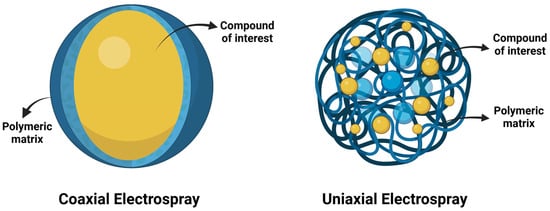
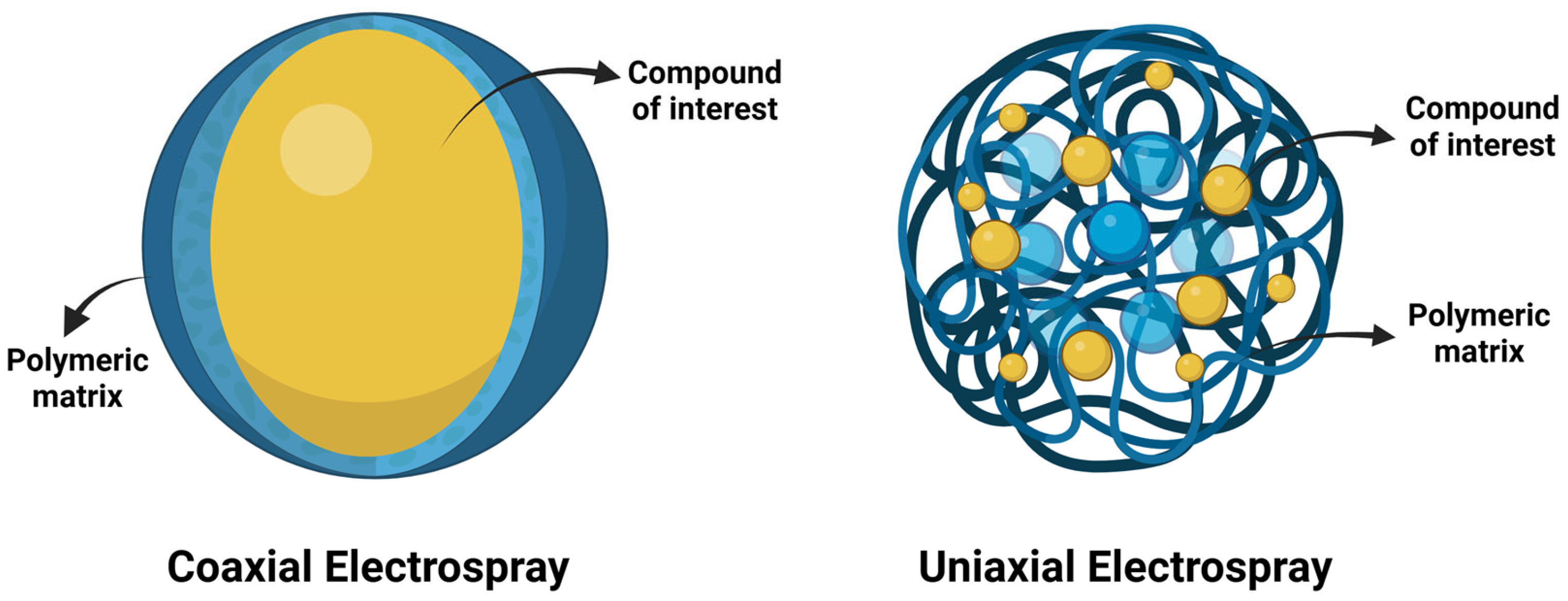
Nanoparticle morphology and encapsulation efficiency vary according to the methodology employed. Figure 1 illustrates both uniaxial and core–shell particle systems. In the uniaxial method, a polymeric solution distributes both the polymer and bioactive compound within the resulting polymeric matrix. In contrast, the core–shell system provides enhanced protection by encapsulating the compound within the particle core [2,3,7,17]. In the present study, uniaxial electrospray was employed to produce gelatin-based nanoparticles containing curcumin. This approach provides a simple strategy for incorporating curcumin into a gelatin matrix while allowing the influence of formulation parameters on nanoparticle characteristics to be evaluated.
Figure 1.
The entrapment or encapsulation of biocompounds using uniaxial or coaxial methodology. Created in BioRender. Tapia, V. (2026) https://BioRender.com/ehlidry [18].
Curcumin, a natural polyphenol extracted from Curcuma longa, exhibits significant biological properties, including antioxidant, anti-inflammatory, and antimicrobial activities [19]. Irshad et al. reported that C. longa extracts exhibit significant antibacterial activity against Escherichia coli and Staphylococcus aureus, as well as antioxidant activity exceeding 90% in DPPH radical scavenging assays. Despite these properties, curcumin’s practical application is limited by poor aqueous solubility and low bioavailability [20]. To address these constraints, Gómez-Estaca et al. encapsulated curcumin within gelatin-based matrices, achieving an 11.3-fold increase in bioaccessibility while preserving its radical-scavenging capacity in complex systems [21]. Furthermore, Kavukcu et al. demonstrated that incorporating curcumin nanoparticles into biopolymeric scaffolds effectively controls inflammatory and microbial processes, highlighting their potential for tissue engineering and wound healing [15]. Nanoencapsulation using biopolymer-based systems is an effective strategy to overcome these limitations, thereby improving solubility, stability, and bioaccessibility [4,22].
Recent studies have increasingly focused on biopolymers, bioactive compounds, and their potential applications across food and biomedical fields. Curcumin encapsulated or entrapped within polymeric matrices has been extensively investigated as a strategy to enhance its stability, bioavailability, and functional performance while preserving its biological properties. In the food sector, Aliabbasi et al. developed curcumin-loaded nanoparticles and investigated their release characteristics to improve the stability and functionality of this bioactive compound [23]. Different nanoparticle formulations were examined to identify systems with enhanced delivery performance. The findings suggested that nanoencapsulation could be a promising approach for food applications, as it may provide improved protection of curcumin while enabling its controlled release in functional and active food systems. In the biomedical field, Cao et al. investigated the influence of different preparation methods on curcumin-loaded materials and evaluated their release behavior [24]. Their results showed that fabrication techniques, such as electrospinning, can effectively preserve curcumin’s biological properties, including its antioxidant activity, while promoting sustained release from composite matrices. These findings highlight the importance of processing strategies in developing functional delivery systems for bioactive compounds. Golmakani et al. developed gelatin/fucoidan-based electrospun composites incorporating curcumin as a bioactive compound [25]. The resulting material exhibited promising antimicrobial and antioxidant properties across different evaluations. The authors proposed this formulation for biomedical applications, particularly as a facial mask, demonstrating the potential of curcumin-containing biopolymeric systems in the development of multifunctional healthcare materials.
Despite growing interest in electrospraying for the fabrication of biopolymer-based nanoparticles, several limitations persist in gelatin-based systems. In particular, the influence of formulation parameters, such as polymer and bioactive compound concentrations, on the rheological behavior precursor solutions and the properties of the resulting nanoparticles remains poorly understood. This study aimed to produce gelatin-curcumin nanoparticles via uniaxial electrospraying and evaluate the influence of formulation parameters (gelatin and curcumin concentrations) on the rheological behavior of precursor solutions and the physicochemical properties of the resulting nanoparticles. These findings provide insight into the relationships among solution properties, processing conditions, and nanoparticle characteristics, thereby contributing to the development of efficient carrier systems for bioactive compounds.
In addition, understanding the physicochemical properties of these systems is important for identifying their potential applications in both the food and biomedical fields. A better understanding of their behavior may provide useful information about their stability, functionality, and overall performance, which could support the development of new materials with specific technological and biological applications.
2. Materials and Methods
2.1. Chemical Reagents
Type B Bovine Gelatin (225 g Bloom, 95% w/w protein content) was purchased from Sigma–Aldrich (St. Louis, MO, USA). Curcumin was obtained from commercial Curcuma longa (turmeric) powder purchased from Comercial Zazueta (Hermosillo, Sonora, México). The extract was obtained via Ultrasound-Microwave-Assisted Extraction (UMAE) following a modified UMAE methodology by Liew et al. [26]. The extraction was performed in an ultrasonic microwave reaction system (Nanjing Xianou Instruments Manufacture Co., Ltd., Nanjing, China) operated at 600 W microwave power and 400 rpm agitation [26]. Glacial acetic acid was purchased from J.T. Baker (Sanford, MA, USA), and distilled water was used.
2.2. Preparation of Gelatin Solutions
Gelatin solutions at concentrations of 5, 8, 10, 12, and 15% (w/v) were prepared using 20% (v/v) acetic acid under magnetic agitation (IKAA C-MAG HS7) at 25 °C. Gelatin-curcumin solutions were prepared using a modified method reported by Rodríguez-Félix et al. (2019) [27]. Curcumin extract was incorporated at concentrations of 1, 3, and 5% (w/v) into gelatin solutions at a fixed concentration of 8% (w/v), maintaining the same preparation conditions as those used for the unloaded gelatin solutions.
2.3. Electrospray Process
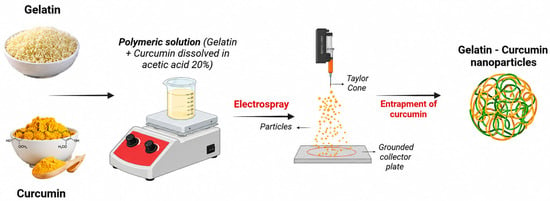
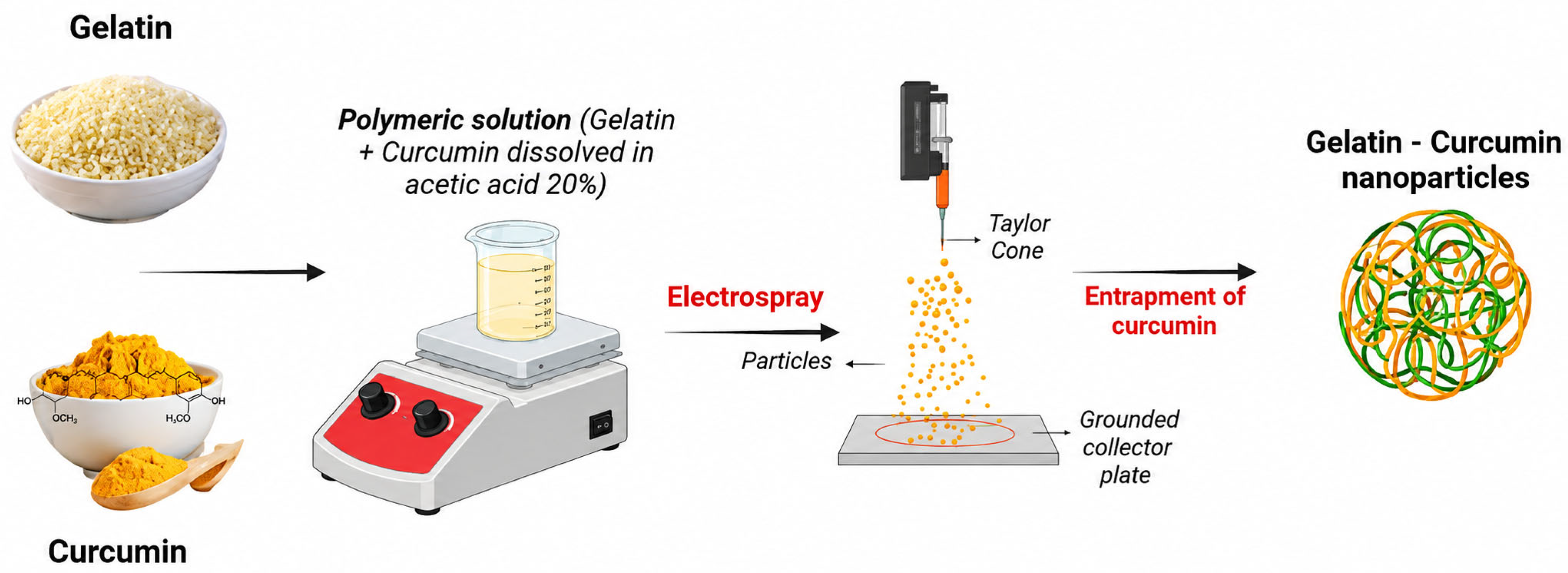
Polymeric nanoparticles were synthesized via electrospraying. Gelatin solutions were loaded into a 10 mL syringe with a stainless-steel needle and processed using a controlled pump (model L-F6, Tongli Tech, Baoding, China) and high-voltage source (model CZE 1000R, Spellman, Hauppauge, NY, USA). The experimental design evaluated the effects of gelatin and curcumin concentrations, while the electrospraying process parameters were maintained constant throughout all experiments. Specifically, the applied voltage, flow rate, and tip-to-collector distance were fixed at 15 kV, 0.1 mL/h, and 10 cm, respectively. These conditions were selected based on previously reported studies from Tapia-Hernández et al. and Rodríguez-Félix et al. and prior experimental experience within our research group, in which these parameters provided stable electrospraying conditions and reproducible particle formation [27,28]. Maintaining these parameters constant allowed the effects of formulation composition to be evaluated under comparable processing conditions. Figure 2 illustrates the electrospraying process.
Figure 2.
Schematic description of the synthesis of gelatin-curcumin nanoparticles by the electrospraying method. Created in BioRender. Tapia, V. (2026) https://BioRender.com/e9bexi6 [29].
2.4. Determination of the Rheological Behavior of the Solutions
Rheological analysis was performed using a MCR-102 rheometer (Anton Paar, Graz, Austria) with a concentric cylinder geometry (1 mm gap) under a shear rate ramp from 0.1 to 100 s−1 at 25 °C. Gelatin solutions (5–15% w/v) were prepared and analyzed via rheometry to classify their flow behavior as Newtonian or non-Newtonian. Shear stress data were fitted to the Power Law model (Equation (1)):
where τ is the shear stress (Pa), Y is the shear rate (s−1), n is the Power Law index, and K is the consistency parameter (Pa∙s). Solutions with n = 1 are considered Newtonian fluids, while n values < 1 are considered non-Newtonian fluids, and values > 1 indicate thickening behavior.
τ = K Yn
2.5. Characterization of the Nanoparticles
2.5.1. Scanning Electron Microscopy (SEM)
The morphological characterization of the material obtained was conducted using a Scanning Electron Microscopy (SEM) JEOL model JSM-7601F (Tokyo, Japan). Samples were coated with gold and analyzed using an acceleration voltage of 10 kV.
2.5.2. Dynamic Light Scattering (DLS)
Particle size distribution and the polydispersity index (PDI) were determined by Dynamic Light Scattering (DLS) using a Zetasizer Nano ZS 3600 instrument (Malvern Instruments, Malvern, UK), equipped with a 633 nm laser and operating at a scattering angle of 173°. Approximately 1 mL of each dispersion was placed in a disposable acrylic cuvette, and particle size distributions were reported on an intensity basis. Zeta potential measurements were performed using a dip cell (ZEN1002; Malvern Instruments, Malvern, UK), and zeta potential was calculated from the measured electrophoretic mobility using the Smoluchowski approximation.
2.5.3. Fourier Transform Infrared (FTIR)
FTIR was performed using a Thermo Scientific™ Nicolet™ iS™5 spectrometer (Thermo Fisher Scientific, Madison, WI, USA) equipped with an attenuated total reflection (ATR) accessory. For each sample, 32 scans were collected in triplicate over the spectral range of 4000–500 cm−1 at a spectral resolution of 8 cm−1. For each sample, 32 scans were collected in triplicate over the spectral range of 4000–500 cm−1 at a spectral resolution of 8 cm−1. The FTIR analysis was conducted following a procedure adapted from Barreras-Urbina et al. [30], with the acquisition conditions described above.
2.5.4. Thermogravimetric Analysis (TGA)
The thermogravimetric analysis was carried out using a Discovery modular system from TA Instruments (TA Instruments, New Castle, DE, USA). The samples were heated from 30 °C to 600 °C at a heating rate of 10 °C min−1 under an air atmosphere, with a gas flow rate of 25 mL min−1.
2.5.5. Experimental Design and Statistical Analysis
Descriptive statistics were used to analyze the particle size distribution of gelatin and gelatin-curcumin nanoparticles obtained by Dynamic Light Scattering (DLS) (Malvern Instruments, Malvern, UK). Gelatin and curcumin extract concentrations were considered experimental factors. Mean comparisons were performed using Tukey’s test at a 95% confidence level. All statistical analyses were conducted using InfoStat software (version 2021).
3. Results and Discussion
3.1. Rheological Behavior
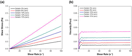
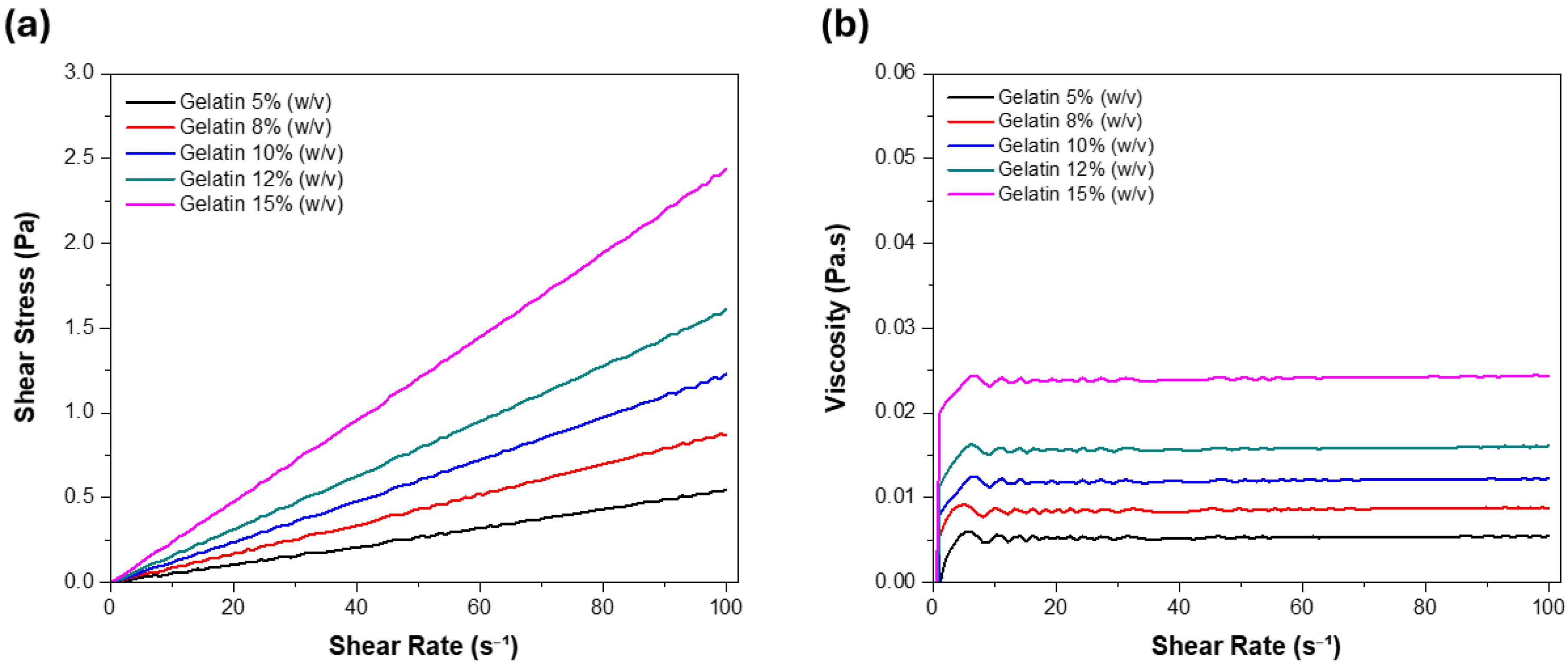
Figure 3 presents the rheograms of gelatin solutions (5–15% w/v) in 20% acetic acid, demonstrating a linear relationship between shear stress and shear rate. This proportionality is characteristic of Newtonian fluids and is consistent with previous reports for dilute gelatin systems [9]. The Power Law model (Table 1) confirms this behavior, with a flow index (n ≈ 1) and a high coefficient of determination (R2 = 0.99), indicating constant viscosity across the evaluated shear range.
Figure 3.
(a) Rheograms of shear stress versus shear rate of gelatin solutions at 5, 8, 10, 12, 15% (w/v), (b) Rheograms of time versus viscosity of gelatin solutions at different concentrations with a continuous ramp of 0 to 100 s−1.
Table 1.
Power Law parameter of the gelatin solution at different concentrations.
This behavior is attributed to the disruption of gelatin intermolecular interactions in acidic media. In the presence of acetic acid, the protonation of amino groups reduces electrostatic interactions and weakens hydrogen bonding between gelatin chains, thereby limiting the formation of structured networks [31,32]. Consequently, the polymer chains behave as isolated or weakly interacting entities, resulting in a constant viscosity independent of shear rate, which is characteristic of Newtonian fluids [33].
Figure 4 shows viscosity as a function of time at shear rates of 20–100 s−1. No significant variation was observed, confirming the absence of time-dependent behavior. This temporal stability indicates that the system maintains consistent flow properties, which are essential for homogeneous electrospray. Conversely, viscosity increased with gelatin concentration, likely due to enhanced intermolecular interactions and greater overlap between polymer chains at higher concentrations. This behavior is consistent with previous reports on gelatin and other biopolymer solutions, in which viscosity increases with concentration due to coil overlap and enhanced chain interactions [5,6].
Figure 4.
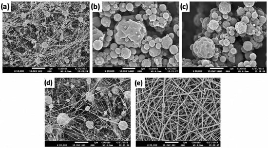
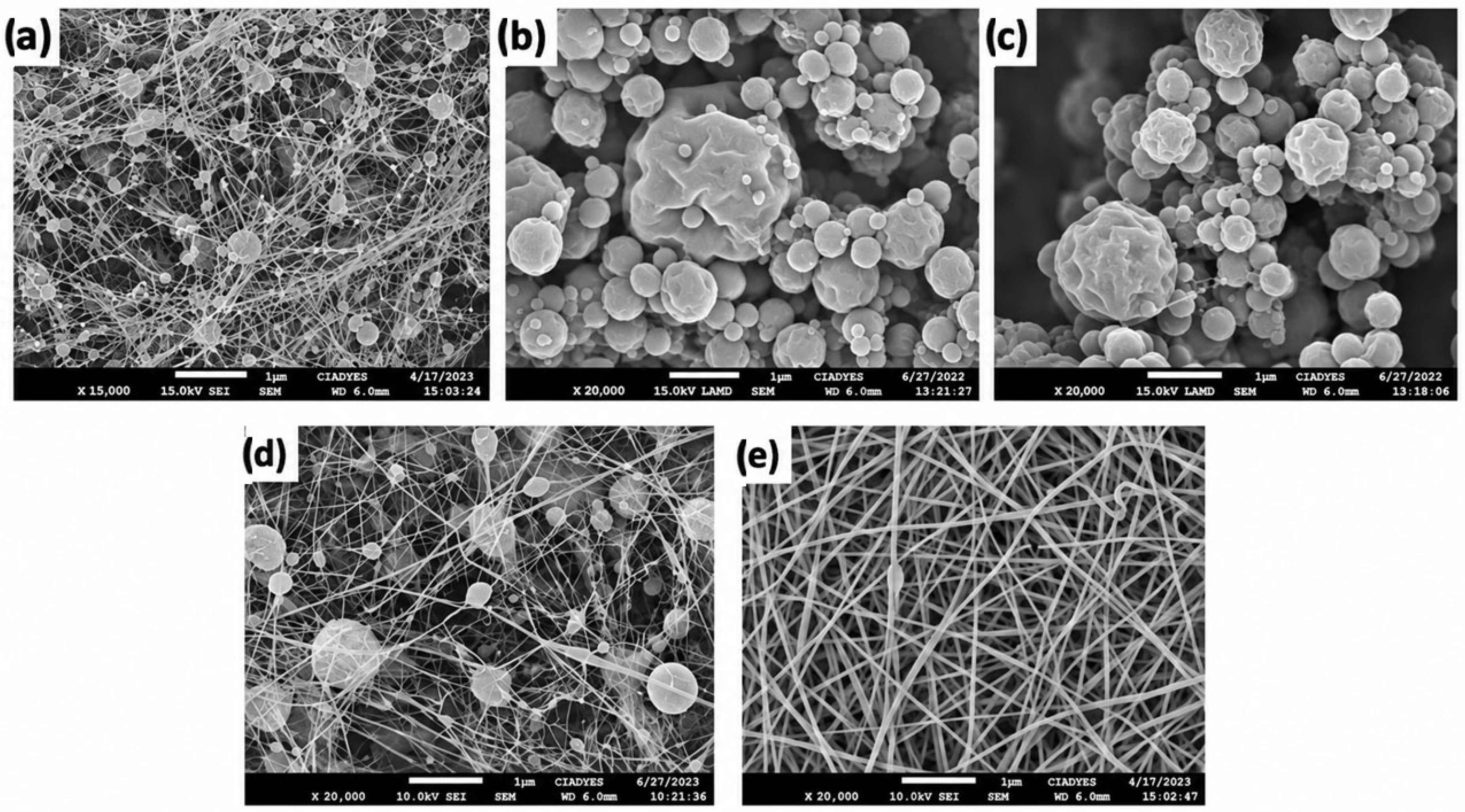
SEM images of gelatin structures obtained through electrohydrodynamic processing of gelatin solutions at concentrations of (a) 5%, (b) 8%, (c) 10%, (d) 12%, and (e) 15% (w/v).
3.2. Characterization of the Nanoparticles
3.2.1. Scanning Electron Microscopy (SEM) and Dynamic Light Scattering (DLS)
The morphologies observed in Figure 4 were strongly influenced by gelatin concentration and electrospraying conditions. At a low gelatin concentration (5% w/v), discontinuous and poorly defined structures formed due to low viscosity and insufficient intermolecular interactions, which led to unstable jet behavior. Increasing concentration to 8% and 10% (w/v) resulted in spherical particles with average diameters of 477.10 ± 118.26 nm and 570.13 ± 34.60 nm, respectively (Table 2). However, irregular surfaces and partial coalescence indicated operation near the transition between droplet formation and jet elongation, consistent with behaviors reported for biopolymer systems processed under electrohydrodynamic conditions [34]. Table 2 shows that the gelatin nanoparticles had an average diameter of 570.13 ± 34.60 nm, with a high polydispersity, indicating a heterogeneous size distribution. As reported by Rodríguez-Félix et al., polydispersity increases as the index approaches 1, while values near 0 indicate more uniform systems [27]. This variability is consistent with the observed morphological irregularities and transitional jet behavior. The zeta potential was 9 mV, suggesting limited colloidal stability due to low electrostatic repulsion. Castro et al. reported that particles with Z ≥ 30 mV are considered highly stable, whereas values below ±5 mV favor aggregation, indicating that the system is prone to particle association [35].
Table 2.
Effect of gelatin concentration on the particle size, PDI, zeta potential, and morphology of gelatin nanoparticles.
These results suggest that the system operates within a semidilute unentangled regime, where polymer chains interact without forming a fully entangled network. Under these conditions, the viscosity is sufficient to stabilize the jet while allowing it to break into discrete droplets. This regime is particularly suitable for electrospraying, as reduced viscoelastic resistance promotes jet fragmentation and minimizes the formation of continuous fibers [2,36]. At a higher gelatin concentration (15% w/v), a clear transition toward fiber formation was observed. This indicates increased chain overlap and entanglement, which suppresses jet breakup and promotes continuous elongation, consistent with previous reports [17,37].
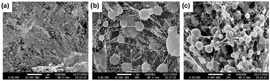
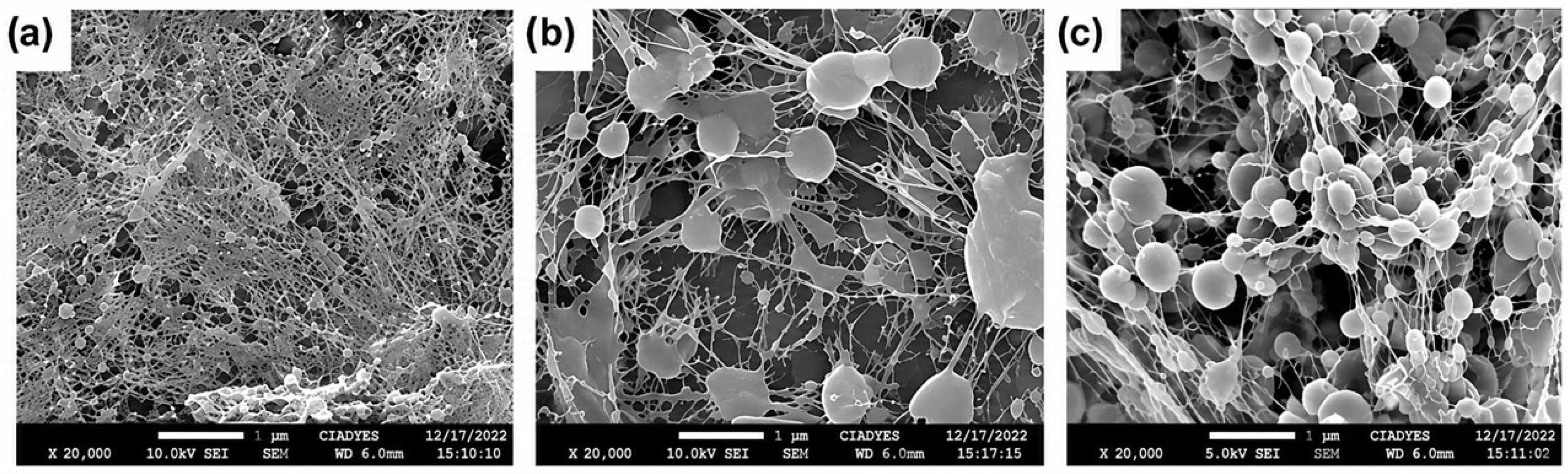
Beyond formulation parameters, processing conditions such as applied voltage, flow rate, and tip-to-collector distance critically determine particle morphology. Applied voltage influences charge density and jet stretching; higher voltages increase Coulombic repulsion, favoring smaller particle sizes. Conversely, higher flow rates result in larger, more polydisperse particles due to incomplete solvent evaporation. Similarly, the tip-to-collector distance influences solvent evaporation and particle solidification; an insufficient distance may result in particle deformation or aggregation [2,6,21,34]. In the present study, these processing parameters were maintained constant to isolate the effects of gelatin and curcumin extract concentrations on particle characteristics, as described in Section 2.3. For the curcumin-loaded systems in Figure 5, prepared at a fixed gelatin concentration of 8% (w/v), particle morphology and size were strongly dependent on curcumin extract concentration. As shown in Table 3, the average diameters were 622.16 ± 142.3 nm, 196.30 ± 6.23 nm, and 343.56 ± 63.39 nm for 1%, 3%, and 5% (w/v) concentrations, respectively. Particle size exhibited a non-linear dependence on curcumin extract content; the smallest particles with lower polydispersity were observed at 3% (w/v), indicating improved jet stability and more efficient distribution, which favored droplet breakup.
Figure 5.
SEM images of gelatin (8% w/v) structures obtained through electrohydrodynamic processing of aqueous solutions with different curcumin extract concentrations: (a) 1% (w/v), (b) 3% (w/v), and (c) 5% (w/v).
Table 3.
Particle size, polydispersity index (PDI), zeta potential, and morphology of gelatin (8% w/v) nanoparticles loaded with different curcumin extract concentrations.
In contrast, larger particle sizes at 1% (w/v) and 5% (w/v) were observed compared with the 3% (w/v) formulation. This non-linear variation may be associated with changes in physicochemical properties of the precursor solution induced by increasing curcumin extract concentration, which can affect the electrohydrodynamic behavior of the jet and, consequently, particle formation [38,39]. Previous studies have shown that the incorporation or increasing concentration of curcumin can modify the size of polymeric or protein-based nanoparticles. In electrosprayed systems, the presence of curcumin has been associated with changes in particle size, potentially due to alterations in the electrical properties of the precursor solution and the resulting electrostatic stretching of the jet [40]. Similarly, curcumin concentration has been reported to influence the size of gelatin-based nanoparticles, with increasing curcumin concentrations resulting in larger particles under certain formulation conditions [41]. These findings support the possibility that changes in curcumin extract concentration contributed to the variation in particle size observed in the present study, although the specific contribution of solution conductivity and viscosity was not directly evaluated in this study.
All samples exhibited spherical particles interconnected by thin filaments, forming a “pearl necklace” morphology characteristic of intermediate viscoelastic regimes [40]. Furthermore, environmental factors such as humidity may influence particle morphology, as delayed solvent evaporation can promote structural deformation [41,42]. PDI values (0.60–0.79) indicated a relatively broad particle size distribution, with the lowest PDI observed at 3%. In contrast, the zeta potential increased in magnitude with increasing curcumin extract concentration, reaching −28.56 ± 1.25 mV at 5% (w/v), suggesting a lower tendency toward particle aggregation due to greater electrostatic repulsion [38,39]. Unlike the larger, more polydisperse gelatin-curcumin particles (up to 1200 nm) reported by Gómez-Estaca et al., our 3% (w/v) formulation exhibited the smallest particle size (196.30 ± 6.23 nm) and the lowest PDI among the curcumin-loaded formulations [21]. Although the 3% (w/v) formulation exhibited the smallest mean particle size (196.30 ± 6.23 nm) and the lowest PDI among the curcumin extract-loaded formulations, the 5% (w/v) formulation was selected based primarily on its higher zeta potential magnitude, as a lower tendency toward particle aggregation was considered particularly relevant for formulation selection. Overall, these results indicate that curcumin extract concentration influenced the particle size distribution and surface charge of the electrosprayed structures.
3.2.2. Fourier Transform Infrared (FTIR)
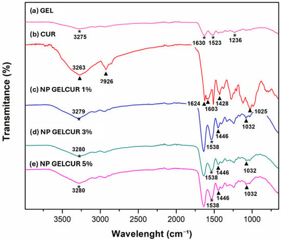
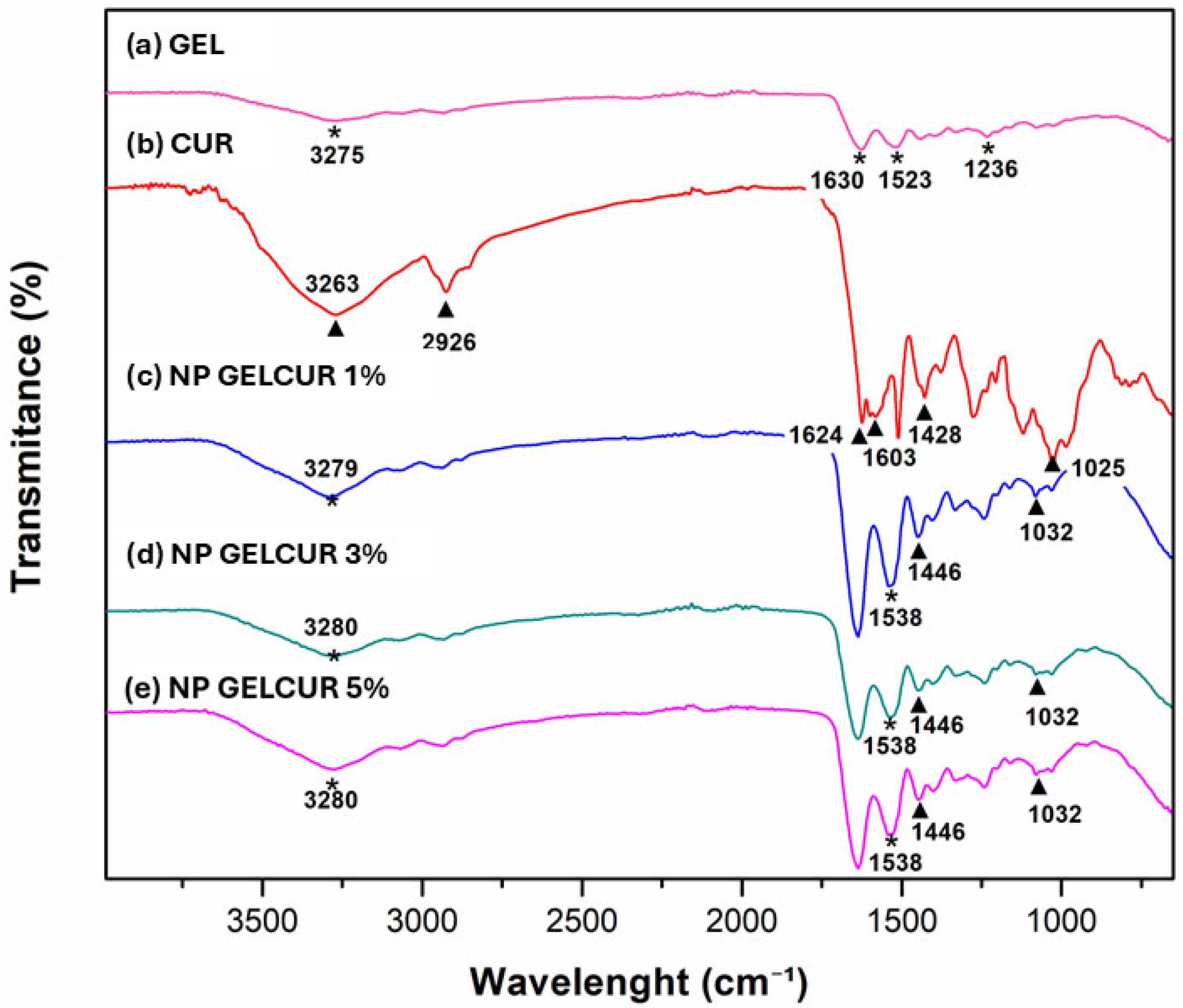
Figure 6 presents the FTIR spectra of 8% (w/v) gelatin and 5% (w/v) curcumin extract, highlighting their primary functional groups. The gelatin spectrum exhibits characteristic amide bands: amide A at 3275 cm−1 (N-H stretching), amide I at 1628 cm−1 (C=O and C-N stretching), amide II at 1523 cm−1 (N-H bending), and amide III at 1235 cm−1 (C-N stretching), consistent with previous reports [6,43]. These bands correspond to the protein backbone and reflect its secondary structure. Unlike globular proteins, gelatin derived from collagen denaturation adopts a flexible linear conformation. Under the experimental conditions used in this work (acidic medium and electrospraying), gelatin chains predominantly adopt a random coil configuration [9,11]. This structural state is essential for nanoparticle formation as it prevents reorganization into triple helices and avoids gelation, thereby enabling the formation of stable jets and subsequent droplet breakup during electrospraying [21,43].
Figure 6.
FTIR spectra of gelatin, curcumin extract, and gelatin–curcumin electrosprayed structures containing 1%, 3%, and 5% (w/v) curcumin. The principal characteristic bands of gelatin (*) and curcumin (▲) are indicated together with their corresponding wavenumbers. Relevant spectral shifts associated with gelatin–curcumin interactions are also indicated in the composite formulations.
For the curcumin extract, characteristic absorption bands were identified at 3263 cm−1 (O-H stretching), 2926 cm−1 (aliphatic C-H stretching), 1624 cm−1 (C=O stretching), 1603 cm−1 (aromatic C=C vibration), and 1428 cm−1 (phenolic C-O stretching), consistent with Asadi et al. (2021) [44]. Figure 6 also shows the FTIR spectra of gelatin-curcumin structures (1%, 3%, and 5% w/v), where slight shifts in amide A (3275 to 3279–3280 cm−1) and amide II (1523 to 1538 cm−1) were observed, suggesting changes in the hydrogen bonding environment and possible interactions between gelatin and curcumin, particularly though hydrogen bonding between hydroxyl groups of curcumin and gelatin amide groups [3,11,43]. Furthermore, shifts in the phenolic C-O band (from 1428 to 1447 cm−1), also with changes in the bands associated with ether/ methyl groups (1029–1032 cm−1, 2929–2932 cm−1), further support the presence of molecular interactions between the curcumin extract and the gelatin matrix.
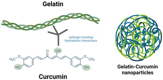
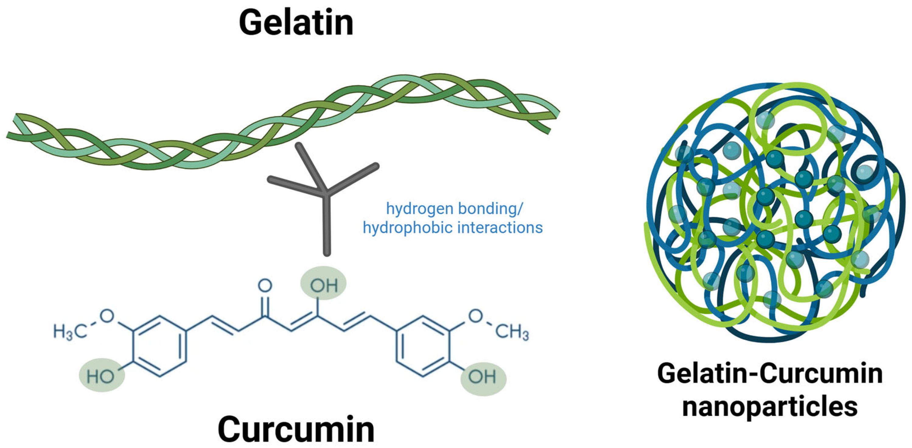
At the molecular level, nanoparticle formation is driven by the assembly of gelatin and curcumin. Under acidic conditions, gelatin adopts a random coil conformation, facilitating its interaction with small molecules. During electrospray, the curcumin extract is entrapped within the gelatin matrix, primarily through hydrogen bonding between curcumin’s hydroxyl groups and gelatin’s amide groups, supplemented by hydrophobic interactions. Noncovalent interactions promote the formation of a stabilized molecular assembly in which curcumin is dispersed within the polymeric matrix (Figure 7). This assembly enhances nanoparticle integrity and curcumin stability by limiting exposure to environmental factors [21,45,46,47].
Figure 7.
Representation of the gelatin-curcumin assembly on the nanoparticles. Created in BioRender. Tapia, V. (2026) https://BioRender.com/j1urys0 [48].
3.2.3. Thermogravimetric Analysis (TGA)
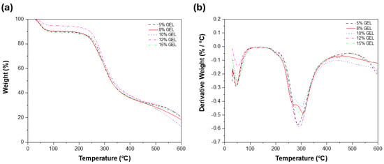
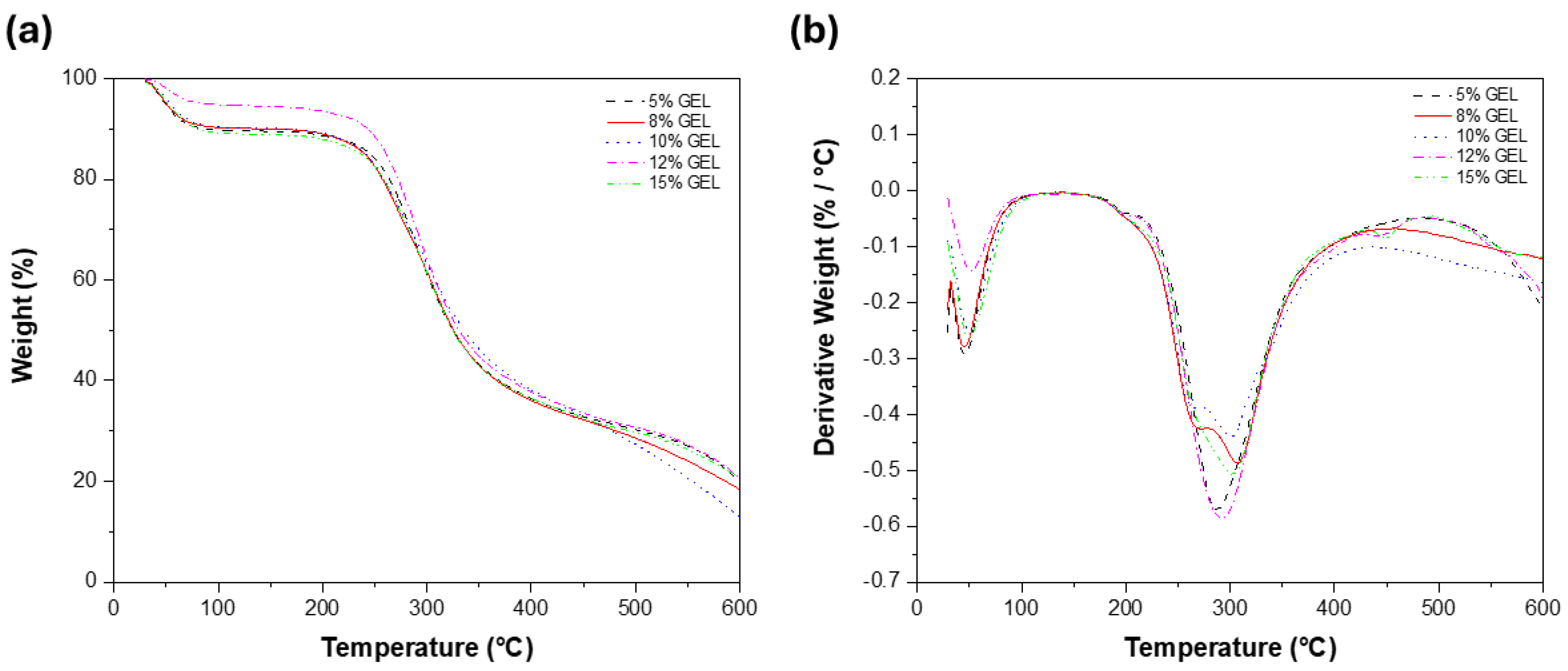
Figure 8 shows the TGA and DTG curves for gelatin solutions at 5–15% concentration. All samples exhibited similar thermal degradation profiles consisting of three main stages, indicating that gelatin concentration had little effect on the overall degradation mechanism. Table 4 shows that the first weight-loss stage (30–90 °C) was associated with the evaporation of physically adsorbed moisture, followed by a slight mass loss between 85 and 230 °C, attributed to the removal of strongly bound water and low-molecular-weight compounds. The main degradation event occurred between 220 and 380 °C, due to the breakdown of peptide bonds and decomposition of the gelatin backbone. Among the evaluated formulations, the 12% gelatin sample exhibited a shift in the degradation peak toward higher temperatures and the highest weight loss (55%), indicating enhanced thermal stability.
Figure 8.
Thermogravimetric analysis of gelatin nanoparticles (5%, 8%, 10%, 12%, and 15%). (a) Mass loss thermogram (TGA) and (b) derivative thermogram (DTG).
Table 4.
Thermogravimetric analysis (TGA) of gelatin nanoparticles.
In contrast, the 10% gelatin sample exhibited the greatest weight loss during the final degradation stage (370–600 °C), suggesting the formation of less thermally stable residue. These results align with the three-stage degradation profile of Type A gelatin, where structural water is removed below 200 °C and peptide bond cleavage occurs between 200 and 400 °C [8]. The improved thermal stability of the 12% gelatin formulation is attributed to increased molecular chain entanglement in the semidilute regime, which enhances protein network cohesion and delays thermal degradation [5]. All formulations remained thermally stable below approximately 220 °C, demonstrating their suitability for moderate-temperature processing.
4. Conclusions
This study demonstrates the feasibility of using uniaxial electrospraying to fabricate gelatin-based structures containing curcumin extract. The selected gelatin formulation enabled the production of spherical nanoparticles. At the same time, curcumin extract incorporation reduced particle size and surface charge of resulting structures, highlighting its influence on nanoparticle organization and surface charge. Although the 3% (w/v) curcumin extract formulation exhibited the smallest mean particle size and the lowest PDI among the curcumin-containing formulations, the 5% (w/v) formulation showed the highest absolute zeta potential (−28.56 ± 1.25 mV), suggesting a lower tendency toward particle aggregation. FTIR analysis supported the presence of interactions between gelatin and curcumin, while thermal analysis provided information on the thermal behavior of the electrosprayed formulations. Overall, these results demonstrate the influence of formulation composition on the physicochemical and morphological characteristics of gelatin–curcumin structures produced by uniaxial electrospraying. Further studies should evaluate encapsulation efficiency, storage stability, release behavior, and biological performance to determine their suitability for bioactive compound delivery applications.
Author Contributions
A.V.T.-G., writing—original draft preparation and formal analysis; C.G.B.-U., formal analysis, validation, methodology, supervision; L.A.-V., supervision; T.J.M.-S., visualization, resources; A.Z.G.-V., visualization and supervision; F.R.-F., supervision and investigation; J.A.T.-H., resources and formal analysis; I.Y.L.-P., visualization. B.E.L.-C., visualization; M.J.M.-V., supervision; I.E.Q.-R., visualization. All authors have read and agreed to the published version of the manuscript.
Funding
This research received no external funding.
Institutional Review Board Statement
Not applicable.
Data Availability Statement
The data are contained within the article.
Acknowledgments
The authors express their gratitude to the University of Sonora for its academic support and institutional resources. Special thanks to the Centro de Investigación en Alimentación y Desarrollo (CIAD) for providing access to its facilities and enabling participation in the bioactive compound extraction process. The authors also acknowledge the technical assistance of William Santiago González and Víctor Rejón-Moo at CINVESTAV-IPN Unidad Mérida in performing the SEM and FTIR measurements. These analyses were performed at Laboratorio Nacional CONAHCYT LANNBIO-Cinvestav-Mérida (PROY No. 321119). The authors also acknowledge BioRender for assistance in creating selected schematic illustrations presented in this manuscript.
Conflicts of Interest
The authors declare no conflicts of interest.
References
- Dinparvar, S.; Bagirova, M.; Allahverdiyev, A.M.; Abamor, E.S.; Safarov, T.; Aydogdu, M.; Aktas, D. A nanotechnology-based new approach in the treatment of breast cancer: Biosynthesized silver nanoparticles using Cuminum cyminum L. seed extract. J. Photochem. Photobiol. B Biol. 2020, 208, 111902. [Google Scholar] [CrossRef] [Scilit]
- Bhushani, J.A.; Anandharamakrishnan, C. Electrospinning and electrospraying techniques: Potential food based applications. Trends Food Sci. Technol. 2014, 38, 21–33. [Google Scholar] [CrossRef] [Scilit]
- Gómez-Mascaraque, L.G.; Tordera, F.; Fabra, M.J.; Martínez-Sanz, M.; Lopez-Rubio, A. Coaxial electrospraying of biopolymers as a strategy to improve protection of bioactive food ingredients. Innov. Food Sci. Emerg. Technol. 2019, 51, 2–11. [Google Scholar] [CrossRef] [Scilit]
- Castro-Coelho, S.C.; Estevinho, B.N.; Rocha, F. Encapsulation in food industry with emerging electrohydrodynamic techniques: Electrospinning and electrospraying—A review. Food Chem. 2021, 339, 127850. [Google Scholar] [CrossRef] [Scilit]
- Erencia, M.; Cano, F.; Tornero, J.A.; Macanás, J.; Carrillo, F. Resolving the electrospinnability zones and diameter prediction for the electrospinning of the gelatin/water/acetic acid system. Langmuir 2014, 30, 7198–7205. [Google Scholar] [CrossRef] [Scilit]
- Tapia-Hernández, J.A.; Del-Toro-Sánchez, C.L.; Cinco-Moroyoqui, F.J.; Ruiz-Cruz, S.; Juárez, J.; Castro-Enríquez, D.D.; Barreras-Urbina, C.G.; López-Ahumada, G.A.; Rodríguez-Félix, F. Gallic acid-loaded zein nanoparticles by electrospraying process. J. Food Sci. 2019, 84, 818–831. [Google Scholar] [CrossRef] [Scilit]
- Feng, X.; Dai, H.; Ma, L.; Fu, Y.; Yu, Y.; Zhou, H.; Guo, T.; Zhang, Y.; Zhu, H.; Wang, H. Properties of Pickering emulsion stabilized by food-grade gelatin nanoparticles: Influence of the nanoparticles concentration. Colloids Surf. B Biointerfaces 2020, 196, 111294. [Google Scholar] [CrossRef] [Scilit]
- Gómez-Mascaraque, L.G.; Lagarón, J.M.; López-Rubio, A. Electrosprayed gelatin submicroparticles as edible carriers for the encapsulation of polyphenols of interest in functional foods. Food Hydrocoll. 2015, 49, 42–52. [Google Scholar] [CrossRef] [Scilit]
- Ahmed, J. Rheological properties of gelatin and advances in measurement. In Advances in Food Rheology and Its Applications; Woodhead Publishing: Cambridge, UK, 2017; pp. 377–404. [Google Scholar]
- Zhang, J.; Wang, G.; Liang, Q.; Cai, W.; Zhang, Q. Rheological and microstructural properties of gelatin B/tara gum hydrogels: Effect of protein/polysaccharide ratio, pH and salt addition. LWT Food Sci. Technol. 2019, 103, 108–115. [Google Scholar] [CrossRef] [Scilit]
- Khan, M.R.; Sadiq, M.B. Importance of gelatin, nanoparticles and their interactions in the formulation of biodegradable composite films: A review. Polym. Bull. 2020, 78, 4047–4073. [Google Scholar] [CrossRef] [Scilit]
- Yang, Z.; Chaieb, S.; Hemar, Y. Gelatin-based nanocomposites: A review. Polym. Rev. 2021, 61, 765–813. [Google Scholar] [CrossRef] [Scilit]
- Alipal, J.; Mohd Pu’Ad, N.A.S.; Lee, T.C.; Nayan, N.H.M.; Sahari, N.; Basri, H.; Idris, M.I.; Abdullah, H.Z. A review of gelatin: Properties, sources, process, applications, and commercialisation. Mater. Today Proc. 2021, 42, 240–250. [Google Scholar] [CrossRef] [Scilit]
- Lu, Y.; Luo, Q.; Chu, Y.; Tao, N.; Deng, S.; Wang, L.; Li, L. Application of gelatin in food packaging: A review. Polymers 2022, 14, 436. [Google Scholar] [CrossRef] [Scilit]
- Kavukcu, S.B.; Çakır, S.; Karaer, A.; Türkmen, H.; Rethinam, S. Curcumin nanoparticles supported gelatin-collagen scaffold: Preparation, characterization, and in vitro study. Toxicol. Rep. 2021, 8, 1475–1479. [Google Scholar] [CrossRef] [Scilit]
- Baydin, T.; Aarstad, O.A.; Dille, M.J.; Hattrem, M.N.; Draget, K.I. Long-term storage stability of type A and type B gelatin gels: The effect of Bloom strength and co-solutes. Food Hydrocoll. 2022, 127, 107535. [Google Scholar] [CrossRef] [Scilit]
- Rathore, P.; Schiffman, J.D. Beyond the single-nozzle: Coaxial electrospinning enables innovative nanofiber chemistries, geometries, and applications. ACS Appl. Mater. Interfaces 2021, 13, 48–66. [Google Scholar] [CrossRef] [Scilit]
- Tapia, V. The Entrapment or Encapsulation of Biocompounds Using Uniaxial or Coaxial Methodology. Created in BioRender. 2026. Available online: https://BioRender.com/ehlidry (accessed on 19 September 2026).
- Khanh, L.L.; Truc, N.T.; Dat, N.T.; Nghi, N.T.P.; Van Toi, V.; Hoai, N.T.T.; Quyen, T.N.; Loan, T.T.T.; Hiep, N.T. Gelatin-stabilized composites of silver nanoparticles and curcumin: Characterization, antibacterial and antioxidant study. Sci. Technol. Adv. Mater. 2019, 20, 276–290. [Google Scholar] [CrossRef] [Scilit]
- Irshad, S.; Muazzam, A.; Shahid, Z.; Dalrymple, M.B. Curcuma longa (Turmeric): An auspicious spice for antibacterial, phytochemical and antioxidant activities. Pak. J. Pharm. Sci. 2018, 31, 2689–2696. [Google Scholar] [CrossRef] [Scilit]
- Gómez-Estaca, J.; Gavara, R.; Hernández-Muñoz, P. Encapsulation of curcumin in electrosprayed gelatin microspheres enhances its bioaccessibility and widens its uses in food applications. Innov. Food Sci. Emerg. Technol. 2015, 29, 302–307. [Google Scholar] [CrossRef] [Scilit]
- Karimi, A.; Askari, G.; Yarmand, M.S.; Salami, M.; Emam-Djomeh, Z. Development, modification and characterization of ursolic acid-loaded gelatin nanoparticles through electrospraying technique. Food Bioprod. Process. 2020, 124, 329–341. [Google Scholar] [CrossRef] [Scilit]
- Aliabbasi, N.; Mehrabi, S.; Kheirandish, M.; Gashtasbi, S.; Mokhtarian, M.; Hosseini-Isfahani, M.; Vakilinezami, A.; Vakilinezami, P.; Mostaghim, T.; Rezaeinia, H. The novel nano-electrospray delivery of curcumin via ultrasound assisted Balangu (Lallemantia royleana) hydrocolloid-chickpea protein interaction. Food Chem. 2025, 484, 144388. [Google Scholar] [CrossRef] [Scilit]
- Cao, M.; Zhou, F.; Cheng, S.; Wang, X.; Yuan, Z.; Huang, X.; Shu, Z. Effects of preparation method on the release kinetics of curcumin-loaded composites. React. Funct. Polym. 2025, 217, 106488. [Google Scholar] [CrossRef] [Scilit]
- Golmakani, M.-T.; Zamansani, E.; Hajjari, M.M.; Moosavi-Nasab, M.; Niakousari, M. Curcumin-loaded gelatin/fucoidan electrospun composite: Physicochemical characterization and antioxidative application. Food Chem. X 2025, 27, 102495. [Google Scholar] [CrossRef] [Scilit]
- Liew, S.Q.; Teoh, W.H.; Yusoff, R.; Ngoh, G.C. Comparisons of process intensifying methods in the extraction of pectin from pomelo peel. Chem. Eng. Process.—Process Intensif. 2019, 143, 107586. [Google Scholar] [CrossRef] [Scilit]
- Rodríguez-Félix, F.; Del-Toro-Sánchez, C.L.; Cinco-Moroyoqui, F.J.; Juárez, J.; Ruiz-Cruz, S.; López-Ahumada, G.A.; Carvajal-Millan, E.; Castro-Enríquez, D.D.; Barreras-Urbina, C.G.; Tapia-Hernández, J.A. Preparation and characterization of quercetin-loaded zein nanoparticles by electrospraying and study of in vitro bioavailability. J. Food Sci. 2019, 84, 2883–2897. [Google Scholar] [CrossRef] [Scilit]
- Tapia-Hernández, J.A.; Torres-Chávez, P.I.; Ramírez-Wong, B.; Rascón-Chu, A.; Plascencia-Jatomea, M.; Barreras-Urbina, C.G.; Rangel-Vázquez, N.A.; Rodríguez-Félix, F. Micro- and nanoparticles by electrospray: Advances and applications in foods. J. Agric. Food Chem. 2015, 63, 4699–4707. [Google Scholar] [CrossRef] [Scilit]
- Tapia, V. Schematic Description of the Synthesis of Gelatin-Curcumin Nanoparticles by the Electrospraying Method. Created in BioRender. 2026. Available online: https://BioRender.com/e9bexi6 (accessed on 19 September 2026).
- Barreras-Urbina, C.G.; Plascencia-Jatomea, M.; Wong-Corral, F.J.; Pérez-Tello, M.; Ledesma-Osuna, A.I.; Tapia-Hernández, J.A.; Castro-Enríquez, D.D.; Rueda-Puente, E.O.; Rodríguez-Félix, F. Simple method to obtaining a prolonged-release system of urea based on wheat gluten: Development and characterization. Polym. Bull. 2020, 77, 6525–6541. [Google Scholar] [CrossRef] [Scilit]
- Ghorani, B.; Emadzadeh, B.; Rezaeinia, H.; Russell, S.J. Improvements in gelatin cold water solubility after electrospinning and associated physicochemical, functional and rheological properties. Food Hydrocoll. 2020, 104, 105740. [Google Scholar] [CrossRef] [Scilit]
- Yang, T.; Yang, H.; Fan, Y.; Li, B.; Hou, H. Interactions of quercetin, curcumin, epigallocatechin gallate and folic acid with gelatin. Int. J. Biol. Macromol. 2018, 118, 124–131. [Google Scholar] [CrossRef] [Scilit]
- Goudie, K.J.; McCreath, S.J.; Parkinson, J.A.; Davidson, C.M.; Liggat, J.J. Investigation of the influence of pH on the properties and morphology of gelatin hydrogels. J. Polym. Sci. 2023, 61, 2316–2332. [Google Scholar] [CrossRef] [Scilit]
- Zhang, S.; Campagne, C.; Salaün, F. Influence of solvent selection in the electrospraying process of polycaprolactone. Appl. Sci. 2019, 9, 402. [Google Scholar] [CrossRef] [Scilit]
- Castro, G.M.M.A.; Passos, T.S.; Nascimento, S.S.D.C.; Medeiros, I.; Araújo, N.K.; Maciel, B.L.L.; Padilha, C.E.; Ramalho, A.M.Z.; Sousa Júnior, F.C.; de Assis, C.F. Gelatin nanoparticles enable water dispersibility and potentialize the antimicrobial activity of Buriti (Mauritia flexuosa) oil. BMC Biotechnol. 2020, 20, 55. [Google Scholar] [CrossRef] [Scilit]
- Lee, H.; An, S.; Kim, S.; Jeon, B.; Kim, M.; Kim, I.S. Readily functionalizable and stabilizable polymeric particles with controlled size and morphology by electrospray. Sci. Rep. 2018, 8, 15725. [Google Scholar] [CrossRef] [Scilit]
- Thakkar, S.; Misra, M. Electrospray particles as drug delivery systems: Concepts, strategies, and applications. In Biomedical Applications of Electrospinning and Electrospraying; Woodhead Publishing: Cambridge, UK, 2021; pp. 245–275. [Google Scholar]
- Titus, D.; Samuel, E.J.J.; Roopan, S.M. Nanoparticle characterization techniques. In Green Synthesis, Characterization and Applications of Nanoparticles; Elsevier: Amsterdam, The Netherlands, 2019; pp. 303–319. [Google Scholar]
- Lunardi, C.N.; Gomes, A.J.; Rocha, F.S.; De Tommaso, J.; Patience, G.S. Experimental methods in chemical engineering: Zeta potential. Can. J. Chem. Eng. 2021, 99, 627–639. [Google Scholar] [CrossRef] [Scilit]
- Chhouk, K.; Diono, W.; Kanda, H.; Goto, M. Micronization for Enhancement of Curcumin Dissolution via Electrospraying Technique. ChemEngineering 2018, 2, 60. [Google Scholar] [CrossRef] [Scilit]
- Xia, Y.; Xu, R.; Ye, S.; Yan, J.; Kumar, P.; Zhang, P.; Zhao, X. Microfluidic Formulation of Curcumin-Loaded Multiresponsive Gelatin Nanoparticles for Anticancer Therapy. ACS Biomater. Sci. Eng. 2023, 9, 3402–3413. [Google Scholar] [CrossRef] [Scilit]
- Wang, X.; Xu, L.; Zheng, G.; Jiang, J.; Sun, D.; Li, W. Formation of suspending beads-on-a-string structure in electrohydrodynamic printing process. Mater. Des. 2021, 204, 109692. [Google Scholar] [CrossRef] [Scilit]
- Torkamani, A.E.; Syahariza, Z.A.; Norziah, M.H.; Mahmood, W.A.K.; Juliano, P. Production and characterization of gelatin spherical particles formed via electrospraying and encapsulated with polyphenolic antioxidants from Momordica charantia. Food Bioprocess Technol. 2018, 11, 1943–1954. [Google Scholar] [CrossRef] [Scilit]
- Asadi, M.; Salami, M.; Hajikhani, M.; Emam-Djomeh, Z.; Aghakhani, A.; Ghasemi, A. Electrospray production of curcumin-walnut protein nanoparticles. Food Biophys. 2021, 16, 15–26. [Google Scholar] [CrossRef] [Scilit]
- Avossa, J.; Herwig, G.; Toncelli, C.; Itel, F.; Rossi, R.M. Electrospinning based on benign solvents: Current definitions, implications and strategies. Green Chem. 2022, 24, 2347–2375. [Google Scholar] [CrossRef] [Scilit]
- Derkach, S.R.; Voron’ko, N.G.; Sokolan, N.I.; Kolotova, D.S.; Kuchina, Y.A. Interactions between gelatin and sodium alginate: UV and FTIR studies. J. Dispers. Sci. Technol. 2020, 41, 690–698. [Google Scholar] [CrossRef] [Scilit]
- Yang, H.; Duan, L.; Li, Q.; Tian, Z.; Li, G. Experimental and modeling investigation on the rheological behavior of collagen solution as a function of acetic acid concentration. J. Mech. Behav. Biomed. Mater. 2018, 77, 125–134. [Google Scholar] [CrossRef] [Scilit]
- Tapia, V. Representation of the Gelatin-Curcumin Assembly on the Nanoparticles. Created in BioRender. 2026. Available online: https://BioRender.com/j1urys0 (accessed on 19 September 2026).

Disclaimer/Publisher’s Note: The statements, opinions and data contained in all publications are solely those of the individual author(s) and contributor(s) and not of MDPI and/or the editor(s). MDPI and/or the editor(s) disclaim responsibility for any injury to people or property resulting from any ideas, methods, instructions or products referred to in the content. |
© 2026 by the authors. Licensee MDPI, Basel, Switzerland. This article is an open access article distributed under the terms and conditions of the Creative Commons Attribution (CC BY) license.