


Abstract
Bleeding remains a leading cause of death in trauma and surgery, which drives the search for effective local hemostatic materials. Chitosan is a promising candidate because of its biocompatibility, biodegradability, and intrinsic hemostatic activity, yet chitosan hydrogels remain poorly characterized in terms of hemocompatibility. This study assessed the hemocompatibility and hemostatic activity of chitosan hydrogels with varying degrees of crosslinking and their complexes with ficin. Hydrogels were synthesized using 1,4-butanediol diglycidyl ether and characterized by ζ-potential measurements. Hemocompatibility was evaluated through hemolysis, hemoglobin spectroscopy, erythrocyte cytoarchitecture, and lymphocyte viability assays. Ficin was immobilized by adsorption and covalent binding, and hemostatic properties were assessed via swelling and kinetic blood coagulation. The hydrogels showed a statistically significant inverse dose-dependent hemolytic effect, attributed to their low colloidal stability. In contrast, hemoglobin spectra, erythrocyte cytoarchitecture, and lymphocyte viability showed no statistically significant changes. Adsorption immobilization provided higher protein loading and proteolytic activity than covalent binding. Ficin-containing compositions significantly accelerated blood coagulation compared with pristine hydrogels. These findings confirm the potential of the developed hydrogels and their ficin complexes as hemocompatible hemostatic materials.
1. Introduction
Bleeding remains a leading cause of death in trauma, surgery, and disaster medicine. A substantial share of these deaths results from blood loss occurring before patients reach a medical facility [1,2]. Effective local hemostatic materials that can rapidly stop bleeding are therefore a priority in biomedical engineering. To date, various hemostatic agents based on cellulose, collagen, gelatin, and synthetic polymers have entered clinical practice [3]. However, many of them have significant limitations, including high cost, immunogenicity, limited biodegradation, and insufficient efficacy in massive bleeding [4,5].
In recent decades, chitosan—a biopolymer obtained by deacetylation of chitin—has attracted considerable research interest. Chitosan combines several valuable properties, including biocompatibility, biodegradability, antimicrobial activity, and pronounced hemostatic capacity [6,7]. Its hemostatic mechanism is attributed to its polycationic nature, which arises from primary amino groups. These groups enable electrostatic interactions with the negatively charged membranes of red blood cells and platelets, promoting their aggregation and activating the coagulation cascade. Importantly, the form in which chitosan is used—powder or particles, sponge, hydro- or cryogel, nanofiber, and so on—strongly influences its hemostatic efficacy. Previous studies have shown that crosslinked chitosan forms, such as sponges and hydrogels, provide the highest hemostatic efficiency [6]. Native chitosan, however, has limited mechanical strength and degrades rapidly under physiological conditions, which significantly restricts its use as a wound dressing or hemostatic material. Crosslinking of polymer chains, required to form a hydrogel or sponge, addresses these issues. It improves mechanical stability, allows the degree of swelling and the biodegradation rate to be tuned, and enables control over porosity, sorption capacity, and cell adhesion. Crosslinking also gives these materials a structural resemblance to the extracellular matrix [8,9].
Another strategy for improving chitosan-based hemostatic materials is to functionalize them with substances that have intrinsic coagulant activity. For example, incorporating calcium alginate—a source of coagulation factor IV—into chitosan granules cuts the hemostatic time on liver laceration by nearly half [10]. Chitosan sponges containing fibrinogen and thrombin—coagulation factors I and IIa, which are serine proteases—have also been reported [11]. These materials show excellent hemostatic properties, fully coagulating blood within 75 s. This is nearly four times more effective than a chitosan sponge without incorporated proteases. However, thrombin and fibrinogen are expensive and difficult to obtain. This drives the search for cheaper protein components with similar effects. Plant cysteine proteases are particularly attractive in this regard. They are readily available, show high proteolytic activity, have broad substrate specificity, and can degrade necrotic tissue [12]. The most widely studied plant cysteine proteases are papain, bromelain, and ficin, all of which share a conserved catalytic triad of Cys, His, and Asn residues and a similar overall fold [13]. Papain, derived from Carica papaya latex, has been extensively characterized and is widely used in industry, but it exhibits relatively low thermal stability and is rapidly inactivated under acidic conditions [14]. Bromelain, obtained from pineapple stem, shows high proteolytic activity and anti-inflammatory properties, but it is a complex mixture of several proteases and non-protease components, which complicates its standardization and limits the reproducibility of formulations [15]. Ficin, isolated from fig latex, offers several practical advantages for the present application. It combines high specific activity with a relatively simple purification profile, and it remains active over a broader pH range than papain, including near-neutral conditions relevant to blood contact [16]. Notably, ficin is known to cleave fibrin, activate human coagulation factor X, and exhibit plasminogen-like activity [17,18,19], which directly supports its role as a cascade-activating component of a hemostatic material. Ficin also possesses antibacterial and antibiofilm activity of its own [20], which further expands the functionality of the resulting hemostatic agent. These combined properties make ficin a more suitable choice than papain or bromelain for developing multifunctional hemostatic materials.
Developing a hemostatic agent that contains ficin, or any other protease, is essentially an enzyme immobilization problem. This means that techniques and expertise from the field of enzyme immobilization can be applied directly. The most common approaches are adsorption and covalent immobilization. Adsorption immobilization is simple and cost-effective, but it may lead to enzyme desorption under physiological conditions [21]. Covalent immobilization provides more stable attachment of the enzyme, but it often causes partial loss of catalytic activity due to conformational changes in the protein. It also requires additional reagents, such as glutaraldehyde [22,23]. Careful optimization of the immobilization conditions and method is therefore usually required [24,25].
Several studies have already explored the immobilization of ficin on chitosan-based matrices. For example, Baidamshina et al. reported the adsorption-based immobilization of ficin on acid-soluble chitosan and demonstrated its anti-biofilm and wound-healing activity [26]. Other work from our group has examined ficin immobilization on chitosan graft copolymers with N-vinylimidazole, on carboxymethyl chitosan, and on 2-(4-acetamido-2-sulfanilamide) chitosan, focusing primarily on the preservation of proteolytic activity and the structural features of the resulting complexes [27,28,29]. In these studies, the carrier was either a soluble chitosan derivative or a non-crosslinked chitosan matrix, and the primary focus was on the enzymatic and antibacterial properties of the immobilized ficin.
1,4-Butanediol diglycidyl ether (BDDE) has also been used to crosslink chitosan into cryogels and other hydrogel architectures, mainly for tissue engineering and drug delivery applications [30]. However, these studies did not address the immobilization of ficin or the hemostatic performance of the resulting materials. To the best of our knowledge, the combination of BDDE-crosslinked chitosan hydrogels with immobilized ficin has not been reported so far.
In contrast, the present work addresses a different formulation space and a different functional goal. First, we use cross-linked chitosan hydrogels (Cross-Cs) prepared by covalent crosslinking of chitosan with BDDE at two different crosslinker ratios, which allows the hydrogel network properties (charge, swelling, colloidal stability) to be systematically varied. Second, we evaluate the hemocompatibility of the resulting materials across multiple parameters, including hemolysis, hemoglobin spectral characteristics, erythrocyte cytoarchitecture, and lymphocyte viability. Third, we demonstrate that the ficin-loaded hydrogels significantly accelerate whole-blood coagulation. To our knowledge, this is the first study that combines a systematic evaluation of BDDE-crosslinked chitosan hydrogels, a comprehensive hemocompatibility assessment, and the use of immobilized ficin to activate the natural coagulation cascade.
Despite extensive research on chitosan-based hemostatic agents, chitosan hydrogels remain poorly characterized. Their effects on key hemocompatibility parameters—hemolysis, hemoglobin spectral properties, red blood cell morphology, and lymphocyte viability—are largely unknown. This gap in knowledge motivated the present study, which aimed to comprehensively assess the hemocompatibility and hemostatic activity of chitosan hydrogels with varying degrees of crosslinking, as well as their complexes with ficin.
2. Materials and Methods
2.1. Materials
Chitosan with a molecular weight of 200 kDa and a degree of deacetylation of 0.91 (Bioprogress, Moscow, Russia) and 1,4-butanediol diglycidyl ether (>98%, Shanghai Macklin Biochemical Technology Co., Ltd., Shanghai, China) were used to synthesize the chitosan hydrogels. Ficin (F4165), azocasein (A2765), and glutaraldehyde (50 wt.% aqueous solution, W512303) were purchased from Sigma-Aldrich (St. Louis, MO, USA). Trypan blue, resazurin, and Ficoll were purchased from Shanghai Macklin Biochemical Technology Co., Ltd. (Shanghai, China). All reagents were used as received.
2.2. Synthesis and Characterization of Chitosan Hydrogels
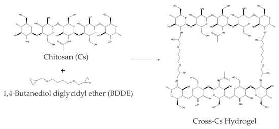
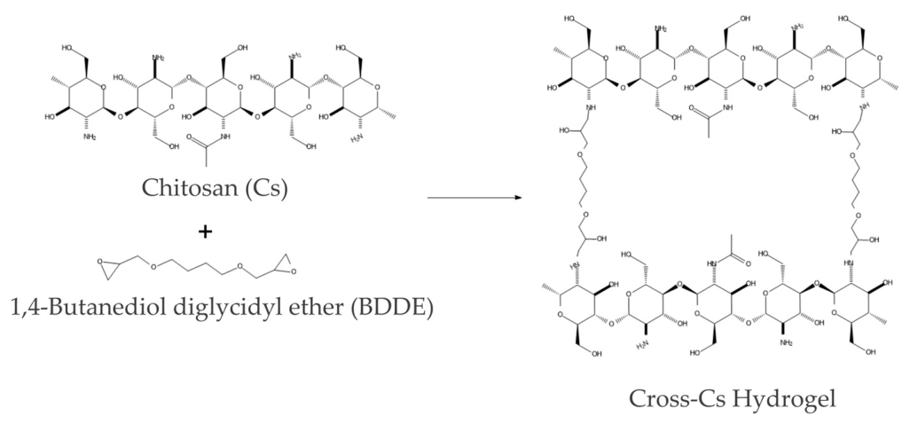
Chitosan (Cs; 1 g) and 40 mL of a 2% acetic acid solution were placed in a thermostated reactor equipped with a stirrer and a reflux condenser. After the chitosan had completely dissolved, 0.875 or 1.75 mL of BDDE was added to obtain Cross-Cs-1-1 (Cs:BDDE molar ratio = 1:1) and Cross-Cs-1-2 (Cs:BDDE molar ratio = 1:2), respectively. The mixture was kept at 80 °C for 2 h. Once the reaction was complete, the resulting gel was collected and purified by dialysis against distilled water through a regenerated cellulose membrane (molecular weight cut-off 3.5 kDa) for three days. The product was then freeze-dried to constant weight, with a yield of 84–87%. Scheme 1 illustrates the reaction.
Scheme 1.
Crosslinking of chitosan with BDDE.
The ζ-potential of the chitosan hydrogels was measured on 0.05% (w/v) aqueous solutions using a Zetasizer Nano ZS instrument (Malvern Panalytical B.V., Almelo, The Netherlands).
The swelling behavior of the chitosan hydrogels was studied in distilled water. A hydrogel sample (~10 mg) was placed in 10 mL of distilled water and kept at 25 °C for 24 h to reach equilibrium swelling. The swollen hydrogel was then filtered off, the excess liquid was carefully removed from its surface with filter paper, and the sample was weighed. The swelling ratio (SR, g/g) was calculated as [31,32]:
where m1 and m0 are the masses of the swollen and dry hydrogel, respectively (g). Measurements were performed in triplicate for each sample.
The crosslinking density of the hydrogels was estimated using the scaling relationship derived from the Flory–Rehner theory [33], which relates the effective crosslinking density (νe) to the equilibrium swelling ratio:
where Q is the equilibrium swelling ratio (g/g). Because the classical Flory–Rehner equation with χ = 0.63 [34] for the chitosan–water system yields non-physical results for strongly swelling polyelectrolyte hydrogels (the denominator approaches zero and the ionic contribution of quaternized chitosan is not accounted for), the scaling relationship was used to compare the relative crosslinking densities of the synthesized hydrogels within the same polymer family. The crosslinking density ratio between two samples was calculated as:
The equilibrium swelling ratios of Cross-Cs-1-1 and Cross-Cs-1-2 in distilled water were 15 and 9 g/g, respectively. Using the scaling relationship, the crosslinking density of Cross-Cs-1-2 was estimated to be approximately 2.2 times higher than that of Cross-Cs-1-1. This is consistent with the twofold excess of BDDE used in the synthesis of Cross-Cs-1-2 and confirms that the crosslinker content effectively controls the network density of the resulting hydrogels.
2.3. Blood Sampling
Venous blood was obtained from healthy volunteers aged 18–45 years. The inclusion criteria were age 18–45 years, good general health, and written informed consent. The exclusion criteria were a history of hematological, cardiovascular, hepatic, or renal disease; recent infection or antibiotic treatment within the previous 30 days; use of anticoagulant or antiplatelet medication; pregnancy or lactation; and any known bleeding disorder. Blood was obtained from 30 independent healthy donors (16 female and 14 male). For each experiment, blood from a single donor was used without pooling, so that biological replicates represent independent donors rather than repeated measurements from the same sample. Each experiment was performed with blood from at least 10 independent donors. All procedures complied with good clinical practice and the ethical requirements for blood collection. The study followed the principles of the Declaration of Helsinki (7th revision, 2013) and the ethical guidelines of the Voronezh Regional Blood Center. Blood was pretreated with an anticoagulant mixture (citric acid, sodium citrate, and dextrose).
2.4. Red Blood Cell Hemolysis Assay
To prepare the red blood cell (RBC) suspension, 1 mL of whole blood was mixed with 9 mL of 0.9% NaCl solution and centrifuged at 1500 rpm for 15 min. The supernatant was carefully removed, and the procedure was repeated twice to ensure maximum purity of the RBC suspension.
The absorbance of the initial RBC suspension was adjusted to ~1.2 a.u. at λ = 495 nm. An aliquot of the RBC suspension was diluted with an equal volume of the chitosan hydrogel suspension (A0), distilled water (Al), or 0.9% NaCl (Ac). After incubation at 37 °C for 20 min, the absorbance of all samples was measured at λ = 495 nm using a UV-2550PC spectrophotometer (Shimadzu Scientific Instruments Inc., Kyoto, Japan).
The percentage of non-lysed RBCs was calculated using the following equation [35]:
2.5. Hemoglobin Spectra
To isolate hemoglobin, 200 µL of the RBC suspension was lysed by adding 1800 µL of distilled water. The resulting mixture was centrifuged at 10,000 rpm for 20 min. The absorbance of the supernatant was adjusted to 0.8 a.u. at λ = 414 nm. Absorption spectra of the hemoglobin solutions were recorded on a UV-2550PC spectrophotometer (Shimadzu Scientific Instruments Inc., Kyoto, Japan) over a wavelength range of 190–900 nm, with a spectral slit width of 1 nm and a scanning step of 1 nm, in eight replicates.
The content of oxyhemoglobin (HbO2), deoxyhemoglobin (Hb), and methemoglobin (MtHb) was calculated using the following equations [36]:
where [HbO2], [Hb], and [MtHb] are the concentrations of the hemoglobin forms (mol/L), and A577, A569, and A500 are the absorbance values of the hemoglobin solution at 577, 569, and 500 nm, respectively.
[HbO2] = (1.7A577 − 0.13A569 − 0.15A500)/10−4,
[Hb] = (−1.6A577 + 2.5A569 + 0.33A500)/10−4,
[MtHb] = (0.2A577 − 0.4A569 + 0.33A500)/10−4,
2.6. RBC Cytoarchitecture
For cytoarchitecture assessment, the RBC suspension was mixed with a 0.1% chitosan hydrogel dispersion at a 1:1 volume ratio and incubated at 37 °C for 30 min. The cells were then fixed in 2.5% (w/v) glutaraldehyde for 1 h. After fixation, the suspension was placed onto a specially prepared aluminum slide and dehydrated through a graded ethanol series (25%, 50%, 70%, 80%, 90%, and 100%). The samples were subsequently air-dried at 37 °C [37]. Dried cells were sputter-coated with gold and examined using a JSM 6380 LV scanning electron microscope equipped with an INKA 250 microanalysis system (JEOL Ltd., Tokyo, Japan). An untreated RBC suspension served as the reference sample. Cell shapes were classified according to the approach described by Bessis [38]. For each sample, 500–1500 cells were counted in randomly selected fields. The numbers of intact cells (Nint), reversibly transformed cells (Nrt), and irreversibly transformed cells (Nit) were recorded and expressed as percentages. The following indices were calculated: transformation index (TI) = (Nrt + Nit)/Nint; reversible transformation index (RTI) = Nrt/Nint; and irreversible transformation index (ITI) = Nit/Nint, where Nint, Nrt, and Nit are the counts of intact, reversibly transformed, and irreversibly transformed cells, respectively [39].
2.7. Lymphocytes Viability
Lymphocytes were isolated by density gradient centrifugation of donor blood using Ficoll-Urografin (ρ = 1.077 g/cm3) [40]. The lymphocyte suspension was treated with chitosan hydrogel dispersions as described in Section 2.6 for 1 h. The number of viable cells in the suspensions was determined by trypan blue exclusion and by the resazurin assay [41,42].
2.8. Ficn Immobilization on Chitosan Hydrogels
2.8.1. Adsorption Immobilization
A ficin solution (200 mL, 1 mg/mL in 0.05 M Tris-HCl buffer, pH 7.4, or borate buffer with KCl, pH 9.0) was added to 1 g of the chitosan hydrogel. The mixture was incubated for 3 h at 25 °C and then centrifuged at 1500× g for 10 min. After incubation, the resulting preparation was washed with 0.05 M Tris-HCl buffer (pH 7.4) or borate buffer with KCl (pH 9.0) until no protein was detected in the washings. Protein content was monitored on an SF-2000 spectrophotometer (LOMO Systems, St. Petersburg, Russia) at λ = 280 nm.
2.8.2. Covalent Immobilization
A ficin solution (20 mL, 1 mg/mL in 0.05 M Tris-HCl buffer, pH 7.4) and 10 mL of 5% glutaraldehyde were added to 1 g of the chitosan hydrogel. The mixture was incubated for 1 h at 4 °C and then centrifuged at 1500× g for 10 min. After incubation, the resulting preparation was purified by dialysis against 0.05 M Tris-HCl buffer (pH 7.4) through a cellophane membrane with a molecular weight cut-off of 25 kDa until no protein was detected in the washings. Protein content was monitored on an SF-2000 spectrophotometer (LOMO Systems, St. Petersburg, Russia) at λ = 280 nm.
Protein content and total protease activity in the azocasein hydrolysis reaction were assessed as described in [43,44].
2.9. Evaluating Hemostatic Properties of Chitosan Hydrogels and Ficin–Chitosan Hydrogel Compositions
2.9.1. Swelling Behavior of Chitosan Hydrogels
A chitosan hydrogel sample (~10 mg) was placed in 200 µL of human blood. After 120 s, the sample was removed from the blood and weighed. Measurements were performed in six replicates for each polymer. The swelling ratio of the hydrogel was calculated using Equation (1).
2.9.2. Kinetic Whole Blood Coagulation
Samples of chitosan hydrogels or hydrogel-immobilized ficin (2 mg each) were placed on watch glasses, and 200 µL of human blood was added. For each sample, seven watch glasses were prepared. To initiate aggregation, 25 µL of 0.2 mol/L calcium chloride solution was added, and the time of clot formation onset was recorded. Every 5 min, one of the glasses was immersed in 100 mL of distilled water, and the absorbance of the resulting solution was measured at 545 nm using an SF-2000 spectrophotometer (LOMO Systems, St. Petersburg, Russia).
2.10. Statistical Assay
Each experiment was repeated at least six times. Statistical analysis was performed using the Stadia 8.0 Professional software package (http://protein.bio.msu.ru/~akula/Podr2~1.htm; accessed on 29 September 2026). Because all measured parameters followed a normal distribution, differences between control and experimental groups were assessed using Student’s t-test, with a significance threshold of p < 0.05.
Each experiment was performed using blood from at least 10 independent donors. Biological replicates therefore correspond to independent donors, not repeated measurements from the same sample.
3. Results and Discussion
3.1. Blood Compatibility Evaluation of Chitosan Hydrogels
Any material intended for blood contact must combine high hemostatic efficacy with pronounced hemocompatibility [45,46,47]. Its primary assessment includes hemolytic activity, preservation of the oxygen-transport function of hemoglobin, and resistance of red blood cells to deformation [48]. Red blood cell destruction and hemoglobin oxidation to methemoglobin not only reduce the oxygen-carrying capacity of blood but may also trigger adverse reactions such as inflammation and thrombosis. Lymphocyte viability is another key criterion, as these immune cells are among the first to contact an implanted material. This study therefore provides a comprehensive assessment of the hemocompatibility of chitosan hydrogels and their ficin complexes, covering hemolysis, hemoglobin spectral characteristics, erythrocyte cytoarchitecture, and lymphocyte viability.
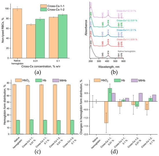
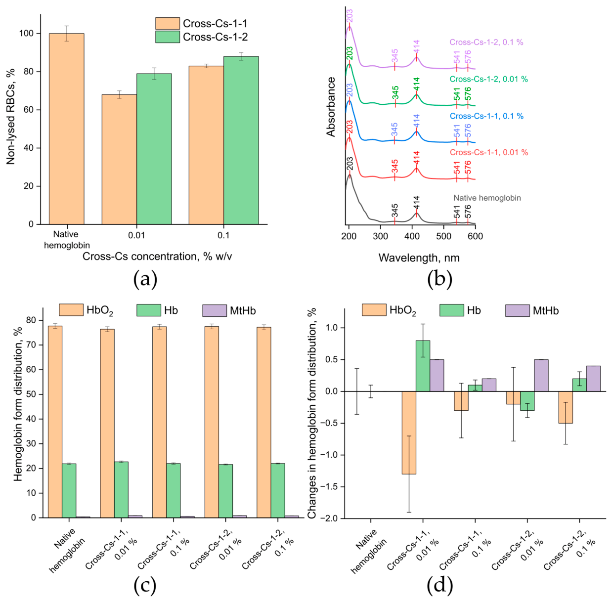
Erythrocyte hemolysis showed an inverse dose-dependent effect for both chitosan hydrogels (Figure 1a). A decrease in hydrogel concentration statistically significantly activated hemolytic processes. Thus, 0.1% Cross-Cs-1-2 caused the death of 12% of RBCs, whereas a reduction to 0.01% destroyed 21% of the cells. Similar findings were observed for Cross-Cs-1-1, where a decrease in concentration increased the number of dead red blood cells from 17% to 32%. This behavior can be explained by the low stability of chitosan hydrogel particles suspended in an aqueous medium. Native chitosan carries a surface charge of approximately 20–25 mV, which is insufficient to maintain colloidal stability. This charge arises from partial protonation of the primary amino groups of the polysaccharide. During hydrogel synthesis, the BDDE crosslinking agent reacts with the NH2 groups of chitosan, reducing their steric accessibility for protonation. As a result, the surface charge of the resulting structure drops to ~10–15 mV, further reducing the aggregative stability of the suspension. It is well known [49] that an increase in the concentration of suspended chitosan particles with a relatively low charge disrupts the colloidal system and causes the polysaccharide to precipitate as a separate phase.
Figure 1.
Chitosan hydrogel interactions with RBCs and hemoglobin: (a) percentage of RBC hemolysis; (b) hemoglobin spectra; (c) hemoglobin form distribution; (d) changes in the distribution of hemoglobin forms.
The observed concentration dependence of the hemolytic properties of chitosan hydrogels can therefore be interpreted as follows. A high hydrogel concentration (0.1%) produces a low-stability suspension that rapidly aggregates under the experimental conditions. Most of the hydrogel then precipitates and leaves the reaction zone, so it does not interact with RBCs, and up to 88% of the cells remain intact. Suspensions containing 0.01% hydrogel are less prone to self-aggregation and retain more suspended particles capable of interacting with RBCs. As a result, RBC adsorption on the surface of hydrogel particles is more efficient than in 0.1% suspensions, which increases the number of destroyed red blood cells to 32%.
This interpretation is consistent with literature data showing that chitosan nanoparticles with low ζ-potential (+5 mV) exhibit minimal hemolysis (≤7%) at high concentrations (2000 µg/mL) due to aggregation [50], whereas particles with high positive charge (+48 mV) cause pronounced hemolysis [51]. The same study showed that chitosan particles dispersed in saline (+20–25 mV) are hemocompatible, which supports the role of surface charge in modulating hemolytic activity [51]. In addition, crosslinking of chitosan has been reported to reduce its hemolytic activity by limiting the accessibility of amino groups [52]. Taken together, these findings support the hypothesis that the inverse concentration dependence observed here is governed by the colloidal stability of the hydrogel particles rather than by their intrinsic chemical composition.
The same hypothesis is supported by a comparison of erythrocyte hemolysis results obtained with hydrogels containing different amounts of the crosslinking agent. Cross-Cs-1-2 was synthesized with twice as much crosslinking agent as Cross-Cs-1-1. As a result, Cross-Cs-1-2 has a lower surface charge (10 mV) and therefore lower colloidal stability than Cross-Cs-1-1 (15 mV). Cross-Cs-1-2 suspensions are thus more prone to phase separation than systems based on Cross-Cs-1-1. This reduces the number of interactions between Cross-Cs-1-2 hydrogel particles and red blood cells that lead to cell destruction.
According to the ASTM classification, materials with hemolysis above 5% are considered hemolytic, while those between 2% and 5% are classified as moderately hemolytic [53]. The Cross-Cs hydrogels therefore fall into the hemolytic category, with hemolysis ranging from 12% to 32%. Importantly, however, hemolysis decreased at higher hydrogel concentrations, which points to the colloidal stability of the dispersion, rather than the chemical nature of the material, as the dominant factor. It should also be noted that the ASTM classification was originally developed for nanoparticles (ASTM E2524) and rigid blood-contacting materials (ASTM F756), and does not take into account the concentration-dependent colloidal behavior, high water content, or porous structure characteristic of hydrogel dispersions.
The next stage of the study focused on the UV/VIS spectra of hemoglobin. Figure 1b shows the spectrum of native hemoglobin, with the following absorption bands: 203 nm (protein peak), 345 nm (δ-band), 414 nm (Soret band, or γ-band), 541 nm (β-band), and 576 nm (α-band). This indicates that the spectrum corresponds to oxyhemoglobin [54].
After interaction with chitosan hydrogels, the spectral characteristics of oxyhemoglobin remained unchanged regardless of hydrogel concentration (Figure 1b). This indicates that the structural features of the hydrogels do not affect the spectroscopic properties of oxyhemoglobin. This may be due to their low ζ-potential values and the limited mobility of the polymer networks. This conclusion is important for understanding the mechanisms of interaction between biological macromolecules and chitosan hydrogels in various biomedical applications.
The analysis of hemoglobin form distribution after interaction with chitosan hydrogels is also highly informative for assessing the blood compatibility of the synthesized materials. Native hemoglobin, used as a control, showed the expected stable distribution, with a predominance of the functional form HbO2 (77.8%) and a minimal fraction of the oxidized, inactive form MtHb (0.43%). The hydrogels did not significantly affect the content of liganded forms (Figure 1c). A closer look at the data in Figure 1d reveals several notable trends. For Cross-Cs-1-2 at 0.1%, the changes in hemoglobin form content were minimal: HbO2 decreased by only 0.5%, while Hb and MtHb changed within the range of 0.2–0.4%. A similar pattern was observed for Cross-Cs-1-2 at 0.01%, where HbO2 decreased by 0.2%, Hb increased by 0.3%, and MtHb increased by 0.5%. For Cross-Cs-1-1 at 0.1%, the changes were also modest: HbO2 decreased by 0.3%, Hb increased by 0.1%, and MtHb increased by 0.2%. By contrast, Cross-Cs-1-1 at 0.01% produced the most pronounced shift: HbO2 decreased by 1.3%, while Hb and MtHb increased by 0.8% and 0.5%, respectively. Importantly, in all cases, the magnitude of these changes remained below 1.5%, indicating that the hydrogels do not substantially perturb the oxygen-transport function of hemoglobin. These findings further confirm that the magnitude of the ζ-potential is the dominant factor governing erythrocyte hemolysis and hemoglobin form distribution. Cross-Cs-1-2, which had the lowest ζ-potential value, showed the smallest deviations in hemoglobin form distribution from native hemoglobin across the entire range of studied concentrations. Overall, this supports the potential use of the synthesized chitosan hydrogels as hemostatic agents.
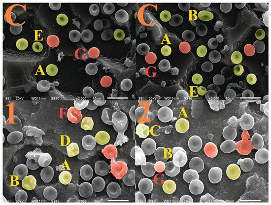
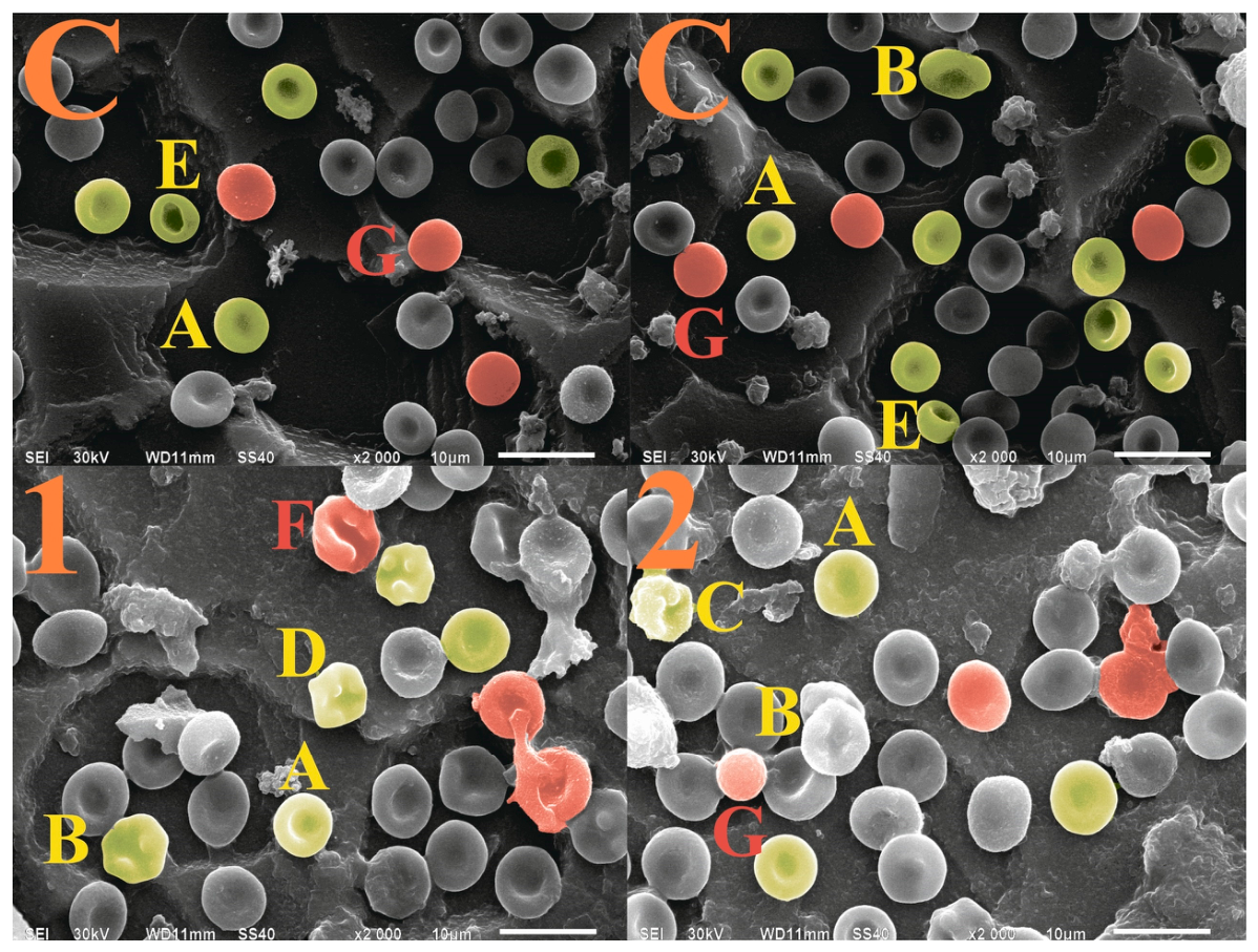
Changes in RBC cytoarchitecture induced by interaction with chitosan hydrogels were examined. Native donor RBCs showed 95.1% discocytes, 3.8% reversibly deformed cells, and 1.1% irreversibly deformed cells (Figure 2). The transformation indices were as follows: TI—0.05, RTI—0.03, and ITI—0.01 (Table 1).
Figure 2.
SEM images of RBCs: C—control samples of native RBCs; 1—Cross-Cs-1-1; 2—Cross-Cs-1-2. A—discocytes; reversibly transformed cells (yellow cells and letters): B—echinocytes (type I), C—echinocytes (type II), D—echinocytes (type III), E—stomatocytes; irreversibly transformed cells (red cells and letters): F—spherostomatocytes (type I), G—spherostomatocytes (type II).
Table 1.
Parameters of RBC cytoarchitecture.
After erythrocytes were incubated with 0.01% suspensions of Cross-Cs-1-1 and Cross-Cs-1-2 for 20 min at 37 °C, the number of discocytes decreased from 95.1% in native RBCs to 85.2% and 87.2%, respectively. Reversibly deformed cells accounted for 8.6% and 10.3%, while irreversibly deformed erythrocytes increased from 1.1% in native RBCs to 6.2% and 2.5%, respectively. Although these changes did not reach statistical significance under the conditions tested, they indicate a shift in the erythrocyte shape distribution that may be biologically meaningful. The increase in irreversibly deformed cells, particularly for Cross-Cs-1-1 (6.2% versus 2.5% for Cross-Cs-1-2), suggests that this hydrogel causes more pronounced membrane perturbation, consistent with its higher surface charge (15 mV versus 10 mV) and lower colloidal stability. Such morphological changes are known to precede hemolysis and may reflect early damage to the erythrocyte membrane. At the same time, the relatively high proportion of discocytes remaining after exposure (85–87%) indicates that the majority of cells retained their normal shape. The biological significance of these changes should therefore be considered in the context of the intended application, and further studies with longer exposure times or higher hydrogel concentrations may be needed to fully assess the long-term effects on erythrocyte morphology.
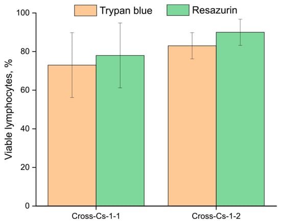
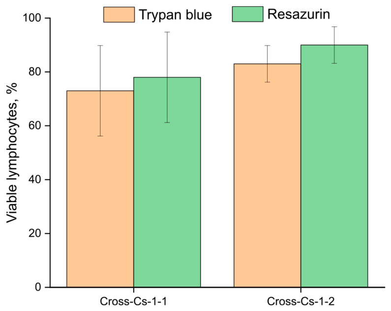
The final hemocompatibility test of the synthesized chitosan hydrogels assessed their effect on lymphocyte viability using trypan blue exclusion and the resazurin assay. With Cross-Cs-1-2, the percentage of viable lymphocytes was somewhat higher, ranging from 83% to 90% depending on the assay method. By contrast, samples with Cross-Cs-1-1 showed viability of 73–78% (Figure 3). No statistically significant changes in lymphocyte viability were detected. This further highlights the potential of the obtained hydrogels as hemostatic agents.
Figure 3.
Assessment of lymphocyte viability.
Overall, the comprehensive hemocompatibility assessment showed that interaction of the chitosan hydrogels with blood does not cause critical disturbances to the oxygen-transport function of hemoglobin, erythrocyte cytoarchitecture, or lymphocyte viability. The ζ-potential magnitude of the hydrogels is the dominant factor governing both their colloidal stability and their interaction with blood cells. The smallest deviations in hemoglobin spectral characteristics and hemoglobin form distribution from native values were observed for Cross-Cs-1-2, which had the lowest ζ-potential. These results confirm the potential of the synthesized chitosan hydrogels as hemocompatible hemostatic materials.
3.2. Ficin Immobilization on Chitosan Hydrogels
Ficin is a plant cysteine protease with high proteolytic activity, which gives it a broad spectrum of biological effects. In particular, it activates human coagulation factor X [55]. Unlike protein coagulation factors such as fibrinogen and thrombin, ficin is more readily available. This makes ficin-based hemostatic agents an attractive option for producing effective, low-cost materials. However, to function effectively within a hemostatic agent, ficin must be immobilized on chitosan hydrogels under carefully selected conditions. These conditions need to strike a balance between protein loading and the catalytic activity of the enzyme.
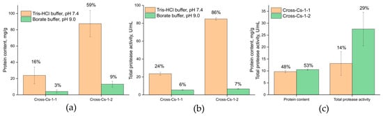
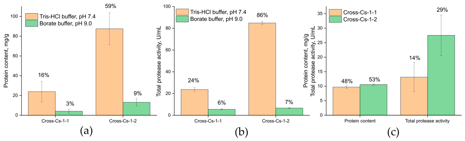
Ficin was immobilized on chitosan hydrogels in two different media: 0.05 M Tris-HCl buffer at pH 7.4 and 0.05 M borate buffer with added KCl at pH 9.0 (referred to below as borate buffer, pH 9.0). Protein analysis of the resulting heterogeneous preparations revealed that the highest ficin loading (in mg per g of support) was achieved on Cross-Cs-1-2 in 0.05 M Tris-HCl buffer at pH 7.4 (Figure 4a). Under these conditions, 59% of the initial protein was bound to the support. By contrast, when 0.05 M borate buffer at pH 9.0 was used, the protein immobilization yield did not exceed 10% of the initial protein content in solution.
Figure 4.
Various options of ficin immobilization on chitosan hydrogels: (a) protein content of adsorptively immobilized ficin. Protein immobilization yield is shown above the bars (100% corresponds to the total ficin content in the sorption media); (b) total protease activity of adsorptively immobilized ficin. Activity immobilization yield is shown above the bars (100% corresponds to the free ficin activity under the experimental conditions); (c) protein content and total protease activity of covalently immobilized ficin. Protein and activity immobilization yields are shown above the bars.
This observation can be explained as follows. Ficin has an isoelectric point of approximately 9.0 [56]. Below this pH, the net charge of ficin is positive; above it, negative. In borate buffer at pH 9.0, electrostatic forces contribute only minimally to the ficin–chitosan hydrogel interaction, so the protein binds to the hydrogel surface mainly through weak hydrogen bonds and van der Waals interactions. At pH 7.4, the contribution of electrostatic forces to the ficin–hydrogel interaction increases, improving the sorption efficiency and increasing the amount of protein bound to the hydrogel surface. At the same time, the chitosan hydrogels themselves carry a net positive surface charge (10 mV for Cross-Cs-1-2 and 15 mV for Cross-Cs-1-1). Consequently, electrostatic repulsion between the hydrogels and ficin can occur at pH 7.4. However, Cross-Cs-1-2 has a lower positive surface charge, which ultimately results in more efficient sorption of ficin from solution by this hydrogel.
The total protease activity immobilization yields followed the same trend, with the highest value again observed for Cross-Cs-1-2 (Figure 4b). This is consistent with its higher protein content. In all other cases, the total protease activity immobilization yields did not exceed 30% of the free ficin activity, pointing to the limited practical potential of such immobilized ficin preparations. Taken together, the protein and activity immobilization yields suggest that 0.05 M Tris-HCl buffer at pH 7.4 is the more promising medium for sorption immobilization of ficin on chitosan hydrogels. Covalent immobilization of ficin on chitosan hydrogels was therefore carried out in this medium.
Protein analysis of the covalently bound ficin preparations revealed that the highest ficin loading (in mg per g of support) was achieved with Cross-Cs-1-2. This preparation also showed higher catalytic activity (Figure 4c). However, its total protease activity immobilization yield was almost two times lower than that of adsorptively immobilized ficin. This is likely due to the rigid attachment of ficin to the hydrogel surface, which restricts the steric accessibility of the enzyme active site for catalysis.
A comparison of sorption and covalent immobilization of ficin on the chitosan hydrogel matrix clearly shows that adsorption immobilization is more promising in terms of both key parameters: protein loading in the complexes and their total proteolytic activity. Moreover, adsorption immobilization is simple, cost-effective, rapid, and does not require toxic activators. This makes it preferable to covalent binding for a range of biotechnological and medical applications. Adsorptively immobilized ficin was therefore used in the subsequent studies to evaluate the hemostatic properties of ficin–chitosan hydrogel compositions.
3.3. Hemostatic Properties of Chitosan Hydrogels and Ficin–Chitosan Hydrogel Compositions
One of the most common mechanisms of action of local hemostatic agents is the “plug effect” [57]. In this mechanism, the hemostatic material swells upon contact with blood and compresses the damaged vessels. This makes it worthwhile to examine how the synthesized hydrogels swell in a blood environment. After 120 s of incubation in blood, the swelling ratio (SR) of Cross-Cs-1-1 was 15 ± 1, whereas that of Cross-Cs-1-2 was 13 ± 2. Both hydrogels therefore swell fairly rapidly in blood, reaching SR values that are statistically indistinguishable. The trend toward lower swelling for Cross-Cs-1-2 may be explained by its higher crosslinking density, which restricts network expansion in the blood environment.
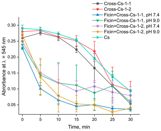
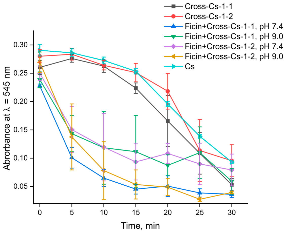
The next step in evaluating the hemostatic properties of the obtained hydrogels and ficin–hydrogel compositions was to assess kinetic blood coagulation. To this end, changes in absorption at λ = 545 nm were monitored as a function of contact time between the hemostatic agent and blood (Figure 5).
Figure 5.
Kinetic blood coagulation induced by chitosan, Cross-CS hydrogels and ficin–Cross-CS compositions.
As blood cell aggregates formed, the light absorption intensity decreased with increasing interaction time between blood and the chitosan hydrogels or ficin–chitosan hydrogel compositions. However, the coagulation rate differed between samples. A closer look at the kinetic curves in Figure 5 reveals several important trends. Native chitosan (Cs) produced the steepest initial drop in absorbance, reaching a low plateau within the first 5–10 min. This indicates that soluble chitosan is a highly effective coagulant in this assay, which is consistent with its strong polycationic nature and its ability to aggregate blood cells. Pristine hydrogels Cross-Cs-1-1 and Cross-Cs-1-2, by contrast, showed only a gradual decrease in absorbance over 30 min. This points to a relatively slow coagulation process driven mainly by the intrinsic hemostatic properties of the crosslinked chitosan network. The difference between soluble Cs and the Cross-Cs hydrogels may be attributed to the reduced accessibility of amino groups in the crosslinked network and to the lower colloidal stability of the hydrogel dispersions, which limits their interaction with blood cells. By contrast, all ficin-containing compositions produced a much steeper initial drop in optical density than the pristine hydrogels, especially within the first 5–10 min. This confirms that the immobilized enzyme actively accelerates blood cell aggregation. Among the studied samples, ficin + Cross-Cs-1-1 (immobilized at pH 7.4) and ficin + Cross-Cs-1-2 (immobilized at pH 9.0) reached the lowest absorbance values by the end of the experiment. This suggests that these two compositions provide the most efficient coagulation. Interestingly, these samples differ in both the hydrogel type and the immobilization medium. This indicates that coagulation efficiency depends on a combination of factors, including the surface charge of the hydrogel, the conformation of the adsorbed enzyme, and the accessibility of its active site. Overall, the kinetic coagulation data confirm that the developed ficin–chitosan hydrogel compositions are promising hemostatic materials capable of accelerating blood clotting.
The hemostatic performance of the developed materials appears to result from the combined action of two complementary mechanisms. The Cross-Cs hydrogels themselves contribute through a passive, “plug-like” mechanism: upon contact with blood, they swell rapidly and form a physical barrier that concentrates blood cells and coagulation factors at the wound site, thereby promoting clot formation. This effect is consistent with the swelling behavior of the hydrogels and with the gradual decrease in absorbance observed for the pristine hydrogels in the kinetic coagulation assay. However, this mechanism alone is relatively slow, as indicated by the moderate coagulation rate of the ficin-free hydrogels.
The immobilized ficin introduces an active, cascade-activating component. Upon release from the hydrogel, ficin can cleave fibrin and activate human coagulation factor X [17,18,19], thereby accelerating the conversion of prothrombin to thrombin and the subsequent formation of a fibrin network. This is consistent with the much steeper initial drop in absorbance observed for all ficin-containing compositions, especially within the first 5–10 min, compared with the pristine hydrogels. The released enzyme therefore acts as a biochemical accelerator of the coagulation cascade, while the hydrogel provides the physical scaffold and the sustained release profile.
Taken together, these two mechanisms appear to act synergistically: the hydrogel concentrates blood components at the wound site, and the released ficin accelerates the cascade that leads to fibrin formation. This combination may explain why the ficin-containing hydrogels achieved the lowest absorbance values by the end of the experiment and why the effect was most pronounced for ficin + Cross-Cs-1-1 (immobilized at pH 7.4) and ficin + Cross-Cs-1-2 (immobilized at pH 9.0). Further studies, including direct measurements of factor Xa and thrombin generation, will be needed to confirm this proposed mechanism.
To place the performance of the developed ficin-loaded Cross-Cs hydrogels in the context of previously reported chitosan-based hemostatic systems, a comparative analysis was performed using the parameters most consistently reported in the literature (Table 2). Because direct comparison is complicated by differences in test protocols, blood sources, and material formats, the comparison focuses on coagulation/clotting time, hemolysis, swelling capacity, and cytocompatibility.
Table 2.
Parameters of blood cells.
Several limitations of this study should be acknowledged. First, all experiments were performed in vitro using donor blood and model assays, and no in vivo validation was carried out. The behavior of the developed hydrogels in a living organism may differ substantially from that observed under controlled in vitro conditions, where blood flow, endothelial interactions, immune responses, and wound-healing processes all contribute to the overall hemostatic outcome. Second, the physicochemical characterization of the hydrogels was limited to ζ-potential measurements and swelling studies; a more detailed analysis of the network structure, mechanical properties, and degradation behavior would provide a more complete picture of the material. Third, the release of ficin from the hydrogels was monitored indirectly through the proteolytic activity of the release medium, and direct measurements of enzyme release kinetics and stability under physiological conditions were not performed. Fourth, the proposed mechanism of ficin-mediated coagulation activation, in which the released enzyme activates factor X and promotes fibrin formation, is inferred from the kinetic coagulation data and from literature reports rather than being directly demonstrated. Direct measurements of factor Xa and thrombin generation would be required to confirm this mechanism. Finally, the hemostatic performance of the developed materials was not compared with that of clinically established hemostatic agents such as Celox™, QuikClot®, or Surgicel®, which limits the assessment of their practical potential. These limitations will be addressed in future studies, which will include in vivo evaluation, detailed physicochemical and mechanical characterization, direct measurements of enzyme release and stability, biochemical confirmation of the coagulation mechanism, and benchmarking against commercial hemostatic materials.
Thus, the obtained hydrogels combine rapid swelling in blood with efficient coagulation, and their functionalization with ficin significantly enhances hemostatic performance. These results support the potential of the developed compositions for use as local hemostatic agents.
4. Conclusions
In this study, chitosan hydrogels with varying degrees of crosslinking were synthesized and evaluated for their hemocompatibility and hemostatic activity in vitro, both alone and in complexes with the plant cysteine protease ficin.
The experimental data obtained in this work can be summarized as follows. The hydrogels did not cause statistically significant changes in hemoglobin spectral characteristics, erythrocyte cytoarchitecture, or lymphocyte viability under the conditions tested. Hemolysis was found to depend inversely on hydrogel concentration, decreasing from 32% to 17% for Cross-Cs-1-1 and from 21% to 12% for Cross-Cs-1-2 as the concentration increased from 0.01% to 0.1%. The ζ-potential of the hydrogels correlated with their crosslinking degree, decreasing from 15 mV for Cross-Cs-1-1 to 10 mV for Cross-Cs-1-2.
Ficin was successfully immobilized on the chitosan hydrogels by both adsorption and covalent binding. The experimental data showed that adsorption immobilization provided higher protein loading (59% versus 29% of the initial protein) and higher total proteolytic activity (86% versus 29% of free ficin activity) than covalent binding. The optimal medium for adsorption immobilization was 0.05 M Tris-HCl buffer at pH 7.4. These differences are attributed to the rigid attachment of the enzyme and the restricted accessibility of its active site in the covalently bound preparation.
The kinetic coagulation experiments demonstrated that ficin-containing compositions accelerated blood cell aggregation compared to pristine hydrogels, with ficin + Cross-Cs-1-1 (immobilized at pH 7.4) and ficin + Cross-Cs-1-2 (immobilized at pH 9.0) showing the most pronounced decrease in absorbance.
Several aspects of this work remain interpretive rather than directly demonstrated. The decrease in hemolysis at higher hydrogel concentrations is proposed to result from particle aggregation and precipitation, although the colloidal behavior of the dispersions was not characterized in detail. The correlation between ζ-potential and blood-cell interactions observed here is consistent with, but does not by itself prove, a causal role of surface charge. Likewise, the proposed synergistic mechanism between the hydrogel scaffold and the released ficin, in which the hydrogel concentrates blood components and the enzyme activates the coagulation cascade, is inferred from the kinetic data and from literature reports and requires direct biochemical confirmation.
It should be emphasized that all experiments were performed in vitro using donor blood and model assays, and the results cannot be directly extrapolated to in vivo conditions. Further studies should include direct measurements of factor Xa and thrombin generation, detailed characterization of the colloidal state of the dispersions, and in vivo evaluation of hemostatic efficacy, biodegradation, tissue response, and long-term safety. These steps will be necessary to substantiate the potential of the developed materials as hemostatic agents.
Author Contributions
Conceptualization, V.G.A. and M.G.H.; methodology V.G.A. and M.G.H.; investigation, A.V.S., Y.A.R., A.N.D., S.S.G., V.A.K. and S.M.P.; resources, E.V.B.; writing—original draft preparation, M.S.L.; writing—review and editing, M.S.L., E.V.B., V.G.A. and M.G.H.; visualization, M.S.L.; supervision, V.G.A. and M.G.H.; project administration, V.G.A. and M.G.H.; funding acquisition, V.G.A. All authors have read and agreed to the published version of the manuscript.
Funding
This research was funded by Russian Science Foundation, grant number RSF-25-74-20024, project “Hybrid Multifunctional Hemostatic Compositions Based on Modified Chitosans and Protein–Polysaccharide Complexes: Influence of Composition on Structural and Functional Properties and Features of Interaction with Blood and Its Formed Element”.
Institutional Review Board Statement
The study was conducted in accordance with the Declaration of Helsinki, and approved by the Institutional Ethics Committee of Voronezh State University (protocol code 5001-03, dated 23 September 2026).
Informed Consent Statement
Informed consent was obtained from all subjects involved in the study.
Data Availability Statement
The original contributions presented in the study are included in the article, and further inquiries can be directed to the corresponding author.
Acknowledgments
SEM data were obtained using the equipment at the Research Core Centre of Voronezh State University. Donor blood samples were obtained from the Voronezh Regional Blood Center.
Conflicts of Interest
The authors declare no conflicts of interest.
References
- Eastridge, B.J.; Holcomb, J.B.; Shackelford, S. Outcomes of Traumatic Hemorrhagic Shock and the Epidemiology of Preventable Death from Injury. Transfusion 2019, 59, 1423–1428. [Google Scholar] [CrossRef] [Scilit] [PubMed]
- Marrone, M.; Bellantuono, L.; Stellacci, A.; Misceo, F.; Silvestre, M.; Zotti, F.; Dell’Erba, A.; Bellotti, R. Haemorrhage and Survival Times: Medical–Legal Evaluation of the Time of Death and Relative Evidence. Diagnostics 2023, 13, 732. [Google Scholar] [CrossRef] [Scilit] [PubMed]
- Jamali, B.; Nouri, S.; Amidi, S. Local and Systemic Hemostatic Agents: A Comprehensive Review. Cureus 2024, 16, e72312. [Google Scholar] [CrossRef] [Scilit] [PubMed]
- Chen, X.Y.; Xiao, A.; Wang, Y.; Lin, S.-X.; Rodrigue, D. A Review on Hemostatic Materials: Past, Present and Future Developments. Biomater. Adv. 2026, 185, 214830. [Google Scholar] [CrossRef] [Scilit] [PubMed]
- Su, J.; Liu, C.; Sun, A.; Yan, J.; Sang, F.; Xin, Y.; Zhao, Y.; Wang, S.; Dang, Q. Hemostatic and Antimicrobial Properties of Chitosan-Based Wound Healing Dressings: A Review. Int. J. Biol. Macromol. 2025, 306, 141570. [Google Scholar] [CrossRef] [Scilit] [PubMed]
- Zhang, S.; Lei, X.; Lv, Y.; Wang, L.; Wang, L.-N. Recent Advances of Chitosan as a Hemostatic Material: Hemostatic Mechanism, Material Design and Prospective Application. Carbohyd. Polym. 2024, 327, 121673. [Google Scholar] [CrossRef] [Scilit] [PubMed]
- Hu, Z.; Zhang, D.-Y.; Lu, S.-T.; Li, P.-W.; Li, S.-D. Chitosan-Based Composite Materials for Prospective Hemostatic Applications. Mar. Drugs 2018, 16, 273. [Google Scholar] [CrossRef] [Scilit] [PubMed]
- Roas-Escalona, N.; Becquart, F.; Delair, T.; Dutertre, F. Chitosan-Based Hydrogels: Influence of Crosslinking Strategy on Rheological Properties. Carbohyd. Polym. 2024, 341, 122329. [Google Scholar] [CrossRef] [Scilit] [PubMed]
- Gheorghiță, D.; Moldovan, H.; Robu, A.; Bița, A.-I.; Grosu, E.; Antoniac, A.; Corneschi, I.; Antoniac, I.; Bodog, A.D.; Băcilă, C.I. Chitosan-Based Biomaterials for Hemostatic Applications: A Review of Recent Advances. Int. J. Mol. Sci. 2023, 24, 10540. [Google Scholar] [CrossRef] [Scilit] [PubMed]
- Wu, X.; Tang, Z.; Liao, X.; Wang, Z.; Liu, H. Fabrication of Chitosan@calcium Alginate Microspheres with Porous Core and Compact Shell, and Application as a Quick Traumatic Hemostat. Carbohyd. Polym. 2020, 247, 116669. [Google Scholar] [CrossRef] [Scilit] [PubMed]
- Shi, Y.; Yu, W.; Liang, X.; Cheng, J.; Cao, Y.; Liu, M.; Fang, Y.; Yang, Z.; Liu, H.; Wei, H.; et al. Interpenetrating Network Expansion Sponge Based on Chitosan and Plasma for Ultrafast Hemostasis of Arterial Bleeding Wounds. Carbohyd. Polym. 2023, 307, 120590. [Google Scholar] [CrossRef] [Scilit] [PubMed]
- Balakireva, A.V.; Kuznetsova, N.V.; Petushkova, A.I.; Savvateeva, L.V.; Zamyatnin, A.A., Jr. Trends and Prospects of Plant Proteases in Therapeutics. Curr. Med. Chem. 2019, 26, 465–486. [Google Scholar] [CrossRef] [Scilit] [PubMed]
- Schechter, I.; Berger, A. On the Active Site of Proteases. III. Mapping the Active Site of Papain; Specific Peptide Inhibitors of Papain. Biochem. Biophys. Res. Commun. 1968, 32, 898–902. [Google Scholar] [CrossRef] [Scilit] [PubMed]
- Fernández-Lucas, J.; Castañeda, D.; Hormigo, D. New Trends for a Classical Enzyme: Papain, a Biotechnological Success Story in the Food Industry. Trends Food Sci. Technol. 2017, 68, 91–101. [Google Scholar] [CrossRef] [Scilit]
- Kumar, V.; Mangla, B.; Javed, S.; Ahsan, W.; Kumar, P.; Garg, V.; Dureja, H. Bromelain: A Review of Its Mechanisms, Pharmacological Effects and Potential Applications. Food Funct. 2023, 14, 8101–8128, Correction in Food Funct. 2025, 16, 6733. https://doi.org/10.1039/D5FO90061A. [Google Scholar] [CrossRef] [Scilit] [PubMed]
- Morellon-Sterling, R.; El-Siar, H.; Tavano, O.L.; Berenguer-Murcia, Á.; Fernández-Lafuente, R. Ficin: A Protease Extract with Relevance in Biotechnology and Biocatalysis. Int. J. Biol. Macromol. 2020, 162, 394–404. [Google Scholar] [CrossRef] [Scilit] [PubMed]
- Yang, H.R.; Hwang, D.H.; Prakash, R.L.M.; Kim, J.-H.; Hong, I.-H.; Kim, S.; Kim, E.; Kang, C. Exploring the Fibrin(Ogen)Olytic, Anticoagulant, and Antithrombotic Activities of Natural Cysteine Protease (Ficin) with the κ-Carrageenan-Induced Rat Tail Thrombosis Model. Nutrients 2022, 14, 3552. [Google Scholar] [CrossRef] [Scilit] [PubMed]
- Whitaker, J.R. Properties of the Proteolytic Enzymes of Commercial Ficin. J. Food Sci. 1957, 22, 483–493. [Google Scholar] [CrossRef] [Scilit]
- Hamed, M.B.; El-Badry, M.O.; Kandil, E.I.; Borai, I.H.; Fahmy, A.S. A Contradictory Action of Procoagulant Ficin by a Fibrinolytic Serine Protease from Egyptian Ficus Carica Latex. Biotechnol. Rep. 2020, 27, e00492. [Google Scholar] [CrossRef] [Scilit] [PubMed]
- Baidamshina, D.R.; Trizna, E.Y.; Goncharova, S.S.; Sorokin, A.V.; Lavlinskaya, M.S.; Melnik, A.P.; Gafarova, L.F.; Kharitonova, M.A.; Ostolopovskaya, O.V.; Artyukhov, V.G.; et al. The Effect of Ficin Immobilized on Carboxymethyl Chitosan on Biofilms of Oral Pathogens. Int. J. Mol. Sci. 2023, 24, 16090. [Google Scholar] [CrossRef] [Scilit] [PubMed]
- Holyavka, M.G.; Artyukhov, V.G. Prospects and Problems in Enzyme Immobilization Methodology: Comprehensive Review. Biophys. Rev. 2025, 17, 1471–1509. [Google Scholar] [CrossRef] [Scilit] [PubMed]
- Prabhakar, T.; Giaretta, J.; Zulli, R.; Rath, R.J.; Farajikhah, S.; Talebian, S.; Dehghani, F. Covalent Immobilization: A Review from an Enzyme Perspective. Chem. Eng. J. 2025, 503, 158054. [Google Scholar] [CrossRef] [Scilit]
- Hassan, M.E.; Zhu, X.; De Souza, E.F.; Elnashar, M.M.; Lu, F. Enzyme Immobilization Advances: A Key to Unlocking Renewable Bioenergy Potential. Green Chem. 2025, 27, 11289–11311. [Google Scholar] [CrossRef] [Scilit]
- Robescu, M.S.; Bavaro, T. A Comprehensive Guide to Enzyme Immobilization: All You Need to Know. Molecules 2025, 30, 939. [Google Scholar] [CrossRef] [Scilit] [PubMed]
- Wang, S.; Li, R.; Xie, Y.; Zhang, H.; Huang, X.; Chen, S.; Xu, W.; Liu, Y. Recent Progress on Enzyme Immobilization: Materials, Strategies, and Applications. Food Bioeng. 2026, 5, 220–243. [Google Scholar] [CrossRef] [Scilit]
- Baidamshina, D.R.; Koroleva, V.A.; Trizna, E.Y.; Pankova, S.M.; Agafonova, M.N.; Chirkova, M.N.; Vasileva, O.S.; Akhmetov, N.; Shubina, V.V.; Porfiryev, A.G.; et al. Anti-Biofilm and Wound-Healing Activity of Chitosan-Immobilized Ficin. Int. J. Biol. Macromol. 2020, 164, 4205–4217. [Google Scholar] [CrossRef] [Scilit] [PubMed]
- Sorokin, A.V.; Olshannikova, S.S.; Lavlinskaya, M.S.; Holyavka, M.G.; Faizullin, D.A.; Zuev, Y.F.; Artukhov, V.G. Chitosan Graft Copolymers with N-Vinylimidazole as Promising Matrices for Immobilization of Bromelain, Ficin, and Papain. Polymers 2022, 14, 2279. [Google Scholar] [CrossRef] [Scilit] [PubMed]
- Holyavka, M.; Redko, Y.; Goncharova, S.; Lavlinskaya, M.; Sorokin, A.; Kondratyev, M.; Artyukhov, V. Novel Hybrid Catalysts of Cysteine Proteases Enhanced by Chitosan and Carboxymethyl Chitosan Micro- and Nanoparticles. Polymers 2024, 16, 3111. [Google Scholar] [CrossRef] [Scilit] [PubMed]
- Olshannikova, S.S.; Malykhina, N.V.; Lavlinskaya, M.S.; Sorokin, A.V.; Yudin, N.E.; Vyshkvorkina, Y.M.; Lukin, A.N.; Holyavka, M.G.; Artyukhov, V.G. Novel Immobilized Biocatalysts Based on Cysteine Proteases Bound to 2-(4-Acetamido-2-Sulfanilamide) Chitosan and Research on Their Structural Features. Polymers 2022, 14, 3223. [Google Scholar] [CrossRef] [Scilit] [PubMed]
- Privar, Y.; Skatova, A.; Maiorova, M.; Golikov, A.; Boroda, A.; Bratskaya, S. Tuning Mechanical Properties, Swelling, and Enzymatic Degradation of Chitosan Cryogels Using Diglycidyl Ethers of Glycols with Different Chain Length as Cross-Linkers. Gels 2024, 10, 483. [Google Scholar] [CrossRef] [Scilit] [PubMed]
- Chakraborty, A.; Al Mamun, A.; Xia, D.; Vu, T.; Zahid, A.A.; Luo, W.; Paul, A. Engineering a Dietary Fiber-Based Injectable Hydrogel for Blood-Clotting Applications. Cell Rep. Phys. Sci. 2026, 7, 103330. [Google Scholar] [CrossRef] [Scilit]
- Lavlinskaya, M.S.; Sorokin, A.V. Enhancement of Water Uptake in Composite Superabsorbents Based on Carboxymethyl Cellulose Through Porogen Incorporation and Lyophilization. Gels 2024, 10, 797. [Google Scholar] [CrossRef] [Scilit] [PubMed]
- Flory, P.J. Principles of Polymer Chemistry; 19. print.; Cornell University Press: Ithaca, NY, USA, 2006. [Google Scholar]
- Kumar, B.; Feller, J.-F.; Castro, M.; Lu, J. Conductive Bio-Polymer Nano-Composites (CPC): Chitosan-Carbon Nanotube Transducers Assembled via Spray Layer-by-Layer for Volatile Organic Compound Sensing. Talanta 2010, 81, 908–915. [Google Scholar] [CrossRef] [Scilit] [PubMed]
- Sæbø, I.; Bjørås, M.; Franzyk, H.; Helgesen, E.; Booth, J. Optimization of the Hemolysis Assay for the Assessment of Cytotoxicity. Int. J. Mol. Sci. 2023, 24, 2914. [Google Scholar] [CrossRef] [Scilit] [PubMed]
- Artyukhov, V.G.; Kalaeva, E.A.; Putintseva, O.V.; Polyubez’eva, A.I. The modification of structural and functional properties of human hemoglobin induced by nitroglycerin under different oxygen regime conditions. Biomed. Khim. 2016, 62, 251–258. [Google Scholar] [CrossRef] [Scilit] [PubMed]
- Zhong, R.; Han, D.; Wu, X.; Wang, H.; Li, W.; He, Z.; Zhang, X.; Liu, J. An Evaluation of Morphological Changes and Deformability of Suspended Red Blood Cells Prepared Using Whole Blood with Different Hemoglobin Levels of Tibetans. Transfus. Med. Hemother. 2021, 48, 210–219. [Google Scholar] [CrossRef] [Scilit] [PubMed]
- Bessis, M. Red Cell Shape: Physiology, Pathology, Ultrastructure; Springer: Berlin/Heidelberg, Germany, 1974. [Google Scholar]
- Bezrodnykh, E.A.; Holyavka, M.G.; Belyaeva, T.N.; Pankova, S.M.; Artyukhov, V.G.; Antonov, Y.A.; Berezin, B.B.; Blagodatskikh, I.V.; Tikhonov, V.E. Viability and Surface Morphology of Human Erythrocytes upon Interaction with Chitosan Derivatives. ACS Appl. Bio Mater. 2025, 8, 1909–1920. [Google Scholar] [CrossRef] [Scilit] [PubMed]
- Rowland-Jones, S.L. Lymphocytes: A Practical Approach, 2nd ed.; Practical Approach Series; Oxford University Press, Incorporated: Oxford, UK, 2000. [Google Scholar]
- Eremeev, A.V.; Pikina, A.S.; Vladimirova, T.V.; Bogomazova, A.N. Methods for Assessing the Viability of Cells Cultured in Vitro in 2D and 3D Structures. Genes Cells 2023, 18, 5–21. [Google Scholar] [CrossRef] [Scilit]
- O’Brien, J.; Wilson, I.; Orton, T.; Pognan, F. Investigation of the Alamar Blue (Resazurin) Fluorescent Dye for the Assessment of Mammalian Cell Cytotoxicity. Eur. J. Biochem. 2000, 267, 5421–5426. [Google Scholar] [CrossRef] [Scilit] [PubMed]
- Holyavka, M.G.; Goncharova, S.S.; Artyukhov, V.G. Various Options for Covalent Immobilization of Cysteine Proteases—Ficin, Papain, Bromelain. Int. J. Mol. Sci. 2025, 26, 547. [Google Scholar] [CrossRef] [Scilit] [PubMed]
- Koroleva, V.; Lavlinskaya, M.; Holyavka, M.; Penkov, N.; Zuev, Y.; Artyukhov, V. Thermal Inactivation, Denaturation and Aggregation Processes of Papain-Like Proteases. Chem. Biodiv. 2024, 21, e202401038. [Google Scholar] [CrossRef] [Scilit] [PubMed]
- Weber, M.; Steinle, H.; Golombek, S.; Hann, L.; Schlensak, C.; Wendel, H.P.; Avci-Adali, M. Blood-Contacting Biomaterials: In Vitro Evaluation of the Hemocompatibility. Front. Bioeng. Biotechnol. 2018, 6, 99. [Google Scholar] [CrossRef] [Scilit] [PubMed]
- Nalezinková, M. In Vitro Hemocompatibility Testing of Medical Devices. Thromb. Res. 2020, 195, 146–150. [Google Scholar] [CrossRef] [Scilit] [PubMed]
- Panda, P.K.; Sadeghi, K.; Park, K.; Seo, J. Regeneration Approach to Enhance the Antimicrobial and Antioxidant Activities of Chitosan for Biomedical Applications. Polymers 2022, 15, 132. [Google Scholar] [CrossRef] [Scilit] [PubMed]
- McNamee, A.P.; Timms, D.; Nestler, F.; Bartnikowski, N.; Smith, A.; Tansley, G.D.; Simmonds, M.J. In Vitro Hemocompatibility of the BiVACOR Total Artificial Heart in Continuous and Pulsatile Flow. Artif. Organs 2026, 50, 84–93. [Google Scholar] [CrossRef] [Scilit] [PubMed]
- Szymańska, E.; Winnicka, K. Stability of Chitosan—A Challenge for Pharmaceutical and Biomedical Applications. Mar. Drugs 2015, 13, 1819–1846. [Google Scholar] [CrossRef] [Scilit] [PubMed]
- Zheng, M.; Han, B.; Yang, Y.; Liu, W. Synthesis, Characterization and Biological Safety of O-Carboxymethyl Chitosan Used to Treat Sarcoma 180 Tumor. Carbohyd. Polym. 2011, 86, 231–238. [Google Scholar] [CrossRef] [Scilit]
- Kojima, T.; Yamada, H.; Yamamoto, T.; Matsushita, Y.; Fukushima, K. Dyeing Regions of Oxidative Hair Dyes in Human Hair Investigated by Nanoscale Secondary Ion Mass Spectrometry. Colloids Surf. B Biointerfaces 2013, 106, 140–144. [Google Scholar] [CrossRef] [Scilit] [PubMed]
- Sánchez-Cid, P.; Gónzalez-Ulloa, G.; Alonso-González, M.; Jiménez-Rosado, M.; Rafii-El-Idrissi Benhnia, M.; Romero, A.; Ostos, F.J.; Perez-Puyana, V.M. Influence of Natural Crosslinkers on Chitosan Hydrogels for Potential Biomedical Applications. Macromol. Mater. Eng. 2023, 308, 2300195. [Google Scholar] [CrossRef] [Scilit]
- Jesus, S.; Schmutz, M.; Som, C.; Borchard, G.; Wick, P.; Borges, O. Hazard Assessment of Polymeric Nanobiomaterials for Drug Delivery: What Can We Learn From Literature So Far. Front. Bioeng. Biotechnol. 2019, 7, 261. [Google Scholar] [CrossRef] [Scilit] [PubMed]
- Martínez-Mancera, F.D.; Hernández-López, J.L. In Vitro Observation of Direct Electron Transfer of Human Haemoglobin Molecules on Glass/Tin-Doped Indium Oxide Electrodes. J. Mex. Chem. Soc. 2017, 59, 302–307. [Google Scholar] [CrossRef] [Scilit]
- Richter, G.; Schwarz, H.P.; Dorner, F.; Turecek, P.L. Activation and Inactivation of Human Factor X by Proteases Derived from Ficus Carica. Br. J. Haematol. 2002, 119, 1042–1051. [Google Scholar] [CrossRef] [Scilit] [PubMed]
- Žiemytė, M.; Escudero, A.; Díez, P.; Ferrer, M.D.; Murguía, J.R.; Martí-Centelles, V.; Mira, A.; Martínez-Máñez, R. Ficin–Cyclodextrin-Based Docking Nanoarchitectonics of Self-Propelled Nanomotors for Bacterial Biofilm Eradication. Chem. Mater. 2023, 35, 4412–4426. [Google Scholar] [CrossRef] [Scilit] [PubMed]
- Tomaiuolo, M.; Matzko, C.N.; Poventud-Fuentes, I.; Weisel, J.W.; Brass, L.F.; Stalker, T.J. Interrelationships between Structure and Function during the Hemostatic Response to Injury. Proc. Natl. Acad. Sci. USA 2019, 116, 2243–2252. [Google Scholar] [CrossRef] [Scilit] [PubMed]

Disclaimer/Publisher’s Note: The statements, opinions and data contained in all publications are solely those of the individual author(s) and contributor(s) and not of MDPI and/or the editor(s). MDPI and/or the editor(s) disclaim responsibility for any injury to people or property resulting from any ideas, methods, instructions or products referred to in the content. |
© 2026 by the authors. Licensee MDPI, Basel, Switzerland. This article is an open access article distributed under the terms and conditions of the Creative Commons Attribution (CC BY) license.