


Simple Summary
Pancreatic neuroendocrine neoplasms (PanNENs) are uncommon tumors that can behave very differently from one patient to another. Because many PanNENs express somatostatin receptors, molecular imaging can identify these receptors and help determine whether a patient may benefit from peptide receptor radionuclide therapy (PRRT). This review summarizes how modern molecular imaging, including somatostatin receptor PET and 18F-FDG PET, can characterize tumor biology and heterogeneity and support individualized treatment decisions. We also discuss the current evidence for PRRT, its integration with surgery, liver-directed treatments, systemic therapies, and active surveillance, as well as the importance of patient selection and toxicity monitoring. New approaches such as individualized dosimetry, somatostatin receptor antagonists, alpha-emitting radionuclides, radiomics, and artificial intelligence may further improve the precision of radionuclide therapy. The future of PanNEN theranostics is likely to move beyond fixed treatment strategies toward adaptive approaches that repeatedly integrate molecular imaging, tumor biology, treatment response, and patient-specific factors to optimize treatment over the course of disease.
Abstract
Pancreatic neuroendocrine neoplasms (PanNENs) are a heterogeneous group of tumors characterized by variable biological behavior and frequent overexpression of somatostatin receptors, making them ideal candidates for theranostic approaches. Over the past two decades, advances in molecular imaging and peptide receptor radionuclide therapy (PRRT) have fundamentally transformed the diagnosis and management of advanced PanNENs, establishing nuclear medicine as a central component of precision oncology. This review provides a comprehensive overview of the evolving role of theranostics in PanNENs, with particular emphasis on recent developments in nuclear medicine and radiation-based precision medicine. We discuss the biological foundation of molecular imaging, current clinical evidence supporting PRRT, and emerging strategies for personalized radionuclide therapy based on quantitative imaging, patient selection, and individualized dosimetry. Furthermore, we highlight next-generation radiopharmaceuticals, including somatostatin receptor antagonists and α-emitting radionuclides, as well as novel diagnostic tracers that are expanding the scope of theranostic applications. The review also examines the growing impact of artificial intelligence, radiomics, and computational modeling in image analysis, treatment planning, and adaptive radionuclide therapy. Finally, we discuss future perspectives toward adaptive precision theranostics, in which molecular imaging, multi-omics integration, advanced dosimetry, and artificial intelligence converge to support dynamic, patient-specific treatment strategies. Continued technological innovation and multidisciplinary collaboration are expected to further establish theranostics as a cornerstone of personalized management for patients with PanNENs.
1. Introduction
Pancreatic neuroendocrine neoplasms (PanNENs) are biologically diverse tumors arising from pancreatic endocrine cells. Although accounting for only 1–2% of pancreatic malignancies, their diagnosed incidence has steadily risen over the past two decades owing to advancements in imaging, endoscopic ultrasonography, pathology, and clinical awareness [1,2]. Unlike pancreatic ductal adenocarcinoma, which is primarily managed by anatomical staging, PanNENs exhibit marked heterogeneity in differentiation, proliferation, hormonal function, and molecular profile, necessitating individualized management [3].
The rapid evolution of nuclear medicine has transformed this field. Somatostatin receptor scintigraphy, once used merely for localization, has evolved into positron emission tomography/computed tomography (PET/CT) using radiolabeled somatostatin analogues—particularly 68GA-DOTATATE and related tracers. This transition has dramatically enhanced lesion detection, staging accuracy, and tumor biology characterization [4,5].
This progress has catalyzed theranostics—integrating diagnostic imaging with targeted radionuclide therapy. Somatostatin receptor imaging identifies patients candidate for peptide receptor radionuclide therapy (PRRT), directly linking diagnosis to treatment [6]. The pivotal NETTER-1 trial established 177Lu-DOTATATE’s efficacy in progressive somatostatin receptor-positive neuroendocrine tumors [7]. Although focused on midgut tumors, growing evidence supports PRRT’s clinical benefits in PanNENs, leading to its inclusion in international guidelines [8,9,10].
Modern theranostics extends beyond standard PRRT. Dual-tracer PET combining somatostatin receptor and 18F-FDG PET evaluates receptor expression alongside metabolic activity, uncovering tumor heterogeneity and guiding therapeutic selection [11]. Meanwhile, therapy continues to advance: personalized dosimetry optimizes radiation delivery while mitigating toxicity, somatostatin receptor antagonists offer superior tumor uptake, and -emitters (225Ac, 212Pb) show promise in treatment-resistant disease [12,13,14]. Combination strategies with systemic therapies are also actively explored.
Nuclear medicine has thus shifted from an adjunctive diagnostic tool to a cornerstone of precision oncology for PanNENs. This review summarizes current evidence and future perspectives in molecular imaging and theranostics for PanNENs.
2. Review Methodology
2.1. Literature Search Strategy
This narrative review was conducted to summarize the current and emerging evidence on theranostic approaches for pancreatic neuroendocrine neoplasms (PanNENs), with particular emphasis on molecular imaging, somatostatin receptor (SSTR) imaging, peptide receptor radionuclide therapy (PRRT), individualized dosimetry, novel radiopharmaceuticals, and computational approaches. A structured literature search was performed using PubMed/MEDLINE, Embase, and Web of Science. The search strategy combined terms related to PanNENs and neuroendocrine tumors with terms related to molecular imaging and radionuclide therapy, including “pancreatic neuroendocrine tumor,” “pancreatic neuroendocrine neoplasm,” “PanNET,” “somatostatin receptor,” “SSTR PET,” “DOTATATE,” “DOTATOC,” “FDG PET,” “peptide receptor radionuclide therapy,” “PRRT,” “177Lu-DOTATATE,” “dosimetry,” “radiopharmaceutical,” “SSTR antagonist,” “alpha-emitter,” “FAPI,” “radiomics,” and “artificial intelligence.”
2.2. Study Selection and Evidence Prioritization
Because this article is a narrative review rather than a systematic review or meta-analysis, studies were selected according to their clinical relevance, methodological quality, and contribution to the understanding of theranostics in PanNENs. Particular priority was given to prospective clinical trials, PanNEN-specific cohorts, systematic reviews and meta-analyses, international clinical practice guidelines, and studies addressing clinically relevant aspects of molecular imaging, PRRT efficacy, toxicity, dosimetry, and treatment sequencing.
Importantly, evidence was interpreted according to tumor origin. Studies conducted specifically in PanNENs were prioritized when discussing pancreatic disease. Evidence derived from midgut neuroendocrine tumors, other gastroenteropancreatic neuroendocrine tumors, pulmonary neuroendocrine tumors, or mixed neuroendocrine tumor populations was considered supportive or indirect evidence and was explicitly distinguished from PanNEN-specific evidence whenever appropriate.
2.3. Scope of the Review
The review was organized around four major clinical and translational domains: (i) molecular imaging for biological characterization and treatment selection; (ii) PRRT and its current clinical applications; (iii) individualized treatment strategies, including patient selection, dosimetry, toxicity monitoring, and treatment sequencing; and (iv) emerging theranostic technologies, including novel radiopharmaceuticals, radiomics, artificial intelligence, multi-omics integration, and adaptive precision theranostics.
Both established clinical applications and emerging investigational approaches were considered. For established therapies, emphasis was placed on clinically validated evidence and guideline-supported recommendations. For emerging technologies, the available preclinical, early-phase, and translational evidence was reviewed with particular attention to current limitations and the need for prospective clinical validation.
2.4. Evidence Interpretation and Clinical Context
Given the biological heterogeneity of PanNENs, the evidence was interpreted in the context of tumor differentiation, WHO grade and Ki-67 index, SSTR expression, 18F-FDG uptake, tumor burden, disease distribution, hormonal function, organ function, previous treatment, and disease kinetics. Particular attention was paid to situations in which molecular imaging may influence treatment selection or multidisciplinary sequencing among surgery, liver-directed therapies, systemic therapies, and PRRT.
The literature was synthesized qualitatively rather than quantitatively. No formal meta-analysis or pooled statistical analysis was performed. The purpose of this approach was to integrate established clinical evidence with emerging technological developments and to identify unresolved questions that may guide future prospective studies of personalized theranostics in PanNENs.
3. Molecular Imaging and Biological Characterization of PanNENs
Pancreatic neuroendocrine neoplasms (PanNENs) are characterized by substantial biological heterogeneity in differentiation, proliferative activity, hormonal function, receptor expression, and metastatic behavior. Accordingly, anatomical imaging alone may be insufficient to characterize the biological phenotype of individual lesions. Molecular imaging provides complementary information by visualizing somatostatin receptor (SSTR) expression and glucose metabolism in vivo, thereby enabling non-invasive assessment of tumor heterogeneity and supporting individualized treatment selection. In particular, the integration of SSTR-targeted imaging with 18F-fluorodeoxyglucose (FDG) PET/CT has established a biological imaging framework that links tumor phenotype with theranostic eligibility [4,5,11,15,16].
3.1. Somatostatin Receptor PET
SSTR imaging is the molecular foundation of theranostics in well-differentiated PanNENs. SSTRs, particularly SSTR2, are frequently overexpressed in well-differentiated neuroendocrine tumors and can be targeted using radiolabeled somatostatin analogues. The development of positron emission tomography/computed tomography (PET/CT) with 68Ga-labeled DOTA-conjugated somatostatin analogues, including 68Ga-DOTATATE, 68Ga-DOTATOC, and 68Ga-DOTANOC, has substantially improved lesion detection, staging, and receptor characterization compared with conventional 111In-pentetreotide scintigraphy [4,5,17].
SSTR PET provides whole-body assessment of receptor expression and is particularly valuable for identifying metastatic disease, detecting small lesions, defining the extent of receptor-positive tumor burden, and selecting patients for peptide receptor radionuclide therapy (PRRT). Quantitative PET parameters, including standardized uptake values and measures of receptor-positive tumor volume, may provide additional information regarding tumor burden, biological phenotype, prognosis, and potential treatment response [4,5,18].
Importantly, SSTR positivity should not be interpreted as a binary characteristic. Receptor expression may vary both between patients and among individual lesions within the same patient. Such interlesional heterogeneity is clinically relevant because lesions with strong SSTR expression may remain highly susceptible to receptor-targeted radionuclide therapy, whereas SSTR-poor lesions may exhibit limited uptake and consequently reduced radiation delivery. Thus, SSTR PET should be regarded not only as a staging modality but also as a functional biomarker that informs the potential therapeutic relevance of receptor targeting [4,11,16].
For patients being considered for PRRT, SSTR PET therefore represents the diagnostic component of the theranostic paradigm: the same molecular target used to visualize disease is subsequently exploited to deliver targeted radionuclide therapy. However, receptor expression alone does not fully capture tumor aggressiveness, and complementary metabolic imaging may be required to identify biologically discordant disease [4,6,11].
3.2. 18F-FDG PET and Assessment of Tumor Aggressiveness
18F-FDG PET/CT provides complementary information by assessing glucose metabolism rather than receptor expression. Whereas SSTR PET generally reflects the differentiated neuroendocrine phenotype and receptor availability, FDG uptake is associated with increased glycolytic activity and, in many PanNENs, more aggressive biological behavior. Consequently, FDG PET can identify lesions with high metabolic activity that may not be adequately characterized by SSTR imaging alone [11,15,16].
The clinical value of FDG PET is particularly relevant in patients with heterogeneous or rapidly progressive disease. High FDG uptake in the presence of preserved SSTR expression may indicate biological heterogeneity, whereas intense FDG uptake with weak or absent SSTR expression may suggest more aggressive disease with reduced suitability for SSTR-directed therapy. Conversely, tumors demonstrating strong SSTR uptake with relatively low FDG uptake generally exhibit a more differentiated phenotype and may be particularly appropriate for PRRT [11,16].
This complementary relationship is clinically important because histological grade and Ki-67 proliferation index are obtained from tissue and may not fully represent the biological heterogeneity of metastatic disease. A single biopsy may therefore underestimate the presence of more aggressive subclones. Functional imaging provides a whole-body perspective and can reveal discordant lesions that may require a different therapeutic strategy. Thus, FDG PET should be considered a complementary biological assessment rather than simply an alternative to SSTR PET [11,15,16].
Importantly, FDG PET is not uniformly required for all patients with PanNENs. Its greatest clinical value is likely to occur when tumor biology is uncertain, disease is clinically aggressive, tumor grade is relatively high, progression occurs despite treatment, or heterogeneous lesions are suspected. In these settings, FDG PET may help identify patients for whom PRRT alone is unlikely to provide adequate disease control [11,16].
3.3. Dual-Tracer PET: Integrating Receptor Expression and Metabolic Activity
The combination of SSTR PET and FDG PET provides a more comprehensive assessment of PanNEN biology than either modality alone. SSTR PET identifies receptor-positive disease that may be amenable to receptor-targeted therapy, whereas FDG PET identifies metabolically active disease associated with more aggressive behavior. Their integration therefore provides simultaneous information on therapeutic target expression and tumor aggressiveness [11,16].
The NETPET approach was developed to integrate these two complementary imaging phenotypes into a unified framework. Patients with strong SSTR uptake and absent or limited FDG uptake represent a receptor-positive, metabolically less aggressive phenotype, whereas increasing discordance between SSTR and FDG uptake may indicate biologically heterogeneous or more aggressive disease. In metastatic gastroenteropancreatic neuroendocrine neoplasms, the NETPET score has demonstrated prognostic relevance, supporting the clinical value of dual-tracer imaging for biological risk stratification [11,16].
For PanNENs, the principal clinical implication is that SSTR positivity should not automatically be equated with PRRT sensitivity. A patient may have extensive SSTR-positive disease but also harbor FDG-avid, SSTR-low lesions that are biologically distinct. In such circumstances, treatment selection should consider the dominant biological phenotype, the distribution of heterogeneous lesions, tumor burden, Ki-67 index, disease kinetics, and available alternative treatments rather than relying on a single imaging parameter [11,16].
Dual-tracer imaging may therefore serve as a bridge between diagnosis and multidisciplinary treatment planning. SSTR-dominant disease may favor PRRT when other eligibility criteria are satisfied, whereas strongly FDG-dominant disease may prompt consideration of systemic chemotherapy or other approaches. Mixed phenotypes may require combination or sequential strategies, particularly when disease is anatomically or biologically heterogeneous [11,16].
Nevertheless, the evidence supporting dual-tracer imaging is stronger for prognostic and biological stratification than for prospectively validated treatment selection. Accordingly, NETPET-based or similar imaging classifications should currently be considered complementary decision-support tools rather than definitive treatment algorithms.
3.4. Functioning Versus Non-Functioning PanNENs
PanNENs can be broadly categorized as functioning or non-functioning according to whether hormone secretion produces a clinically recognizable hormonal syndrome. Functioning PanNENs include insulinoma, gastrinoma, glucagonoma, VIPoma, somatostatinoma, and several rarer hormone-producing tumors. In contrast, non-functioning PanNENs do not produce a clinically overt hormonal syndrome and are frequently detected because of mass effect, incidental imaging findings, or metastatic disease [19,20].
This distinction is clinically relevant to molecular imaging because imaging assessment must be interpreted together with the hormonal phenotype, tumor grade, disease distribution, and therapeutic objectives. In functioning PanNENs, molecular imaging contributes not only to localization and staging but also to characterization of the extent of receptor-positive disease that may be considered for receptor-targeted therapy. At the same time, control of hormone-mediated symptoms remains an essential component of treatment, and tumor-directed therapy should not be considered independently of endocrine management [19].
In non-functioning PanNENs, molecular imaging is particularly important for defining disease extent and biological behavior because patients may remain asymptomatic despite substantial tumor burden. SSTR PET can identify receptor-positive disease potentially suitable for PRRT, whereas FDG PET may provide additional information when aggressive biology or intrapatient heterogeneity is suspected. Contemporary management therefore requires integration of functional status with histological grade, Ki-67 index, SSTR expression, FDG uptake, tumor burden, and disease kinetics rather than classification according to hormonal function alone [20].
Functioning status may also become important during radionuclide therapy. In patients with functioning tumors, changes in hormone production and the potential for acute hormone-related complications should be considered during treatment planning and monitoring. Thus, endocrine stabilization and appropriate multidisciplinary management should accompany molecular imaging and radionuclide treatment when clinically indicated [19].
Overall, the biological characterization of PanNENs is best conceptualized as a multidimensional process integrating histology, Ki-67 proliferation, hormonal function, SSTR expression, glucose metabolism, tumor burden, and disease kinetics. SSTR PET identifies the molecular target, FDG PET provides complementary information on metabolic aggressiveness, and dual-tracer imaging reveals spatial and biological heterogeneity. Together, these modalities establish the molecular imaging foundation for individualized theranostic decision-making in PanNENs [4,5,11,15,16].
The principal molecular imaging modalities used to characterize receptor expression, tumor aggressiveness, and biological heterogeneity in PanNENs, and their implications for theranostic decision-making, are summarized in Table 1.
Table 1.
Current molecular imaging modalities for theranostic management of pancreatic neuroendocrine neoplasms.
4. PRRT: Evidence and Current Clinical Applications
Peptide receptor radionuclide therapy (PRRT) has become an important treatment option for advanced, well-differentiated, somatostatin receptor (SSTR)-positive pancreatic neuroendocrine neoplasms (PanNENs). By coupling a somatostatin analogue with a therapeutic radionuclide, PRRT selectively delivers radiation to SSTR-expressing tumor cells and provides a direct therapeutic application of molecular imaging. However, the level of evidence differs substantially according to tumor origin. In particular, the pivotal randomized evidence for 177Lu-DOTATATE was generated predominantly in midgut neuroendocrine tumors, whereas evidence supporting its use in PanNENs has largely derived from prospective cohorts, retrospective studies, and meta-analyses. This distinction is important when interpreting the clinical role of PRRT in pancreatic disease [7,8,21].
4.1. PanNEN-Specific Evidence
The evidence supporting PRRT in PanNENs has progressively expanded despite the absence, until recently, of a dedicated phase III randomized trial restricted exclusively to pancreatic tumors. Multiple retrospective and prospective studies have demonstrated clinically meaningful tumor control in patients with advanced, well-differentiated, SSTR-positive PanNENs, particularly in those with progressive or unresectable metastatic disease. The international NETTER-R retrospective study specifically evaluated 177Lu-DOTATATE in 110 patients with advanced PanNENs. Among patients with evaluable radiological response, the median progression-free survival (PFS) was 24.8 months and the objective response rate was 40.3%, while the median overall survival was 41.4 months. Treatment discontinuation because of treatment-emergent adverse events was not observed, supporting the feasibility of PRRT in routine clinical practice [22].
These findings are consistent with earlier PanNEN cohorts and meta-analyses, which have generally reported substantial disease control and durable progression-free survival following 177Lu-DOTATATE. A systematic review and meta-analysis specifically comparing 177Lu-DOTATATE with everolimus in advanced pancreatic neuroendocrine tumors reported higher objective response and disease control rates and longer PFS with PRRT, although the underlying evidence was predominantly observational and should therefore be interpreted cautiously [22,23].
The PanNEN-specific evidence therefore supports PRRT as an effective treatment for appropriately selected patients, but it does not establish that PRRT should universally precede all other systemic or locoregional treatments. Treatment selection should remain individualized according to tumor grade, Ki-67 index, SSTR expression, FDG uptake, disease burden, rate of progression, symptoms, previous treatment, and the feasibility of surgery or liver-directed therapy.
4.2. Evidence from Midgut and Broader GEP-NETs
The strongest randomized evidence establishing the efficacy of 177Lu-DOTATATE comes from the phase III NETTER-1 trial, which enrolled patients with progressive, advanced, well-differentiated midgut neuroendocrine tumors. 177Lu-DOTATATE plus long-acting octreotide produced a marked improvement in PFS and objective response compared with high-dose octreotide alone. At the primary analysis, the estimated 20-month PFS rate was 65.2% with 177Lu-DOTATATE versus 10.8% with control, and the objective response rate was 18% versus 3%, respectively. [7] These findings established PRRT as a major therapeutic modality for SSTR-positive advanced neuroendocrine tumors, but the predominantly midgut population limits direct extrapolation to PanNENs.
Subsequent evidence from broader gastroenteropancreatic neuroendocrine tumor (GEP-NET) populations has further supported the activity of PRRT beyond midgut disease. Importantly, however, mixed-site studies should be interpreted according to the proportion and clinical characteristics of pancreatic tumors included. Thus, evidence from midgut and mixed GEP-NET populations should be regarded as supportive rather than equivalent to PanNEN-specific evidence when making pancreatic treatment decisions.
More recently, the phase III NETTER-2 trial evaluated first-line 177Lu-DOTATATE plus long-acting octreotide in previously untreated patients with advanced, well-differentiated, SSTR-positive grade 2–3 GEP-NETs. The trial demonstrated a significant improvement in median PFS from 8.5 to 22.8 months compared with high-dose octreotide, supporting the potential role of PRRT earlier in the treatment course for selected GEP-NETs. However, because NETTER-2 included tumors from multiple gastrointestinal and pancreatic sites, its results should not be interpreted as definitive evidence for first-line PRRT in every PanNEN [23].
Thus, the current evidence hierarchy can be summarized as follows: NETTER-1 provides definitive randomized evidence in midgut NETs; NETTER-2 expands randomized evidence to selected advanced GEP-NETs; and PanNEN-specific studies provide direct but predominantly non-randomized evidence for pancreatic disease. This distinction should be maintained when formulating PanNEN-specific treatment recommendations.
4.3. Guidelines and Current Clinical Position
International guidelines support PRRT as an important treatment option for unresectable or metastatic, well-differentiated, SSTR-positive GEP-NETs, particularly following disease progression on somatostatin analogues. The ESMO Clinical Practice Guidelines and ENETS consensus recommendations recognize PRRT as an established therapeutic modality in appropriately selected patients [8,21]. The current evidence also supports multidisciplinary integration of PRRT with surgery, liver-directed treatment, somatostatin analogues, targeted therapies such as everolimus and sunitinib, and chemotherapy rather than viewing PRRT as an isolated treatment pathway.
The practical position of PRRT in PanNENs can therefore be defined by several principles. First, PRRT is most firmly established for progressive, well-differentiated, SSTR-positive, unresectable or metastatic disease. Second, SSTR expression should be sufficiently intense and homogeneous to support meaningful tumor targeting. Third, tumor biology assessed by Ki-67 and complementary FDG PET should be incorporated into treatment selection. Finally, organ function, bone marrow reserve, previous therapies, tumor burden, symptoms, and expected disease trajectory should be considered before treatment [8,21].
The availability of randomized evidence for earlier-line PRRT in GEP-NETs has expanded the potential therapeutic window, but the optimal sequencing of PRRT relative to CAPTEM, targeted therapy, liver-directed therapy, and cytoreductive surgery remains incompletely defined specifically for PanNENs. Consequently, first-line or early-line PRRT should currently be considered selectively rather than automatically applied to all newly diagnosed advanced PanNENs.
4.4. Patient Selection for PRRT
Patient selection is the central determinant of PRRT efficacy. SSTR positivity on molecular imaging is necessary, but SSTR positivity alone is not sufficient to predict clinical benefit. Tumor differentiation, Ki-67 index, disease distribution, tumor burden, FDG uptake, renal function, bone marrow reserve, performance status, and previous treatments should be integrated into the decision-making process [8,21].
SSTR PET should demonstrate adequate receptor expression in target lesions, with uptake exceeding an appropriate reference background. Current practical standards recommend multidisciplinary assessment and laboratory evaluation before each treatment cycle, including renal function, liver function, hemoglobin, white blood cell count, and platelet count [24].
Dual-tracer imaging is particularly useful when tumor biology is heterogeneous. Strong SSTR uptake with relatively low FDG uptake generally indicates a more differentiated phenotype and a favorable biological substrate for PRRT, whereas highly FDG-avid lesions with reduced or heterogeneous SSTR expression may indicate aggressive disease that is less likely to respond adequately to PRRT alone [11,16,21].
Other clinically relevant selection factors include hepatic tumor burden, bone metastases, renal function, and prior chemotherapy. Patients with extensive tumor burden may have a reduced therapeutic window because of greater exposure of normal organs and bone marrow. Conversely, patients with limited-volume, strongly SSTR-positive disease may achieve prolonged disease control with PRRT. Importantly, these observations are primarily derived from observational and retrospective datasets and should not be interpreted as definitive predictive thresholds.
For functioning PanNENs, hormonal activity must also be incorporated into treatment planning. PRRT can provide tumor and hormonal control in selected functioning tumors, but patients at risk of hormone-related complications require appropriate endocrine stabilization and monitoring during treatment. PRRT should therefore be administered within a multidisciplinary framework involving nuclear medicine, medical oncology, endocrinology when appropriate, radiology, and surgery. The clinical workflow for molecular imaging-guided PRRT integrates patient evaluation, SSTR and FDG PET-based biological phenotyping, treatment eligibility assessment, individualized treatment planning, response evaluation, and adaptive management within a multidisciplinary framework (Figure 1).
Figure 1.
Clinical Workflow for Molecular Imaging-Guided PRRT in Pancreatic Neuroendocrine Neoplasms. The workflow integrates clinical assessment, molecular imaging, biological phenotyping, PRRT eligibility assessment, dosimetry and treatment planning, response evaluation, and adaptive management. Patient evaluation includes tumor grade, Ki-67 index, symptoms and hormonal activity, performance status, and organ function. SSTR and 18F-FDG PET/CT provide complementary information on receptor expression, metabolic activity, and tumor heterogeneity. PRRT eligibility is determined by SSTR-positive disease, biological profile, organ function, bone marrow reserve, and multidisciplinary assessment. Treatment planning incorporates activity selection, renal protection, and hematologic and biochemical monitoring. Treatment response is subsequently evaluated using molecular imaging, laboratory tests, and toxicity assessment, allowing continued treatment, treatment modification, PRRT retreatment, alternative therapy, or surveillance according to the evolving clinical and biological status.
4.5. Toxicity and Monitoring
The overall safety profile of 177Lu-DOTATATE is favorable, but toxicity remains an important component of patient selection and long-term management. Acute and subacute adverse events include nausea, vomiting, fatigue, abdominal symptoms, and transient hematologic toxicity. Hematologic adverse events, particularly lymphopenia, thrombocytopenia, anemia, and neutropenia, are generally transient but may become clinically important in heavily pretreated patients or those with limited baseline marrow reserve [7,24].
Amino acid co-infusion is an essential component of standard PRRT administration because it reduces renal tubular reabsorption of the radiolabeled peptide and thereby decreases renal radiation exposure. However, amino acid solutions themselves can produce substantial nausea and vomiting, particularly with high-osmolality formulations. Antiemetic prophylaxis and appropriate infusion strategies are therefore important for treatment tolerability [24].
Renal toxicity is generally uncommon with contemporary 177Lu-DOTATATE protocols using renal protection, but renal function should be assessed before each cycle and during long-term follow-up. Patients with pre-existing renal impairment require particular caution, and treatment modification or prolonged intervals between cycles may be necessary if renal function deteriorates [24].
Hematologic toxicity is also influenced by cumulative radiation exposure and baseline marrow reserve. Prior chemotherapy, particularly extensive or alkylating-agent exposure, extensive bone metastases, and previous hematologic toxicity may increase concern for marrow complications. Persistent cytopenias should prompt more frequent laboratory assessment and consideration of hematological evaluation [24].
A rare but clinically important late complication is therapy-related myeloid neoplasia, including myelodysplastic syndrome (MDS) and acute leukemia. Long-term studies of 177Lu-DOTATATE have demonstrated that these events occur infrequently, but their delayed onset warrants prolonged surveillance. The final long-term analysis of NETTER-1 continued to support a favorable benefit–risk profile, with the incidence of MDS remaining below 3%.
Functioning PanNENs require an additional safety consideration: hormonal crisis. Rapid tumor injury or manipulation of hormone-producing tumor cells can potentially precipitate acute hormone-related complications. Practical PRRT standards therefore recommend attention to hormonal status and preparedness for acute endocrine complications, particularly in symptomatic or high-risk patients [24].
Practical monitoring framework
Before each PRRT cycle, patients should undergo assessment of complete blood cell counts and bone marrow reserve, serum creatinine and renal function, liver function, performance status, and clinical symptoms. Interval imaging should be performed when clinically indicated to assess disease status and distribution, while SSTR expression should be considered in the context of the current disease burden. In patients with functioning PanNENs, hormonal activity and the potential for hormone-related complications should also be evaluated before treatment.
During treatment, patients should be monitored for nausea, vomiting, infusion-related symptoms, hematologic toxicity, renal complications, and hormone-related events. After completion of PRRT, continued clinical and laboratory surveillance is necessary, with particular attention to persistent cytopenias and renal dysfunction. Long-term follow-up should also consider delayed therapy-related myeloid neoplasms [24].
Thus, PRRT should be viewed not simply as a fixed four-cycle treatment but as a longitudinal therapeutic process in which patient selection, molecular imaging, organ protection, toxicity monitoring, response assessment, and subsequent treatment decisions are continuously integrated. This concept provides the clinical foundation for individualized dosimetry and adaptive theranostic strategies discussed in the subsequent sections. The key clinical evidence and guideline recommendations for PRRT in PanNENs, with distinctions between PanNEN-specific and broader NET evidence, are summarized in Table 2.
Table 2.
Representative clinical evidence supporting peptide receptor radionuclide therapy in pancreatic neuroendocrine neoplasms.
5. PRRT in the Multidisciplinary Treatment Sequence
The clinical value of peptide receptor radionuclide therapy (PRRT) in pancreatic neuroendocrine neoplasms (PanNENs) depends not only on its intrinsic antitumor activity but also on its position within the broader treatment sequence. PanNEN management requires integration of tumor grade, Ki-67 index, SSTR expression, FDG uptake, disease distribution, tumor burden, symptoms, disease kinetics, and the feasibility of surgery or liver-directed treatment. Accordingly, PRRT should be regarded as one component of a multidisciplinary treatment strategy rather than as a universally preferred treatment at a predefined line of therapy [8,9,10,21].
5.1. Surgery and Cytoreductive Treatment
Surgery remains the preferred potentially curative treatment for localized, resectable well-differentiated PanNENs. In selected patients with metastatic disease, particularly those with limited hepatic metastases, cytoreductive surgery or liver metastasectomy may also provide meaningful disease control and symptom relief. The North American Neuroendocrine Tumor Society consensus recognizes the potential role of surgical resection and cytoreduction in appropriately selected patients with pancreatic neuroendocrine tumors, emphasizing tumor burden, distribution of metastatic disease, biological behavior, and the possibility of achieving meaningful cytoreduction [9].
PRRT should therefore not automatically replace surgery in patients with resectable or potentially resectable disease. Instead, the possibility of achieving complete or substantial cytoreduction should be evaluated before systemic radionuclide therapy. In selected patients with liver-dominant disease, PRRT may be considered when complete surgical resection is not feasible, when disease is multifocal or bilobar, or when tumor biology and SSTR expression favor receptor-targeted therapy. Conversely, patients with limited metastatic disease that can be effectively resected or ablated may benefit from a surgery-centered strategy followed by surveillance or additional systemic treatment when indicated [8,9].
An important area of ongoing investigation is the use of PRRT as a component of multimodal downstaging or conversion strategies. In selected SSTR-positive tumors, tumor reduction following PRRT could theoretically facilitate subsequent cytoreductive surgery; however, prospective evidence defining the role of preoperative PRRT for conversion to resectability remains limited. Therefore, such approaches should currently be individualized within a multidisciplinary tumor board rather than considered standard practice.
5.2. Liver-Directed Therapy
The liver is the most common site of distant metastasis in advanced PanNENs, and liver-directed therapies remain important for patients with liver-dominant disease. Depending on tumor distribution, hepatic reserve, symptoms, and biological behavior, treatment options include surgical resection, ablation, transarterial embolization, transarterial chemoembolization, and selective internal radiation therapy [8,9].
The choice between liver-directed therapy and PRRT should be based on the pattern of hepatic disease rather than on the presence of liver metastases alone. Patients with limited, technically resectable lesions may be considered for metastasectomy or ablation, whereas patients with multifocal or bilobar disease may be better suited to intra-arterial approaches or systemic radionuclide therapy. PRRT may be particularly attractive when liver metastases demonstrate strong and relatively homogeneous SSTR expression and when disease is not amenable to complete local treatment [8,9,21].
Sequential or combined approaches may be appropriate in selected patients. For example, local treatment of dominant or symptomatic hepatic lesions may be followed by PRRT for residual systemic SSTR-positive disease. Conversely, systemic PRRT may be considered before subsequent liver-directed intervention when reduction of overall tumor burden or treatment of extrahepatic disease is required. However, evidence defining the optimal sequence remains limited, and prospective comparative studies are needed.
5.3. Somatostatin Analogues
Somatostatin analogues (SSAs), including octreotide and lanreotide, remain fundamental components of PanNEN management because of their antiproliferative effects in selected well-differentiated, SSTR-positive tumors and their ability to control hormone-related symptoms in functioning tumors [8].
The relationship between SSAs and PRRT is complementary rather than competitive. In patients receiving PRRT, long-acting SSA administration is commonly incorporated into treatment protocols, while short-acting formulations may be used for symptom control when clinically appropriate. The pivotal NETTER-1 trial established the efficacy of 177Lu-DOTATATE in progressive midgut NETs despite continued long-acting octreotide, providing an important clinical precedent for integrating SSA therapy with PRRT [7].
For functioning PanNENs, continuation or optimization of SSA therapy may remain important for hormonal symptom control even when PRRT is being administered. Thus, PRRT should generally be considered an extension of receptor-directed treatment rather than a simple replacement for SSA therapy.
5.4. CAPTEM and Cytotoxic Chemotherapy
Capecitabine plus temozolomide (CAPTEM) is an important systemic treatment option for advanced pancreatic NETs, particularly when tumor shrinkage is clinically desirable, disease is relatively aggressive, or a rapid response is required. The randomized phase II ECOG-ACRIN E2211 trial demonstrated significantly longer PFS with CAPTEM than with temozolomide alone in patients with advanced low- or intermediate-grade pancreatic NETs, supporting CAPTEM as an active cytotoxic regimen for pancreatic disease. [25].
The decision between CAPTEM and PRRT should therefore incorporate both tumor biology and the therapeutic objective. PRRT may be favored when disease is strongly SSTR-positive, relatively well differentiated, and suitable for receptor-targeted therapy, particularly when prolonged disease control rather than rapid tumor shrinkage is the primary objective. CAPTEM may be particularly attractive when tumor burden is high, disease is rapidly progressive, substantial tumor reduction is required, or the biological phenotype suggests that PRRT alone may be insufficient.
Importantly, these approaches should not be viewed as mutually exclusive. Sequential treatment with CAPTEM and PRRT may be considered in selected patients, although the optimal order remains uncertain and may depend on disease kinetics, SSTR expression, marrow reserve, previous treatments, and the anticipated toxicity of subsequent therapy.
Poorly differentiated pancreatic neuroendocrine carcinoma (NEC) represents a distinct biological entity and should not be managed according to the same treatment algorithm as well-differentiated PanNETs. Platinum-based chemotherapy, typically using a platinum agent combined with etoposide, remains a principal systemic treatment approach for advanced poorly differentiated NEC, whereas PRRT generally has a more limited role when SSTR expression is absent or heterogeneous [8].
5.5. Everolimus and Sunitinib
Targeted therapies provide additional systemic options for progressive well-differentiated PanNENs. Everolimus, an mTOR inhibitor, demonstrated a significant improvement in PFS compared with placebo in the phase III RADIANT-3 trial involving advanced, progressive pancreatic neuroendocrine tumors [26]. Sunitinib, a multikinase inhibitor targeting pathways including VEGF signaling, similarly demonstrated significant improvement in PFS in advanced pancreatic neuroendocrine tumors in a randomized phase III trial [27].
These agents are particularly relevant when disease is progressive but remains relatively indolent, when SSTR expression is inadequate for PRRT, or when an oral targeted treatment is clinically preferable. In contrast, strongly SSTR-positive disease with appropriate biological characteristics may favor PRRT, particularly when receptor-targeted radiation is expected to provide meaningful systemic tumor control. The choice between PRRT, everolimus, and sunitinib should therefore be individualized rather than determined solely by treatment line.
The comparative sequencing of PRRT with targeted therapy remains incompletely defined. In particular, there is insufficient PanNEN-specific prospective evidence to establish whether PRRT should systematically precede everolimus or sunitinib, or vice versa. Treatment selection should therefore consider SSTR and FDG imaging, tumor growth rate, tumor burden, comorbidities, anticipated toxicity, patient preference, and access to subsequent therapies.
5.6. Surveillance and Active Observation
Surveillance remains an appropriate strategy for selected patients with indolent, low-volume, asymptomatic, well-differentiated PanNENs, particularly when immediate treatment is unlikely to provide a meaningful clinical benefit [8]. Active observation should not be interpreted as therapeutic inaction but as structured monitoring that allows treatment to be initiated when tumor progression, symptoms, increasing tumor burden, or biological deterioration becomes clinically relevant.
Molecular imaging may strengthen surveillance strategies by providing information beyond anatomical tumor size. Changes in SSTR expression, FDG uptake, or the emergence of discordant lesions may indicate biological evolution and prompt reconsideration of treatment. However, the optimal frequency and modality of molecular imaging during surveillance remain incompletely standardized.
Surveillance may also be appropriate after successful local treatment, cytoreductive surgery, liver-directed therapy, or completion of PRRT when there is no evidence of clinically significant progression. Continued monitoring should integrate cross-sectional imaging, biochemical assessment when appropriate, symptoms, hormonal function in functioning PanNENs, and tumor kinetics.
5.7. Integrating PRRT into a Multidisciplinary Treatment Algorithm
The optimal position of PRRT is therefore determined by the interaction between resectability, tumor biology, SSTR expression, disease distribution, disease kinetics, and treatment goals. Surgery should remain central for resectable disease, while cytoreductive or liver-directed approaches may be appropriate for selected metastatic patterns. SSAs provide an important foundation for hormonal and antiproliferative management, whereas CAPTEM, everolimus, and sunitinib provide alternative or sequential systemic strategies according to tumor biology and clinical objectives. Platinum-based chemotherapy should generally be reserved for poorly differentiated NEC or other aggressive phenotypes in which PRRT is unlikely to provide adequate control [8,9,10].
Within this framework, PRRT is particularly well suited to progressive, well-differentiated, strongly SSTR-positive disease in which receptor-targeted radiation is expected to provide meaningful systemic control. Conversely, high FDG uptake, rapid progression, substantial tumor burden, SSTR heterogeneity, or poor differentiation may shift treatment toward chemotherapy or other non-PRRT strategies. Dual-tracer imaging can therefore serve as an important bridge between molecular characterization and treatment sequencing [11,18,21].
Ultimately, the role of PRRT should be determined dynamically rather than by a fixed treatment-line paradigm. A multidisciplinary approach allows treatment to evolve from surveillance or local therapy to systemic therapy, PRRT, retreatment, or alternative modalities as tumor biology and treatment response change. This treatment philosophy provides the clinical basis for the adaptive precision theranostic framework developed in later sections of this review.
6. Personalized PRRT
Peptide receptor radionuclide therapy (PRRT) has traditionally been administered according to standardized activity schedules, typically consisting of repeated fixed activities of 177Lu-DOTATATE. However, absorbed radiation dose varies substantially among patients because of differences in tumor burden, SSTR expression, pharmacokinetics, renal function, and radiosensitivity. Personalized PRRT therefore aims to move beyond a fixed-activity paradigm by integrating quantitative molecular imaging, individualized dosimetry, tumor heterogeneity, treatment response, and organ tolerance into treatment planning [28,29].
6.1. Individualized Dosimetry
Unlike external-beam radiotherapy, radionuclide therapy historically relied largely on administered activity rather than directly measured absorbed dose. Individualized dosimetry addresses this limitation by estimating the radiation absorbed by tumors and organs at risk, particularly the kidneys and bone marrow. Serial post-treatment SPECT/CT or PET/CT can be used to generate time–activity curves and estimate cumulative absorbed doses, thereby providing a quantitative basis for treatment optimization [28,29,30].
Kidney dosimetry has been particularly important because the kidneys represent a major dose-limiting organ in PRRT. Individualized 177Lu-DOTATATE treatment based on kidney dosimetry has demonstrated the feasibility of adapting administered activity according to patient-specific radiation exposure [28]. Similarly, prospective dosimetry-guided studies have suggested that tumor absorbed dose is associated with treatment response, whereas organ dosimetry can help identify patients in whom treatment intensification may be possible without exceeding acceptable toxicity [29].
The potential advantage of dosimetry-guided PRRT is therefore not simply to increase the administered activity, but to optimize the therapeutic index. Patients with favorable tumor-to-organ dose distributions may potentially receive more effective tumor irradiation, whereas patients with high renal or marrow exposure may require activity reduction, treatment delay, or alternative strategies. However, the clinical thresholds that should trigger dose escalation or reduction remain incompletely standardized, and the relationship between absorbed dose and clinical outcome is influenced by tumor biology, receptor heterogeneity, and treatment history [28,29].
Recent prospective evidence has strengthened the feasibility of individualized dosimetry. A phase II study demonstrated the feasibility and safety of individualized 177Lu-DOTATATE treatment based on dosimetric assessment, supporting the concept that patient-specific absorbed-dose information can be incorporated into routine PRRT planning. (i) More recently, the DUONEN trial has further investigated dosimetry-guided PRRT with a focus on individualized treatment and safety, although the clinical benefit of dosimetry-guided dose optimization over conventional fixed-activity protocols remains to be established prospectively [25].
Thus, individualized dosimetry should currently be regarded as an important precision tool rather than a universally established replacement for standard PRRT protocols. Its greatest potential may lie in patients with substantial interpatient variability in tumor burden, renal function, or marrow reserve and in situations where treatment intensification or de-escalation is being considered.
6.2. Tumor Heterogeneity and Spatially Discordant Disease
Tumor heterogeneity represents one of the major challenges to successful PRRT. SSTR expression may vary substantially among metastatic lesions within the same patient, and biologically aggressive lesions may demonstrate increased FDG uptake with reduced or heterogeneous SSTR expression. Consequently, the absorbed radiation dose delivered to individual lesions may differ markedly even when the administered activity is identical [11,16,31].
Dual-tracer PET provides a clinically useful framework for characterizing this heterogeneity. Strong SSTR uptake with relatively low FDG uptake generally represents a differentiated, receptor-rich phenotype that is favorable for PRRT, whereas intense FDG uptake with weak or heterogeneous SSTR expression may identify aggressive subclones that are less likely to achieve adequate control with PRRT alone [11,16].
This spatial heterogeneity has important implications for personalized treatment. A patient may simultaneously harbor highly SSTR-positive lesions that are excellent PRRT targets and SSTR-low/FDG-avid lesions that are relatively radioresistant. In such cases, the overall response may underestimate persistent aggressive disease, while conventional whole-patient response assessment may fail to capture lesion-specific treatment resistance. Quantitative molecular imaging and lesion-level dosimetry may therefore provide a more informative assessment of treatment efficacy than global administered activity alone [31].
Tumor heterogeneity also provides a biological explanation for acquired resistance. Loss or reduction in SSTR expression, increasing glycolytic activity, dedifferentiation, and clonal selection may occur during disease progression. Consequently, repeat molecular imaging at clinically meaningful points may identify changes in tumor phenotype that require modification of the treatment strategy rather than continuation of the same PRRT regimen [11,16,31].
Importantly, heterogeneity should not automatically preclude PRRT. Instead, the clinical question should be whether the majority of clinically relevant disease remains sufficiently SSTR-positive to derive meaningful benefit and whether discordant lesions can be addressed by surgery, liver-directed therapy, chemotherapy, targeted therapy, or other emerging approaches. This reinforces the importance of multidisciplinary treatment planning.
6.3. Adaptive Treatment
The concept of adaptive PRRT extends individualized dosimetry by incorporating changes in tumor biology and treatment response throughout the treatment course. Standard PRRT protocols generally prescribe predefined treatment cycles at fixed intervals, whereas adaptive treatment seeks to modify treatment intensity, timing, or subsequent therapeutic strategy according to updated clinical and molecular information [28,29].
A practical adaptive approach may integrate serial SSTR imaging, FDG PET when clinically indicated, tumor burden, hematologic and renal function, clinical symptoms, and previous treatment response. Patients demonstrating persistent strong SSTR expression and favorable tumor control may continue the planned PRRT course, whereas patients developing marked FDG-avid progression, SSTR loss, significant toxicity, or rapid disease progression may require treatment modification or transition to another therapeutic modality.
Adaptive treatment should therefore not be interpreted simply as administering more PRRT. It encompasses several possible decisions, including continuation of treatment, modification of administered activity, extension or shortening of treatment intervals, treatment interruption, early transition to another systemic or liver-directed therapy, and consideration of PRRT retreatment after a clinically meaningful treatment-free interval.
The potential role of individualized dosimetry is particularly relevant to this adaptive framework. Serial dosimetric information may allow treatment intensity to be adjusted according to the cumulative radiation delivered to tumors and organs at risk rather than relying solely on the number of administered cycles. However, prospective evidence demonstrating that adaptive dose modification improves survival or quality of life remains limited. Therefore, adaptive PRRT should currently be regarded as an emerging precision strategy rather than an established evidence-based algorithm [28,29,32].
6.4. PRRT Retreatment
Disease progression after an initial response or prolonged disease stabilization following PRRT raises the possibility of retreatment. Retreatment may be considered in selected patients who previously derived meaningful clinical benefit, retain sufficient SSTR expression, and maintain adequate renal and bone marrow reserve. The rationale is based on the observation that recurrent disease may remain receptor-positive and therefore susceptible to additional radionuclide exposure [33].
Retrospective studies have reported that selected patients can achieve additional disease control following salvage or retreatment PRRT. The existing literature, however, is heterogeneous with respect to patient selection, interval between treatment courses, administered activity, and definitions of response. In a recent U.S. real-world study of 31 patients receiving 177Lu-DOTATATE retreatment after disease progression, the median PFS following retreatment was 9.6 months, and clinically significant grade 3–4 nephrotoxicity was not observed. These findings support the feasibility of retreatment but do not establish it as a standard approach for all patients with post-PRRT progression [34].
Retreatment should therefore be considered selectively. Favorable characteristics include a meaningful initial response or prolonged disease control, a substantial interval before progression, persistent SSTR expression, absence of rapidly progressive SSTR-negative disease, and adequate renal and hematologic reserve. Conversely, early progression during or shortly after initial PRRT, marked SSTR loss, rapidly increasing FDG uptake, or substantial cumulative organ toxicity should favor alternative treatment strategies.
Importantly, the optimal role of PRRT retreatment remains an active clinical question. The ongoing phase II NET RETREAT trial is directly comparing 177Lu-DOTATATE retreatment with usual systemic treatment options in patients with metastatic or unresectable GEP-NETs that have progressed after previous PRRT, highlighting the current uncertainty regarding the preferred strategy after PRRT failure.
6.5. Toward Dose- and Biology-Adapted PRRT
Personalized PRRT can therefore be conceptualized as an iterative process in which tumor biology, molecular target expression, radiation dose, organ tolerance, and treatment response are continuously integrated. Rather than assuming that every patient should receive the same cumulative activity, the goal is to identify the treatment intensity and duration most likely to achieve meaningful tumor control while preserving renal and hematologic function.
In this framework, molecular imaging acts as a dynamic biological sensor, dosimetry provides a quantitative measure of radiation exposure, and longitudinal response assessment determines whether the current therapeutic strategy remains appropriate. This approach provides the conceptual bridge from conventional PRRT toward adaptive precision theranostics, while recognizing that prospective validation is still required before individualized dose adaptation and retreatment can be routinely implemented as standardized clinical algorithms.
Emerging radiopharmaceuticals and next-generation theranostic strategies that may expand targetability, overcome treatment resistance, or improve individualized treatment of PanNENs are summarized in Table 3.
Table 3.
Emerging radiopharmaceuticals and next-generation theranostic strategies for pancreatic neuroendocrine neoplasms.
7. Emerging Radiopharmaceuticals and Current Computational Applications
The development of theranostics in pancreatic neuroendocrine neoplasms (PanNENs) is extending beyond conventional SSTR agonist-based PRRT. Next-generation approaches aim to increase tumor targeting, overcome heterogeneous or acquired resistance, and improve the quantitative interpretation of molecular imaging. In parallel, artificial intelligence (AI) and radiomics are increasingly being applied to molecular imaging for automated analysis, biological characterization, treatment-response prediction, and clinical decision support. Unlike the future-oriented adaptive framework discussed later, these technologies represent emerging or currently investigational applications that are already being evaluated in clinical and translational settings.
7.1. Somatostatin Receptor Antagonists
Conventional SSTR-targeted radiopharmaceuticals are based primarily on receptor agonists, which bind SSTRs and undergo receptor-mediated internalization. SSTR antagonists provide a different targeting strategy because they do not require receptor internalization yet can bind to a broader population of receptor conformations. This may result in greater tumor accumulation and improved tumor-to-background contrast compared with conventional agonists [21,35].
Preclinical and early clinical studies have demonstrated the potential of SSTR antagonists for both diagnostic imaging and radionuclide therapy. In particular, 177Lu-satoreotide tetraxetan (177Lu-DOTA-JR11) has shown promising antitumor activity in patients with advanced SSTR-positive neuroendocrine tumors, supporting the feasibility of antagonist-based therapeutic targeting [36].
The potential advantage of antagonists is particularly relevant to tumors with heterogeneous receptor expression, in which increasing the number of accessible receptor-binding sites could enhance radiation delivery. However, available clinical evidence remains relatively early, and the optimal relationship between receptor density, antagonist uptake, absorbed tumor dose, and clinical outcome has not yet been established. Furthermore, hematologic toxicity remains an important consideration in therapeutic antagonist approaches [36].
For PanNENs, SSTR antagonists should therefore currently be considered a next-generation theranostic platform rather than a replacement for established 177Lu-DOTATATE therapy. Their potential clinical value may be greatest in patients with persistent SSTR expression but suboptimal response to conventional agonist-based PRRT, although this hypothesis requires prospective validation.
7.2. α-Emitting Radionuclides
α-emitting radionuclides represent another strategy for overcoming resistance to conventional β-emitting PRRT. Alpha particles have a high linear energy transfer (LET) and produce dense ionization over a very short tissue range, resulting in highly potent DNA damage. This physical property provides a potential advantage in tumors that are relatively resistant to conventional β-particle irradiation [12,13].
Several α-emitting radionuclides, including 225Ac, 212Pb, and 213Bi, are being investigated in SSTR-targeted radiopharmaceutical therapy. Early clinical experience has reported encouraging antitumor activity in patients with advanced neuroendocrine neoplasms, including those who have progressed following conventional PRRT [12,13,37].
The potential role of α-PRRT is therefore particularly relevant to patients with SSTR-positive disease that has developed resistance to 177Lu-based therapy. However, the available evidence remains largely early-phase and heterogeneous with respect to radionuclide, targeting ligand, administered activity, treatment schedule, and patient population. Safety, particularly renal and hematologic toxicity, also requires careful assessment because the high biological potency of α-particles may narrow the therapeutic window if tumor targeting is insufficient [37].
Accordingly, α-emitting radionuclides should currently be regarded as investigational salvage or next-generation therapies, rather than established alternatives to conventional PRRT. Their future role will depend on prospective studies demonstrating clinically meaningful improvements in tumor control, survival, or quality of life.
7.3. FAPI PET and Microenvironment-Directed Imaging
SSTR imaging primarily characterizes the neuroendocrine receptor phenotype of PanNENs, whereas fibroblast activation protein inhibitor (FAPI) imaging provides complementary information related to the tumor microenvironment. FAPI-based PET targets fibroblast activation protein, which is expressed by activated fibroblasts and stromal components in a variety of malignant tumors. This approach may therefore visualize biological features that are not adequately represented by SSTR or FDG imaging [38].
The potential relevance of FAPI PET to PanNENs is particularly interesting in tumors with low or heterogeneous SSTR expression, dedifferentiated components, or substantial stromal remodeling. In such settings, FAPI imaging may provide complementary information regarding the tumor microenvironment and potentially identify disease components that are poorly characterized by conventional receptor-based imaging [38].
However, the clinical evidence supporting FAPI PET in PanNENs remains substantially less mature than that for SSTR PET or FDG PET. Its diagnostic performance, prognostic significance, and ability to guide treatment selection have not yet been established sufficiently to support routine clinical use. Moreover, FAPI uptake reflects stromal biology rather than a tumor-specific neuroendocrine target, and therefore its interpretation requires careful integration with histology and other molecular imaging modalities.
FAPI PET should consequently be considered a complementary investigational imaging strategy, particularly for biologically heterogeneous or SSTR-low disease, rather than a replacement for SSTR-based theranostic imaging.
7.4. Radiomics as a Quantitative Imaging Application
Radiomics converts routinely acquired PET/CT or SPECT/CT images into high-dimensional quantitative data describing tumor intensity, texture, shape, spatial distribution, and intratumoral heterogeneity. Unlike conventional metrics such as maximum standardized uptake value, radiomics attempts to characterize the spatial complexity of the entire tumor and may therefore provide additional information regarding biological phenotype [39,40].
In PanNENs, radiomic features derived from SSTR PET have been investigated for associations with tumor grade, receptor expression, treatment response, and prognosis. Integration of radiomic features with dual-tracer PET may be particularly useful because it can capture both receptor expression and metabolic heterogeneity across individual lesions [11,16,39,41].
Radiomics may also be relevant to PRRT selection. For example, a tumor with high average SSTR uptake may nevertheless contain spatially heterogeneous regions with low receptor expression or increased metabolic activity. Conventional SUV-based assessment may overlook such spatial information, whereas radiomic analysis may identify imaging patterns associated with aggressive disease or reduced treatment sensitivity. Thus, radiomics has the potential to refine patient selection beyond simple binary assessment of SSTR positivity.
Nevertheless, radiomic applications remain limited by substantial methodological heterogeneity. Differences in image acquisition, reconstruction parameters, segmentation methods, feature extraction, feature selection, and statistical modeling can affect reproducibility. External validation and harmonization across institutions are therefore essential before radiomic biomarkers can be incorporated into routine PanNEN treatment algorithms.
7.5. Artificial Intelligence in Current Theranostic Practice
AI is increasingly being applied to molecular imaging to automate tasks that are traditionally labor-intensive or subject to interobserver variability. Deep-learning approaches, particularly convolutional neural networks, can support lesion detection, organ and tumor segmentation, and quantitative assessment of tumor burden [42,43].
In the context of PanNEN theranostics, AI can integrate SSTR PET, FDG PET, CT, MRI, pathology, laboratory parameters, and clinical variables to generate multidimensional predictive models. Such models have been investigated for tumor grading, progression-risk estimation, treatment-response prediction, and prognostic stratification [39,43,44].
AI may also support PRRT patient selection by integrating quantitative molecular imaging with clinical and laboratory variables. This is potentially important because SSTR positivity alone does not fully predict treatment benefit. Models incorporating SSTR expression, FDG uptake, tumor burden, radiomic features, and clinical characteristics may identify patients with a high probability of response as well as patients who are unlikely to benefit sufficiently from PRRT despite preserved receptor expression [44].
Another emerging application is AI-assisted dosimetry. Conventional dosimetry requires repeated imaging, manual or semiautomated segmentation, image registration, and kinetic analysis. AI-based methods may automate lesion and organ segmentation and facilitate voxel-based dose calculation, potentially improving the efficiency and reproducibility of individualized treatment planning [45].
However, the current evidence should be interpreted cautiously. Most AI studies remain retrospective, single-center, or based on relatively small datasets, and many models lack independent external validation. Differences in scanners, acquisition protocols, reconstruction algorithms, patient populations, and endpoint definitions may limit generalizability. Importantly, improved predictive accuracy does not necessarily demonstrate improved clinical outcomes. Prospective validation is therefore required before AI-derived biomarkers can be used to determine PRRT eligibility or treatment intensity in routine practice.
7.6. Clinical Position of Emerging Computational and Radiopharmaceutical Approaches
The current landscape can therefore be viewed as a transition from established receptor-based theranostics toward increasingly quantitative and biologically integrated approaches. SSTR antagonists aim to improve receptor targeting, α-emitting radionuclides seek to overcome radioresistance, and FAPI PET provides complementary information regarding the tumor microenvironment. Radiomics and AI, meanwhile, transform molecular imaging from primarily qualitative assessment into quantitative decision-support information [12,13,39,45].
Importantly, these technologies should currently be regarded as complementary to established SSTR PET, FDG PET, and 177Lu-DOTATATE-based PRRT, rather than replacements for them. Their clinical value will ultimately depend not only on improved imaging performance or predictive accuracy, but on whether they improve treatment selection, reduce toxicity, prolong disease control, or improve patient-centered outcomes. This distinction is essential when translating promising computational and radiopharmaceutical technologies into clinical practice. AI is becoming integral to every theranostic phase—from lesion detection to individualized dosimetry, response assessment, and surveillance. As these technologies mature, theranostics will evolve from receptor-guided therapy into an adaptive computational oncology platform. The clinical workflow of artificial intelligence and radiomics in nuclear medicine is summarized in Figure 2.
Figure 2.
Artificial intelligence and radiomics in nuclear medicine: from quantitative image analysis to adaptive precision theranostics. The figure illustrates the clinical workflow of artificial intelligence (AI) and radiomics in theranostic management of pancreatic neuroendocrine neoplasms. Following multimodal molecular imaging, AI-assisted image segmentation enables extraction of high-dimensional radiomic features that quantitatively characterize tumor phenotype and biological heterogeneity. These imaging biomarkers are integrated with clinical, pathological, and molecular data using machine learning algorithms to support treatment response prediction, individualized dosimetry, patient selection, and optimization of peptide receptor radionuclide therapy (PRRT). Continuous incorporation of treatment outcomes into AI models establishes a feedback-driven learning health system that enables adaptive precision theranostics and supports increasingly personalized nuclear medicine.
8. Future Perspectives: Toward Adaptive Precision Theranostics
The next phase of theranostic development in pancreatic neuroendocrine neoplasms (PanNENs) will require integration of molecular imaging with increasingly comprehensive biological, clinical, and computational information. Although SSTR PET, FDG PET, individualized dosimetry, radiomics, and artificial intelligence are progressively expanding the precision of current PRRT, future theranostics will need to account for the dynamic evolution of both tumor biology and host factors. The ultimate goal is to move from treatment personalization at a single time point toward a continuously updated system in which diagnosis, treatment selection, response assessment, and subsequent intervention are dynamically interconnected.
8.1. Multi-Omics Integration
Molecular imaging provides a spatial and whole-body representation of tumor phenotype, whereas genomic, transcriptomic, proteomic, and metabolomic analyses provide complementary information at the molecular level. Integrating these data streams could provide a more comprehensive biological characterization of PanNENs than any single modality. In particular, alterations involving MEN1, DAXX, ATRX, and mTOR-related pathways may provide biological context for imaging phenotypes and potentially explain differences in tumor differentiation, metastatic behavior, and treatment response [46].
The integration of multi-omics with SSTR and FDG PET may therefore enable a transition from receptor-based patient selection toward multidimensional biological phenotyping. For example, persistent SSTR expression in a tumor with molecular features associated with aggressive biology may represent a different therapeutic context from strong SSTR expression in a genomically stable, indolent tumor. Similarly, discordance between imaging phenotype and molecular characteristics may identify biologically evolving disease that requires closer surveillance or alternative treatment.
Liquid biopsy approaches, including circulating tumor DNA (ctDNA) and circulating tumor cells, may further enable longitudinal assessment of tumor evolution without repeated tissue biopsy. Combining serial liquid biopsy with molecular imaging could allow changes in tumor biology to be detected earlier and could provide complementary evidence of treatment response or emerging resistance. However, the clinical utility of such integrated approaches in PanNENs remains investigational, and prospective validation is required before multi-omics-guided PRRT can be incorporated into routine treatment algorithms [47].
Importantly, multi-omics should not be viewed simply as an accumulation of additional biomarkers. Its future value will depend on whether molecular information can be translated into actionable decisions regarding radionuclide selection, treatment sequencing, dosimetry, surveillance, and retreatment.
8.2. Digital Twins and Virtual Treatment Simulation
Digital twins represent a potentially transformative extension of computational precision medicine. A medical digital twin is a continuously updated computational representation of an individual that integrates clinical, imaging, molecular, and longitudinal treatment data. In theranostics, such a model could incorporate SSTR and FDG PET, dosimetry, tumor burden, pathology, genomic information, renal function, hematologic reserve, treatment history, and observed response [48].
The theoretical value of a digital twin is that it could simulate alternative treatment scenarios before they are implemented clinically. For example, a virtual model might estimate the expected tumor radiation dose, organ exposure, disease trajectory, or treatment response associated with different radionuclides, administered activities, treatment intervals, or sequential treatment strategies. This could transform treatment planning from selection of a predefined protocol into comparison of individualized therapeutic scenarios.
However, digital twins remain largely conceptual or investigational in oncology. Major challenges include integration of heterogeneous data, model calibration, longitudinal updating, uncertainty quantification, interoperability, and validation against real clinical outcomes. Recent reviews emphasize that digital twins have substantial potential for precision health but that evidence demonstrating direct improvement in patient outcomes remains limited [49].
For PanNENs, digital twins may be particularly attractive because the disease is characterized by substantial interpatient and intrapatient heterogeneity, prolonged disease trajectories, repeated imaging, and multiple potential treatment sequences. Nevertheless, their clinical adoption will require prospective validation demonstrating that virtual treatment simulation improves decision-making and patient outcomes rather than simply increasing computational complexity.
8.3. Federated AI for Multicenter Precision Theranostics
The rarity of PanNENs creates a fundamental challenge for artificial intelligence: individual institutions often have insufficient numbers of patients to train robust models, particularly for uncommon biological phenotypes, rare toxicities, and retreatment scenarios. Centralized pooling of patient-level imaging, genomic, and clinical data is also constrained by privacy, regulatory, and governance requirements.
Federated AI offers a potential solution by allowing institutions to collaboratively train machine-learning models while keeping patient-level data within local secure environments. Instead of transferring raw imaging or clinical data to a central repository, participating centers exchange model parameters or other privacy-preserving information. This approach may allow geographically diverse PanNEN populations to contribute to model development while reducing the need for direct data sharing [50].
The concept has already moved beyond purely theoretical discussion in precision oncology. The European DigiONE initiative demonstrated a federated learning approach designed to identify optimal cancer treatments using standardized routine electronic health records while preserving local data control.
For PanNENs, federated AI could ultimately support multicenter models integrating SSTR PET, FDG PET, radiomics, dosimetry, pathology, genomics, treatment exposure, and longitudinal outcomes. Such models could be particularly valuable for rare clinical scenarios for which conventional single-center datasets are inadequate.
Nevertheless, federated learning does not automatically eliminate bias or heterogeneity. Differences in scanners, acquisition protocols, patient populations, clinical practice, data quality, and endpoint definitions can produce substantial interinstitutional variation. Therefore, federated models will require rigorous external validation, harmonized data standards, transparent governance, and continuous monitoring of model performance.
8.4. Learning Health Systems
A learning health system (LHS) represents the organizational framework through which clinical practice continuously generates evidence that is subsequently used to improve future care. In the context of PanNEN theranostics, every patient encounter could contribute information regarding molecular phenotype, administered activity, dosimetry, toxicity, response, treatment sequence, and long-term outcome.
Such an ecosystem could create a continuous feedback loop:
molecular imaging → treatment → response → data generation → model refinement → improved treatment selection.
This concept is particularly relevant to PanNENs because randomized trials are difficult to conduct for every clinically important treatment sequence, particularly in rare subgroups and after multiple prior therapies. Real-world data can complement randomized evidence by capturing treatment patterns and outcomes in broader clinical populations, although observational data should not be regarded as a substitute for randomized trials.
AI could function as an analytical engine within such a system by integrating longitudinal imaging, dosimetry, pathology, molecular data, electronic health records, and patient-reported outcomes. Continuous learning could then identify patterns associated with response, resistance, toxicity, and treatment sequencing. However, any clinical learning system must incorporate safeguards against algorithmic drift, biased datasets, inappropriate causal inference, and unvalidated treatment recommendations.
The concept is therefore not simply to create a continuously learning algorithm, but to establish a clinically governed learning ecosystem in which computational predictions remain subject to multidisciplinary clinical judgment, prospective validation, ethical oversight, and patient-centered decision-making.
8.5. Next-Generation Theranostics
Future theranostics may ultimately extend beyond SSTR-directed approaches. Next-generation radiopharmaceuticals could target alternative tumor-associated or microenvironmental molecules, potentially expanding radionuclide therapy to tumors with low, heterogeneous, or lost SSTR expression. Novel α-emitting radionuclides, receptor antagonists, FAPI-directed approaches, and other emerging targets may provide complementary therapeutic options for biologically distinct disease [35,51].
A more advanced paradigm would allow molecular phenotype to determine the radionuclide and targeting ligand, rather than assuming that a single radiopharmaceutical is appropriate throughout the disease course. For example, strongly SSTR-positive disease could remain appropriate for conventional PRRT, whereas SSTR-low/FDG-avid progression might trigger evaluation of alternative targets or non-radionuclide systemic therapy. The same patient could therefore receive different theranostic strategies at different stages of disease as the molecular phenotype evolves.
This concept represents an important shift from static theranostics to dynamic theranostics. The relevant question would no longer be simply whether a patient is eligible for PRRT, but rather:
Which molecular target should be treated, with which radionuclide, at what dose, and at what point in the disease trajectory?
8.6. Toward an Adaptive Precision Theranostic Ecosystem
The ultimate vision is the convergence of molecular imaging, multi-omics, individualized dosimetry, next-generation radiopharmaceuticals, radiomics, AI, digital twins, federated learning, and learning health systems into a continuously adaptive theranostic ecosystem.
In such a system, molecular imaging would function as a dynamic biological sensor; multi-omics would provide molecular context; dosimetry would quantify treatment exposure; AI would integrate multidimensional information; digital twins would simulate therapeutic scenarios; and learning health systems would continuously incorporate real-world outcomes into subsequent decisions.
The objective would not be to replace established clinical evidence or multidisciplinary judgment. Rather, these technologies would provide increasingly precise information to support decisions that remain clinically accountable. The system would continuously reassess whether treatment should be continued, intensified, modified, combined, discontinued, or replaced according to evolving tumor and host characteristics.
For PanNENs, this framework is particularly attractive because the disease may evolve over many years and may contain spatially and temporally heterogeneous populations of tumor cells. A single biopsy, a single PET scan, or a single treatment response may therefore provide only a partial representation of the patient’s disease state.
The conceptual framework of this next-generation approach is illustrated in Figure 3, in which comprehensive biological profiling, computational integration, longitudinal monitoring, and next-generation theranostics converge toward adaptive treatment optimization.
Figure 3.
Future perspectives: toward adaptive precision theranostics for pancreatic neuroendocrine neoplasms. The figure illustrates a conceptual framework for the next generation of theranostic management in pancreatic neuroendocrine neoplasms. Comprehensive molecular profiling, including multimodal imaging and multi-omics technologies, provides the biological foundation for individualized therapeutic planning. Artificial intelligence integrates quantitative imaging, radiomic features, clinical variables, and real-world data to support predictive modeling, personalized dosimetry, and dynamic treatment optimization. Continuous response assessment through molecular imaging, liquid biopsy, clinical outcomes, and patient-reported measures enables adaptive modification of radionuclide therapy throughout the disease course. Supported by enabling technologies such as next-generation radiopharmaceuticals, digital twin modeling, cloud-based learning health systems, and advanced computational platforms, this adaptive ecosystem facilitates continuous knowledge generation and iterative therapeutic refinement. Ultimately, adaptive precision theranostics aims to deliver the right radiopharmaceutical to the right patient at the right dose and the right time, maximizing therapeutic efficacy while minimizing toxicity and improving long-term patient outcomes.
Ultimately, adaptive precision theranostics seeks to deliver the right radiopharmaceutical to the right patient, at the right dose, at the right time, while continuously balancing tumor control, toxicity, treatment burden, and patient goals. This represents a potential evolution from protocol-based radionuclide therapy toward a dynamic, learning, and biologically informed model of precision oncology.
The convergence of quantitative molecular imaging, individualized dosimetry, advanced radiopharmaceuticals, radiomics, AI, and multi-omics is reshaping theranostics into an integrated precision oncology ecosystem. Molecular imaging will serve as a dynamic biological sensor, AI will guide multidimensional decisions, and dosimetry will optimize radiation exposure. The future vision of adaptive precision theranostics for pancreatic neuroendocrine neoplasms is illustrated in Figure 3.
9. Current Recommendations and Unresolved Clinical Questions
9.1. Current Recommendations
The current evidence supports a structured, multidisciplinary approach to theranostic management of PanNENs. SSTR PET should be considered the central molecular imaging modality for staging, receptor characterization, and assessment of suitability for SSTR-targeted therapy, particularly PRRT. 18F-FDG PET provides complementary information regarding tumor aggressiveness and biological heterogeneity and is particularly useful when tumor grade is high, disease is rapidly progressive, or discordant biological behavior is suspected. Dual-tracer imaging should therefore be considered when SSTR expression alone does not adequately characterize the clinical phenotype [4,11,16,21].
PRRT should currently be considered primarily for patients with progressive, well-differentiated, unresectable or metastatic, strongly SSTR-positive PanNENs in whom receptor-targeted radiation is expected to provide meaningful tumor control. Importantly, SSTR positivity alone should not determine treatment selection. Tumor grade, Ki-67 index, tumor burden, disease kinetics, FDG uptake, renal function, bone marrow reserve, performance status, and previous treatments should be integrated into the decision-making process [8,11,21].
Surgery remains central for localized and resectable disease, while cytoreductive surgery or liver-directed therapy may be appropriate for selected patients with limited or liver-dominant metastatic disease. PRRT should therefore be considered within a multidisciplinary treatment sequence rather than as an automatic replacement for local therapies. For advanced disease, treatment selection among PRRT, somatostatin analogues, CAPTEM, everolimus, sunitinib, liver-directed therapy, and other systemic approaches should be individualized according to tumor biology, treatment goals, and disease kinetics.
Renal protection with amino acid infusion and systematic monitoring of renal and hematologic function should be incorporated into PRRT administration. Before each treatment cycle, patients should undergo assessment of blood counts, renal and hepatic function, performance status, clinical symptoms, and treatment-related toxicity. Long-term follow-up should include surveillance for persistent cytopenias, renal dysfunction, and rare therapy-related myeloid neoplasms. Functioning PanNENs require additional assessment of hormonal activity and appropriate management of hormone-related complications [21].
Individualized dosimetry represents an important emerging component of precision PRRT. Although fixed-activity regimens remain widely used, patient-specific differences in tumor burden, receptor expression, pharmacokinetics, and organ tolerance support the rationale for incorporating dosimetric information when available. At present, however, dosimetry-guided dose escalation or reduction should be considered a precision strategy rather than a universally established standard of care [28,29,32].
Finally, response assessment should extend beyond conventional anatomical tumor measurements. Changes in SSTR expression, FDG uptake, tumor burden, symptoms, renal function, and hematologic toxicity may provide complementary information regarding treatment efficacy and emerging resistance. Patients with persistent SSTR-positive disease and meaningful prior benefit may be considered for retreatment in selected circumstances, whereas rapid progression, SSTR loss, or marked FDG-dominant disease should prompt consideration of alternative therapeutic strategies.
9.2. Unresolved Clinical Questions
Despite substantial progress, several clinically important questions remain unanswered. These questions are particularly relevant because much of the evidence supporting PRRT in PanNENs remains derived from retrospective studies, prospective cohorts, or broader NET populations rather than randomized trials specifically restricted to pancreatic tumors [8,21].
When should PRRT be used relative to CAPTEM?
CAPTEM and PRRT are both active treatments for advanced well-differentiated PanNENs, but there is insufficient PanNEN-specific randomized evidence to establish a universal sequence. CAPTEM may be attractive when rapid tumor shrinkage is required or disease is relatively aggressive, whereas PRRT may be favored when SSTR expression is strong and prolonged receptor-targeted disease control is the principal objective. The optimal sequence may therefore depend on tumor biology, disease kinetics, tumor burden, and treatment goals rather than treatment line alone.
Which Ki-67 range derives the greatest benefit from PRRT?
Ki-67 is an important marker of tumor proliferation and prognosis, but its role as a predictive biomarker for PRRT remains incompletely defined. Well-differentiated G1–G2 tumors generally represent the population with the strongest evidence base, while selected G3 well-differentiated tumors may also benefit when SSTR expression remains substantial. The optimal Ki-67 threshold for PRRT selection, particularly in G3 disease, requires further prospective investigation.
How should FDG-positive/SSTR-positive mixed disease be treated?
Discordant SSTR and FDG uptake represents one of the most important challenges in personalized PRRT. Patients may harbor strongly SSTR-positive lesions alongside FDG-avid, SSTR-low lesions, reflecting intrapatient biological heterogeneity. It remains uncertain whether PRRT alone is sufficient in such patients, whether dominant aggressive lesions should receive local or systemic treatment, or whether combination or sequential approaches provide superior outcomes [11,16].
What degree of SSTR heterogeneity should preclude PRRT?
Current clinical practice generally requires adequate SSTR expression in clinically relevant disease, but there is no universally validated quantitative threshold defining when heterogeneous receptor expression renders PRRT inappropriate. Future studies should determine whether quantitative measures such as whole-body SSTR-positive tumor volume, total lesion uptake, or lesion-level heterogeneity can provide clinically actionable thresholds [18,31].
Which dosimetry thresholds are clinically actionable?
Although higher tumor absorbed doses have been associated with improved response and organ dosimetry can help characterize toxicity risk, the dose thresholds that should trigger treatment intensification, reduction, or discontinuation remain incompletely standardized. It remains unclear whether dosimetry-guided treatment improves clinically meaningful outcomes compared with conventional fixed-activity protocols. Prospective trials are required to establish whether individualized dosimetry should become a routine treatment standard [28,29,32].
Can PRRT be safely and effectively moved to earlier lines of therapy?
Randomized evidence from broader GEP-NET populations has raised the possibility of earlier-line PRRT, but the optimal timing specifically for PanNENs remains uncertain. Earlier treatment could potentially prevent accumulation of tumor burden and preserve organ function, but it may also expose patients with relatively indolent disease to radionuclide-related toxicity before other effective therapies are exhausted. PanNEN-specific comparative studies are needed to determine whether early PRRT improves long-term outcomes.
Which patients should undergo PRRT retreatment?
Retreatment is increasingly considered in patients who previously achieved meaningful disease control and retain sufficient SSTR expression and organ reserve. However, the optimal interval between treatment courses, cumulative radiation exposure, patient selection criteria, and expected benefit remain uncertain. Prospective studies are required to determine whether retreatment should become a standardized therapeutic option or remain a highly selected salvage strategy.
Does AI improve clinical outcomes or only prediction?
AI and radiomics can potentially improve lesion characterization, response prediction, dosimetry, and patient selection, but most current studies primarily demonstrate diagnostic or predictive performance. Whether AI-guided decisions actually improve progression-free survival, overall survival, quality of life, toxicity, or treatment efficiency remains largely unproven. Future studies should therefore move beyond model accuracy toward clinical utility and prospective outcome validation [52].
Can molecular imaging become a dynamic biomarker of treatment resistance?
A further unresolved question is whether serial SSTR and FDG imaging can detect clinically meaningful biological evolution early enough to alter treatment outcomes. Loss of SSTR expression, increasing FDG uptake, or the emergence of spatially discordant lesions may represent early signs of resistance. However, the optimal timing of repeat molecular imaging and the specific imaging changes that should trigger treatment modification remain undefined.
9.3. A Practical Decision Framework
Taken together, current evidence supports a practical framework in which molecular phenotype, tumor biology, disease distribution, and treatment goals are evaluated together rather than sequentially in isolation.
For a patient with localized resectable PanNEN, surgery remains the principal treatment strategy. For selected patients with limited metastatic disease, cytoreductive surgery or liver-directed treatment should be considered when meaningful tumor reduction is technically achievable. For advanced unresectable disease, strong SSTR expression supports consideration of PRRT, whereas marked FDG avidity, rapid progression, or SSTR-negative disease may favor chemotherapy, targeted therapy, or other approaches. In functioning tumors, hormonal control must be incorporated throughout the treatment course.
The resulting decision process can be understood as a continuous, multidisciplinary process in which tumor biology and molecular phenotype are first evaluated to define the most appropriate treatment goal. Treatment is then selected and sequenced according to the patient’s disease characteristics, followed by systematic assessment of therapeutic response, toxicity, and changes in tumor biology. These findings are subsequently reassessed to determine whether treatment should be continued, modified, intensified, or replaced as the disease evolves.
This framework emphasizes that PRRT eligibility should not be regarded as a single binary decision. Rather, it represents a dynamic clinical assessment that may change as tumor phenotype, disease burden, treatment response, and patient condition evolve.
9.4. From Current Recommendations to Adaptive Precision Theranostics
The current standard of care already incorporates several elements of precision theranostics: molecular imaging for target identification, FDG PET for biological characterization, individualized patient selection, renal protection, toxicity monitoring, and multidisciplinary treatment sequencing. The next step is to integrate these components longitudinally.
The emerging model therefore extends beyond a simple image-and-treat approach. It begins with molecular imaging to characterize the disease phenotype, followed by selection of the most appropriate therapeutic strategy based on tumor biology and patient-specific factors. Treatment response and toxicity are then systematically measured and reassessed, allowing subsequent management to be adapted dynamically as the tumor and the patient’s clinical condition evolve. This distinction is central to the future development of PanNEN theranostics. The objective is not to replace established therapies or clinical judgment, but to continuously refine treatment selection according to the evolving molecular and clinical state of each patient.
10. Conclusions
Theranostics is rapidly reshaping the management of pancreatic neuroendocrine neoplasms (PanNENs) by linking molecular imaging with targeted radionuclide therapy. SSTR PET has become central to receptor characterization and PRRT selection, while 18F-FDG PET provides complementary information regarding tumor aggressiveness and biological heterogeneity. Their integration enables a more refined assessment of disease phenotype than either modality alone.
PRRT is now an established treatment option for selected patients with progressive, well-differentiated, SSTR-positive, unresectable or metastatic PanNENs. However, its clinical application should not be based solely on SSTR expression or a predefined treatment line. PanNEN-specific evidence should be distinguished from evidence derived from midgut and broader GEP-NET populations, and treatment decisions should incorporate tumor grade, Ki-67 index, disease burden, disease kinetics, FDG uptake, organ function, previous treatment, and patient goals. Surgery, cytoreductive and liver-directed therapies, somatostatin analogues, CAPTEM, targeted therapies, and surveillance remain important components of an individualized multidisciplinary strategy.
The field is also moving from fixed-activity PRRT toward more personalized approaches incorporating individualized dosimetry, lesion-level heterogeneity, adaptive treatment strategies, and selected retreatment. Emerging radiopharmaceuticals, including SSTR antagonists and α-emitting radionuclides, may further expand therapeutic options, while FAPI imaging, radiomics, and artificial intelligence may improve biological characterization and treatment selection. Nevertheless, many of these approaches remain investigational and require prospective validation demonstrating clinical benefit beyond improved imaging or predictive performance.
Looking forward, the convergence of multi-omics, digital twins, federated AI, learning health systems, and next-generation theranostics may enable a continuously evolving model of precision treatment. Rather than treating molecular imaging as a static gatekeeper for PRRT, future theranostics may use longitudinal imaging and multimodal data to repeatedly reassess tumor phenotype, treatment response, toxicity, and therapeutic opportunities.
Ultimately, the goal is to move from static receptor-based treatment selection to dynamic, adaptive precision theranostics. For PanNENs, this paradigm has the potential to integrate tumor biology, molecular imaging, individualized radiation delivery, multidisciplinary treatment sequencing, and longitudinal patient-centered assessment into a continuously learning therapeutic strategy. The future of PanNEN theranostics will therefore depend not simply on developing more powerful radiopharmaceuticals, but on determining which target should be treated, with which agent, at what dose, and at what point in the evolving disease trajectory.
Author Contributions
Conceptualization, T.O. and A.N.; methodology, T.O., R.I., M.T., R.Y., Y.N. and A.N.; literature investigation, T.O., R.I., M.T., R.Y., Y.N. and A.N.; writing—original draft preparation, T.O.; writing—review and editing, R.I., M.T., R.Y., Y.N. and A.N.; visualization, R.Y. and A.N.; supervision, A.N.; project administration, T.O. All authors have read and agreed to the published version of the manuscript.
Funding
This research received no external funding.
Institutional Review Board Statement
Not applicable. This study was a narrative review and did not involve human participants, human-derived samples, or animal experiments.
Informed Consent Statement
Not applicable.
Data Availability Statement
No new data were created or analyzed in this study. Data sharing is not applicable to this article.
Acknowledgments
The authors thank the Kochi Organization for Medical Reformation and Renewal for its institutional support during manuscript preparation.
Conflicts of Interest
The authors declare no conflicts of interest.
References
- Dasari, A.; Shen, C.; Halperin, D.; Zhao, B.; Zhou, S.; Xu, Y.; Shih, T.; Yao, J.C. Trends in the Incidence, Prevalence, and Survival Outcomes in Patients With Neuroendocrine Tumors in the United States. JAMA Oncol. 2017, 3, 1335–1342. [Google Scholar] [CrossRef] [Scilit]
- Yao, J.C.; Hassan, M.; Phan, A.; Dagohoy, C.; Leary, C.; Mares, J.E.; Abdalla, E.K.; Fleming, J.B.; Vauthey, J.N.; Rashid, A.; et al. One hundred years after "carcinoid": Epidemiology of and prognostic factors for neuroendocrine tumors in 35,825 cases in the United States. J. Clin. Oncol. 2008, 26, 3063–3072. [Google Scholar] [CrossRef] [Scilit]
- WHO Classification of Tumours Editorial Board. Digestive System Tumours, 5th ed.; International Agency for Research on Cancer: Lyon, France, 2019. [Google Scholar]
- Ebner, R.; Sheikh, G.T.; Brendel, M.; Ricke, J.; Cyran, C.C. ESR Essentials: Role of PET/CT in neuroendocrine tumors—Practice recommendations by the European Society for Hybrid, Molecular and Translational Imaging. Eur. Radiol. 2025, 35, 1903–1912. [Google Scholar] [CrossRef] [Scilit]
- Bozkurt, M.F.; Virgolini, I.; Balogova, S.; Beheshti, M.; Rubello, D.; Decristoforo, C.; Ambrosini, V.; Kjaer, A.; Delgado-Bolton, R.; Kunikowska, J.; et al. Guideline for PET/CT imaging of neuroendocrine neoplasms with 68Ga-DOTA-conjugated somatostatin receptor targeting peptides and 18F-DOPA. Eur. J. Nucl. Med. Mol. Imaging 2017, 44, 1588–1601. [Google Scholar] [CrossRef] [Scilit]
- Sgouros, G.; Bodei, L.; McDevitt, M.R.; Nedrow, J.R. Radiopharmaceutical therapy in cancer: Clinical advances and challenges. Nat. Rev. Drug Discov. 2020, 19, 589–608. [Google Scholar] [CrossRef] [Scilit]
- Strosberg, J.; El-Haddad, G.; Wolin, E.; Hendifar, A.; Yao, J.; Chasen, B.; Mittra, E.; Kunz, P.L.; Kulke, M.H.; Jacene, H.; et al. Phase 3 Trial of 177Lu-Dotatate for Midgut Neuroendocrine Tumors. N. Engl. J. Med. 2017, 376, 125–135. [Google Scholar] [CrossRef] [Scilit]
- Pavel, M.; Öberg, K.; Falconi, M.; Krenning, E.P.; Sundin, A.; Perren, A.; Berruti, A.; ESMO Guidelines Committee. Gastroenteropancreatic neuroendocrine neoplasms: ESMO Clinical Practice Guidelines for diagnosis, treatment and follow-up. Ann. Oncol. 2020, 31, 844–860. [Google Scholar] [CrossRef] [Scilit]
- Howe, J.R.; Merchant, N.B.; Conrad, C.; Keutgen, X.M.; Hallet, J.; Drebin, J.A.; Minter, R.M.; Lairmore, T.C.; Tseng, J.F.; Zeh, H.J.; et al. The North American Neuroendocrine Tumor Society Consensus Paper on the Surgical Management of Pancreatic Neuroendocrine Tumors. Pancreas 2020, 49, 1–33. [Google Scholar] [CrossRef] [Scilit]
- Shah, M.H.; Goldner, W.S.; Halfdanarson, T.R.; Bergsland, E.; Berlin, J.D.; Halperin, D.; Chan, J.; Kulke, M.H.; Benson, A.B.; Blaszkowsky, L.S.; et al. NCCN Guidelines Insights: Neuroendocrine and Adrenal Tumors, Version 2.2018. J. Natl. Compr. Canc Netw. 2018, 16, 693–702. [Google Scholar] [CrossRef] [Scilit]
- Chan, D.L.; Hayes, A.R.; Karfis, I.; Conner, A.; Furtado O’Mahony, L.; Mileva, M.; Bernard, E.; Roach, P.; Marin, G.; Pavlakis, N.; et al. Dual [68Ga]DOTATATE and [18F]FDG PET/CT in patients with metastatic gastroenteropancreatic neuroendocrine neoplasms: A multicentre validation of the NETPET score. Br. J. Cancer 2023, 128, 549–555. [Google Scholar] [CrossRef] [Scilit]
- Kratochwil, C.; Apostolidis, L.; Rathke, H.; Apostolidis, C.; Bicu, F.; Bruchertseifer, F.; Choyke, P.L.; Haberkorn, U.; Giesel, F.L.; Morgenstern, A. Dosing 225Ac-DOTATOC in patients with somatostatin-receptor-positive solid tumors: 5-year follow-up of hematological and renal toxicity. Eur. J. Nucl. Med. Mol. Imaging 2021, 49, 54–63. [Google Scholar] [CrossRef] [Scilit]
- Virgolini, I.J.; Di Santo, G.; Santo, G. Peptide receptor radionuclide therapy in neuroendocrine tumours: Advances, combination strategies, and future directions. Eur. J. Nucl. Med. Mol. Imaging 2026, 53, 4335–4347. [Google Scholar] [CrossRef] [Scilit]
- Sundlöv, A.; Gleisner, K.S.; Tennvall, J.; Ljungberg, M.; Warfvinge, C.F.; Holgersson, K.; Hallqvist, A.; Bernhardt, P.; Svensson, J. Phase II trial demonstrates the efficacy and safety of individualized, dosimetry-based 177Lu-DOTATATE treatment of NET patients. Eur. J. Nucl. Med. Mol. Imaging 2022, 49, 3830–3840. [Google Scholar] [CrossRef] [Scilit]
- Zhang, S.; Wang, X.; Gao, X.; Chen, X.; Li, L.; Li, G.; Liu, C.; Miao, Y.; Wang, R.; Hu, K. Radiopharmaceuticals and their applications in medicine. Signal Transduct. Target Ther. 2025, 10, 1. [Google Scholar] [CrossRef] [Scilit]
- Chan, D.L.; Pavlakis, N.; Schembri, G.P.; Bernard, E.J.; Hsiao, E.; Hayes, A.; Barnes, T.; Diakos, C.; Khasraw, M.; Samra, J.; et al. Dual Somatostatin Receptor/FDG PET/CT Imaging in Metastatic Neuroendocrine Tumours: Proposal for a Novel Grading Scheme with Prognostic Significance. Theranostics 2017, 7, 1149–1158. [Google Scholar] [CrossRef] [Scilit]
- Krenning, E.P.; Kwekkeboom, D.J.; Bakker, W.H.; Breeman, W.A.; Kooij, P.P.; Oei, H.Y.; van Hagen, M.; Postema, P.T.; de Jong, M.; Reubi, J.C.; et al. Somatostatin receptor scintigraphy with [111In-DTPA-D-Phe1]- and [123I-Tyr3]-octreotide: The Rotterdam experience with more than 1000 patients. Eur. J. Nucl. Med. 1993, 20, 716–731. [Google Scholar] [CrossRef] [Scilit]
- Werner, R.A.; Weich, A.; Kircher, M.; Solnes, L.B.; Javadi, M.S.; Higuchi, T.; Buck, A.K.; Pomper, M.G.; Rowe, S.P.; Lapa, C. The theranostic promise for Neuroendocrine Tumors in the late 2010s—Where do we stand, where do we go? Theranostics 2018, 8, 6088–6100. [Google Scholar] [CrossRef] [Scilit]
- Hofland, J.; Falconi, M.; Christ, E.; Castaño, J.P.; Faggiano, A.; Lamarca, A.; Perren, A.; Petrucci, S.; Prasad, V.; Ruszniewski, P.; et al. European Neuroendocrine Tumor Society 2023 guidance paper for functioning pancreatic neuroendocrine tumour syndromes. J. Neuroendocrinol. 2023, 35, e13318. [Google Scholar] [CrossRef] [Scilit]
- Kos-Kudła, B.; Castaño, J.P.; Denecke, T.; Grande, E.; Kjaer, A.; Koumarianou, A.; de Mestier, L.; Partelli, S.; Perren, A.; Stättner, S.; et al. European Neuroendocrine Tumour Society (ENETS) 2023 guidance paper for nonfunctioning pancreatic neuroendocrine tumours. J. Neuroendocrinol. 2023, 35, e13343. [Google Scholar] [CrossRef] [Scilit]
- Hicks, R.J.; Kwekkeboom, D.J.; Krenning, E.; Bodei, L.; Grozinsky-Glasberg, S.; Arnold, R.; Borbath, I.; Cwikla, J.; Toumpanakis, C.; Kaltsas, G.; et al. ENETS Consensus Guidelines for the Standards of Care in Neuroendocrine Neoplasia: Peptide Receptor Radionuclide Therapy with Radiolabeled Somatostatin Analogues. Neuroendocrinology 2017, 105, 295–309. [Google Scholar] [CrossRef] [Scilit]
- Clement, D.; Navalkissoor, S.; Srirajaskanthan, R.; Courbon, F.; Dierickx, L.; Eccles, A.; Lewington, V.; Mitjavila, M.; Percovich, J.C.; Lequoy, B.; et al. Efficacy and safety of 177Lu-DOTATATE in patients with advanced pancreatic neuroendocrine tumours: Data from the NETTER-R international, retrospective study. Eur. J. Nucl. Med. Mol. Imaging 2022, 49, 3529–3537. [Google Scholar] [CrossRef] [Scilit]
- Singh, S.; Halperin, D.; Myrehaug, S.; Herrmann, K.; Pavel, M.; Kunz, P.L.; Chasen, B.; Tafuto, S.; Lastoria, S.; Capdevila, J.; et al. [177Lu]Lu-DOTA-TATE plus long-acting octreotide versus high-dose long-acting octreotide for the treatment of newly diagnosed, advanced grade 2-3, well-differentiated, gastroenteropancreatic neuroendocrine tumours (NETTER-2): An open-label, randomised, phase 3 study. Lancet 2024, 403, 2807–2817. [Google Scholar] [CrossRef] [Scilit]
- Hope, T.A.; Abbott, A.; Colucci, K.; Bushnell, D.L.; Gardner, L.; Graham, W.S.; Lindsay, S.; Metz, D.C.; Pryma, D.A.; Stabin, M.G.; et al. NANETS/SNMMI Procedure Standard for Somatostatin Receptor-Based Peptide Receptor Radionuclide Therapy with 177Lu-DOTATATE. J. Nucl. Med. 2019, 60, 937–943. [Google Scholar] [CrossRef] [Scilit]
- Kunz, P.L.; Graham, N.T.; Catalano, P.J.; Nimeiri, H.S.; Fisher, G.A.; Longacre, T.A.; Suarez, C.J.; Martin, B.A.; Yao, J.C.; Kulke, M.H.; et al. Randomized Study of Temozolomide or Temozolomide and Capecitabine in Patients With Advanced Pancreatic Neuroendocrine Tumors (ECOG-ACRIN E2211). J. Clin. Oncol. 2023, 41, 1359–1369. [Google Scholar] [CrossRef] [Scilit]
- Yao, J.C.; Shah, M.H.; Ito, T.; Bohas, C.L.; Wolin, E.M.; Van Cutsem, E.; Hobday, T.J.; Okusaka, T.; Capdevila, J.; de Vries, E.G.; et al. Everolimus for advanced pancreatic neuroendocrine tumors. N. Engl. J. Med. 2011, 364, 514–523. [Google Scholar] [CrossRef] [Scilit]
- Raymond, E.; Dahan, L.; Raoul, J.L.; Bang, Y.J.; Borbath, I.; Lombard-Bohas, C.; Valle, J.; Metrakos, P.; Smith, D.; Vinik, A.; et al. Sunitinib malate for the treatment of pancreatic neuroendocrine tumors. N. Engl. J. Med. 2011, 364, 501–513. [Google Scholar] [CrossRef] [Scilit]
- Sundlöv, A.; Sjögreen-Gleisner, K.; Svensson, J.; Ljungberg, M.; Olsson, T.; Bernhardt, P.; Tennvall, J. Individualised 177Lu-DOTATATE treatment of neuroendocrine tumours based on kidney dosimetry. Eur. J. Nucl. Med. Mol. Imaging 2017, 44, 1480–1489. [Google Scholar] [CrossRef] [Scilit]
- Garske-Román, U.; Sandström, M.; Fröss Baron, K.; Lundin, L.; Hellman, P.; Welin, S.; Johansson, S.; Khan, T.; Lundqvist, H.; Eriksson, B.; et al. Prospective observational study of 177Lu-DOTA-octreotate therapy in 200 patients with advanced metastasized neuroendocrine tumours (NETs): Feasibility and impact of a dosimetry-guided study protocol on outcome and toxicity. Eur. J. Nucl. Med. Mol. Imaging 2018, 45, 970–988. [Google Scholar] [CrossRef] [Scilit]
- Dickson, J.C.; Armstrong, I.S.; Gabiña, P.M.; Denis-Bacelar, A.M.; Krizsan, A.K.; Gear, J.M.; Van den Wyngaert, T.; de Geus-Oei, L.F.; Herrmann, K. EANM practice guideline for quantitative SPECT-CT. Eur. J. Nucl. Med. Mol. Imaging 2023, 50, 980–995. [Google Scholar] [CrossRef] [Scilit]
- Roll, W.; Weckesser, M.; Seifert, R.; Bodei, L.; Rahbar, K. Imaging and liquid biopsy in the prediction and evaluation of response to PRRT in neuroendocrine tumors: Implications for patient management. Eur. J. Nucl. Med. Mol. Imaging 2021, 48, 4016–4027. [Google Scholar] [CrossRef] [Scilit]
- Del Prete, M.; Buteau, F.A.; Beauregard, J.M. Personalized 177Lu-octreotate peptide receptor radionuclide therapy of neuroendocrine tumours: A simulation study. Eur. J. Nucl. Med. Mol. Imaging 2017, 44, 1490–1500. [Google Scholar] [CrossRef] [Scilit]
- van der Zwan, W.A.; Brabander, T.; Kam, B.L.R.; Teunissen, J.J.M.; Feelders, R.A.; Hofland, J.; Krenning, E.P.; de Herder, W.W. Salvage peptide receptor radionuclide therapy with [177Lu-DOTA,Tyr3]octreotate in patients with bronchial and gastroenteropancreatic neuroendocrine tumours. Eur. J. Nucl. Med. Mol. Imaging 2019, 46, 704–717. [Google Scholar] [CrossRef] [Scilit]
- Delpassand, E.S.; Yazdi, S.M.; Ghantoji, S.; Nakasato, A.; Strickland, C.; Nunez, R.; Shafie, A.; Cork, S.; Byrne, C.; Tang, J.; et al. Effectiveness and Safety of Retreatment with 177Lu-DOTATATE in Patients with Progressive Neuroendocrine Tumors: A Retrospective Real-World Study in the United States. J. Nucl. Med. 2024, 65, 746–752. [Google Scholar] [CrossRef] [Scilit]
- Günther, T.; Tulipano, G.; Dournaud, P.; Bousquet, C.; Csaba, Z.; Kreienkamp, H.J.; Lupp, A.; Korbonits, M.; Castaño, J.P.; Wester, H.J.; et al. International Union of Basic and Clinical Pharmacology. CV. Somatostatin Receptors: Structure, Function, Ligands, and New Nomenclature. Pharmacol. Rev. 2018, 70, 763–835. [Google Scholar] [CrossRef] [Scilit]
- Wild, D.; Grønbæk, H.; Navalkissoor, S.; Haug, A.; Nicolas, G.P.; Pais, B.; Ansquer, C.; Beauregard, J.M.; McEwan, A.; Lassmann, M.; et al. A phase I/II study of the safety and efficacy of [177Lu]Lu-satoreotide tetraxetan in advanced somatostatin receptor-positive neuroendocrine tumours. Eur. J. Nucl. Med. Mol. Imaging 2023, 51, 183–195. [Google Scholar] [CrossRef] [Scilit]
- Yamaguchi, N.; Wei, J.J.; Isomoto, H. Clinical application of targeted α-emitter therapy in gastroenteropancreatic neuroendocrine neoplasms. J. Gastroenterol. 2025, 60, 809–819. [Google Scholar] [CrossRef] [Scilit]
- Juweid, M.E.; Al-Qasem, S.F.; Khuri, F.R.; Gallamini, A.; Lohmann, P.; Ziellenbach, H.J.; Mottaghy, F.M. Beyond fluorodeoxyglucose: Molecular imaging of cancer in precision medicine. CA Cancer J. Clin. 2025, 75, 226–242. [Google Scholar] [CrossRef] [Scilit]
- Belge Bilgin, G.; Bilgin, C.; Burkett, B.J.; Orme, J.J.; Childs, D.S.; Thorpe, M.P.; Halfdanarson, T.R.; Johnson, G.B.; Kendi, A.T.; Sartor, O. Theranostics and artificial intelligence: New frontiers in personalized medicine. Theranostics 2024, 14, 2367–2378. [Google Scholar] [CrossRef] [Scilit]
- Lambin, P.; Rios-Velazquez, E.; Leijenaar, R.; Carvalho, S.; van Stiphout, R.G.; Granton, P.; Zegers, C.M.; Gillies, R.; Boellard, R.; Dekker, A.; et al. Radiomics: Extracting more information from medical images using advanced feature analysis. Eur. J. Cancer 2012, 48, 441–446. [Google Scholar] [CrossRef] [Scilit]
- Staal, F.C.R.; Aalbersberg, E.A.; van der Velden, D.; Wilthagen, E.A.; Tesselaar, M.E.T.; Beets-Tan, R.G.H.; Maas, M. GEP-NET radiomics: A systematic review and radiomics quality score assessment. Eur. Radiol. 2022, 32, 7278–7294. [Google Scholar] [CrossRef] [Scilit]
- Hosny, A.; Parmar, C.; Quackenbush, J.; Schwartz, L.H.; Aerts, H.J.W.L. Artificial intelligence in radiology. Nat. Rev. Cancer 2018, 18, 500–510. [Google Scholar] [CrossRef] [Scilit]
- Aerts, H.J. The Potential of Radiomic-Based Phenotyping in Precision Medicine: A Review. JAMA Oncol. 2016, 2, 1636–1642. [Google Scholar] [CrossRef] [Scilit]
- Laudicella, R.; Comelli, A.; Liberini, V.; Vento, A.; Stefano, A.; Spataro, A.; Crocè, L.; Baldari, S.; Bambaci, M.; Deandreis, D.; et al. [68Ga]DOTATOC PET/CT Radiomics to Predict the Response in GEP-NETs Undergoing [177Lu]DOTATOC PRRT: The “Theragnomics” Concept. Cancers 2022, 14, 984. [Google Scholar] [CrossRef] [Scilit]
- Visvikis, D.; Lambin, P.; Beuschau Mauridsen, K.; Hustinx, R.; Lassmann, M.; Rischpler, C.; Shi, K.; Pruim, J. Application of artificial intelligence in nuclear medicine and molecular imaging: A review of current status and future perspectives for clinical translation. Eur. J. Nucl. Med. Mol. Imaging 2022, 49, 4452–4463. [Google Scholar] [CrossRef] [Scilit]
- Lipkova, J.; Chen, R.J.; Chen, B.; Lu, M.Y.; Barbieri, M.; Shao, D.; Vaidya, A.J.; Chen, C.; Zhuang, L.; Williamson, D.F.K.; et al. Artificial intelligence for multimodal data integration in oncology. Cancer Cell 2022, 40, 1095–1110. [Google Scholar] [CrossRef] [Scilit]
- Liu, F.; Beck, S.; Yang, L.; Luo, H.; Zhang, K. Advancing AI for multi-omics and clinical data integration in basic and translational cancer research. Nat. Rev. Cancer 2026, 26, 497–512. [Google Scholar] [CrossRef] [Scilit]
- Björnsson, B.; Borrebaeck, C.; Elander, N.; Gasslander, T.; Gawel, D.R.; Gustafsson, M.; Jörnsten, R.; Lee, E.J.; Li, X.; Lilja, S.; et al. Digital twins to personalize medicine. Genome Med. 2019, 12, 4. [Google Scholar] [CrossRef] [Scilit]
- Laubenbacher, R.; Mehrad, B.; Shmulevich, I.; Trayanova, N. Digital twins in medicine. Nat. Comput. Sci. 2024, 4, 184–191. [Google Scholar] [CrossRef] [Scilit]
- Mahon, P.; Chatzitheofilou, I.; Dekker, A.; Fernández, X.; Hall, G.; Helland, A.; Traverso, A.; Van Marcke, C.; Vehreschild, J.; Ciliberto, G.; et al. A federated learning system for precision oncology in Europe: DigiONE. Nat. Med. 2024, 30, 334–337. [Google Scholar] [CrossRef] [Scilit]
- Aboagye, E.O.; Barwick, T.D.; Haberkorn, U. Radiotheranostics in oncology: Making precision medicine possible. CA Cancer J. Clin. 2023, 73, 255–274. [Google Scholar] [CrossRef] [Scilit]
- Topol, E.J. High-performance medicine: The convergence of human and artificial intelligence. Nat. Med. 2019, 25, 44–56. [Google Scholar] [CrossRef] [Scilit] [PubMed]
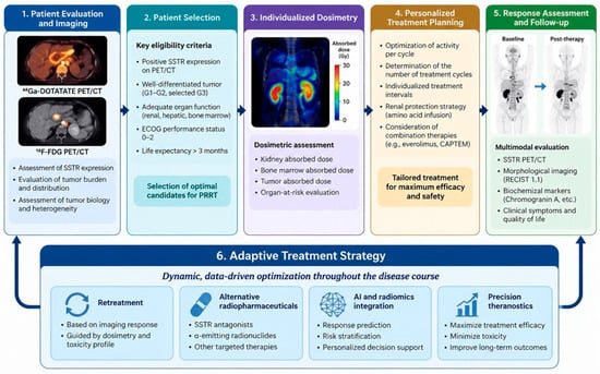
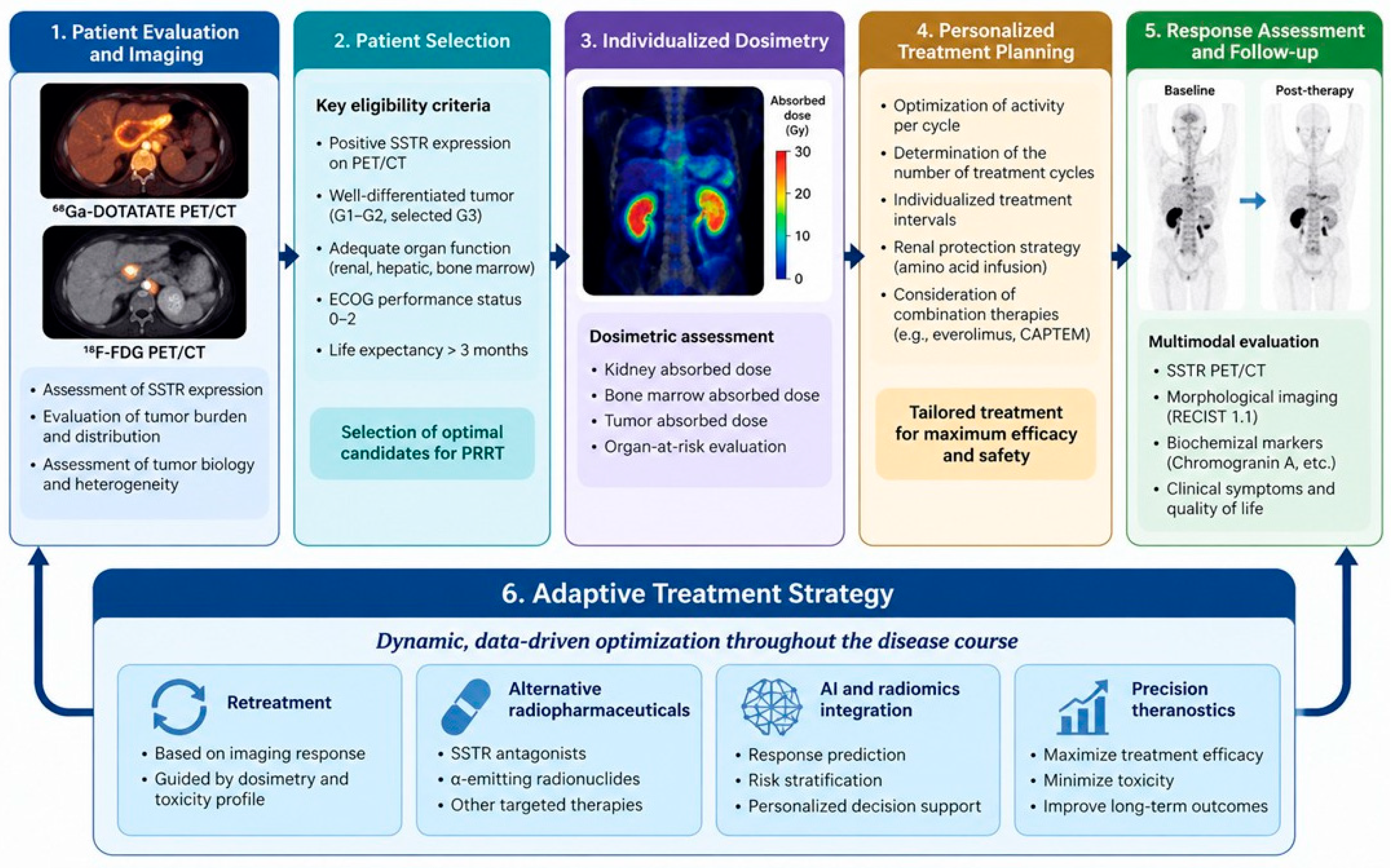
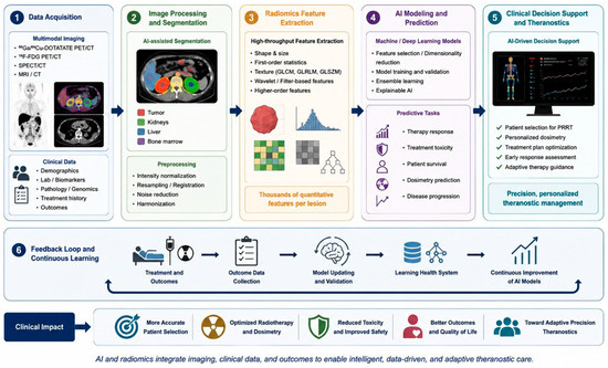
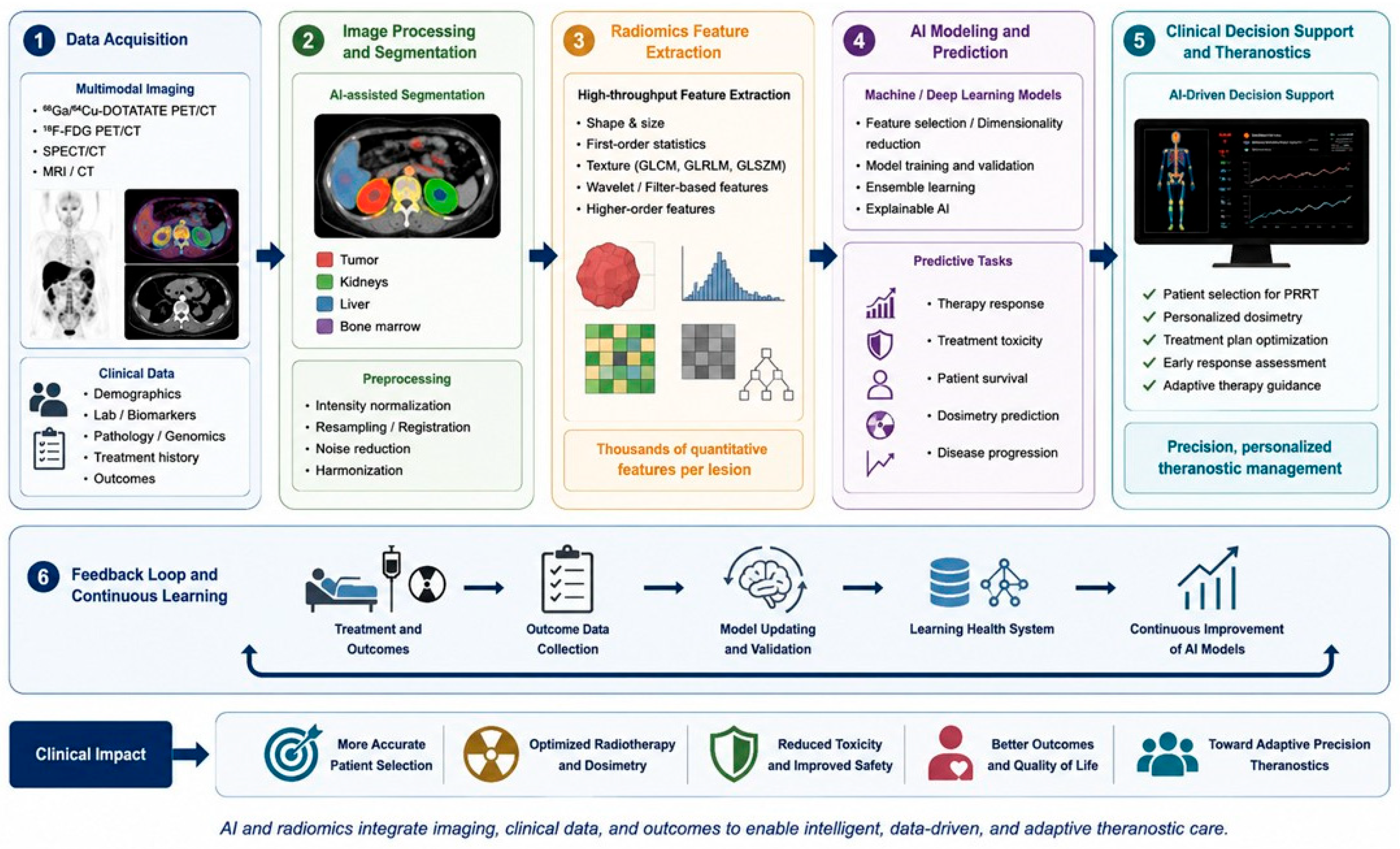
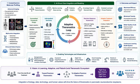
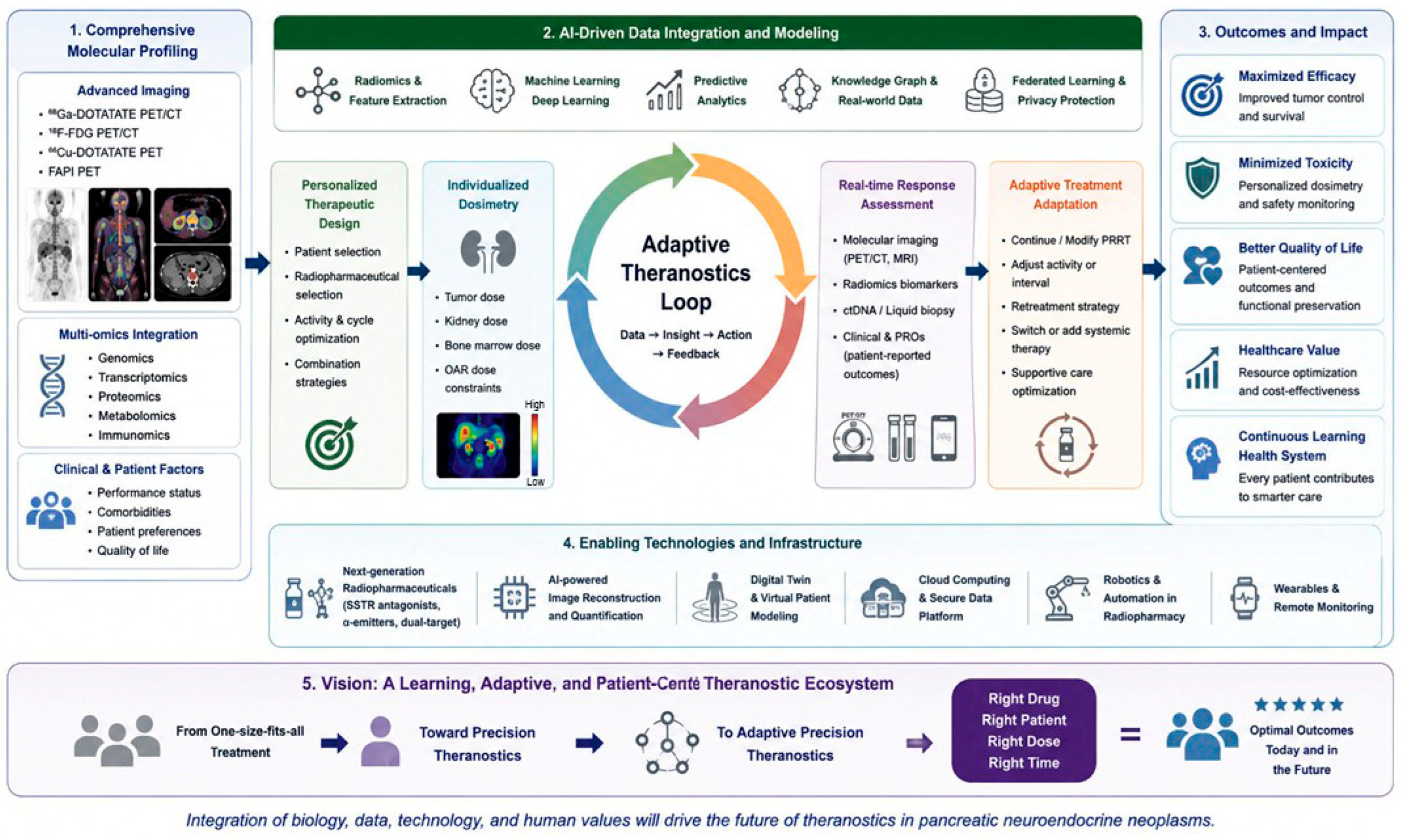

Disclaimer/Publisher’s Note: The statements, opinions and data contained in all publications are solely those of the individual author(s) and contributor(s) and not of MDPI and/or the editor(s). MDPI and/or the editor(s) disclaim responsibility for any injury to people or property resulting from any ideas, methods, instructions or products referred to in the content. |
© 2026 by the authors. Licensee MDPI, Basel, Switzerland. This article is an open access article distributed under the terms and conditions of the Creative Commons Attribution (CC BY) license.