


Abstract
Bio-based plastics, such as polylactic acid, offer an alternative to petroleum-based plastics and a prospect to address the plastic pollution problem. While mounting evidence suggests that microplastics pose a human health threat, the risk posed by bio-based plastics remains unknown. Here, we use Caenorhabditis elegans to investigate the effects of secondary microplastic material derived from 3D-printed polylactic acid plastic on fertility and lifespan. We created and characterized microplastics from a 3D-printed item using cryogenic milling. Using these microplastics, we exposed C. elegans and assessed lifespan, reproduction, and various stress responses. Our studies demonstrate that exposure to 1 µg/L polylactic acid microplastic material reduces fertility and alters gonad structure. When examining germline integrity, we find chromosomal disorganization in the gonad after polylactic acid microplastic exposure and increased apoptotic cell death, which correlates with DAF-16/FOXO and gst-4 oxidative stress responses. Although lifespan decreases with exposure to microplastics of different polymer types, we did not observe a change in lifespan with polylactic acid microplastic material exposure. Overall, we find that exposure to microplastic material derived from a 3D-printed polylactic acid item reduces fertility and is accompanied by germline alterations, including chromosome aberrations, oxidative stress, and cell death.
1. Introduction
While bio-based plastics offer an alternative to petroleum-based plastics and help address the pervasive problem of plastic pollution, their health risks remain understudied. Bio-based plastics hold promise for reducing plastic waste due to their ability to degrade and their perceived environmental and health benefits over conventional plastics [1,2].
The first bio-based plastic was synthesized in the 1930s, though its widespread use has only recently increased significantly [1]. Lactic acid (LA) was first isolated by the chemist Wilhelm Scheele and was later polymerized by Wallace Hume Carothers as polylactic acid (PLA) [1]. Early production was cost-prohibitive, and it did not gain widespread use until the 1960s, when it was used as absorbable sutures and drug-delivery matrices in biomedicine [3,4,5]. Since being approved by the Food and Drug Administration for biomedical uses in the 1970s, PLA and other bio-based plastics have been used in food packaging [2,6,7] and, more recently, as filaments for 3D printing [8]. PLA currently accounts for the largest share of filaments used in 3D printing, and its use is expected to increase [8].
PLA is a bio-based, biodegradable plastic derived from fermented plant sugars [1]. It offers advantages such as reduced use of conventional petroleum-based plastics and applications in biomedical and food fields. PLA biodegrades through hydrolysis of its ester backbone [9]. Under high temperatures and humidity, this process produces lactic acid oligomers and, finally, the lactic acid monomer, which some microbes can metabolize into carbon dioxide, water, and biomass [9,10]. However, it does not fully degrade under most real-world environmental conditions and can lead to PLA microplastic formation [10]. As PLA requires very specific conditions to degrade (i.e., humidity (>50%) and temperature (>55 °C) [9], it can persist in the environment [11] and produce secondary microplastics (MP; particles < 5 mm [12]) and nanoplastics (NP; particles < 1 µm [12]). MPs and NPs of a wide variety of polymer types have been found in animal and human tissues [13,14,15,16]. Overwhelming evidence points to MPs’ detrimental effects on health and development. Across a wide range of animal models, MPs and NPs reduce lifespan and reproductive success and can act as carriers, amplifying their detrimental effects [17,18,19,20].
Much of the evidence on MP and NP exposure relies on pristine primary MP stocks because they are readily available [21,22]. However, these pristine MPs do not adequately represent the secondary microplastics found in the environment. Evidence shows that MPs with irregular size, shape, and surface texture have more detrimental effects than their pristine counterparts [21,23,24]. Irregular secondary MPs form through the breakdown of larger items, wear during product use, or generation in waste management [25]. UV exposure, mechanical wear, or fragmentation of larger plastic particles can drive this breakdown [25]. Additionally, primary and secondary MPs can contain manufacturing additives or absorb and carry environmental chemicals, which can be transferred to animals after ingestion [17,26]. Thus, studying secondary (irregular) MPs has direct implications for human health and remains an understudied area of MP and NP exposure research compared with pristine plastic counterparts.
Discarded waste from 3D printing can become persistent MPs that warrant further study. The 3D printing waste stream is emerging and poorly characterized because the process is decentralized [27]. Waste from 3D printing can come from degradation of the 3D-printed item after it is discarded or from waste generated during production [27,28,29]. One study found that 34% of the materials used in 3D printing ended up as waste, largely from support structures and failed prints [28]. Although additive manufacturing often reduces scrap waste compared to subtractive methods, printed parts can still add to the waste stream [29]. Despite efforts to reduce and recycle 3D-printed plastic waste, it is expected to increase in both industrial and personal manufacturing, and recycling these materials comes with significant challenges [30]. Therefore, we must ask how MPs from these processes can harm health, as studies on the health effects of MPs derived from 3D-printed waste remain limited.
The assumption that PLA is “safe” is being challenged with emerging evidence that PLA-MPs can cause neurotoxicity, intestinal damage, inflammation [31,32], and reduce fertility [33,34,35]. Here we use the well-established Caenorhabditis elegans (C. elegans) model [36] to investigate the effects of irregular secondary PLA-MPs on reproduction and lifespan. PLA-MPs were made from an item produced with a 3D printer to mimic common types of waste that could lead to the production of MPs through improper disposal. Secondary MPs were made using a mechanical method that introduces no additional chemicals beyond those already present in the item to examine the effects of PLA-MPs with heterogeneous shape, surface texture, surface structure, and size. PLA-MP exposure levels of 1 µg/L were used, as they are within the range of other studies asking about MP effects and that mimic concentrations determined from human samples, such as blood [13,19,33,37,38]. The main aim of this study was to determine the physiological effects of secondary MP material made from a PLA-3D-printed item. This study is novel because bio-based plastics can still produce persistent MPs with the potential to cause health effects in animals and humans, and yet they remain understudied. We hypothesized that MP material derived from a PLA-based 3D-printed item would form a heterogeneous, irregular pool and cause physiological defects in C. elegans. We found that exposure to PLA-MP material reduced brood size, altered gonad development, induced apoptosis, and triggered an oxidative stress response in the germline.
2. Materials and Methods
2.1. Creation of Secondary Microplastics
A plastic cone was printed on an A1 Mini 3D printer (Bambu Labs, Shenzhen, Guangdong Province, China). Orange 1.75 mm PLA filament (PrintBed, U.S. NatureWorks, Plymouth, MN, USA) was used to make the item. A 2.5 g plastic cone fragment was collected and placed in a Freezer/Mill CG-200 (Cole-Parmer, Metuchen, NJ, USA). We adapted the cryogenic milling protocol from the Cole-Parmer Sample Preparation for Cryogenic Grinding of Polymers with the CG-200 Freezer/Mill. Using the thermoplastic setting, the following protocol was used: 15 min precool, 12 grinding cycles (2 min each), 2 min cool, and a rate of 12 cycles per second. This cycle was repeated 3 times. The plastic particles were then placed in a sieve stack (starting with the largest, Sieve No. 40 [425 µm], Sieve No. 60 [250 µm], Sieve No. 80 [180 µm], Sieve No. 100 [150 µm], and ending with the smallest, Sieve No. 63 µm [63 µm], Standard Sieves Dual Manufacturing stainless steel mesh, ASTM E11 Compliant Sieves, Franklin Park, IL, USA), and the stack was manually shaken for 10 min. Using a series of sieves aids fractionation and improves sieving efficiency. Fragments of MPs that passed through the 63-micron opening were collected for use in all experiments. All MPs are stored in glass containers to reduce potential MP contamination from other sources.
2.2. Characterization of Microplastics
ATR-IR analysis was used to determine the plastic and chemical profile. Using the Bruker Alpha Platinum ATR, a scoop of PLA-MPs was placed on the crystal part of the instrument and scanned at a resolution of 4 cm−1, 16 scans, and a range of 400–4000 cm−1. MP size and surface were characterized by Scanning Electron Microscopy (SEM; JEOL JSM-6010 PLUS/LA, Peabody, MA, USA), as previously described [17]. Samples were sputter-coated with 2 nm gold particles prior to imaging (Quorum Technologies EMS150T ES, Lewes, UK). MP size (area, smallest width, longest width, and thickness) was measured using ImageJ software (version 1.54).
GCxGC-TOFMS analysis was performed to identify the chemical components of the PLA filament used to create MPs. PLA filament was cut into ~1 cm pieces, and 200 mg was weighed out. Next, 1.0 mL of dichloromethane (DCM) was added for every 100 mg of sample (extraction factor 10 mL/g). Samples were allowed to sit for four hours, then the solvent was removed. The supernatant was transferred to a vial for storage and further preparation. A solvent blank was also prepared. All extracts were prepared at a 10-fold dilution level for analysis by adding 100 µL of extract to an autosampler vial containing 900 µL of DCM and 10.0 µL of internal standard cocktail. The internal standard cocktail contains 100 µg/mL of 1,4-Dichlorobenzene-D4, Naphthalene-D8, Acenaphthene-d10, Phenanthrene-D10, Chrysene-D12 and Perylene-D12. The final internal standard concentration is 1 µg/mL.
GCxGC-TOFMS analysis of DCM extracts was performed using an Agilent 7890 gas chromatograph coupled to a LECO HRT GCxGC (LECO, St. Joseph, MI, USA). Injection volume was 1.0 µL. The inlet temperature was 275 °C, and the inlet mode was splitless with a 1 min purge. Separation was achieved using two columns. The primary column (1st Dimension) was an RXi-1MS (30 m × 0.25 mm × 0.25 µm; Restek, Bellefonte, PA, USA), and the second column (2nd Dimension) was an RXi-17SilMS (1.3 m × 0.25 mm × 0.25 µm; Restek, Bellefonte, PA, USA). The first column was held at 45 °C for 3 min, ramped to 330 °C at a rate of 8 °C/min, and held for 5.0 min. The second column and modulator were offset by 5.0 and 8.0 degrees Celsius, respectively, relative to the primary oven. Helium carrier flow was set to a constant flow of 1.2 mL/min. The transfer line temperature was set at 300 °C. The modulation period was 4 s (1.0 s hot, 1.0 s cold, with 2 cycles per modulation period) throughout the entire run. The mass spectrometer was operated using electron ionization (EI) at 70 eV. The ion source temperature was set at 225 °C. Spectra were collected from 45 to 650 m/z with a scan time of 100 spectra/s. The filament was turned on at 210 s.
2.3. Preparation of E. coli OP50 Food and PLA Microplastics
MPs were suspended in Escherichia coli (E. coli, strain OP50, grown in LB broth), then seeded on Nematode Growth Media (NGM) plates (Teknova, N1105, Hollister, CA, USA), and allowed to dry overnight before adding C. elegans to the plates for exposure as previously described [17]. To prepare PLA-MP E. coli suspensions, PLA-MPs were suspended in a 1% solution in 1× phosphate-buffered saline (PBS), then diluted into E. coli OP50. At each dilution step, the solution was sonicated for 15 min (Emerson Branson 3800, Danbury, CT, USA). Control E. coli, not containing MPs, was sonicated and diluted in parallel. This was done as previously described to ensure that E. coli are still viable [17]. For the 1 µg/L PLA exposure, 200 µL of suspension was added to each 60 mm plate (nominal PLA mass = 0.2 ng per plate), while 500 µL was added to each 100 mm plate (nominal PLA mass = 0.5 ng per plate). These values represent the nominal mass added based on suspension concentration and volume; particle number per plate was not quantified. The exposure material consisted of irregular particles spanning a broad size distribution; therefore, particle number cannot be accurately converted from nominal mass without assumptions about particle geometry, thickness, density, and the post-sonication particle-size distribution. Accordingly, exposure is reported as nominal mass concentration and nominal mass delivered per plate rather than particle number. The actual number of particles delivered to individual plates was not measured and represents a limitation of the exposure assessment. Wide-bore tips were used to transfer liquid. In-house prepared PLA-MPs were used for all experiments.
2.4. Maintenance of C. elegans
Caenorhabditis elegans (C. elegans) strains, N2 (wild type), TJ356 (zIs356 [daf-16p::daf-16a/b::GFP + rol-6(su1006)]), CL2166 (dvIs19 [gst-4p::GFP::NLS] III), and MD701 (bcIs39 [lim-7p::ced-1::GFP + lin-15(+)] that were used in this study were provided by the Caenorhabditis Genetics Center. C. elegans were grown on NGM plates seeded with E. coli at 20 °C.
2.5. Exposure Design
Bleach-synchronized L1 larvae were exposed to either E. coli or E. coli containing secondary 1 µg/L PLA-MP material on NGM plates prepared within 48 h. A concentration of 1 µg/L MP was used for all exposure studies, as this level of exposure is within the range of other studies and falls within the concentration range reported in previous MP studies and is comparable on a mass-concentration basis to concentrations reported in some human samples such as blood [13,17,19,20,37,38]. However, differences in polymer composition, particle size and number, and analytical methods limit direct comparison among studies. At least 2–3 biological replicates were performed for each assay. For stress assays and gonad chromosome analysis, L1-synchronized larvae were placed onto each condition, incubated for 72 h at 20 °C, and then imaged. For the gst-4 promoter-driven GFP oxidative stress positive control, C. elegans were exposed to either M9 or 50 mM paraquat in M9 and incubated at 20 °C for 1 h before dissecting the gonads and imaging live, adapted from [39]. For all experiments, C. elegans were monitored during exposure and prior to endpoint analysis to ensure they were at the same stage. A schematic of exposure design and endpoints is available as Supplemental Figure S1.
2.6. Lifespan Assay
Lifespan assays were performed at 20 °C. Approximately 50 synchronized L1 larvae were placed on the corresponding treatment conditions for 48 h and then transferred to new plates containing the same treatment with 100 µM 5′-fluorodeoxyuridine (RPI, 50-91-9, Mount Prospect, IL, USA). C. elegans were counted as dead or alive every day until the number of live C. elegans reached zero, as described previously [17]. Three independent lifespan assays were performed. Day 0 is adult day 1. During the lifespan assay, if a nematode dies due to an unnatural event (lost on pick or injured), it is censored and not included in the count.
2.7. Evaluation of Reproductive Toxicity
To determine the total number of eggs laid per C. elegans and the percentage hatched, bleach-synchronized L1 larvae were seeded onto either control (E. coli) or 1 µg/L PLA-MPs in E. coli OP50. After 48 h from initial seeding, L4 larval C. elegans were then singled onto respective plates. The following day, L4 singling was repeated from the previous day’s plate, and both plates were kept. After 24 h, L1s and embryos were counted from the first day of separating, then repeated every day, counting the L4-free plate, until egg laying stopped (around 6–7 days), as previously described [17,39].
2.8. Imaging and Microscopy
All fluorescent and bright-field images were captured using an EVOS M5000 (Invitrogen, Bothell, WA, USA). Specifically, Carnoy’s-fixed and Hoechst-stained whole C. elegans images were taken with a Zeiss LSM 910/Axio Observer 7 (Zeiss, Jena, Germany). For live imaging, whole C. elegans (DAF-16::GFP and CED-1::GFP) were transferred to an agarose pad on a slide and immobilized with levamisole (Vector Laboratories, SP-5000-18, Newark, CA, USA). For live imaging of dissected gonads (gst-4p::GFP expression), gonads were dissected from C. elegans and imaged. Protocols were adapted from previously described studies [17,39]. We used representative SEM images to measure particle size (longest and shortest length and area) with ImageJ measurement tools. All particles were measured in at least 5 representative images ranging from 30 to 40× magnification. All image processing and analysis were performed using ImageJ software (version 1.54). For the gonad area and length, FIJI (version 2.9.0) was used for analysis.
2.9. Activation of Stress Response
To determine whether PLA-MP exposure induces stress activation, the strain TJ356, expressing DAF-16 fused to GFP, was exposed to PLA-MPs as described. Live imaging was completed within 5 min of paralysis. Each C. elegans was classified by the localization of DAF-16::GFP expression (cytoplasmic, intermediate, or nuclear), as previously described [17,40,41].
2.10. Oxidative Stress Assay
To determine whether PLA-MP exposure leads to oxidative stress, the strain CL2166, expressing GFP under the gst-4 promoter, was exposed as described. This reporter strain has GFP fused to the promoter of glutathione S-transferase 4, and oxidative stress induces GFP expression. Following exposure, C. elegans were transferred into a drop of M9 on a positively charged slide. Gonads were dissected and isolated from the remaining carcasses to minimize background autofluorescence, as previously described [39]. Dissected gonads were imaged within 10 min of dissection. Imaging settings were consistent across samples. Fluorescent intensities were determined in ImageJ software. A set of gst-4p::GFP C. elegans was treated for 1 h with or without 50 mM paraquat (PQ) (in M9) and was used as positive and negative controls, respectively, adapted from [39].
2.11. Germ Cell Apoptosis
To determine if PLA-MP exposure leads to cell death in C. elegans, wild-type C. elegans were exposed to 1 µg/L PLA-MPs in E. coli or E. coli only for 72 h. This protocol was adapted from Henderson et al. [39]. Adult C. elegans were washed from plates and incubated with 25 μg/mL of acridine orange (Abcam, ab270791, Waltham, MA, USA). Tubes were wrapped in foil to protect from light and placed on a rotator at room temperature for 1 h. C. elegans were then transferred to NGM plates with E. coli and protected from light for 1 h. Stained C. elegans were then mounted on 4% agarose pads with levamisole and imaged. MD701 C. elegans (expressing a CED-1::GFP fusion protein in sheath cells) were exposed to 1 µg/L PLA-MPs in E. coli or E. coli only. L1-synchronized larvae were placed onto each condition and live-imaged 72 h later.
2.12. Chromosome Organization
After PLA-MP and control exposure (as described above), adult C. elegans were picked and placed onto a concave slide containing a drop of M9. Bacteria were washed away by pipetting off and discarding the original M9 drop, then replacing it with the new M9, ensuring the C. elegans were untouched. The C. elegans were then pipetted onto a positively charged slide with a drop of M9 + 1 mM levamisole. Within 2 min, the gonads were dissected using a 27G needle to cut off the heads, just below the pharynx. The excess paralytic liquid was pipetted out and wicked away. Following dissection, gonads were fixed in 4% paraformaldehyde (PFA) in 1× phosphate-buffered saline (PBS) for 20 min. After incubation, excess liquid was removed. The sample was then washed three times for 3 min each with PTX solution (10× PBS, 10% Triton X). Next, C. elegans were stained with Hoechst for 5 min, followed by one wash with PTX. A mounting medium was added prior to adding a coverslip. These were then imaged using an EVOS fluorescence microscope and subsequently quantified. This protocol was adapted from Henderson et al. [39].
2.13. Gonad Morphology
Analysis of adult gonad morphology after PLA-MP and control exposure (as described above) was performed by fixing adult C. elegans with Carnoy’s fixative (6 parts ethanol: 3 parts chloroform: 1 part glacial acetic acid). C. elegans were wicked into a minimal volume of M9 buffer on a microscope slide, and excess liquid was wicked away. C. elegans were then fixed by dropping 2–3 drops of Carnoy’s fixative onto the slide from a height of 2 inches, and the slide was allowed to dry. C. elegans were then rehydrated in 20 μL of M9 for 1 h. After wicking away excess M9, 10 μL of staining solution (1:10,000 Hoechst 33342 (20 mM, 62249, Thermo Scientific, Waltham, MA, USA) was added and incubated for 5 min. Excess staining solution was wicked away, and the remaining area was washed once with M9. Mounting medium was added, and the slide was covered with a coverslip. Slides were left overnight at 4 °C before imaging. Carnoy’s-fixed and Hoechst-stained C. elegans were used to measure gonad area and length, as adapted from Shao et al. [24].
2.14. Statistical Analysis
Statistical analysis and figure design were performed with GraphPad Prism 8 (GraphPad Software version 11, San Diego, CA, USA) or Excel. Data were checked for normality between treatments, and statistical analysis was performed using a Mann–Whitney test (brood size, oxidative stress, apoptosis, and gonad measurements). For gonadal analyses, each gonad was obtained from a separate individual C. elegans animal, and individual worms were considered independent biological observations. Multiple worms were analyzed for each treatment condition, and experiments were independently repeated three times on different days. Overall response patterns were visually examined across the three independent experimental runs to confirm consistency among experiments. For analysis of mitotic germ cell nuclear diameter, multiple nuclei were measured within the gonad of each individual worm. Because multiple nuclear measurements were obtained from each gonad, nuclear diameter was analyzed at two levels. First, individual nuclear diameter measurements were compared among treatment groups on a per-nucleus basis. Second, nuclear diameter measurements were averaged within each gonad to generate a single mean nuclear diameter for each individual worm, and statistical comparisons were performed using these per-gonad means. Thus, the per-nucleus analysis evaluated the distribution of individual nuclear measurements, whereas the per-gonad analysis used the individual worm as the unit of analysis. A chi-square test was performed to evaluate the localization of DAF-16 in TJ356 using raw frequency counts, with data presented as percentages. Fisher’s exact test with 95% C.I. was performed to analyze the raw counts of Hoechst staining data. Chromosome organization defects were analyzed using two-tailed Fisher’s exact tests. For each defect category, the 2 × 2 table consisted of treatment (control versus PLA-MP) by defect status (present versus absent), with each gonad counted once for a given defect category. Because a single gonad could exhibit multiple abnormalities, defect categories were analyzed independently. Reported p-values were not adjusted for multiple comparisons. Experiments were all carried out with a minimum of 2–3 biological replicates (N) and the total number of C. elegans (n) examined, which were used in statistical analysis as described.
3. Results and Discussion
3.1. Creation and Characterization of PLA Secondary Microplastics
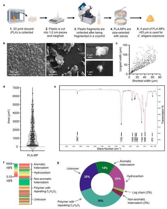
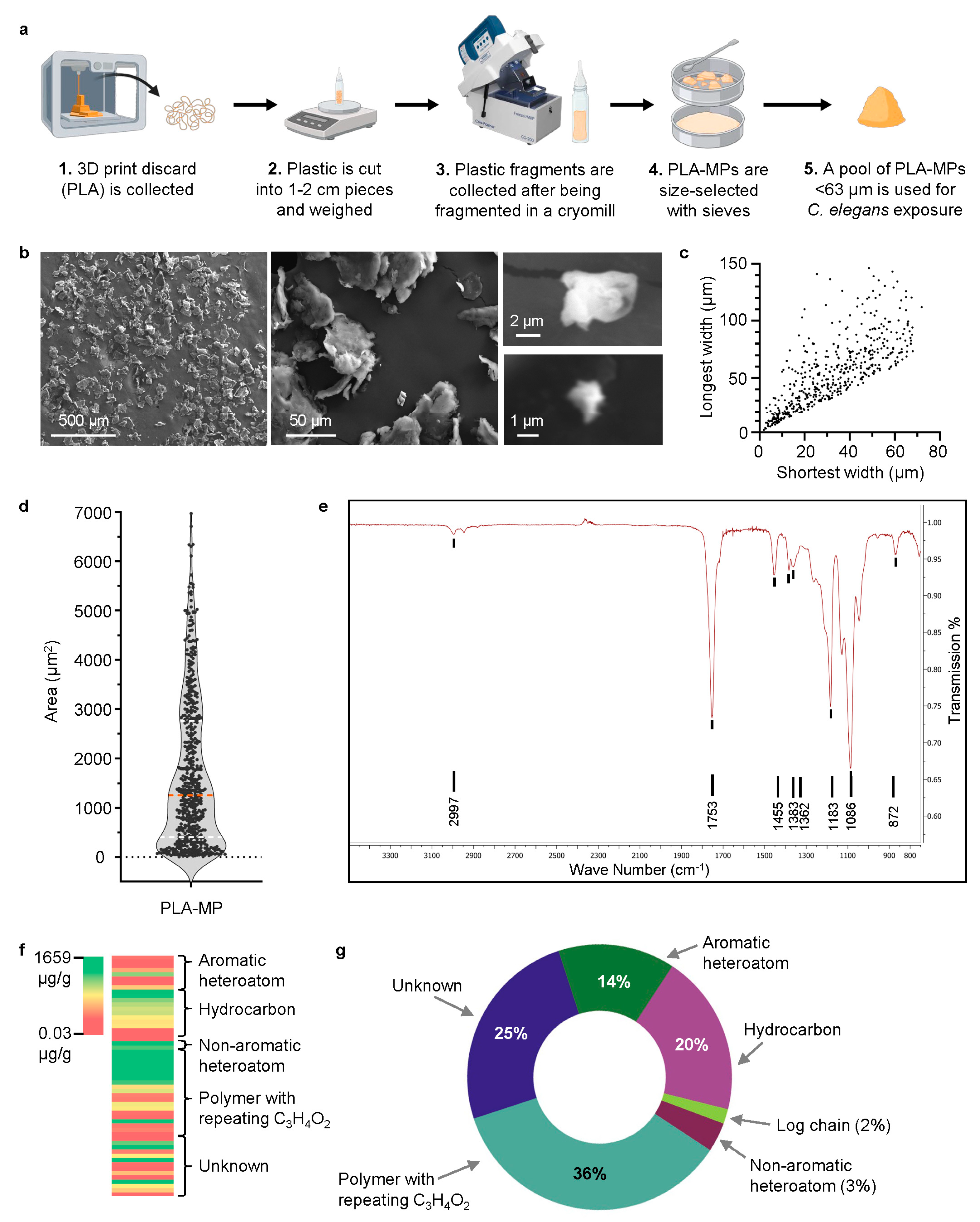
Secondary polylactic acid (PLA) microplastics (MPs) were produced from discarded 3D-printed items (Figure 1a) and characterized (particle size and surface structure) by scanning electron microscopy (SEM). PLA-MPs were irregular in size and shape, with an overabundance of flat irregular shapes (Figure 1b). Quantification of particle size using the shortest and longest surface (Figure 1c, Supplemental Figure S2) showed a variety of sizes ranging from 1 to 150 µm (Figure 1c, Supplemental Figure S2). The area of the largest surface visible showed a broad distribution, with a large portion of the population skewing towards a smaller overall area (Figure 1d). We mechanically separated MPs with a 63 µm sieve, but larger fragments can pass through (as seen, up to 150 µm) if one side is 63 µm or less. In our work, we chose to generate environmentally realistic secondary MPs from plastic waste, rather than relying on homogeneous commercially available MPs. Secondary MPs can be produced in the laboratory with a variety of techniques, such as ultraviolet (UV) exposure, chemical treatment, and mechanical breakdown [23,42,43,44]. Mechanical methods are a top-down approach that use physical fragmentation of plastic through processes such as cutting, grinding, milling, and cryogenic treatment. The particles produced by these processes mimic the irregular characteristics of environmental plastics [42,44]. The fragments we produced had irregular, heterogeneous morphologies, as observed in the mechanical breakdown of other polymer types, and sieving allowed us to narrow the particle-size range tested [44,45]. It is important to note that during our suspension of MPs in the C. elegans food source, we performed multiple sonication steps. Sonication has been used to fragment materials and create MPs [42]. Therefore, the final PLA-MP material suspension presented to C. elegans may contain a larger proportion of smaller particles than SEM determined, because this step occurred before sonication. MP exposure can be through ingestion, dermal contact, and inhalation [46,47]. MPs of various polymers and mixtures have been detected in prepared food products, raw fruits and vegetables, drinking water, and beverages [48,49,50,51]. Many studies of MP exposure focus on ingestion. When considering ingestion as a source of exposure, C. elegans is limited by the size of its mouth (~4 µm in adults) [52,53]. The provided food source, E. coli, is approximately 1 µm in size. In the wild, C. elegans will consume a variety of microbial food sources, though they show a preference for smaller food particles, and ingestion of larger items can lead to reduced feeding or obstruction [52]. Regarding MPs, C. elegans have been shown to ingest those up to 5 µm in diameter [54]. Based on SEM size-distribution data, an estimate of particles small enough to be ingested (≤5 µm, the smallest particle size) indicates that less than 3% of particles fall within this range. This estimate is limited by potential bias toward imaging larger particles, which were easier to identify. Exposure to PLA-MP material could therefore have occurred through ingestion of an irregular pool of PLA-MPs, which may include larger particles (>5 µm). Thus, causing inefficient ingestion and indirect toxicity through feeding interference or dermal exposure. Additionally, leachates, not the plastic particles themselves, could cause and/or contribute to physiological defects. We refer to our exposure conditions as PLA-MP material exposure, as we cannot rule out that the effects are due to the MPs themselves, the leachates, or a combination.
Figure 1.
Creation and characterization of secondary MPs from 3D printer PLA waste. (a) Diagram of the process of making a less than 63 µm pool of PLA-MPs. Created with BioRender.com. (b) Representative SEM images of PLA-MPs to determine size, shape, and surface characteristics. (c) PLA-MP particle size distribution, n = 510. (d) PLA-MP particle area distribution, n = 510. (e) ATR spectrum of PLA-MP. (f) GCxGC-TOFMS analysis of PLA filament used to create MPs. Concentration (µg/g) of chemicals and their classification. (g) Pie chart representation of the classification of chemical types determined by GCxGC-TOFMS analysis of PLA filament.
Next, we examined the chemical properties of the 3D-printer-extruded and cryo-milled PLA-MP particles. ATR-FT-IR spectra showed a similar IR profile (Figure 1e) to that observed for reported PLA samples [6]. Some plastic polymers undergo physical changes that alter their ATR/FTIR spectra after UV degradation, a process that facilitates the secondary creation of MPs [23,55]. No chemical differences were observed, which is consistent with studies showing that 3D-printed PLA materials are not altered by the extraction and heating processes used in 3D printing [56]. PLA-MPs were created using a cryogenic mill to mimic mechanical degradation without chemical or UV treatment, allowing examination of the consequences of non-pristine MP particles without additional chemical treatment. To further characterize the additives in the PLA filament, GC×GC–TOFMS (comprehensive two-dimensional gas chromatography coupled with time-of-flight mass spectrometry) was used. The analysis confirmed the presence of PLA polymer, yet over 50 components, including aromatic heteroatoms, hydrocarbons, non-aromatic heteroatoms, polymers with repeating C3H4O2, and 14 unknown chemicals, are also present (Figure 1f,g, and Supplemental Figure S3).
3.2. C. elegans Exposed to PLA-MP Material Leads to Reduced Fertility
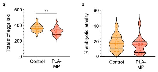
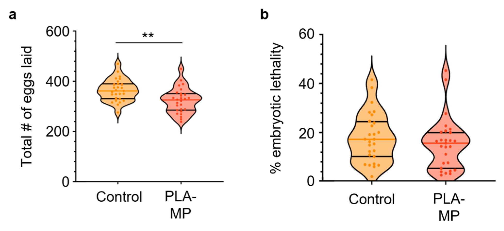
To determine whether PLA-MP material affects reproductive toxicity, L1-stage C. elegans were continuously exposed to 1 µg/L PLA-MPs. There was a significant decrease in the total number of eggs laid when exposed to PLA-MPs (p = 0.0021) (Figure 2a). However, there was no significant change in embryonic lethality with PLA-MP exposure (Figure 2b, Supplemental Figure S4). MPs of various polymers at similar concentrations have been shown to reduce brood size [17,18,23,33,57]. In contrast to our results with a pool of non-pristine irregular PLA-MPs, no reduction in brood size was seen with 1 µg/L exposure to ~3 µm pristine PLA-MPs, whereas higher concentrations (10 and 100 µg/L PLA-MPs) reduced brood size [33]. Although embryonic lethality did not change, as with pristine PLA-MPs at higher concentrations, brood size decreased under our exposure conditions.
Figure 2.
Total eggs laid and embryonic lethality of C. elegans exposed to PLA-MP material. (a) Number of eggs laid per C. elegans with E. coli only or with PLA-MPs. N = 4, n = 27, 27. # = number (b) Percent embryonic lethality. Violin plots show the mean with the standard deviation. Mann–Whitney (two-sample) tests were used for statistical analysis (** p ≤ 0.01).
Differences from studies using pristine PLA-MPs may reflect differences in particle morphology, exposure characteristics, and/or the chemical composition of the materials tested. Multiple studies show that irregular MPs have a more detrimental effect on fertility than their pristine counterparts. Shao et al. demonstrated that secondary MPs produced via UV irradiation reduced brood size compared with pristine PLA-MPs [24]. Compared with our irregular cryo-milled PLA-MPs, this work showed that uniform-sized PLA-MPs produced by UV irradiation were slightly smaller, had a more rugged surface, and exhibited altered chemical bonds compared with those not treated with UV [24]. While there are differences in the production of secondary MPs (UV irradiation vs. mechanical) and distribution by particle size (more homogeneous vs. heterogeneous), we note that the pool of ingestible PLA-MPs in our study was significantly smaller than that used in Shao et al. (2024) [24]. Differences in particle morphology may contribute to the different reproductive outcomes observed across studies; however, we cannot separate the contribution of shape from differences in particle number, size distribution, or chemical composition in the present study. Reduced fertility may be due to irregular MPs and/or chemicals contained in the plastic itself. MPs can serve as vectors, transporting chemicals into animals [26,58,59], and the chemicals they can harbor may cause more detrimental effects than the individual components alone [17]. While leachates of 10 and 100 μg/L PLA-MP did not cause obvious reproductive toxicity [33], this may be due to differences in PLA-MP material sources and the additives they contain.
3.3. PLA-MP Material Exposure Alters Gonad Development
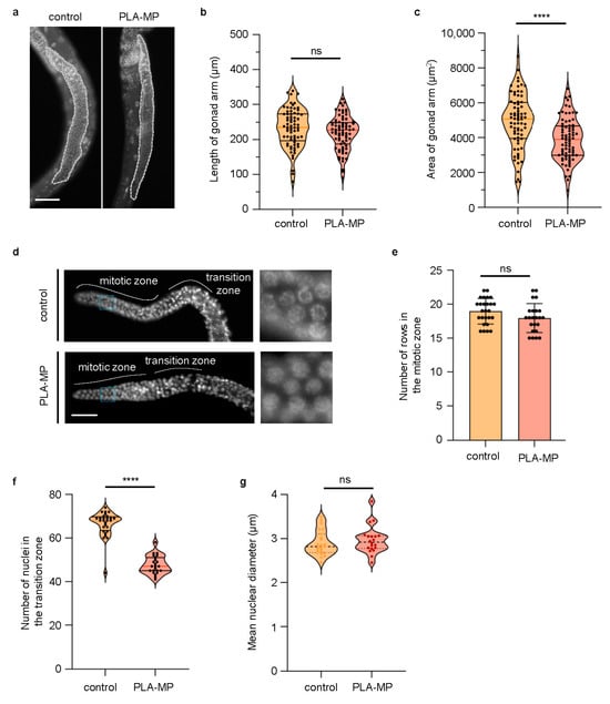
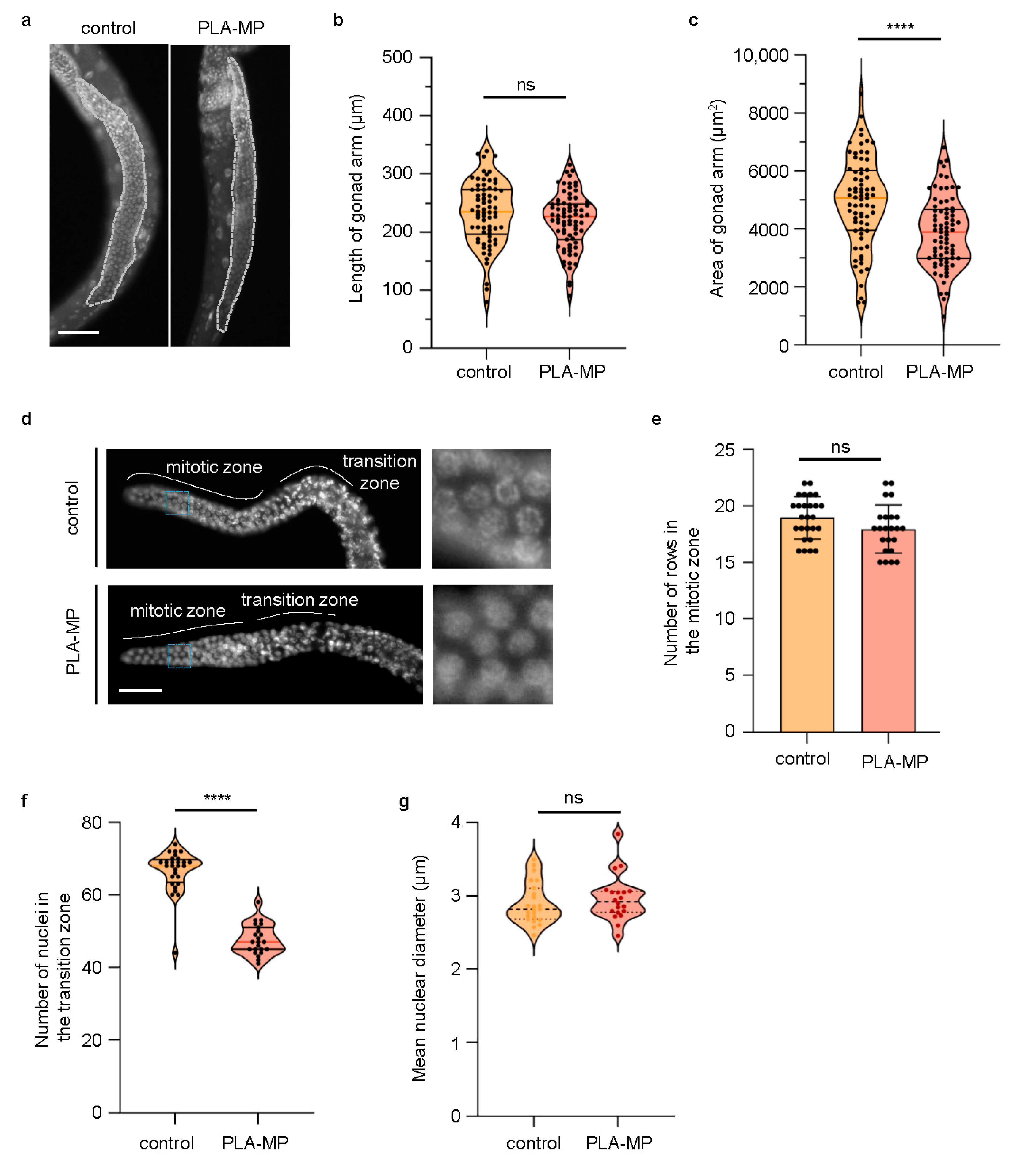
Given the decrease in reproductive capacity, a subsequent examination of alterations in germline development following PLA-MP material exposure was carried out to determine a potential mechanism underlying reduced fertility. Exposure of C. elegans to 1 µg/L PLA-MPs led to a reduced gonad area (p < 0.0001) and a non-significant reduction in the length of the gonad (Figure 3a–c). Additionally, PLA-MP exposure did not alter the number of rows of mitotic cells per gonad, but it did reduce the number of cells in the transition zone (p < 0.0001; Figure 3d–f). PLA-MP exposure significantly increased nuclear diameter of mitotic nuclei when analyzed on a per-nucleus basis (p < 0.0001); however, when nuclear diameter measurements were averaged within each gonad, and individual worms were used as the unit of analysis, the increase was not statistically significant (Supplemental Figures S3 and S4c). Shao et al. showed similar reductions in gonad development with exposure to higher levels (10 and 100 µg/L) of uniform 3 µm-diameter PLA-MPs [33]. 1 µg/L 3 µm-diameter PLA-MPs, or leachates of 10 and 100 μg/L PLA-MPs, did not affect gonad development in their study [33]. These differences likely reflect differences in MP size, shape, surface characteristics, and chemical composition. Another study by Shao et al. compared pristine and UV-aged PLA-MPs and found greater toxicity to reproductive capacity and gonad development in UV-aged PLA-MPs, which had altered chemical and physical properties [24]. Compared with pristine MPs, exposure to irregular MPs is associated with enhanced reproductive defects and altered gonad development, consistent with results from studies using weathered PLA-MPs and other MP types [23,34,60]. The observed decrease in gonad size (Figure 3) parallels those observed with polystyrene (PS) MPs [61]. Furthermore, across multiple species, MP toxicity alters gonad structure, development, and reproductive capacity [18], as seen in other non-MP-related toxicity studies [62,63].
Figure 3.
Alteration of gonad development with exposure to PLA-MP material. (a) Representative images of Hoechst staining in whole adult C. elegans fed E. coli without (control) and with PLA-MPs. The dotted line indicates the measured gonad region. Scale bar = 50 µm. (b) Quantification of the length of the gonad arm, N = 3, n = 70, 75. ns = not significant. (c) Quantification of the area of the gonad arm, N = 3, n = 72, 76. (d) Representative images of Hoechst staining in dissected gonads from adult C. elegans fed E. coli without (control) and with PLA-MPs, left whole gonad, and right zoom-in on pre-meiotic nuclei. The blue box indicates the image expanded to the right. Scale bar = 25 µm. (e) Quantification of the number of rows in the mitotic zone. N = 2, n = 28, 20. (f) Quantification of nuclei in the transition zone. N = 2, n = 28, 20. (g) Quantification of the average per gonad, mean nuclear diameter (μm) in C. elegans, following control or PLA-MP exposure. N = 3, n = 23, 20; p = 0.461 by an unpaired two-tailed Welch’s t-test. **** p ≤ 0.0001.
3.4. PLA-MP Material Exposure Leads to an Increase in Chromosome Organization Defects and Apoptosis in the Gonad
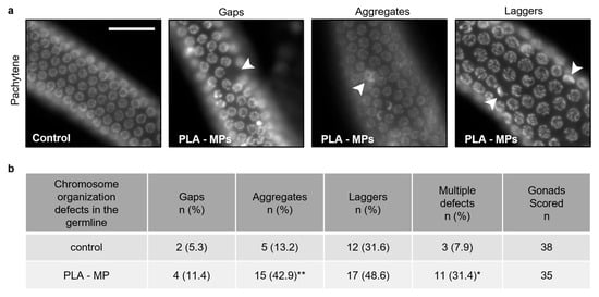
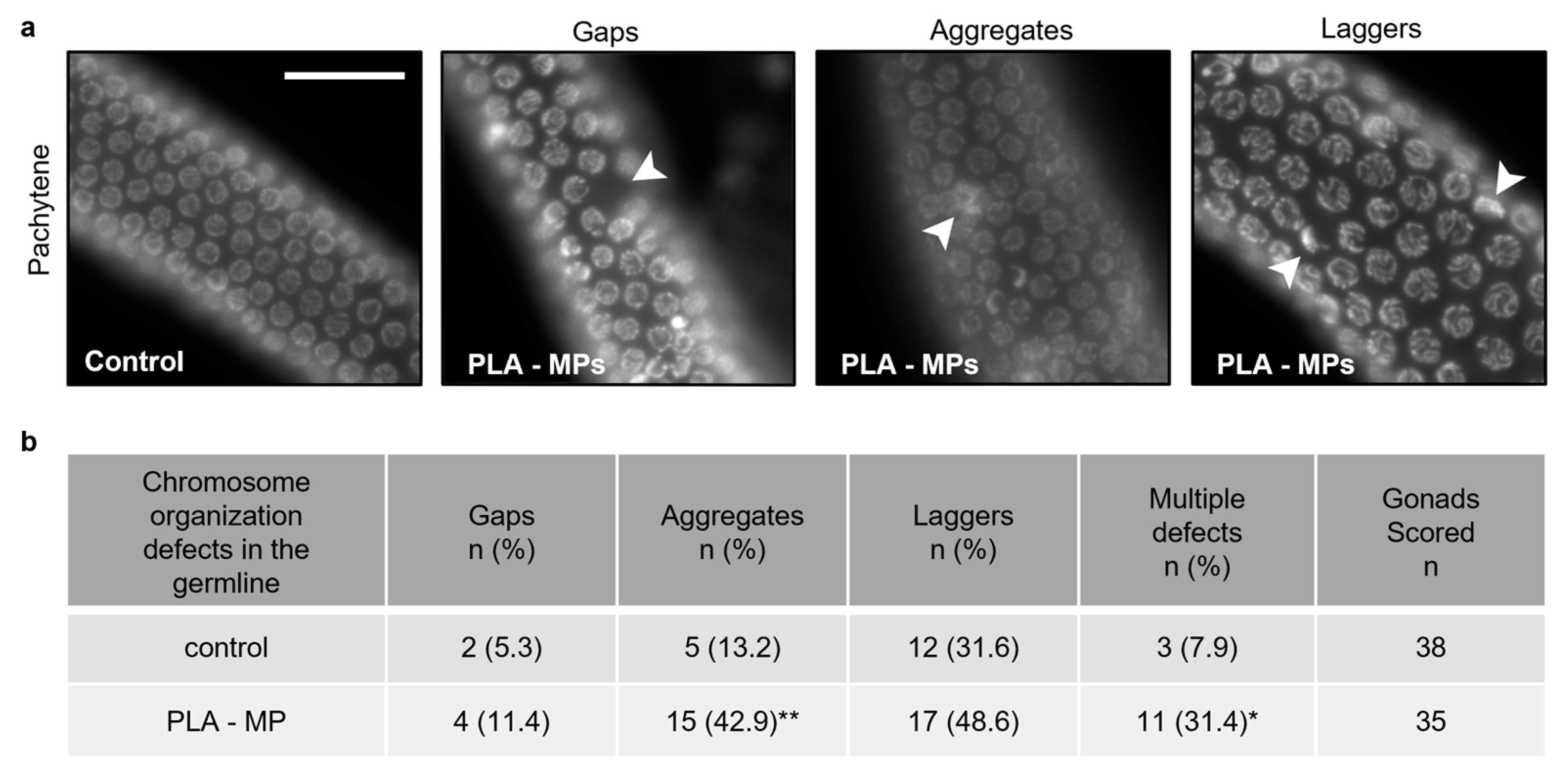
Reduced fertility in C. elegans, leading to gonad defects, can be attributed to defects in chromosome organization and apoptosis in the gonad [39,64]. Fertility defects can result from meiotic errors that lead to autosomal chromosome segregation errors and the formation of aneuploid gametes in C. elegans [65]. To assess whether defects in chromosome organization reduce oocyte development and contribute to reduced fertility, gonads of PLA-MP material-exposed C. elegans were Hoechst-stained. Three categories were defined to represent chromosomal defects that can serve as indicators of DNA damage [64]. Laggers are categorized as leptotene/zygotene-like nuclei commonly found in the transition zone, yet they persist into the pachytene stage [64]. Aggregates are multinucleated clumps, and gaps are areas of reduced nuclear density [64]. Exposure to PLA-MPs significantly increased aggregates (p = 0.0078) and the presence of multiple defects (p = 0.016) (Figure 4). However, PLA-MP exposure did not significantly increase the number of gaps or laggers (Figure 4). Among C. elegans with more than one type of defect, 31.4% of observed gonads had multiple defects after PLA-MP exposure, compared with 7.9% in the control (p = 0.016). Therefore, PLA-MP material exposure resulted in more C. elegans with multiple defects. Shin et al. and Henderson et al. report similar germline chromosomal abnormalities resulting from exposure to soluble chemicals (phthalates and other toxicants) [39,64]. Defects in meiotic chromosome organization may contribute to reduced fertility with PLA-MP material exposure, possibly due to the physical MPs themselves and/or combined exposure to the chemicals they carry. Our results on defects in chromosome organization in the meiotic germline of PLA-MP-exposed C. elegans are consistent with evidence that MPs are genotoxic across species. Polystyrene microplastics (PS-MPs) delay porcine oocyte maturation by disrupting spindle architecture and metaphase I chromosome alignment [66]. PS-MPs similarly led to chromosomal instability in mammalian cells by increasing centriole numbers and altering microtubule-kinetochore attachments [67]. Specifically, in C. elegans, PS-MP exposure leads to oocyte-specific chromosome aberrations and germline apoptosis [68]. Furthermore, chemicals from similar classes identified in the PLA used to generate MP (e.g., phthalates) cause similar germline chromosomal abnormalities [39,64]. Together with established germline toxicity assays showing chemically induced meiotic chromosome defects in C. elegans, these findings support the idea that exposure to secondary PLA-MP material from a 3D-printed item is associated with altered meiotic chromosome organization during meiosis and contributes to reproductive decline.
Figure 4.
Observed defects in the chromosomal organization of C. elegans germline. (a) Representative images of Hoechst staining in adult C. elegans fed E. coli without (control) and with PLA-MP material. Arrowheads indicate defects. Scale bar = 25 µm. (b) Quantification of defects observed (gaps, aggregates, laggers) in Hoechst-stained gonad nuclei in C. elegans with exposure to their regular E. coli diet only or with 1 µg/L PLA-MPs. N = 3, n = 38, 35. Two-tailed Fisher’s exact test was used for statistical analysis (* p ≤ 0.05, ** p ≤ 0.01).
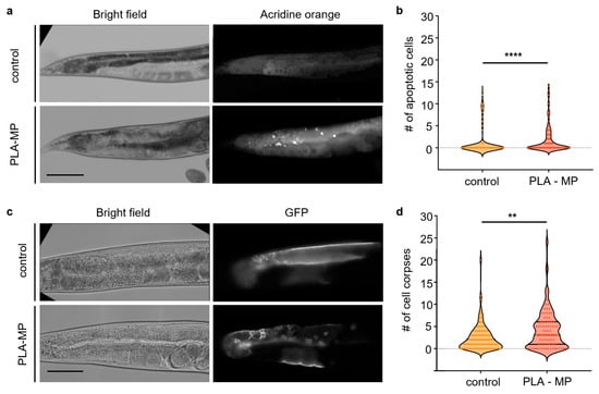
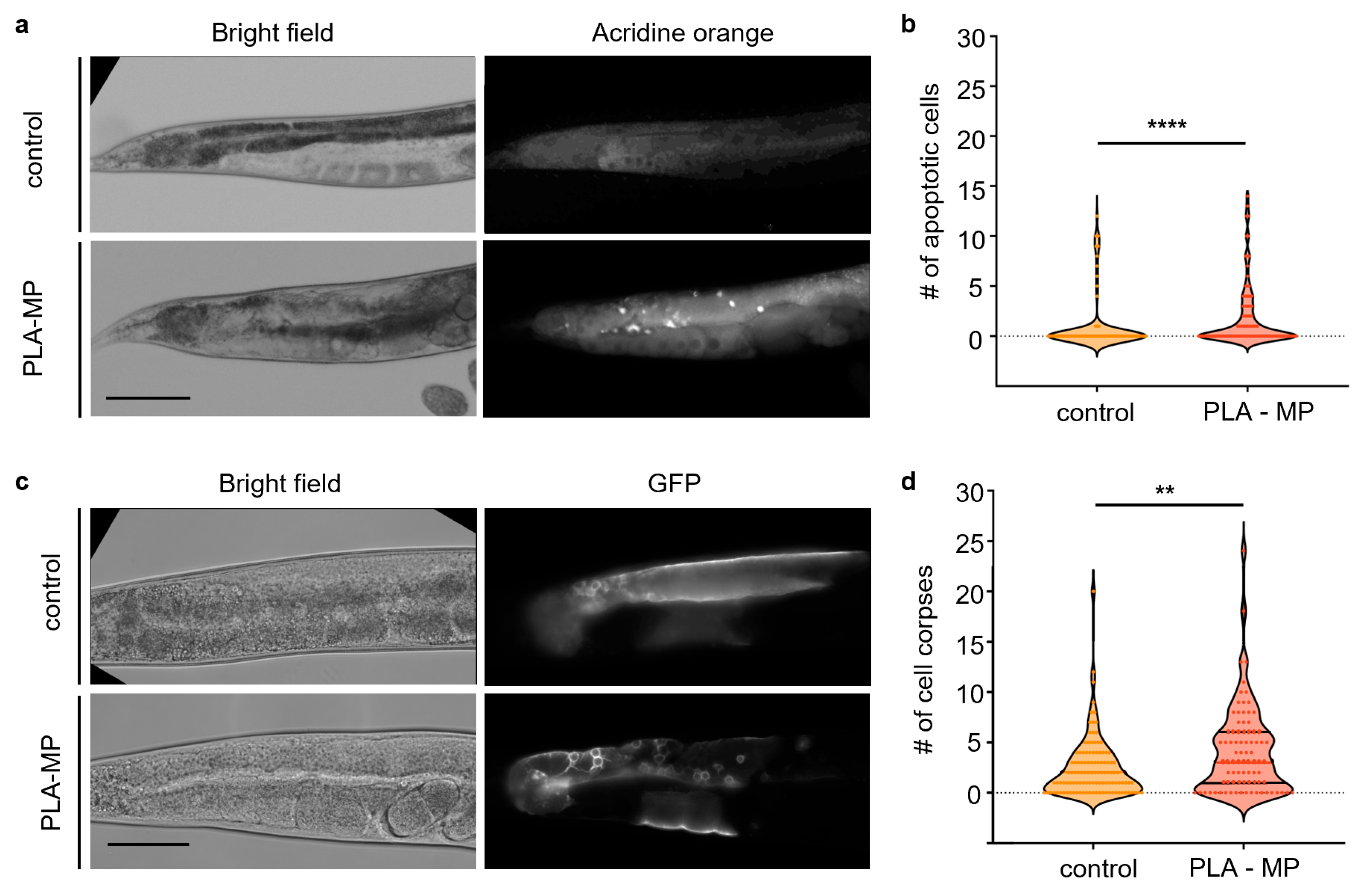
Chromatin that has collapsed in the late pachytene region has previously been found to correlate with apoptosis in germ cells. Moreover, chromosome disorganization has been linked to DNA damage from exposure to soluble toxicants [39,64], and MP studies observe DNA-damage-induced germline apoptosis [33,60,68]. Therefore, we next examined the germline in PLA-MP material-exposed C. elegans for cell death and apoptosis. To determine whether PLA-MP material exposure increases germ cell apoptosis, acridine orange staining and expression of a functional CED-1::GFP fusion protein in the sheath cells of a reporter C. elegans strain were monitored. Acridine orange-stained apoptotic cells increased significantly after PLA-MP material exposure compared with the control (p < 0.0001) (Figure 5a,b). In addition, cell corpses increased when exposed to PLA-MPs (p = 0.0042) (Figure 5c,d). Acridine orange staining and CED-1::GFP reporter expression both quantify apoptosis, but they measure slightly different stages of the cell death process [69]. CED-1::GFP reporter expression is more sensitive and represents an earlier stage of apoptosis than acridine orange staining, which depends on permeability and retention [69]. The increased number of cell corpses detected by CED-1::GFP expression (Figure 5d), compared with the number of apoptotic cells detected by acridine orange (Figure 5b), likely reflects these differences. Pristine uniform 3 µm PLA-MPs (10 and 100 µg/L) led to germline apoptosis, while 1 µg/L PLA-MP did not [33]. Consistent with our results, UV-aged PLA-MPs induced germline apoptosis across all exposure levels (1, 10, and 100 µg/L) [34]. This indicates that irregular secondary PLA-MP material causes detrimental germline defects and apoptosis at lower exposure levels than pristine PLA-MPs.
Figure 5.
Increased apoptotic cells and cell death in the germline of PLA-MP material-exposed C. elegans. (a) Representative images of acridine orange staining in adult C. elegans fed E. coli with and without PLA-MPs. Scale bar = 100 µm. (b) Quantification of acridine orange staining of apoptotic cells in C. elegans with exposure to their regular E. coli diet only or with 1 µg/L PLA-MPs. N = 3, n = 102, 107. (c) Representative images of MD701 and ced-1::GFP expression in adult C. elegans fed E. coli with and without PLA-MPs. Scale bar = 50 µm. # = number. (d) Quantification of MD701, ced-1::GFP apoptotic cells in C. elegans with exposure to their regular E. coli diet only or with 1 µg/L PLA-MPs. N = 4, n = 115, 84. The Mann–Whitney test was used for statistical analysis. (** p ≤ 0.01; **** p ≤ 0.0001). # = number.
3.5. PLA-MP Material Exposure Leads to DAF-16-Induced Stress in C. elegans and Oxidative Stress in the Gonad
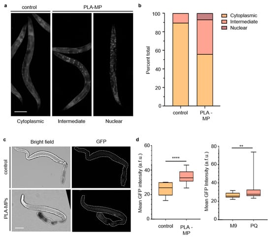
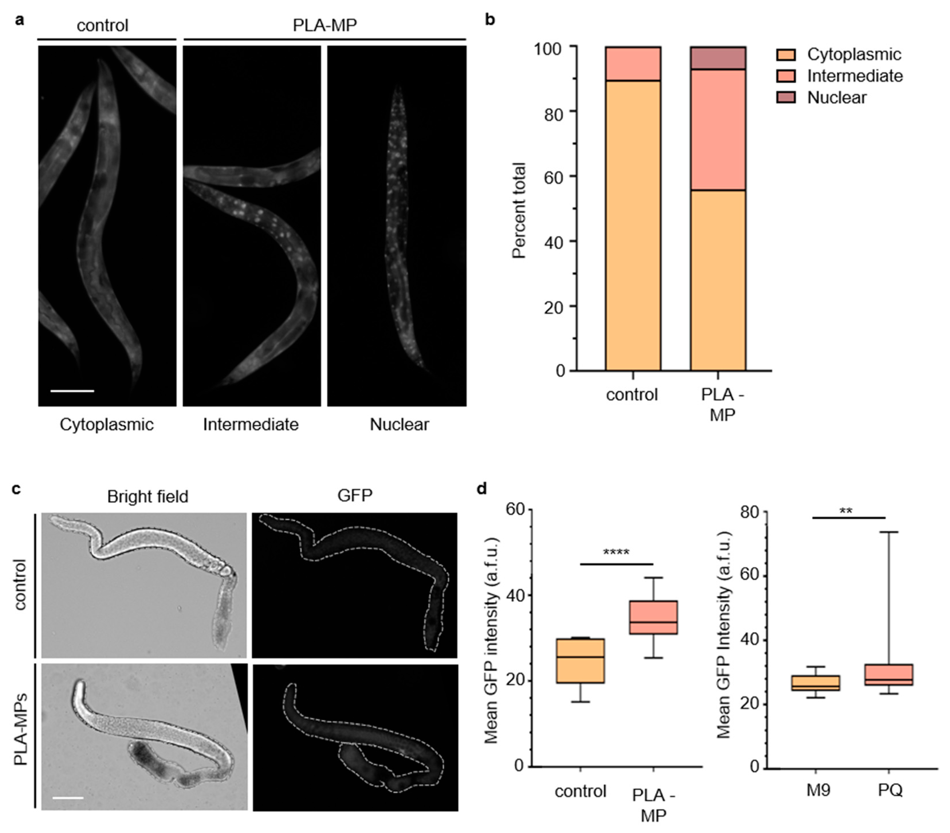
PLA has been found to cause inflammation and increase reactive oxygen species (ROS) levels [24,32,33,35], both of which are often associated with DNA damage [32], cell death [70], and reproductive decline [71,72]. Additionally, a wide range of MP types, sizes, and exposure levels have been shown to cause stress via DAF-16/FOXO signaling and gst-4 [17,23]. These pathways are central to the MP-induced stress response. Thus, we next asked whether the DAF-16 or the gst-4 stress responses were activated upon exposure to secondary PLA-MP material. Exposure of C. elegans to PLA-MP material increased the percentage of C. elegans with intermediate and nuclear expression of DAF-16 (p < 0.0001) (Figure 6a,b). To test whether oxidative stress played a role in the germline, an oxidative stress response reporter strain containing gst-4p::GFP, in which GFP expression is driven by the glutathione S-transferase 4 (gst-4) promoter [73]. When this reporter strain was exposed to 1 µg/L PLA-MPs, we observed a significant increase in mean GFP signal intensity in the gonads (p < 0.0001) (Figure 6c,d). The response increased, as also observed with paraquat, an herbicide that induces oxidative stress (Figure 6d) [74,75].
Figure 6.
Multiple stress response pathways are activated in C. elegans after exposure to PLA-MP material. (a) Representative images of DAF-16::GFP expression in adult C. elegans, showing cytoplasmic (left), intermediary (center), and nuclear localization (right). Scale bar = 100 µm. (b) Quantification of DAF-16::GFP localization in adult C. elegans with exposure to their regular E. coli diet or with 1 µg/L PLA-MPs. N = 3, n = 122, 210. A chi-square test was used for statistical analysis (p < 0.0001). (c) Representative images of gst-4 promoter-driven GFP expression in a dissected gonad from C. elegans without and with exposure to PLA-MP. The dotted line indicates the shape of the gonad. Scale bar = 50 µm. (d) Quantification of the mean GFP intensity from C. elegans expressing gst-4 promoter-driven GFP without and with exposure to PLA-MP. a.f.u. = arbitrary fluorescence units. Error bars represent standard deviation (SD). **** p < 0.0001, ** p = 0.0100 by the two-tailed Mann–Whitney test, 95% C.I. n = 18–46 gonads per condition. N = 3, biological replicates.
3.6. Exposure to Polylactic Acid Microplastic Material Does Not Reduce Lifespan
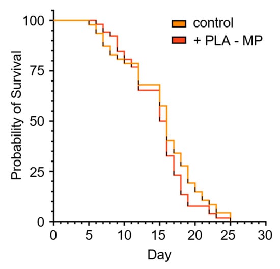
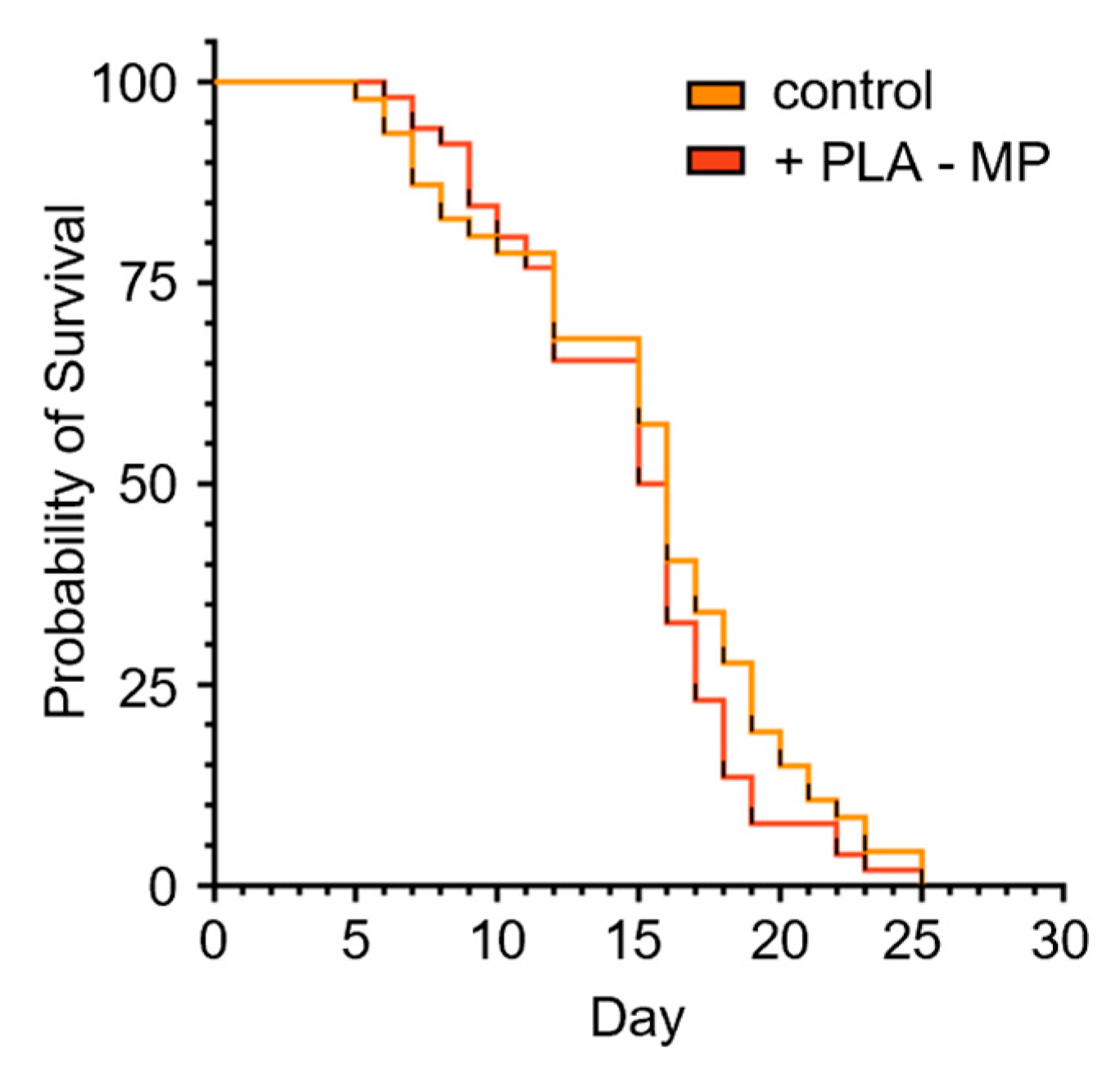
MP exposure shows varying effects on lifespan, which may be due to polymer type, size, and concentration. In many cases, MP exposure has been demonstrated to reduce lifespan in C. elegans [17,23,38,54]. Additionally, activation of the DAF-16 stress response can be associated with increased lifespan [41,76,77,78]. Extended lifespan induced by stress events is linked to multiple pathways, including the insulin/insulin-like growth factor (IGF-1) receptor signaling pathway and diet [78]. Due to mixed reports on longevity in C. elegans and the potential for larger (>5 µm) particles to obstruct the pharynx and thus alter nutrition [52], as well as studies indicating intestine-specific stress due to MP exposure [79], we next asked whether PLA-MP material exposure would change the lifespan in C. elegans. Exposure of C. elegans to PLA-MP material did not alter their lifespan (Figure 7 and Supplemental Figure S5). There are only a limited number of studies on the lifespan of MPs composed of PLA or bio-based plastics. PLA-MPs reduced lifespan in the rotifer Brachionus plicatilis, and PLA-MPs (100 µg/L) reduced lifespan in C. elegans [33,80]. These studies used different concentrations and types of PLA-MPs [33]. In contrast to the B. plicatilis study, Shao et al. reported reduced lifespan with 3 µm-diameter PLA-MPs, but only at higher exposure levels (100 µg/L), and PLA-MP leachates did not affect lifespan [33]. These varying effects on lifespan may reflect differences in PLA-MP exposure levels and particle properties.
Figure 7.
Lifespan of C. elegans exposed to PLA-MP material. Survival assay for C. elegans exposed to E. coli (no MPs) or 1 µg/L PLA-MPs continuously. n = 47, 52. Not significant, p = 0.249. Log-rank (Mantel–Cox).
4. Limitations
Several limitations should be considered when interpreting these results. The exposure was based on nominal PLA material mass rather than measured MP particle number. Given the heterogeneous particle dimensions and the possibility of additional fragmentation during sonication, the SEM measurements obtained before preparation of the final exposure suspension cannot be used to determine the actual particle number delivered to each plate. Thus, the nominal mass concentration should not be interpreted as a measured particle concentration. Future development and availability of small-particle analysis technologies may overcome this limitation. In addition, we examined a single exposure concentration, limiting conclusions about dose–response relationships. Future studies should examine multiple exposure concentrations to determine a dose–response relationship. Furthermore, because the 3D-printed PLA material that was used to create MPs contained multiple chemical components and leachates were not studied, the observed effects cannot be attributed specifically to the PLA polymer, particle properties, or associated chemical additives. This is an important area to pursue in our future studies. Lastly, although animals were monitored to confirm a comparable developmental stage at the time of analysis, developmental progression was not quantified. Therefore, the possibility that subtle differences in developmental timing contributed to the gonad morphology results cannot be excluded.
5. Conclusions
Here we show that non-pristine, irregular PLA-MP material can trigger germline damage, apoptosis, and reproductive decline. We show that exposure of C. elegans to a heterogeneous pool of 1 µg/L PLA-MP material (varying in size, shape, and surface texture and containing a mixture of chemical additives) reduces fertility but not lifespan. Although we did not observe an effect on lifespan from PLA-MP material exposure, we observed reduced fertility and germline changes. The germline is potentially more sensitive than somatic tissues under our exposure conditions. The fertility reduction correlates with germline alterations, including chromosome aberrations, oxidative stress, and cell death. Therefore, irregular secondary PLA-MP material is an emerging pollutant that can affect fertility and germline health. Our work complements the emerging literature on pollutants with similar endpoints for particle-based environmental pollutants and highlights that particle-based pollutants may carry chemicals that can lead to negative health outcomes.
We presented a method to create and mimic environmentally relevant PLA-MPs using a cryo-mill method to examine physiological defects and their underlying causes. We focused on using mechanically processed secondary MPs to better mimic those found in the environment. In reality, human and animal MP exposure is represented by various polymer types, mixtures (polymer and additive), absorption of environmental chemicals, and multiple methods of degradation (mechanical, UV, chemical, etc.). GC×GC-TOFMS analysis of PLA filament used to create the 3D-printed item showed that the material contained additives, many of which are unknown chemicals. Future work will systematically examine the chemical classes present and aim to determine how each may, separately or in combination, contribute to toxicity. Additionally, future work can methodically add UV aging and chemical exposure to simulate more advanced environmental degradation. Future work on the effects of MP exposure that represents those found in the environment and to which humans and animals are exposed is of major importance. The microplastics field needs to share results transparently, use rigorous and reproducible experimentation, and develop new analytical techniques to identify, analyze, and study the MPs to which humans and animals are exposed.
Supplementary Materials
The following supporting information can be downloaded at https://www.mdpi.com/article/10.3390/pollutants6040069/s1. Supplemental Figure S1: Schematic of assays using various C. elegans strains. Supplemental Figure S2: Frequency distribution of microplastic sizes. Supplemental Figure S3: Chemicals identified after GC×GC–TOFMS analysis of PLA filament used to create the 3D-printed item that was used for MP creation. Supplemental Figure S4: Number of eggs hatched, unhatched, and quantification of the mean nuclear diameter (μm) in C. elegans following control or PLA-MP material exposure. Supplemental Figure S5: Lifespan of C. elegans exposed to PLA-MP material.
Author Contributions
Conceptualization, J.C.H.; methodology, J.C.H., C.A.O.M., D.M.M., R.J.R. and R.L.P.; validation, J.C.H. and R.L.P.; formal analysis, J.C.H., C.A.O.M. and R.L.P.; investigation, J.C.H., C.A.O.M., D.F., R.J.R., D.M.M., M.F.G., L.R.S., V.P., A.G. and R.L.P.; resources, J.C.H.; data curation, J.C.H., C.A.O.M. and D.M.M.; writing—original draft preparation, J.C.H. and C.A.O.M.; writing—review and editing, J.C.H.; visualization, J.C.H. and C.A.O.M.; supervision, J.C.H.; project administration, J.C.H.; funding acquisition, J.C.H. All authors have read and agreed to the published version of the manuscript.
Funding
This research was funded by the National Institutes of Health, NIGMS (R16GM150406) and NIGMS (S10GM158739). C. Maldonado was funded by an NIH grant, T34GM149455. A. Gutierrez was funded by a National Science Foundation grant, HRD 2207388.
Data Availability Statement
The raw data supporting the conclusions of this article will be made available by the authors on request.
Acknowledgments
Many undergraduate student researchers have helped move this project forward, and we thank them for their time and valuable input. We thank K. Favela and Southwest Research Institute for support in collecting and analyzing the GC×GC–TOFMS data. We thank S. Oxley for her advice and discussions. We also thank St. Mary’s University and Texas State University for their support. St. Mary’s University funds were provided by the Department of Biological Sciences, the Benjamin F. Biaggini Endowment, and the Office of Student Research and Inquiry SURF program. C. elegans strains were provided by the CGC, which is funded by the NIH Office of Research Infrastructure Programs (P40 OD010440).
Conflicts of Interest
The authors declare no conflicts of interest.
Abbreviations
| C. elegans | Caenorhabditis elegans |
| DAF-16 | Forkhead box O (FOXO) protein |
| E. coli | Escherichia coli |
| GC×GC–TOFMS | Comprehensive two-dimensional gas chromatography coupled with time-of-flight mass spectrometry |
| GFP | Green fluorescent protein |
| GST-4 | Glutathione S-transferase 4 |
| LA | Lactic acid |
| L1 | Larval stage 1 |
| L4 | Larval stage 4 |
| MP | Microplastic |
| NGM | Nematode growth medium agarose |
| NP | Nanoplastic |
| PBS | phosphate-buffered saline |
| PFA | paraformaldehyde |
| PLA | Polylactic acid |
| PS | Polystyrene |
| PQ | Paraquat |
| ROS | Reactive oxygen species |
| SEM | Scanning electron microscopy |
| UV | Ultraviolet |
References
- Khouri, N.G.; Bahú, J.O.; Blanco-Llamero, C.; Severino, P.; Concha, V.O.C.; Souto, E.B. Polylactic acid (PLA): Properties, synthesis, and biomedical applications—A review of the literature. J. Mol. Struct. 2024, 1309, 138243. [Google Scholar] [CrossRef] [Scilit]
- Hasoon, K.T.N.; Panicker, U.G. Beyond fossil plastics: Next-generation PLA-based bio-packaging for industrial applications-advances, challenges, and data-driven insights. RSC Adv. 2026, 16, 15095–15118. [Google Scholar] [CrossRef] [Scilit] [PubMed]
- Tyler, B.; Gullotti, D.; Mangraviti, A.; Utsuki, T.; Brem, H. Polylactic acid (PLA) controlled delivery carriers for biomedical applications. Adv. Drug Deliv. Rev. 2016, 107, 163–175. [Google Scholar] [CrossRef] [Scilit] [PubMed]
- Singhvi, M.S.; Zinjarde, S.S.; Gokhale, D.V. Polylactic acid: Synthesis and biomedical applications. J. Appl. Microbiol. 2019, 127, 1612–1626. [Google Scholar] [CrossRef] [Scilit] [PubMed]
- Giammona, G.; Craparo, E.F. Biomedical Applications of Polylactide (PLA) and Its Copolymers. Molecules 2018, 23, 980. [Google Scholar] [CrossRef] [Scilit] [PubMed]
- Moldovan, A.; Cuc, S.; Prodan, D.; Rusu, M.; Popa, D.; Taut, A.C.; Petean, I.; Bomboş, D.; Doukeh, R.; Nemes, O. Development and Characterization of Polylactic Acid (PLA)-Based Nanocomposites Used for Food Packaging. Polymers 2023, 15, 2855. [Google Scholar] [CrossRef] [Scilit] [PubMed]
- de Albuquerque, T.L.; Junior, J.E.M.; de Queiroz, L.P.; Ricardo, A.D.S.; Rocha, M.V.P. Polylactic acid production from biotechnological routes: A review. Int. J. Biol. Macromol. 2021, 186, 933–951. [Google Scholar] [CrossRef] [Scilit] [PubMed]
- Joseph, T.M.; Kallingal, A.; Suresh, A.M.; Mahapatra, D.K.; Hasanin, M.S.; Haponiuk, J.; Thomas, S. 3D printing of polylactic acid: Recent advances and opportunities. Int. J. Adv. Manuf. Technol. 2023, 125, 1015–1035. [Google Scholar] [CrossRef] [Scilit] [PubMed]
- Gioia, C.; Giacobazzi, G.; Vannini, M.; Totaro, G.; Sisti, L.; Colonna, M.; Marchese, P.; Celli, A. End of Life of Biodegradable Plastics: Composting versus Re/Upcycling. ChemSusChem 2021, 14, 4167–4175. [Google Scholar] [CrossRef] [Scilit] [PubMed]
- Fogašová, M.; Figalla, S.; Danišová, L.; Medlenová, E.; Hlaváčiková, S.; Vanovčanová, Z.; Omaníková, L.; Baco, A.; Horváth, V.; Mikolajová, M.; et al. PLA/PHB-Based Materials Fully Biodegradable under Both Industrial and Home-Composting Conditions. Polymers 2022, 14, 4113. [Google Scholar] [CrossRef] [Scilit] [PubMed]
- Mo, A.; Zhang, Y.; Gao, W.; Jiang, J.; He, D. Environmental fate and impacts of biodegradable plastics in agricultural soil ecosystems. Appl. Soil Ecol. 2023, 181, 104667. [Google Scholar] [CrossRef] [Scilit]
- Frias, J.P.G.L.; Nash, R. Microplastics: Finding a consensus on the definition. Mar. Pollut. Bull. 2019, 138, 145–147. [Google Scholar] [CrossRef] [Scilit] [PubMed]
- Leslie, H.A.; van Velzen, M.J.M.; Brandsma, S.H.; Vethaak, A.D.; Garcia-Vallejo, J.J.; Lamoree, M.H. Discovery and quantification of plastic particle pollution in human blood. Environ. Int. 2022, 163, 107199. [Google Scholar] [CrossRef] [Scilit] [PubMed]
- Nihart, A.J.; Garcia, M.A.; El Hayek, E.; Liu, R.; Olewine, M.; Kingston, J.D.; Castillo, E.F.; Gullapalli, R.R.; Howard, T.; Bleske, B.; et al. Author Correction: Bioaccumulation of microplastics in decedent human brains. Nat. Med. 2025, 31, 1114–1119. [Google Scholar] [CrossRef] [Scilit] [PubMed]
- Hu, C.J.; A Garcia, M.; Nihart, A.; Liu, R.; Yin, L.; Adolphi, N.; Gallego, D.F.; Kang, H.; Campen, M.J.; Yu, X. Microplastic presence in dog and human testis and its potential association with sperm count and weights of testis and epididymis. Toxicol. Sci. 2024, 200, 235–240. [Google Scholar] [CrossRef] [Scilit] [PubMed]
- Montano, L.; Raimondo, S.; Piscopo, M.; Ricciardi, M.; Guglielmino, A.; Chamayou, S.; Gentile, R.; Gentile, M.; Rapisarda, P.; Conti, G.O.; et al. First evidence of microplastics in human ovarian follicular fluid: An emerging threat to female fertility. Ecotoxicol. Environ. Saf. 2025, 291, 117868. [Google Scholar] [CrossRef] [Scilit] [PubMed]
- Maldonado, C.A.O.; Mares, D.M.; Garcia, P.C.; Gamez, M.F.; Flores, M.R.; Friudenberg, A.D.; Peterson, R.L.; Harr, J.C. Microplastic-Mediated Delivery of Di-butyl Phthalate Alters C. elegans Lifespan and Reproductive Fidelity. Microplastics 2025, 4, 96. [Google Scholar] [CrossRef] [Scilit] [PubMed]
- Jewett, E.; Arnott, G.; Connolly, L.; Vasudevan, N.; Kevei, E. Microplastics and Their Impact on Reproduction-Can we Learn From the C. elegans Model? Front. Toxicol. 2022, 4, 748912. [Google Scholar] [CrossRef] [Scilit] [PubMed]
- Peng, M.; Felix, R.C.; Canario, A.V.M.; Power, D.M. The physiological effect of polystyrene nanoplastic particles on fish and human fibroblasts. Sci. Total Environ. 2024, 914, 169979. [Google Scholar] [CrossRef] [Scilit] [PubMed]
- Liu, Z.; Yu, P.; Cai, M.; Wu, D.; Zhang, M.; Chen, M.; Zhao, Y. Effects of microplastics on the innate immunity and intestinal microflora of juvenile Eriocheir sinensis. Sci. Total Environ. 2019, 685, 836–846. [Google Scholar] [CrossRef] [Scilit] [PubMed]
- Völkl, M.; Jérôme, V.; Weig, A.; Jasinski, J.; Meides, N.; Strohriegl, P.; Scheibel, T.; Freitag, R. Pristine and artificially-aged polystyrene microplastic particles differ in regard to cellular response. J. Hazard Mater. 2022, 435, 128955. [Google Scholar] [CrossRef] [Scilit] [PubMed]
- Gouda, M.Z.; Xu, E.G.; Ateia, M. Why Pristine, Aged, and Real-World Microplastics Are All Essential for Environmental Research. Environ. Sci. Technol. 2026, 60, 7–10. [Google Scholar] [CrossRef] [Scilit] [PubMed]
- León, R.E.; Salcedo, V.A.A.; Miguel, F.J.N.S.; Tardio, C.R.A.L.; Briceño, A.A.T.; Fouilloux, S.F.C.; Barbosa, M.d.M.; Barros, C.A.S.; Waldman, W.R.; Espinosa-Bustos, C.; et al. Photoaged polystyrene nanoplastics exposure results in reproductive toxicity due to oxidative damage in Caenorhabditis elegans. Environ. Pollut. 2024, 348, 123816. [Google Scholar] [CrossRef] [Scilit] [PubMed]
- Shao, Y.; Hua, X.; Li, Y.; Wang, D. Comparison of reproductive toxicity between pristine and aged polylactic acid microplastics in Caenorhabditis elegans. J. Hazard Mater. 2024, 466, 133545. [Google Scholar] [CrossRef] [Scilit] [PubMed]
- Thompson, R.C.; Courtene-Jones, W.; Boucher, J.; Pahl, S.; Raubenheimer, K.; Koelmans, A.A. Twenty years of microplastic pollution research-what have we learned? Science 2024, 386, eadl2746. [Google Scholar] [CrossRef] [Scilit] [PubMed]
- Teuten, E.L.; Saquing, J.M.; Knappe, D.R.; Barlaz, M.A.; Jonsson, S.; Björn, A.; Rowland, S.J.; Thompson, R.C.; Galloway, T.S.; Yamashita, R.; et al. Transport and release of chemicals from plastics to the environment and to wildlife. Philos. Trans. R. Soc. Lond B Biol. Sci. 2009, 364, 2027–2045. [Google Scholar] [CrossRef] [Scilit] [PubMed]
- Prasad, G.; Arunav, H.; Dwight, S.; Ghosh, M.B.; Jayadev, A.; Nair, D.I. Advancing Sustainable Practices in Additive Manufacturing: A Comprehensive Review on Material Waste Recyclability. Sustainability 2024, 16, 10246. [Google Scholar] [CrossRef] [Scilit]
- Song, R.; Telenko, C. Material and energy loss due to human and machine error in commercial FDM printers. J. Clean. Prod. 2017, 148, 895–904. [Google Scholar] [CrossRef] [Scilit]
- Zhu, C.; Li, T.; Mohideen, M.M.; Hu, P.; Gupta, R.; Ramakrishna, S.; Liu, Y. Realization of Circular Economy of 3D Printed Plastics: A Review. Polymers 2021, 13, 744. [Google Scholar] [CrossRef] [Scilit] [PubMed]
- Maraveas, C.; Kyrtopoulos, I.V.; Arvanitis, K.G. Evaluation of the Viability of 3D Printing in Recycling Polymers. Polymers 2024, 16, 1104. [Google Scholar] [CrossRef] [Scilit] [PubMed]
- Qian, Q.; Pu, Q.; Li, L.; Wu, J.; Cheng, G.; Cheng, Y.; Wang, X.; Wang, H. Polylactic acid microplastics before and after aging induced neurotoxicity in zebrafish by disrupting the microbiota-gut-brain axis. J. Hazard Mater. 2025, 488, 137306. [Google Scholar] [CrossRef] [Scilit] [PubMed]
- Alaraby, M.; Abass, D.; Farre, M.; Hernandez, A.; Marcos, R. Are bioplastics safe? Hazardous effects of polylactic acid (PLA) nanoplastics in Drosophila. Sci. Total Environ. 2024, 919, 170592. [Google Scholar] [CrossRef] [Scilit] [PubMed]
- Shao, Y.; Wang, Y.; Hua, X.; Li, Y.; Wang, D. Polylactic acid microparticles in the range of mug/L reduce reproductive capacity by affecting the gonad development and the germline apoptosis in Caenorhabditis elegans. Chemosphere 2023, 336, 139193. [Google Scholar] [CrossRef] [Scilit] [PubMed]
- Wu, J.; Shao, Y.; Hua, X.; Li, Y.; Wang, D. Photo-aged polylactic acid microplastics causes severe transgenerational decline in reproductive capacity in C. elegans: Insight into activation of DNA damage checkpoints affected by multiple germline histone methyltransferases. Environ. Pollut. 2025, 382, 126697. [Google Scholar] [CrossRef] [Scilit] [PubMed]
- Gutiérrez-Rial, D.; Lagoa, A.; Villar, I.; Feijoo, P.; Mariño, F.; Garrido, J.; Soto, B.; Mato, S. Short- and medium-term effects of biodegradable microplastics (PLA and PHB) on earthworm development and reproduction. Ecotoxicology 2025, 34, 701–711. [Google Scholar] [CrossRef] [Scilit] [PubMed]
- Bhagat, J.; Nishimura, N.; Shimada, Y. Worming into a robust model to unravel the micro/nanoplastic toxicity in soil: A review on Caenorhabditis elegans. TrAC Trends Anal. Chem. 2021, 138, 116235. [Google Scholar] [CrossRef] [Scilit]
- Lenz, R.; Enders, K.; Nielsen, T.G. Microplastic exposure studies should be environmentally realistic. Proc. Natl. Acad. Sci. USA 2016, 113, E4121–E4122. [Google Scholar] [CrossRef] [Scilit] [PubMed]
- Qiu, Y.; Liu, Y.; Li, Y.; Li, G.; Wang, D. Effect of chronic exposure to nanopolystyrene on nematode Caenorhabditis elegans. Chemosphere 2020, 256, 127172. [Google Scholar] [CrossRef] [Scilit] [PubMed]
- Henderson, A.L.; Karthikraj, R.; Berdan, E.L.; Sui, S.H.; Kannan, K.; Colaiacovo, M.P. Exposure to benzyl butyl phthalate (BBP) leads to increased double-strand break formation and germline dysfunction in Caenorhabditis elegans. PLoS Genet. 2024, 20, e1011434. [Google Scholar] [CrossRef] [Scilit] [PubMed]
- Ke, T.; Soares, F.A.A.; Santamaria, A.; Bowman, A.B.; Skalny, A.V.; Aschner, M. N,N’ bis-(2-mercaptoethyl) isophthalamide induces developmental delay in Caenorhabditis elegans by promoting DAF-16 nuclear localization. Toxicol. Rep. 2020, 7, 930–937. [Google Scholar] [CrossRef] [Scilit] [PubMed]
- Leite, N.R.; de Araújo, L.C.A.; Rocha, P.d.S.d.; Agarrayua, D.A.; Ávila, D.S.; Carollo, C.A.; Silva, D.B.; Estevinho, L.M.; Souza, K.d.P.; dos Santos, E.L. Baru Pulp (Dipteryx alata Vogel): Fruit from the Brazilian Savanna Protects against Oxidative Stress and Increases the Life Expectancy of Caenorhabditis elegans via SOD-3 and DAF-16. Biomolecules 2020, 10, 1106. [Google Scholar] [CrossRef] [Scilit] [PubMed]
- von der Esch, E.; Lanzinger, M.; Kohles, A.J.; Schwaferts, C.; Weisser, J.; Hofmann, T.; Glas, K.; Elsner, M.; Ivleva, N.P. Simple Generation of Suspensible Secondary Microplastic Reference Particles via Ultrasound Treatment. Front. Chem. 2020, 8, 169. [Google Scholar] [CrossRef] [Scilit] [PubMed]
- Li, X.; Wu, X.; Lee, J.H.; Liu, J. Preparation of Microplastics and Nanoplastics for Environmental Research. Environ. Sci. Technol. 2026, 60, 16368–16389. [Google Scholar] [CrossRef] [Scilit] [PubMed]
- Boettcher, H.; Kukulka, T.; Cohen, J.H. Methods for controlled preparation and dosing of microplastic fragments in bioassays. Sci. Rep. 2023, 13, 5195. [Google Scholar] [CrossRef] [Scilit] [PubMed]
- Gardon, T.; Paul-Pont, I.; Le Moullac, G.; Soyez, C.; Lagarde, F.; Huvet, A. Cryogrinding and sieving techniques as challenges towards producing controlled size range microplastics for relevant ecotoxicological tests. Environ. Pollut. 2022, 315, 120383. [Google Scholar] [CrossRef] [Scilit] [PubMed]
- Enyoh, C.E.; Shafea, L.; Verla, A.W.; Verla, E.N.; Qingyue, W.; Chowdhury, T.; Paredes, M. Microplastics Exposure Routes and Toxicity Studies to Ecosystems: An Overview. Environ. Anal. Health Toxicol. 2020, 35, e2020004. [Google Scholar] [CrossRef] [Scilit] [PubMed]
- GESAMP. GESAMP Joint Group of Experts on the Scientific Aspects of Marine Environmental Protection. Sources, Fate and Effects of Microplastics in the Marine Environment: Part 2 of a Global Assessment. GESAMP Rep. Stud. 2016, 93, 220. [Google Scholar]
- Danopoulos, E.; Twiddy, M.; Rotchell, J.M. Microplastic contamination of drinking water: A systematic review. PLoS ONE 2020, 15, e0236838. [Google Scholar] [CrossRef] [Scilit] [PubMed]
- De-la-Torre, G.E. Microplastics: An emerging threat to food security and human health. J. Food Sci. Technol. 2019, 57, 1601–1608. [Google Scholar] [CrossRef] [Scilit] [PubMed]
- Yates, J.; Deeney, M.; Rolker, H.B.; White, H.; Kalamatianou, S.; Kadiyala, S. A systematic scoping review of environmental, food security and health impacts of food system plastics. Nat. Food 2021, 2, 80–87. [Google Scholar] [CrossRef] [Scilit] [PubMed]
- Yates, J.; Deeney, M.; Muncke, J.; Almroth, B.C.; Dignac, M.-F.; Castillo, A.C.; Courtene-Jones, W.; Kadiyala, S.; Kumar, E.; Stoett, P.; et al. Plastics matter in the food system. Commun. Earth Environ. 2025, 6, 176. [Google Scholar] [CrossRef] [Scilit] [PubMed]
- De, A.; Sahu, A.K.; Singh, V.K. Bite size of Caenorhabditis elegans regulates feeding, satiety and development on a diet of live yeast. J. Biosci. 2025, 50, 67. [Google Scholar] [CrossRef] [Scilit]
- Gubert, P.; Gubert, G.; de Oliveira, R.C.; Fernandes, I.C.O.; Bezerra, I.C.; de Ramos, B.; de Lima, M.F.; Rodrigues, D.T.; da Cruz, A.F.N.; Pereira, E.C.; et al. Caenorhabditis elegans as a Prediction Platform for Nanotechnology-Based Strategies: Insights on Analytical Challenges. Toxics 2023, 11, 239. [Google Scholar] [CrossRef] [Scilit] [PubMed]
- Shang, X.; Lu, J.; Feng, C.; Ying, Y.; He, Y.; Fang, S.; Lin, Y.; Dahlgren, R.; Ju, J. Microplastic (1 and 5 mum) exposure disturbs lifespan and intestine function in the nematode Caenorhabditis elegans. Sci. Total Environ. 2020, 705, 135837. [Google Scholar] [CrossRef] [Scilit] [PubMed]
- Yu, Y.; Li, X.; Zhang, R.; Guo, W. Insights into the morphology-dependent adsorption of aged polystyrene nanoplastics on manganese oxides. Colloids Surf. A Physicochem. Eng. Asp. 2023, 679, 132565. [Google Scholar] [CrossRef] [Scilit]
- Pop, M.A.; Croitoru, C.; Bedő, T.; Geamăn, V.; Radomir, I.; Cos, N.M.; Zaharia, S.M.; Chicos, L.A.; Milos, A.I. Structural changes during 3D printing of bioderived and synthetic thermoplastic materials. J. Appl. Polym. Sci. 2019, 136, 47382. [Google Scholar] [CrossRef] [Scilit]
- Lei, L.; Wu, S.; Lu, S.; Liu, M.; Song, Y.; Fu, Z.; Shi, H.; Raley-Susman, K.M.; He, D. Microplastic particles cause intestinal damage and other adverse effects in zebrafish Danio rerio and nematode Caenorhabditis elegans. Sci. Total Environ. 2018, 619–620, 1–8. [Google Scholar] [CrossRef] [Scilit] [PubMed]
- Rochman, C.M.; Hoh, E.; Kurobe, T.; Teh, S.J. Ingested plastic transfers hazardous chemicals to fish and induces hepatic stress. Sci. Rep. 2013, 3, 3263. [Google Scholar] [CrossRef] [Scilit] [PubMed]
- Browne, M.A.; Niven, S.J.; Galloway, T.S.; Rowland, S.J.; Thompson, R.C. Microplastic moves pollutants and additives to worms, reducing functions linked to health and biodiversity. Curr. Biol. 2013, 23, 2388–2392. [Google Scholar] [CrossRef] [Scilit] [PubMed]
- Xu, T.; Chen, H.; Zhang, L.; Xie, D.; Tan, S.; Guo, H.; Xiang, M.; Yu, Y. Aged polystyrene microplastics cause reproductive impairment via DNA-damage induced apoptosis in Caenorhabditis elegans. Chemosphere 2024, 362, 142519. [Google Scholar] [CrossRef] [Scilit] [PubMed]
- Qu, M.; Qiu, Y.; Kong, Y.; Wang, D. Amino modification enhances reproductive toxicity of nanopolystyrene on gonad development and reproductive capacity in nematode Caenorhabditis elegans. Environ. Pollut. 2019, 254, 112978. [Google Scholar] [CrossRef] [Scilit] [PubMed]
- Guedon, R.; Maremonti, E.; Armant, O.; Galas, S.; Brede, D.A.; Lecomte-Pradines, C. A systems biology analysis of reproductive toxicity effects induced by multigenerational exposure to ionizing radiation in C. elegans. Ecotoxicol. Environ. Saf. 2021, 225, 112793. [Google Scholar] [CrossRef] [Scilit] [PubMed]
- Chen, H.; Wang, C.; Li, H.; Ma, R.; Yu, Z.; Li, L.; Xiang, M.; Chen, X.; Hua, X.; Yu, Y. A review of toxicity induced by persistent organic pollutants (POPs) and endocrine-disrupting chemicals (EDCs) in the nematode Caenorhabditis elegans. J. Environ. Manag. 2019, 237, 519–525. [Google Scholar] [CrossRef] [Scilit] [PubMed]
- Shin, N.; Cuenca, L.; Karthikraj, R.; Kannan, K.; Colaiacovo, M.P. Assessing effects of germline exposure to environmental toxicants by high-throughput screening in C. elegans. PLoS Genet. 2019, 15, e1007975. [Google Scholar] [CrossRef] [Scilit] [PubMed]
- Lui, D.Y.; Colaiacovo, M.P. Meiotic development in Caenorhabditis elegans. Adv. Exp. Med. Biol. 2013, 757, 133–170. [Google Scholar] [CrossRef] [Scilit] [PubMed]
- Cui, Z.; Zhang, J.; Zhang, J.; Zhong, J.; Wu, J.; Miao, Y.; Wang, H. Microplastics (MPs) exposure impairs porcine oocyte quality by triggering oxidative stress-directed DNA damage and apoptosis with metabolomic alterations. Ecotoxicol. Environ. Saf. 2025, 300, 118461. [Google Scholar] [CrossRef] [Scilit] [PubMed]
- Park, J.; Son, J.; Kim, J.; Cha, B.S.; Kim, W.; Jung, J.; Yune, T.Y.; Kim, S.S.; Choi, S.; Kim, D.; et al. Carboxylate polystyrene nanoplastics disrupt mitotic progression and induce chromosomal instability. Ecotoxicol. Environ. Saf. 2025, 307, 119407. [Google Scholar] [CrossRef] [Scilit] [PubMed]
- Yu, C.W.; Luk, T.C.; Liao, V.H. Long-term nanoplastics exposure results in multi and trans-generational reproduction decline associated with germline toxicity and epigenetic regulation in Caenorhabditis elegans. J. Hazard Mater. 2021, 412, 125173. [Google Scholar] [CrossRef] [Scilit] [PubMed]
- Lant, B.; Derry, W.B. Fluorescent visualization of germline apoptosis in living Caenorhabditis elegans. Cold Spring Harb. Protoc. 2014, 2014, 420–427. (In English) [Google Scholar] [CrossRef] [Scilit] [PubMed]
- Villalpando-Rodriguez, G.E.; Gibson, S.B. Reactive Oxygen Species (ROS) Regulates Different Types of Cell Death by Acting as a Rheostat. Oxid. Med. Cell. Longev. 2021, 2021, 9912436. [Google Scholar] [CrossRef] [Scilit] [PubMed]
- Agarwal, A.; Gupta, S.; Sharma, R.K. Role of oxidative stress in female reproduction. Reprod. Biol. Endocrinol. 2005, 3, 28. [Google Scholar] [CrossRef] [Scilit] [PubMed]
- Chen, X.; Liu, D. ROS: Their roles in diminished ovarian reserve. Front. Endocrinol. 2026, 17, 1823452. [Google Scholar] [CrossRef] [Scilit] [PubMed]
- Detienne, G.; Van de Walle, P.; De Haes, W.; Schoofs, L.; Temmerman, L. SKN-1-independent transcriptional activation of glutathione S-transferase 4 (GST-4) by EGF signaling. Worm 2016, 5, e1230585. [Google Scholar] [CrossRef] [Scilit] [PubMed][Green Version]
- Keith, S.A.; Amrit, F.R.; Ratnappan, R.; Ghazi, A. The C. elegans healthspan and stress-resistance assay toolkit. Methods 2014, 68, 476–486. [Google Scholar] [CrossRef] [Scilit] [PubMed]
- McCarthy, S.; Somayajulu, M.; Sikorska, M.; Borowy-Borowski, H.; Pandey, S. Paraquat induces oxidative stress and neuronal cell death; neuroprotection by water-soluble Coenzyme Q10. Toxicol. Appl. Pharmacol. 2004, 201, 21–31. [Google Scholar] [CrossRef] [Scilit] [PubMed]
- Senchuk, M.M.; Dues, D.J.; E Schaar, C.; Johnson, B.K.; Madaj, Z.B.; Bowman, M.J.; E Winn, M.; Van Raamsdonk, J.M. Activation of DAF-16/FOXO by reactive oxygen species contributes to longevity in long-lived mitochondrial mutants in Caenorhabditis elegans. PLoS Genet. 2018, 14, e1007268. [Google Scholar] [CrossRef] [Scilit] [PubMed]
- Lin, K.; Hsin, H.; Libina, N.; Kenyon, C. Regulation of the Caenorhabditis elegans longevity protein DAF-16 by insulin/IGF-1 and germline signaling. Nat. Genet. 2001, 28, 139–145. [Google Scholar] [CrossRef] [Scilit] [PubMed]
- Zhou, K.I.; Pincus, Z.; Slack, F.J. Longevity and stress in Caenorhabditis elegans. Aging 2011, 3, 733–753. [Google Scholar] [CrossRef] [Scilit] [PubMed]
- Yu, Y.; Chen, H.; Hua, X.; Dang, Y.; Han, Y.; Yu, Z.; Chen, X.; Ding, P.; Li, H. Polystyrene microplastics (PS-MPs) toxicity induced oxidative stress and intestinal injury in nematode Caenorhabditis elegans. Sci. Total Environ. 2020, 726, 138679. [Google Scholar] [CrossRef] [Scilit] [PubMed]
- Ali, W.; Jeong, H.; Kim, D.-H.; Lee, J.-S.; Zinck, P.; Souissi, S.; Lee, J.-S. Adverse effects of environmentally relevant microplastics on in vivo endpoints, oxidative stress, and mitogen-activated protein kinase signaling pathway and multixenobiotic resistance system in the marine rotifer Brachionus plicatilis. Sci. Total Environ. 2025, 958, 178027. [Google Scholar] [CrossRef] [Scilit] [PubMed]
Disclaimer/Publisher’s Note: The statements, opinions and data contained in all publications are solely those of the individual author(s) and contributor(s) and not of MDPI and/or the editor(s). MDPI and/or the editor(s) disclaim responsibility for any injury to people or property resulting from any ideas, methods, instructions or products referred to in the content. |
© 2026 by the authors. Licensee MDPI, Basel, Switzerland. This article is an open access article distributed under the terms and conditions of the Creative Commons Attribution (CC BY) license.