


Abstract
This review aims to present an updated, comprehensive analysis of data on the diversity and challenges of current approaches to the screening and monitoring of subjects at risk of T1D, as the earliest interventions during the course of the disease. Previously, screening for T1D was justified only for research purposes. A major turning point occurred when teplizumab, an immunomodulatory drug that delays the onset of overt T1D, was approved. Nowadays, there is a growing number of screening initiatives, and this trend is spreading fast across the world. In this context, novel recommendations emphasize the need for the wider identification of subjects at risk of T1D, suggesting that screening should not include only first-degree relatives of persons with T1D. Furthermore, current experts’ opinions have shifted the detection of T1D risk in the direction of ultimate goal-screening in the general population. Also, subjects at risk should be monitored, undergo metabolic testing, be informed about their risk, and be educated about the disease. Currently, there is a diversity in approaches to the screening and monitoring of subjects at risk of T1D, predominantly in the pediatric population. Several knowledge gaps persist in this area of investigation, especially in recommendations and potential benefits for the adult population. However, the scientific community is focusing on developing and adapting screening and monitoring strategies to suit particular countries, aiming to make them more universal while refining the definition of individual risk for T1D. Nevertheless, the screening and monitoring of subjects at risk should be the earliest interventions focused on delaying T1D.
1. Introduction
The prevalence of type 1 diabetes (T1D) is growing globally, and it will increase by up to 141% in the next 20 years [1]. Simultaneously, the incidence of T1D has been rising over the last few decades, among different geographic regions. In general, earlier reports have shown that very young children, under the age of 5, have the fastest increase in annual incidence [2]. However, the newer data (2002–2018) suggest that the annual percentage change increased by 2%, and that this increase is progressive with age, culminating at 2.75% for the 15–19 age group [3]. Moreover, recent data about the incidence among prepubertal children, teenagers, and young adults (10–24-year-olds) showed that global T1D incidence in this age group increased over the past three decades, with the most rapid increase in young adults aged 20–24 years [4].
On the other hand, an analysis of adult-onset T1D incidence in a systematic review of population-based studies from 32 countries and regions until 2022 reported dominantly insufficient data and the highest incidence of adult-onset T1D in Scandinavian countries, with a higher incidence in men. Simultaneously, the authors pointed out that it remained unclear whether the incidence of adult-onset T1D decreased with increasing age, and whether it had changed over time at all [5]. Still, a previous study reported a lower incidence in subjects 20–64 years of age than in youth up to 19 years, but the total number of new cases in adults from 2001 to 2015 was higher in adults than in youth [6]. Interestingly, more than half of the subjects with recent-onset T1D in 2024 were adults, making that population heterogeneous in clinical presentation and risk of T1D development [7].
The rising incidence of T1D in children is largely based on the more profound role of environmental factors [8,9] in triggering the autoimmune response in genetically susceptible subjects. In contrast, the increase in adults is related to improved diagnostic accuracy and the rise in obesity prevalence, which, via insulin resistance, drives additional β-cell stress [10]. Still, the pediatric population is the focus of screening and longitudinal monitoring, while the adult population remains under-screened for risk of T1D. Another, separate group is the population of patients with other autoimmune diseases who have a high risk of T1D development. Consequently, patients with Hashimoto thyroiditis, Graves’ disease, and Addison’s disease have 6.1-, 9.9-, and 11.7-fold higher T1D incidence rates, respectively, in comparison to the general population [11]. It should also be noted that T1D develops mainly before other autoimmune diseases in children and adults [12].
In light of these alarming findings, efforts to expand and improve the detection of subjects at risk of T1D have intensified recently, with the intention to delay disease onset. This review aims to present an updated, comprehensive analysis of data on the diversity and challenges of current approaches to screening and monitoring for subjects at risk of T1D, as the earliest interventions during the course of the disease.
2. Materials and Methods
This narrative review is based on a literature search of PubMed, Medline, and Google Scholar from January 2015 to December 2025. The references written earlier were included based on their scientific importance, clinical relevance or conceptual suitability. The search strategy was conducted using MeSH and free-text terms to identify relevant articles. We used the combination of the following terms: “type 1 diabetes” AND “stages” AND “screening” AND “monitoring”, also “prediction”, “first degree relatives of patients with Type 1 diabetes”, “antibodies”, “prevention”.
Furthermore, we analyzed the current guidelines of the American Diabetes Association, as well as the websites of consortia and research organizations for T1D prevention, screening programs, and trials (INNODIA, Breakthrough T1D, EDENT1FI, GPPAD) to explore new information. We also searched Clinicaltrials.gov using the condition filter prediabetes/Type 1 diabetes and other terms.
Inclusion criteria were the following:
- Papers focusing on screening and monitoring strategies and initiatives in pre-T1D;
- Studies addressing the course of prediabetes in T1D;
- Peer-reviewed articles in English.
The manuscripts are included based on the level of evidence, importance to the area of investigation, and recency of publication. We selected only peer-reviewed articles in English, randomized clinical trials, observational trials, and systematic and comprehensive reviews.
First, we systematically and thoroughly searched for publications, identified 129 publications, then eliminated 26 based on title and abstract, and evaluated the relevant ones in detail. The exclusion criteria were: non-English publications, duplicates, papers that do not fit into the scope of this review (related to screening of microvascular complications of T1D), editorials and perspectives. Finally, 99 full-text manuscripts were analyzed for this review, prioritized for inclusion due to their scientific contribution to this field and conceptual fit in narrative synthesis.
3. Discussion
3.1. Screening for T1D: From Risk of Disease to Early Stages of Disease
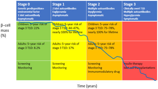
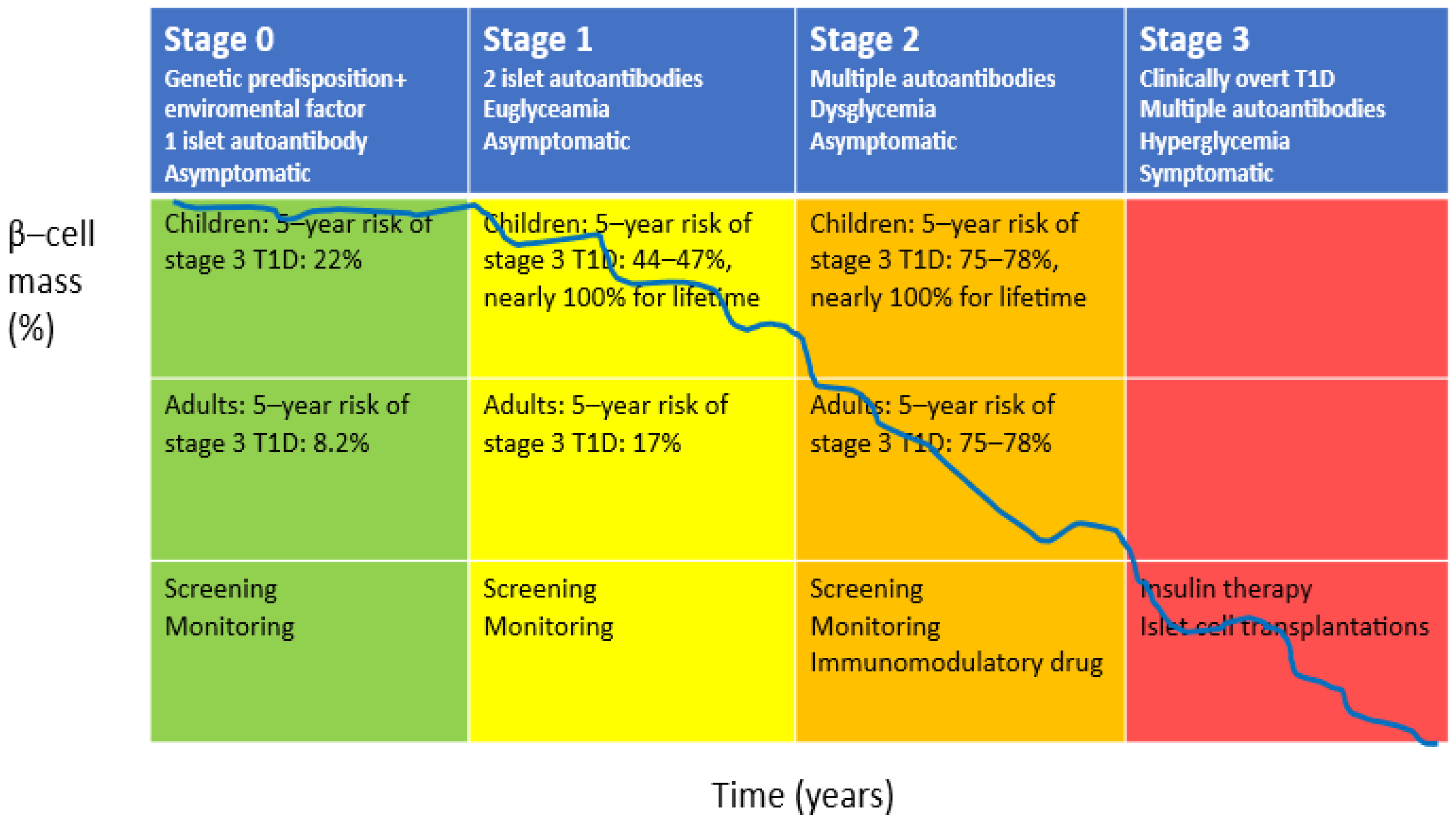
The complex and multifactorial pathogenesis of T1D underlies the diversity in risk and clinical manifestations of T1D, which makes the screening and monitoring of subjects at risk quite challenging [13,14,15,16,17]. Previously, it was suggested that islet autoantibodies, insulin autoantibodies (IAA), insulinoma antigen 2 (IA2), glutamic acid decarboxylase 65 (GAD65) and zinc transporter 8 (ZnT8), can be detected years before the clinical onset of T1D in the peripheral blood of subjects at risk for T1D development. These autoantibodies became standardized biomarkers for the prediction and diagnosis of early stages of T1D [18]. The classification of prediabetes that simultaneously and comprehensively includes impairments in β-cell mass/function and clinical aspects of T1D progression defines stages (0–3) based on the number of islet autoantibodies and glucose tolerance status (Figure 1) [19]. The precise β-cell mass in different stages of pre-T1D is not yet defined. However, it is reported that the risk of overt T1D in subjects with two autoantibodies is almost 100% over their lifetime, whereas, for the presence of one autoantibody, the risk is moderate, at 15% over the next 10 years [18,19,20]. Consequently, the real start of T1D is stage 1, and it is the path of no return to the clinical onset of T1D in the pediatric population. On the other hand, the true significance of the detection of multiple autoantibodies in the adult population is not yet well understood.
Figure 1.
Stages of type 1 diabetes (T1D), risk of stage 3 T1D and current interventional strategies through the course of the disease [18,19,20].
A huge step forward in T1D healthcare was recently made in the USA, when ICD-10 incorporated new codes for T1D presymptomatic, stage 1 E10.A01 and stage 2 E10.A02. In this sense, the early stages of pre-T1D are now recognized as disease stages and can be used for the screening and diagnosis of T1D.
Keeping in mind that there were no effective prevention measures, screening for risk of T1D was previously justified only for research [21,22]. Opponents to screening insisted on the low positive predictive value of tests, high costs, unresolved ethical issues, and challenges like rising anxiety and societal stigma [23,24].
The major turning point came with teplizumab (Tzield®), the first immunomodulatory drug that can postpone stage 3 in children older than 8 years and adults at stage 2 [25]. In this context, a precise definition of stages could help to detect subjects at risk, to monitor them through the stages as an early interventional strategy and offer them disease-modifying drugs, and consequently to extend the timeline from stage 1 to stage 3 T1D (Figure 1) [26].
Previous preventive studies have focused on the population of first-degree relatives (FDRs) of people with T1D, as they have a higher relative risk of T1D than the general population [27,28]. On the other hand, less than 15% of recent-onset T1D patients have FDR with T1D [29]. Therefore, screening in the general population arises as a promising option, especially considering the results obtained from screening for T1D in the general population, where it was shown that the progression to T1D is similar in the general population and in FDRs [30]. In this context, several diabetes organizations recommend screening for T1D in the wider population at risk for T1D, especially in people with autoimmune diseases (primarily celiac or thyroid disease) and genetic risk for T1D, as well as in non-obese adults with impaired glycemic tolerance at age < 35 years [31,32,33].
3.2. Screening Programs Currently Running in Europe
The screening of the risk for T1D is generally based on the detection of islet autoantibodies or genetic risk scores, or a combination of genetic and immunological markers [34]. Nowadays, there is a progressively increasing number of screening initiatives, and this trend is spreading fast across Europe. The Innovative approach towards understanding and arresting type 1 diabetes—INNODIA—is the largest platform focused on screening the FDRs of T1D patients and recruiting persons at risk of T1D in prevention clinical trials [35]. In addition, European action for the Diagnosis of Early Non-Clinical Type 1 diabetes For disease Interception—EDENT1FI—is a newer network aimed at identifying children at risk of T1D through public health screening in Sweden, Denmark, Germany, Italy, Portugal, the Czech Republic, the UK and Poland, between November 2023 and October 2028. They plan to screen 200,000 subjects aged 1–17 years for islet autoantibodies from capillary blood and, after confirmation of the presence of antibodies, to monitor them with OGTT, HbA1c or continuous glucose monitoring (CGM). Every country has its local project, adapted for their needs and possibilities—Sweden/Denmark (Dia Union), Germany (Fr1da), Italy (D1Ce), the Czech Republic (βetty), the UK (ELSA), Poland and Portugal (EDEN1FI)—with planned screening numbers, ages, places (primary care, hospitals, home), and regions included in project [36]. For example, in the UK, the ELSA feasibility and acceptability study was planned to screen more than 20,000 children aged 3–13 years old from the general population [37]. In the UK, during the INGR1D2 study, genetic risk for T1D was detected in over 31,000 infants.
Furthermore, the Global Platform for the Prevention of Autoimmune Diabetes—GPPAD—is a consortium of European research organizations aimed at the detection of infants with genetic risk for T1D, so as to offer them participation in primary prevention clinical trials. In this context, a respectable number of more than 600,000 infants have undergone screening, and over 2400 children are participating in the GPPAD prevention studies to date [38]. Additionally, the European pre-T1D Registry involves investigators who register subjects with pre-T1D, aiming to inform them about monitoring and current options for treatment to delay the disease. This organization is coordinating with GPPAD, INNODIA and EDENT1FI. Simultaneously, Italy has recently adopted an interesting screening program at the national level for children in the general population. Pediatric primary care physicians are the first point of free-of-charge screening and communication with parents. They screen children at ages 2–3, 6–7, and 10–11 for both islet autoantibodies and genetic markers from capillary blood and refer all autoantibody-positive children to pediatric T1D expert regional centers, responsible for further education and metabolic monitoring (CGM, not OGTT). The program will be evaluated by physicians and parents using questionnaires [39,40]. On the other hand, France launched a National Programme for the screening of T1D in FDRs aged 2–45 years, which includes testing for ≥2 autoantibodies (IAA, anti-GAD, anti-IA-2). If screening is initially negative, they intend to repeat the screening every 4 years until the age of 12. Islet autoantibody-positive subjects will be followed through the monitoring of OGTT, glycemia and/or HbA1c [41].
The spectrum of the described programs was broadened significantly in the past couple of years, and the screening for T1D in families of persons with T1D was revived, which spread the idea of the need for screening in the general population, with a focus on children. Interestingly, there is a drastic imbalance between screening initiatives in the pediatric and adult populations in Europe. In fact, there is only one screening program for the risk of T1D, focused exclusively on adults aged 18–70 in the general population, named Type 1 Diabetes Risk in Adults (T1DRA) [42], and it is being conducted in the UK.
3.3. Screening Programs Currently Available Outside of Europe
Currently, the USA, as the country with the leading prevalence of T1D in the world, performs several parallel screening programs. TrialNet is the oldest free program in the USA for the early detection of subjects at risk for T1D, including them in prevention clinical trials. The program is focused on FDRs, aged 2–45 years, and on younger second-degree relatives, aged 2–20, or on subjects from the general population, aged 2–45 years, who tested positive for one islet autoantibody in person or with an at-home kit [43].
Simultaneously, there is a growing number of programs in the general population. Autoimmunity Screening for Kids—ASK—is a free screening program in the USA, Colorado, for T1D and gluten enteropathy for subjects ages 1–17, from the general population [44]. The first results showed a drastic reduction in DKA incidence in Colorado (from 60% to 5%) in screened and monitored children with pre-T1D [45]. Moreover, the PLEDGE study [Population Level Estimate of type 1 Diabetes risk Genes in Children] is being performed across all Sanford clinics in the USA, conducting general population screening for T1D and celiac autoantibodies so as to document its economic justifications and introduction into standard pediatric care. They will enroll ~33,000 children, <6 years or 9–16 years of age, into this program [46]. Furthermore, the Virginia PrIMeD Project has a part focusing on the detection of risk for T1D, analyzing genetic markers for T1D as the first step and then guiding those with genetic risk to further autoantibody testing. In this context, they included around 3700 children (ages 2–16 years) from pediatric clinics, and testing was performed on saliva samples [47].
Simultaneously, Combined Antibody Screening for Celiac and Diabetes Evaluation (CASCADE) offers free screening for T1D and gluten enteropathy in the state of Washington for children newborn to 8 months or 4 to 7 years of age [48]. Additionally, Australia has Type 1 Screen, an organization funded by Breakthrough Type 1 (formerly JDRF), offering free screening for islet antibodies from capillary blood for FDR as well as for the general population. To date, they have screened 3843 subjects, finding 193 positive subjects [49]. Interestingly, they published a pilot study examining three models of population-based screening. Screening was conducted from 2022 to 2024, and it involved the detection of genetic risk markers using newborns’ capillary blood and then the islet autoantibody after 11 months of age; the detection of genetic risk markers using the saliva of babies 6–12 months of age and then the islet autoantibody after 10 months of age; or the detection of the islet autoantibody using capillary blood from children aged 2, 6 or 10 years. Therefore, three cohorts were to be analyzed, comprising a total of up to 9000 children. Moreover, they would analyze the participation rate for each cohort, as well as costs, risk perception, anxiety and physician opinions [50]. Another running program is the VISION-T1D program, a prospective cohort study performed in 2024–2025 in Saudi Arabia, focusing on the detection of islet autoantibodies in children aged 2–18 years, with optional genetic testing [51]. Finally, the Antibody Detection Israeli Research (ADIR) program, started in 2021 with support from Breakthrough T1D, represents a national screening initiative, aiming to involve around 35,000 children aged 9 months to preschool age across Israel for islet antibody screening. ADIR will implement new technology for autoantibody detection using capillary blood, making it easy to use in the general population. They have screened 17,000 children up until now; 0.47% had multiple islet antibodies, while 120 children had one islet antibody [52].
Having considered all this data, it can be concluded that numerous screening approaches exist worldwide, focusing on the extended detection of risk in the general population. In order to enhance their functionality and usability, some of these screening programs include another autoimmune disease, celiac enteropathy, alongside T1D. An important feature of these programs is the simplicity with which genetic and immunological markers can be obtained. Interestingly, the programs predominantly use capillary blood, though some of them use saliva. In this context, it is reported that capillary testing for islet antibodies is the preferred method for screening children for T1D, with the most suitable timing being, according to parents, during preschool vaccination visits, despite concerns regarding the perceived volume of the capillary sample [53]. In addition, simplicity must be a priority, as the preferred screening program is the one that can be easily implemented in everyday clinical practice [54,55]. Recently, pediatricians at the primary care level have expressed concerns about screening for T1D, noting that they are not familiar with selecting and interpreting tests, and highlighting unresolved issues related to the psychosocial impact on families and the additional workload that interferes with their routine clinical practice [56]. Additionally, endocrinology specialists have reported that family stress, costs, and uncertainty regarding treatment are primary barriers to T1D screening [57].
It should be noted that there are differences regarding age categories and the number of children recruited in these programs. This is very important, considering that the detection of persistent islet antibodies is happening mainly during the first two years of life [58,59]. If we summarize different approaches to screening by age groups, generally, screening in pediatric populations is beneficial at ages 2, 6, and 10, because it will identify at least 80% of cases with risk [58]. However, it still raises concerns, keeping in mind the remaining 20% of children at risk who stay undetected. On the other hand, this might be a potential explanation for the fact that screening for T1D after the age of 18 is a less available option in screening programs.
Fr1da and GPPAD are programs targeting the general pediatric population, but using different screening methods: Fr1da uses islet autoantibodies and GPPAD uses genetic markers. Additionally, from the perspective of the target population, INNODIA, Trial NET and the French screening programs still focus on FDRs as the primary target population for the screening of T1D, even though data from the Fr1da study demonstrated the same risk in subjects in stage 2, irrespective of family relation to a patient with T1D. Furthermore, there are differences in the inclusion criteria between the INNODIA and GPPAD screening programs. While INNODIA includes children and adults up to 45 years of age who have newly diagnosed T1D and their FDRs, to explore the natural history of T1D and guide patients in tertiary prevention studies, GPPAD strictly includes infants up to 7 months and guides those with a high genetic risk score (>10% predicted genetic risk to develop multiple islet autoantibodies up to 6 years of life) to primary prevention studies. INNODIA, for now, has recruited patients for MELD ATG (exploring immunomodulatory drug antithymocyte globulin) and DIAGNODE 3 (exploring anti-GAD vaccine + supplementation with vitamin D), while GPPAD recruited infants for PoInT (exploring oral insulin), SynTIA (probiotics), and AVAnT1A (exploring vaccination against SARS-CoV during the first 9 months of life). In that context, the primary goal for INNODIA was to protect C peptide levels, while GPPAD aims at modulating the natural course of pre-T1D and postponing or blocking the onset of autoimmunity in genetically susceptible infants.
On the other hand, a common feature of all screening initiatives focusing on the pediatric population is that they include children up to the age 6 and have monitoring and education about T1D symptoms as necessary components of screening programs.
To conclude, the current screening initiatives are still missing data on the best assay for capillary blood islet autoantibody screening, in terms of precision, sensitivity, and cost. Apart from that, there are no screening programs in Asia, Africa and South America, mainly due to the low prevalence of T1D in this part of the world at this moment.
Finally, there are no clear recommendations about the timing and frequency of screening in the adult population. Data from multicenter cohorts pointed out that screening strategies must adapt to the adult population’s specificities. Besides T1DRA, the only initiative including exclusively adults, there are few international screening networks focusing on children which include transition into adult care and the young adult population. In that context, the TrialNet Pathway to Prevention recently published the results of approximately 100,000 adult relatives and demonstrated that adults more frequently screen positive for a single autoantibody (4.0%) in comparison to children (2.6%), although they are less likely to have multiple detected autoantibodies (0.83% vs. 2.8%) [60]. Moreover, INNODIA, a European partnership across 27 partners in 13 countries and more than 50 accredited medical centers, as well as EDENT1FI, the latest European initiative across 13 countries, are stratifying risk and feasibility for general population screening that includes transition into adult care [35].
Trial Net multicenter data demonstrated different progression patterns for adults compared to the pediatric population during follow-up: adults with stage 1 T1D have a significantly lower 5-year progression risk (17%) than children (47%). Still, once adults reach stage 2, their 5-year risk rapidly increases to 78%, being comparable with the pediatric cohort [60] (Figure 1). However, since age is the major modulator of genetic risk for T1D [58], it would be reasonable to screen once in a lifetime, after 18 years of age, especially in families with T1D, and particularly for GADA. In adult-onset T1D, GADA is the primary immunological marker, frequently detected as the only antibody or at the highest titer. It has been shown that adding ZnT8A to a panel of GADA and IA-2A increases diagnostic sensitivity in adults by 5–10% [61]. Furthermore, adult trials preferred high-affinity assays, such as electrochemiluminescence, because they are more precise in detecting disease-relevant antibodies from low-titer, non-progressive antibodies, which are more frequent in adults [62]. But a major obstacle in using islet autoantibody screening in adults is the lack of a sufficient number of longitudinal studies analyzing progression to stage 3 T1D. Consequently, for adults, autoantibody tests are frequently done with non-fasting C-peptide measurements. Previous studies have shown that adults with newly diagnosed T1D experience a milder decline in the endogenous insulin reserve than children (40% annually vs. 50% annually) [63]. Finally, there are no studies assessing the risk of T1D in the general population in adults older than 45 years [64].
It was reported that the 5-year risk of T1D in single positive islet autoantibody adults is similar to the 5-year risk of type 2 diabetes development in adults ≥ 40 years in the general population (5% vs. 4%) [65]. Previously, it was demonstrated that no further islet autoantibody testing is needed after the confirmation of multiple islet autoantibodies in adults, as the loss of islet autoantibodies after testing positive for multiple antibodies does not alter future risk of T1D [66].
Eventually, there seems to be no consensus about the timing and type of screening, and the next few years will sharpen recommendations, which should be adjusted for every country. Obviously, there are numerous advantages in detecting and monitoring subjects at risk of T1D.
3.4. Monitoring of Subjects at Risk for T1D: Again, Heterogeneity in Approaches
The main advantages and barriers for the screening and monitoring of subjects at risk of T1D are summarized in Table 1.
Table 1.
The main advantages and barriers of screening and monitoring of risk of T1D [23,24,31,56,67,68].
There are several advantages of monitoring subjects at risk: optimal HbA1c levels at diagnosis of T1D, a reduced frequency of insulin therapy at the onset of disease, a reduced incidence of ketosis, and, most significantly, a reduced incidence of diabetic ketoacidosis (DKA), a life-threatening condition [31]. If we compare the cohort of children screened from the general population in the Fr1da study and the Di Meglio cohort of children diagnosed with clinical T1D without prior screening, Fr1da children had fewer incidences of DKA and better HbA1c and C peptide levels [67] at the clinical onset of T1D. It is suggested that screening might be cost-effective if it decreases DKA incidence and HbA1c levels at diagnosis, keeping in mind that it is associated with better glycemic control during the course of overt disease and consequently lower costs due to the reduced incidence of chronic complications [69]. Additionally, based on findings of the Fr1da trial, the length of hospitalization at diagnosis would be reduced by 18%, and the number of days with symptoms and children with weight loss before diagnosis would be reduced by 60% and 71%, respectively, which represents additional economic benefits [68].
At this moment, although it is strongly recommended, there is no clear consensus about the monitoring of subjects at risk, and there are few recommendations, as summarized in Table 2.
Table 2.
Current recommendations for monitoring of subjects at risk of T1D.
In 2024, members of the Fr1da, GPPAD, and INNODIA consortia gave expert consensus suggesting random plasma glucose and HbA1c testing every 6 months for children in stage 1, followed by 2 h OGTT, with frequency depending on the age, from 1 to 3 years. However, for stage 2 children, irrespective of age, follow-up should be every 3 months, with random plasma glucose, CGM, or HbA1c testing performed, as well as education on the symptoms and signs of hyperglycemia and at-home glycemic monitoring. It is interesting that repeated 2 h OGTT was not routinely recommended for stage 2 children, but it was suggested to be performed every 6 months when HbA1c ≥ 6% [70].
Afterwards, ISPAD, as a leading global organization focusing on the improvement of care for the pediatric population with diabetes, published consensus guidelines, which include an update on staging for T1D, as well as a comprehensive perception of the screening and monitoring of at-risk children, adolescents, and young adults [71]. They have pointed out that children in stages 1 and 2 are in the early T1D stage of the disease and not just at risk for the disease. Moreover, they suggested screening for islet autoantibodies twice during childhood because it will detect the majority of children who will progress to overt T1D, and it is the most cost-effective option. Additionally, glycemic monitoring should be mandatory from stage 1, using 2 h OGTT as the first choice, otherwise using HbA1c (except for very young children), venous or capillary random glycemia, or CGM. Besides this, considering the pediatric population, the experts suggested using the self-monitoring of blood glucose and urinary glucose testing as monitoring techniques, which are simple to execute. The frequency of testing should be adjusted according to the age and degree of risk for progression. For children up to 3 years old in stage 0 with one autoantibody on islet antigens, monitoring for antibodies, random glycemia or HbA1c should be performed twice a year for the next 3 years, then yearly for another 3 years. For older children, testing every year for the next 3 years is suggested. Simultaneously, in stage 1, glycemic monitoring should be performed quarterly for children up to 3 years old, twice a year for children 3–9 years old, and annually for children older than 9 years. Interestingly, in stage 2, frequent testing—every 3 months—is suggested for children, while for young adults the testing should be performed every 6 months [71].
Furthermore, a group of experts published recommendations for primary care physicians and for secondary/tertiary physicians on monitoring people at risk for T1D [72]. These are briefly based on the mandatory metabolic monitoring of subjects starting from stage 0 through stages 1 and 2, with age-dependent visit frequency ranging from 6 months to 3 years. The 2 h OGTT is considered the gold standard, but they also suggested more accessible methods for the general population, such as capillary or venous glycemia and HbA1c (Table 2). Interestingly, they pointed out that adults should be screened every 3 years, as in screening programs for type 2 diabetes, except in high-risk vulnerable populations, where annual testing is recommended (FDRs, presence of another autoimmune disease, dysglycemia, or stress hyperglycemia). Stage 1 adults should be tested annually using HbA1c, and, if there is no disease progression over 5 years, follow-up is recommended every 2 years. Additionally, stage 2 adults should be monitored every 6 months with HbA1c and a 2 h OGTT or CGM. Primary care physicians should be educated and motivated to follow subjects with a single autoantibody, while those in stage 1 or 2 should be referred to secondary/tertiary care levels [72].
Finally, the latest ADA recommendations are simple and clear, suggesting using HbA1C every 6 months and 2 h OGTT annually for stages 1 and 2, with further adaptation depending on individual risk [73].
Obviously, there are disagreements about the frequency and method of monitoring children in stage 1, which vary from strictly every 3 months for very young children with the highest risk, to more liberally at 12–36 months in older children and adults. On the other hand, all experts agree that children in stage 2 should be monitored frequently, every 3 months.
A possible explanation for these differences in monitoring methods is mainly based on the fact that there are only a few studies about the prediction precision of different methods and the easiness of performing them. In this context, the metabolic monitoring of subjects in stages 1 and 2 may be conducted using glycemia, HbA1c, 2 h OGTT, or CGM. Previously, it was indicated that a rise in HbA1c within the normal range by more than 10% over 3–12 months is a valid predictor of progression to stage 3 [74]. The growing usage of diabetes technology has mandated the use of CGM to assess the risk of progression to stage 3. The results of the ASK study in a pediatric population with two positive autoantibodies indicated that, if only 10% of the time is spent above normoglycemia, the risk of progression to stage 3 in the next year is as high as 80% [75]. At the same time, the data obtained in adolescents in stage 1 who were followed in the TrialNet study showed that, if only 5% of the time is spent above 7.8 mmol/L, the risk of progression in the next 2 years is 40%, and, if the limit is 8.8 mmol/L, the risk rises to over 60% [76]. In a recent ENDIA study, preschool children monitored for 10 years in stage 1 who progressed to stage 3 had higher glycemic variability indices and a longer time spent in hyperglycemia on CGM compared to antibody-negative children [77]. Finally, data from a longitudinal study monitoring FDRs in stage 2 shed light on the quality of different glycemic parameters as predictors for progression to stage 3. The analysis of baseline data showed that OGTT and CGM performed similarly, while HbA1c had worse predictive potential. On the other hand, longitudinal data showed the better predictive power of repeated OGTT than CGM for progression to stage 3. However, for the first time, further analysis highlighted the combination of CGM and HbA1c as informative and similar to repeated OGTT, yet easier to conduct in the long-term monitoring of progression rate to stage 3 [78].
3.5. Challenges in Screening and Monitoring: Factors Influencing Risk for Progression to Stage 3
A growing number of data suggests that the current definition of stages and risk prediction is challenging, and that it is a very important issue from the perspective of the motivation of subjects to be screened and monitored. Previous studies have shown that the main risk factors for the development of stage 1 and further deterioration and progression to stage 3 are different, numerous and complex [79], and consist of genetic, immunological, metabolic and environmental factors.
In this sense, the T1D genetic risk score was proven to have excellent prognostic power for the risk of progression to stage 3 [80]. Moreover, age is the primary determinant and main modifier of progression to overt T1D, inversely related to T1D risk in subjects with multiple islet antibodies [81,82]. Simultaneously, immunological factors influencing this risk include not only the number, but also the type, titer, and adherence of islet autoantibodies. The type of autoantibody is important, as IAA shows fast, while GADA shows slow progression in children; however, in adults, Zn T8 shows rapid progression [83,84]. Furthermore, IA-2A+ subjects had higher genetic risk scores and a higher risk for progression in overt T1D, persistently, during both stages in pre-T1D [85]. In this sense, subjects with a single IA-2+ should be observed regularly, uniformly and periodically in the same way as those with numerous islet autoantibodies. Simultaneously, a higher titer and affinity of islet autoantibodies were associated with rapid progression [86,87]. In the pediatric population, a higher titer of IAA and IA2, but not GADA, was associated with a higher risk of progression to stage 3 in patients with one persistent islet autoantibody [88]. Furthermore, a novel tool for stratifying risk for stage 3, based on a machine learning approach and utilizing numerous autoantibody features, suggests that changes in dominantly IA-2A titers during the first 12 years of life may modulate the risk of progression to overt T1D [89]. Additionally, it is reported that the interaction between parts of autoantibodies and ligands, dictated by certain gene products, may influence progression to stage 3 T1D [90]. Recently, it was suggested that it is possible to define subpopulations of children at genetic risk for T1D with different risks of progression to T1D, based on autoantibody profiles, seroconversion age, and the sequence of appearance of autoantibodies during follow-up [91].
Considering previous data, detecting only the type of islet autoantibody is insufficient and not precise enough for defining pre-T1D stages or for risk stratification by stage. In that context, motivating the population for screening and subjects at risk for monitoring might be difficult without further increasing the power of the existing biomarkers and uncovering new predictive biomarkers.
In the context of other biomarkers, the data obtained in the GPPAD POInT study demonstrated postprandial glycemia spikes in children progressing to stage 1, even 8 weeks before seroconversion [92], indicating that β-cell mass/function markers drive the progression rate independently of immunological markers. So, the pro-insulin-to-C-peptide ratio and glucose/C-peptide values from OGTT have an excellent correlation with progression to stage 3, and might be used to individually stratify risk for the deterioration of disease [93,94,95]. Furthermore, unmethylated DNA, as a marker of β-cell death, has the potential to be a biomarker of the progression rate to stage 3 [96]. Nowadays, scientists are working to improve the precision prediction of T1D using new stratifying methods, novel biomarkers and “omics” approaches [97,98,99].
Recently, the joint group of experts published recommendations simultaneously in the most prominent journals in diabetology, aimed at narrowing gaps of uncertainties in T1D. They suggested three concepts for exploring the problem of heterogeneity in T1D: the palette concept regarding numerous and distinct risk factors, the gradient concept regarding the staging system and its insufficiency to clarify individual risk and particularly risk in the adult population, and the network-threshold concept suggesting the precise counting of risk markers and detecting their threshold for the triggering of T1D progression [100].
3.6. Unresolved Issues and Perspectives in Screening and Monitoring for T1D
There are definitely numerous differences in approaches when it comes to the screening and especially to the monitoring of subjects at risk for T1D. This research area has several knowledge gaps. The first knowledge gap is the population of adults with pre-T1D, as all the findings we have are based mainly on the data obtained from the pediatric population. The second knowledge gap refers to the importance of single autoantibody positivity for progression to stage 3, especially in the population of adults. The issue linked with this is the issue of the mechanisms of protection from disease progression. The final knowledge gap relates to the upper and lower age limits for screening for T1D in the adult population and the number and type of antibodies.
Moreover, regarding the metabolic monitoring of adults with single islet autoantibody positivity, the range of HbA1c 5.7–6.4% might not be an optimal definition of risk for progression in stage 3, keeping in mind that nearly 40% of adults ≥ 40 years have these HbA1c levels [65]. Consequently, some experts suggested HbA1c ≥ 6%, in adults for more frequent monitoring [64].
In addition, another area that is rapidly developing is the exploration of novel biomarkers showing disease progression for the precise phenotyping of risk.
Furthermore, we know the average risk for progression to T1D through the stages, but individual risk is not precisely defined. It is not known whether all individuals who become dysglycemic, especially adults, progress to T1D [101]. Intriguingly, subjects in stage 2 can revert to normoglycemia [102]. In addition, it is unknown whose disease would progress and when the progression will take place. The definition of stages with autoantibodies and glycemia is necessary for the detection of subjects at risk, but further personalization of risk and sharpening of individual prediction must be performed [103,104]. In that context, biomarkers of β-cell function heterogeneity, stress and death should be further defined [104,105]. Moreover, the precise perception of risk for T1D is fundamental for parents’ or subjects’ decision-making about participation in preventive trials, and for giving consent to take teplizumab. A recent review suggested that understanding the psychosocial implications of T1D-risk screening is as critical to its success as the improved understanding of the purpose, procedures and consequences of screening [106]. Additionally, defining screening programs is dependent on resources, population and clinical settings [69]. However, costs may depend on T1D prevalence, the incidence of DKA and screening test accuracy in a specific country. Finally, an intensive scientific effort is still required to develop a sustainable method for the expansion of screening as a combination of risk stratification in families, screening for risk in the general population and personalization of risk [107,108].
4. Conclusions
There is currently an expansion of screening and vigilant monitoring of subjects at risk for T1D. Presently, approaches to screening and monitoring at-risk subjects vary, and they primarily focus on the pediatric population, with a serious gap in recommendations and potential benefits for the adult population. It is important to develop and adapt screening and monitoring strategies tailored to different countries, aiming to make them more universally applicable while refining the definition of individual risk for T1D. Nonetheless, the screening and monitoring of subjects at risk should be integrated with early interventions aimed at delaying T1D.
Author Contributions
Conceptualization, T.M. and A.J.; methodology, T.M.; validation, T.M., A.J. and N.M.L.; formal analysis, T.M.; investigation, T.M.; writing—original draft preparation, T.M. and A.J.; writing—review and editing, T.M., A.J. and N.M.L.; supervision, A.J. All authors have read and agreed to the published version of the manuscript.
Funding
This research received no external funding.
Institutional Review Board Statement
Not applicable.
Informed Consent Statement
Not applicable.
Data Availability Statement
No new data were created or analyzed in this study. Data sharing is not applicable to this article.
Conflicts of Interest
The authors declare no conflicts of interest.
Abbreviations
The following abbreviations are used in this manuscript:
| T1D | Type 1 diabetes |
| FDR | First-degree relative |
| IAA | Insulin autoantibodies |
| IA2 | Insulinoma antigen 2 autoantibodies |
| GAD65 | Glutamic acid decarboxylase 65 autoantibodies |
| ZnT8 | Zinc transporter 8 autoantibodies |
| ISPAD | The International Society for Pediatric and Adolescent Diabetes |
References
- Gregory, G.A.; Robinson, T.I.G.; Linklater, S.E.; Wang, F.; Colagiuri, S.; de Beaufort, C.; Donaghue, K.C.; International Diabetes Federation Diabetes Atlas Type 1 Diabetes in Adults Special Interest Group; Magliano, D.J.; Maniam, J.; et al. Global incidence, prevalence, and mortality of type 1 diabetes in 2021 with projection to 2040: A modelling study. Lancet Diabetes Endocrinol. 2022, 10, 741–760, Erratum in Lancet Diabetes Endocrinol. 2022, 10, e11. https://doi.org/10.1016/S2213-8587(22)00280-7. PMID: 36113507. [Google Scholar] [CrossRef] [Scilit] [PubMed]
- EURODIAB ACE Study Group. Variation and trends in incidence of childhood diabetes in Europe. Lancet 2000, 355, 873–876, Erratum in Lancet 2000, 356, 1690. PMID: 10752702. [Google Scholar] [CrossRef] [Scilit] [PubMed]
- Wagenknecht, L.E.; Lawrence, J.M.; Isom, S.; Jensen, E.T.; Dabelea, D.; Liese, A.D.; Dolan, L.M.; Shah, A.S.; Bellatorre, A.; Sauder, K.; et al. Trends in incidence of youth-onset type 1 and type 2 diabetes in the USA, 2002-18: Results from the population-based SEARCH for Diabetes in Youth study. Lancet Diabetes Endocrinol. 2023, 11, 242–250. [Google Scholar] [CrossRef] [Scilit] [PubMed] [PubMed Central]
- Gong, B.; Yang, W.; Xing, Y.; Lai, Y.; Shan, Z. Global, regional, and national burden of type 1 diabetes in adolescents and young adults. Pediatr. Res. 2025, 97, 568–576. [Google Scholar] [CrossRef] [Scilit]
- Harding, J.L.; Wander, P.L.; Zhang, X.; Li, X.; Karuranga, S.; Chen, H.; Sun, H.; Xie, Y.; Oram, R.A.; Magliano, D.J.; et al. The Incidence of Adult-Onset Type 1 Diabetes: A Systematic Review From 32 Countries and Regions. Diabetes Care 2022, 45, 994–1006, Erratum in Diabetes Care 2023, 46, 2084. https://doi.org/10.2337/dc23-er11. PMID: 35349653; PMCID: PMC9016739. [Google Scholar] [CrossRef] [Scilit] [PubMed] [PubMed Central]
- Rogers, M.A.M.; Kim, C.; Banerjee, T.; Lee, J.M. Fluctuations in the incidence of type 1 diabetes in the United States from 2001 to 2015: A longitudinal study. BMC Med. 2017, 15, 199. [Google Scholar] [CrossRef] [Scilit]
- International Diabetes Federation. IDF Diabetes Atlas, 11th ed.; International Diabetes Federation: Brussels, Belgium, 2025. [Google Scholar]
- Gale, E.A.M. The Rise of Childhood Type 1 Diabetes in the 20th Century. Diabetes 2002, 51, 3353–3361. [Google Scholar] [CrossRef] [Scilit]
- Egro, F.M. Why is type 1 diabetes increasing? J. Mol. Endocrinol. 2013, 51, R1–R13. [Google Scholar] [CrossRef] [Scilit]
- Leslie, R.D.; Evans-Molina, C.; Freund-Brown, J.; Buzzetti, R.; Dabelea, D.; Gillespie, K.M.; Goland, R.; Jones, A.G.; Kacher, M.; Phillips, L.S.; et al. Adult-Onset Type 1 Diabetes: Current Understanding and Challenges. Diabetes Care 2021, 44, 2449–2456. [Google Scholar] [CrossRef] [Scilit] [PubMed] [PubMed Central]
- Thomas, N.; Vaidya, B.; Leslie, R.D.; Agardh, D.; Oram, R.; Dabelea, D.; Singh, A.; Chantzichristos, D.; Rewers, M. Association of Individual or Family History of Autoimmune Disease with Future Development of Type 1 Diabetes. Diabetes/Metab. Res. Rev. 2026, 42, E70110. [Google Scholar] [CrossRef] [Scilit]
- Hughes, J.W.; Riddlesworth, T.D.; DiMeglio, L.A.; Miller, K.M.; Rickels, M.R.; McGill, J.B. Autoimmune Diseases in Children and Adults with Type 1 Diabetes from the T1D Exchange Clinic Registry. J. Clin. Endocrinol. Metab. 2016, 101, 4931–4937. [Google Scholar] [CrossRef] [Scilit] [PubMed] [PubMed Central]
- Ilonen, J.; Lempainen, J.; Veijola, R. The heterogeneous pathogenesis of type 1 diabetes mellitus. Nat. Rev. Endocrinol. 2019, 15, 635–650. [Google Scholar] [CrossRef] [Scilit]
- Van Belle, T.L.; Coppieters, C.T.; von Herrath, M.G. Type 1 Diabetes: Etiology, Immunology, and Therapeutic Strategies. Physiol. Rev. 2011, 91, 79–118. [Google Scholar] [CrossRef] [Scilit]
- Michels, A.W.; Redondo, M.J.; Atkinson, M.A. The pathogenesis, natural history, and treatment of type 1 diabetes: Time (thankfully) does not stand still. Lancet Diabetes Endocrinol. 2022, 10, 90–92. [Google Scholar] [CrossRef] [Scilit] [PubMed] [PubMed Central]
- Krischer, J.P.; Lynch, K.F.; Schatz, D.A.; Ilonen, J.; Lernmark, Å.; Hagopian, W.A.; Rewers, M.J.; She, J.X.; Simell, O.G.; Toppari, J.; et al. The 6 year incidence of diabetes-associated autoantibodies in genetically at-risk children: The TEDDY study. Diabetologia 2015, 58, 980–987. [Google Scholar] [CrossRef] [Scilit] [PubMed] [PubMed Central]
- Atkinson, M.A.; Roep, B.O.; Posgai, A.; Wheeler, D.C.S.; Peakman, M. The challenge of modulating β-cell autoimmunity in type 1 diabetes. Lancet Diabetes Endocrinol. 2019, 7, 52–64. [Google Scholar] [CrossRef] [Scilit] [PubMed] [PubMed Central]
- Ziegler, A.G.; Rewers, M.; Simell, O.; Simell, T.; Lempainen, J.; Steck, A.; Winkler, C.; Ilonen, J.; Veijola, R.; Knip, M.; et al. Seroconversion to Multiple Islet Autoantibodies and Risk of Progression to Diabetes in Children. JAMA 2013, 309, 2473–2479. [Google Scholar] [CrossRef] [Scilit] [PubMed]
- Insel, R.A.; Dunne, J.L.; Atkinson, M.A.; Chiang, J.L.; Dabelea, D.; Gottlieb, P.A.; Greenbaum, C.J.; Herold, K.C.; Krischer, J.P.; Lernmark, Å.; et al. Staging presymptomatic type 1 diabetes: A scientific statement of JDRF, the Endocrine Society, and the American Diabetes Association. Diabetes Care 2015, 38, 1964–1974. [Google Scholar] [CrossRef] [Scilit] [PubMed] [PubMed Central]
- Sims, E.K.; Besser, R.E.J.; Dayan, C.; Geno Rasmussen, C.; Greenbaum, C.; Griffin, K.J.; Hagopian, W.; Knip, M.; Long, A.E.; Martin, F.; et al. Screening for Type 1 Diabetes in the General Population: A Status Report and Perspective. Diabetes 2022, 71, 610–623. [Google Scholar] [CrossRef] [Scilit] [PubMed] [PubMed Central]
- Wilson, J.M.G.; Jungner, G. Principles and Practice of Screening for Disease; World Health Organisation: Geneva, Switzerland, 1968. [Google Scholar]
- Knip, M. Should We Screen for Risk of Type 1 Diabetes? Diabetes Care 2008, 31, 622–623. [Google Scholar] [CrossRef] [Scilit] [PubMed]
- Schatz, D.; Krischer, J.; She, J. To Screen or Not to Screen for Pre-Type 1 Diabetes? Horm. Res. 2002, 57, 12–17. [Google Scholar] [CrossRef] [Scilit] [PubMed]
- Smith, L.B.; Lynch, K.F.; Driscoll, K.A.; Johnson, S.B.; TEDDY Study Group. Parental monitoring for type 1 diabetes in genetically at-risk young children: The TEDDY study. Pediatr. Diabetes 2021, 22, 717–728. [Google Scholar] [CrossRef] [Scilit] [PubMed] [PubMed Central]
- Ziegler, A.G.; Cengiz, E.; Kay, T.W.H. The future of type 1 diabetes therapy. Lancet 2025, 406, 1520–1534, Erratum in Lancet 2025, 406, 2220. https://doi.org/10.1016/S0140-6736(25)02217-2. [Google Scholar] [CrossRef] [Scilit] [PubMed]
- Quattrin, T.; Mastrandrea, L.D.; Walker, L.S.K. Type 1 diabetes. Lancet 2023, 401, 2149–2162. [Google Scholar] [CrossRef] [Scilit] [PubMed]
- Bonifacio, E. Predicting Type 1 Diabetes Using Biomarkers. Diabetes Care 2015, 38, 989–996. [Google Scholar] [CrossRef] [Scilit]
- American Diabetes Association Professional Practice Committee for Diabetes*. 2. Diagnosis and Classification of Diabetes: Standards of Care in Diabetes—2026. Diabetes Care 2026, 49, S27–S49. [Google Scholar] [CrossRef] [Scilit]
- Besser, R.E.J.; Ng, S.M.; Robertson, E.J. Screening children for type 1 diabetes. BMJ 2021, 375, e067937. [Google Scholar] [CrossRef] [Scilit]
- Weiss, A.; Zapardiel-Gonzalo, J.; Voss, F.; Jolink, M.; Stock, J.; Haupt, F.; Kick, K.; Welzhofer, T.; Heublein, A.; Winkler, C.; et al. Progression likelihood score identifies substages of presymptomatic type 1 diabetes in childhood public health screening. Diabetologia 2022, 65, 2121–2131, Erratum in Diabetologia 2022, 65, 2175. https://doi.org/10.1007/s00125-022-05798-z. PMID: 36028774; PMCID: PMC9630406. [Google Scholar] [CrossRef] [Scilit]
- Simmons, K.M.W.; Frohnert, B.I.; O’Donnell, H.K.; Bautista, K.; Geno Rasmussen, C.; Gerard Gonzalez, A.; Steck, A.K.; Rewers, M.J. Historical Insights and Current Perspectives on the Diagnosis and Management of Presymptomatic Type 1 Diabetes. Diabetes Technol. Ther. 2023, 25, 790–799. [Google Scholar] [CrossRef] [Scilit]
- Holt, R.I.G.; DeVries, J.H.; Hess-Fischl, A.; Hirsch, I.B.; Kirkman, M.S.; Klupa, T.; Ludwig, B.; Nørgaard, K.; Pettus, J.; Renard, E.; et al. The management of type 1 diabetes in adults. A consensus report by the American Diabetes Association (ADA) and the European Association for the Study of Diabetes (EASD). Diabetologia 2021, 64, 2609–2652, Erratum in Diabetologia 2022, 65, 255. https://doi.org/10.1007/s00125-021-05600-6. PMID: 34590174; PMCID: PMC8481000. [Google Scholar] [CrossRef] [Scilit]
- Scheiner, G.; Weiner, S.; Kruger, D.; Pettus, J. Screening for Type 1 Diabetes: Role of the Diabetes Care and Education Specialist. ADCES Pract. 2022, 10, 20–25. [Google Scholar] [CrossRef] [Scilit]
- Joglekar, M.V.; Kaur, S.; Pociot, F.; Hardikar, A.A. Prediction of progression to type 1 diabetes with dynamic biomarkers and risk scores. Lancet Diabetes Endocrinol. 2024, 12, 483–492. [Google Scholar] [CrossRef] [Scilit] [PubMed]
- INNODIA. A Private-Public Partnership Against Type 1 Diabetes. 12 December 2025. Available online: http://www.innodia.eu/ (accessed on 29 December 2025).
- Hoffmann, L.; Kohls, M.; Arnolds, S.; Achenbach, P.; Bergholdt, R.; Bonifacio, E.; Bosi, E.; Gündert, M.; Hoefelschweiger, B.K.; Hummel, S.; et al. EDENT1FI Master Protocol for screening of presymptomatic early-stage type 1 diabetes in children and adolescents. BMJ Open 2025, 15, e088522. [Google Scholar] [CrossRef] [Scilit] [PubMed]
- Quinn, L.M.; Dias, R.P.; Greenfield, S.M.; Richter, A.G.; Garstang, J.; Shukla, D.; Acharjee, A.; Gkoutos, G.; Oram, R.; Faustini, S.; et al. Protocol for a feasibility and acceptability study for UK general population paediatric type 1 diabetes screening-the EarLy Surveillance for Autoimmune diabetes (ELSA) study. Diabet. Med. 2025, 42, e15490. [Google Scholar] [CrossRef] [Scilit] [PubMed] [PubMed Central]
- The Global Platform for Preventing Type 1 Diabetes. GPPAD. 12 December 2025. Available online: http://www.gppad.org/ (accessed on 29 December 2025).
- Vincentini, O.; Pricci, F.; Silano, M.; Agrimi, U.; Iacoponi, F.; Villa, M.; Porfilio, C.; Sedile, A.I.; Ulivi, F.; Catassi, C.; et al. Study protocol of D1Ce Screen: A pilot project of the Italian national screening program for type 1 diabetes and coeliac disease in the paediatric population. PLoS ONE 2025, 20, E0328624. [Google Scholar] [CrossRef] [Scilit]
- Cherubini, V.; Mozzillo, E.; Iafusco, D.; Bonfanti, R.; Ripoli, C.; Pricci, F.; Vincentini, O.; Agrimi, U.; Silano, M.; Ulivi, F.; et al. Follow-up and monitoring programme in children identified in early-stage type 1 diabetes during screening in the general population of Italy. Diabetes Obes. Metab. 2024, 26, 4197–4202. [Google Scholar] [CrossRef] [Scilit] [PubMed]
- Mallone, R.; Bismuth, E.; Thivolet, C.; Benhamou, P.Y.; Hoffmeister, N.; Collet, F.; Nicolino, M.; Reynaud, R.; Beltrand, J. Screening and care for preclinical stage 1-2 type 1 diabetes in first-degree relatives: French expert position statement. Diabetes Metab. 2025, 51, 101603. [Google Scholar] [CrossRef] [Scilit] [PubMed]
- The Type 1 Diabetes Risk in Adults (T1DRA) Study. T1DRA. (n.d.). Available online: https://t1dra.bristol.ac.uk (accessed on 29 December 2025).
- Type 1 Diabetes Screening & Early Detection. Breakthrough T1D. 22 December 2025. Available online: https://www.breakthrought1d.org/early-detection/#screening-options (accessed on 29 December 2025).
- Ask Research Program/Autoimmunity Screening for Kids/Denver, CO. Ask. (n.d.). Available online: https://www.askhealth.org/ (accessed on 29 December 2025).
- Alonso, G.T.; Coakley, A.; Pyle, L.; Manseau, K.; Thomas, S.; Rewers, A. Diabetic ketoacidosis at diagnosis of type 1 diabetes in Colorado children, 2010–2017. Diabetes Care 2020, 43, 17–121. [Google Scholar] [CrossRef] [Scilit]
- Clinicaltrials.gov. (n.d.). Available online: https://clinicaltrials.gov/study/NCT04477928 (accessed on 29 December 2025).
- Primed | Strategic Investment Fund—The University of Virginia. (n.d.). Available online: https://sif.virginia.edu/primed (accessed on 29 December 2025).
- Clinicaltrials.gov. (n.d.-a). Available online: https://clinicaltrials.gov/study/NCT04677699 (accessed on 29 December 2025).
- Home. Type1Screen. (n.d.). Available online: https://type1screen.org/ (accessed on 24 December 2025).
- Bell, K.J.; Brodie, S.; Couper, J.J.; Colman, P.; Davis, E.; Deed, G.; Hagopian, W.; Haynes, A.; Hendrieckx, C.; Henry, A.; et al. Protocol for the Australian Type 1 Diabetes National Screening Pilot: Assessing the feasibility and acceptability of three general population screening models in children. Diabet. Med. 2024, 41, e15419. [Google Scholar] [CrossRef] [Scilit] [PubMed]
- Algadi, I.S.; AlRuthia, Y.; Mujammami, M.H.; Aburisheh, K.H.; Alotaibi, M.; Al Issa, S.; Al-Saif, A.A.; Seftel, D.; Tsai, C.T.; Al Khalifah, R.A. Early Detection of Type 1 Diabetes in First-Degree Relatives in Saudi Arabia (VISION-T1D): Protocol for a Pilot Implementation Study. JMIR Res. Protoc. 2025, 14, e70575. [Google Scholar] [CrossRef] [Scilit] [PubMed] [PubMed Central]
- Oron, T.; Cortez, F.J.; Shtaif, B.; Robinson, P.V.; Yackobovitch-Gavan, M.; Tandel, D.; Seftel, D.; Phillip, M.; Tsai, C.T.; Gat-Yablonski, G. Detection of Islet Autoantibodies in Whole Blood by Antibody Detection by Agglutination-PCR (ADAP) Technology Is Sensitive and Suitable for General Population Screening Programs. Pediatr. Diabetes 2024, 2024, 4238394. [Google Scholar] [CrossRef] [Scilit]
- Scudder, C.; Townson, J.; Bowen-Morris, J.; Gillespie, K.; Evans, P.; Jones, S.; Thomas, N.P.B.; Stanford, J.; Fox, R.; Todd, J.A.; et al. General population screening for type 1 diabetes using islet autoantibodies at the preschool vaccination visit: A proof-of-concept study (the T1Early study). Arch. Dis. Child. 2024, 109, 812–817. [Google Scholar] [CrossRef] [Scilit] [PubMed] [PubMed Central]
- Rewers, M. Health economic considerations of screening for early type 1 diabetes. Diabetes Obes. Metab. 2025, 27, 69–77. [Google Scholar] [CrossRef] [Scilit] [PubMed] [PubMed Central]
- Leichter, S.B.; Felton, J.L.; Geno Rasmussen, C.; Rizzuto, P.; Bellini, N.; Ebekozien, O.; Schulman-Rosenbaum, R. Establishing Screening Programs for Presymptomatic Type 1 Diabetes: Practical Guidance for Diabetes Care Providers. J. Clin. Endocrinol. Metab. 2025, 110, 2371–2382. [Google Scholar] [CrossRef] [Scilit] [PubMed]
- March, C.A.; Schoemer, P.; Naame, E.; Libman, I. Are Pediatric Primary Care Providers Ready for Type 1 Diabetes Screening and Monitoring? A Survey from a Large, U.S. Pediatric Network. Diabetes Care 2025, 48, e134–e135. [Google Scholar] [CrossRef] [Scilit] [PubMed]
- Ospelt, E.; Hardison, H.; Rioles, N.; Noor, N.; Weinstock, R.S.; Cossen, K.; Mathias, P.; Smego, A.; Mathioudakis, N.; Ebekozien, O.; et al. Understanding Providers’ Readiness and Attitudes Toward Autoantibody Screening: A Mixed-Methods Study. Clin. Diabetes 2024, 42, 17–26. [Google Scholar] [CrossRef] [Scilit] [PubMed] [PubMed Central]
- Bonifacio, E.; Ziegler, A.G. Type 1 diabetes risk factors, risk prediction and presymptomatic detection: Evidence and guidance for screening. Diabetes Obes. Metab. 2025, 27, 28–39. [Google Scholar] [CrossRef] [Scilit] [PubMed] [PubMed Central]
- Ghalwash, M.; Dunne, J.L.; Lundgren, M.; Rewers, M.; Ziegler, A.G.; Anand, V.; Toppari, J.; Veijola, R.; Hagopian, W. Type 1 Diabetes Intelligence Study Group.Two-Age Islet-Autoantibody Screening for Childhood Type 1 Diabetes: A Prospective Cohort Study. Lancet Diabetes Endocrinol. 2022, 10, 589–596. [Google Scholar] [CrossRef] [Scilit]
- Templeman, E.L.; Ferrat, L.A.; Thomas, N.; Speake, C.; Wherrett, D.K.; Sherr, J.; Wentworth, J.M.; Redondo, M.J.; Parik, H.M.; Felton, J.L.; et al. Contrasting Adult and Pediatric Populations in a Cohort of At-Risk Relatives in The T1D TrialNet Pathway to Prevention Study. Diabetes Care 2025, 48, 1571–1580. [Google Scholar] [CrossRef] [Scilit] [PubMed]
- Yang, L.; Luo, S.; Huang, G.; Peng, J.; Li, X.; Yan, X.; Lin, J.; Wenzlau, J.M.; Davidson, H.W.; Hutton, J.C.; et al. The diagnostic value of zinc transporter 8 autoantibody (ZnT8A) for type 1 diabetes in Chinese. Diabetes Metab. Res. Rev. 2010, 26, 579–584. [Google Scholar] [CrossRef] [Scilit] [PubMed] [PubMed Central]
- Gu, Y.; Jia, X.; Vartak, T.; Miao, D.; Dong, F.; Jerram, S.T.; Rewers, M.; Ferrara, A.; Lawrence, J.M.; Yu, L.; et al. Improving clinical utility of GAD65 autoantibodies by electrochemiluminescence assay and clinical phenotype when identifying autoimmune adult-onset diabetes. Diabetologia 2021, 64, 2052–2060. [Google Scholar] [CrossRef] [Scilit] [PubMed] [PubMed Central]
- Shields, B.M.; McDonald, T.J.; Oram, R.; Hill, A.; Hudson, M.; Leete, P.; Pearson, E.R.; Richardson, S.J.; Morgan, N.G.; Hattersley, A.T.; et al. C-Peptide Decline in Type 1 Diabetes Has Two Phases: An Initial Exponential Fall and a Subsequent Stable Phase. Diabetes Care 2018, 41, 1486–1492. [Google Scholar] [CrossRef] [Scilit] [PubMed] [PubMed Central]
- Thomas, N.; Tatovic, D.; Jones, A.; Narendran, P. Managing adults with screen-detected islet autoantibody positivity: A pragmatic framework. Lancet Diabetes Endocrinol. 2025, 13, 980–986. [Google Scholar] [CrossRef] [Scilit]
- Rodgers, L.R.; Hill, A.V.; Dennis, J.M.; Craig, Z.; May, B.; Hattersley, A.T.; McDonald, T.J.; Andrews, R.C.; Jones, A.; Shields, B.M. Choice of HbA1c threshold for identifying individuals at high risk of type 2 diabetes and implications for diabetes prevention programmes: A cohort study. BMC Med. 2021, 19, 184. [Google Scholar] [CrossRef] [Scilit] [PubMed]
- So, M.; O’Rourke, C.; Bahnson, H.T.; Greenbaum, C.J.; Speake, C. Autoantibody reversion: Changing risk categories in multiple autoantibody-positive individuals. Diabetes Care 2020, 43, 913–917. [Google Scholar] [CrossRef] [Scilit]
- Hummel, S.; Carl, J.; Friedl, N.; Winkler, C.; Kick, K.; Stock, J.; Reinmüller, F.; Ramminger, C.; Schmidt, J.; Lwowsky, D.; et al. Children diagnosed with presymptomatic type 1 diabetes through public health screening have milder diabetes at clinical manifestation. Diabetologia 2023, 66, 1633–1642. [Google Scholar] [CrossRef] [Scilit] [PubMed] [PubMed Central]
- Bonifacio, E.; Winkler, C.; Achenbach, P.; Ziegler, A.G. Effect of population-wide screening for presymptomatic early-stage type 1 diabetes on paediatric clinical care. Lancet Diabetes Endocrinol. 2024, 12, 376–378. [Google Scholar] [CrossRef] [Scilit] [PubMed]
- Duca, L.M.; Wang, B.; Rewers, M.; Rewers, A. Diabetic ketoacidosis at diagnosis of type 1 diabetes predicts poor long-term glycemic control. Diabetes Care 2017, 40, 1249–1255. [Google Scholar] [CrossRef] [Scilit]
- Hendriks, A.E.J.; Marcovecchio, M.L.; Besser, R.E.J.; Bonifacio, E.; Casteels, K.; Elding Larsson, H.; Gemulla, G.; Lundgren, M.; Kordonouri, O.; Mallone, R.; et al. Clinical care advice for monitoring of islet autoantibody-positive individuals with presymptomatic type 1 diabetes. Diabetes Metab. Res. Rev. 2024, 40, e3777. [Google Scholar] [CrossRef] [Scilit] [PubMed]
- Haller, M.J.; Bell, K.J.; Besser, R.E.J.; Casteels, K.; Couper, J.J.; Craig, M.E.; Elding Larsson, H.; Jacobsen, L.; Lange, K.; Oron, T.; et al. ISPAD Clinical Practice Consensus Guidelines 2024: Screening, Staging, and Strategies to Preserve Beta-Cell Function in Children and Adolescents with Type 1 Diabetes. Horm. Res. Paediatr. 2024, 97, 529–545. [Google Scholar] [CrossRef] [Scilit] [PubMed] [PubMed Central]
- Phillip, M.; Achenbach, P.; Addala, A.; Albanese-O’Neill, A.; Battelino, T.; Bell, K.J.; Besser, R.E.J.; Bonifacio, E.; Colhoun, H.M.; Couper, J.J.; et al. Consensus guidance for monitoring individuals with islet autoantibody-positive pre-stage 3 type 1 diabetes. Diabetologia 2024, 67, 1731–1759, Erratum in Diabetologia 2024, 67, 2012–2014. https://doi.org/10.1007/s00125-024-06266-6. PMID: 38910151; PMCID: PMC11410955. [Google Scholar] [CrossRef] [Scilit] [PubMed]
- American Diabetes Association Professional Practice Committee for Diabetes. Prevention or Delay of Diabetes and Associated Comorbidities: Standards of Care in Diabetes—2026. Diabetes Care 2026, 49, S50–S60. [Google Scholar] [CrossRef] [Scilit]
- Helminen, O.; Aspholm, S.; Pokka, T.; Hautakangas, M.R.; Haatanen, N.; Lempainen, J.; Ilonen, J.; Simell, O.; Knip, M.; Veijola, R. HbA1c Predicts Time to Diagnosis of Type 1 Diabetes in Children at Risk. Diabetes 2015, 64, 1719–1727. [Google Scholar] [CrossRef] [Scilit] [PubMed]
- Steck, A.K.; Dong, F.; Geno Rasmussen, C.; Bautista, K.; Sepulveda, F.; Baxter, J.; Yu, L.; Frohnert, B.I.; Rewers, M.J.; ASK Study Group. CGM Metrics Predict Imminent Progression to Type 1 Diabetes: Autoimmunity Screening for Kids (ASK) Study. Diabetes Care 2022, 45, 365–371. [Google Scholar] [CrossRef] [Scilit] [PubMed]
- Wilson, D.M.; Pietropaolo, S.L.; Acevedo-Calado, M.; Huang, S.; Anyaiwe, D.; Scheinker, D.; Steck, A.K.; Vasudevan, M.M.; McKay, S.V.; Sherr, J.L.; et al. CGM Metrics Identify Dysglycemic States in Participants from the TrialNet Pathway to Prevention Study. Diabetes Care 2023, 46, 526–534. [Google Scholar] [CrossRef] [Scilit] [PubMed] [PubMed Central]
- Haynes, A.; Tully, A.; Smith, G.J.; Penno, M.A.S.; Craig, M.E.; Wentworth, J.M.; Huynh, T.; Colman, P.G.; Soldatos, G.; Anderson, A.J.; et al. Early Dysglycemia Is Detectable Using Continuous Glucose Monitoring in Very Young Children at Risk of Type 1 Diabetes. Diabetes Care 2024, 47, 1750–1756. [Google Scholar] [CrossRef] [Scilit] [PubMed] [PubMed Central]
- Desouter, A.K.; Keymeulen, B.; Van de Velde, U.; Van Dalem, A.; Lapauw, B.; De Block, C.; Gillard, P.; Seret, N.; Balti, E.V.; Van Vooren, E.R.; et al. Repeated OGTT Versus Continuous Glucose Monitoring for Predicting Development of Stage 3 Type 1 Diabetes: A Longitudinal Analysis. Diabetes Care 2025, 48, 528–536. [Google Scholar] [CrossRef] [Scilit] [PubMed] [PubMed Central]
- Krischer, J.P.; Liu, X.; Lernmark, Å.; Hagopian, W.A.; Rewers, M.J.; She, J.X.; Toppari, J.; Ziegler, A.G.; Akolkar, B.; TEDDY Study Group. Predictors of the initiation of islet autoimmunity and progression to multiple autoantibodies and clinical diabetes: The TEDDY study. Diabetes Care 2022, 45, 2271–2281. [Google Scholar] [CrossRef] [Scilit] [PubMed]
- Redondo, M.J.; Geyer, S.; Steck, A.K.; Sharp, S.; Wentworth, J.M.; Weedon, M.N.; Antinozzi, P.; Sosenko, J.; Atkinson, M.; Pugliese, A.; et al. A Type 1 Diabetes Genetic Risk Score Predicts Progression of Islet Autoimmunity and Development of Type 1 Diabetes in Individuals at Risk. Diabetes Care 2018, 41, 1887–1894. [Google Scholar] [CrossRef] [Scilit] [PubMed] [PubMed Central]
- Jacobsen, L.M.; Bocchino, L.; Evans-Molina, C.; DiMeglio, L.; Goland, R.; Wilson, D.M.; Atkinson, M.A.; Aye, T.; Russell, W.E.; Wentworth, J.M.; et al. The risk of progression to type 1 diabetes is highly variable in individuals with multiple autoantibodies following screening. Diabetologia 2020, 63, 588–596. [Google Scholar] [CrossRef] [Scilit] [PubMed] [PubMed Central]
- So, M.; O’Rourke, C.; Ylescupidez, A.; Bahnson, H.T.; Steck, A.K.; Wentworth, J.M.; Bruggeman, B.S.; Lord, S.; Greenbaum, C.J.; Speake, C. Characterising the age-dependent effects of risk factors on type 1 diabetes progression. Diabetologia 2022, 65, 684–694. [Google Scholar] [CrossRef] [Scilit]
- So, M.; Speake, C.; Steck, A.K.; Lundgren, M.; Colman, P.G.; Palmer, J.P.; Herold, K.C.; Greenbaum, C.J. Advances in Type 1 Diabetes Prediction Using Islet Autoantibodies: Beyond a Simple Count. Endocr. Rev. 2021, 42, 584–604. [Google Scholar] [CrossRef] [Scilit] [PubMed]
- Ziegler, A.G.; Pflueger, M.; Winkler, C.; Achenbach, P.; Akolkar, B.; Krischer, J.P.; Bonifacio, E. Accelerated progression from islet autoimmunity to diabetes is causing the escalating incidence of type 1 diabetes in young children. J. Autoimmun. 2011, 37, 3–7. [Google Scholar] [CrossRef] [Scilit] [PubMed] [PubMed Central]
- Sims, E.K.; Cuthbertson, D.; Ferrat, L.A.; Bosi, E.; Evans-Molina, C.; DiMeglio, L.A.; Nathan, B.M.; Ismail, H.M.; Jacobsen, L.M.; Redondo, M.J.; et al. IA-2A positivity increases risk of progression within and across established stages of type 1 diabetes. Diabetologia 2025, 68, 993–1004. [Google Scholar] [CrossRef] [Scilit] [PubMed] [PubMed Central]
- Felton, J.L.; Redondo, M.J.; Oram, R.A.; Speake, C.; Long, S.A.; Onengut-Gumuscu, S.; Rich, S.S.; Monaco, G.S.F.; Harris-Kawano, A.; Perez, D.; et al. Islet autoantibodies as precision diagnostic tools to characterise heterogeneity in type 1 diabetes: A systematic review. Commun. Med. 2024, 4, 66. [Google Scholar] [CrossRef] [Scilit] [PubMed] [PubMed Central]
- Bender, C.; Schlosser, M.; Christen, U.; Ziegler, A.G.; Achenbach, P. GAD autoantibody affinity in schoolchildren from the general population. Diabetologia 2014, 57, 1911–1918. [Google Scholar] [CrossRef] [Scilit] [PubMed]
- Steck, A.K.; Vehik, K.; Bonifacio, E.; Lernmark, A.; Ziegler, A.G.; Hagopian, W.A.; She, J.; Simell, O.; Akolkar, B.; Krischer, J.; et al. Predictors of Progression from the Appearance of Islet Autoantibodies to Early Childhood Diabetes: The Environmental Determinants of Diabetes in the Young (TEDDY). Diabetes Care 2015, 38, 808–813. [Google Scholar] [CrossRef] [Scilit] [PubMed] [PubMed Central]
- Mistry, S.; Gouripeddi, R.; Raman, V.; Facelli, J.C. Stratifying risk for onset of type 1 diabetes using islet autoantibody trajectory clustering. Diabetologia 2023, 66, 520–534. [Google Scholar] [CrossRef] [Scilit] [PubMed] [PubMed Central]
- Zhao, L.P.; Papadopoulos, G.K.; Skyler, J.S.; Parikh, H.M.; Kwok, W.W.; Lybrand, T.P.; Bondinas, G.P.; Moustakas, A.K.; Wang, R.; Pyo, C.W.; et al. Profiling Associations Between IGHG-FCGR Ligand-Receptor Interactions and Disease Progression from Stage 1 and 2 to Stage 3 Type 1 Diabetes. Diabetes 2025, 75, 379–388. [Google Scholar] [CrossRef] [Scilit] [PubMed]
- Ghalwash, M.; Anand, V.; Ng, K.; Dunne, J.L.; Lou, O.; Lundgren, M.; Hagopian, W.A.; Rewers, M.; Ziegler, A.G.; Veijola, R.; et al. Data-Driven Phenotyping of Presymptomatic Type 1 Diabetes Using Longitudinal Autoantibody Profiles. Diabetes Care 2024, 47, 1424–1431. [Google Scholar] [CrossRef] [Scilit] [PubMed] [PubMed Central]
- Warncke, K.; Weiss, A.; Achenbach, P.; von dem Berge, T.; Berner, R.; Casteels, K.; Groele, L.; Hatzikotoulas, K.; Hommel, A.; Kordonouri, O.; et al. Elevations in blood glucose before and after the appearance of islet autoantibodies in children. J. Clin. Investig. 2022, 132, e162123. [Google Scholar] [CrossRef] [Scilit]
- Triolo, T.M.; Pyle, L.; Seligova, S.; Yu, L.; Simmons, K.; Gottlieb, P.; Evans-Molina, C.; Steck, A.K. Proinsulin: C-peptide ratio trajectories over time in relatives at increased risk of progression to type 1 diabetes. J. Transl. Autoimmun. 2021, 4, 100089. [Google Scholar] [CrossRef] [Scilit] [PubMed]
- Sosenko, J.M.; Skyler, J.S.; Mahon, J.; Krischer, J.P.; Beam, C.A.; Boulware, D.C.; Greenbaum, C.J.; Rafkin, L.E.; Cowie, C.; Cuthbertson, D.; et al. The application of the diabetes prevention trial-type 1 risk score for identifying a preclinical state of type 1 diabetes. Diabetes Care 2012, 35, 1552–1555. [Google Scholar] [CrossRef] [Scilit] [PubMed] [PubMed Central]
- Sosenko, J.M.; Skyler, J.S.; DiMeglio, L.A.; Beam, C.A.; Krischer, J.P.; Greenbaum, C.J.; Boulware, D.; Rafkin, L.E.; Matheson, D.; Herold, K.C.; et al. A New Approach for Diagnosing Type 1 Diabetes in Autoantibody-Positive Individuals Based on Prediction and Natural History. Diabetes Care 2015, 38, 271–276. [Google Scholar] [CrossRef] [Scilit]
- Herold, K.C.; Usmani-Brown, S.; Ghazi, T.; Lebastchi, J.; Beam, C.A.; Bellin, M.D.; Ledizet, M.; Sosenko, J.M.; Krischer, J.P.; Palmer, J.P.; et al. β-cell death during type 1 diabetes development in at-risk individuals. J. Clin. Investig. 2015, 125, 1163–1173. [Google Scholar] [CrossRef] [Scilit]
- You, L.; Ferrat, L.A.; Oram, R.A.; Parikh, H.M.; Steck, A.K.; Krischer, J.; Redondo, M.J.; Type 1 Diabetes TrialNet Study Group. Identification of type 1 diabetes risk phenotypes using an outcome-guided clustering analysis. Diabetologia 2024, 67, 2507–2517. [Google Scholar] [CrossRef] [Scilit] [PubMed] [PubMed Central]
- Carr, A.L.J.; Evans-Molina, C.; Oram, R.A. Precision medicine in type 1 diabetes. Diabetologia 2022, 65, 1854–1866. [Google Scholar] [CrossRef] [Scilit] [PubMed] [PubMed Central]
- Xhonneux, L.-P.; Knight, O.; Lernmark, Å.; Bonifacio, E.; Hagopian, W.A.; Rewers, M.J.; She, J.X.; Toppari, J.; Parikh, H.; Smith, K.G.C.; et al. Transcriptional networks in at-risk individuals identify signatures of type 1 diabetes progression. Sci. Transl. Med. 2021, 13, Eabd5666. [Google Scholar] [CrossRef] [Scilit]
- Evans-Molina, C.; Dor, Y.; Lernmark, A.; Mathieu, C.; Millman, J.R.; Mirmira, R.G.; Pociot, F.; Redondo, M.J.; Rich, S.S.; Richardson, S.J.; et al. The Heterogeneity of Type 1 Diabetes: Implications for Pathogenesis, Prevention, and Treatment—2024 Diabetes, Diabetes Care, and Diabetologia Expert Forum. Diabetes 2025, 74, 1730–1747. [Google Scholar] [CrossRef] [Scilit] [PubMed]
- Sosenko, J.M. Staging the progression to type 1 diabetes with prediagnostic markers. Curr. Opin. Endocrinol. Diabetes Obes. 2016, 23, 297–305. [Google Scholar] [CrossRef] [Scilit] [PubMed] [PubMed Central]
- Skyler, J.S.; Krischer, J.P.; Wolfsdorf, J.; Cowie, C.; Palmer, J.P.; Greenbaum, C.; Cuthbertson, D.; Rafkin-Mervis, L.E.; Chase, H.P.; Leschek, E. Effects of oral insulin in relatives of patients with type 1 diabetes: The Diabetes Prevention Trial—Type 1. Diabetes Care 2005, 28, 1068–1076. [Google Scholar] [CrossRef] [Scilit] [PubMed]
- Roep, B.O. Personalised prevention therapy in type 1 diabetes. Lancet 2025, 406, 2515–2517. [Google Scholar] [CrossRef] [Scilit] [PubMed]
- Jacobsen, L.M.; Atkinson, M.A.; Sosenko, J.M.; Gitelman, S.E. Time to reframe the disease staging system for type 1 diabetes. Lancet Diabetes Endocrinol. 2024, 12, 924–933. [Google Scholar] [CrossRef] [Scilit] [PubMed] [PubMed Central]
- Martinez, M.M.; Spiliopoulos, L.; Salami, F.; Agardh, D.; Toppari, J.; Lernmark, A.; Kero, J.; Veijola, R.; Tossavainen, P.; Palmu, S.; et al. Heterogeneity of beta-cell function in subjects with multiple islet autoantibodies in the TEDDY family prevention study—TEFA. Clin. Diabetes Endocrinol. 2021, 7, 23. [Google Scholar] [CrossRef] [Scilit]
- Johnson, S.B.; O’Donnell, H.; Smith, L.B.; Melin, J. Psychological Impact of Screening for Risk of Type 1 Diabetes: An Update. Curr. Diabetes Rep. 2025, 25, 57. [Google Scholar] [CrossRef] [Scilit] [PubMed]
- Danne, T.; Kapellen, T.M.; Widholz, S.A.; Wabitsch, M.; Ziegler, R. Optimizing Type 1 Diabetes Screening in People with Family History: A German Perspective. J. Diabetes Sci. Technol. 2025, 10, 19322968251383911. [Google Scholar] [CrossRef] [Scilit] [PubMed] [PubMed Central]
- Mameli, C.; Triolo, T.M.; Chiarelli, F.; Rewers, M.; Zuccotti, G.; Simmons, K.M. Lessons and gaps in the prediction and prevention of type 1 diabetes. Pharmacol. Res. 2023, 193, 106792. [Google Scholar] [CrossRef] [Scilit] [PubMed]

Disclaimer/Publisher’s Note: The statements, opinions and data contained in all publications are solely those of the individual author(s) and contributor(s) and not of MDPI and/or the editor(s). MDPI and/or the editor(s) disclaim responsibility for any injury to people or property resulting from any ideas, methods, instructions or products referred to in the content. |
© 2026 by the authors. Licensee MDPI, Basel, Switzerland. This article is an open access article distributed under the terms and conditions of the Creative Commons Attribution (CC BY) license.