


Abstract
Background: Adipose tissue grafting is a regenerative medicine approach based on the use of autologous adipose tissue showing significant potential for the treatment of diabetic foot lesions. In subjects with diabetes, impaired wound healing, peripheral neuropathy, and vascular insufficiency contribute to the development of chronic ulcers and osteomyelitis, complicating traditional treatment strategies. Materials and Methods: We conducted a retrospective analysis of our center’s database, including all subjects treated for chronic diabetic foot ulcers who underwent adipose grafting with a follow-up of at least six months. For the control group, we included patients who received artificial dermis grafts rather than adipose grafts. Results: We identified 45 patients in the adipose group and 39 in the control group. Baseline characteristics were similar between the two groups for: mean age, diabetes duration, creatinine level, HbA1c, peripheral neuropathy, peripheral vascular disease and osteomyelitis. Coronary artery disease was prevalent in the control group. Wound location demonstrates a prevalent heel involvement for the adipose group. No patients experienced severe complications due to adipose graft: four patients (9%) developed a hematoma at the adipose tissue harvesting site, which was resolved spontaneously. At a mean follow-up of 12 ± 5 months, 30 (67%) patients achieved healing with a mean healing time of 202 ± 82 days, without requiring further surgical interventions. The control group achieved 51% of healing (p = 0.04), with a mean healing time longer than in the adipose graft group: 275 ± 132 days (p = 0.02). Moreover adipose graft permitted the best performance in rearfoot lesions. Conclusions: Adipose grafting is a safe and minimally invasive procedure with no major adverse events. Our data demonstrate its efficacy in promoting healing, even in chronic lesions with osteomyelitis and those located on the heel. Efficacy was found to be superior to that of the artificial dermis graft group.
1. Background
Diabetic foot ulcers (DFUs) represent one of the most devastating and costly complications of diabetes mellitus, exerting a significant impact on patient morbidity and mortality [1]. The multifactorial pathogenesis of DFUs—encompassing neuropathy, ischemia, and impaired immune response—contributes to their propensity for chronicity and poor healing outcomes. Although standard treatments, such as debridement, specialized dressings, and infection management, remain the cornerstone of therapy, they are often insufficient to address the complex tissue defects and the unfavorable biochemical environment typical of chronic diabetic wounds [2]. In this context, regenerative medicine aims to recover and regenerate tissue lost or damaged due to infection or ischemia. Adipose grafting, which utilizes the patient’s own autologous adipose tissue, has emerged as a promising therapeutic resource [3]. Traditionally employed in plastic and reconstructive surgery for volume and contour restoration, adipose tissue has demonstrated regenerative properties that extend beyond a simple filling effect. Adipocytes function as a source of energy and structural support, while the stromal vascular fraction (SVF) provides a heterogeneous mixture of regenerative cells, including adipose-derived stem cells (ADSCs), endothelial cells, pericytes, and smooth muscle cells [4].
ADSCs are primarily responsible for the therapeutic potential of adipose grafting; by secreting growth factors, they promote angiogenesis, reducing the chronic inflammatory state of the wound, and stimulating cellular proliferation and migration [5,6]. The application of adipose grafting in diabetic foot ulcers remains limited to very small case series [7]. ADSCs have been employed to enhance blood supply in non-revascularizable patients, showing evidence of symptomatic improvement and wound healing, albeit in limited cohorts (39 patients across 4 studies) [8,9,10]. Other applications have focused on accelerating wound healing, with evidence indicating improved healing times and closure rates [11,12], or on promoting recovery following debridement or minor amputations [13]. Patients most likely to benefit from ADSC therapy include those with long-standing ulcers unresponsive to conventional treatments, those with ulcers complicated by underlying vascular insufficiencies, and patients for whom surgical options are limited due to significant comorbidities. In such scenarios, ADSCs offer an innovative approach to stimulate healing and improve overall wound outcomes [14]. Beyond safety, several points remain to be clarified by current studies regarding the application of adipose grafting in diabetic foot lesions: long-term follow-up, evidence of post-healing recurrence, limb salvage rates, and the influence of metabolic control.
2. Materials and Methods
This retrospective study was conducted by querying the database of a tertiary referral center for diabetic foot treatment from January 2021 to January 2025.
Inclusion Criteria: age over 18 years; diagnosis of diabetes mellitus; presence of lower limb ulcers showing no signs of healing (defined as lack of wound epithelialization, granulation tissue formation, or wound contraction) for at least 4 weeks, or the presence of tissue loss extending beyond the dermis; and a transcutaneous oxygen pressure (TcPO2) greater than 30 mmHg or the presence of a pedal pulse detected via Doppler ultrasound at the wound site during the screening process. We included patients with diabetic foot lesions involving tissue loss localized to any part of the foot, absence of active ischemia, and who were both able and willing to provide informed consent.
Exclusion Criteria: Patients with unresolved acute infection, cellulitis, or osteomyelitis—as diagnosed by clinical signs of infection, magnetic resonance imaging, or microbiological culture results—were excluded. Patients with a history of infection were eligible only after complete clinical and microbiological resolution of the infectious process. Additionally, patients diagnosed with malignant tumors or those unable to tolerate off-loading requirements were also excluded.
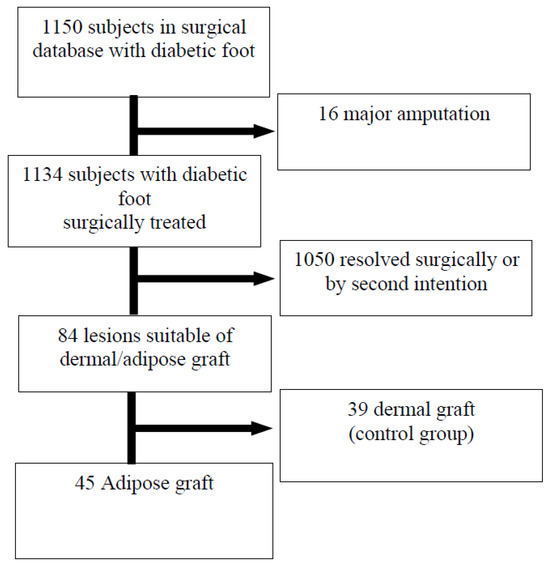
Enrollment. Out of 1150 individuals with DM surgically treated for diabetic foot, 84 presented conditions suitable for a dermal or adipose graft (Figure 1). Enrollment was based on a sequential clinical protocol. The control group (dermal graft) consists of patients treated primarily between January 2021 and December 2022, a period during which adipose grafting was not yet available at our facility. Starting from January 2023, following the implementation of the water-jet-assisted liposuction technique, adipose grafting became the primary regenerative approach for eligible patients. Importantly, the clinical team, debridement protocols, and post-operative off-loading standards remained unchanged throughout the entire study period (2021–2025) to ensure the highest possible comparability between the two cohorts.
Figure 1.
Flowchart.
In the intervention group, 45 patients (40 M, 5 F) with Diabetic Foot Ulcers (DFUs) were included; in the control group, we included 39 patients (33 M, 6 F) with Diabetic Foot Ulcers (DFUs), who underwent artificial dermal grafting.
Study description. All ulcers underwent surgical debridement (sharp debridement) to remove necrotic or non-viable tissue. In subjects with acute infection, the infectious condition was resolved through surgical debridement and antibiotic therapy until the infection was cleared. All patients underwent vascular assessment with Color-Doppler and TcPO2. In case of ischemia, revascularization was performed, confirming success by at least one patent artery at the ankle or a post-procedure TcPO2 > 30 mmHg.
Once adequate wound cleansing and resolution of the infectious process were achieved, we performed the adipose tissue graft.
Water-jet-Assisted Liposuction. The collection of autologous adipose tissue was performed using a water-jet-assisted liposuction system (Body-jet® eco, Human Med AG, Schwerin, Germany), strictly adhering to the manufacturer’s clinical guidelines [15]. The procedure began with a minimal percutaneous incision in the abdominal region to allow the insertion of a 3.8 mm Biofill harvesting cannula. To facilitate the gentle separation of adipocytes from the surrounding stromal matrix, a tumescent solution composed of saline and epinephrine was infiltrated. This was coupled with a synchronized suction mechanism maintained at a constant negative pressure of −375 mmHg. The harvested lipoaspirate was initially gathered in a dedicated sterile canister before being transferred into 50 mL syringes under aseptic conditions. Tissue refinement was limited to a natural decantation process to separate the viable fat from the fluid phase. To preserve the biological integrity of the stromal vascular fraction (SVF), the entire workflow—from initial harvesting to final preparation—was completed in less than 120 min. Finally, the processed adipose graft was delivered through precise infiltration both along the ulcer margins and directly onto the granular surface of the wound bed.
Artificial dermal graft. The preparation of the lesion is the same as described for the adipose graft. After surgical debridement (sharp debridement) and resolution of the infectious process, we performed the dermal graft [16]. The wound underwent wound bed reactivation, hemostasis control, and antiseptic irrigation. Subsequently, Hyalomatrix [17] was applied and fixed to the edges of the lesion with metal staples. Relaxation incisions were made in the dermis, and the site was dressed with sterile gauze.
Post-Operative Management. Topical treatment involved the application of povidone-iodine as an antiseptic. Initial post-operative protocols required the use of casts or non-weight-bearing footwear. To facilitate ongoing pressure offloading, clinicians utilized removable braces, casts, or specialized therapeutic shoes, tailored to the specific nature and site of the wound as well as the patient’s mobility. Once complete epithelialization was achieved, all participants were fitted with either prefabricated or custom-molded orthopedic shoes.
Definitions. Wound healing was defined as the complete epithelialization of the ulcer surface, without drainage or need for further dressings. Forefoot was defined as the anterior part of the foot area including the phalanges and metatarsal bones and associated soft tissue structures, while the hindfoot or rearfoot included the calcaneus and the talus and associated soft tissue structures.
Statistical Analysis. Statistical processing was conducted with the SPSS software suite (v. 12.0.2; SPSS Inc., Chicago, IL, USA). We presented continuous data as either mean ± standard deviation or median (interquartile range), determined by the distribution’s normality. To evaluate differences between the dermal and adipose graft cohorts, we employed Student’s t-test or the Mann–Whitney U test as appropriate; categorical data were analyzed via Fisher’s exact or Chi-square tests. Healing rates over time were mapped using Kaplan–Meier survival curves, with group disparities assessed through the log-rank test. Furthermore, a Cox proportional hazards regression was performed to isolate independent factors influencing healing time, accounting for baseline clinical variables. Findings are reported as Hazard Ratios (HR) with 95% Confidence Intervals (CI), and a p-value below 0.05 denoted statistical significance. Model discrimination was verified using the C-index.
Ethical Approval. This research adhered to the principles outlined in the 1964 Declaration of Helsinki and all subsequent revisions. Data collection was governed by a general consent framework, which patients acknowledged during their initial consultation. Through this agreement, participants granted permission for their clinical information to be utilized for epidemiological research, disease analysis, and educational purposes, all directed toward enhancing medical knowledge, therapeutic outcomes, and preventive strategies.
3. Results
Our study included 45 people with diabetes (40 M, 5 F) treated with an adipose graft. Of these, 41 had type 2 diabetes and 4 type 1; the mean age was 69.5 ± 10 years (mean ± SD), and five (11%) were female. The mean HbA1c was 7.6 ± 1.6%. Regarding lower limb complications, peripheral neuropathy was present in 43 people (95%), while 31 (69%) had peripheral artery disease, 28 of whom required pre-operative endovascular revascularization. End-stage renal disease was present in one participant, and coronary artery disease (CAD) in six (Table 1). The control group consisted of 39 individuals (33 males, six females) with diabetes treated with an artificial dermal graft. Age, duration of diabetes, glycemic control, prevalence of neuropathy and arteriopathy, and degree of renal impairment were similar to those in the intervention group. Only ischemic heart disease was significantly more prevalent in the control group (46% vs. 13%, p < 0.01).
Table 1.
Basal characteristics.
Wound characteristics. Two groups presented comparable lesions in terms of Texas classification: both grade and stage were superimposable. The presence of osteomyelitis was also similar between the groups (73% vs. 84%, p = ns). The group treated with adipose graft had significantly longer wound duration (7.6 ± 8 vs. 4.2 ± 6 months, p = 0.03), indicating a subgroup of lesions with greater chronicity and, consequently, increased healing difficulty. Regarding location, the adipose graft group showed a higher frequency of heel lesions (53% vs. 38%, p = 0.04), which are among the most challenging to heal.
Safety. No major complications were observed during the adipose graft procedure. Four minor complications (9%) occurred, consisting of hematomas at the adipose tissue harvest site that resolved spontaneously without requiring intervention.
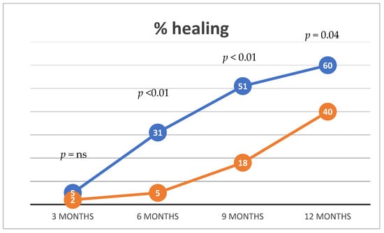
Outcomes. After a mean follow-up of 12 ± 5 months, complete wound healing was achieved in 30 subjects (67%) in the adipose graft group, compared with 51% in the artificial dermis group (p = 0.04). Healing time was significantly shorter in the adipose graft group: 202 ± 82 days vs. 275 ± 132 days (p = 0.02). Analysis of healing rates showed that, after the initial assessment at 3 months (5% vs. 2%, p = ns), the adipose graft provided significantly higher healing percentages compared with the dermal graft: at 6 months (31% vs. 5%, p < 0.01), at 9 months (51% vs. 18%, p < 0.01), and at 12 months (60% vs. 40%, p = 0.04) (Table 2, Figure 2).
Table 2.
Outcome.
Figure 2.
Healing time and rate. Blu line adipose group, orange line standard care.
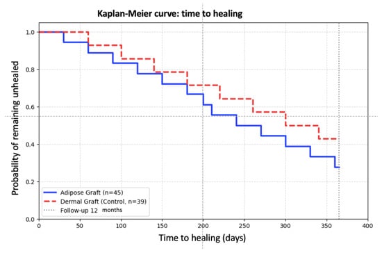
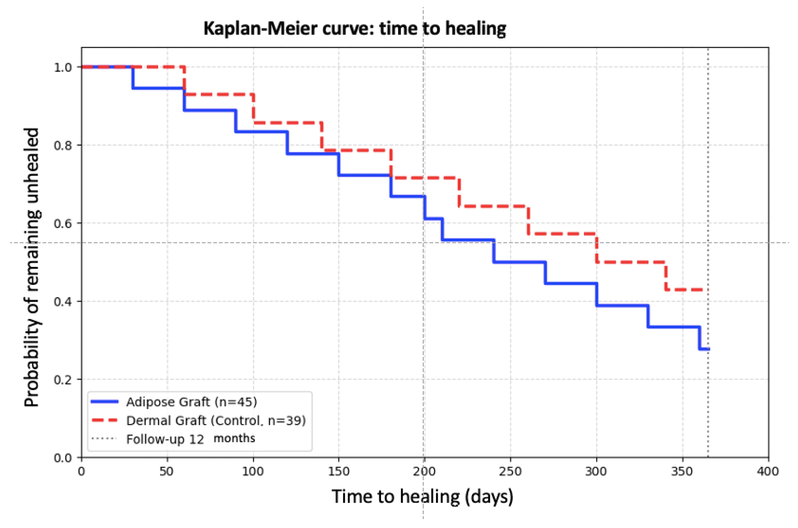
The Kaplan–Meier analysis reveals an early divergence between the two curves, which becomes clinically evident after the first 90–120 days (Figure 3). At 12 months (365 days), the estimated cumulative healing rate is ~65% for the Adipose group compared to ~50% for the control group, confirming the superior regenerative potential of autologous adipose tissue grafting. Comparison of the two curves using the Log-rank test (p = 0.02) indicates a statistically significant regenerative efficacy in favor of the adipose graft.
Figure 3.
Kaplan–Meier curves.
Wound location: subgroup analysis by wound location revealed similar outcomes for forefoot lesions between the adipose graft and control groups: healing rate 76% vs. 68% (p = ns), healing time (days) 206 ± 94 vs. 262 ± 147 (p = ns). In contrast, for hindfoot lesions, the adipose graft demonstrated significantly superior performance: healing rate 56% vs. 27% (p < 0.001), healing time (days) 197 ± 69 vs. 322 ± 79 (p < 0.01).
Glycemic control. We analyzed the impact of glycemic control on the healing rate and healing times: glycated hemoglobin levels did not affect the clinical outcome.
Limb salvage. Following the adipose graft procedure, 15 patients (33%) did not achieve healing. Three of these patients subsequently healed after a second intervention: two underwent Syme amputations, one a transmetatarsal amputation. Control group: One ray amputation, two underwent transmetatarsal amputations, one above the knee amputation. Moreover, to obtain healing of the stump we performed only one skin graft in the adipose group and nine in the control group.
Relapse. Both groups presented low relapse rate. At 12 ± 5 month follow-ups, one patient in the adipose group (2%) experienced relapse compared to 3 people (8%) in the control group (p = ns) demonstrating the efficacy of both approaches in healing stumps.
Mortality. Mortality at follow-up was similar between the two groups: Six deaths (13%) in the adipose graft group and seven deaths (18%) in the control group (p = ns).
Multivariate Cox analysis confirmed that adipose tissue graft treatment is independently associated with a higher probability of complete healing (HR 2.18; 95% CI 1.31–3.63; p = 0.003), after adjusting for ulcer duration (HR 0.92 per additional month; p = 0.004), hindfoot localization (HR 0.68; p = 0.102), and the presence of coronary heart disease (HR 0.79; p = 0.375). The model demonstrated good discriminative ability (C-index 0.69). These findings indicate that the beneficial effect of adipose grafting persists even in the most chronic and complex cases, although lesion duration remains an independent negative predictor.
Healing rates showed that the adipose graft provided significantly higher healing percentages compared with the dermal graft: at 6 months (31% vs. 5%, p < 0.01), at 9 months (51% vs. 18%, p < 0.01), and at 12 months (60% vs. 40%, p = 0.04).
The Kaplan–Meier curves illustrate the time-to-heal for both groups. The Adipose Graft group (blue line) achieved faster healing compared to the Dermal Graft control group (red dashed line). Marks on the curves indicate censored data at the 12-month follow-up.
4. Discussion
The results of this study demonstrate that adipose tissue grafting is effective in promoting wound healing and reducing the time to wound resolution in the treatment of diabetic foot ulcers, thereby decreasing the need for additional surgical interventions. These findings align with those reported in a recent systematic review and meta-analysis, where the group treated with autologous adipose tissue grafting exhibited a significantly higher rate of complete healing compared to the standard care group [7]. However, it should be emphasized that the existing literature primarily consists of preliminary studies with very small sample sizes, which further underscores the value of our study, as it presents the largest case series of patients treated to date [7]. Furthermore, the comparison with a control group with comparable characteristics, notwithstanding a higher prevalence of ischemic heart disease which did not appear to negatively affect the comparison, enhances the validity of our findings.
Moreover, these results were obtained from a cohort of complex, difficult-to-heal wounds that are commonly encountered in clinical practice but underrepresented in published studies: long-duration lesions, a high prevalence of peripheral vasculopathy, and heel involvement in over 50% of cases. The application of adipose grafting proved particularly decisive in the treatment of hindfoot lesions, which often represent a significant challenge for achieving healing, yielding markedly higher healing rates and shorter healing times compared to dermal grafting. This finding positions adipose tissue grafting as a valuable tool for managing complex hindfoot ulcers, where effective reconstructive alternatives are limited [18].
Limb salvage was one of the primary outcomes of this study, given that calcaneal ulcer location carries a high risk of major amputation [19]. The use of adipose grafting limited major amputations to only two patients (4%), a noteworthy result in terms of limb salvage potential, particularly considering that both major amputations were Syme procedures, involving only foot removal at the ankle level. In light of the lesion characteristics and the degree of local compromise, this outcome is encouraging and supports the consideration of adipose grafting as an effective therapy for limb salvage.
Another key finding from our study is the safety of the procedure, a critical requirement for the broader adoption of adipose grafting. No serious complications were observed, and minor complications were limited to self-resolving hematomas at the harvest site, without the need for any intervention, consistent with previous reports [20]. The impact of anesthesia was negligible, as the procedure requires only local anesthesia and does not necessitate general anesthesia. No pain issues were reported during or after the procedure.
Healing time represents a fundamental parameter in the management of diabetic foot ulcers. Studies on adipose grafting have reported reductions in wound size starting from the first weeks after application [21,22,23], although complete healing often requires longer periods. In our analysis, the mean healing time exceeded 6 months, which is consistent with the baseline severity of the lesions, characterized by significant tissue loss and bone exposure in the majority of cases (>90%). The peak in healing observed after 3 months post-treatment aligns with other studies [24] and can be attributed to the physiology of adipose grafting, where regenerative stimulation reaches its maximum expression between 2 and 3 months after application. In the literature, faster healing times have been reported only in less complex lesions, such as neuropathic, recent-onset, superficial ulcers without severe peripheral arteriopathy [11,12,25]. Comparison with dermal substitutes highlights the advantage of adipose grafting: The healing rate was statistically significantly higher already at 6 months post-procedure (31% vs. 5%), with this benefit maintained at 9 and 12 months. Furthermore, achieving healing with adipose grafting does not require additional procedures, such as skin grafts, resulting in benefits for patients and cost savings for healthcare systems.
Finally, thanks to the long-term follow-up, this study provides important insights into the quality of healing and long-term safety, demonstrating a low recurrence rate (only one patient) and mortality rates comparable to the control group. These data confirm the possibility of safely applying adipose grafting in diabetic patients, regardless of metabolic control.
A significant difference was observed in the prevalence of Coronary Artery Disease (CAD), which was higher in the control group (46% vs. 13%). While CAD is a recognized marker of systemic frailty, it did not influence mortality in our cohort. Furthermore, the multivariate Cox regression analysis demonstrated that CAD was not an independent predictor of healing (p = 0.45). This suggests that the superior outcomes observed in the intervention group are likely attributable to the regenerative properties of the adipose graft rather than the baseline cardiovascular profile of the patients.
Study Limitations: this study has some limitations that should be acknowledged. First, its retrospective nature and single center, the non-randomized allocation of patients may introduce selection bias, although clinical protocols remained consistent over the study period. Second, while the sample size is relevant for this specific intervention, larger prospective trials are needed to confirm these findings. Finally, although we adjusted for wound location and depth, unmeasured factors such as the initial baseline surface area of the lesion and patient compliance with off-loading may have influenced the outcomes.
5. Conclusions
Adipose tissue grafting is a safe, minimally invasive procedure that demonstrates superior efficacy compared to artificial dermal grafts in the treatment of chronic diabetic foot ulcers. Our findings reveal significantly higher healing rates (67% vs. 51%) and shorter healing times, with particularly pronounced benefits in hindfoot lesions—traditionally the most challenging to treat. Furthermore, multivariate analysis identified adipose grafting as a strong independent predictor of healing (HR 2.18). Consequently, this regenerative approach represents a new paradigm in limb salvage, proving effective even in chronic cases complicated by osteomyelitis.
Author Contributions
Conceptualization, R.D.R. and C.M.; methodology, C.M.; software, R.D.R.; validation, R.A., B.B. and E.B.; formal analysis, A.M.; investigation, R.A.; resources, R.D.R.; data curation, R.D.R.; writing—original draft preparation, R.D.R.; writing—review and editing, C.M., M.N. and G.P.; visualization, E.B.; supervision, R.A.; project administration, R.D.R. All authors have read and agreed to the published version of the manuscript.
Funding
This research received no external funding.
Institutional Review Board Statement
This study was conducted in accordance with the Helsinki Declaration of 1964 and its later amendments. According to the Italian Medicines Agency det. 20 March 2008 on retrospective observational studies on anonymous data, preemptive approval by an ethics committee was not mandatory and given that the study collected anonymous data, not referable to specific individuals, approval by one or more ethical committee(s) was not requested.
Informed Consent Statement
We used a general consent system that was signed by patients on first visit, in which they agreed to their clinical data being used for clinical research, epidemiology, disease study and training.
Data Availability Statement
The original contributions presented in the study are included in the article, further inquiries can be directed to the corresponding author.
Conflicts of Interest
The authors declare no conflicts of interest.
References
- Da Ros, R.; Assaloni, R. Burden of Infected Diabetic Foot Ulcers on Hospital Admissions and Costs in a Third-Level Center. Diabetology 2024, 5, 141–150. [Google Scholar] [CrossRef] [Scilit]
- OuYang, H.; Tang, Y. Platelet-rich plasma for the treatment of diabetic foot ulcer: A systematic review. Front. Endocrinol. 2023, 14, 1256081. [Google Scholar] [CrossRef] [Scilit]
- Gentile, P.; Sterodimas, A. Allogenic Use of Stromal Vascular Fraction and Decellularized Extracellular Matrices as Advanced Therapy Medicinal Products in Tissue Regeneration. Int. J. Mol. Sci. 2020, 21, 4982. [Google Scholar] [CrossRef] [Scilit] [PubMed]
- Conese, M.; Annacontini, L. The Role of Adipose-Derived Stem Cells, Dermal Regenerative Templates, and Platelet-Rich Plasma in Tissue Engineering-Based Treatments of Chronic Skin Wounds. Stem Cells Int. 2020, 9, 7056261. [Google Scholar] [CrossRef] [Scilit] [PubMed]
- Farabi, B.; Roster, K. The Efficacy of Stem Cells in Wound Healing: A Systematic Review. Int. J. Mol. Sci. 2024, 25, 3006. [Google Scholar] [CrossRef] [Scilit]
- Nagarajan, P.; Rajarathinam, M. From Fat to Healing: How Adipose-Derived Stem Cells Are Changing the Game for Diabetic Foot Ulcers-A Perspective. Health Sci. Rep. 2025, 8, e71385. [Google Scholar] [CrossRef] [Scilit]
- Elsharkawi, M.; Ghoneim, B. Role of Adipose Derived Stem Cells in Patients with Diabetic Foot Ulcers: Systematic Review and Meta-Analysis of Randomised Controlled Trials. Int. J. Low. Extrem. Wounds 2025, 24, 542–549. [Google Scholar] [CrossRef] [Scilit]
- Bura, A.; Planat-Benard, V. Phase I trial: The use of autologous cultured adipose-derived stroma/stem cells to treat patients with non-revascularizable critical limb ischemia. Cytotherapy 2014, 16, 245–257. [Google Scholar] [CrossRef] [Scilit] [PubMed]
- Carstens, M.H.; Gómez, A. Non-reconstructable peripheral vascular disease of the lower extremity in ten patients treated with adipose-derived stromal vascular fraction cells. Stem Cell Res. 2017, 18, 14–21. [Google Scholar] [CrossRef] [Scilit] [PubMed]
- Raposio, E.; Bertozzi, N. Adipose-derived stem cells for treatment of chronic cutaneous ulcers in patients with critical limb ischemia: A pilot study. Eur. J. Plast. Surg. 2018, 41, 459–464. [Google Scholar] [CrossRef] [Scilit]
- Han, S.K.; Kim, H.R. The treatment of diabetic foot ulcers with uncultured, processed lipoaspirate cells: A pilot study. Wound Repair. Regen. 2010, 18, 342–348. [Google Scholar] [CrossRef] [Scilit]
- Namgoong, S.; Yoon, I.J. A Pilot Study Comparing a Micronized Adipose Tissue Niche versus Standard Wound Care for Treatment of Neuropathic Diabetic Foot Ulcers. J. Clin. Med. 2022, 11, 5887. [Google Scholar] [CrossRef] [Scilit]
- Leone, N.; Andreoli, F. Clinical Outcomes and Characterisation of the Autologous Adipose Tissue Harvested With Superficial Enhanced Fluid Fat Injection Method for Treatment of Diabetic Foot Ulcer Undergoing Minor Amputation (SEFFIDiFA Trial). Int. Wound J. 2025, 22, e70784. [Google Scholar] [CrossRef] [Scilit]
- Tseng, S.L.; Lin, K. Adipose-derived stem cells in diabetic foot care: Bridging clinical trials and practical application. World J. Diabetes 2024, 15, 1162–1177. [Google Scholar] [CrossRef] [Scilit]
- Meyer, J.; Salamon, A. Isolation and differentiation potential of human mesenchymal stem cells from adipose tissue harvested by water jet-assisted liposuction. Aesthet. Surg. J. 2015, 35, 1030–1039. [Google Scholar] [CrossRef] [Scilit]
- Sidoti, G.B.; Iurilli, M. New frontiers of dermal repair and regeneration for a modern approach in wound healing. Ital. J. Wound Care 2025, 9, 12–20. [Google Scholar] [CrossRef] [Scilit]
- Caravaggi, C.; De Giglio, R. HYAFF 11-based autologous dermal and epidermal grafts in the treatment of noninfected diabetic plantar and dorsal foot ulcers: A prospective, multicenter, controlled, randomized clinical trial. Diabetes Care 2003, 26, 2853–2859. [Google Scholar] [CrossRef] [Scilit]
- Álvaro-Afonso, F.J.; García-Álvarez, Y. Advances in Dermoepidermal Skin Substitutes for Diabetic Foot Ulcers. Curr. Vasc. Pharmacol. 2020, 18, 182–192. [Google Scholar] [CrossRef] [Scilit] [PubMed]
- Dos Santos, V.P.; da Silveira, D.R. Risk factors for primary major amputation in diabetic patients. Sao Paulo Med. J. 2006, 124, 66–70. [Google Scholar] [CrossRef] [Scilit] [PubMed]
- Tanios, E.; Ahmed, T. Efficacy of Adipose-Derived Stromal Vascular Fraction cells in the Management of Chronic Ulcers: A Randomized Clinical Trial. Regen. Med. 2021, 16, 975–988. [Google Scholar] [CrossRef] [Scilit]
- Thamm, O.C.; Eschborn, J. Sublesional fat grafting leads to a temporary improvement of wound healing in chronic leg ulcers: A prospective, randomised clinical trial. Wound Repair Regen. 2023, 31, 663–670. [Google Scholar] [CrossRef] [Scilit]
- Moon, K.C.; Suh, H.S. Potential of Allogeneic Adipose-Derived Stem Cell-Hydrogel Complex for Treating Diabetic Foot Ulcers. Diabetes 2019, 68, 837–846. [Google Scholar] [CrossRef] [Scilit] [PubMed]
- Smith, O.J.; Leigh, R. Fat grafting and platelet-rich plasma for the treatment of diabetic foot ulcers: A feasibility-randomised controlled trial. Int. Wound J. 2020, 17, 1578–1594. [Google Scholar] [CrossRef] [Scilit] [PubMed]
- Lonardi, R.; Leone, N. Autologous micro-fragmented adipose tissue for the treatment of diabetic foot minor amputations: A randomized controlled single-center clinical trial (MiFrAADiF). Stem Cell Res. Ther. 2019, 10, 223. [Google Scholar] [CrossRef] [Scilit] [PubMed]
- Yastı, A.Ç.; Akgun, A.E. Graft of 3D bioprinted autologous minimally manipulated homologous adipose tissue for the treatment of diabetic foot ulcer. Wounds 2023, 35, E22–E28. [Google Scholar] [CrossRef] [Scilit]

Disclaimer/Publisher’s Note: The statements, opinions and data contained in all publications are solely those of the individual author(s) and contributor(s) and not of MDPI and/or the editor(s). MDPI and/or the editor(s) disclaim responsibility for any injury to people or property resulting from any ideas, methods, instructions or products referred to in the content. |
© 2026 by the authors. Licensee MDPI, Basel, Switzerland. This article is an open access article distributed under the terms and conditions of the Creative Commons Attribution (CC BY) license.