


Abstract
Atherosclerosis, manifesting as acute myocardial infarction, stroke and peripheral artery disease, is the main cause of death worldwide. Conventional risk factors contributing to the development and progression of atherosclerosis are well established, including diabetes mellitus, hypertension, hypercholesterolemia, smoking and obesity. In recent decades, other lipid molecules have been identified as risk factors for atherosclerosis and arterial calcification, including lipoprotein (a) and apolipoproteins. Despite the available evidence for the association between those biomarkers and atherosclerosis in the general population, their impact on diabetic patients is incompletely characterized. This review aims to summarize the current evidence on the relationship between lipoprotein (a), apolipoproteins and atherosclerotic cardiovascular disease in diabetic patients. By integrating genetic, epidemiological, and mechanistic data, this review highlights the dual and context-dependent associations of lipoprotein (a) with incident type 2 diabetes and atherosclerotic cardiovascular risk, supporting more nuanced interpretation of Lp(a) in diabetes-related risk assessment.
1. Introduction
Atherosclerosis, manifesting as acute myocardial infarction (AMI), stroke and peripheral artery disease (PAD), remains the leading global cause of morbidity and mortality [1,2]. Among conventional risk factors for atherosclerosis, hypertension, diabetes mellitus (DM), hypercholesterolemia, smoking, obesity, and nonmodifiable factors (age and family history) were shown to be the most important. The impact of these risk factors proved to be related to sex, with age having the highest impact in men compared to hypertension in menopausal women [3]. Atherosclerotic cardiovascular disease (ASCVD) is strongly associated with components of metabolic syndrome, defined as abdominal obesity, insulin resistance, impaired glucose metabolism, hypertension, and dyslipidemia [4]. Although LDL-C may appear “optimal” in some patients with metabolic syndrome/diabetes, discordance with apoB and non-HDL-C is common; thus, apoB and non-HDL-C are practical and complementary metrics for estimating atherogenic particle burden and residual ASCVD risk [5]. Likewise, in patients with diabetes, several factors contribute to the CV risk, including hyperglycemia, insulin resistance, dyslipidemia, low-grade inflammation, obesity and hypertension [6]. Accordingly, diabetes is treated as a high-risk ASCVD state, with lipid lowering targets and intensity differing by risk category (primary vs. secondary prevention); in this setting, Lp(a) testing may further refine residual risk beyond standard lipid measures. In recent decades, type 2 DM has been confirmed as a global epidemic with an increased prevalence, even in young populations [7,8]. It has been shown, in several epidemiological studies, to be closely related to CVDs, including coronary artery disease, heart failure and stroke, with their associated high mortalities [1]. Even with good glycemic control and LDL-C lowering, patients with diabetes remain at high risk for ASCVD. This residual risk is driven by factors beyond LDL-C, including atherogenic particle burden (apoB-containing lipoproteins), inflammation/thrombosis, and genetically determined Lp(a). Accordingly, apoB is increasingly used as an integrated marker of atherogenic particles, and elevated Lp(a) is used as a factor that can refine risk assessment in DM beyond traditional lipid profiling [9,10]. The impact of DM on atherosclerosis pathophysiology is through the harm created by the advanced glycation end-products, increased oxidative stress, activation of protein kinase C (PKC), circulating chronic inflammation, non-coding RNAs, and epigenetic modification [2]. In T2 DM, 40% to 63% of β-cell mass is reduced, depending on disease duration [3]. Such cell death is associated with glucotoxicity, lipotoxicity, glucolipotoxicity and apoptosis [4]. Many observational studies have shown that individuals with DM have elevated markers of inflammation, including C-reactive protein (CRP), interleukin-6 (IL-6), and plasminogen activator inhibitor 1 (PAI-1) [5]. In DM, non-enzymatic glycation and oxidative stress can modify lipoproteins and impair HDL function. In particular, diabetes-related modification of apoA-I may reduce HDL’s anti-inflammatory and cholesterol efflux capacity, contributing to endothelial dysfunction and accelerated atherosclerosis [6]. These qualitative changes in lipoproteins provide mechanistic context for why apolipoproteins and Lp(a) may add clinically relevant information beyond traditional lipid measures in diabetic patients [7]. This review aims to summarize current evidence on the relationship between lipoprotein (a), apolipoproteins and ASCVD in diabetic patients.
2. Material and Methods
We conducted a systematic literature search following key elements of the systematic review methodology. This review followed key elements of the PRISMA (Preferred Reporting Items for Systematic Reviews and Meta-Analyses) framework to guide literature identification, selection, and reporting [11]. Two independent reviewers (A.L and V.B.M.) developed and performed the literature search strategy using predefined Medical Subject Headings (MeSHs). We searched PubMed, Google Scholar, Web of Science, Scopus, and the Cochrane Library up to 15 March 2025. The PubMed/MEDLINE search strategy was: ((lipoprotein (a)[Title/Abstract] OR Lp(a)[Title/Abstract] OR “Lipoprotein (a)”[MeSH Terms]) OR (apolipoprotein[Title/Abstract] OR ApoB[Title/Abstract] OR ApoA1[Title/Abstract] OR “Apolipoproteins”[MeSH Terms])) AND (diabetes[Title/Abstract] OR “diabetes mellitus”[MeSH Terms] OR T2D[Title/Abstract] OR “type 2 diabetes”[Title/Abstract] OR T1D[Title/Abstract] OR “type 1 diabetes”[Title/Abstract]) AND (atherosclerosis[Title/Abstract] OR ASCVD[Title/Abstract] OR “cardiovascular diseases”[MeSH Terms] OR coronary[Title/Abstract] OR stroke[Title/Abstract] OR “peripheral artery disease”[Title/Abstract]). Studies were included if they met the following criteria: (1) original research articles, (2) human studies involving adults, (3) assessing lipoprotein (a) and/or apolipoproteins (ApoA-I, ApoB, ApoE), (4) evaluating associations with diabetes mellitus and/or ASCVD outcomes, (5) observational studies, randomized controlled trials, or Mendelian randomization studies, (6) published in peer-reviewed journals, and (7) published in English. We excluded articles published before 2010 and articles with inconclusive findings. A 2010 cut-off was used to prioritize contemporary evidence aligned with modern definitions, Lp(a) reporting/assays, and current prevention practice; earlier landmark studies were used for background only.
Data Extraction. Data from eligible studies were extracted using a standardized extraction form. Key factors used were: author(s) and year of publication, study design and sample size, main findings and outcomes of interest, limitations and biases noted by the author
Quality Assessment. Study quality was assessed using design-appropriate tools: randomized trials were appraised using the Cochrane Risk of Bias tool, and observational studies were evaluated based on key methodological domains (selection, confounding control, outcome assessment, and reporting). Quality judgments were used to inform interpretation of the evidence.
3. Results and Discussion
The selection process and eligibility criteria were structured based on the PRISMA 2020 guidelines [11]. A total of 1582 articles were initially identified. After screening for duplicates, and after applying the predefined inclusion and exclusion criteria, 55 full-text articles were assessed for eligibility; 7 studies met the inclusion criteria and were included in the final analysis (Table 1, Figure S1 Supplement).
Risk of Bias Assessment
The risk of bias assessment of the seven included studies is summarized in Table S1 (Supplementary Materials). Overall, most studies were judged to have a low risk of bias in the outcome measurement and reporting domains. Moderate risk was observed primarily in the randomization process for observational and post hoc analyses, reflecting inherent study design limitations. High risk ratings were mainly related to non-randomized designs rather than methodological flaws in outcome assessment. Overall, the quality of the included evidence was considered acceptable for narrative synthesis.
3.1. Lipoprotein (a): Structure and Function
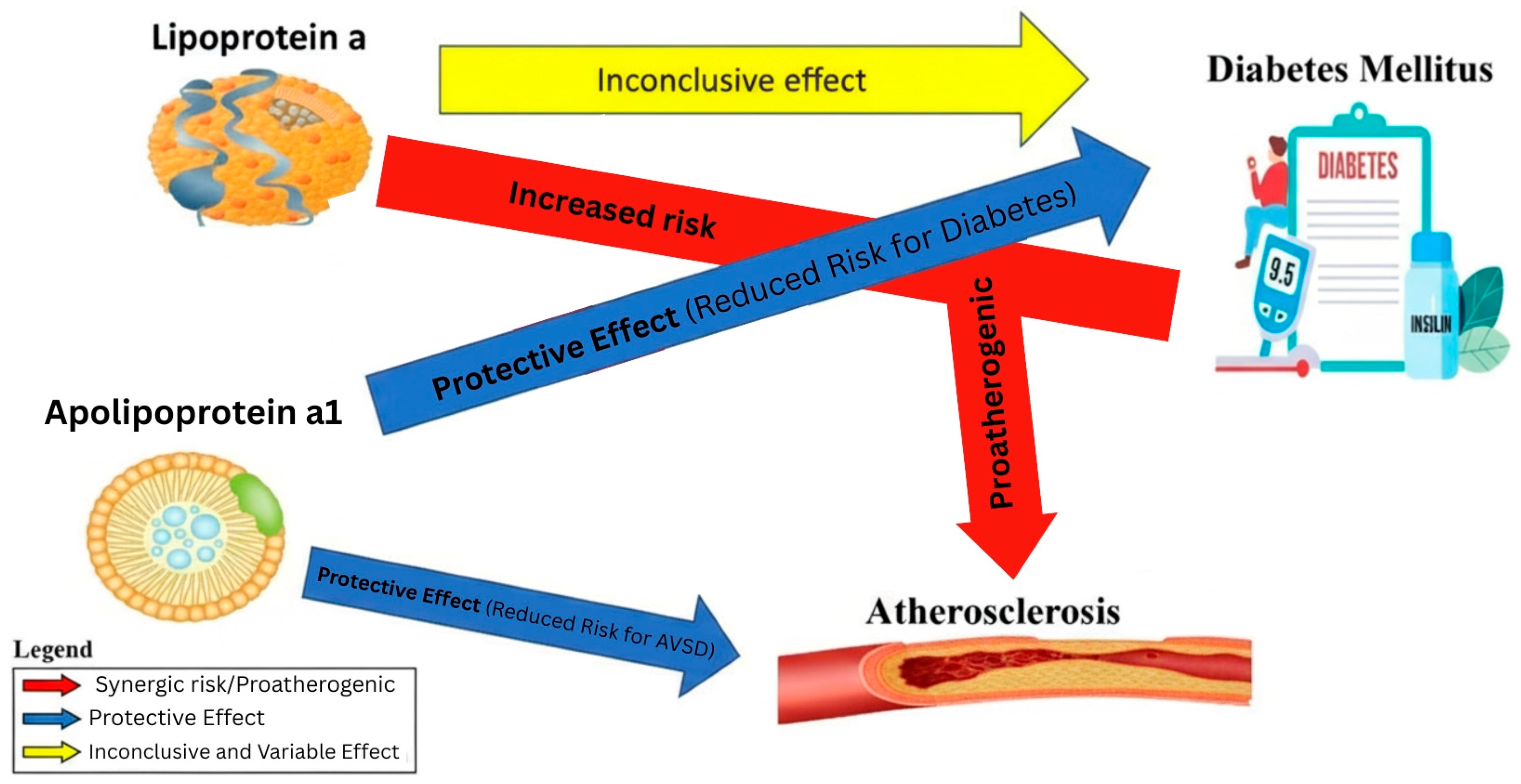
Lp(a) levels are largely genetically determined and are only minimally influenced by lifestyle, which is relevant when counseling patients with diabetes [10]. Importantly, in contrast to its well-established association with ASCVD, several epidemiological and genetic studies have reported a paradoxical association whereby very low Lp(a) concentrations are linked to a higher risk of incident type 2 diabetes, which complicates clinical interpretation of Lp(a) in diabetic populations [12,13]. The apolipoprotein (a) component is thought to be central to Lp(a)’s harmful effects, particularly due to its similarity to plasminogen, which may interfere with the body’s natural ability to dissolve blood clots [14]. Lp(a) consists of a lipid-rich core containing cholesterol esters and triglycerides, surrounded by a surface layer that includes phospholipids, free cholesterol, and other neutral lipids. Levels below 30 mg/dL are generally considered normal, but the risk of CV complications increases substantially once concentrations exceed 50–75 mg/dL. It is estimated that one in five people globally have Lp(a) levels above this threshold. Both the European Atherosclerosis Society and American guidelines consider levels ≥50 mg/dL (or ≥125 nmol/L) to confer increased risk for atherosclerotic CVD [15]. Because the relationship between mass (mg/dL) and particle concentration (nmol/L) depends on apo(a) isoform size, conversion between units is not fixed; therefore, reporting Lp(a) in nmol/L is preferred when available. Most recently, it has been shown that while both low Lp(a) values (<75 nmol/L) and high values (>189 nmol/L) remained stable over time, a significant number of individuals with Lp(a) ranging between 75 and 189 nmol/L experienced clinically significant fluctuations in related risk [16]. Nevertheless, Lp(a) is generally considered stable and is usually measured once, with repeat testing mainly occurring due to major inflammatory or hepatic/renal changes [10]. Lp(a) particles are more atherogenic than other apo B-containing lipoproteins due to their ability to carry up to 90% of all oxidized phospholipids (OxPL) and also due to their long circulation half-lives [17] (Figure 1).
Figure 1.
Schematic representation of the effects of lipoprotein (a) and apolipoprotein on atherosclerosis and diabetes.
Geographically, the distribution of Lp(a) levels is not uniform across populations. African descents typically exhibit the highest levels, while those from East and Southeast Asia have the lowest levels. In diabetes, these ancestry-related differences suggest that uniform Lp(a) thresholds may not confer equivalent ASCVD risk across populations, highlighting the need for ancestry-aware interpretation. Populations in Europe, South Asia, and Latin America tend to fall somewhere in between. Moreover, studies have shown that postmenopausal women may have up to 17% higher Lp(a) levels than men, likely due to hormonal shifts influencing lipid metabolism [18].
Beyond the structural and genetic aspects, Lp(a) plays a distinct role in what clinicians call residual CV risk—the risk that persists even after LDL cholesterol is effectively lowered. This is especially relevant in patients with T2 DM, where a mix of metabolic dysfunction, chronic inflammation, and endothelial damage amplifies CV risk. Lp(a), along with triglyceride-rich lipoproteins and other remnant particles, contributes to this residual burden [19]. Clinically, this residual risk persists despite standard-of-care lipid-lowering therapy (statins with or without ezetimibe, and PCSK9 inhibitors in selected patients) and has led to the recognition of additional risk enhancers, such as elevated Lp(a) and apoB-containing lipoproteins to guide more intensive or individualized prevention strategies in T2DM [20]. Lp(a) has atherogenic potential due to its proatherogenic apo(a) component, proinflammatory oxidized phospholipid content, and prothrombotic effects, owing to its ability to inhibit plasminogen. The phospholipids in Lp(a) undergo oxidation and are transported to catabolic sites, where they can cause damage. In vitro studies have shown effects, including endothelial disruption, increased platelet adhesion and smooth muscle cell proliferation [21]. In patients with diabetes, high Lp(a) levels have been linked more consistently to macrovascular complications, while associations with microvascular endpoints (retinopathy, nephropathy, neuropathy) are less consistent. Curiously, some studies have suggested that very low Lp(a) levels might be associated with a higher risk of developing T2 DM, although the mechanisms behind this inverse relationship remain unclear [21,22]. Lp(a) has also been shown to be associated with aortic valve calcification and the development and progression of aortic stenosis [23].
3.2. Apolipoproteins: Structure and Function
Apolipoproteins are essential proteins embedded in lipoprotein particles, serving not only as structural scaffolds but also as regulators of lipid metabolism. They influence how lipoproteins interact with receptors and enzymes, and several of them play particularly important roles in the development of atherosclerosis. These include apoB-100, apoB-48, apoA-I, apoC-II, apoC-III, apoE, and apo(a). Apolipoprotein B-100 is the main structural component of very-low-density lipoproteins (VLDLs), intermediate-density lipoproteins (IDLs), low-density lipoprotein cholesterol (LDL) and Lp(a). Apolipoprotein B-48 is an isoform of apolipoprotein B-100 which is an integral part of chylomicrons. Apolipoprotein A-I is a structural part of the HDL (high-density lipoprotein) particle and has an important role in reverse cholesterol transport. Apolipoprotein C-II, apolipoprotein C-III and apolipoprotein E are involved in the metabolism of triglyceride-rich lipoproteins. Finally, apolipoprotein (a) binds covalently to apolipoprotein B-100 to form Lp(a) [21]. Apolipoproteins are lipid transport mediators, and they also provide structural integrity and protection of the lipid core in the lipoprotein complex. Chylomicrons are one of the types of lipoproteins that have the function of transporting dietary lipids from the digestive tract to the liver via circulation. The liver absorbs the lipids and combines them with apo B-100 to form VLDLs, which are enriched with triglycerides. Lipoprotein lipase is an enzyme that breaks down VLDLs into intermediate-density IDLs, and finally, when the VLDL molecule loses triglycerides, an LDL molecule with a higher cholesterol content is formed. Apo B is retained in the particle and is recognized by receptors found on the surface of many cells in the body, which stimulate cholesterol uptake. LDL and Apo B cholesterol transporters are vital for cell membrane physiology and steroid hormone production. Excess LDL, on the other hand, can cause fatty deposits (plaques) on arterial walls, as well as hardening and scarring of blood vessels, resulting in atherosclerosis [22]. Apo A-I is the primary apolipoprotein in HDL. It plays a key role in maintaining HDL structure and in facilitating the removal of excess cellular cholesterol via the ATP-binding cassette transporter-1 (ABCA1) [24]. In diabetes, although HDL-C itself is not a therapeutic target, qualitative dysfunction of apo A-I may help explain impaired HDL functionality and residual ASCVD risk, despite apparently normal HDL-C levels. A naturally occurring variant of apo A-I is Apo A-I Milano, which contains very low levels of HDL-C and high triglyceride levels but is associated with a very low incidence of CVD [25]. From a diabetes prevention and ASCVD risk assessment perspective, apo B and the apo B/apo A-I ratio have particular clinical relevance, because they reflect the total burden of atherogenic particles and may better capture residual risk when LDL-C alone appears to be “controlled” [26,27].
3.3. Lipoprotein (a) as Atherogenic Risk Factor
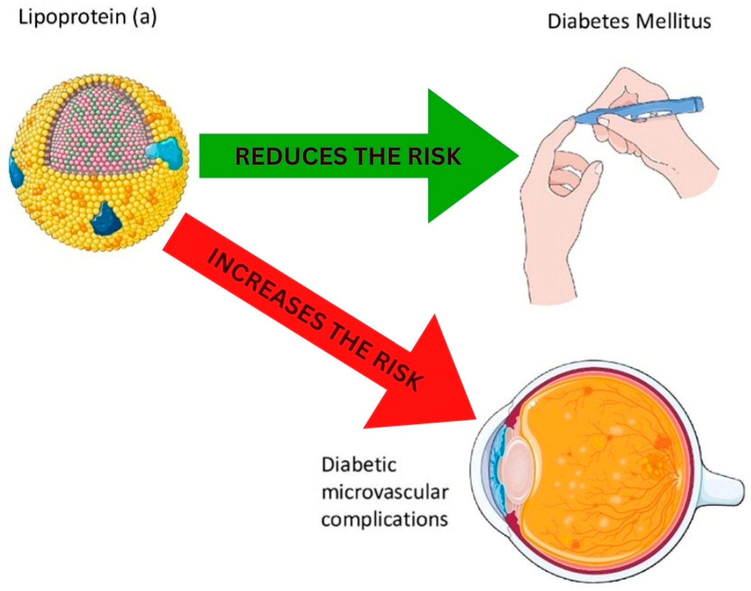
Hyperlipidemia is characterized by elevated plasma levels of LDL cholesterol (LDL-C), VLDL cholesterol (VLDL-C) and triglycerides, often accompanied by HDL cholesterol (HDL -C). In diabetes, the characteristic “diabetic dyslipidemia” pattern includes hypertriglyceridemia, low HDL-C, increased LDL-C, and frequent discordance between LDL-C and apo B/non–HDL-C [28] (Figure 2). Plasma lipoproteins carry certain amounts of lipid oxidation products (LOPs), which can negatively affect normal physiological functions and promote atherosclerotic processes. Recent studies suggest that plasma lipoproteins actively transport LOPs, with LDLs directing them to peripheral tissues and high-density lipoproteins (HDLs) facilitating reverse transport. It has been proposed that the transport of LOPs by specific lipoproteins may contribute to the atherosclerosis-related effects of both LDL and HDL [29]. Studies evaluating Lp(a) in relation to diabetes have reported associations that are independent of BMI, HbA1c, and triglycerides; however, these analyses primarily address the presence/incident diabetes rather than ASCVD outcomes. A study involving 477 patients with T2 DM (including men and women of premenopausal and postmenopausal age) and 105 control participants found that individuals with DM had significantly higher serum Lp(a) levels compared to controls. Also, serum Lp(a) levels in DM patients were significantly higher in women compared to men, in contrast to Lp(a) levels in the control group, which did not differ between males and females. Furthermore, there was no significant difference in Lp(a) levels between premenopausal and postmenopausal women with diabetes [30]. The general population in the Heart Study in the City of Copenhagen and the Copenhagen General Population Study, which included plasma Lp(a) concentrations and took into account the isoforms, showed that it is less likely that Lp(a) levels are causally related to T2 DM, but a causal relationship with the size of the large Lp(a) isoform cannot be excluded [24]. Clinically, these data suggest that Lp(a) is not clearly causal for T2DM development, but it is causal for ASCVD; therefore, in patients with diabetes, Lp(a) primarily helps refine ASCVD risk on top of an already high baseline risk. An inverse association between Lp(a) concentration and incident T2DM has been reported. However, low Lp(a) should not be considered protective for ASCVD, since elevated Lp(a) is still associated with increased ASCVD risk. According to this, an increase in incidence rates of DM at an Lp(a) concentration cut-off of 1 mg/dL compared to higher cut-offs suggests that the relative deficiency of Lp(a) may be involved in the risk [30]. The Bruneck Study, a prospective population-based survey on the epidemiology and pathogenesis of atherosclerosis, supports the hypothesis that Lp(a) concentration is inversely related to the development of new-onset type 2 DM. The study found that an increased risk of diabetes was associated with individuals whose Lp(a) concentrations fell within the two lowest quintiles (mean Lp(a) levels < 7 mg/dL) [31]. According to Gudbjartsson DF et al., the molar concentration of Lp(a) fully accounted for its association with CAD, with no residual connection to apo(a) size. Homozygous carriers of loss-of-function mutations had little or no Lp(a) and showed an increased risk of type 2 DM. The study concluded that the molar concentration of Lp(a) is the key factor influencing CVD risk, and that low Lp(a) concentrations (in the bottom 10%) are associated with an elevated risk of T2 DM [12]. Lowering elevated Lp(a) toward the population median is a plausible strategy to reduce CAD risk, but both CV benefit and any impact on diabetes risk require confirmation in outcome trials [32]. Available data suggest no clear association between Lp(a) and incident microvascular complications in some T2DM cohorts [33]. However, evidence remains limited and inconsistent, and further studies are needed before excluding a potential role of Lp(a) in microvascular endpoints. Compensatory hyperinsulinemia in early insulin resistance, followed by relative insulin deficiency with β-cell failure, has been hypothesized to influence Lp(a) levels; however, this remains a mechanistic hypothesis rather than a proven causal pathway [34]. A study offers supporting evidence of the association between genetically predicted increased insulin concentrations and decreased Lp(a) levels, suggesting that hyperinsulinemia, commonly seen in T2 DM, may partly explain the inverse relationship between low Lp(a) concentrations and an elevated risk of T2 DM [35]. In contrast, another study found no evidence of a causality effect of insulin in the Lp(a)-T2 DM association [36]. In population-based prospective cohorts, Lp(a) levels decline in the years preceding incident T2DM and show an inverse association with the homeostatic model assessment (HOMA) index [37]. In T1DM patients, plasma Lp(a) levels have been reported to correlate with markers of glycemic control and early microvascular involvement. In children with T1 DM, plasma Lp(a) levels have been associated with HbA1c and showed a positive trend with albuminuria. A cross-sectional study revealed a significant association between Lp(a) levels and vascular complications in patients with T1 DM. However, no significant difference in plasma Lp(a) levels was found between men and women [38].
Figure 2.
Lipoprotein (a) and diabetes mellitus.
3.4. Apo A1 as Protective Factor
ApoA-I and HDL exhibit antiatherogenic potential by inhibiting inflammation in endothelial cells and macrophages. However, the anti-inflammatory properties of apoA-I and HDL are lost in patients with T2 DM [39]. In diabetes, due to non-enzymatic glycation of apoA-I by reactive α-oxoaldehydes such as methylglyoxal and 3-deoxyglucosone, this function is lost, and its circulating half-life is also reduced. Activation of the enzyme myeloperoxidase also reduces the ability of apoA-I to produce NO (nitric oxide), which promotes vasorelaxation. A low apoA-I level was associated with insulin resistance in people with impaired glucose tolerance [39,40].
In one trial, a single infusion of synthetic reconstituted discoid HDLs composed of apoA-I complexed with phospholipid molecules transiently increased HDL, apoA-I, and plasma insulin levels and decreased blood glucose levels in patients with T2D. Although cholesterol ester transfer protein (CETP) inhibition increases HDL-C and apoA-I levels, large ASCVD outcome trials have shown mixed or neutral effects on cardiovascular events; therefore, these biomarker changes should not be interpreted as having proven clinical benefits [12]. Experimental data suggest that Lp(a)-related pathways may influence pancreatic β-cell function, potentially involving pancreatic duodenal homeobox 1 (Pdx1)-dependent mechanisms. However, these observations are preliminary and should be regarded as hypothesis-generating rather than established clinical pathways. One study examined the association between apolipoprotein A1 (ApoA1), high-density lipoprotein cholesterol (HDL-C), and their ratio with inflammatory markers in Chinese adults diagnosed with coronary artery disease (CAD). The authors analyzed clinical and laboratory data to determine whether these lipid-related markers were linked to systemic inflammation. They found that lower levels of ApoA1 and HDL-C, as well as alterations in their ratio, were significantly associated with higher inflammatory marker levels. The findings suggest that ApoA1, HDL-C, and their ratio may serve as useful indicators of inflammatory status and cardiovascular risk in patients with CAD [40].
The study by Raimon Ferré et al. demonstrates that higher levels of HDL cholesterol (HDL-C) and apolipoprotein A-I (apoA-I) are strongly associated with improved vasoreactivity in small peripheral arteries. The authors show that individuals with lower HDL-C and apoA-I exhibit impaired endothelium-dependent vasodilation, suggesting a direct functional link between HDL particles and microvascular health. These findings support the concept that HDL and apoA-I contribute to cardiovascular protection not only through lipid transport but also via modulation of endothelial function and vascular reactivity [41].
3.5. Therapeutic Intervention in Lp(a)
Although Lp(a) contributes to the development of CV disease in patients with DM, few trials have investigated the benefits of reducing Lp(a) within this patient population. Guidelines generally treat elevated Lp(a) as a risk-enhancing factor, although diabetes-specific integration into risk algorithms and treatment thresholds remains limited [42].
Protein convertase subtilisin/kexin type 9 (PCSK9) inhibitors are a group of drugs that reduce the concentration of LDL cholesterol (LDL-C) significantly (by 50–60%), and they also modestly reduce the concentration of Lp(a) by 20–25%. The reduction in ASCVD events observed in outcome trials is driven predominantly by the magnitude of LDL-C lowering, with Lp(a) lowering likely contributing only a minor additional effect. A study used alirocumab (PCSK9 inhibitor) and showed that at low baseline Lp(a) concentrations, alirocumab had a minimal effect on Lp(a) levels and showed a trend toward reducing the estimated risk of incident T2 DM compared to placebo. On the other hand, at high baseline Lp(a) levels, alirocumab produced significant reductions in Lp(a) concentrations and showed a trend toward increasing the estimated risk of incident T2 DM compared with placebo. Treatment and baseline Lp(a) had a significant interactive effect on the risk of incident T2 DM. The Lp(a) concentration at which alirocumab had a neutral effect on incident T2 DM was ∼50 mg/dL [43,44]. However, these observations should be interpreted cautiously and considered to be hypothesis-generating rather than definitive evidence of a clinically meaningful effect on diabetes risk. Currently, apo(a)-targeting therapies are in the clinical testing phase, including antisense oligonucleotides and small interfering RNA (siRNA) approaches, and result in a reduction in Lp(a) that can be substantial, with some agents achieving near-complete lowering in early-phase trials [45].
3.6. Emerging Biomarkers and Future Directions
Beyond Lp(a) and classical apolipoproteins, several emerging biomarkers related to inflammation and lipoprotein remodeling have been explored in diabetes and ASCVD; these are discussed here as future research directions rather than established clinical tools.
3.6.1. Phospholipid Transfer Protein as an Emerging Biomarker
Although not a primary focus of this review, phospholipid transfer protein (PLTP) is discussed here as an emerging marker linking apo-containing remodeling, diabetes-related dyslipidemia, and residual ASCVD risk. Phospholipid transfer protein (PLTP) is part of the lipid transfer protein family, which also includes CETP, lipopolysaccharide-binding protein, and bactericidal/permeability protein (BPI). In the past decade, most human studies have demonstrated a positive association between plasma PLTP activity and atherosclerosis [46]. The role of PLTP may be linked to hyperlipidemia, hypercoagulation, obesity, insulin resistance, and type 2 diabetes, although its effect on inflammation remains inconclusive [47].
3.6.2. Lipoprotein-Associated Phospholipase A2 (Lp-PLA2)
Lipoprotein-associated phospholipase A2 (Lp-PLA2) is an inflammatory enzyme/biomarker that is associated with increased ASCVD risk. Additionally, its levels are consistently higher in individuals with T2 DM compared to those without T2 DM [48]. Lipoprotein-associated phospholipase A2 (Lp-PLA2) is a proinflammatory enzyme and serves as a marker of inflammation in the blood vessel wall and thus a risk factor and causative factor for atherosclerosis [49]. LP-PLA2 is associated with DM through various mechanisms, including heightened inflammatory activity resulting from the hydrolysis of oxidized phospholipids, which can promote insulin resistance and increase the risk of T2 DM. Alternatively, elevated Lp-PLA2 levels may serve as a marker of increased adipose tissue inflammation, altered metabolic processes such as increased fatty acid flux, and/or disruptions in adipokines that contribute to the development of insulin resistance and pancreatic β-cell dysfunction. Cross-sectional studies have shown that both Lp-PLA2 activity and/or mass are higher in individuals with diabetes compared to those without diabetes [49,50]. However, Lp-PLA2 is not routinely recommended or used in current diabetes lipid risk management as a standard clinical test, and its role is best viewed as an adjunct inflammatory risk marker rather than a treatment target.
3.6.3. Growth-Differentiating Factor 15 and Circulating Monocyte
As part of future research directions, growth-differentiating factor 15 (GDF-15) is an emerging inflammatory biomarker, and recent studies suggest that its levels correlate with pancreatic β-cell apoptosis, vascular endothelial cell and adipocyte inflammatory damage, and oxidative stress. Circulating GDF-15 levels and the serum ApoB/ApoA1 ratio in T2 DM patients with CAD were higher than those with T2 DM alone. Serum GDF-15 levels and the ApoB/ApoA1 ratio may therefore be helpful in T2 DM patients for predicting CAD and preventing adverse CV events [50,51]. Circulating monocytes are a cluster of blood cells that are modulated by immune factors, including tumor necrosis factor alpha (TNF-α) and Toll-like receptor (TLR) 2, TLR4, and TLR8 ligands that can interact with thrombocytes and endothelial cells, resulting in exaggerated inflammation and increased oxidative stress, and they are involved in systematic inflammation diseases like T2 DM and CV disease. The monocyte-to-HDL-c ratio (MHR) was also identified as a sign of oxidative stress and systemic inflammation, a predictive marker for CV disease. MHR and MAR were negatively associated with HDL-c and ApoA1 (p < 0.05). In addition, a positive correlation between MHR and MAR (monocyte-to-ApoA1 ratio) was observed [51,52].
Table 1.
Design characteristics and messages of included studies.
4. Conclusions
This review provides a comprehensive overview of Lp(a), as well as its structure, function, and role in CVD, particularly in patients with T2 DM. Lp(a) is a genetically determined lipoprotein that shares similarities with LDL cholesterol but includes apolipoprotein (a), thus making it more atherogenic. High levels of Lp(a) are associated with an increased risk of atherosclerotic CVD, and concentrations above 50 mg/dL are considered a significant risk factor. However, genetic variability and differences in apo(a) isoform size and Lp(a) assays/units complicate establishing a clear risk threshold. The article also highlights the intricate relationship between Lp(a) and T2 DM. While Lp(a) has been traditionally linked to CV risk, some studies suggest an inverse association between Lp(a) levels and the risk of developing T2 DM, especially in individuals with low Lp(a) concentrations. Importantly, low Lp(a) should not be interpreted as “protective” for ASCVD, as elevated Lp(a) remains a causal risk factor for ASCVD. This may reflect underlying mechanisms such as insulin resistance and hyperinsulinemia in diabetes, although the exact relationship remains complex. Furthermore, the review explores the role of apolipoproteins in lipoprotein metabolism and their influence on atherosclerosis. Apolipoprotein A-I (ApoA-I), which is critical for HDL-C function and reverse cholesterol transport, appears to exert protective effects against atherosclerosis, although this protective role is diminished in T2 DM due to glycation and other metabolic disturbances. Apolipoprotein A1 is shown to diminish some of the pathological mechanisms of DM in atherosclerosis. This highlights the need for founding more systematic reviews and meta-analyses to clearly establish the relationship between such biomarkers. Key research gaps include defining clinically relevant Lp(a) thresholds for ASCVD risk stratification in diabetes and determining whether Lp(a) lowering confers incremental cardiovascular benefits beyond established apoB- and LDL-C-lowering strategies. It also sheds light on the need for large clinical trials for establishing tested therapeutic options for modifying such biomarkers in patients with DM and atherosclerosis. The review also addresses the emerging biomarkers, such as phospholipid transfer protein (PLTP) and lipoprotein-associated phospholipase A2 (Lp-PLA2), both of which are involved in inflammation and have been reported to be associated with atherosclerosis and insulin resistance in T2 DM. Finally, the article discusses potential therapeutic strategies for elevated Lp(a) levels, such as PCSK9 inhibitors and emerging mRNA-targeting therapies, which seem promising in reducing Lp(a) and CV risk without exacerbating T2 DM.
Supplementary Materials
The following supporting information can be downloaded at https://www.mdpi.com/article/10.3390/diabetology7040072/s1: Figure S1: Flow chart of included studies; Table S1: Assessment of risk of bias in the included studies.
Author Contributions
Conceptualization, V.B.-M. and G.B. (Gani Bajraktari); methodology, A.L.; software, A.L.; validation, G.B. (Guxim Bytyçi), V.B.-M. and R.T.; formal analysis, A.L. and G.B. (Gani Bajraktari); investigation, A.L. and R.T.; resources, G.B. (Gani Bajraktari); data curation, R.T. and G.B. (Guxim Bytyçi); writing—original draft preparation, A.L. and R.T.; writing—review and editing, A.L., G.B. (Gani Bajraktari) and M.Y.H.; visualization, G.B. (Guxim Bytyçi); supervision, G.B. (Gani Bajraktari), M.Y.H. and V.B.-M.; project administration, G.B. (Guxim Bytyçi). All authors have read and agreed to the published version of the manuscript.
Funding
This research received no external funding.
Institutional Review Board Statement
Not applicable.
Informed Consent Statement
Not applicable.
Data Availability Statement
No new data were created or analyzed in this study.
Conflicts of Interest
The authors declare no conflict of interest.
References
- Libby, P.; Buring, J.E.; Badimon, L.; Hansson, G.K.; Deanfield, J.; Bittencourt, M.S.; Tokgözoğlu, L.; Lewis, E.F. Atherosclerosis. Nat. Rev. Dis. Primers 2019, 5, 56. [Google Scholar] [CrossRef] [Scilit] [PubMed]
- Malakar, A.K.; Choudhury, D.; Halder, B.; Paul, P.; Uddin, A.; Chakraborty, S. A review on coronary artery disease, its risk factors, and therapeutics. J. Cell. Physiol. 2019, 234, 16812–16823. [Google Scholar] [CrossRef] [Scilit] [PubMed]
- Sharma, K.; Gulati, M. Coronary artery disease in women: A 2013 update. Glob. Heart 2013, 8, 105–112. [Google Scholar] [CrossRef] [Scilit]
- Paoletti, R.; Bolego, C.; Poli, A.; Cignarella, A. Metabolic syndrome, inflammation and atherosclerosis. Vasc. Health Risk Manag. 2006, 2, 145–152. [Google Scholar] [CrossRef] [Scilit]
- Katsi, V.; Papakonstantinou, I.; Tsioufis, K. Atherosclerosis, diabetes mellitus, and cancer: Common epidemiology, shared mechanisms, and future management. Int. J. Mol. Sci. 2023, 24, 11786. [Google Scholar] [CrossRef] [Scilit]
- Borén, J.; Öörni, K.; Catapano, A.L. The link between diabetes and cardiovascular disease. Atherosclerosis 2024, 394, 117607. [Google Scholar] [CrossRef] [Scilit]
- Khan, M.A.B.; Hashim, M.J.; King, J.K.; Govender, R.D.; Mustafa, H.; Al Kaabi, J. Epidemiology of type 2 diabetes—Global burden of disease and forecasted trends. J. Epidemiol. Glob. Health 2020, 10, 107–111. [Google Scholar] [CrossRef] [Scilit]
- Hill, M.A.; Yang, Y.; Zhang, L.; Sun, Z.; Jia, G.; Parrish, A.R.; Sowers, J.R. Insulin resistance, cardiovascular stiffening and cardiovascular disease. Metabolism 2021, 119, 154766. [Google Scholar] [CrossRef] [Scilit]
- Marx, N.; Federici, M.; Schütt, K.; Müller-Wieland, D.; A Ajjan, R.; Antunes, M.J.; Christodorescu, R.M.; Crawford, C.; Di Angelantonio, E.; Eliasson, B.; et al. 2023 ESCGuidelines for the management of cardiovascular disease in patients with diabetes. Eur. Heart J. 2023, 44, 4043–4140, Erratum in Eur. Heart J. 2023, 44, 5060. https://doi.org/10.1093/eurheartj/ehad774. Erratum in Eur. Heart J. 2024, 45, 518. https://doi.org/10.1093/eurheartj/ehad857. [Google Scholar] [CrossRef] [Scilit] [PubMed]
- Kronenberg, F.; Mora, S.; Stroes, E.S.G.; Ference, B.A.; Arsenault, B.J.; Berglund, L.; Dweck, M.R.; Koschinsky, M.; Lambert, G.; Mach, F.; et al. Lipoprotein(a) in atherosclerotic cardiovascular disease and aortic stenosis: A European Atherosclerosis Society consensus statement. Eur. Heart J. 2022, 43, 3925–3946. [Google Scholar] [CrossRef] [Scilit] [PubMed] [PubMed Central]
- Page, M.J.; McKenzie, J.E.; Bossuyt, P.M.; Boutron, I.; Hoffmann, T.C.; Mulrow, C.D.; Shamseer, L.; Tetzlaff, J.M.; Akl, E.A.; Brennan, S.E.; et al. The PRISMA 2020 statement: An updated guideline for reporting systematic reviews. BMJ 2021, 372, 71. [Google Scholar] [CrossRef] [Scilit] [PubMed] [PubMed Central]
- Gudbjartsson, D.F.; Thorgeirsson, G.; Sulem, P.; Helgadottir, A.; Gylfason, A.; Saemundsdottir, J.; Bjornsson, E.; Norddahl, G.L.; Jonasdottir, A.; Jonasdottir, A.; et al. Lipoprotein(a) concentration and risks of cardiovascular disease and diabetes. J. Am. Coll. Cardiol. 2019, 74, 2982–2994. [Google Scholar] [CrossRef] [Scilit]
- Ma, C.-X.; Ma, X.-N.; Guan, C.-H.; Li, Y.-D.; Mauricio, D.; Fu, S.-B. Cardiovascular disease in type 2 diabetes mellitus: Progress toward personalized management. Cardiovasc. Diabetol. 2022, 21, 74. [Google Scholar] [CrossRef] [Scilit]
- Poznyak, A.; Grechko, A.V.; Poggio, P.; Myasoedova, V.A.; Alfieri, V.; Orekhov, A.N. The diabetes mellitus-atherosclerosis connection: The role of lipid and glucose metabolism and chronic inflammation. Int. J. Mol. Sci. 2020, 21, 1835. [Google Scholar] [CrossRef] [Scilit] [PubMed]
- Butler, A.E.; Janson, J.; Bonner-Weir, S.; Ritzel, R.; Rizza, R.A.; Butler, P.C. Beta-cell deficit and increased beta-cell apoptosis in humans with type 2 diabetes. Diabetes 2003, 52, 102–110. [Google Scholar] [CrossRef] [Scilit] [PubMed]
- Haythorne, E.; Rohm, M.; van de Bunt, M.; Brereton, M.F.; Tarasov, A.I.; Blacker, T.S.; Sachse, G.; dos Santos, M.S.; Exposito, R.T.; Davis, S.; et al. Diabetes causes marked inhibition of mitochondrial metabolism in pancreatic β-cells. Nat. Commun. 2019, 10, 2474. [Google Scholar] [CrossRef] [Scilit]
- La Sala, L.; Prattichizzo, F.; Ceriello, A. The link between diabetes and atherosclerosis. Eur. J. Prev. Cardiol. 2019, 26, 15–24. [Google Scholar] [CrossRef] [Scilit]
- Khalid, M.; Petroianu, G.; Adem, A. Advanced glycation end products and diabetes mellitus: Mechanisms and perspectives. Biomolecules 2022, 12, 542. [Google Scholar] [CrossRef] [Scilit] [PubMed]
- Gomez-Delgado, F.; Raya-Cruz, M.; Katsiki, N.; Delgado-Lista, J.; Perez-Martinez, P. Residual cardiovascular risk: When should we treat it? Eur. J. Intern. Med. 2023, 120, 17–24. [Google Scholar] [CrossRef] [Scilit]
- Pirillo, A.; Svecla, M.; Luigi Catapano, A.; Holleboom, A.G.; Danilo Norata, G. Impact of protein glycosylation on lipoprotein metabolism and atherosclerosis. Cardiovasc. Res. 2021, 117, 1033–1045. [Google Scholar] [CrossRef] [Scilit]
- Nurmohamed, N.S.; Gaillard, E.L.; Malkasian, S.; de Groot, R.J.; Ibrahim, S.; Bom, M.J.; Kaiser, Y.; Earls, J.P.; Min, J.K.; Kroon, J.; et al. Lipoprotein(a) and long-term plaque progression, low-density plaque, and pericoronary inflammation. JAMA Cardiol. 2024, 9, 826–834. [Google Scholar] [CrossRef] [Scilit]
- Bhatia, H.S.; Zheng, K.H.; Garg, P.K.; Guan, W.; Whelton, S.P.; Budoff, M.J.; Tsai, M.Y. Lipoprotein(a) and aortic valve calcification: The multi-ethnic study of Atherosclerosis. JACC Cardiovasc. Imaging 2023, 16, 258–260. [Google Scholar] [CrossRef] [Scilit] [PubMed]
- Ghouse, J.; Ahlberg, G.; Rand, S.A.; Olesen, M.S.; Vilhjalmsson, B.; Stender, S.; Bundgaard, H. Within-person stability of lipoprotein(a) concentration. Eur. Heart J. 2025, 46, 1159–1161. [Google Scholar] [CrossRef] [Scilit]
- Lampsas, S.; Xenou, M.; Oikonomou, E.; Pantelidis, P.; Lysandrou, A.; Sarantos, S.; Goliopoulou, A.; Kalogeras, K.; Tsigkou, V.; Kalpis, A.; et al. Lipoprotein(a) in atherosclerotic diseases: From pathophysiology to diagnosis and treatment. Molecules 2023, 28, 969. [Google Scholar] [CrossRef] [Scilit] [PubMed]
- Tsamoulis, D.; Kosmas, C.E.; Rallidis, L.S. Is inverse association between lipoprotein(a) and diabetes mellitus another paradox in cardiometabolic medicine? Expert Rev. Endocrinol. Metab. 2024, 19, 63–70. [Google Scholar] [CrossRef] [Scilit] [PubMed]
- Singh, K.; Prabhakaran, D. Apolipoprotein B—An ideal biomarker for atherosclerosis? Indian Heart J. 2024, 76, S121–S129. [Google Scholar] [CrossRef] [Scilit] [PubMed]
- Mehta, A.; Shapiro, M.D. Apolipoproteins in vascular biology and atherosclerotic disease. Nat. Rev. Cardiol. 2021, 19, 168–179. [Google Scholar] [CrossRef] [Scilit]
- Alhomoud, I.S.; Talasaz, A.; Mehta, A.; Kelly, M.S.; Sisson, E.M.; Bucheit, J.D.; Brown, R.; Dixon, D.L. Role of lipoprotein(a) in atherosclerotic cardiovascular disease: A review of current and emerging therapies. Pharmacother. J. Hum. Pharmacol. Drug Ther. 2023, 43, 1051–1063. [Google Scholar] [CrossRef] [Scilit]
- Nordestgaard, B.G.; Langsted, A. Lipoprotein(a) and cardiovascular disease. Lancet 2024, 404, 1255–1264. [Google Scholar] [CrossRef] [Scilit]
- Nakhjavani, M.; Morteza, A.; Esteghamati, A.; Khalilzadeh, O.; Zandieh, A.; Safari, R. Serum lipoprotein(a) levels are greater in female than male patients with type-2 diabetes. Lipids 2010, 46, 349–356. [Google Scholar] [CrossRef] [Scilit]
- Paige, E.; Masconi, K.L.; Tsimikas, S.; Kronenberg, F.; Santer, P.; Weger, S.; Willeit, J.; Kiechl, S.; Willeit, P. Lipoprotein(a) and incident type-2 diabetes: Results from the prospective Bruneck Study and a meta-analysis of published literature. Cardiovasc. Diabetol. 2017, 16, 38. [Google Scholar] [CrossRef] [Scilit]
- Nestel, P.; Loh, W.J.; Ward, N.C.; Watts, G.F. New Horizons: Revival of lipoprotein (a) as a risk factor for cardiovascular disease. J. Clin. Endocrinol. Metab. 2022, 107, e4281–e4294. [Google Scholar] [CrossRef] [Scilit]
- Singh, S.S.; Rashid, M.; Lieverse, A.G.; Kronenberg, F.; Lamina, C.; Mulder, M.T.; de Rijke, Y.B.; Sijbrands, E.J.G.; van Hoek, M. Lipoprotein(a) plasma levels are not associated with incident microvascular complications in type 2 diabetes mellitus. Diabetologia 2020, 63, 1248–1257. [Google Scholar] [CrossRef] [Scilit] [PubMed]
- Thomas, D.D.; Corkey, B.E.; Istfan, N.W.; Apovian, C.M. Hyperinsulinemia: An early indicator of metabolic dysfunction. J. Endocr. Soc. 2019, 3, 1727–1747. [Google Scholar] [CrossRef] [Scilit]
- Lejawa, M.; Goławski, M.; Fronczek, M.; Osadnik, T.; Paneni, F.; Ruscica, M.; Pawlas, N.; Lisik, M.; Banach, M. Causal associations between insulin and LP(a) levels in Caucasian population: A Mendelian randomization study. Cardiovasc. Diabetol. 2024, 23, 316. [Google Scholar] [CrossRef] [Scilit] [PubMed]
- Buchmann, N.; Scholz, M.; Lill, C.M.; Burkhardt, R.; Eckardt, R.; Norman, K.; Loeffler, M.; Bertram, L.; Thiery, J.; Steinhagen-Thiessen, E.; et al. Association between lipoprotein(A) level and type 2 diabetes: No evidence for a causal role of lipoprotein(a) and insulin. Acta Diabetol. 2017, 54, 1031–1038. [Google Scholar] [CrossRef] [Scilit]
- Kamstrup, P.R.; Nordestgaard, B.G. Lipoprotein(a) concentrations, isoform size, and risk of type 2 diabetes: A mendelian randomisation study. Lancet Diabetes Endocrinol. 2013, 1, 220–227. [Google Scholar] [CrossRef] [Scilit] [PubMed]
- Littmann, K.; Wodaje, T.; Alvarsson, M.; Bottai, M.; Eriksson, M.; Parini, P.; Brinck, J. The Association of Lipoprotein(a) plasma levels with prevalence of cardiovascular disease and metabolic control status in patients with type 1 diabetes. Diabetes Care 2019, 43, 1851–1858. [Google Scholar] [CrossRef] [Scilit]
- King, T.W.; Cochran, B.J.; Rye, K.-A. Apoa-I and diabetes. Thromb. Vasc. Biol. 2023, 43, 1362–1368. [Google Scholar] [CrossRef] [Scilit]
- Dong, H.; Wang, J.; Hu, P.; Lu, N. Association of Apolipoprotein A1, High Density Lipoprotein Cholesterol, and Their Ratio with Inflammatory Marker in Chinese Adults with Coronary Artery Disease. Angiology 2023, 74, 765–773. [Google Scholar] [CrossRef] [Scilit] [PubMed]
- Ferré, R.; Aragonès, G.; Plana, N.; Merino, J.; Heras, M.; Buixadera, C.; Masana, L. High-density lipoprotein cholesterol and apolipoprotein A1 levels strongly influence the reactivity of small peripheral arteries. Atherosclerosis 2011, 216, 115–119. [Google Scholar] [CrossRef] [Scilit]
- von Eckardstein, A.; Nordestgaard, B.G.; Remaley, A.T.; Catapano, A.L. High-density lipoprotein revisited: Biological functions and clinical relevance. Eur. Heart J. 2023, 44, 1394–1407. [Google Scholar] [CrossRef] [Scilit]
- Mora, S.; Kamstrup, P.R.; Rifai, N.; Nordestgaard, B.G.; Buring, J.E.; Ridker, P.M. Lipoprotein(a) and risk of type 2 diabetes. Clin. Chem. 2010, 56, 1252–1260. [Google Scholar] [CrossRef] [Scilit]
- Barter, P.J.; Rye, K.-A.; Tardif, J.-C.; Waters, D.D.; Boekholdt, S.M.; Breazna, A.; Kastelein, J.J. Effect of Torcetrapib on glucose, insulin, and hemoglobin a1c in subjects in the investigation of lipid level management to understand its impact in atherosclerotic events (illuminate) trial. Circulation 2011, 124, 555–562. [Google Scholar] [CrossRef] [Scilit] [PubMed]
- Schwartz, G.G.; Szarek, M.; Bittner, V.A.; Bhatt, D.L.; Diaz, R.; Goodman, S.G.; Jukema, J.W.; Loy, M.; Manvelian, G.; Pordy, R.; et al. Relation of lipoprotein(a) levels to incident type 2 diabetes and modification by alirocumab treatment. Diabetes Care 2021, 44, 1219–1227. [Google Scholar] [CrossRef] [Scilit] [PubMed]
- Kronenberg, F. Lipoprotein(a): From causality to treatment. Curr. Atheroscler. Rep. 2024, 26, 75–82. [Google Scholar] [CrossRef] [Scilit] [PubMed]
- Schlitt, A.; Blankenberg, S.; Bickel, C.; Lackner, K.J.; Heine, G.H.; Buerke, M.; Werdan, K.; Maegdefessel, L.; Raaz, U.; Rupprecht, H.J.; et al. PLTP activity is a risk factor for subsequent cardiovascular events in CAD patients under statin therapy: The atherogene study. J. Lipid Res. 2008, 50, 723–729. [Google Scholar] [CrossRef] [Scilit]
- Jiang, X.-C. Phospholipid transfer protein: Its impact on lipoprotein homeostasis and atherosclerosis. J. Lipid Res. 2018, 59, 764–771. [Google Scholar] [CrossRef] [Scilit]
- Zalewski, A.; Macphee, C.; Nelson, J.J. Lipoprotein-associated phospholipase A2: A potential therapeutic target for atherosclerosis. Curr. Drug Target-Cardiovasc. Hematol. Disord. 2005, 5, 527–532. [Google Scholar] [CrossRef] [Scilit]
- Noto, H.; Chitkara, P.; Raskin, P. The role of lipoprotein-associated phospholipase A(2) in the metabolic syndrome and diabetes. J. Diabetes Its Complicat. 2006, 20, 343–348. [Google Scholar] [CrossRef] [Scilit]
- Nelson, T.L.; Biggs, M.L.; Kizer, J.R.; Cushman, M.; Hokanson, J.E.; Furberg, C.D.; Mukamal, K.J. Lipoprotein-associated phospholipase A2 (LP-PLA2) and future risk of type 2 diabetes: Results from the Cardiovascular Health Study. J. Clin. Endocrinol. Metab. 2012, 97, 1695–1701. [Google Scholar] [CrossRef] [Scilit] [PubMed]
- Wang, W.; Chen, Z.Y.; Guo, X.L.; Tu, M. Monocyte to high-density lipoprotein and apolipoprotein A1 ratios: Novel indicators for metabolic syndrome in Chinese newly diagnosed type 2 diabetes. Front. Endocrinol. 2022, 13, 935776. [Google Scholar] [CrossRef] [Scilit] [PubMed]
Disclaimer/Publisher’s Note: The statements, opinions and data contained in all publications are solely those of the individual author(s) and contributor(s) and not of MDPI and/or the editor(s). MDPI and/or the editor(s) disclaim responsibility for any injury to people or property resulting from any ideas, methods, instructions or products referred to in the content. |
© 2026 by the authors. Licensee MDPI, Basel, Switzerland. This article is an open access article distributed under the terms and conditions of the Creative Commons Attribution (CC BY) license.