


Abstract
In recent years, research and development in the field of green extraction of bioactive compounds from plants has intensified. This increased focus is driven by market trends, environmental concerns, and consumers’ growing interest in natural and healthy ingredients, as well as bioactive compounds. This development aligns with a global trend toward more sustainable use of natural resources. In this context, macroalgae have been recognized as valuable sources of bioactive compounds with various health benefits. These molecules include proteins, fatty acids, vitamins, and pigments. Phycobiliproteins (PBPs) are pigments and metabolites of particular interest that can be extracted from microalgae. This group of colored proteins, mainly present in cyanobacteria and red algae, is known to have a wide range of potential applications. However, conventional methods for extracting PBPs, such as homogenization, maceration, and freezing, are time-consuming and energy-intensive, often producing unsatisfactory yields. As a result, new extraction technologies have been developed, including ultrasound-assisted extraction, ionic liquid extraction methods, and the use of natural deep eutectic solvents. This review summarizes existing green processes for extracting and purifying PBPs, with the aim of enabling feasible and sustainable valorization of algae. Specifically, it covers various extraction and purification techniques of PBPs, as well as the effects of environmental growth conditions on the production of these metabolites. It also highlights the biological and pharmacological activities of PBPs and explores their potential applications in the food, cosmetic, and biomedical sectors.
1. Introduction
Photosynthetically active radiation—the spectral range of solar radiation between 400 and 700 nm—is captured by photosynthetic organisms and converted into chemical energy. The specific pigments present in a photosynthetic organism depend on its taxonomic group (bacteria, algae, or higher plants) [1,2,3,4,5]. The main pigments involved in photosynthesis belong to three classes: carotenoids, chlorophylls, and PBPs [6]. Each class absorbs light at specific wavelengths, which gives photosynthetic tissues their characteristic colors [7,8]. For instance, most carotenoids absorb light between 400 and 500 nm [9]. Chlorophyll-a has its absorption peaks at approximately 642 nm (red light) and 372 nm (blue light), whereas chlorophyll-b absorbs maximally at about 626 nm (yellow light) and 392 nm (blue light) [10]. PBPs, containing phycobilin pigments, absorb light in the 460–670 nm range [11]. PBPs, owing to their complementary absorption profile, expand the effective spectral range for photosynthesis and serve as key components in the light-harvesting apparatus of cyanobacteria and red algae.
Natural pigments are widely used in the food and cosmetic industries for their coloring potential and the variety of hues they can produce (ranging from yellow and red to green and blue) [12]. Beyond their role as colorants, many pigments also exhibit health-promoting properties, such as antioxidant, anti-inflammatory, antimicrobial, and anti-tumor activities, which make them valuable for functional foods and dietary supplements. Furthermore, these pigments (including PBPs) find applications in pharmaceutical and medical fields, for example, as fluorescent tags and as photosensitizers in cancer photodynamic therapy [13,14,15]. Among them, PBPs stand out not only for their intense fluorescence but also for their structural diversity and biological relevance, which together explain their rapid transition from laboratory molecules to commercially significant bioproducts.
Microalgae are increasingly recognized as promising sources of high-value natural products, particularly pigments. They exhibit high pigment content, rapid growth rates compared to terrestrial plants, and adaptability to diverse environmental conditions. In recent years, research has increasingly focused on optimizing the extraction and purification of pigments from microalgal biomass. Microalgae are recognized as among the most productive and sustainable sources of PBPs. In this context, the efficiency of their recovery is now a key parameter governing the scalability and cost-effectiveness of large-scale production [16].
However, conventional downstream processing methods for obtaining natural ingredients have multiple drawbacks, including high energy consumption, the use of toxic organic solvents, substantial waste generation, low extraction and purification yields, negative environmental impacts, and the possible degradation of thermolabile compounds [17]. To address these issues, while also complying with regulations and satisfying consumer demand for more natural products, industries are exploring greener and more sustainable extraction technologies. Consequently, various innovative extraction techniques have been developed. For example, ultrasound-assisted extraction (UAE), natural deep eutectic solvents (NaDESs), ionic liquids (ILs), microwave-assisted extraction (MAE), and supercritical fluid extraction (SFE) have all emerged as novel green extraction methods. Many of these approaches are particularly suited to PBPs, which are sensitive to temperature, pH, and solvent composition, and therefore benefit from processes designed to preserve structural integrity while improving yield.
In this review, we describe the structure of PBPs and highlight recent advances in their extraction and purification from microalgal sources. We also discuss the biological activities and pharmacological effects of these pigments and summarize their applications in various fields over the past few decades. More specifically, our aim is to place PBPs at the center of this discussion and to examine how greener extraction and purification strategies can be brought together into a single, coherent approach. Unlike earlier reviews, this article looks at how different methods compare in terms of efficiency, scalability, and their ability to preserve PBP stability, while also taking into account factors such as solvent recyclability and regulatory considerations. By connecting practical choices in methodology with the purity levels required for different applications, it aims to offer a clear and useful guide for researchers and industry professionals alike.
2. PBPs
PBPs are water-soluble pigment–proteins that play a crucial role in oxygenic photosynthesis [18]. They serve as accessory light-harvesting pigments in various photosynthetic organisms, including cyanobacteria (blue-green algae), red algae (Rhodophyta) [19], glaucocystophytes, cryptophytes [20,21], and even in cyanelles (endosymbiotic plastid-like organelles) [22].
In cyanobacteria and red algae, the primary light-harvesting antenna is not the chlorophyll-based system found in higher plants and green algae. Instead, these organisms use large multi-subunit protein complexes, first discovered in the 1960s, called phycobilisomes, which are attached to the thylakoid membrane [23]. Phycobilisomes are composed of stacks of PBP subunits assembled into a central core and multiple rod substructures (illustrated in Figure 1) [24,25], stabilized by non-pigmented linker polypeptides [26,27]. This unique antenna system allows cyanobacteria and red algae to capture and utilize light wavelengths that chlorophyll alone cannot efficiently absorb.
Figure 1.
Structural organization of the phycobilisome photosystem center. The diagram illustrates the spatial arrangement of PBP rod elements, such as phycoerythrin (PE), phycocyanin (PC), and allophycocyanin (APC), anchored to the thylakoid membrane. These components form the light-harvesting antenna complex responsible for capturing and transferring solar energy in cyanobacteria and red algae.
PBPs are characterized by their intrinsic fluorescence, which arises from covalently attached tetrapyrrole chromophores known as phycobilins [28]. Four major phycobilin chromophores are well documented: phycoerythrobilin, phycourobilin, phycocyanobilin, and phycoviolobilin [20,29]. Each chromophore absorbs light at distinct wavelengths, imparting different visible colors—red for phycoerythrobilin, yellow for phycourobilin, blue for phycocyanobilin, and violet for phycoviolobilin [25]. PBPs are classified into four main categories—phycoerythrin, phycocyanin, allophycocyanin, and phycoerythrocyanin—based on the type of phycobilin covalently bound to specific cysteine residues within the protein [20,30,31]. Generally, phycoerythrins contain phycoerythrobilin and sometimes phycourobilin, whereas phycocyanin and allophycocyanin contain phycocyanobilin and occasionally phycoviolobilin [24]. These different pigment–protein complexes extend the range of light wavelengths that can be captured for photosynthesis and contribute to the vibrant colors of the organisms that contain them, allowing adaptation to different light environments.
Figure 1 illustrates the general structure of a phycobilisome, including the arrangement of PBPs in the core and rods.
2.1. Phycocyanin (PC)
Phycocyanin is an intense blue, water-soluble PBP commonly extracted from cyanobacteria [32]. This pigment–protein consists of alpha (α) and beta (β) subunits in approximately a 1:1 ratio (with typical subunit molecular weights of ~15–20 kDa for α and ~17–22 kDa for β) [33]. Each of these subunits carries a phycocyanobilin chromophore covalently attached, which largely determines the pigment’s light-absorption properties [34,35]. Phycocyanin has a strong absorption maximum in the 610–620 nm range and an emission maximum around 645 nm, giving it a bright fluorescence [36]. Based on subtle differences in spectral properties and origin, phycocyanin is further distinguished into subtypes such as R-phycocyanin (found in red algae, with sub-variants R-phycocyanin I and R-phycocyanin II) and C-phycocyanin (from cyanobacteria) [37,38].
2.2. Phycoerythrin (PE)
Phycoerythrin is a pink to red (sometimes purplish) PBP, well known as a natural pigment used in foods and nutraceuticals [39,40]. It is abundant in red algae and also found in certain cryptophytes and cyanobacteria [37]. Phycoerythrins usually contain phycoerythrobilin and can also have phycourobilin as chromophores, which allow them to absorb green-yellow light. Depending on its source and specific absorption spectrum, PE is categorized into forms such as C-phycoerythrin (C-PE), which has a primary absorption peak around 562 nm; B-phycoerythrin (B-PE), which shows two absorption peaks at roughly 545 nm and 565 nm; and R-phycoerythrin (R-PE), characterized by absorption peaks near 499 nm, 545 nm, and 565 nm [41]. R-PE from red algae is further subdivided into four closely related subtypes (R-PE I to IV), while C-PE from cyanobacteria is subdivided into C-PE I and II, reflecting slight variations in their chromophore content and spectral properties [38].
2.3. Phycoerythrocyanin (PEC)
Phycoerythrocyanin is a less common PBP found in certain cyanobacteria (but notably absent in red algae) [42,43]. It is sometimes considered an alternative form of phycoerythrin, as it shares some structural similarities with phycoerythrin but has distinct chromophores. Phycoerythrocyanin typically assembles into either a trimer ((αβ)_3) or hexamer ((αβ)_6) structure [37]. Uniquely, the β subunits of phycoerythrocyanin carry two phycocyanobilin chromophores (attached at specific cysteine residues, e.g., positions 84 and 153), while the α subunit carries one phycoviolobilin chromophore [25,37]. These chromophores give phycoerythrocyanin a characteristic bluish-purple hue. The absorption and fluorescence properties of phycoerythrocyanin can vary with its oligomeric state (trimer vs. hexamer) [44]. In general, phycoerythrocyanin shows a major absorption peak around ~560 nm (often with a secondary shoulder near 600 nm) [43], and its fluorescence emission typically peaks at approximately 625 nm [44].
2.4. Allophycocyanin (APC)
Allophycocyanin is a turquoise-colored PBP and a major constituent of the phycobilisome core in cyanobacteria and red algae [37,45]. Structurally, allophycocyanin usually exists as a trimer of (αβ) units ((αβ)-3). Each α and β subunit binds a single phycocyanobilin chromophore [25]. Allophycocyanin’s spectral characteristics complement those of other PBPs, with a maximal light absorption at around 651 nm and a secondary shoulder near 620 nm [46]. This allows allophycocyanin to efficiently transfer energy to chlorophyll-a in the photosynthetic reaction center. By acting in concert with the other PBPs, allophycocyanin helps ensure that light energy captured by the phycobilisome is effectively funneled to the photosynthetic reaction centers for conversion into chemical energy.
3. Novel Extraction Technologies and Their Application for Microalgal Pigments Recovery
Isolating PBPs from microalgae involves two main steps: extraction from the cells and purification of the resulting extracts. The choice of extraction technique depends on factors such as cell wall robustness and species composition [47]. Many microalgae possess highly resistant cell walls that protect them from environmental stress but also hinder access to intracellular compounds during extraction [47,48]. While conventional methods (i.e., solvent maceration using buffered water [49,50,51] or physical disruption through freeze–thaw cycles [52]) have long been used, they often suffer from drawbacks including low yield, long processing times, and environmental concerns [22,53].
In response to these limitations, recent advances have introduced two complementary strategies aligned with green chemistry principles. The first involves the use of green solvents, which are environmentally friendly alternatives to conventional organic solvents. These include water, ionic liquids (ILs), deep eutectic solvents (DESs), and natural deep eutectic solvents (NaDESs), all designed to reduce toxicity, improve biodegradability, and comply with regulatory standards. These solvents enhance pigment solubility and selectivity during extraction. They are discussed in detail in Section 3.1. The second strategy focuses on green extraction techniques, which are physical or physicochemical methods that improve extraction efficiency while minimizing energy consumption and solvent use. These include ultrasound-assisted extraction (UAE), microwave-assisted extraction (MAE), supercritical fluid extraction (SFE), pulsed electric field-assisted extraction (PEFAE), pressurized liquid extraction (PLE), and high-pressure homogenization (HPH). These methods are covered in Section 3.2, Section 3.3, Section 3.4 and Section 3.5, with emphasis on their mechanisms, advantages, and applications for PBP recovery. Many of these techniques can be integrated into hybrid workflows to further enhance pigment yield and purity [54,55].
Purity remains a critical parameter for PBPs, with thresholds varying by application: food-grade phycocyanin requires a purity index above 0.7, cosmetic-grade above 1.5, reagent-grade above 3.9, and analytical-grade above 4.0 [38,49,50]. This section also presents recent advances in green extraction of PBPs, beginning with solvent innovations and followed by physical and physicochemical techniques. Table 1, provided at the end of the section, summarizes the main green extraction techniques for PBPs, outlining their principles, advantages, limitations, and specific applications.
3.1. Green Solvents
According to the second principle of green extraction—which advocates the use of environmentally friendly solvents—and in light of strict legislation regarding solvent use in the food, cosmetic, and pharmaceutical sectors (e.g., Directive No. 2016/1855), industries have been compelled to seek alternative solvents to replace conventional organic ones [56,57]. In recent years, several categories of these “green” solvents have been developed and utilized, including water, ionic liquids (ILs), deep eutectic solvents (DESs), NaDESs, supercritical fluids, and bio-based solvents. Bio-based solvents, in particular, are derived from biomass sources such as energy crops or forest products, making them renewable and biodegradable. Figure 2 illustrates the main categories of green solvents.
Figure 2.
Main categories of green solvents used in sustainable extraction and formulation processes. The central icon represents the concept of “green solvents,” emphasizing environmental compatibility. Surrounding it are eight key categories: water, ionic liquids (ILs), deep eutectic solvents (DESs), natural deep eutectic solvents (NaDESs), supercritical fluids (SFEs), bio-solvents, liquid polymers, and fluorinated solvents. These categories reflect current innovations aimed at replacing conventional organic solvents in line with green chemistry principles and regulatory demands.
3.1.1. Ionic Liquid (ILs)
Ionic liquids (ILs) are a class of molten salts characterized by low melting points (below 100 °C) that make them liquid at room temperature (approximately 20–25 °C) [58]. They consist of organic cations paired with organic or inorganic anions [59]. The most commonly used ILs often contain bulky, asymmetrical cations such as N,N′-dialkylimidazolium (an imidazolium ion) or similar structures, including pyridinium, quaternary ammonium (NR XH (4−x))+, phosphonium (PR XH (4−x))+, and sulfonium (SR XH (3−x))+, ions. Figure 3 illustrates the cations and anions most frequently employed in ILs.
Figure 3.
Structure of ionic liquids (ILs): commonly used cations and anions. ILs are salts that remain liquid below 100 °C due to their bulky, asymmetrical ions. They consist of organic cations and organic or inorganic anions. These combinations reduce lattice energy, resulting in low melting points and enabling ILs to function as versatile solvents in green chemistry.
3.1.2. Deep Eutectic Solvents (DESs)
Ionic liquids have been considered as “green” designer solvents and used as alternatives to traditional organic solvents [60,61,62,63,64,65,66,67,68]. However, due to concerns about their poor biocompatibility and biodegradability, some researchers have questioned just how green ILs truly are [69,70]. In response, deep eutectic solvents (DESs) emerged in 2004 as a greener alternative to ILs [71,72,73].
Like ILs, DESs are mixtures of two or more components that, when combined in specific ratios, form a new compound with a significantly lower melting point than those of the individual components—known as a eutectic mixture. However, unlike ILs, which are composed of discrete cations and anions, DESs are typically formed by blending a hydrogen bond donor (HBD) with a hydrogen bond acceptor (HBA). The HBA is often a quaternary ammonium salt, with choline chloride (ChCl) being the most commonly used example [74]. ChCl readily forms eutectic mixtures with a variety of HBDs such as urea, glucose, and oxalic acid [74]. Produced from biomass and widely used in animal feed as a pro-vitamin supplement [75], choline chloride is biodegradable, inexpensive, and non-toxic—traits that make it an ideal HBA for synthesizing green solvents [74]. Some representative HBAs and HBDs are illustrated in Figure 4.
Figure 4.
Common hydrogen bond donors (HBDs) and acceptors (HBAs) used in deep eutectic solvents (DESs) and natural deep eutectic solvents (NaDESs). DESs typically pair HBAs like choline chloride with HBDs such as urea or 4-chlorophenol. NaDESs use natural components like glucose or choline chloride as HBAs, combined with HBDs like malic or lactic acid. These combinations form stable eutectic mixtures through hydrogen bonding, enabling eco-friendly solvent systems.
DESs have already been applied in PBP extraction. Xu et al. [76], for instance, utilized a DES-based aqueous biphasic system to extract and purify R-phycoerythrin (R-PE) from Porphyra yezoensis. Under optimal conditions, this method achieved a final R-PE yield of approximately 70% and a purity ratio of 3.825 [76].
3.1.3. Natural Deep Eutectic Solvents (NaDESs)
Natural deep eutectic solvents (NaDESs) are a relatively new subset of DESs (deep eutectic solvents). They share key characteristics with conventional DESs—namely, they are mixtures of two components that form eutectic combinations with melting points lower than those of either component alone, are non-flammable, and exhibit low vapor pressure. What differentiates NaDESs from standard DESs is the natural origin of their components [77,78]. Typically, one component in a NaDES is an uncharged hydrogen bond donor (HBD) while the other is a non-toxic hydrogen bond acceptor (HBA), such as cholinium chloride. NaDES mixtures are prepared by combining these components in a specific molar ratio so that they interact through extensive hydrogen bonding [79,80]. Figure 4 summarizes some of the most commonly used HBAs and HBDs in the preparation of both DESs and NaDESs.
Historically, NaDESs were first introduced by Choi et al. [81], who described them as a “third” liquid phase naturally present in living cells and organisms—distinct from water and lipids. This intracellular liquid phase, according to Choi’s team, is composed of primary metabolites capable of dissolving numerous natural compounds that are otherwise poorly soluble in water or fats [81]. In fact, this third phase is thought to consist of mixtures of ubiquitous metabolites such as organic acids (e.g., citric, malic, lactic, maleic acids), amino acids, sugars (e.g., fructose, glucose, sucrose), choline chloride, and related compounds [82]. Based on the nature of their constituent components, NaDESs can be classified into four broad groups: (i) those derived from organic acids, (ii) those derived from choline chloride, (iii) mixtures primarily composed of sugars, and (iv) various other combinations [83].
In line with the green chemistry principles of Anastas and Warner [84], NaDESs are gaining prominence as eco-friendly alternatives to synthetic solvents in industries such as food, cosmetics, and pharmaceuticals [85,86,87]. They have proven effective as green solvents for extracting a wide array of bioactive compounds, including PBPs. For example, Hilali et al. [88] extracted the blue pigment phycocyanin from Spirulina using five different glycerol-based NaDES formulations. By varying the extraction time (15–60 min), temperature (25–50 °C), and water content (20–60%), they found that two particular NaDESs—glycerol/glucose/betaine (4:1:1 molar ratio) and glycerol/glucose/proline (4:1:1)—achieved the highest phycocyanin recovery [88].
In another study, Wills et al. [89] explored phycocyanin extraction from freeze-dried Spirulina using both traditional bio-derived solvents (ethyl acetate and dimethyl carbonate) and a glucose/glycerol (Glu/Gly) NaDES under ultrasound-assisted conditions. Using a biomass-to-solvent ratio of 1:20 (w/w) and a 30 min ultrasonication, the Glu/Gly NaDES yielded around 40 mg of phycocyanin per gram of biomass when the glycerol proportion in the solvent was increased to 3 (a 3:1 glycerol-to-glucose ratio). Interestingly, the addition of water to this NaDES dramatically enhanced the extraction efficiency: including 10% and 20% water (v/v) boosted the phycocyanin yield to approximately 61.4 mg/g and 70.8 mg/g of biomass, respectively, compared to about 21.9 mg/g without any added water [89].
3.1.4. Supercritical Fluid Extraction (SFE)
The concept of supercritical fluids dates back to 1822, when Charles Cagniard de la Tour observed unusual changes in a solvent’s behavior at certain pressures and temperatures [90,91,92]. The theoretical underpinnings of supercritical fluids were later described by Johannes van der Waals in 1873 [93], and in 1879 Hannay and Hogarth highlighted their remarkable ability to dissolve both solids and gases [92]. Despite these early discoveries, practical use of supercritical fluids remained limited for many years. Only in the 1970s were they applied industrially, primarily for decaffeinating coffee and extracting hop oils. By the 1980s, supercritical fluid extraction (SFE) gained broader adoption in the food industry as a greener alternative to traditional organic solvents for extracting compounds from plant materials [92,94].
A supercritical fluid is defined as a substance maintained above its critical temperature and pressure—the point beyond which distinct liquid and gas phases do not exist. In simple terms, once a fluid’s temperature and pressure exceed their critical values simultaneously, the fluid enters a supercritical state. In this state, it exhibits properties intermediate between a liquid and a gas: a density near that of liquids, a viscosity closer to that of gases, and diffusivity values between typical liquid and gas ranges [95]. The most commonly used solvent for SFE is carbon dioxide (CO2) in its supercritical form, owing to its conveniently low critical point (31 °C and ~74 bar) and favorable characteristics (CO2 is colorless, odorless, non-flammable, non-toxic, and can be recycled) [96,97]. One limitation, however, is that supercritical CO2 alone is a poor solvent for polar compounds. To overcome this, a small percentage of a polar co-solvent like ethanol is often added to adjust the overall solvent polarity and improve extraction efficiency [98,99].
Supercritical fluid extraction is regarded as a green technique for recovering PBPs. For example, researchers successfully extracted C-phycocyanin from Spirulina maxima using SFE by optimizing various parameters (pressure, temperature, and time). The conditions of 24.13 MPa, 60 °C, and 30 min yielded phycocyanin with the highest antioxidant activity—equivalent to about 410.1 μmol Trolox per mg—which was approximately 1.7-fold greater than that of the untreated biomass [100]. In another study, Deniz et al. [101] optimized SFE conditions for extracting and purifying phycocyanin from Spirulina platensis. They reported that using supercritical CO2 at 250 bar and 60 °C for 45 min, with 10% ethanol as a co-solvent, achieved a phycocyanin yield of 90.74% and a purity of 75.12% [101].
3.2. Microwave-Assisted Extraction (MAE)
Microwave-assisted extraction (MAE) is a relatively modern technique that has gained popularity over the past few decades. The first laboratory applications of microwave energy date back to the 1970s, when Abu-Samra et al. used microwave heating for digesting samples under pressure in metal analysis [102]. Later, in 1986, Ganzler and co-workers demonstrated the use of a household microwave oven to extract bioactive compounds from plant materials [103,104]. Since these pioneering studies, microwave energy has been widely adopted for extracting various bioactive compounds from both plant and animal matrices [105]. Microwaves have even been employed as a pretreatment to disrupt resistant cell walls, thereby enhancing the release of target compounds prior to extraction.
Microwaves are non-ionizing electromagnetic waves with wavelengths roughly between 1 cm and 1 mm (frequencies ~300 MHz to 300,000 MHz) [106,107]. When applied to a sample, microwave energy primarily interacts with polar molecules (especially water) through two mechanisms: ionic conduction and dipole rotation. These interactions cause rapid heating within the material, unlike conventional heating that relies on slow conduction and convection from an external heat source. Furthermore, in conventional extraction methods, heat and mass transfer occur from the outside of the material inward, whereas microwave heating causes energy absorption and mass transfer from the inside of the material outward [108,109]. This inside-out heating results in more efficient and faster extraction compared to traditional techniques [110].
There are two main configurations of microwave extraction systems: open-vessel systems (operating at atmospheric pressure) and closed-vessel systems (operating under controlled pressure and temperature). Closed-vessel MAE systems are often multi-mode, meaning they distribute microwave radiation randomly and uniformly within the cavity. This ensures even irradiation of the sample, accelerating the release and mass transfer of target compounds into the solvent. The elevated pressure in closed systems allows the solvent to reach higher temperatures without boiling, improving extraction efficiency while also reducing the risk of contamination or loss of volatile components.
Advances in microwave technology have led to two distinct MAE approaches: Microwave-Assisted Solvent Extraction (MASE) and Solvent-Free Microwave Extraction (SFME). MASE refers to conventional microwave extraction using a solvent (as described above and shown in Figure 5). In contrast, SFME is a newer method (patented in 2004) designed primarily for extracting essential oils and other volatile compounds without added solvents [111].
Figure 5.
Microwave-Assisted Solvent Extraction (MASE). This technique uses microwave energy to heat a solvent in contact with plant material, accelerating the release of bioactive compounds. The rapid heating improves extraction efficiency while reducing solvent use and processing time.
In an SFME system (Figure 6A), fresh plant material is placed in a glass reactor and microwaved at atmospheric pressure without any solvent. The internal moisture of the plant absorbs microwave energy, heats up, and evaporates, causing the cells to rupture and release their contents. A cooling condenser then liquefies the vapor; the condensed water (along with dissolved or co-distilled bioactives) drips back, effectively using the plant’s own water as the extracting medium [112].
Figure 6.
(A) Solvent-Free Microwave Extraction (SFME) and (B) Microwave Hydrodiffusion and Gravity (MHG). Both techniques use microwave energy to extract compounds from fresh plant material without added solvents. SFME relies on internal moisture and condensation, while MHG uses gravity to continuously drain the extract, simplifying the process and reducing energy use.
Other innovative microwave-based methods include Microwave Hydrodiffusion and Gravity (MHG) (Figure 6B), patented in 2008. Like SFME, MHG is an open-vessel, solvent-free technique conducted at atmospheric pressure. However, MHG combines microwave heating with gravity to drain out the extract continuously, without the need for a cooling system. As plant tissues are heated and compounds are liberated, gravity pulls the liquid extract out of the material, separating it from the biomass. MHG thus enables efficient extraction with simpler equipment, offering advantages over SFME such as lower energy consumption and minimal environmental impact [113].
Numerous studies have explored microwave-assisted extraction (MAE) as an efficient alternative to conventional methods for PBP recovery. For instance, Vali Aftari et al. [114] optimized MAE for extracting C-phycocyanin (C-PC) from Spirulina platensis. Under optimal conditions (pH 7.0, 25 min), MAE yielded a C-PC concentration of 4.54 mg/mL with a purity ratio of 1.27, demonstrating the effectiveness of this technique for rapid and selective extraction [114]. In another study, Huschek et al. [115] employed MAE to extract B-phycoerythrin from Porphyridium purpureum. Using a Doehlert experimental design, they optimized the process to a biomass-to-solvent ratio of 16.8 mg/mL, an irradiation time of 172 s, and an extraction temperature of 30 °C (with a desirability value of 0.82). Species within the genus Porphyridium, including P. purpureum and P. cruentum, are among the most efficient natural producers of B-phycoerythrin, with reported pigment contents typically ranging from about 4.8% to over 13% of dry weight [115,116]. Their relatively simple cell architecture facilitates pigment release under mild extraction conditions, while high expression of biosynthetic enzymes contributes to consistently elevated PBP levels. Beyond pigments, Porphyridium spp. synthesize valuable co-products such as eicosapentaenoic acid, arachidonic acid, and exopolysaccharides, which can be recovered in parallel. These combined biochemical traits make the genus a particularly attractive model for developing integrated, sustainable biorefinery processes. For these reasons, Porphyridium species are frequently used as model systems for evaluating and optimizing green extraction strategies [115,116].
Under these conditions, the MAE yielded about 7.2 mg of PBP, corresponding to an 8.5% extraction yield and approximately 13,961 PA units of B-phycoerythrin per microgram of biomass. Juin et al. [116] further demonstrated the speed and effectiveness of MAE for PBP extraction. Using a Biotage Initiator microwave reactor, they extracted phycoerythrin (PE), phycocyanin (PC), and allophycocyanin (APC) from Porphyridium purpureum. Remarkably, a PE yield of 73.7 μg per mg of biomass was achieved in only 10 s at 40 °C. This ultra-rapid extraction achieves approximately a 180- to 1080-fold reduction in processing time compared to conventional soaking methods, without compromising yield [116].
3.3. Ultrasound-Assisted Extraction (UAE)
Ultrasound (or ultrasonic) waves are mechanical acoustic waves with frequencies (typically 20 kHz to around 10 MHz) above the range of human hearing (on average, 20 Hz to 20 kHz). These waves can propagate through various media—liquids, gases, and solids—creating unique effects due to the intense energy they carry [117]. The concept of ultrasound dates to 1794, when Italian scientist Lazzaro Spallanzani first theorized about echolocation in bats using inaudible sound. Later, in 1880, Jacques and Pierre Curie harnessed the piezoelectric effect to generate and detect ultrasonic waves in air and water [118]. The first practical ultrasonic generator was invented in 1883 by Sir Francis Galton (the ultrasonic whistle), and by 1917, Paul Langevin introduced ultrasound technology (sonar) for underwater detection during World War I [119].
In modern times, especially since the 1990s, ultrasonic technology has been employed in various industries, notably food processing [120]. Thanks to its mechanical and chemical effects, ultrasound has been used as an alternative technique to assist in homogenization, dehydration, filtration, mixing, crystallization, fermentation, and particularly extraction processes [121]. Extraction is a crucial step for recovering bioactive compounds from biological matrices on an industrial scale, and ultrasound-assisted extraction has gained traction as a greener alternative to traditional solvent extraction methods. Key parameters that influence UAE performance include the ultrasonic power, frequency, and amplitude of the waves applied to the sample [119].
The effectiveness of ultrasound in extraction largely stems from acoustic cavitation. Ultrasonic waves traveling through a liquid create rapid pressure oscillations that lead to the formation of microscopic bubbles (cavitation bubbles). When these bubbles grow and collapse violently, they generate localized high-shear forces, shock waves, and micro-jets. This cavitation phenomenon can erode and disrupt cell walls and membranes, thereby releasing cellular contents and enhancing the transfer of target compounds into the surrounding solvent [117,122]. Figure 7 schematically illustrates the formation and collapse of cavitation bubbles. Unlike conventional heating or mechanical agitation, which rely on slower energy transfer, ultrasonic cavitation provides an efficient, non-thermal means of breaking open cells and speeding up mass transfer in extractions.
Figure 7.
Ultrasound-induced cavitation and cell disruption. Ultrasonic waves generate microscopic bubbles that expand and collapse violently, producing shear forces and shock waves. This mechanical action ruptures cell membranes, releasing intracellular compounds and enhancing solvent-based extraction.
UAE equipment generally comes in three formats: ultrasonic baths, probe (horn) sonicators, and ultrasonic reactors (flow cells) (Figure 8) [123]. Each type has its advantages, with baths being simple and good for uniform treatment of multiple samples, probes providing higher energy intensity for small volumes, and flow reactors enabling continuous processing. Cyanobacteria (blue-green algae) and red algae—the primary biological sources of PBPs—have robust cell walls that often hinder the release of intracellular products. Applying ultrasound can effectively rupture or permeabilize these tough cell walls, making it a valuable tool for extracting PBPs from such microalgae [124,125]. In fact, ultrasound-assisted extraction has been shown to significantly improve yields of PBPs in various studies.
Figure 8.
Ultrasound-assisted extraction (UAE) equipment. UAE systems include (A) probe sonicators for high-intensity treatment, (B) ultrasonic baths for uniform sample processing, and (C) flow reactors for continuous extraction. These devices disrupt the microalgae cell walls, enhancing the release of intracellular compounds like PBPs.
Numerous investigations have focused on using ultrasound to extract PBPs. For example, Thaisamak et al. [126] employed temperature-controlled ultrasonication (UAET) to enhance phycocyanin extraction from Spirulina. They tested multiple ultrasonic frequencies (28, 45, and 100 kHz) and extraction temperatures (40, 45, 50 °C), and found that the optimal conditions (which included a high frequency of 100 kHz and a solid–liquid ratio of 1:5 w/v) yielded up to 127.7 mg of phycocyanin per gram of dry biomass, at a concentration of 2.55 mg/mL, in only 30 min. This represented a substantial improvement in both extraction yield and speed compared to conventional methods [126].
In another study, researchers investigated ultrasonic extraction as a step for C-phycocyanin recovery and encapsulation from Arthrospira platensis (spirulina). Using an ultrasound probe (20 kHz) for just 5 min on a mixture of biomass and solvent (1 g: 15 mL), they achieved 50 mg of phycocyanin per gram of biomass extraction (though with a modest purity of ~0.6%) [127]. This brief ultrasonication burst enabled rapid extraction, demonstrating the effectiveness of ultrasound in efficiently liberating phycocyanin from the matrix, which could subsequently be further processed (e.g., for encapsulation) if required.
Ultrasound was also studied in combination with other extraction or pretreatment techniques. Tavanandi et al. [128] explored the synergy of ultrasonication with traditional pretreatments like freeze–thaw cycles and maceration for extracting PBPs. By varying ultrasonication time (0–3 min), amplitude (10–70%), and biomass-to-solvent ratio (1:6 to 1:10), they observed that integrating ultrasound with a freeze–thaw pretreatment gave the best results with a yield 30% higher than freeze–thaw alone. In fact, the freeze–thaw plus ultrasound method achieved an extraction efficiency of around 92%, compared to approximately 70% with just freeze–thaw. Even combining ultrasonication with simple maceration improved the efficiency to about 83.5%, underscoring the benefits of using ultrasound alongside conventional methods [128].
A comparative study by another group evaluated four different cell-disruption pretreatments—freeze/thaw (F/T), enzymatic digestion (EE), ultrasonication (US), and pulsed electric field (PEF)—for extracting C-phycocyanin from dried Spirulina platensis biomass [129]. Among these methods, ultrasound pretreatment (30 min at 40 kHz) led to the highest C-PC yield (approximately 129.5 mg per gram of biomass). The PEF method (performed at 24 kV/cm for 44 pulses) yielded about 84.0 mg/g, which was lower in comparison. In terms of extract purity, the freeze–thaw approach produced the highest purity C-PC (purity ratio ~2.47), followed by ultrasound (~2.15) and PEF (~2.13) [129]. This suggests that while ultrasound pretreatment maximized yield, the gentler freeze–thaw method might preserve protein purity slightly better.
It is worth noting that although UAE generally increases extraction yield and efficiency, overly intense or prolonged ultrasonication can have drawbacks. Some reports have indicated that harsh ultrasound conditions might damage phycocyanin’s structure, potentially reducing its purity or bioactivity. Indeed, Ilter et al. [125] found that a conventional (non-ultrasonic) extraction method yielded phycocyanin with better quality attributes—higher purity, stronger antioxidant activity, and more appealing color—compared to UAE or even MAE. In their study, UAE produced the highest phycocyanin yield but resulted in comparatively lower purity, whereas the classical extraction generated a better balance between yield and quality [125]. This highlights the importance of optimizing ultrasound conditions and, when necessary, combining or comparing it with other methods to ensure that the extracted PBPs maintain their desired functional properties.
3.4. Pulsed Electric Field-Assisted Extraction (PEF)
Pulsed electric field-assisted extraction (PEF) is a non-thermal, innovative technology used to rupture cell membranes and enhance the release of intracellular compounds. In a PEF process, short bursts of high-voltage pulses (on the order of nanoseconds to milliseconds) are applied to a suspension of biomass between two electrodes [130]. Field intensities typically range from 0.1 up to tens of kV/cm, and when these electrical pulses are delivered, they induce a phenomenon known as electroporation. Electroporation (illustrated in Figure 9) refers to the formation of temporary or permanent nanoscale pores in cell membranes due to the electric field. By adjusting the field strength and pulse duration, one can achieve either reversible pore formation (cells survive but become temporarily permeable) or irreversible pore formation (cells are lethally damaged), both of which facilitate the leakage of intracellular contents into the surrounding solvent.
Figure 9.
Electroporation mechanism in Pulsed Electric Field (PEF) extraction. Short, high-intensity electric pulses disrupt cell membranes, allowing intracellular compounds to be released. This non-thermal method enhances extraction efficiency while preserving sensitive biomolecules.
A typical PEF setup consists of a treatment chamber flanked by two electrodes. When a high-voltage difference is applied, an intense electric field is established across the chamber and the cell suspension within. This field disrupts the cell structure, often in a very selective and uniform manner, allowing solvents to penetrate the cells more easily and solubilize target compounds. Because PEF uses electrical energy instead of heat, it avoids thermal degradation of sensitive compounds and is thus considered a gentle, green extraction method.
PEF has been successfully applied to extract pigments and other bioactives from various microalgae [131]. For example, Martinez et al. [132] applied PEF to Spirulina platensis to extract C-phycocyanin, studying how different parameters affect yield and purity. Their results indicated that operating at higher temperatures improved extraction efficiency (for a given yield target) by allowing shorter treatment times or lower field strengths. In another study, Martinez et al. [133] used PEF to extract phycoerythrin from Porphyridium cruentum. They found that within 24 h after treating P. cruentum cells with PEF at 8–10 kV/cm for a total pulse duration of 150 μs, a significant amount of phycoerythrin was released into the medium. In contrast, untreated control cells (even after 48 h of incubation) retained their phycoerythrin, underscoring how an intact cell membrane can prevent pigment release and how effectively PEF breaches that barrier [133].
Other researchers have combined PEF with mild post-treatments to maximize yield. Akaberi et al. [134] treated Arthrospira platensis (spirulina) with PEF and then incubated the biomass in a pH 8 buffer for 24 h at 23 °C. The algae suspension had a biomass loading of roughly 10% (100 g dry weight per kg of suspension), and a total energy input of about 0.56 MJ per kg of dry biomass was delivered via PEF. This pretreatment significantly enhanced the extraction yield of proteins and C-phycocyanin, demonstrating PEF’s value in improving recovery of PBPs [134].
Moreover, PEF can be effectively combined with mechanical disruption techniques. Carullo and co-workers [135] evaluated the extraction of water-soluble proteins and C-phycocyanin from A. platensis by using high-shear homogenization (HSH) and PEF either individually or in tandem. HSH alone (20,000 rpm, specific energy ~96 kJ/kg) and PEF alone (20 kV/cm, specific energy ~100 kJ/kg) both improved yields to some extent; however, the combined HSH + PEF treatment had a synergistic effect. The dual treatment significantly increased the extraction yield of phycocyanin and other soluble proteins compared to either method on its own. An added benefit of this combined approach was an improved recovery of carbohydrates as well, highlighting the potential of integrating PEF with other unit operations in a biorefinery context [135].
In summary, PEF is a promising, eco-friendly extraction technique that uses short, high-intensity electric shocks to perforate cell membranes and liberate valuable intracellular products. Its ability to enhance extraction yields—either alone or in combination with other methods—makes it a compelling option for obtaining PBPs from microalgae while maintaining the quality and functionality of these sensitive biomolecules.
3.5. High-Pressure Homogenization (HPH)
High-pressure homogenization (HPH) is a non-thermal physical process increasingly used in the food, pharmaceutical, and chemical industries [136,137]. It serves multiple purposes, such as dispersion, emulsification, microbial/enzymatic inactivation, mixing, nanoemulsion formation, and crucially, cell disruption for extracting intracellular components. The technique has been shown to be highly effective at breaking cell walls and membranes, making it a powerful tool for releasing target bioactive compounds [137]. Today, HPH is recognized as a potent method for cell disintegration and is frequently employed as a pretreatment to facilitate the downstream recovery of intracellular products [136,138].
The HPH process involves pumping a liquid (containing the biomass) at very high pressure through a narrow gap or valve—on the scale of mere micrometers—using a specialized high-pressure pump (or intensifier). As the fluid rapidly passes through this tiny gap, it undergoes an extreme pressure drop and experiences intense forces, including high shear, turbulence, and cavitation. These forces can dramatically alter the structure of the fluid and any suspended cells or particles within it, effectively tearing apart cell walls and releasing their contents into the surrounding solution [139,140,141].
HPH is relatively underexplored, specifically as an extraction technology, with fewer publications compared to other methods. However, evidence suggests it can achieve high yields and purities for certain compounds, such as PBPs. For example, Ruiz-Dominguez et al. [137] compared HPH and microwave-assisted extraction (MAE) against traditional methods (like maceration and freeze–thaw) for extracting C-phycocyanin from Spirulina maxima. They found that HPH outperformed the other methods, yielding the highest purity C-PC, whereas simple maceration yielded the lowest. This indicates that HPH not only effectively liberates phycocyanin but may do so in a way that preserves its integrity and purity better than some other approaches.
In summary, while HPH is less frequently reported in extraction studies, it holds significant promise for PBP extraction due to its robust cell-disruption capability. By efficiently breaking down tough cell structures without heat, HPH offers a rapid and green means to access valuable intracellular compounds for further purification and use.
A variety of purification strategies have been developed for PBPs, each exploiting differences in physicochemical properties such as charge, size, hydrophobicity, and conformation. These approaches range from simple precipitation and membrane-based concentration to chromatographic separation and emerging liquid-phase partitioning systems. This section presents the main purification techniques in a logical sequence, from primary enrichment to advanced and specialized methods.
Table 1.
Summary of green extraction techniques for PBP recovery: principles, advantages, limitations, and sustainability aspects.
4. Purification of Phycobiliproteins (PBPs)
4.1. Precipitation-Based Purification
Developing simple, cost-effective purification methods for PBPs has become crucial to meet growing demand across various fields. In practice, purifying a target like C-phycocyanin (C-PC) often requires multiple steps that exploit differences in the physical, chemical, and functional properties of PBPs. Key properties leveraged during purification include the proteins’ charge, conformation, hydrophobicity/hydrophilicity, and size [15]. Accordingly, a range of techniques—used individually or in combination—have been applied for PBP purification. These methods span precipitation-based approaches as well as various chromatographic and non-chromatographic techniques [25,38]. Typically, a multi-step strategy is employed to incrementally improve purity at each stage.
One of the most common initial steps is ammonium sulfate precipitation, which serves as a convenient pre-purification or fractionation step for PBPs. This classical “salting-out” approach is widely used both in laboratory protocols and at industrial scale, and it is highly recommended for PBP processing [142]. In a typical procedure, solid ammonium sulfate is gradually added to the crude extract. At a concentration of about 20–30% saturation (w/v), many unwanted proteins and impurities precipitate and can be removed by centrifugation, decanting, or filtration. Subsequently, the ammonium sulfate level is increased (usually to 60–70% saturation), causing most of the PBPs to precipitate as well. The PBP-rich precipitate can then be collected, while residual contaminants remain in solution. Besides ammonium sulfate, other precipitating agents—such as certain polymers, copolymers, or polyelectrolytes—may be used to selectively precipitate PBPs. These agents operate via mechanisms like macromolecular crowding or specific binding interactions that reduce the target protein’s solubility, leading to its selective precipitation [142,143,144]. By carefully choosing and tuning these precipitation conditions, a substantial enrichment of PBPs can be achieved in this initial stage.
4.2. Chromatographic Purification
For higher degrees of purification, various chromatographic methods are commonly employed, as detailed in recent reviews [38]. Conventional chromatographic techniques applied to PBPs include gel filtration (size-exclusion) chromatography, ion-exchange chromatography, hydroxyapatite chromatography, expanded bed adsorption, and hydrophobic interaction chromatography, among others. Ion-exchange chromatography separates proteins based on charge differences (affinity to charged resins) and has been shown to be effective for enriching PBPs, which carry characteristic charges at physiological pH. Size-exclusion chromatography, on the other hand, separates molecules by size and can help isolate PBPs (which have high molecular weights as multimeric complexes) from smaller contaminants. Both ion exchange and gel filtration are widely used at the laboratory scale for PBP purification [18,145]. In one example, Galetovic et al. (2020) [146,147,148] implemented an ion-exchange step (using a DEAE-cellulose column) as a secondary purification step to improve PBP purity after initial extraction. In another, Feng et al. (2008) [148] demonstrated in a patent that a hydroxyapatite column could yield high-purity PBPs in a single step; this method was noted to be simple, low in cost, and high in yield, making it suitable for large-scale production. More recently, innovative approaches such as 3D-printed chromatographic columns have been explored. Scorza et al. [147] developed low-cost, 3D-printed monolithic anion-exchange adsorbers and achieved one-step purification of phycocyanin with promising results and scalability. Membrane-based chromatography is another emerging option. For instance, Lauceri et al. [49] used a PVDF (polyvinylidene fluoride) membrane adsorber to rapidly purify PBPs from Arthrospira platensis and Porphyridium cruentum. This membrane chromatography approach achieved analytical-grade purity for C-PC and B-phycoerythrin and a high degree of purification for allophycocyanin [49]. These chromatographic and adsorptive techniques, while highly effective, often come with increased costs at scale, so they are frequently preceded by bulk enrichment steps like precipitation or membrane filtration to reduce process volume and complexity.
4.3. Membrane Filtration Techniques
Membrane filtration methods, especially ultrafiltration, can be used as both concentration and purification steps for PBPs. Ultrafiltration is particularly popular since it can simultaneously concentrate the proteins and dialyze away small-molecule impurities without requiring additional steps like precipitation or solvent exchange. Because ultrafiltration is essentially a physical sieving process, it avoids diluting the product—unlike precipitation/centrifugation rounds that may necessitate re-dissolving the pellet. It is also readily scalable, capable of handling large volumes of extracts with relatively low energy input [149]. In practice, the PBP extract is passed through an ultrafiltration membrane with a molecular weight cut-off selected to retain the large PBP complexes while allowing smaller contaminants to pass. Choosing an appropriate membrane cut-off is critical; it must be low enough to retain the target PBPs (which are often in the 50–250 kDa range as oligomers) but high enough to let through other proteins or pigments that are smaller, thereby improving purity [14,18]. When optimally implemented, ultrafiltration can significantly boost the purity index of a crude extract and is amenable to continuous, industrial operation.
4.4. Aqueous Two-Phase Systems (ATPSs) and Variants
In recent years, aqueous two-phase systems (ATPSs) have gained attention as an attractive alternative for protein separation, including PBP purification, due to their simplicity and biocompatibility [150,151]. ATPS is essentially a gentle liquid–liquid extraction technique in which two immiscible aqueous phases are formed by mixing certain phase-forming components at sufficiently high concentrations [149,152,153]. Common ATPS formulations include polymer–polymer combinations (such as polyethylene glycol (PEG) with dextran), polymer–salt combinations (e.g., PEG with phosphate or citrate salts), and, more recently, even polymer/ionic liquid or ionic liquid/salt mixtures. PBPs, being highly water-soluble yet relatively large and charged, often preferentially partition into one of the two aqueous phases under appropriate conditions. In particular, ATPS composed of a polymer and a salt have been found especially suitable for enriching C-phycocyanin and related PBPs [154,155]. The appeal of ATPS lies in its ease of scaling up (it can be performed in simple mixers or settlers rather than specialized columns), its rapid phase separation, and its ability to combine extraction and primary purification in one step. Moreover, ATPS tends to maintain proteins in a hydrated, native state (due to the high water content of both phases), potentially preserving activity better than organic solvent extractions.
A novel variant of this approach is the aqueous micellar two-phase system (AMTPS), introduced in recent years as a more biocompatible and cost-effective platform [156]. AMTPS uses a water-rich system containing a small amount of a non-ionic surfactant (such as Triton X-100, Triton X-114, or others) [154]. Below the surfactant’s cloud point temperature, the mixture is a single homogeneous phase. Upon heating above the cloud point, however, the solution separates into two distinct aqueous phases: one phase becomes enriched in the surfactant (and is more hydrophobic), while the other phase is largely surfactant-free (and more hydrophilic) [157,158]. This temperature-triggered phase split creates two environments of differing hydrophobicity within a predominantly water-based system. PBPs—which are water-soluble proteins—will generally prefer the more hydrophilic phase unless modified or bound to hydrophobic ligands, whereas more hydrophobic contaminants (or protein–detergent complexes) may concentrate in the surfactant-rich phase. The AMTPS strategy is advantageous because it requires only minimal amounts of surfactant, making it inexpensive and reducing potential toxicity or environmental impact [157,158]. Furthermore, by adjusting factors like temperature and adding modulators (salts, co-surfactants, or ionic liquids), one can fine-tune the partitioning behavior to improve yields and selectivity [154,157]. AMTPS has shown great potential for processing biomass with very high water content (for example, fresh macroalgae, which can be >85% water) [156,159]. It helps address challenges in handling such water-rich materials by performing extraction and concentration without the need to first dewater or dry the biomass.
Another modern technique, Liquid Biphasic Flotation (LBF), builds upon ATPS by incorporating an air flotation mechanism to enhance separation efficiency [160]. LBF is valued as a cost-effective method for biomolecule recovery due to its operational simplicity, high efficiency, and scalability [154,160]. In LBF, an ATPS is first created (commonly a polymer–salt or alcohol–salt aqueous two-phase system). Then, a stream of gas (usually air) is bubbled through one of the phases (typically the denser, salt-rich phase). Target molecules that exhibit any surfactant-like or surface-active characteristics tend to attach to the rising bubbles. When these bubbles migrate to the interface and enter the upper phase (e.g., the polymer-rich phase), they burst, releasing the concentrated target molecules into that top layer. In effect, LBF uses buoyant air bubbles to rapidly transport and concentrate the desired proteins into one phase, speeding up the partitioning process. This approach creates a pair of immiscible yet water-heavy phases (both phases are largely aqueous: one may be polymer-rich and the other salt-rich, for example). Because of the mild, aqueous environment, LBF maintains high biocompatibility, thereby preserving protein functionality, and it typically achieves good selectivity with minimal loss of target activity [161]. The method is relatively new but shows promise for PBP purification, as it can handle large volumes and might be integrated into continuous processing schemes.
Overall, ATPS and its variants (including AMTPS and LBF) offer an integrated extraction-purification approach. They combine steps of extraction, concentration, and primary purification into a single process, which can significantly shorten processing time and simplify workflows. These techniques have proven to be effective alternatives to more labor-intensive multi-step chromatographic purification in many cases [38,162]. Additional benefits include their gentle handling of biomolecules (thanks to the aqueous environment), easy scale-up using simple equipment, and high recovery yields and purity levels achievable in a short time frame. Many of the phase-forming components (polymers, salts, surfactants, etc.) can also be recycled and reused, improving the economic viability and sustainability of these methods [163].
4.5. Triphasic Partitioning (TPP)
Apart from liquid-phase separations, triphasic partitioning (TPP) has also been explored for PBP purification. Triphasic partitioning is a non-chromatographic fractionation technique that is simple and easily scalable, often achieving substantial purification in a single step [164]. TPP involves adding a specific salt (usually solid ammonium sulfate) and an organic co-solvent (commonly tert-butanol) to an aqueous protein solution, which causes the mixture to separate into three fractions [165]. Under the right conditions, this yields a lower aqueous phase that retains very polar components (e.g., water-soluble proteins, carbs), a top organic-rich phase that extracts hydrophobic compounds (such as pigments and lipids into the t-butanol), and an interfacial precipitate containing aggregated proteins [166,167]. PBPs, depending on how the TPP is tuned, can either precipitate at the interface or remain in one of the liquid layers, enabling their separation from many impurities in one operation. By adjusting parameters like the ammonium sulfate saturation, pH (to exploit isoionic precipitation near the protein’s isoelectric point), and the ratio of t-butanol, TPP can be optimized to maximize PBP recovery and purity. The appeal of TPP lies in its speed and the fact that it does not require specialized chromatography columns—it leverages basic chemical principles to achieve purification.
4.6. Emerging and Hybrid Techniques
Finally, some novel purification methodologies have been proposed and tested at laboratory scale for PBPs in recent years. These include techniques like centrifugal precipitation chromatography (which combines aspects of centrifugation and chromatography) [168], various electrophoretic elution methods (using electric fields to isolate proteins) [169], and even the use of vortex flow reactors to enhance adsorption-based separations [170]. While such approaches can offer high purification factors or unique benefits, they often come with increased complexity, challenging scalability, or high costs. Thus, despite their technical merits, many of these advanced methods are not yet practical for large-scale or industrial PBP purification [142].
In summary, a wide array of purification strategies is available for PBPs, each with its own advantages and limitations. In practice, the choice of method (or combination of methods) depends on the required purity, scale, available resources, and the specific end-use of the PBPs, with an overarching goal of maximizing yield and quality in a cost-efficient, sustainable manner.
4.7. Purity Assessment and Quality Criteria
Purification is determined from the ratio of the maximum absorbance at the wavelengths of the PBP (A-PC) and the total amount of proteins present in the extract (280 nm):
For use as a food or cosmetic colorant, C-PC generally needs to reach a purity ratio above 0.7. Higher purity grades are required for analytical and reagent purposes, typically around 3.9 for reagent-grade and 4.0 or above for analytical-grade C-PC [171,172].
Table 2 provides a comparative overview of the main purification strategies reported for PBPs. It summarizes representative studies describing precipitation, membrane separation, and chromatographic methods, together with their corresponding purity ratios and recovery yields. These data reflect the variability of results obtained with different techniques and serve as a basis for comparing process efficiency across algal sources and experimental conditions.
Table 2.
Summary of purification methods for PBPs from different microalgal sources. The table lists representative purification techniques, corresponding purity ratios, and recovery yields, illustrating methodological variability among studies.
5. Biological Activities and Pharmacological Effects of PBPs
Beyond their role in light-harvesting, PBPs exhibit a wide range of biological effects, including antioxidant, anti-inflammatory, immunomodulatory, anticancer, hepatoprotective, and metabolic activities. These actions stem mainly from their tetrapyrrole chromophores and amino acid composition, which confer strong redox capacity and influence key cellular signaling pathways. The following sections summarize the principal pharmacological properties of PBPs, focusing on their antioxidant, anti-inflammatory, and anticancer mechanisms, as well as their emerging biotechnological and therapeutic applications.
5.1. Antioxidant Activities
Oxidative stress, defined as an imbalance between reactive oxygen species (ROS) production and antioxidant defenses, contributes to the pathogenesis of numerous human disorders [180,181,182,183,184,185]. In aerobic organisms, ROS arise continuously from mitochondrial respiration and immune activity and are normally neutralized by endogenous enzymes or dietary antioxidants. When this balance is lost, excess radicals attack biomolecules such as lipids, proteins, and DNA, initiating cascades of cellular damage [184,185].
Among natural antioxidants, PBPs, especially C-phycocyanin and phycoerythrin, stand out for their potent ROS-scavenging abilities. This is largely due to their tetrapyrrole chromophores, which are chemically active and directly participate in neutralizing radicals [186,187,188,189]. Interestingly, the structure of phycocyanobilin closely resembles biliverdin, allowing it to interact with biliverdin reductase and form phycocyanorubin. This compound is known to inhibit NADPH oxidase, a major source of ROS in cells [190,191]. Additionally, the amino acid makeup of PBPs, which is rich in glutamic acid, glycine, and arginine, enhances their ability to bind metals and quench free radicals [192].
These biochemical properties translate into real-world protective effects. In kidney models, C-phycocyanin helps counteract oxidative damage caused by oxalate, restoring antioxidant enzyme levels and reducing lipid peroxidation [193,194,195,196,197]. Similar benefits are seen in cases of heavy metal exposure, where PBPs reduce ROS buildup, protect kidney function, and prevent cell death [198,199,200,201]. In the liver, both C-phycocyanin and phycoerythrin have been shown to combat damage from carbon tetrachloride (CCl4) by neutralizing toxic radicals and stabilizing liver enzyme levels, thereby reducing inflammation and tissue injury [202,203,204,205,206]. Comparable liver protection has also been observed against other toxins, including thioacetamide, radiation, and harmful plant compounds [187,205,206].
The brain also benefits from these proteins. In models of neurodegenerative diseases like Parkinson’s and Alzheimer’s, PBPs help reduce oxidative stress in neurons, protect mitochondria, and slow down the loss of dopamine-producing cells and the buildup of amyloid plaques [207,208,209,210,211,212,213]. Nutrigenomic studies suggest that these effects may be linked to changes in the expression of genes sensitive to oxidative stress [212,213,214,215]. In models mimicking multiple sclerosis, phycocyanin has been shown to reduce lipid peroxidation in brain tissue and help preserve myelin and nerve fibers [216,217,218,219,220,221].
Taken together, these findings show that PBPs protect cells through a combination of mechanisms: direct radical scavenging, enzyme modulation, and preservation of cellular redox homeostasis. Their effects are consistently observed across liver, kidney, and brain systems, highlighting their broad therapeutic potential.
5.2. Anti-Inflammatory and Immunomodulatory Activities
Inflammation is a protective biological response triggered by harmful stimuli such as pathogens, toxins, or damaged cells. It plays a central role in eliminating these threats and initiating tissue repair [222,223]. However, persistent or excessive inflammation can result in elevated levels of pro-inflammatory mediators (e.g., cytokines, nitric oxide, prostaglandins), contributing to chronic inflammatory conditions [224].
PBPs, particularly C-phycocyanin, have been shown to interfere with inflammatory signaling pathways. They inhibit Toll-like receptor (TLR) activation and downstream NF-κB signaling, leading to reduced expression of inducible nitric oxide synthase (iNOS), cyclooxygenase-2 (COX-2), and cytokines such as TNF-α, IL-1β, and IL-6 [223,224,225,226]. In animal models, spirulina-derived phycocyanin reduced inflammation in carrageenan-induced paw edema and cotton pellet granuloma assays, with effects comparable to non-steroidal anti-inflammatory drugs [225,226]. In vitro, phycocyanin suppressed nitrite and TNF-α production in LPS-stimulated macrophages without increasing IL-10 levels, indicating a direct inhibition of pro-inflammatory outputs [227]. It also influenced macrophage polarization, promoting arginine metabolism consistent with a shift away from the M1 (pro-inflammatory) phenotype [228]. Additionally, phycocyanin has demonstrated analgesic effects in models such as acetic acid-induced writhing and hot plate tests, which are commonly used to assess pain responses linked to inflammation [225,226,229].
In the gastrointestinal tract, inflammation control is critical due to constant exposure to external agents. In dextran sulfate sodium (DSS)-induced colitis models, phycocyanobilin reduced colonic inflammation, lowered cytokine levels, and preserved epithelial integrity [230,231,232]. These effects were associated with activation of cannabinoid receptor 2 and inhibition of the p38 MAPK/MK2 pathway, both involved in barrier repair and mucosal protection [233]. A clinical study reported that spirulina supplementation improved quality-of-life measures in patients with ulcerative colitis, including reductions in stress and sleep disturbances [234]. Data on Crohn’s disease remain limited.
Beyond the anti-inflammatory effects, PBPs also exhibit immunomodulatory properties. They have been shown to enhance erythropoietin-dependent hematopoiesis [235], increase macrophage phagocytic activity [236], and support adaptive immunity by promoting antibody production and adjusting cytokine profiles [237,238]. In a mouse model of experimental autoimmune encephalomyelitis, phycocyanin upregulated regulatory T cell markers (FoxP3, CD25, IL-10) and reduced disease severity, with evidence of remyelination and axonal preservation [220,239,240]. In vitro, phycocyanin suppressed allospecific Th1 responses without impairing antigen presentation by dendritic cells, suggesting potential relevance in transplantation contexts [241]. Similarly, in allergic models, C-phycocyanin reduced antigen-specific IgE levels and histamine release from mast cells while increasing mucosal IgA concentrations [242,243,244]. These effects were observed in ovalbumin-induced ear swelling and mast cell degranulation assays, indicating reduced allergic inflammation and enhanced mucosal defense [243,244].
Finally, emerging studies have explored the impact of spirulina and its PBPs on neurological conditions. Spirulina contains tryptophan, a precursor of serotonin, and has been proposed to support mental health through antioxidant and anti-inflammatory mechanisms. Some reports have even explored spirulina supplementation in the context of neurological and psychiatric conditions, including depression, bipolar disorder, anxiety, schizophrenia, and substance use disorders, focusing on its impact on serotonin levels and neuroprotective mechanisms [245]. In a rat model of schizophrenia, Spirulina platensis supplementation reduced behavioral symptoms and restored protein expression (e.g., APO-E, RTN-4) in the prefrontal cortex [246,247,248]. These effects were associated with reduced oxidative stress and microglial activation [245].
5.3. Anticancer Activities
Cancer cells are characterized by uncontrolled proliferation and reduced apoptotic activity [249]. Therapeutic strategies targeting these features include chemotherapy, radiotherapy, gene therapy, immunotherapy, and surgery. As many anticancer agents originate from natural compounds [250], C-phycocyanin, the major PBP from Spirulina, has been investigated for its effects against liver, lung, breast, and colon cancers, as well as leukemias, in both cell and animal models [251,252]. In vitro, phycocyanin has been reported to inhibit DNA synthesis and induce cell-cycle arrest at the G0/G1 phase [253]. Treated cells exhibit morphological changes consistent with apoptosis, including shrinkage, chromatin condensation, DNA fragmentation, and membrane blebbing. Selective cytotoxicity toward cancer cells has been observed, with minimal effects on non-cancerous cells at high concentrations [253,254].
At the molecular level, cancer cells often show reduced expression of pro-apoptotic factors (e.g., p53, Bax, Fas, caspases) and elevated levels of anti-apoptotic proteins such as Bcl-2. Phycocyanin has been reported to downregulate Bcl-2, promote cytochrome C release from mitochondria, increase ROS levels, and activate the intrinsic apoptosis pathway. Concurrent activation of caspases, Bax, and p53, along with a decrease in the Fas/FasL ratio, has also been documented, indicating involvement of both intrinsic and extrinsic apoptotic mechanisms [255,256].
Chronic inflammation contributes to tumor development and progression. Persistent activation of NF-κB and COX-2 supports cell survival, angiogenesis, and metastasis [257]. Additional factors such as TNF-α, IL-1, IL-6, IL-17, IL-11, prostaglandins, and STAT3 are involved in maintaining a tumor-promoting microenvironment [258]. Phycocyanin has been shown to inhibit NF-κB activation and reduce COX-2 expression [259,260], which may influence inflammation-associated pathways linked to tumorigenesis.
5.4. Antidiabetic and Hepatoprotective Activities
PBPs have been shown to modulate glucose and lipid metabolism and to protect liver tissue from oxidative and inflammatory injury. In diabetic models, C-phycocyanin and its chromophore phycocyanobilin improve insulin sensitivity, regulate key enzymes involved in carbohydrate metabolism, and reduce hyperglycemia-induced oxidative stress. In liver injury and non-alcoholic fatty liver disease, PBP administration reduces lipid accumulation, restores antioxidant enzyme levels, and limits inflammatory infiltration, indicating hepatoprotective potential. These effects on hepatic and glucose metabolism are closely connected to broader metabolic regulation.
Obesity is defined as excessive accumulation of adipose tissue and is associated with metabolic complications such as type 2 diabetes mellitus (T2DM), atherosclerosis, non-alcoholic fatty liver disease (NAFLD), and related comorbidities. The severity of these conditions is influenced by fat distribution and the individual’s oxidative and inflammatory status. Visceral adipose tissue, located around internal organs, is metabolically and endocrinologically active and contributes to chronic low-grade inflammation. Combined with poor dietary habits, this inflammatory state increases pro-oxidant molecule production and exacerbates oxidative stress. Persistent inflammation and oxidative stress are known to disrupt metabolic homeostasis and contribute to chronic disease development.
PBPs have been studied for their potential to counteract obesity and associated metabolic disturbances. Preclinical and clinical studies report that microalgae rich in PBPs reduce body weight and visceral fat accumulation [261,262]. These effects are linked to changes in gene expression: downregulation of genes involved in adipogenesis and lipogenesis [263] and upregulation of genes related to thermogenesis and fatty acid oxidation [264], resulting in reduced fat storage and increased energy expenditure.
T2DM is characterized by impaired insulin signaling and chronic hyperglycemia. Phycocyanobilin, the chromophore of C-phycocyanin, has been shown to lower fasting blood glucose, improve insulin sensitivity, and reduce inflammation and oxidative stress in diabetic models [265]. Mechanistic studies indicate enhanced insulin signaling via increased IRS-1 tyrosine phosphorylation, reduced IRS-1 serine phosphorylation, and elevated glucokinase levels in liver and pancreas [266]. In vitro, C-phycocyanin protects INS-1 pancreatic β-cells exposed to methylglyoxal—a cytotoxic byproduct that impairs β-cell function—through activation of the PI3K/Akt pathway and modulation of FoxO1, supporting cell survival under diabetic stress [267].
NAFLD, the leading cause of chronic liver disease in Western countries [268], begins with lipid accumulation in hepatocytes and may progress to non-alcoholic steatohepatitis (NASH), which involves inflammation, cellular injury, and fibrosis [269]. C-phycocyanin activates the AMPK signaling pathway in the liver, reducing lipid accumulation and enhancing antioxidant capacity [270]. Treated models show decreased hepatic fat content, increased endogenous antioxidants, reduced immune cell infiltration, and lower fibrosis levels compared to controls [191,224,271].
Atherosclerosis, characterized by plaque formation in arteries, is associated with dyslipidemia, inflammation, and oxidative damage. Oxidized low-density lipoprotein (LDL) and endothelial inflammation promote lipid and immune cell accumulation in arterial walls [272]. In hyperlipidemic models, phycocyanin supplementation reduces total and oxidized LDL levels and lowers markers of oxidative stress and inflammation [224,273]. Phycocyanobilin activates heme oxygenase-1 (HO-1), which degrades pro-oxidant heme and generates antioxidant molecules such as bilirubin [274]. It also modulates endothelial function by maintaining nitric oxide (NO) signaling, regulating endothelial nitric oxide synthase (eNOS), and reducing oxidative disruptions. These include downregulation of NADPH oxidase subunits (e.g., p22phox, NOX) and adhesion molecules such as VCAM-1 [274]. By preserving eNOS activity and limiting oxidative damage, PBPs support vasodilation and endothelial integrity. Additionally, phycocyanin has been reported to inhibit pathological glutathionylation of eNOS, a modification that impairs NO production, thereby contributing to blood pressure regulation and vascular protection [275].
5.5. Applications of PBPs
Synthetic chemicals are increasingly scrutinized for their potential adverse effects on health and the environment. This has led to growing demand for natural products, which are generally perceived as safer and associated with fewer side effects. In response, industries are actively seeking natural alternatives to synthetic additives and therapeutics.
In this context, PBPs are being investigated for diverse applications in biotechnology and biomedicine. For example, their intense fluorescence supports their use as natural pigments in diagnostic assays. Likewise, they are also incorporated into nutraceutical supplements and pharmaceutical formulations targeting various health conditions and used in cosmetics for antioxidative and anti-aging functions.
In the food industry, PBPs, and particularly C-phycocyanin and phycoerythrin, are already employed as natural colorants, such as the blue dye used in confections and beverages. Their potential as functional ingredients is also under investigation due to associated health benefits. Figure 10 provides an overview of these applications, illustrating how PBPs are being developed for use in pharmacology, cosmetics and food products. The combination of biological efficacy and low toxicity supports their consideration as candidates for next-generation natural products across multiple commercial sectors [250,251,252,253,254,255,256,257,258,259,260].
Figure 10.
Biological activities and applications of PBPs (PBPs). Their intense fluorescence and low toxicity enable diverse uses in food as natural colorants, cosmetics as antioxidants, and biomedicine as therapeutic agents and diagnostic tools.
5.5.1. Use in Food Industry
Phycocyanin extracted from marine organisms is noted for its functional efficacy and low toxicity, supporting its use in functional foods and nutraceuticals. In recent years, microalgal biomass from species such as Spirulina, Nostoc, and Chlorella has been incorporated into food products as supplements, natural additives, and ingredients. These applications are based on reported health-related properties of microalgae. One example is Algama, a French company founded in 2013, which develops food products using microalgae, such as Spirulina, as alternatives to animal proteins. Between 2015 and 2019, approximately 13,090 new food and beverage products containing algae-derived components were launched globally, with 79% in the food category and 21% in beverages [276].
5.5.2. Use in Cosmetics
The cosmetics industry has seen increased demand for naturally derived ingredients, driven by consumer awareness of health and environmental concerns. This has led manufacturers to replace or supplement synthetic compounds with natural alternatives. Microalgal biomass provides pigments, lipids, vitamins, and minerals suitable for cosmetic formulations. Phycocyanin and phycoerythrin are included in certain Japanese herbal cosmetics that have received approval from the U.S. Food and Drug Administration (FDA) and are marketed in Japan [277]. The DIC Corporation has developed a natural blue dye, “Lina Blue,” derived from Spirulina, which is used in various cosmetic products. Additional companies have introduced algae-based items such as soaps, creams, lotions, eyeliners, and lipsticks. For example, Aubrey Organics released a Blue-Green Algae Hair Rescue Conditioning Mask formulated to support hair repair and reduce split ends [278,279]. Similarly, Tropic Skincare developed the Pure Lagoon blemish prevention serum, which contains microalgae extracts aimed at skin nourishment and pore refinement.
5.5.3. Use in Fluorescent Industry
PBPs are also widely used in research and diagnostics due to their strong fluorescent properties. Their covalently bound chromophores confer large Stokes shifts, high quantum yields, strong fluorescence emission, and water solubility. These characteristics make them suitable as fluorophores in applications such as antibody labeling for immunoassays, flow cytometry, fluorescence microscopy, single-cell analysis, and fluorescence-activated cell sorting (FACS) [280,281,282,283,284].
For instance, allophycocyanin is commonly used as a fluorescent probe for detecting apoptosis [38,279] while phycoerythrin, often stabilized by γ-subunits, is frequently used as a labeling agent due to its brightness and stability [38,279]. In Yan et al. [20] study, PE and APC were used to label human neutrophils for flow cytometric analysis, with PE achieving a detection rate of 98.4% compared to 75.0% for APC. The use of PE improved assay speed and accuracy, which is beneficial for rapid diagnostics such as neutropenia screening. In another application, R-phycoerythrin (R-PE) was conjugated to immunoglobulin G (IgG) on magnetic nanoparticles for protein detection. This method leveraged R-PE’s brightness and surface area to reduce assay time by approximately 20 min per step [267].
Future developments include adapting PBPs for advanced imaging technologies, such as near-infrared fluorescent probes, to expand their utility in bioimaging and medical diagnostics.
6. Conclusions
PBPs, with their vibrant pigmentation and bioactivity of interest, have emerged as valuable natural products with multifaceted applications. Recent progress in extraction techniques—particularly the adoption of greener solvents such as ILs, DES/NADES, and supercritical CO2, together with milder physical processes like microwave- or ultrasound-assisted extraction, pulsed electric fields, and high-pressure homogenization—has made it possible to recover PBPs more efficiently while reducing energy demand and environmental impact. Complementing these advances, refined purification strategies, including selective precipitation, membrane filtration, chromatography, and new aqueous two-phase systems, can now yield PBPs at purities adapted to a wide range of uses, from food-grade colorants to analytical reagents. Once isolated, PBPs demonstrate a broad spectrum of bioactivities. They act as potent antioxidants and anti-inflammatory agents and have shown neuroprotective, hepatoprotective, and immunomodulatory potential, with promising effects also reported in cancer and metabolic disorder models. These bioactive properties, combined with their intrinsic coloring and fluorescence, underpin an expanding portfolio of industrial uses. From natural blue dyes in food and cosmetics to fluorescent probes in biomedical diagnostics, PBPs continue to illustrate how biological resources can connect sustainable processing with high-value applications. Despite the numerous advantages of new extraction methods, their use on an industrial scale remains to be optimized and requires more experiments and studies. Likewise, the biological activities of PBPs warrant further exploration, particularly through preclinical investigations that assess how purity and concentration influence their effects across different applications. This review offers a practical and integrated perspective by linking extraction and purification strategies to the specific requirements of different applications. It presents a comparative overview of green technologies suited to the physicochemical sensitivity of PBPs and situates these methods within both industrial and therapeutic contexts to guide future research and development. In summary, PBPs stand out as a prime example of bio-based compounds whose environmentally friendly production and diverse functionalities position them at the forefront of innovation in food, healthcare, and technology sectors.
Author Contributions
Conceptualization, P.P. and J.A.; methodology, S.M., M.T., A.G. and A.W.; validation, J.A., F.V., F.B., A.E. and P.P.; formal analysis, S.M., M.T., A.G., A.W., J.A. and F.B.; investigation, S.M., J.A., M.T., A.G., A.W., F.B. and F.V. resources, J.A. and P.P.; data curation, S.M., J.A., M.T., A.G., A.W., F.B., F.V. and A.E.; writing—original draft preparation, S.M., J.A., M.T., C.S., A.G., A.W., F.B. and F.V.; writing—review and editing, J.A., C.S., A.E., F.B., F.V. and P.P.; visualization, S.M., J.A., M.T., C.S., A.G., A.W. and F.B.; supervision, J.A., A.E., F.B., F.V. and P.P.; project administration, J.A. and P.P.; funding acquisition, P.P. All authors have read and agreed to the published version of the manuscript.
Funding
This research received no external funding.
Data Availability Statement
The original contributions presented in this study are included in the article. Further inquiries can be directed to the corresponding author.
Acknowledgments
We thank Karine Portet and Gaelle Vesta for their logistical support in the writing of the present manuscript.
Conflicts of Interest
The authors declare no conflicts of interest.
References
- Ng, I.-S.; Keskin, B.B.; Tan, S.-I. A critical review of genome editing and synthetic biology applications in metabolic engineering of microalgae and cyanobacteria. Biotechnol. J. 2020, 15, 1900228. [Google Scholar] [CrossRef] [Scilit] [PubMed]
- Hirooka, S.; Miyagishima, S.-Y.; Fujiwara, T.; Takahashi, S.; Sakamoto, T.; Kajiwara, K.; Sato, N.; Yamaguchi, H.; Ichinose, T.M.; Kamikawa, R.; et al. Life cycle and functional genomics of the unicellular red alga Galdieria for elucidating algal and plant evolution and industrial use. Proc. Natl. Acad. Sci. USA 2022, 119, e2210665119. [Google Scholar] [CrossRef] [Scilit]
- Mattanovich, D.; Ata, Ö.; Gassler, T. Microbes saving the world? How microbial carbon dioxide fixation contributes to storing carbon in goods of our daily life. In Good Microbes in Medicine, Food Production, Biotechnology, Bioremediation, and Agriculture; John Wiley & Sons, Ltd.: Hoboken, NJ, USA, 2022; pp. 265–275. [Google Scholar]
- Hachicha, R.; El-Mabrouk, M.; Medimegh, W.; M’barek, K.B.H.; Bacha, B.B.H.; Soufi, S.; Smaoui, S.; Abid, I.; Said, S.B.H. Biomolecules from microalgae and cyanobacteria: Applications and market survey. Appl. Sci. 2022, 12, 1924. [Google Scholar] [CrossRef] [Scilit]
- Bollati, E.; D’Angelo, C.; Kline, D.I.; Mitchell, B.G.; Wiedenmann, J. Development of a multi-excitation fluorescence (MEF) imaging method to improve the information content of benthic coral reef surveys. Coral Reefs 2021, 40, 1831–1847. [Google Scholar] [CrossRef] [Scilit]
- Muñoz-Sánchez, A.; García-Mendoza, J.L.; Gárate-Soto, I.; Ocampo-Cervantes, J.A.; López-Guzmán, P.G. One-step purification of R-phycoerythrin from the red edible seaweed Grateloupia turuturu. J. Appl. Phycol. 2016, 28, 1821–1831. [Google Scholar]
- Khammee, P.; Unpaprom, Y.; Subhasaen, U.; Ramaraj, R. Potential evaluation of yellow cotton (Cochlospermum regium) pigments for dye sensitized solar cells application. Glob. J. Sci. Eng. 2020, 8, 16–21. [Google Scholar] [CrossRef] [Scilit]
- Tan, X.; Zhang, D.; Duan, Z.; Parajuli, K.; Hu, J. Effects of light color on interspecific competition between Microcystis aeruginosa and Chlorella pyrenoidosa in batch experiment. Environ. Sci. Pollut. Res. 2020, 27, 344–352. [Google Scholar] [CrossRef] [Scilit]
- Scott, K.J. Detection and measurement of carotenoids by UV/VIS spectrophotometry. Curr. Protoc. Food Anal. Chem. 2001, F2.2.1–F2.2.10. [Google Scholar] [CrossRef] [Scilit]
- Milne, B.F.; Toker, Y.; Rubio, A.; Nielsen, S.B. Unraveling the intrinsic color of chlorophyll. Angew. Chem. Int. Ed. 2015, 54, 2170–2173. [Google Scholar] [CrossRef] [Scilit] [PubMed]
- Hu, I.-C. Production of potential coproducts from microalgae. In Biofuels from Algae, 2nd ed.; Elsevier: Amsterdam, The Netherlands, 2019; pp. 345–358. [Google Scholar]
- Rocha, R.; Juen, L.; da Silva, O.J. Colour preference of juvenile Amazon River prawn, Macrobrachium amazonicum (Heller, 1862) (Caridea, Palaemonidae), under laboratory conditions. Aquac. Res. 2020, 51, 2518–2522. [Google Scholar]
- Baby, A.R.; Morocho-Jácome, A.L. Dermocosmetic applications of microalgal pigments. In Advances in Applied Microbiology; Academic Press: Cambridge, MA, USA, 2021; Volume 117, pp. 63–93. [Google Scholar]
- Ashaolu, T.J.; Ashaolu, S.O.; Hanafiah, S.A.; Jise, H.A.; Razak, S.B.A. Phycocyanin, a super functional ingredient from algae; properties, purification characterization, and applications. Int. J. Biol. Macromol. 2021, 193, 2320–2331. [Google Scholar] [CrossRef] [Scilit]
- De Morais, M.G.; da Fontoura Prates, D.; Moreira, J.B.; Duarte, J.H.; Costa, J.A.V. Phycocyanin from microalgae: Properties, extraction and purification, with some recent applications. Ind. Biotechnol. 2018, 14, 30–37. [Google Scholar] [CrossRef] [Scilit]
- Kannaujiya, V.K.; Kumar, D.; Singh, V.; Sinha, R.P. Advances in PBPs research: Innovations and commercialization. In Natural Bioactive Compounds; Academic Press: Cambridge, MA, USA, 2021; pp. 57–81. [Google Scholar]
- Madji, S.; Abert-Vian, M.; Perino, S.; Al-Mascati, Z.Y.N.; El-Shazly, S.; Chemat, F. para-menthane as a stable terpene derived from orange by-products as a novel solvent for green extraction and solubilization of natural substances. Molecules 2019, 24, 2170. [Google Scholar] [CrossRef] [Scilit]
- Chen, H.; Qi, H.; Xiong, P. PBPs—A family of algae-derived biliproteins: Productions, characterization and pharmaceutical potentials. Mar. Drugs 2022, 20, 450. [Google Scholar] [CrossRef] [Scilit] [PubMed]
- Aslam, A.; Fazal, T.; Zaman, Q.u.; Shan, A.; Rehman, F.; Iqbal, J.; Rashid, N.; Rehman, M.S.U. Biorefinery of microalgae for nonfuel products. In Microalgae Cultivation for Biofuels Production; Academic Press: Cambridge, MA, USA, 2020; pp. 197–209. [Google Scholar]
- Qiang, X.; Wang, L.; Niu, J.; Gong, X.; Wang, G. PBP as fluorescent probe and photosensitizer: A systematic review. Int. J. Biol. Macromol. 2021, 193, 1910–1917. [Google Scholar] [CrossRef] [Scilit] [PubMed]
- Ardiles, P.; Cerezal-Mezquita, P.; Salinas-Fuentes, F.; Órdenes-Aenishanslins, D.; Renato-Sandoval, G.; Ruiz-Domínguez, M.C. Biochemical composition and phycoerythrin extraction from red microalgae: A comparative study using green extraction technologies. Processes 2020, 8, 1628. [Google Scholar] [CrossRef] [Scilit]
- Sarkarat, R.; Mohamadnia, S.; Tavakoli, O. Recent advances in non-conventional techniques for extraction of PBPs and carotenoids from microalgae. Braz. J. Chem. Eng. 2023, 40, 85–104. [Google Scholar] [CrossRef] [Scilit]
- Gantt, E.; Conti, S.F. The ultrastructure of Porphyridium cruentum. J. Cell Biol. 1965, 26, 365–381. [Google Scholar] [CrossRef] [Scilit]
- Singh, R.P.; Kirilovsky, D.; Singh, P.K. Cyanobacterial photosynthetic reaction center in wobbly light: Modulation of light energy by orange carotenoid proteins (OCPs). In Cyanobacterial Lifestyle and Its Applications in Biotechnology; Academic Press: Cambridge, MA, USA, 2022; pp. 41–60. [Google Scholar]
- Dagnino-Leone, J.; Figueroa, C.P.; Castañeda, M.L.; Youlton, A.D.; Vallejos-Almirall, A.; Agurto-Muñoz, A.; Pérez, J.P.; Agurto-Muñoz, C. PBPs: Structural aspects, functional characteristics, and biotechnological perspectives. Comput. Struct. Biotechnol. J. 2022, 20, 1506–1527. [Google Scholar] [CrossRef] [Scilit]
- Berera, R. Light-Harvesting Proteins and Complexes in Cyanobacteria Acclimated to Far-Red and Low-Light Conditions. Ph.D. Dissertation, Vrije Universiteit Amsterdam, Amsterdam, The Netherlands, 2017. [Google Scholar]
- Ughy, B. Light Harvesting and State Transitions in Cyanobacteria. Ph.D. Dissertation, The University of British Columbia, Vancouver, BC, Canada, 2005. [Google Scholar]
- Mysliwa-Kurdziel, B.; Solymosi, K. Phycobilins and PBPs used in food industry and medicine. Mini Rev. Med. Chem. 2017, 17, 1173–1193. [Google Scholar] [CrossRef] [Scilit]
- Kumar, V.; Sonani, R.R.; Sharma, M.; Gupta, G.D.; Madamwar, D. Crystal structure analysis of C-phycoerythrin from marine cyanobacterium Phormidium sp. A09DM. Photosynth. Res. 2016, 129, 17–28. [Google Scholar] [CrossRef] [Scilit]
- Sonani, R.R.; Rastogi, R.P.; Patel, R.; Madamwar, D. Recent advances in production, purification and applications of PBPs. World J. Biol. Chem. 2016, 7, 100–109. [Google Scholar] [CrossRef] [Scilit]
- Puzorjov, A.; McCormick, A.J. PBPs from extreme environments and their potential applications. J. Exp. Bot. 2020, 71, 3827–3842. [Google Scholar] [CrossRef] [Scilit]
- Liu, R.; Qin, S.; Li, W. Phycocyanin: Anti-inflammatory effect and mechanism. Biomed. Pharmacother. 2022, 153, 113362. [Google Scholar] [CrossRef] [Scilit]
- Nakajima, K.; Tsuzuki, M.; Iwane, M.; Kawano, Y.; Ohta, S. Efficient extraction and preservation of thermotolerant phycocyanins from red alga Cyanidioschyzon merolae. J. Biosci. Bioeng. 2021, 131, 189–194. [Google Scholar] [CrossRef] [Scilit] [PubMed]
- Eisenberg, I.; Yochelis, S.; Ben-Harosh, R.; Paltiel, Y. Concentration-based self-assembly of phycocyanin. Photosynth. Res. 2017, 134, 39–49. [Google Scholar] [CrossRef] [Scilit]
- Zhang, Z.; Cho, S.; Dadmohammadi, Y.; Li, Y.; Abbaspourrad, A. Improvement of the storage stability of C-phycocyanin in beverages by high-pressure processing. Food Hydrocoll. 2021, 110, 106055. [Google Scholar] [CrossRef] [Scilit]
- Li, W.; Quint, M.; Boussiba, S. (Eds.) PBPs: Recent Developments and Future Applications; Springer: Singapore, 2017. [Google Scholar]
- Ma, J.; Hu, J.; Sha, X.; Meng, D.; Yang, R. PBPs, the pigment-protein complex form of natural food colorants and bioactive ingredients. Crit. Rev. Food Sci. Nutr. 2023, 63, 10619–10641. [Google Scholar]
- Li, W.; Su, Y.; Wang, Q.; Li, Y.; Qin, S. PBPs: Molecular structure, production, applications, and prospects. Biotechnol. Adv. 2019, 37, 340–353. [Google Scholar] [CrossRef] [Scilit] [PubMed]
- Meng, D.; Yao, D.; Ma, J.; Zhang, H.; Ji, H.; Hu, J.; Sha, X.; Yang, R. Self-assembly of phycoerythrin with oligochitosan by electrostatic interaction for stabilization of phycoerythrin. J. Agric. Food Chem. 2021, 69, 12818–12827. [Google Scholar] [CrossRef] [Scilit] [PubMed]
- Zhang, Y.; Wang, Y.; Zhang, S.; Meng, D.; Yao, D.; Hu, J.; Ma, J.; Zhang, H.; Sha, X.; Yang, R. The structural characterization and color stabilization of the pigment protein-phycoerythrin glycosylated with oligochitosan. Food Hydrocoll. 2023, 136, 108241. [Google Scholar] [CrossRef] [Scilit]
- Gargouch, N.; Karkouch, I.; Elleuch, J.; Elkahoui, S.; Michaud, P.; Abdelkafi, S.; Laroche, C.; Fendri, I. Enhanced B-phycoerythrin production by the red microalga Porphyridium marinum: A powerful agent in industrial applications. Int. J. Biol. Macromol. 2018, 120, 2106–2114. [Google Scholar] [CrossRef] [Scilit]
- Pagels, F.; Salvaterra, D.; Amaro, H.M.; Guedes, A.C. Pigments from microalgae. In Handbook of Microalgae-Based Processes and Products; Academic Press: Cambridge, MA, USA, 2020; pp. 465–492. [Google Scholar]
- Freitas, M.V.; Pacheco, D.; Cotas, J.; Mouga, T.; Afonso, C.; Pereira, L. Red seaweed pigments from a biotechnological perspective. Phycology 2022, 2, 1–29. [Google Scholar] [CrossRef] [Scilit]
- Bryant, D.A. Phycoerythrocyanin and phycoerythrin: Properties and occurrence in cyanobacteria. J. Gen. Microbiol. 1982, 128, 835–844. [Google Scholar] [CrossRef] [Scilit]
- Hirose, Y.; Katayama, T.; Sugiura, M.; Iwasaki, T.K.; Ohbayashi, M.; Takeda, R.; Hori, M.; Shimada, T.; Bryant, D.A. Diverse chromatic acclimation processes regulating phycoerythrocyanin and rod-shaped phycobilisome in cyanobacteria. Mol. Plant 2019, 12, 715–725. [Google Scholar] [CrossRef] [Scilit] [PubMed]
- Díaz, C.R.; Soto, C.R.; Robledo-Oyarce, J.F.; Campos-Valenzuela, R.A.; Bórquez, R. Use of PBPs from atacama cyanobacteria as food colorants in a dairy beverage prototype. Foods 2020, 9, 244. [Google Scholar]
- Vinothkanna, A.; Sekar, S. Diagnostic applications of PBPs. In Pigments from Microalgae Handbook; Springer International Publishing: Cham, Swizterland, 2020; pp. 585–610. [Google Scholar]
- Ahirwar, A.; Patel, R.K.; Sharma, N.K.; Singh, A.P.; Kumar, A.; Gupta, R.P.; Singh, A.G.; Singh, P.K. Nanotechnological approaches to disrupt the rigid cell walled microalgae grown in wastewater for value-added biocompounds: Commercial applications, challenges, and breakthrough. Biomass Convers. Biorefin. 2023, 13, 15159–15175. [Google Scholar] [CrossRef] [Scilit]
- Lauceri, R.; Chini Zittelli, G.; Torzillo, G. A simple method for rapid purification of PBPs from Arthrospira platensis and Porphyridium cruentum biomass. Algal Res. 2019, 44, 101685. [Google Scholar] [CrossRef] [Scilit]
- Lauceri, R.; Cavone, C.; Chini Zittelli, G.; Kamburska, L.; Musazzi, S.; Torzillo, G. High purity grade phycocyanin recovery by decoupling cell lysis from the pigment extraction: An innovative approach. Food Bioprocess Technol. 2023, 16, 320–331. [Google Scholar] [CrossRef] [Scilit]
- Munier, M.; Dumay, J.; Morançais, M.; Jaouen, P.; Fleurence, J. Variation in the biochemical composition of the edible seaweed Grateloupia turuturu Yamada harvested from two sampling sites on the brittany coast (France): The influence of storage method on the extraction of the seaweed pigment R-phycoerythrin. J. Chem. 2013, 2013, 568548. [Google Scholar] [CrossRef] [Scilit]
- Glazer, A.N.; Cohen-Bazire, G. Subunit structure of the PBPs of blue-green algae. Proc. Natl. Acad. Sci. USA 1971, 68, 1398–1401. [Google Scholar] [CrossRef] [Scilit]
- Ventura, S.P.M.; e Silva, F.A.; Quental, M.V.; Mondal, D.; Freire, M.G.; Coutinho, J.A.P. Ionic-liquid-mediated extraction and separation processes for bioactive compounds: Past, present, and future trends. Chem. Rev. 2017, 117, 6984–7052. [Google Scholar] [CrossRef] [Scilit] [PubMed]
- Samarathunga, J.; Wijesekara, I.; Jayasinghe, M. Seaweed proteins as a novel protein alternative: Types, extractions, and functional food applications. Food Rev. Int. 2024, 40, 24–49. [Google Scholar] [CrossRef] [Scilit]
- Khadhraoui, B.; Ummat, V.; Tiwari, B.K.; Fabiano-Tixier, A.S.; Chemat, F. Review of ultrasound combinations with hybrid and innovative techniques for extraction and processing of food and natural products. Ultrason. Sonochem. 2021, 76, 105625. [Google Scholar] [CrossRef] [Scilit]
- Zill-e-Huma; Vian, M.A.; Fabiano-Tixier, A.-S.; Elmaataoui, M.; Dangles, O.; Chemat, F. A remarkable influence of microwave extraction: Enhancement of antioxidant activity of extracted onion varieties. Food Chem. 2011, 127, 1472–1480. [Google Scholar] [CrossRef] [Scilit]
- Chemat, F.; Vian, M.A.; Cravotto, G. Review of alternative solvents for green extraction of food and natural products: Panorama, principles, applications and prospects. Molecules 2019, 24, 3007. [Google Scholar] [CrossRef] [Scilit]
- Egorova, K.S.; Gordeev, E.G.; Ananikov, V.P. Biological activity of ionic liquids and their application in pharmaceutics and medicine. Chem. Rev. 2017, 117, 7132–7189. [Google Scholar] [CrossRef] [Scilit] [PubMed]
- Nitsos, C.; Filali, R.; Taidi, B.; Lemaire, J. Current and novel approaches to downstream processing of microalgae: A review. Biotechnol. Adv. 2020, 45, 107650. [Google Scholar] [CrossRef] [Scilit] [PubMed]
- Kurnia, K.A.; Kamarudin, H.; Sarwono, A.; Abdul Mutalib, M.I.; Man, Z.; Bustam, M.A. Physicochemical properties of the protic ionic liquid bis(2-hydroxyethyl)methylammonium formate. J. Solut. Chem. 2012, 41, 1802–1811. [Google Scholar] [CrossRef] [Scilit]
- Lim, J.R.; Chua, L.S.; Mustaffa, A.A. Ionic liquids as green solvent and their applications in bioactive compounds extraction from plants. Process Biochem. 2022, 122, 292–306. [Google Scholar] [CrossRef] [Scilit]
- Stettner, T.; Balducci, A. Protic ionic liquids in energy storage devices: Past, present and future perspective. Energy Storage Mater. 2021, 40, 402–414. [Google Scholar] [CrossRef] [Scilit]
- Khataei, M.M.; Epi, S.B.H.; Lood, R.; Spégel, P.; Yamini, Y.; Turner, C. A review of green solvent extraction techniques and their use in antibiotic residue analysis. J. Pharm. Biomed. Anal. 2022, 209, 114487. [Google Scholar] [CrossRef] [Scilit] [PubMed]
- Merone, G.M.; Piccin, R.A.; da Silva, V.G.; Paes, A.P.; de Oliveira, F.S.; do Lago, V.G.L. Ionic liquids in analytical chemistry: Applications and recent trends. Curr. Anal. Chem. 2021, 17, 1340–1355. [Google Scholar] [CrossRef] [Scilit]
- Suarez Garcia, E.; Suarez Ruiz, C.A.; Tilaye, T.; Eppink, M.H.M.; Wijffels, R.H.; van den Berg, C. Fractionation of proteins and carbohydrates from crude microalgae extracts using an ionic liquid based-aqueous two phase system. Sep. Purif. Technol. 2018, 204, 56–65. [Google Scholar] [CrossRef] [Scilit]
- Rodrigues, R.D.P.; de Castro, F.C.; de Santiago-Aguiar, R.S.; Rocha, M.V.P. Ultrasound-assisted extraction of PBPs from Spirulina (Arthrospira) platensis using protic ionic liquids as solvent. Algal Res. 2018, 31, 454–462. [Google Scholar] [CrossRef] [Scilit]
- Rodrigues, R.D.P.; e Silva, A.S.; Carlos, T.A.V.; Bastos, A.K.P.; de Santiago-Aguiar, R.S.; Rocha, M.V.P. Application of protic ionic liquids in the microwave-assisted extraction of PBPs from Arthrospira platensis with antioxidant activity. Sep. Purif. Technol. 2020, 252, 117448. [Google Scholar] [CrossRef] [Scilit]
- Hashemi, B.; Zohrabi, P.; Dehdashtian, S. Application of green solvents as sorbent modifiers in sorptive-based extraction techniques for extraction of environmental pollutants. TrAC Trends Anal. Chem. 2018, 109, 50–61. [Google Scholar] [CrossRef] [Scilit]
- Taghizadeh, M.; Taghizadeh, A.; Vatanpour, V.; Ganjali, M.R.; Saeb, M.R. Deep eutectic solvents in membrane science and technology: Fundamental, preparation, application, and future perspective. Sep. Purif. Technol. 2021, 258, 118015. [Google Scholar] [CrossRef] [Scilit]
- Babu, A.S.; Sangeetha, A.; Jaganmohan, R. Green solvents for food processing applications. In Food Chemistry; John Wiley & Sons, Ltd.: Hoboken, NJ, USA, 2021; pp. 341–374. [Google Scholar]
- Florindo, C.; Lima, F.; Ribeiro, B.D.; Marrucho, I.M. Deep eutectic solvents: Overcoming 21st century challenges. Curr. Opin. Green Sustain. Chem. 2019, 18, 31–36. [Google Scholar] [CrossRef] [Scilit]
- Liu, J.; Li, X.; Row, K.H. Development of deep eutectic solvents for sustainable chemistry. J. Mol. Liq. 2022, 362, 119654. [Google Scholar] [CrossRef] [Scilit]
- Richu; Sharmhal, A.; Kumar, A.; Kumar, A. Insights into the applications and prospects of ionic liquids towards the chemistry of biomolecules. J. Mol. Liq. 2022, 368, 120580. [Google Scholar] [CrossRef] [Scilit]
- Carmine, G.D.; Abbott, A.P.; D’Agostino, C. Deep eutectic solvents: Alternative reaction media for organic oxidation reactions. React. Chem. Eng. 2021, 6, 582–598. [Google Scholar] [CrossRef] [Scilit]
- Smith, E.L.; Abbott, A.P.; Ryder, K.S. Deep eutectic solvents (DESs) and their applications. Chem. Rev. 2014, 114, 11060–11082. [Google Scholar] [CrossRef] [Scilit]
- Xu, Y.; Wang, Q.; Hou, Y. Efficient purification of R-phycoerythrin from marine algae (Porphyra yezoensis) based on a deep eutectic solvents aqueous two-phase system. Mar. Drugs 2020, 18, 618. [Google Scholar] [CrossRef] [Scilit]
- Shishov, A.; Bulatov, A.; Locatelli, M.; Carradori, S.; Andruch, V. Application of deep eutectic solvents in analytical chemistry. A review. Microchem. J. 2017, 135, 33–38. [Google Scholar] [CrossRef] [Scilit]
- Ruesgas-Ramón, M.; Figueroa-Espinoza, M.C.; Durand, E. Application of deep eutectic solvents (DES) for phenolic compounds extraction: Overview, challenges, and opportunities. J. Agric. Food Chem. 2017, 65, 3591–3601. [Google Scholar] [CrossRef] [Scilit] [PubMed]
- Radošević, K.; Bubalo, M.; Srček, I.; Vrbanc, D.; Radojčić Redovniković, I.; Jakovljević, T. Natural deep eutectic solvents as beneficial extractants for enhancement of plant extracts bioactivity. LWT 2016, 73, 45–51. [Google Scholar] [CrossRef] [Scilit]
- Wu, K.; Ren, J.; Wang, Q.; Nuerjiang, M.; Xia, X.; Bian, C. Research progress on the preparation and action mechanism of natural deep eutectic solvents and their application in food. Foods 2022, 11, 3528. [Google Scholar] [CrossRef] [Scilit]
- Choi, Y.H.; van Spronsen, J.; Dai, F.; Verberne, M.; Hollmann, F.; Arends, I.W.C.E.; Witkamp, G.-J.; Verpoorte, R. Are natural deep eutectic solvents the missing link in understanding cellular metabolism and physiology? Plant Physiol. 2011, 156, 1701–1705. [Google Scholar] [CrossRef] [Scilit]
- González, C.G.; Mustafa, N.R.; Wilson, E.G.; Verpoorte, R.; Choi, Y.H. Application of natural deep eutectic solvents for the “green” extraction of vanillin from vanilla pods. Flavour Fragr. J. 2018, 33, 91–96. [Google Scholar] [CrossRef] [Scilit]
- de los Ángeles Fernández, M.; Boiteux, J.; Espino, M.; Gomez, F.J.; Silva, M.F. Natural deep eutectic solvents-mediated extractions: The way forward for sustainable analytical developments. Anal. Chim. Acta 2018, 1038, 1–10. [Google Scholar] [CrossRef] [Scilit]
- Anastas, P.T.; Warner, J.C. Green Chemistry: Theory and Practice; Oxford University Press: Oxford, UK, 1998. [Google Scholar]
- Palos-Hernández, A.; Gutiérrez Fernández, M.Y.; Escuadra Burrieza, J.; Pérez-Iglesias, J.L.; González-Paramás, A.M. Obtaining green extracts rich in phenolic compounds from underexploited food by-products using natural deep eutectic solvents. Opportunities and challenges. Sustain. Chem. Pharm. 2022, 29, 100773. [Google Scholar] [CrossRef] [Scilit]
- Dai, Y.; van Spronsen, J.; Witkamp, G.-J.; Verpoorte, R.; Choi, Y.H. Natural deep eutectic solvents as new potential media for green technology. Anal. Chim. Acta 2013, 766, 61–68. [Google Scholar] [CrossRef] [Scilit]
- Choi, Y.H.; Verpoorte, R. Green solvents for the extraction of bioactive compounds from natural products using ionic liquids and deep eutectic solvents. Curr. Opin. Food Sci. 2019, 26, 87–93. [Google Scholar] [CrossRef] [Scilit]
- Hilali, S.; Wils, L.; Chevalley, A.; Clément-Larosière, B. Glycerol-based NaDES as green solvents for ultrasound-assisted extraction of phycocyanin from Arthrospira platensis—RSM optimization and ANN modelling. Biomass Convers. Biorefin. 2022, 12, 157–170. [Google Scholar] [CrossRef] [Scilit]
- Wils, L.; L’houtellier, C.; Tchoreloff, P.; Legrand, F.-X.; Le, M.; Cledat, D.; Sohier, H.; Delobel, A.; Petit, J.-M.; Morançais, M.; et al. Natural deep eutectic solvent formulations for spirulina: Preparation, intensification, and skin impact. Algal Res. 2021, 56, 102317. [Google Scholar] [CrossRef] [Scilit]
- Abou Elmaaty, T.; Sayed-Ahmed, K.; Elsisi, H.; Magdi, M. Optimization of extraction of natural antimicrobial pigments using supercritical fluids: A review. Processes 2022, 10, 2111. [Google Scholar] [CrossRef] [Scilit]
- Buzzeo, M.C.; Evans, R.G.; Compton, R.G. Electrochemistry in supercritical fluids. Philos. Trans. R. Soc. A 2004, 362, 2599–2613. [Google Scholar]
- Uwineza, P.A.; Waśkiewicz, A. Recent advances in supercritical fluid extraction of natural bioactive compounds from natural plant materials. Molecules 2020, 25, 3847. [Google Scholar] [CrossRef] [Scilit] [PubMed]
- Berche, B.; Henkel, M.; Kenna, R. Critical phenomena: 150 years since Cagniard de la Tour. J. Phys. Stud. 2009, 13, 3001. [Google Scholar] [CrossRef] [Scilit]
- Kulazynski, M.; Stolarski, M.; Faltynowicz, H.; Narowska, B.; Swiatek, L.; Lukaszewicz, M. Supercritical fluid extraction of vegetable materials. In Supercritical Fluid Technology in Oil and Lipid Chemistry; AOCS Press: Champaign, IL, USA, 1996; pp. 52–63. [Google Scholar]
- Khanra, S.; Mondal, M.; Halder, G. Downstream processing of microalgae for pigments, protein and carbohydrate in industrial application: A review. Food Bioprod. Process. 2018, 110, 43–60. [Google Scholar] [CrossRef] [Scilit]
- Hosikian, A.; Lim, S.; Halim, R.; Danquah, M.K. Chlorophyll extraction from microalgae: A review on the process engineering aspects. Int. J. Chem. Eng. 2010, 2010, 391632. [Google Scholar] [CrossRef] [Scilit]
- Cheng, X.; Qi, Z.; Burdyny, T.; Kong, T.; Sinton, D. Low pressure supercritical CO2 extraction of astaxanthin from Haematococcus pluvialis demonstrated on a microfluidic chip. Bioresour. Technol. 2018, 250, 481–485. [Google Scholar] [CrossRef] [Scilit] [PubMed]
- Saini, R.K.; Keum, Y.-S. Carotenoid extraction methods: A review of recent developments. Food Chem. 2018, 240, 90–103. [Google Scholar] [CrossRef] [Scilit]
- Uwineza, P.A.; Gramza-Michałowska, A.; Bryła, M.; Waśkiewicz, A. Antioxidant activity and bioactive compounds of Lamium album flower extracts obtained by supercritical fluid extraction. Appl. Sci. 2021, 11, 7419. [Google Scholar] [CrossRef] [Scilit]
- Dejsungkranont, M.; Chen, H.-H.; Sirisansaneeyakul, S. Enhancement of antioxidant activity of C-phycocyanin of Spirulina powder treated with supercritical fluid carbon dioxide. Agric. Nat. Resour. 2017, 51, 347–354. [Google Scholar] [CrossRef] [Scilit]
- Deniz, I.; Ozen, M.O.; Yesil-Celiktas, O. Supercritical fluid extraction of phycocyanin and investigation of cytotoxicity on human lung cancer cells. J. Supercrit. Fluids 2016, 108, 13–18. [Google Scholar] [CrossRef] [Scilit]
- Llompart, M.; Garcia-Jares, C.; Celeiro, M.; Dagnac, T. Extraction|Microwave-Assisted Extraction. In Encyclopedia of Analytical Science, 3rd ed.; Worsfold, P., Poole, C., Townshend, A., Miró, M., Eds.; Academic Press: Cambridge, MA, USA, 2019; pp. 67–77. [Google Scholar]
- Kapoore, R.; Butler, T.; Pandhal, J.; Vaidyanathan, S. Microwave-assisted extraction for microalgae: From biofuels to biorefinery. Biology 2018, 7, 18. [Google Scholar] [CrossRef] [Scilit] [PubMed]
- Ganzler, K.; Salgó, A.; Valkó, K. Microwave extraction. A novel sample preparation method for chromatography. J. Chromatogr. A 1986, 371, 299–306. [Google Scholar] [CrossRef] [Scilit]
- Both, S.; Strube, J.; Cravatto, G. Mass transfer enhancement for solid–liquid extractions. In Green Extraction of Natural Products; John Wiley & Sons, Ltd.: Hoboken, NJ, USA, 2015; pp. 101–144. [Google Scholar]
- Dhirubhai, D.V.; Oza, N. Natural source for excipients—An overview. Pharma Sci. Monit. 2019, 10, 16–20. [Google Scholar]
- Moret, S.; Conchione, C.; Srbinovska, A.; Lucci, P. Microwave-based technique for fast and reliable extraction of organic contaminants from food, with a special focus on hydrocarbon contaminants. Foods 2019, 8, 503. [Google Scholar] [CrossRef] [Scilit]
- Wang, L.; Weller, C.L. Recent advances in extraction of nutraceuticals from plants. Trends Food Sci. Technol. 2006, 17, 300–312. [Google Scholar] [CrossRef] [Scilit]
- Kim, T.; Lee, J.; Lee, G. Microwave heating of carbon-based solid materials. Carbon Lett. 2014, 15, 15–24. [Google Scholar] [CrossRef] [Scilit]
- Chemat, F.; Cravotto, G. Microwave-Assisted Extraction for Bioactive Compounds: Theory and Practice; Springer Science & Business Media: New York, NY, USA, 2012. [Google Scholar]
- Chemat, F.; Lucchesi, M.; Smadia, J. Solvent-Free Microwave Extraction of Volatile Natural Substances. U.S. Patent US20040187340A1, 30 September 2004. [Google Scholar]
- Lucchesi, M.E.; Chemat, F.; Smadja, J. Solvent-free microwave extraction of essential oil from aromatic herbs: Comparison with conventional hydro-distillation. J. Chromatogr. A 2004, 1043, 323–327. [Google Scholar] [CrossRef] [Scilit]
- Vian, M.A.; Fernandez, X.; Visinoni, F.; Chemat, F. Microwave hydrodiffusion and gravity, a new technique for extraction of essential oils. J. Chromatogr. A 2008, 1190, 14–17. [Google Scholar] [CrossRef] [Scilit]
- Aftari, V.; Rezaei, K.; Mortazavi, A.; Bandani, A.R. The optimized concentration and purity of Spirulina platensis C-phycocyanin: A comparative study on microwave-assisted and ultrasound-assisted extraction methods. J. Food Process. Preserv. 2016, 40, 1339–1351. [Google Scholar] [CrossRef] [Scilit]
- Huschek, G.; Rawel, H.M.; Schweikert, T.; Henkel-Oberländer, J.; Sagu, S.T. Characterization and optimization of microwave-assisted extraction of B-phycoerythrin from Porphyridium purpureum using response surface methodology and Doehlert design. Bioresour. Technol. Rep. 2022, 19, 101212. [Google Scholar] [CrossRef] [Scilit]
- Juin, C.; Jubeau, M.; Bourseau, P.; Jaouen, P.; El, G.B.; Nicolau, E.; Harscoat-Schiavo, F. Microwave-assisted extraction of PBPs from Porphyridium purpureum. Appl. Biochem. Biotechnol. 2015, 175, 1–15. [Google Scholar] [CrossRef] [Scilit]
- Vernès, L.; Vian, M.; Chemat, F. Ultrasound and microwave as green tools for solid-liquid extraction. In Liquid-Phase Extraction; Elsevier: Amsterdam, The Netherlands, 2020; pp. 355–374. [Google Scholar]
- Abdool, Z. Ultrasound Imaging in Urogynaecology. Master’s Thesis, University of Pretoria, Pretoria, South Africa, 2008. [Google Scholar]
- Agarwal, A.; Maleki, A.; Nguyen, Q.D. History and principles of ocular ultrasonography. In Clinical Atlas of Ophthalmic Ultrasound; Springer International Publishing: Cham, Switzerland, 2019; pp. 1–6. [Google Scholar]
- Bates, D.; Patist, A. Industrial applications of high power ultrasonics in the food, beverage and wine industry. In Case Studies in Novel Food Processing Technologies; Woodhead Publishing: Sawston, UK, 2010; pp. 119–138. [Google Scholar]
- Kiani, H.; Zhang, Z.; Delgado, A.; Sun, D.-W. Ultrasound assisted nucleation of some liquid and solid model foods during freezing. Food Res. Int. 2011, 44, 2915–2921. [Google Scholar] [CrossRef] [Scilit]
- Shawky, E.; Zhu, W.; Tian, J. A review of innovative extraction technologies for protein recovery from plant-based by-products: A step toward zero-waste processing. Int. J. Biol. Macromol. 2025, 315 Pt 1, 144301. [Google Scholar] [CrossRef] [Scilit] [PubMed]
- Quintero Quiroz, J.; Naranjo Duran, A.M.; Silva Garcia, M.; Ciro Gomez, G.L.; Rojas Camargo, J.J. Ultrasound-assisted extraction of bioactive compounds from annatto seeds, evaluation of their antimicrobial and antioxidant activity, and identification of main compounds by LC/ESI-MS analysis. Int. J. Food Sci. 2019, 2019, 3721828. [Google Scholar] [CrossRef] [Scilit] [PubMed]
- Xi, X.; Liu, S.; Chen, X.; Lu, Y.; Wang, G.; Han, B. Effect of ultrasonic extraction on production and structural changes of C-phycocyanin from marine Spirulina maxima. Int. J. Mol. Sci. 2018, 19, 220. [Google Scholar] [CrossRef] [Scilit] [PubMed]
- İlter, I.; Akyıl, S.; Demirel, Z.; Koç, M.; Conk-Dalay, M.; Kaymak-Ertekin, F. Optimization of phycocyanin extraction from Spirulina platensis using different techniques. J. Food Compos. Anal. 2018, 70, 78–88. [Google Scholar] [CrossRef] [Scilit]
- Thaisamak, P.; Jaturonglumlert, S.; Varith, J.; Taip, F.S.; Nitatwichit, C. Kinetic model of ultrasonic-assisted extraction with controlled temperature of C-phycocyanin from S. platensis. GEOMATE J. 2019, 16, 176–183. [Google Scholar] [CrossRef] [Scilit]
- Pan-utai, W.; Iamtham, S. Extraction, purification and antioxidant activity of PBP from Arthrospira platensis. Process Biochem. 2019, 82, 189–198. [Google Scholar] [CrossRef] [Scilit]
- Tavanandi, H.A.; Mittal, R.; Chandrasekhar, J.; Raghavarao, K.S.M.S. Simple and efficient method for extraction of C-Phycocyanin from dry biomass of Arthospira platensis. Algal Res. 2018, 31, 239–251. [Google Scholar] [CrossRef] [Scilit]
- Berrouane, N.E.H.; Mofid, A.; Aimer, S.; Yahia, A.; Amoura, H.; Kadri, N.; Khelifi, L.; Ouahiba, B.; Bencheikh, D. Freeze–thaw-, enzyme-, ultrasound- and pulsed electric field-assisted extractions of C-phycocyanin from Spirulina platensis dry biomass. Food Measure 2022, 16, 1625–1635. [Google Scholar] [CrossRef] [Scilit]
- Soto-Sierra, L.; Stoykova, P.; Nikolov, Z.L. Extraction and fractionation of microalgae-based protein products. Algal Res. 2018, 36, 175–192. [Google Scholar] [CrossRef] [Scilit]
- Poojary, M.; Orlien, V.; Passamonti, P.; Tylewicz, U. Impact of Pulsed Electric Fields on Enzymes. In Handbook of Electroporation; Springer International Publishing: Cham, Switzerland, 2016; pp. 1–21. [Google Scholar]
- Martínez, J.M.; Luengo, E.; Saldaña, G.; Álvarez, I.; Raso, J. C-phycocyanin extraction assisted by pulsed electric field from Artrosphira platensis. Food Res. Int. 2017, 99, 1042–1047. [Google Scholar] [CrossRef] [Scilit]
- Martínez, J.M.; Delso, C.; Álvarez, I.; Raso, J. Pulsed electric field permeabilization and extraction of phycoerythrin from Porphyridium cruentum. Algal Res. 2019, 37, 51–56. [Google Scholar] [CrossRef] [Scilit]
- Akaberi, S.; Krust, D.; Müller, G.; Frey, W.; Gusbeth, C. Impact of incubation conditions on protein and C-Phycocyanin recovery from Arthrospira platensis post- pulsed electric field treatment. Bioresour. Technol. 2020, 306, 123099. [Google Scholar] [CrossRef] [Scilit]
- Carullo, D.; Donsì, F.; Ferrari, G.; Pataro, G. Extraction improvement of water-soluble compounds from Arthrospira platensis through the combination of high-shear homogenization and pulsed electric fields. Algal Res. 2021, 57, 102341. [Google Scholar] [CrossRef] [Scilit]
- Comuzzo, P.; Calligaris, S. Potential Applications of High Pressure Homogenization in Winemaking: A Review. Beverages 2019, 5, 56. [Google Scholar] [CrossRef] [Scilit]
- Liu, D.; Ding, L.; Sun, J.; Boussetta, N.; Vorobiev, E. Yeast cell disruption strategies for recovery of intracellular bio-active compounds—A review. Innov. Food Sci. Emerg. Technol. 2016, 36, 181–192. [Google Scholar] [CrossRef] [Scilit]
- Plazzotta, S.; Manzocco, L. Effect of ultrasounds and high pressure homogenization on the extraction of antioxidant polyphenols from lettuce waste. Innov. Food Sci. Emerg. Technol. 2018, 50, 11–19. [Google Scholar] [CrossRef] [Scilit]
- Middelberg, A.P.J. Process-scale disruption of microorganisms. Biotechnol. Adv. 1995, 13, 491–551. [Google Scholar] [CrossRef] [Scilit] [PubMed]
- Kleinig, A.R.; Middelberg, A.P.J. On the mechanism of microbial cell disruption in high-pressure homogenisation. Chem. Eng. Sci. 1998, 53, 891–898. [Google Scholar] [CrossRef] [Scilit]
- Floury, J.; Bellettre, J.; Legrand, J.; Desrumaux, A. Analysis of a new type of high pressure homogeniser. A study of the flow pattern. Chem. Eng. Sci. 2004, 59, 843–853. [Google Scholar] [CrossRef] [Scilit]
- Martins, M.; Soares, B.P.; Santos, J.H.P.M.; Bharmoria, P.; Acosta, M.A.T.; Dias, A.C.R.V.; Coutinho, J.A.P.; Ventura, S.P.M. Sustainable Strategy Based on Induced Precipitation for the Purification of PBPs. ACS Sustain. Chem. Eng. 2021, 9, 3942–3954. [Google Scholar] [CrossRef] [Scilit]
- Hammerschmidt, N.; Hobiger, S.; Jungbauer, A. Continuous polyethylene glycol precipitation of recombinant antibodies: Sequential precipitation and resolubilization. Process Biochem. 2016, 51, 325–332. [Google Scholar] [CrossRef] [Scilit]
- Ingham, K.C. Protein precipitation with polyethylene glycol. Methods Enzymol. 1984, 104, 351–356. [Google Scholar]
- Yan, S.-G.; Ren, Y.-X.; Wang, Z.-G.; Han, D.-M.; Chen, Y.-H. Single-step chromatography for simultaneous purification of C-phycocyanin and allophycocyanin with high purity and recovery from Spirulina (Arthrospira) platensis. J. Appl. Phycol. 2011, 23, 1–6. [Google Scholar] [CrossRef] [Scilit]
- Galetović, A.; Castillo, C.C.; Araya, K.S.; Sossa, C.A.; Cornejo, F.C.; Lobo, V.M.V. Use of PBPs from Atacama Cyanobacteria as Food Colorants in a Dairy Beverage Prototype. Foods 2020, 9, 244. [Google Scholar] [CrossRef] [Scilit]
- Scorza, L.C.T.; Simon, U.; Wear, M.; Zouliatis, A.; Dimartino, S.; McCormick, A.J. Evaluation of novel 3D-printed monolithic adsorbers against conventional chromatography columns for the purification of c-phycocyanin from Spirulina. Algal Res. 2021, 55, 102253. [Google Scholar] [CrossRef] [Scilit]
- Feng, Y.; Li, X.; Wen, Y.; Bu, Y. Method for Preparing High Purity PBP with Primary Column Chromatography. China Patent 200810134122, 16 July 2008. [Google Scholar]
- Manirafasha, E.; Ndikubwimana, T.; Zeng, X.; Lu, Y.; Jing, K. PBP: Potential microalgae derived pharmaceutical and biological reagent. Biochem. Eng. J. 2016, 109, 282–296. [Google Scholar] [CrossRef] [Scilit]
- Yau, Y.K.; Ooi, C.W.; Ng, E.-P.; Lan, J.C.-W.; Ling, T.C.; Show, P.L. Current applications of different type of aqueous two-phase systems. Bioresour. Bioprocess. 2015, 2, 49. [Google Scholar] [CrossRef] [Scilit]
- Vicente, F.A.; Santos, J.H.P.M.; Pereira, A.M.M.; Tavares, A.P.M.; Ventura, S.P.M. Integration of aqueous (micellar) two-phase systems on the proteins separation. BMC Chem. Eng. 2019, 1, 4. [Google Scholar] [CrossRef] [Scilit]
- Nascimento, S.S.; Santos, V.S.V.; Watanabe, E.O.; Ferreira, J.S. Assessment of the purification of PBPs in cyanobacteria through aqueous two-phase systems with different proportions of PEG/salt. Food Bioprod. Process. 2020, 119, 345–349. [Google Scholar] [CrossRef] [Scilit]
- Porav, A.S.; Patil, G.S.; Hande, V.K.; Shingare, S.R.; Barjibhe, S.S.; Raghavarao, K.S.M.S. Sequential aqueous two-phase system for simultaneous purification of cyanobacterial PBPs. Bioresour. Technol. 2020, 315, 123794. [Google Scholar] [CrossRef] [Scilit]
- Phong, W.N.; Show, P.L.; Chow, Y.H.; Ling, T.C. Recovery of biotechnological products using aqueous two phase systems. J. Biosci. Bioeng. 2018, 126, 273–281. [Google Scholar] [CrossRef] [Scilit]
- Patil, G.; Raghavarao, K.S.M.S. Aqueous two phase extraction for purification of C-phycocyanin. Biochem. Eng. J. 2007, 34, 156–164. [Google Scholar] [CrossRef] [Scilit]
- Seručnik, M.; Vicente, F.A.; Brečko, Ž.; Coutinho, J.A.P.; Ventura, S.P.M.; Žnidaršič-Plazl, P. Development of a Microfluidic Platform for R-Phycoerythrin Purification Using an Aqueous Micellar Two-Phase System. ACS Sustain. Chem. Eng. 2020, 8, 17097–17105. [Google Scholar] [CrossRef] [Scilit] [PubMed]
- Vicente, F.A.; Bernardes, C.V.C.S.; Santos, J.H.P.M.; Passos, C.P.L.F.L.; Freire, M.G.; Coutinho, J.A.P. Purification of Immunoglobulin Y from egg yolk using thermoresponsive aqueous micellar two-phase systems comprising ionic liquids. Sep. Purif. Technol. 2022, 288, 120589. [Google Scholar] [CrossRef] [Scilit]
- Vicente, F.A.; Passos, H.; Neves, C.M.S.S.; Dias, A.M.A.; Pinho, S.P.; Coutinho, J.A.P.; Freire, M.G. Impact of Surface Active Ionic Liquids on the Cloud Points of Nonionic Surfactants and the Formation of Aqueous Micellar Two-Phase Systems. J. Phys. Chem. B 2017, 121, 8742–8755. [Google Scholar] [CrossRef] [Scilit]
- Angeli, P.; Ortega, E.G.; Tsaoulidis, D.; Earle, M. Intensified Liquid-Liquid Extraction Technologies in Small Channels: A Review. Johns. Matthey Technol. Rev. 2019, 63, 299–310. [Google Scholar] [CrossRef] [Scilit]
- Chew, K.W.; Chia, S.R.; Krishnamoorthy, R.; Tao, Y.; Chu, D.-T.; Show, P.L. Liquid biphasic flotation for the purification of C-phycocyanin from Spirulina platensis microalga. Bioresour. Technol. 2019, 288, 121519. [Google Scholar] [CrossRef] [Scilit] [PubMed]
- Leong, H.Y.; Chang, C.-K.; Lim, J.W.; Show, P.L.; Lin, D.-Q.; Chang, J.-S. Liquid Biphasic Systems for Oil-Rich Algae Bioproducts Processing. Sustainability 2019, 11, 4682. [Google Scholar] [CrossRef] [Scilit]
- Cuellar-Bermudez, S.P.; Aguilar-Hernandez, I.; Cardenas-Chavez, D.L.; Ornelas-Soto, N.; Romero-Ogawa, M.A.; Parra-Saldivar, R. Extraction and purification of high-value metabolites from microalgae: Essential lipids, astaxanthin and PBPs. Microb. Biotechnol. 2015, 8, 190–209. [Google Scholar] [CrossRef] [Scilit]
- Ji, L.; Liu, Y.; Luo, J.; Fan, J. Freeze-thaw-assisted aqueous two-phase system as a green and low-cost option for analytical grade B-phycoerythrin production from unicellular microalgae. Algal Res. 2022, 67, 102831. [Google Scholar] [CrossRef] [Scilit]
- Gupta, M.N.; Roy, I. Applications of three phase partitioning and macro-(affinity ligand) facilitated three phase partitioning in protein refolding. In Three Phase Partitioning; Elsevier: Amsterdam, The Netherlands, 2021; pp. 197–222. [Google Scholar]
- Amorim, M.L.; Soares, J.; Coimbra, J.S.D.R.; Leite, M.D.O.; Albino, L.F.T.; Martins, M.A. Microalgae proteins: Production, separation, isolation, quantification, and application in food and feed. Crit. Rev. Food Sci. Nutr. 2021, 61, 1976–2002. [Google Scholar] [CrossRef] [Scilit]
- Gagaoua, M.; Hafid, K. Three Phase Partitioning System, an Emerging Non-Chromatographic Tool for Proteolytic Enzymes Recovery and Purification. Biosens. J. 2016, 5, 134. [Google Scholar] [CrossRef]
- Gul, A.; Khan, S.; Arain, H.; Khan, H.; Ishrat, U.; Siddiqui, M. Three-phase partitioning as an efficient one-step method for the extraction and purification of bromelain from pineapple crown waste. J. Food Process. Preserv. 2022, 46, e16973. [Google Scholar] [CrossRef] [Scilit]
- Gu, D.; Zhang, Y.; Li, D.; Cong, J.; Xu, F.; Wang, H.; Ouyang, W.; Zhang, T. Purification of R-phycoerythrin from Gracilaria lemaneiformis by centrifugal precipitation chromatography. J. Chromatogr. B 2018, 1087–1088, 138–141. [Google Scholar] [CrossRef] [Scilit] [PubMed]
- Malairaj, S.; Muthu, S.; Gopal, V.B.; Perumal, P.; Ramasamy, R. Qualitative and quantitative determination of R-phycoerythrin from Halymenia floresia (Clemente) C. Agardh by polyacrylamide gel using electrophoretic elution technique. J. Chromatogr. A 2016, 1454, 120–126. [Google Scholar] [CrossRef] [Scilit]
- Ibáñez González, M.J.; Mazzuca Sobczuk, T.; Redondo Miranda, R.M.; Molina Grima, E.; Cooney, C. A Novel Vortex Flow Reactor for the Purification of B-Phycoerythrin from Porphyridium cruentum. Master’s Thesis, University of Almería, Almería, Spain, 2016. [Google Scholar]
- Yu, Z.; Zhao, W.; Sun, H.; Mou, H.; Liu, J.; Yu, H.; Dai, L.; Kong, Q.; Yang, S. Phycocyanin from microalgae: A comprehensive review covering microalgal culture, phycocyanin sources and stability. Food Res. Int. 2024, 186, 114362. [Google Scholar] [CrossRef] [Scilit] [PubMed]
- Senthilkumar, N.; Priya, S.S.; Sudha, D.; Vasanthi, P.M.; Pugazlendhi, N.M.; Murugeswari, P.N. Isolation and characterization of macromolecular protein R-Phycoerythrin from Portieria hornemannii. Int. J. Biol. Macromol. 2013, 55, 150–160. [Google Scholar] [CrossRef] [Scilit]
- Gorgich, M.; Passos, M.L.; Mata, T.M.; Martins, A.A.; Saraiva, M.L.M.; Caetano, N.S. Enhancing extraction and purification of phycocyanin from Arthrospira sp. with lower energy consumption. Energy Rep. 2020, 6, 312–318. [Google Scholar] [CrossRef] [Scilit]
- Kumar, D.; Dhar, D.W.; Pabbi, S.; Kumar, N.; Walia, S. Extraction and purification of C-phycocyanin from Spirulina platensis (CCC540). Indian J. Plant Physiol. 2014, 19, 184–188. [Google Scholar] [CrossRef] [Scilit]
- Lauceri, R.; Zittelli, G.C.; Maserti, B.; Torzillo, G. Purification of phycocyanin from Arthrospira platensis by hydrophobic interaction membrane chromatography. Algal Res. 2018, 35, 333–340. [Google Scholar] [CrossRef] [Scilit]
- Li, W.; Pu, Y.; Gao, N.; Tang, Z.; Song, L.; Qin, S. Efficient purification protocol for bioengineering allophycocyanin trimer with N-terminus histag. Saudi J. Biol. Sci. 2017, 24, 451–458. [Google Scholar] [CrossRef] [Scilit]
- Nguyen, H.P.T.; Hwang, H.J.; Kim, H.G.; Choi, J.-S.; Kang, D.-H.; Hong, S.-J. Purification of R-phycoerythrin from a marine macroalga Gracilaria gracilis by anion-exchange chromatography. J. Appl. Phycol. 2020, 32, 553–561. [Google Scholar] [CrossRef] [Scilit]
- Kamble, S.P.; Vikhe, G.P.; Chamle, D.R. Extraction and purification of phycoerythrin-a natural colouring agent from Spirulina platensis. J. Pharm. Chem. Biol. Sci. 2018, 6, 78–84. [Google Scholar]
- Eshaghzadeh, H.; Shahbazi, M.; Akhavan, S.R.; Pereira, L. Efficient Purification and Characterization of Phycoerythrin from Caspian Sea Red Macroalgae (Osmundea caspica). Prepr. Res. Sq. 2022. [Google Scholar] [CrossRef] [Scilit]
- Jones, D.P. Redefining oxidative stress. Antioxid. Redox Signal. 2006, 8, 1865–1879. [Google Scholar] [CrossRef] [Scilit] [PubMed]
- Rajendran, P.; Nandakumar, N.; Rengarajan, T.; Palaniswami, R.; Gnanadhas, E.N.; Lakshminarasaiah, U.; Gopas, J.; Nishigaki, I. Antioxidants and human diseases. Clin. Chim. Acta 2014, 436, 332–347. [Google Scholar] [CrossRef] [Scilit] [PubMed]
- Tan, B.L.; Norhaizan, M.E.; Liew, W.-P.-P. Nutrients and Oxidative Stress: Friend or Foe? Oxid. Med. Cell. Longev. 2018, 2018, 9719584. [Google Scholar] [CrossRef] [Scilit]
- Pagels, F.; Guedes, A.C.; Amaro, H.M.; Kijjoa, A.; Vasconcelos, V. PBPs from cyanobacteria: Chemistry and biotechnological applications. Biotechnol. Adv. 2019, 37, 422–443. [Google Scholar] [CrossRef] [Scilit]
- Somogyi, A.; Rosta, K.; Pusztai, P.; Tulassay, Z.; Nagy, G. Antioxidant measurements. Physiol. Meas. 2007, 28, R41–R55. [Google Scholar] [CrossRef] [Scilit]
- Sies, H. Oxidative stress: A concept in redox biology and medicine. Redox Biol. 2015, 4, 180–183. [Google Scholar] [CrossRef] [Scilit]
- Bhat, V.B.; Madyastha, K.M. Scavenging of peroxynitrite by phycocyanin and phycocyanobilin from Spirulina platensis: Protection against oxidative damage to DNA. Biochem. Biophys. Res. Commun. 2001, 285, 262–266. [Google Scholar] [CrossRef] [Scilit]
- Vadiraja, B.B.; Gaikwad, N.W.; Madyastha, K.M. Hepatoprotective effect of C-phycocyanin: Protection for carbon tetrachloride and R-(+)-pulegone-mediated hepatotoxicty in rats. Biochem. Biophys. Res. Commun. 1998, 249, 428–431. [Google Scholar] [CrossRef] [Scilit]
- Zhou, Z.-P.; Liu, Y.-Y.; Zhai, F.-Z.; Wang, Y.-Q.; Xu, L.-Y. Factors That Effect Antioxidant Activity of C-Phycocyanins from Spirulina platensis. J. Food Biochem. 2005, 29, 313–322. [Google Scholar] [CrossRef] [Scilit]
- Terry, M.J.; Maines, M.D.; Lagarias, J.C. Inactivation of phytochrome- and PBP-chromophore precursors by rat liver biliverdin reductase. J. Biol. Chem. 1993, 268, 26099–26106. [Google Scholar] [CrossRef] [Scilit]
- Sedlak, T.W.; Snyder, S.H. Bilirubin Benefits: Cellular Protection by a Biliverdin Reductase Antioxidant Cycle. Pediatrics 2004, 113, 1776–1782. [Google Scholar] [CrossRef] [Scilit]
- McCarty, M.F.; Barroso-Aranda, J.; Contreras, F. Genistein and phycocyanobilin may prevent hepatic fibrosis by suppressing proliferation and activation of hepatic stellate cells. Med. Hypotheses 2009, 72, 330–332. [Google Scholar] [CrossRef] [Scilit] [PubMed]
- Wu, H.-L.; Wang, G.-H.; Xiang, W.-Z.; Li, T.; He, H. Stability and Antioxidant Activity of Food-Grade Phycocyanin Isolated from Spirulina platensis. Int. J. Food Prop. 2016, 19, 2349–2362. [Google Scholar] [CrossRef] [Scilit]
- Joshi, S.; Peck, A.B.; Khan, S.R. NADPH oxidase as a therapeutic target for oxalate induced injury in kidneys. Oxid. Med. Cell. Longev. 2013, 2013, 462361. [Google Scholar] [CrossRef] [Scilit]
- Huang, H.-S.; Ma, M.-C.; Chen, C.-F.; Chen, J. Lipid peroxidation and its correlations with urinary levels of oxalate, citric acid, and osteopontin in patients with renal calcium oxalate stones. Urology 2003, 62, 1123–1128. [Google Scholar] [CrossRef] [Scilit] [PubMed]
- Khand, F.D.; Gordge, M.P.; Robertson, W.G.; Noronha-Dutra, A.A.; Hothersall, J.S. Mitochondrial superoxide production during oxalate-mediated oxidative stress in renal epithelial cells. Free Radic. Biol. Med. 2002, 32, 1339–1350. [Google Scholar] [CrossRef] [Scilit]
- Farooq, S.M.; Asokan, D.; Kalaiselvi, P.; Sakthivel, R.; Varalakshmi, P. Prophylactic role of phycocyanin: A study of oxalate mediated renal cell injury. Chem. Biol. Interact. 2004, 149, 1–7. [Google Scholar] [CrossRef] [Scilit]
- Farooq, S.M.; Subashini, E.; Nagarajan, N.; Boopathi, S.; Perumal, R.; MubarakAli, D.; Asokan, D. C-phycocyanin confers protection against oxalate-mediated oxidative stress and mitochondrial dysfunctions in MDCK cells. PLoS ONE 2014, 9, e93056. [Google Scholar] [CrossRef] [Scilit]
- Rodríguez-Sánchez, R.; Ortiz-Butrón, R.; Blas-Valdivia, V.; Hernández-García, A.; Cano-Europa, E. PBPs or C-phycocyanin of Arthrospira (Spirulina) maxima protect against HgCl(2)-caused oxidative stress and renal damage. Food Chem. 2012, 135, 2359–2365. [Google Scholar] [CrossRef] [Scilit] [PubMed]
- Cano-Europa, E.; Blas-Valdivia, V.; Hernández-García, A.; Ortiz-Butrón, R.; Franco-Colín, M.; Pedraza-Chaverrí, J.; Pérez-Rojas, J.M. PBPs from Pseudanabaena tenuis rich in c-phycoerythrin protect against HgCl2-caused oxidative stress and cellular damage in the kidney. J. Appl. Phycol. 2010, 22, 495–501. [Google Scholar] [CrossRef] [Scilit]
- Elshazly, M.O.; El-Rahman, S.S.A.; Morgan, A.M.; Ali, M.E. The Remedial Efficacy of Spirulina platensis versus Chromium-Induced Nephrotoxicity in Male Sprague-Dawley Rats. PLoS ONE 2015, 10, e0126780. [Google Scholar] [CrossRef] [Scilit] [PubMed]
- Tan, S.; Tan, X.; Chi, Z.; Zhang, D.; Li, W. In vitro assessment of the toxicity of lead (Pb2+) to phycocyanin. Chemosphere 2018, 192, 171–177. [Google Scholar] [CrossRef] [Scilit]
- Weber, L.W.D.; Boll, M.; Stampfl, A. Hepatotoxicity and Mechanism of Action of Haloalkanes: Carbon Tetrachloride as a Toxicological Model. Crit. Rev. Toxicol. 2003, 33, 105–136. [Google Scholar] [CrossRef] [Scilit]
- Soni, B.; Visavadiya, N.P.; Madamwar, D. Ameliorative action of cyanobacterial phycoerythrin on CCl4-induced toxicity in rats. Toxicology 2008, 248, 59–65. [Google Scholar] [CrossRef] [Scilit]
- Osman, A.; Salama, A.; Emam Mahmoud, K.; Sitohy, M. Alleviation of carbon tetrachloride-induced hepatocellular damage and oxidative stress in rats by Anabaena oryzae phycocyanin. J. Food Biochem. 2021, 45, e13562. [Google Scholar] [CrossRef] [Scilit]
- Sathyasaikumar, K.V.; Swapna, I.; Reddy, P.V.; Murthy, C.R.; Srujana, G.; Sathyavathi, M.; Reddanna, P. Co-administration of C-Phycocyanin ameliorates thioacetamide-induced hepatic encephalopathy in Wistar rats. J. Neurol. Sci. 2007, 252, 67–75. [Google Scholar] [CrossRef] [Scilit]
- Liu, Q.; Li, W.; Qin, S. Therapeutic effect of phycocyanin on acute liver oxidative damage caused by X-ray. Biomed. Pharmacother. 2020, 130, 110553. [Google Scholar] [CrossRef] [Scilit]
- Sokoloff, L. Energetics of Functional Activation in Neural Tissues. Neurochem. Res. 1999, 24, 321–329. [Google Scholar] [CrossRef] [Scilit]
- Chaturvedi, R.K.; Beal, M.F. Mitochondrial approaches for neuroprotection. Ann. N. Y. Acad. Sci. 2008, 1147, 395–412. [Google Scholar] [CrossRef] [Scilit]
- Hemmati-Dinarvand, M.; Momeni, Z.; Elahi, M.; Hosseinkhani, M.; Ghasemnejad-Berenji, M.; Rezaei, N.; Mojtahedi, Z. Oxidative stress and Parkinson’s disease: Conflict of oxidant-antioxidant systems. Neurosci. Lett. 2019, 709, 134296. [Google Scholar] [CrossRef] [Scilit]
- Breijyeh, Z.; Karaman, R. Comprehensive Review on Alzheimer’s Disease: Causes and Treatment. Molecules 2020, 25, 5789. [Google Scholar] [CrossRef] [Scilit]
- Chen, Z.; Zhong, C. Oxidative stress in Alzheimer’s disease. Neurosci. Bull. 2014, 30, 271–281. [Google Scholar] [CrossRef] [Scilit]
- Pabon, M.M.; Jernberg, J.N.; Morganti, J.; Contreras, J.; Hudson, C.E.; Klein, R.L.; Bickford, P.C. A spirulina-enhanced diet provides neuroprotection in an α-synuclein model of Parkinson’s disease. PLoS ONE 2012, 7, e45256. [Google Scholar] [CrossRef] [Scilit] [PubMed]
- Liu, Y.; Jovcevski, B.; Pukala, T.L. C-Phycocyanin from Spirulina Inhibits α-Synuclein and Amyloid-β Fibril Formation but Not Amorphous Aggregation. J. Nat. Prod. 2019, 82, 66–73. [Google Scholar] [CrossRef] [Scilit] [PubMed]
- Oh, J.H.; Kim, E.-Y.; Nam, T.-J. Phycoerythrin-Derived Tryptic Peptide of a Red Alga Pyropia yezoensis Attenuates Glutamate-Induced ER Stress and Neuronal Senescence in Primary Rat Hippocampal Neurons. Mol. Nutr. Food Res. 2018, 62, e1700469. [Google Scholar] [CrossRef] [Scilit] [PubMed]
- Imai, Y.; Koseki, Y.; Hirano, M.; Nakamura, S. Nutrigenomic Studies on the Ameliorative Effect of Enzyme-Digested Phycocyanin in Alzheimer’s Disease Model Mice. Nutrients 2021, 13, 4431. [Google Scholar] [CrossRef] [Scilit]
- Losy, J. Is MS an inflammatory or primary degenerative disease? J. Neural Transm. 2013, 120, 1459–1462. [Google Scholar] [CrossRef] [Scilit]
- Cross, A.H.; Manning, P.T.; Stern, M.K.; Misko, T.P. Evidence for the production of peroxynitrite in inflammatory CNS demyelination. J. Neuroimmunol. 1997, 80, 121–130. [Google Scholar] [CrossRef] [Scilit] [PubMed]
- Cross, A.H.; Manning, P.T.; Keeling, R.M.; Schmidt, R.E.; Misko, T.P. Peroxynitrite formation within the central nervous system in active multiple sclerosis. J. Neuroimmunol. 1998, 88, 45–56. [Google Scholar] [CrossRef] [Scilit] [PubMed]
- Haider, L.; Fischer, M.F.; Frischer, J.M.; Bauer, J.; Höftberger, H.P.; Botond, S.; Kennedy, P.G.E.; Lassmann, H. Oxidative damage in multiple sclerosis lesions. Brain 2011, 134, 1914–1924. [Google Scholar] [CrossRef] [Scilit]
- Pentón-Rol, G.; Marín-Prida, J.F.; Pardo-Andreu, L.; Martínez-Sánchez, M.; Cervantes-Llanos, G.; Serafín, M.R.; Asís, D.G.F.; Noda, L.C.G.; Rodríguez, M.A.L.; Padrón, G.G.; et al. C-Phycocyanin ameliorates experimental autoimmune encephalomyelitis and induces regulatory T cells. Int. Immunopharmacol. 2011, 11, 29–38. [Google Scholar] [CrossRef] [Scilit] [PubMed]
- Pentón-Rol, G.; Marín-Prida, J.F.; Pentón-Arias, G.; Aguilar, L.M.V.; Saavedra, P.A.L.; Pardo-Andreu, L.; Díaz-Reyes, P.A.; Farrill, J.F.T.O.; González, M.A.R.; Noda, L.C.G.; et al. Comparative Neuroregenerative Effects of C-Phycocyanin and IFN-Beta in a Model of Multiple Sclerosis in Mice. J. Neuroimmune Pharmacol. 2016, 11, 153–167. [Google Scholar] [CrossRef] [Scilit] [PubMed]
- Pentón-Rol, G.; Marín-Prida, J.; McCarty, M.F. C-Phycocyanin-derived Phycocyanobilin as a Potential Nutraceutical Approach for Major Neurodegenerative Disorders and COVID-19-induced Damage to the Nervous System. Curr. Neuropharmacol. 2021, 19, 2250–2275. [Google Scholar] [CrossRef] [Scilit] [PubMed]
- Medzhitov, R. Origin and physiological roles of inflammation. Nature 2008, 454, 428–435. [Google Scholar] [CrossRef] [Scilit]
- Li, Y. The Bioactivities of Phycocyanobilin from Spirulina. J. Immunol. Res. 2022, 2022, 4008991. [Google Scholar] [CrossRef] [Scilit]
- Abu-Taweel, G.M.; Dkhil, M.A.; Al-Quraishy, S.; Esmat, A.; Abdel-Daim, M.A. Spirulina consumption effectively reduces anti-inflammatory and pain related infectious diseases. J. Infect. Public Health 2019, 12, 777–782. [Google Scholar] [CrossRef] [Scilit]
- Quader, S.H.; Islam, S.U.; Saifullah, A.; Majumder, F.U. Evaluation of anti-inflammatory and analgesic activities of Spirulina platensis. Pharma Innov. 2013, 2, 11–17. [Google Scholar]
- Cherng, S.-C.; Cheng, S.-N.; Tarn, A.; Chou, T.-C. Anti-inflammatory activity of c-phycocyanin in lipopolysaccharide-stimulated RAW 264.7 macrophages. Life Sci. 2007, 81, 1431–1435. [Google Scholar] [CrossRef] [Scilit]
- Ghennai, A.; Koceir, E.A.; Aouichat-Bouguerra, S.; Bouderba, S. Arthrospira platensis improves macrophages arginine metabolism and prevents insulin resistance in Psammomys obesus. Prog. Nutr. 2017, 19, 436–441. [Google Scholar]
- Shih, C.-M.; Cheng, S.-N.; Wong, C.-S.; Kuo, Y.-L.; Chou, T.-C. Antiinflammatory and antihyperalgesic activity of C-phycocyanin. Anesth. Analg. 2009, 108, 1303–1310. [Google Scholar] [CrossRef] [Scilit]
- Garcia, F.A.d.O.; Sales-Campos, H.; Yuen, V.G.; Viana, G.S.B.; Oliveira, C.J.F.; McNeill, J.H. Arthrospira (Spirulina) platensis Attenuates Dextran Sulfate Sodium-induced Colitis in Mice by Suppressing Key Pro-inflammatory Cytokines. Korean J. Gastroenterol. 2020, 76, 150–158. [Google Scholar] [CrossRef] [Scilit]
- Guo, W.; Zeng, M.; Zhu, S.; Li, S.; Qian, Y.; Wu, H. Phycocyanin ameliorates mouse colitis via phycocyanobilin-dependent antioxidant and anti-inflammatory protection of the intestinal epithelial barrier. Food Funct. 2022, 13, 3294–3307. [Google Scholar] [CrossRef] [Scilit]
- Pan, D.; Huang, B.; Gan, Y.; Gao, C.; Liu, Y.; Tang, Z. Phycocyanin Ameliorates Colitis-Associated Colorectal Cancer by Regulating the Gut Microbiota and the IL-17 Signaling Pathway. Mar. Drugs 2022, 20, 260. [Google Scholar] [CrossRef] [Scilit] [PubMed]
- El-Maadawy, W.H.; Hafiz, E.; Okasha, H.; Osman, N.A.; Ali, G.H.; Hussein, R.A. Phycocyanin stimulates ulcerative colitis healing via selective activation of cannabinoid receptor-2, intestinal mucosal healing, Treg accumulation, and p38MAPK/MK2 signaling inhibition. Life Sci. 2022, 305, 120741. [Google Scholar] [CrossRef] [Scilit]
- Moradi, S.; Zobeiri, M.; Feizi, A.; Clark, C.C.T.; Entezari, M.H. The effects of spirulina (Arthrospira platensis) supplementation on anthropometric indices, blood pressure, sleep quality, mental health, fatigue status and quality of life in patients with ulcerative colitis: A randomised, double-blinded, placebo-controlled trial. Int. J. Clin. Pract. 2021, 75, e14472. [Google Scholar] [PubMed]
- Liu, Q.; Huang, Y.; Zhang, R.; Cai, T.; Cai, Y. Medical Application of Spirulina platensis Derived C-Phycocyanin. Evid. Based Complement. Altern. Med. 2016, 2016, 7803846. [Google Scholar] [CrossRef] [Scilit]
- Wu, Q.; Liu, L.; Miron, A.; Klímová, B.; Wan, D.; Kuča, K. The antioxidant, immunomodulatory, and anti-inflammatory activities of Spirulina: An overview. Arch. Toxicol. 2016, 90, 1817–1840. [Google Scholar] [CrossRef] [Scilit] [PubMed]
- Grover, P.; Bhatnagar, A.; Kumari, N.; Narayan Bhatt, A.; Kumar Nishad, D.; Purkayastha, J. C-Phycocyanin-a novel protein from Spirulina platensis- In vivo toxicity, antioxidant and immunomodulatory studies. Saudi J. Biol. Sci. 2021, 28, 1853–1859. [Google Scholar] [CrossRef] [Scilit]
- Chen, H.-W.; Yang, T.-S.; Chen, M.-J.; Chang, Y.-C.; Wang, E.I.-C.; Ho, C.-L.; Chou, J.C.; Lu, C.-H. Purification and immunomodulating activity of C-phycocyanin from Spirulina platensis cultured using power plant flue gas. Process Biochem. 2014, 49, 1337–1344. [Google Scholar] [CrossRef] [Scilit]
- Pentón-Rol, G.; Marín-Prida, J.; Falcón-Cama, V. C-Phycocyanin and Phycocyanobilin as Remyelination Therapies for Enhancing Recovery in Multiple Sclerosis and Ischemic Stroke: A Preclinical Perspective. Behav. Sci. 2018, 8, 15. [Google Scholar] [CrossRef] [Scilit]
- Cervantes-Llanos, M.; Marín-Prida, J.; Pardo-Andreu, L.; Padrón-Yaquis, G.; Pentón-Arias, G.; Sert-Kun, M.; Rubial-García, A.; Lagumersindez-Denis, N.; Pentón-Rol, G. Beneficial effects of oral administration of C-Phycocyanin and Phycocyanobilin in rodent models of experimental autoimmune encephalomyelitis. Life Sci. 2018, 194, 130–138. [Google Scholar] [CrossRef] [Scilit]
- Basdeo, S.A.; H-Brennan, D.; Doyle, S.L.; Glennon-Abott, Z.; Killeen, T.; Feighery, C.; O’Farrelly, C.; O’Brien, E.C.; Volkov, Y.; Roche, H.M.; et al. Suppression of human alloreactive T cells by linear tetrapyrroles; relevance for transplantation. Transl. Res. 2016, 178, 81–94.e2. [Google Scholar] [CrossRef] [Scilit]
- Liu, Q.; Wang, S.; Chen, X.; Sun, Y.; Lin, S.; Pan, J. Anti-allergic activity of R-phycocyanin from Porphyra haitanensis in antigen-sensitized mice and mast cells. Int. Immunopharmacol. 2015, 25, 465–473. [Google Scholar] [CrossRef] [Scilit] [PubMed]
- Remirez, D.; Ledón, N.; González, R. Role of histamine in the inhibitory effects of phycocyanin in experimental models of allergic inflammatory response. Mediat. Inflamm. 2002, 11, 81–85. [Google Scholar] [CrossRef] [Scilit] [PubMed]
- Nemoto-Kawamura, C.; Hirahashi, T.; Nagai, T.; Yamada, H.; Katoh, T.; Hayashi, O. Phycocyanin enhances secretary IgA antibody response and suppresses allergic IgE antibody response in mice immunized with antigen-entrapped biodegradable microparticles. J. Nutr. Sci. Vitaminol. 2004, 50, 129–136. [Google Scholar] [CrossRef] [Scilit] [PubMed]
- Sasie, S. Spirulina as a main source of tryptophan for mental illness: Improving level of serotonin through tryptophan supplementation. Glob. J. Med. Public Health 2018, 7, 1–5. [Google Scholar]
- Shahzad, S.; Ali, S.; Haider, S.; Khaliq, S.; Gilani, S.J.; Khan, M.; Tabassum, S.; Haleem, D.J. Spirulina platensis (Blue-green algae): A miracle from sea combats the oxidative stress and improves behavioral deficits in an animal model of Schizophrenia. Pak. J. Pharm. Sci. 2020, 33, 1847–1853. [Google Scholar]
- Haider, S.; Shahzad, S.; Khaliq, S.; Gilani, S.J.; Batool, T.; Tabassum, S.; Haleem, D.J. Spirulina platensis reduces the schizophrenic-like symptoms in rat model by restoring altered APO-E and RTN-4 protein expression in prefrontal cortex. Life Sci. 2021, 277, 119417. [Google Scholar] [CrossRef] [Scilit] [PubMed]
- Piovan, A.; Filippini, R.; Argentini, C.; Moro, S.; Giusti, P.; Zusso, M. The Effect of C-Phycocyanin on Microglia Activation Is Mediated by Toll-like Receptor 4. Int. J. Mol. Sci. 2022, 23, 1440. [Google Scholar] [CrossRef] [Scilit] [PubMed]
- Hanahan, D.; Weinberg, R.A. Hallmarks of cancer: The next generation. Cell 2011, 144, 646–674. [Google Scholar] [CrossRef] [Scilit] [PubMed]
- Tan, W.; Lu, J.; Huang, M.; Li, Y.; Chen, M.; Wu, G.; Gong, J.; Zhong, Z.; Xu, Z.; Dang, Y.; et al. Anti-cancer natural products isolated from chinese medicinal herbs. Chin. Med. 2011, 6, 27. [Google Scholar] [CrossRef] [Scilit]
- Jiang, L.; Wang, Y.; Yin, Q.; Liu, G.; Liu, H.; Huang, Y.; Li, B. Phycocyanin: A Potential Drug for Cancer Treatment. J. Cancer 2017, 8, 3416–3429. [Google Scholar] [CrossRef] [Scilit]
- Braune, S.; Krüger-Genge, A.; Kammerer, S.; Jung, F.; Küpper, J.-H. Phycocyanin from Arthrospira platensis as Potential Anti-Cancer Drug: Review of In Vitro and In Vivo Studies. Life 2021, 11, 91. [Google Scholar] [CrossRef] [Scilit]
- Basha, O.M.; Hafez, R.A.; El-Ayouty, Y.M.; Mahrous, K.F.; Bareedy, M.H.; Salama, A.M. C-Phycocyanin inhibits cell proliferation and may induce apoptosis in human HepG2 cells. Egypt J. Immunol. 2008, 15, 161–167. [Google Scholar]
- Oftedal, L.; Selheim, F.; Wahlsten, M.; Sivonen, K.; Døskeland, S.O.; Herfindal, L. Marine Benthic Cyanobacteria Contain Apoptosis-Inducing Activity Synergizing with Daunorubicin to Kill Leukemia Cells, but not Cardiomyocytes. Mar. Drugs 2010, 8, 2659–2672. [Google Scholar] [CrossRef] [Scilit]
- Li, B.; Gao, M.-H.; Zhang, X.-C.; Chu, X.-M. Molecular immune mechanism of C-phycocyanin from Spirulina platensis induces apoptosis in HeLa cells in vitro. Biotechnol. Appl. Biochem. 2006, 43, 155–164. [Google Scholar] [CrossRef] [Scilit]
- Ying, J.; Chen, F.; Li, Y.; Wang, M. Effects of phycocyanin on apoptosis of human laryngeal cancer HEP-2 cells. Zhongguo Bing li Sheng li Za Zhi 2015, 31, 1189–1196. [Google Scholar]
- Meissner, L.F.; Chuprevich, T.W.; Inhorn, S.L. Nucleotide specificity of Giemsa banding. Lancet 1973, 2, 94–95. [Google Scholar] [CrossRef] [Scilit] [PubMed]
- Rao, S.K.; Malla, S.G.; Prasad, B.S.S.; Kumar, P.B.K.K.; Gondi, V.K.; Alapati, J.K.V.P.; Pothani, K.K.K.; Mundulru, V.L.; Voleti, M.A.G.B.; Sitaram, M.P.V.S.; et al. Pro-inflammatory genes as biomarkers and therapeutic targets in oral squamous cell carcinoma. J. Biol. Chem. 2010, 285, 32512–32521. [Google Scholar] [CrossRef] [Scilit] [PubMed]
- Zhu, C.; Wang, Y.; Yan, X.; Li, C.; Wang, Y.; Chen, F. Selenium-Containing Phycocyanin from Se-Enriched Spirulina platensis Reduces Inflammation in Dextran Sulfate Sodium-Induced Colitis by Inhibiting NF-κB Activation. J. Agric. Food Chem. 2016, 64, 5060–5070. [Google Scholar] [CrossRef] [Scilit]
- Saini, M.K.; Sanyal, S.N. Targeting angiogenic pathway for chemoprevention of experimental colon cancer using C-phycocyanin as cyclooxygenase-2 inhibitor. Biochem. Cell Biol. 2014, 92, 206–218. [Google Scholar] [CrossRef] [Scilit]
- Gómez-Zorita, S.; Trepiana, J.; González-Arceo, M.; Aguirre, L.; Milton-Laskibar, I.; González, M.; Kajarabille, N.; Léniz, A.; Portillo, M.P. Anti-Obesity Effects of Microalgae. Int. J. Mol. Sci. 2019, 21, 41. [Google Scholar] [CrossRef] [Scilit] [PubMed]
- Moradi, S.; Ziaei, R.; Foshati, S.; Mohammadi, H.; Nachvak, S.M.; Rouhani, M.H. Effects of Spirulina supplementation on obesity: A systematic review and meta-analysis of randomized clinical trials. Complement. Ther. Med. 2019, 47, 102211. [Google Scholar] [CrossRef] [Scilit]
- Saidi, H.; Bounihi, A.; Bouazza, A.; Hichami, A.; Koceir, E.H.A.; Khan, N.A. Spirulina reduces diet-induced obesity through downregulation of lipogenic genes expression in Psammomys obesus. Arch. Physiol. Biochem. 2022, 128, 1001–1009. [Google Scholar] [CrossRef] [Scilit]
- Seo, Y.-J.; Kim, K.-J.; Choi, J.; Koh, E.-J.; Lee, B.-Y. Spirulina maxima Extract Reduces Obesity through Suppression of Adipogenesis and Activation of Browning in 3T3-L1 Cells and High-Fat Diet-Induced Obese Mice. Nutrients 2018, 10, 712. [Google Scholar] [CrossRef] [Scilit]
- Ou, Y.; Lin, L.; Yang, X.; Pan, Q.; Cheng, X. Antidiabetic potential of phycocyanin: Effects on KKAy mice. Pharm. Biol. 2013, 51, 539–544. [Google Scholar] [CrossRef] [Scilit]
- Ou, Y.; Ren, Z.; Wang, J.; Yang, X. Phycocyanin ameliorates alloxan-induced diabetes mellitus in mice: Involved in insulin signaling pathway and GK expression. Chem. Biol. Interact. 2016, 247, 49–54. [Google Scholar] [CrossRef] [Scilit]
- Gao, Y.; Liao, G.; Xiang, C.; Yang, X.; Cheng, X.; Ou, Y. Effects of phycocyanin on INS-1 pancreatic β-cell mediated by PI3K/Akt/FoxO1 signaling pathway. Int. J. Biol. Macromol. 2016, 83, 185–194. [Google Scholar] [CrossRef] [Scilit]
- Brown, W.V.; Fujioka, K.; Wilson, P.W.F.; Woodworth, K.A. Obesity: Why be concerned? Am. J. Med. 2009, 122, S4–S11. [Google Scholar] [CrossRef] [Scilit]
- Brunt, E.M.; Tiniakos, D.G. Histopathology of nonalcoholic fatty liver disease. World J. Gastroenterol. 2010, 16, 5286–5296. [Google Scholar] [CrossRef] [Scilit]
- Ma, P.; Huang, R.; Jiang, J.; Ding, Y.; Li, T.; Ou, Y. Potential use of C-phycocyanin in non-alcoholic fatty liver disease. Biochem. Biophys. Res. Commun. 2020, 526, 906–912. [Google Scholar] [CrossRef] [Scilit]
- Ould Amara-Leffad, L.; Ramdane, H.; Nekhoul, K.; Ouznadji, A.; Koceir, E.A. Spirulina effect on modulation of toxins provided by food, impact on hepatic and renal functions. Arch. Physiol. Biochem. 2019, 125, 184–194. [Google Scholar] [CrossRef] [Scilit]
- Kattoor, A.J.; Pothineni, N.V.K.; Palagiri, D.; Mehta, J.L. Oxidative Stress in Atherosclerosis. Curr. Atheroscler. Rep. 2017, 19, 42. [Google Scholar] [CrossRef] [Scilit]
- Coué, M.; Géraud, L.; Gaci, N.; El-Assar, M.; Nadaud, S.; Vessières, E.; Gratas-Rabbia-Ré, C.; Lemitre, M.; Lemoine, A.; Borderie, D.; et al. Perinatal Administration of C-Phycocyanin Protects Against Atherosclerosis in apoE-Deficient Mice by Modulating Cholesterol and Trimethylamine-N-Oxide Metabolisms. Arterioscler. Thromb. Vasc. Biol. 2021, 41, e512–e523. [Google Scholar] [CrossRef] [Scilit] [PubMed]
- Strasky, Z.; Zemancova, J.; Nemeckova, I.; Rathouska, J.; Wong, R.J.; Muchova, L.; Subhanova, I.; Vanikova, J.; Vanova, K.; Vitek, L. Spirulina platensis and phycocyanobilin activate atheroprotective heme oxygenase-1: A possible implication for atherogenesis. Food Funct. 2013, 4, 1586–1594. [Google Scholar] [CrossRef] [Scilit] [PubMed]
- Rojas-Franco, P.; Montiel-Dávalos, A.; Velázquez-Cruz, R.; Hernández-Pando, R.; Cano-Martínez, A.; Montes-Atenas, V.; Marín-Prida, J.; Padrón-Yaquis, G.; Sánchez-Lozada, L.G.; Osorio-Alonso, H. The Nutraceutical Antihypertensive Action of C-Phycocyanin in Chronic Kidney Disease Is Related to the Prevention of Endothelial Dysfunction. Nutrients 2022, 14, 1464. [Google Scholar] [CrossRef] [Scilit] [PubMed]
- Boukid, F.; Comaposada, J.; Ribas-Agustí, A.; Castellari, M. Development of High-Protein Vegetable Creams by Using Single-Cell Ingredients from Some Microalgae Species. Foods 2021, 10, 2550. [Google Scholar] [CrossRef] [Scilit]
- Prasanna, R.; Sood, A.; Suresh, A.; Nayak, S.; Kaushik, B. Potentials and applications of algal pigments in biology and industry. Acta Bot. Hung. 2007, 49, 131–156. [Google Scholar] [CrossRef] [Scilit]
- Joshi, S.; Kumari, R.; Upasani, V.N. Applications of Algae in Cosmetics: An Overview. Int. J. Innov. Res. Sci. Eng. Technol. 2018, 7, 1269–1278. [Google Scholar]
- Sangeetha, J.; Thangadurai, D. Algal Genetic Resources: Cosmeceuticals, Nutraceuticals, and Pharmaceuticals from Algae; CRC Press: Boca Raton, FL, USA, 2022. [Google Scholar]
- Sekar, S.; Chandramohan, M. PBPs as a commodity: Trends in applied research, patents and commercialization. J. Appl. Phycol. 2008, 20, 113–136. [Google Scholar] [CrossRef] [Scilit]
- Lu, X.; Nan, F.; Feng, J.; Lv, J.; Liu, Q.; Liu, X.; Xie, S. Effects of Different Environmental Factors on the Growth and Bioactive Substance Accumulation of Porphyridium purpureum. Int. J. Environ. Res. Public Health 2020, 17, 2221. [Google Scholar] [CrossRef] [Scilit]
- Li, T.; Xu, J.; Wang, W.; Chen, Z.; Li, C.; Wu, H.; Wu, H.; Xiang, W. A Novel Three-Step Extraction Strategy for High-Value Products from Red Algae Porphyridium purpureum. Foods 2021, 10, 2164. [Google Scholar] [CrossRef] [Scilit] [PubMed]
- Li, C.; Wu, H.; Xiang, W.; Wu, H.; Wang, N.; Wu, J.; Li, T. Comparison of Production and Fluorescence Characteristics of Phycoerythrin from Three Strains of Porphyridium. Foods 2022, 11, 2069. [Google Scholar] [CrossRef] [Scilit] [PubMed]
- Li, T.; Xu, J.; Wu, H.; Jiang, P.; Chen, Z.; Xiang, W. Growth and Biochemical Composition of Porphyridium purpureum SCS-02 under Different Nitrogen Concentrations. Mar. Drugs 2019, 17, 124. [Google Scholar] [CrossRef] [Scilit] [PubMed]
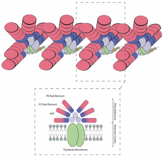
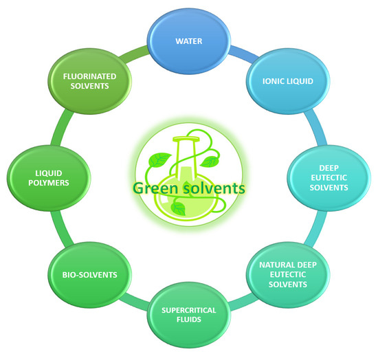
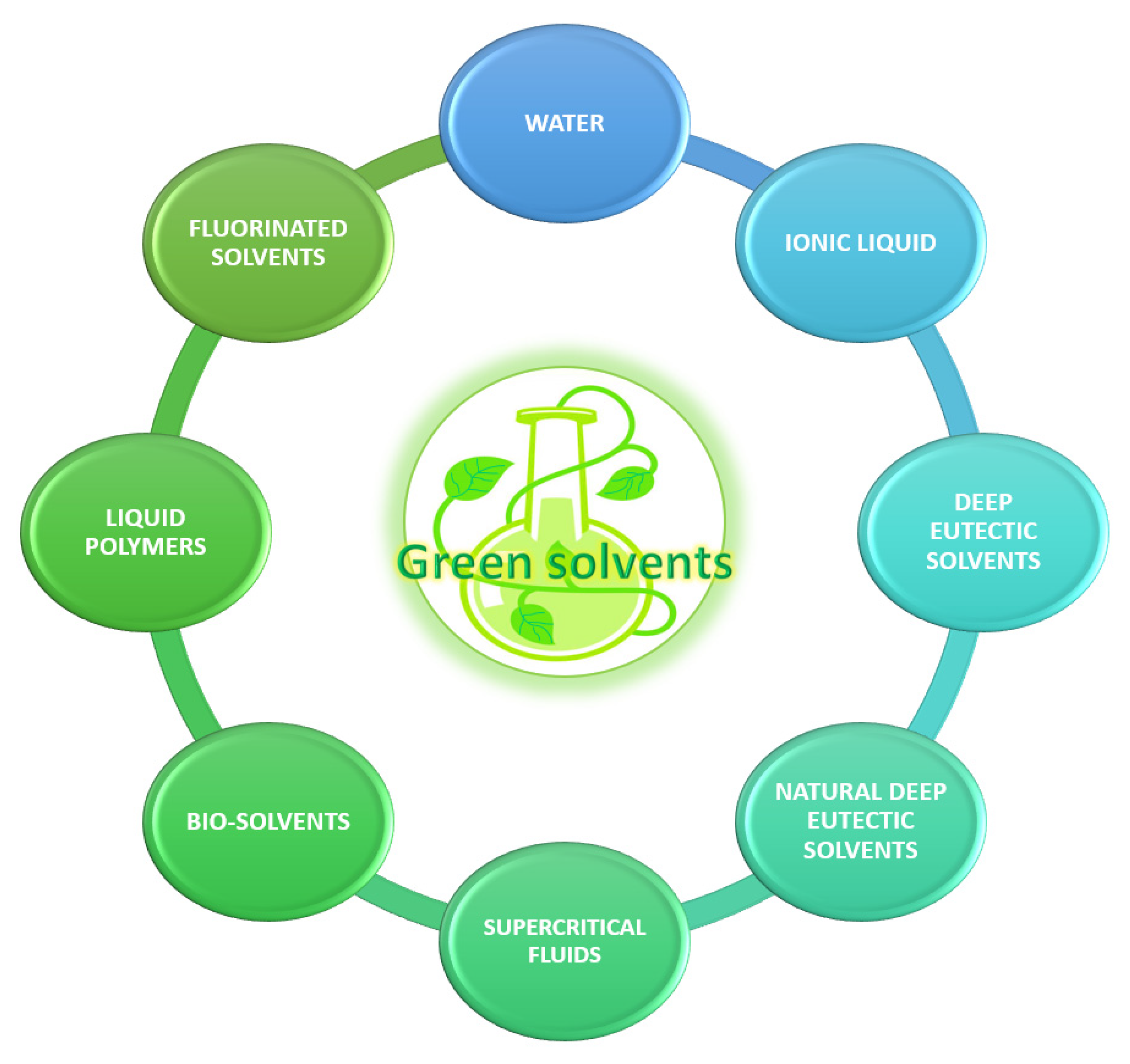
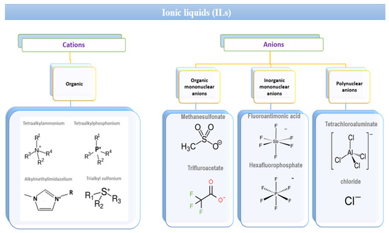
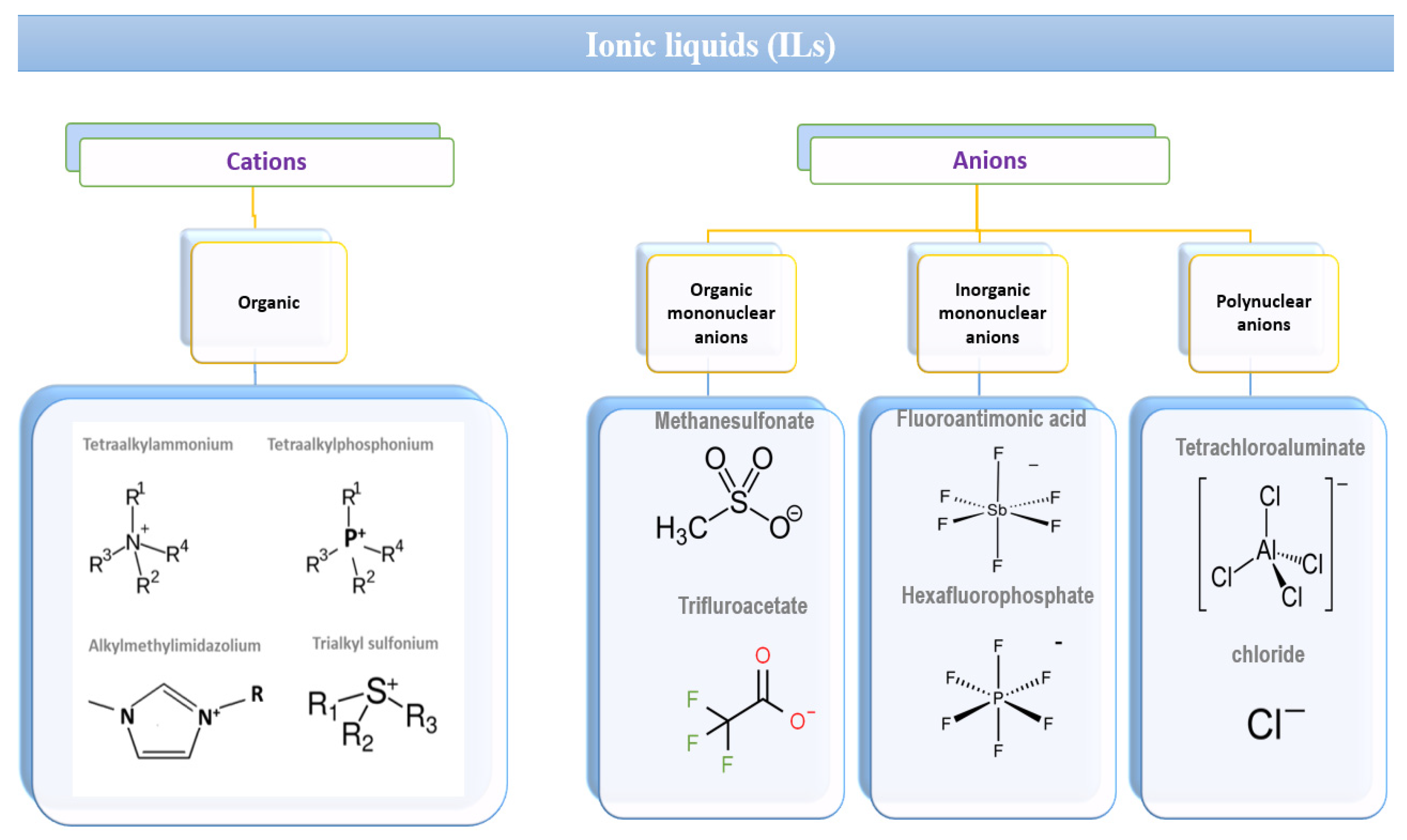
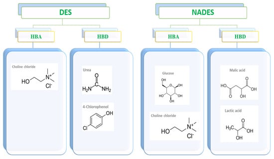
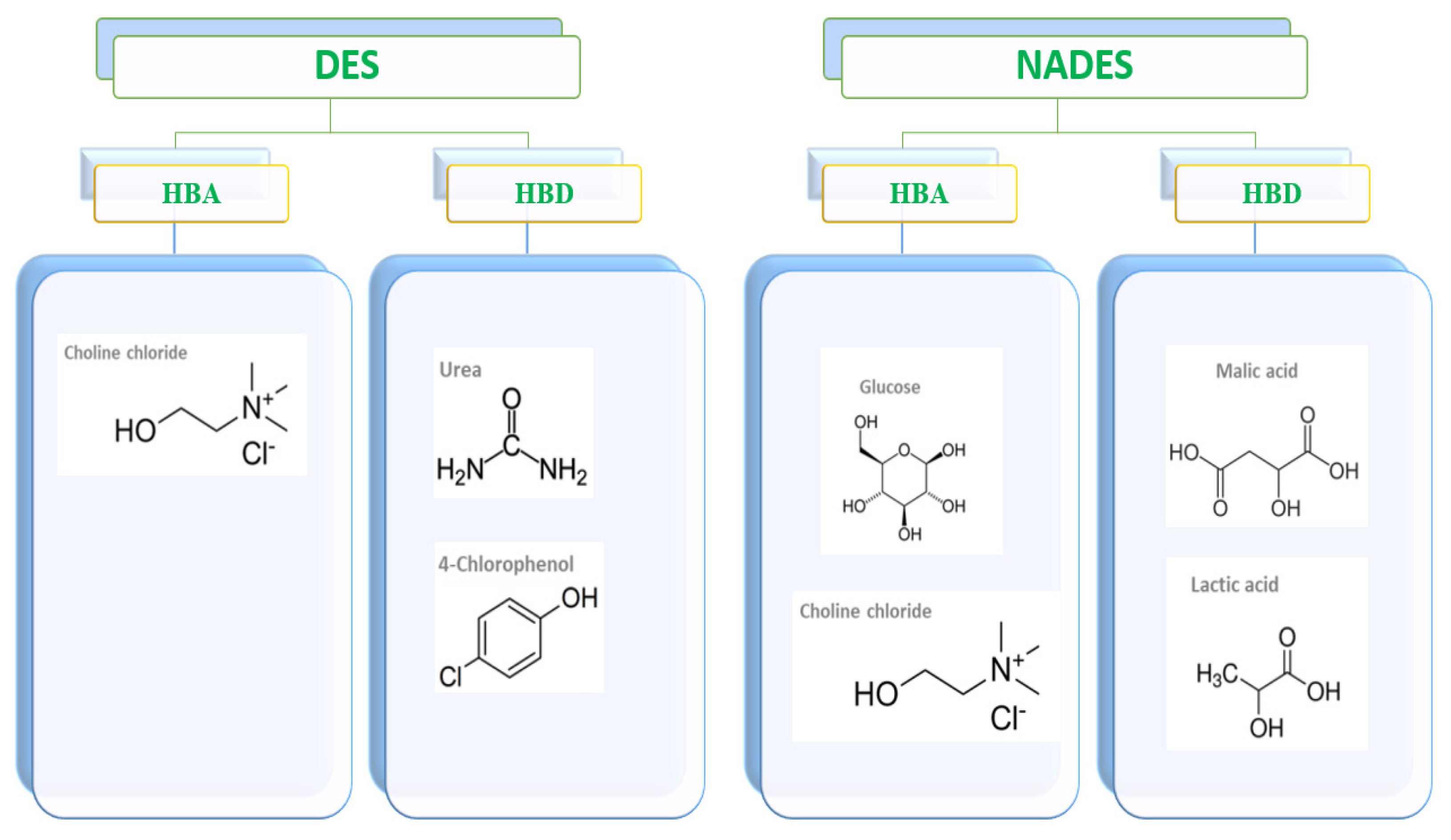
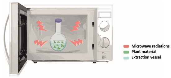
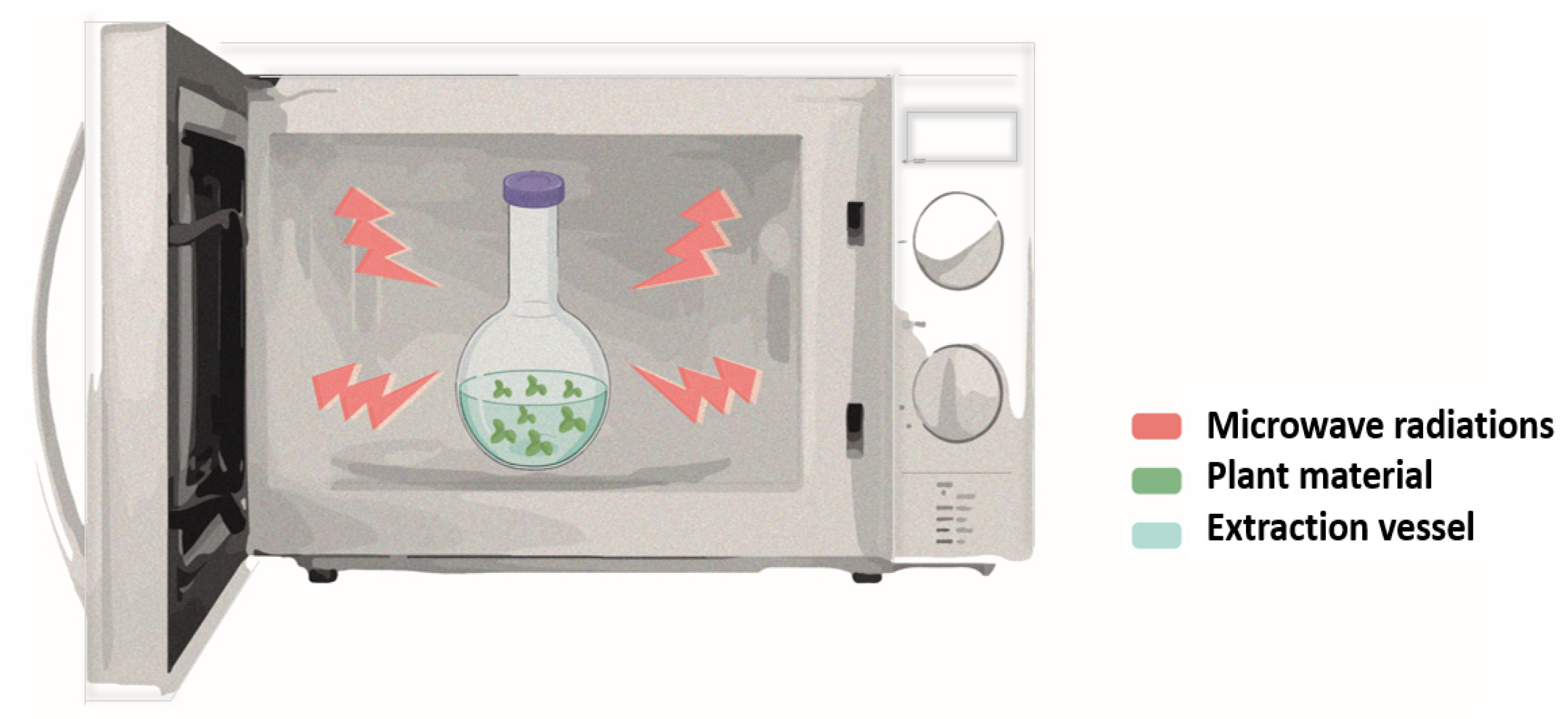
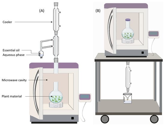
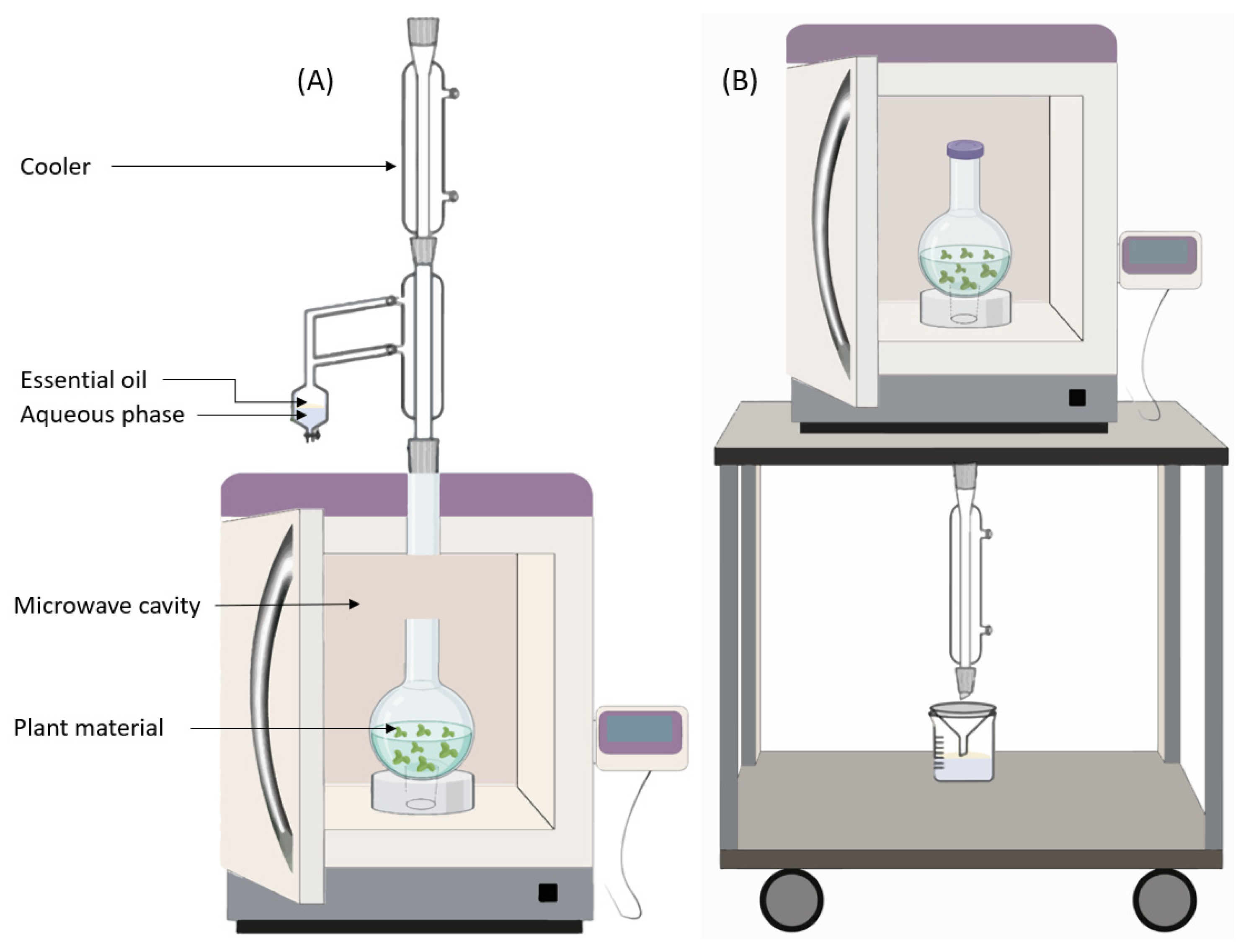
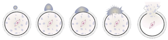
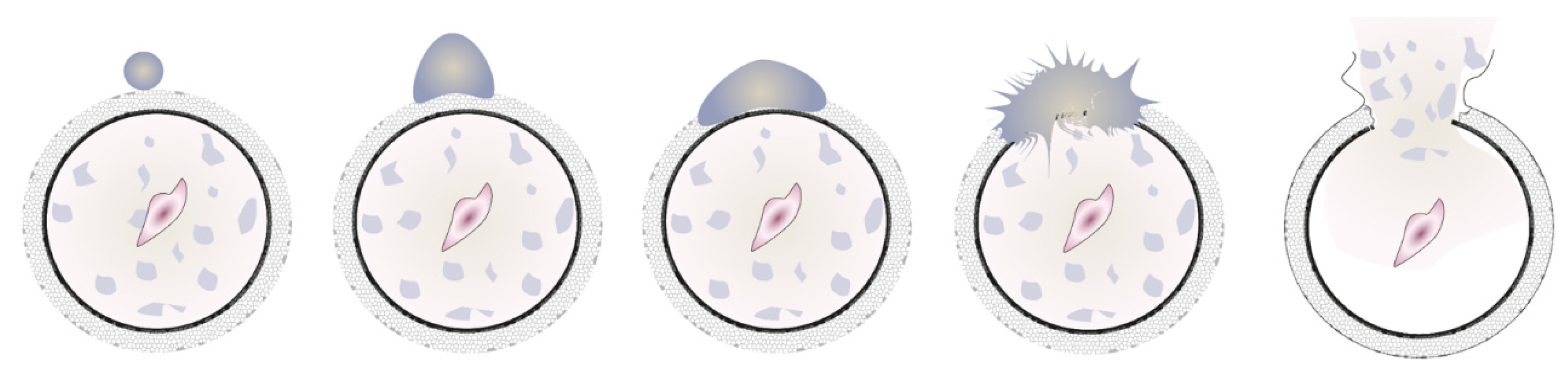
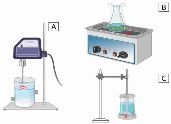
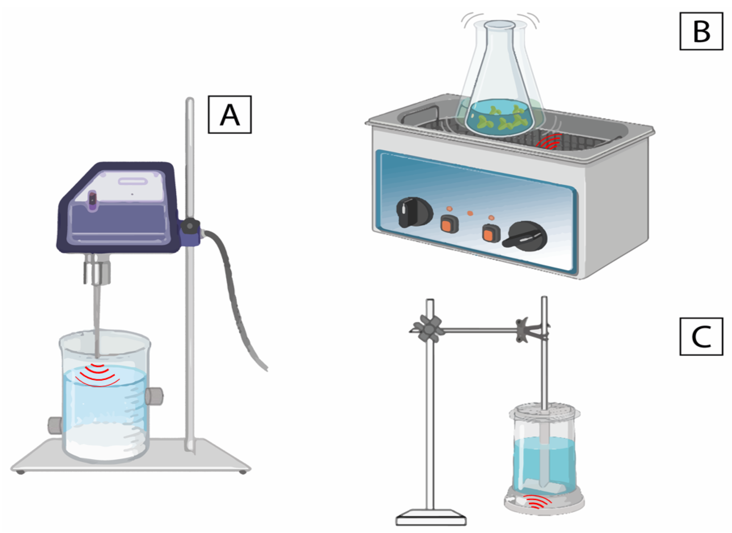
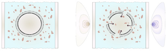
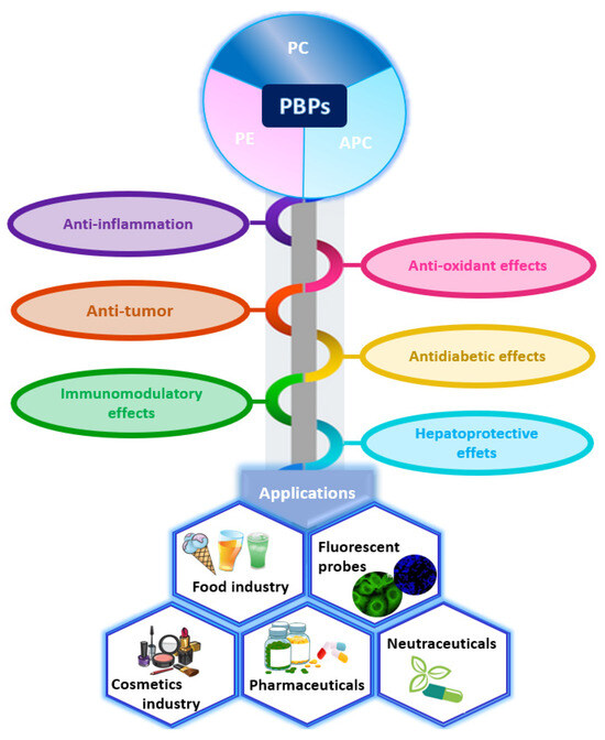
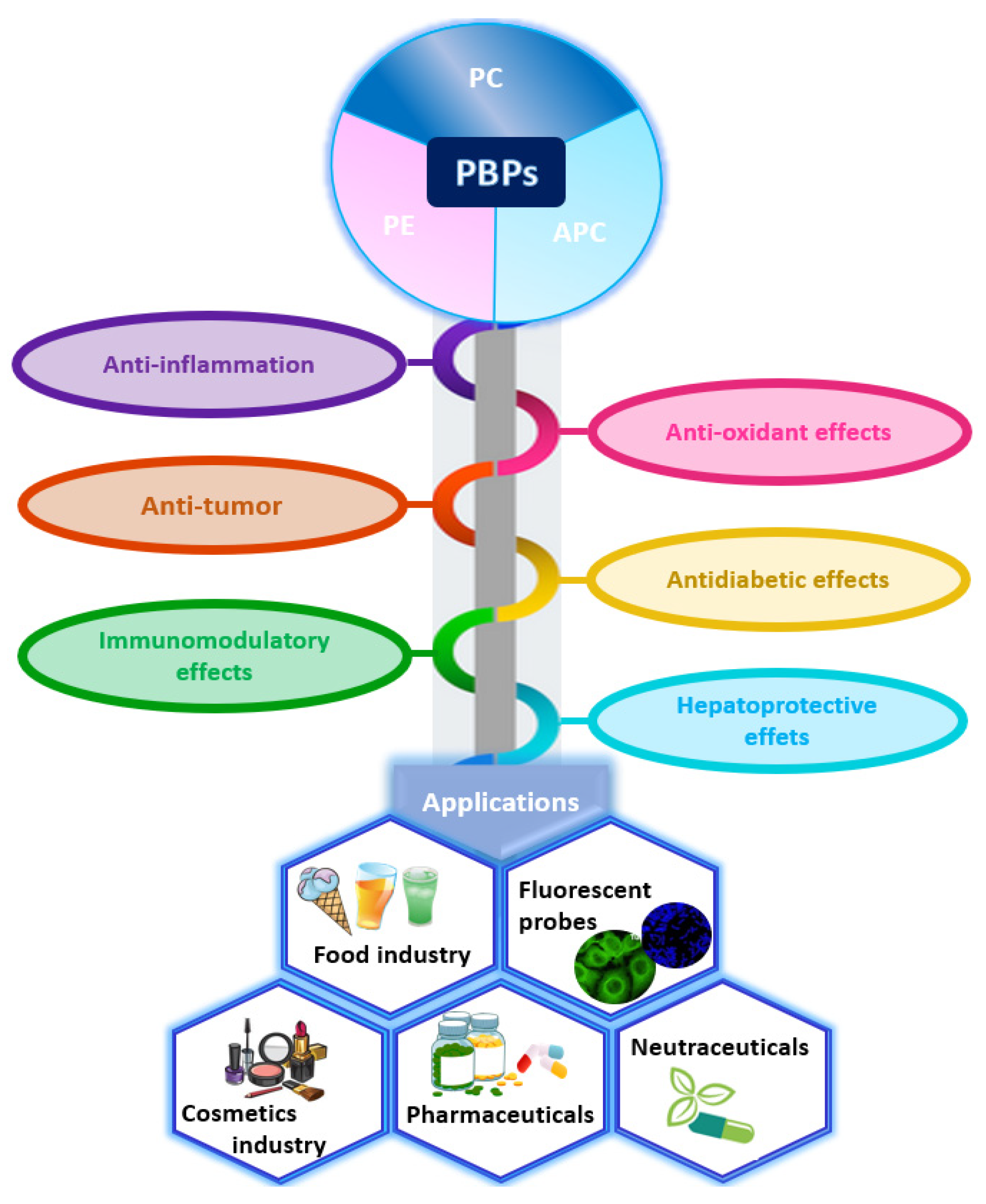

Disclaimer/Publisher’s Note: The statements, opinions and data contained in all publications are solely those of the individual author(s) and contributor(s) and not of MDPI and/or the editor(s). MDPI and/or the editor(s) disclaim responsibility for any injury to people or property resulting from any ideas, methods, instructions or products referred to in the content. |
© 2025 by the authors. Licensee MDPI, Basel, Switzerland. This article is an open access article distributed under the terms and conditions of the Creative Commons Attribution (CC BY) license (https://creativecommons.org/licenses/by/4.0/).