


Abstract
Background: Non-alcoholic fatty liver disease (NAFLD), now largely encompassed by the metabolic dysfunction-associated steatotic liver disease (MASLD) framework, is a leading cause of chronic liver disease worldwide and is closely linked to obesity, insulin resistance, type 2 diabetes (T2D), and cardiometabolic dysfunction. Although hepatic lipid accumulation and metabolic injury remain central to its pathogenesis, increasing evidence implicates the intestinal microbiota as a modulator of disease onset and progression through the gut–liver axis. Objectives: This narrative review summarizes current evidence linking gut microbiota to NAFLD/MASLD, with emphasis on functional microbial alterations relevant to the gut–liver axis, disease severity, microbiota-related biomarkers, and microbiota-targeted interventions. Methods: The literature search for this narrative review was mainly performed using PubMed/MEDLINE (which produced 200 results since 2023). The search addressed the relationship between gut microbiota, NAFLD/MASLD, and gut–liver axis dysfunction. Results: Early studies mainly focused on disease-associated bacterial taxa; however, no consistent microbial signature has been reproduced across populations, disease stages, and methodological platforms. This limitation supports a shift from a taxonomic view of dysbiosis toward a functional interpretation. Different microbial communities may converge on shared pathogenic outputs, including impaired epithelial barrier integrity, microbial translocation, lipopolysaccharide-driven Toll-like receptor 4 activation, bile acid remodelling, altered short-chain fatty acid production, endogenous ethanol generation, choline and trimethylamine-N-oxide metabolism, and tryptophan-derived indole signalling. These mechanisms may influence hepatic steatosis, inflammatory activation, and fibrogenesis. Conclusions: Current data support the microbiota as a biologically plausible disease modifier, but not yet as a standalone diagnostic or therapeutic target; clinical translation will require longitudinal, multi-omics, and function-oriented studies.
1. Introduction
Non-alcoholic fatty liver disease (NAFLD) has traditionally been defined as excessive hepatic fat accumulation in individuals without significant alcohol intake and in the absence of other secondary causes of steatosis. Despite its widespread adoption in clinical and research settings, this terminology has increasingly been criticized because it defines the disease by exclusion and does not explicitly reflect its close relationship with metabolic dysfunction.
In 2020, the term metabolic dysfunction-associated fatty liver disease (MAFLD) was proposed as an alternative diagnostic framework. Unlike NAFLD, MAFLD was based on positive criteria, requiring hepatic steatosis together with overweight or obesity, type 2 diabetes (T2D), or evidence of metabolic dysregulation. This proposal was important because it shifted attention from the exclusion of alcohol-related disease toward the metabolic abnormalities that frequently underlie hepatic steatosis and disease progression.
More recently, an international multisociety Delphi consensus introduced steatotic liver disease (SLD) as an umbrella term and selected metabolic dysfunction-associated steatotic liver disease (MASLD) as the current term replacing NAFLD [1]. MASLD identifies individuals with hepatic steatosis and at least one cardiometabolic risk factor, whereas metabolic dysfunction-associated steatohepatitis (MASH) replaces the previous term non-alcoholic steatohepatitis (NASH). The revised nomenclature represents an effort to improve disease classification by replacing potentially stigmatizing terminology while preserving continuity with the extensive evidence generated under the NAFLD definition. It also provides a more consistent framework for future clinical and translational research. Because most gut microbiota studies were conducted before the introduction of MASLD terminology, the available literature includes studies using NAFLD, MAFLD or MASLD definitions. In this review, NAFLD is used when referring to historical studies or original cohorts defined according to earlier criteria, whereas MASLD is used to reflect the current nomenclature. The combined expression NAFLD/MASLD is used when discussing mechanisms and evidence broadly applicable across these frameworks. MAFLD is retained only when referring to studies that explicitly adopted this diagnostic terminology, and findings from such studies are interpreted in light of the current MASLD framework.
NAFLD/MASLD is among the most prevalent chronic liver diseases worldwide and is strongly associated with obesity, insulin resistance, T2D, dyslipidaemia, and cardiovascular disease [2]. Its clinical spectrum ranges from isolated steatosis to MASH, progressive fibrosis, cirrhosis and hepatocellular carcinoma (HCC) [3]. Among these stages, fibrosis is the strongest predictor of liver-related outcomes. Nevertheless, the mechanisms explaining why only a subset of patients progresses from simple steatosis to inflammatory and fibrotic liver disease remain incompletely understood.
Current models interpret NAFLD/MASLD as a multifactorial condition resulting from the interaction of metabolic, inflammatory, genetic, environmental and microbial factors. Hepatic lipid accumulation is a key initiating event, but it is not sufficient by itself to explain disease progression. Insulin resistance, adipose tissue dysfunction, oxidative stress, mitochondrial impairment, endoplasmic reticulum stress, innate immune activation and gut-derived signals may act simultaneously during the transition from steatosis to steatohepatitis and fibrosis [4]. Within this complex network, the intestinal microbiota has emerged as a biologically plausible interface linking diet, host metabolism, mucosal immunity and liver injury. In this narrative review, both the terms “microbiota” and “microbiome” must not be considered as synonyms; indeed, the former refers to the group of microorganisms (bacteria, viruses, fungi and archaea) populating the anatomical niches, while the latter is the totality of microbial genes, so it includes the broader genetic/functional content.
The involvement of the gut microbiota in NAFLD/MASLD can be understood within the context of the gut–liver axis, a bidirectional system that closely connects intestinal and hepatic functions.
The gut mucosa, made up of epithelial cells linked with intercellular junctions, represents an effective barrier aiming to separate the inner environment from the outer one. This mucosa has an important role because it is involved in the absorption of nutrients but also in the defence against potentially harmful substances; for this reason, it must be highly selective [5,6]. The constitution of this barrier is possible thanks to extracellular components, such as all the digestive enzymes, the gut microbiota components and the mucus layer covering the mucosa which contains the secretory immunoglobulin A and antimicrobial products; intracellular components such as the specialized epithelial cells (Goblet cells, Paneth cells, enterochromaffin cells and M-cells) renewed every 3 to 5 days from the pluripotent stem cells and central nervous system and enteric nervous system which coordinate all the functions [7]. These specialized cells make up the so-called gut-associated lymphoid tissue (GALT), which is involved in the immune response and the tracking and regulation of the intestinal lumen [8].
Because the liver is directly supplied by portal venous blood, it is continuously exposed to compounds originating from the intestine, including nutrients, bile acids, microbial metabolites and structural components of microorganisms [9]. In healthy conditions, several intestinal defence mechanisms, such as the epithelial barrier, mucus layer, antimicrobial peptides, secretory immunoglobulin A, mucosal immune responses and the gut vascular barrier, help prevent excessive passage of bacteria and microbial products into the portal circulation. However, when microbial imbalance is accompanied by impaired barrier integrity, intestinally derived molecules may reach the liver more easily and promote inflammatory as well as fibrogenic pathways.
Several human studies have shown that NAFLD is associated with alterations in both gut microbiota composition and predicted microbial metabolic function. A biopsy-based study reported that NAFLD severity was associated with intestinal dysbiosis and with a shift in the metabolic function of the gut microbiota [10]. Subsequent studies and reviews confirmed that microbial profiles may differ between patients with NAFLD/MASLD and controls, while also emphasizing a major limitation of the field: no single reproducible taxonomic signature has been consistently identified [11,12]. Reported differences in bacterial taxa vary across cohorts and may be influenced by obesity, T2D, diet, ethnicity, geography, medication exposure, fibrosis stage, sequencing platforms, and bioinformatic pipelines [11,12,13]. Moreover, sexual dimorphism has also been recognized as one of the heterogeneity factors; the sex hormones (oestrogens and androgens) can significantly influence the gut microbiota composition, gut barrier integrity and bile acid homeostasis, leading to differences between sexes. At first glance, premenopausal women seem to be partially protected against advanced steatohepatitis and fibrosis due to the oestrogen-driven enrichment of SCFA-producing bacteria and reduced intestinal permeability. Instead, postmenopausal oestrogen and male sex are associated with gut dysbiosis, high endotoxemia and increased MASLD progression. Although the current evidence remains preliminary, sex as a biological variable could have an important role in determining the profile of MASLD [14].
This heterogeneity complicates the translation of microbiome research into clinical practice. If gut microbiota alterations are interpreted only as changes in taxonomic composition, the available evidence may appear fragmented and difficult to reconcile. A functional interpretation may therefore be more informative. Distinct microbial communities may contribute to similar pathogenic pathways if they share altered biological outputs, such as increased production or translocation of pro-inflammatory microbial products, impaired epithelial barrier maintenance, altered bile acid transformation, reduced production of barrier-supportive metabolites or increased generation of hepatotoxic compounds.
Among these mechanisms, intestinal barrier dysfunction and microbial translocation represent one of the most plausible links between dysbiosis and liver injury. Alterations in tight junctions, mucus layer integrity and mucosal immune regulation may increase intestinal permeability, allowing lipopolysaccharide (LPS) and other microbe-associated molecular patterns (MAMPs) to enter the portal circulation [15,16]. LPS, a structural component of the outer membrane of Gram-negative bacteria, may activate Toll-like receptor 4 (TLR4) signalling in Kupffer cells, hepatic stellate cells, and other liver-resident immune cells, promoting nuclear factor kappa B (NF-κB) activation, cytokine release and fibrogenic responses [15,16,17]. This pathway provides a direct microbiological connection between intestinal dysbiosis, innate immune activation and progression of NAFLD/MASLD.
Microbiota-derived metabolites constitute a second major level of interaction between the intestinal ecosystem and the liver. Gut bacteria participate in the conversion of primary into secondary bile acids, thereby influencing farnesoid X receptor (FXR) and Takeda G protein-coupled receptor 5/G protein-coupled bile acid receptor 1 (TGR5/GPBAR1) signalling, both of which regulate lipid metabolism, glucose homeostasis, inflammation and epithelial barrier function [9,16]. Short-chain fatty acids (SCFAs), particularly butyrate, contribute to colonocyte energy metabolism, tight junction regulation and mucosal immune homeostasis, although their role in NAFLD/MASLD appears context-dependent [16]. Other microbial products, including endogenous ethanol, choline-derived metabolites, trimethylamine-N-oxide (TMAO) and indole derivatives, may influence oxidative stress, hepatic lipid accumulation, insulin resistance and immune regulation [12,16].
Taken together, these observations suggest that the intestinal microbiota should be interpreted not only as a source of compositional biomarkers, but also as a functional component of the gut–liver axis in NAFLD/MASLD. This distinction is important because earlier studies largely focused on disease-associated bacterial taxa, whereas fewer investigations have linked microbial composition with barrier dysfunction, microbial translocation, metabolite-mediated signalling and hepatic immune activation as part of the mechanisms involved in disease progression.
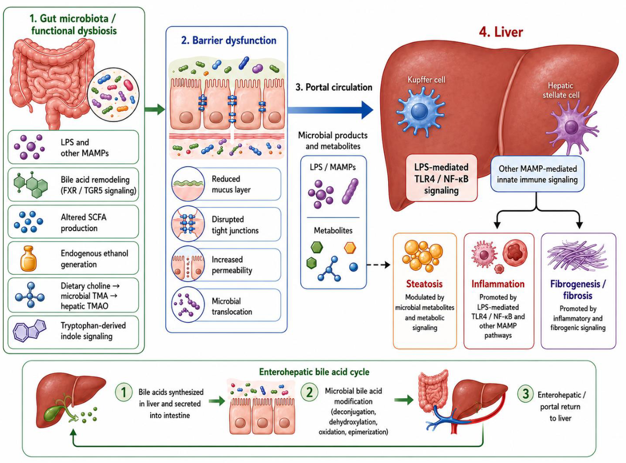
The aim of this narrative review is to summarize and critically interpret current evidence on the relationship between gut microbiota and NAFLD/MASLD, with specific attention to the transition from taxonomic dysbiosis to functional dysbiosis. In particular, this review examines how altered intestinal microbial communities may contribute to hepatic steatosis, inflammation and fibrosis through barrier dysfunction, microbial translocation, LPS-mediated innate immune activation and microbiota-derived metabolites, including bile acids, SCFAs, endogenous ethanol, choline-derived compounds and indole derivatives. By organizing the evidence around these functional mechanisms, this review provides a microbiology-centred framework that may help explain why the gut microbiota remains relevant in NAFLD/MASLD despite the absence of a universal microbial signature. The principal functional mechanisms linking gut microbiota alterations to NAFLD/MASLD progression are summarized in Figure 1.
Figure 1.
Functional gut–liver axis in NAFLD/MASLD. Functional dysbiosis may impair the intestinal mucus layer and epithelial tight junctions, increasing intestinal permeability and the portal translocation of microbial products and metabolites. Lipopolysaccharide (LPS)-mediated Toll-like receptor 4 (TLR4)/nuclear factor kappa B (NF-κB) signalling and other pathways triggered by microbe-associated molecular patterns (MAMPs) may promote hepatic inflammation and fibrogenesis. Microbial bile acid remodelling, altered short-chain fatty acid (SCFA) production, endogenous ethanol generation, choline-derived trimethylamine (TMA)/trimethylamine-N-oxide (TMAO) metabolism, and tryptophan-derived indole signalling may modulate epithelial integrity, hepatic metabolism, steatosis, and immune responses. FXR, farnesoid X receptor; TGR5, Takeda G protein-coupled receptor 5.
2. Methods
This manuscript was designed as a narrative review of the literature on gut microbiota and NAFLD/MASLD. The literature search was mainly performed using PubMed/MEDLINE (which produced 200 results since 2023), with Google Scholar consulted to identify additional full-text articles, highly cited publications, and recent studies relevant to the gut–liver axis.
The search focused on studies addressing the relationship between gut microbiota, NAFLD/MASLD, and gut–liver axis dysfunction, with particular attention to microbial dysbiosis, intestinal barrier impairment, microbial translocation, lipopolysaccharide-mediated signalling, microbiota-derived metabolites, fibrosis progression, microbiota-related biomarkers, and microbiota-targeted interventions.
Priority was given to English-language articles published within the last five years, including human studies, observational studies, randomized controlled trials, systematic reviews, meta-analyses, and relevant narrative reviews. Earlier studies were included when they provided landmark evidence, particularly on biopsy-proven NAFLD, metagenomic signatures of advanced fibrosis, NAFLD-related cirrhosis, or foundational mechanisms of the gut–liver axis.
Studies using NAFLD, MAFLD, or MASLD terminology were considered eligible when their content was relevant to metabolically driven steatotic liver disease and gut–liver axis mechanisms. Terminology was interpreted according to the definitions used in the original studies, while the overall synthesis was aligned with the current MASLD framework.
No formal systematic review protocol, risk-of-bias assessment, or quantitative meta-analysis was performed. Because of the heterogeneity of study designs, populations, diagnostic criteria, sequencing methods, and clinical endpoints, evidence was synthesized qualitatively and organized into thematic sections.
3. Current Evidence on Gut Microbiota Alterations in NAFLD/MASLD
Research on the intestinal microbiota in NAFLD/MASLD has expanded considerably in recent years. Bibliometric evidence indicates that research on metabolic syndrome and MAFLD has progressively moved from classical metabolic descriptors, such as insulin resistance and obesity, toward mechanistic topics including gut microbiota, the gut–liver axis and early intervention strategies [18]. Such bibliometric data should be interpreted as indicators of research trends rather than as direct mechanistic evidence. Nevertheless, they reflect a broader conceptual evolution: NAFLD/MASLD is increasingly viewed not only as the hepatic manifestation of systemic metabolic dysfunction, but also as a condition influenced by intestinal microbial ecology and gut-derived signals.
This interpretation is supported by recent evidence on the role of the gut–liver axis in metabolic liver disease. Schnabl and colleagues suggested that alterations in the gut microbiota may contribute to MASLD by affecting intestinal permeability, bile acid metabolism, inflammatory pathways and the response to therapeutic strategies [19]. In a similar perspective, Vallianou and colleagues described NAFLD/MASLD as a condition closely connected to gut–liver axis dysfunction, where dysbiosis, microbial products and microbiota-derived metabolites may interact with hepatic fat accumulation and inflammatory injury [20]. Consistently, Song and Zhang underlined that impaired communication between the gut and the liver may promote NAFLD progression through microbial and immune-mediated mechanisms [21].
Human microbiome studies indicate that NAFLD/MASLD is often accompanied by alterations in the gut microbial ecosystem. These changes have been described across major bacterial phyla and families, including Bacillota, Bacteroidota, Pseudomonadota, Enterobacteriaceae, Ruminococcaceae and Lachnospiraceae, as well as in genera such as Bacteroides, Prevotella, Escherichia, Veillonella, Faecalibacterium, and Akkermansia [22,23,24,25]. However, the direction and consistency of these findings vary considerably among cohorts. This variability is not surprising, since gut microbiota composition can be shaped by several host- and environment-related factors, including obesity, insulin resistance, T2D, dietary habits, age, ethnicity, medication exposure and fibrosis stage.
Because of this heterogeneity, defining a single microbial profile specifically associated with NAFLD/MASLD remains challenging. Simplified indicators, such as the Bacillota/Bacteroidota ratio, do not adequately reflect the ecological and functional complexity of dysbiosis in metabolic liver disease. Likewise, an increased abundance of Pseudomonadota or Enterobacteriaceae may indicate a greater pro-inflammatory potential of the gut microbiota, but this finding alone cannot account for clinical phenotype, disease progression or response to therapy.
Several studies have attempted to overcome this limitation by integrating microbial composition with clinical, metabolic or methodological variables. Korobeinikova and colleagues compared different analytical strategies for characterizing gut microbiota patterns in NAFLD, showing that methodological choices can influence interpretation of microbial profiles [22]. Yang and colleagues described gut microbiota characteristics in patients diagnosed according to MAFLD criteria, supporting the association between intestinal microbial alterations and metabolically driven hepatic steatosis [23]. Borges-Canha and colleagues reported that gut dysbiosis was linked to severe steatosis and improved diagnostic performance when combined with clinical assessment [24]. Mai and colleagues reviewed the role of gut microbiota in the occurrence and progression of NAFLD, emphasizing that dysbiosis should be interpreted in relation to host metabolic status and disease stage [25].
Overall, current evidence indicates that gut microbiota alterations are common in NAFLD/MASLD, but they do not define a reproducible universal microbial signature. This limitation is not only methodological but also conceptual. The microbiota may be relevant less because of a fixed taxonomic profile and more because distinct microbial configurations can converge on similar pathogenic functions. These include impaired barrier maintenance, microbial translocation, bile acid dysregulation, altered SCFA production, endogenous ethanol generation, choline metabolism, inflammatory signalling and fibrogenic progression.
However, we must have in mind that the correlation of the gut microbiota with NAFLD/MASLD and MAFLD is mainly influenced by a lot of confounding factors that do not allow us to clearly understand the main actor, but diet and common therapies seem the most important ones. A better understanding of the physiology of our microbiota and its interactions with human tissues is mandatory.
4. From Taxonomic Dysbiosis to Functional Dysbiosis
The concept of dysbiosis is commonly used to indicate shifts in microbial diversity or changes in the relative abundance of selected bacterial taxa. In NAFLD/MASLD, however, this taxonomic view describes only one aspect of a more complex process. A more useful perspective is that of functional dysbiosis, which refers to changes in microbial functions capable of influencing host metabolic regulation, intestinal barrier integrity and immune signalling within the liver. This distinction is particularly relevant. Patients with NAFLD/MASLD may differ substantially in their microbial composition, but they can still share similar functional disturbances, such as increased exposure to endotoxins, impaired bile acid metabolism, reduced production of metabolites that support barrier function or enhanced generation of potentially hepatotoxic compounds. At the same time, bacteria belonging to the same genus may exert different biological effects depending on strain-level differences, dietary patterns, the host metabolic profile and the surrounding microbial ecosystem.
Lanthier and Delzenne emphasized that therapeutic and mechanistic approaches to MASLD should not be limited to modifying microbial composition but should also assess whether microbiome changes translate into clinically relevant effects on host metabolism, intestinal permeability, microbial metabolites and liver injury [26]. This function-oriented perspective shifts attention from the identity of individual taxa to the biological activities of the microbial community.
Jing and Jiang similarly framed the crosstalk between T2D and MASLD in terms of microbial outputs, including SCFAs, bile acids, LPS and other MAMPs, branched-chain amino-acid catabolites, TMAO and endogenous ethanol, which may influence the liver through portal circulation or enterohepatic signalling [27].
The gut–microbiota–liver axis therefore provides a useful mechanistic framework for MASLD. Liao and colleagues described bidirectional communication between the gut and liver mediated by microbial communities and their metabolites and discussed interventions targeting intestinal microecology [28]. This framework does not position the microbiota as the sole driver of disease; rather, microbial functions interact with metabolic stress, insulin resistance, hepatic lipid accumulation and immune activation.
Evidence from causal inference studies further supports the need to move beyond simple taxonomy. Yi and colleagues used species-level shotgun metagenomic data, bidirectional Mendelian randomization, meta-analysis and mediation analysis to investigate the relationship between gut microbiota and MAFLD. Their study identified several microbial taxa potentially associated with disease and suggested that deoxycholate may mediate part of the relationship between Bifidobacterium and MAFLD risk [29]. Although these findings require cautious interpretation, they illustrate the value of integrating species-level microbiome data with metabolite-mediated pathways.
Functional dysbiosis provides a practical framework for organizing current evidence. In NAFLD/MASLD, relevant microbial functions include inflammatory ligand production and translocation, maintenance or disruption of the epithelial barrier, bile acid transformation, fermentation and SCFA production, endogenous ethanol generation, choline and TMAO metabolism, tryptophan-derived indole production and microbial modulation of hepatic immune responses.
This model also has translational implications. If the therapeutic goal is only to normalize the abundance of selected taxa, microbiota-targeted interventions will likely remain inconsistent. Conversely, if the goal is to restore microbial functions involved in the gut–liver axis, interventions can be designed around measurable endpoints, such as reduced LPS exposure, improved intestinal permeability, restored bile acid signalling, increased barrier-supportive metabolites, decreased hepatotoxic fermentation products and improved fibrosis-related markers.
5. Intestinal Barrier Dysfunction, Microbial Translocation and LPS-Mediated Liver Injury
The intestinal barrier represents a key interface between gut microbiota and the liver. It includes the mucus layer, epithelial cells, tight junctions, adherens junctions, antimicrobial peptides, secretory immunoglobulin A, mucosal immune cells and the gut vascular barrier. Together, these components regulate the interaction between luminal microorganisms and host tissues, allowing nutrient absorption while limiting uncontrolled microbial translocation.
Commensal bacteria contribute to barrier homeostasis by supporting mucus production, stimulating antimicrobial peptide expression, regulating immunoglobulin A responses and generating metabolites that maintain epithelial integrity. In NAFLD/MASLD, this equilibrium may be disrupted. Dysbiosis may reduce protective microbial functions and increase the production or translocation of inflammatory microbial molecules. Long and colleagues reviewed the relationship between intestinal barrier dysfunction and gut microbiota in NAFLD, emphasizing that impaired epithelial integrity may facilitate the passage of microbial products toward the liver [15]. Saha and Hartmann similarly discussed how gut microbiome alterations affect gut permeability across liver and intestinal diseases, supporting the view that permeability is a central event in gut–liver communication rather than a merely secondary phenomenon [30].
Clinically relevant evidence has also emerged in patients with T2D and MASLD. Forlano and colleagues linked MASLD severity to disruption of gut barrier integrity and altered host–microbiome interactions [31]. This observation is important because T2D is one of the metabolic contexts most strongly associated with MASLD progression because insulin resistance and hyperglycaemia directly disrupt the barrier while independently remodelling the gut microbiota and also independently influencing its composition. Barrier dysfunction may therefore connect metabolic comorbidity, microbial ecology and liver disease severity.
Additional evidence comes from Dallio and colleagues, who evaluated intestinal permeability-related markers in MASLD. Their study reported changes in faecal zonulin, serum lipopolysaccharide-binding protein (LBP), tight junction proteins, LPS, inflammatory cytokines and steatosis-related outcomes after a long-term intervention affecting the gut–liver axis [32]. Although this intervention should not be classified as a classical microbiota-targeted therapy, the study is relevant because it connects permeability markers, LPS exposure, systemic inflammation and liver-related outcomes in a MASLD population.
LPS remains the best-characterized microbial product in this context. As a structural component of the outer membrane of Gram-negative bacteria, LPS provides a direct microbiological link between intestinal dysbiosis and hepatic innate immune activation. Once translocated through a compromised intestinal barrier, LPS may reach the liver through portal circulation and activate TLR4 signalling in Kupffer cells, hepatic stellate cells, liver sinusoidal endothelial cells, and other immune cell populations. This pathway promotes NF-κB activation and the release of inflammatory mediators, including tumour necrosis factor-alpha, interleukin-1 beta, and interleukin-6.
The biological relevance of this mechanism is supported by reviews focused on oxidative stress and inflammatory amplification. Zhou and colleagues described a feedback loop in which gut dysbiosis disrupts barrier function, increases LPS translocation, activates TLR4 signalling and enhances oxidative stress through inflammatory pathways [33]. Kurhaluk and colleagues, although focusing on adolescents, summarized evidence that NAFLD is associated with reduced microbial diversity, enrichment of ethanol- and LPS-producing taxa, altered SCFA profiles and activation of TLR4–NF-κB pathways [34]. These findings are useful because they connect bacterial products, epithelial dysfunction and hepatic inflammation within a microbiological framework.
Other microbial products may also contribute to hepatic injury. Bacterial DNA can activate Toll-like receptor 9, flagellin can activate Toll-like receptor 5, peptidoglycan fragments can engage intracellular pattern-recognition receptors and fungal beta-glucans may stimulate C-type lectin receptors. Although human evidence remains strongest for LPS-related pathways, microbial translocation should be interpreted more broadly as the portal delivery of multiple microbial ligands capable of stimulating hepatic inflammation.
The barrier-translocation model helps explain why compositional microbiome data are often difficult to translate into clinical practice. A microbial community may be relevant not because it contains a specific disease-defining taxon, but because it increases inflammatory ligand exposure or weakens epithelial containment. In this sense, intestinal permeability represents one of the functional points at which microbial ecology becomes hepatic pathology.
6. Microbiota-Derived Metabolites in NAFLD/MASLD
Microbiota-derived metabolites are central to a functional interpretation of the NAFLD/MASLD microbiome. They provide a biochemical bridge between intestinal microbial activity and hepatic metabolism. Unlike taxonomic profiles, which vary considerably across studies, several metabolite pathways recur in the literature, including bile acid remodelling, SCFA production, endogenous ethanol generation, choline/TMAO metabolism and tryptophan-derived indole signalling.
Bile acids are among the clearest examples of bidirectional gut–liver communication. Primary bile acids are synthesized in the liver, secreted into the intestine and modified by bacterial enzymes through deconjugation, dehydroxylation, oxidation and epimerization. These microbial reactions generate secondary bile acids and reshape the bile acid pool. Through receptors such as FXR and TGR5/GPBAR1, bile acids regulate lipid metabolism, glucose homeostasis, intestinal barrier function, inflammation and enterohepatic feedback.
Nimer and colleagues showed that bile acid profiles are associated with histopathological indices and genetic variants involved in NAFLD progression [35]. This supports the concept that bile acids may integrate liver injury, host genetics and microbial metabolism. The mediation analysis by Yi and colleagues further strengthens this view by identifying deoxycholate as a possible mediator between Bifidobacterium and MAFLD risk [29]. These findings suggest that bile acid remodelling may be more informative than the simple presence or absence of specific bacteria.
Among fermentation-derived metabolites, SCFAs represent a major interface between microbial activity, epithelial integrity and hepatic metabolic signalling. Acetate, propionate and butyrate are produced through bacterial fermentation of dietary fibre and resistant starch. Butyrate is particularly relevant because it supports colonocyte energy metabolism, tight junction integrity and mucosal immune regulation.
The role of SCFAs in NAFLD/MASLD should not be interpreted simplistically. Thing and colleagues reported that plasma SCFAs are associated with MASLD, but circulating levels may reflect several overlapping processes, including microbial fermentation, intestinal absorption, hepatic utilization, dietary intake and host metabolic status [36]. Therefore, SCFAs are best understood as context-dependent mediators. Their biological meaning depends on where they are produced, how much is absorbed, how they are metabolized by the liver and whether intestinal barrier function is preserved.
Endogenous ethanol represents another mechanism through which dysbiosis may contribute to hepatic injury. Certain intestinal bacteria can ferment carbohydrates and generate ethanol. Portal exposure to ethanol and acetaldehyde may promote oxidative stress, mitochondrial dysfunction and epithelial barrier impairment. Hoyles and colleagues integrated molecular phenomics and metagenomics in hepatic steatosis and showed that microbial features can cluster with host metabolic traits [37]. Kurhaluk and colleagues also highlighted enrichment of ethanol-producing taxa in adolescent NAFLD, suggesting that endogenous ethanol may be relevant in selected microbial and metabolic contexts [34].
Choline metabolism and TMAO production represent an additional microbiota-dependent pathway. Choline is required for very-low-density lipoprotein assembly and hepatic lipid export. Some gut bacteria metabolize dietary choline into trimethylamine (TMA), which is then oxidized in the liver to TMAO. Excessive microbial choline utilization could theoretically reduce choline availability and favour hepatic lipid accumulation, while TMAO may contribute to cardiometabolic risk and inflammatory signalling. Jing and Jiang included TMAO, nitrogen/methyl flux and branched-chain amino-acid catabolites among microbial outputs relevant to combined metabolic and hepatic endpoints [27].
Tryptophan metabolism adds a further layer of microbiota–host interaction. Gut bacteria can convert tryptophan into indole and indole derivatives, which influence epithelial barrier function and mucosal immunity through pathways such as aryl hydrocarbon receptor (AhR) signalling. Oh and colleagues used network pharmacology to prioritize candidate microbiota-derived metabolites potentially relevant to NAFLD, supporting the concept that microbial metabolites may contribute to disease networks rather than isolated pathways [38].
Overall, metabolite-centred evidence supports the view that the microbiota is not only a community of microorganisms, but also a metabolic interface. Its relevance in NAFLD/MASLD depends on microbial biochemical activity: how the microbiota transforms bile acids, ferments substrates, generates inflammatory or hepatotoxic products, maintains epithelial integrity and modulates immune-metabolic signalling. The principal functional mechanisms linking gut microbiota alterations to NAFLD/MASLD, together with their main mediators, biological effects, representative evidence, and translational implications, are summarized in Table 1.
Table 1.
Principal functional mechanisms linking the gut microbiota to NAFLD/MASLD and their translational implications.
7. Gut Microbiota and Disease Severity: From Steatosis to Fibrosis
A clinically relevant question is whether gut microbiota alterations are associated only with the presence of NAFLD/MASLD or also with disease severity. Available evidence suggests that microbiome-based signals may become more informative when disease progression is considered, particularly in advanced fibrosis and cirrhosis.
Loomba and colleagues identified a gut microbiome-based metagenomic signature for the non-invasive detection of advanced fibrosis in human NAFLD [39]. This study moved beyond simple steatosis and linked microbial patterns to a clinically meaningful disease stage. Caussy and colleagues subsequently described a gut microbiome signature associated with cirrhosis due to NAFLD [40]. These findings suggest that advanced liver disease may be accompanied by more pronounced or more functionally relevant microbial alterations.
At the same time, the diagnostic value of microbiome-based classifiers remains limited. Lang and colleagues compared gut microbiota-based approaches with simple non-invasive tools for predicting advanced fibrosis and found that microbiome classifiers should not be interpreted as replacements for established clinical tools [41]. Microbiome data may contribute to risk stratification, but their current role is more likely complementary than standalone.
Microbiota-related signatures may also be relevant in non-obese NAFLD. Lee and colleagues identified distinct gut microbiome and metabolite profiles associated with significant fibrosis in non-obese NAFLD [42]. This finding suggests that microbial mechanisms are not simply an indirect reflection of severe obesity. Instead, gut microbiota may contribute to disease heterogeneity even in patients without marked adiposity.
Prospective and integrative studies further support the potential of microbial–metabolic markers. Leung and colleagues investigated gut microbiome and metabolite markers for NAFLD risk assessment, suggesting that microbial and metabolic information may improve the understanding of disease development [43]. Zhai and colleagues used bidirectional two-sample Mendelian randomisation to explore associations between gut microbiota and NAFLD/NASH, representing one attempt to move from correlation toward causal inference [44]. Li and colleagues also emphasised the need to distinguish association from causation in gut microbiome–liver disease research [45].
The biological plausibility of a microbiota–fibrosis link is strong. Barrier dysfunction can increase portal exposure to LPS and other microbial products. These signals may sustain hepatic inflammatory activation and contribute to hepatic stellate cell activation. Microbial metabolites, especially bile acids, SCFAs and succinate, may influence the balance between inflammatory resolution and fibrogenic remodelling. Li and colleagues recently framed MASH progression as a process in which gut-derived signals may contribute to macrophage reprogramming and fibrosis through altered SCFAs, secondary bile acids, succinate and endotoxin exposure [46]. Although this topic intersects with immunology, it remains relevant in this review because the initiating signals are gut microbial products.
Thus, the microbiota appears most clinically informative when interpreted in relation to disease stage and function. The relevant question is not whether a patient has a “NAFLD microbiota”, but whether the intestinal ecosystem contributes to barrier leakage, inflammatory ligand exposure, metabolite imbalance and fibrotic progression.
8. Microbiota-Targeted Strategies
The gut microbiota is a modifiable component of the gut–liver axis, which makes it an attractive therapeutic target. However, the therapeutic goal should not be a generic change in faecal microbial composition. In NAFLD/MASLD, a microbiota-targeted intervention is meaningful only if it modifies disease-relevant functions, including intestinal barrier integrity, endotoxin exposure, bile acid transformation, SCFA production, inflammatory signalling, or hepatic lipid accumulation.
Prebiotics and fermentable substrates are among the most biologically plausible interventions because they can influence microbial fermentation and SCFA production. Resistant starch is a relevant example. Ni and colleagues showed that resistant starch supplementation reduced intrahepatic triglyceride content in patients with NAFLD through gut microbiome alterations [47]. More recent work suggests that interindividual variability in gut microbiome composition may influence the response to resistant starch in MASLD [48]. These studies are important because they connect a dietary substrate, microbial function and a liver-related outcome.
Probiotics and synbiotics have been studied extensively, but results remain heterogeneous. The INSYTE trial showed that synbiotic supplementation altered faecal microbiome composition but did not significantly improve liver fat or fibrosis markers [49]. This trial is particularly informative because it demonstrates that microbiome modification alone is not necessarily equivalent to clinical benefit. An intervention may change faecal taxa without correcting the microbial functions that matter for liver disease.
Systematic reviews and meta-analyses provide a broader but still cautious view. Sharpton and colleagues reviewed gut microbiome-targeted therapies in NAFLD and reported potential improvements in selected outcomes, while emphasising heterogeneity across interventions and study designs [50]. Carpi and colleagues similarly reviewed probiotics, prebiotics and synbiotics in NAFLD/NASH, reporting possible benefits but highlighting variability in formulations, treatment duration and endpoints [51]. These limitations are central. The field cannot move forward by treating all probiotics or synbiotics as equivalent; strain specificity, dose, baseline microbiota and disease stage are likely to influence response.
Faecal microbiota transplantation (FMT) represents a broader ecological intervention. Abenavoli and colleagues reviewed FMT as a potential strategy for NAFLD treatment, but emphasised that it remains investigational [52]. He and colleagues more recently reported, in a meta-analysis of FMT in metabolic-associated fatty liver disease, improvements in selected liver inflammation indicators and fat content [53]. However, the certainty and generalizability of these findings remain limited by the small and heterogeneous evidence base. Zhang and colleagues analysed FMT in overweight or obese adults with metabolic disorders and found uncertain effects on systemic metabolic outcomes, with results influenced by methodological assumptions, treatment protocols and donor variability [54]. Taken together, these studies suggest that FMT may have potential, but it cannot yet be considered a standardised therapeutic approach for NAFLD/MASLD.
Microbiome-centred therapeutic reviews increasingly emphasise the need for personalised strategies. Saeed and colleagues reviewed microbiome-centred therapies in MASLD, including prebiotics, probiotics, synbiotics, FMT, engineered bacteria and bacteriophage therapy, highlighting that such interventions may act through improved gut permeability, reduced endotoxin-producing bacteria and altered bile acid metabolism [55]. Bahitham and colleagues synthesised animal and human evidence on MASLD/MASH-related gut microbiome alterations, emphasising microbial taxa, functional metabolites and the need for longitudinal, mechanistically driven studies [56].
Next-generation microbial approaches remain promising but preliminary. Akkermansia muciniphila is of particular interest because of its relationship with mucus layer biology, gut barrier function and metabolic regulation. Asghari and colleagues reviewed preclinical evidence on A. muciniphila in NAFLD models and reported improvements in steatosis, lipid metabolism, insulin sensitivity, tight junction expression and LPS translocation, while emphasising the absence of adequate clinical trials [57]. This distinction is important: A. muciniphila is a biologically attractive target, but it should not yet be presented as an established therapy in human NAFLD/MASLD.
Other dietary approaches may influence gut microbial ecology, but their relevance to NAFLD/MASLD should be interpreted cautiously when liver-specific endpoints are not directly evaluated. For example, dietary polyphenols have been reported to modulate beneficial taxa such as Bifidobacterium, Faecalibacterium and Lactobacillus, but this evidence should be considered supportive of microbiota modulation rather than definitive proof of therapeutic efficacy in MASLD [58].
Therapeutic studies indicate that compositional changes alone are insufficient unless accompanied by measurable functional effects on barrier integrity, endotoxin exposure, microbial metabolites or hepatic outcomes. Future trials should therefore be designed with microbiological and mechanistic endpoints rather than relying only on liver enzymes or stool taxonomic changes.
Beyond dietary and pharmacological strategies, it is also important to consider bariatric surgery (e.g., sleeve gastrectomy, Roux-en-Y gastric bypass). This represents a major natural human intervention model for testing microbiome-liver causality. Indeed, these procedures induce a perturbation of the gut microbiota composition (for example, with the enrichment of species producing SCFA such as Akkermansia muciniphila) and the hepatic metabolism. The subsequent alterations in bile acid flow, luminal pH and the production of gut hormones indicate the restructuring of gut ecosystems and the resolution of hepatic steatosis and inflammation of MASLD [59,60].
9. Knowledge Gaps and Future Directions
Despite rapid progress, several unresolved issues limit the translation of gut microbiota research into clinical practice. Most human studies are cross-sectional and cannot determine whether dysbiosis precedes NAFLD/MASLD, develops as a consequence of metabolic liver disease or acts as an amplifier of progression. Mendelian randomisation studies and mediation analyses are useful attempts to approach causality, but they remain constrained by the complexity of microbiome genetics and by the difficulty of translating statistical causality into biological mechanisms [29,44,45].
Sampling is another critical issue. Most human studies rely on stool samples, but faecal microbiota does not fully represent mucosa-associated communities or small intestinal microbiota. This limitation is particularly relevant because epithelial barrier dysfunction and microbial translocation occur at the mucosal interface. Future studies should integrate faecal profiling with permeability markers, circulating microbial products, bile acid pools and host inflammatory readouts.
Taxonomic resolution also remains insufficient in many studies. Analyses based on 16S rRNA sequencing cannot reliably distinguish strain-level differences or functional gene content. Shotgun metagenomics, metabolomics and bile acid profiling are more suitable for evaluating functional dysbiosis. However, another great limitation lies in the high costs of this multi-omics integration; currently, the use of these technologies is still limited, affecting the study of the topic and the total comprehension of the subject, as well as the translation to clinical application.
The species-level and metabolite-mediated approach used by Yi and colleagues illustrates the direction in which the field is moving [29]. Similar integration of microbiome and metabolite data should become standard in studies aiming to connect microbial ecology with liver outcomes. Indeed, currently most information regards the bacterial community without considering the other components of gut microbiota. This represents a great knowledge gap because only a portion of the gut microbiota is considered, excluding the potential contribution to the progression or modulation of these diseases by the others. However, most recently, a few scientific papers have explored the role of fungi and viruses (both complete organisms and specific antigens), showing their effective role [61,62,63,64].
Clinical heterogeneity further complicates interpretation. NAFLD/MASLD includes patients with different metabolic phenotypes, fibrosis stages, diets, medication exposure and genetic backgrounds. It is unlikely that a single microbial profile or microbiota-targeted therapy will apply to all patients. Baseline microbiome features may influence response to interventions such as resistant starch, probiotics or FMT [48,54]. This supports the development of microbiome-informed patient stratification.
Interventional studies also need more appropriate endpoints. A trial showing changes in faecal microbial abundance without changes in liver fat, fibrosis, permeability, endotoxemia or metabolite profiles provides limited evidence of therapeutic relevance. Conversely, trials that connect microbial changes to functional readouts and hepatic outcomes provide stronger support for causality. Future studies should therefore combine clinical endpoints with microbial metabolic capacity, SCFA profiles, bile acid composition, LPS or LBP levels, zonulin or other permeability markers, inflammatory cytokines and fibrosis-related outcomes.
The future of gut microbiota research in NAFLD/MASLD will depend on a shift from descriptive microbiome associations to functional and stage-aware microbiology. This means studying not only which microorganisms are present, but how they interact with the intestinal barrier, which metabolites they produce, which microbial products reach the liver and how these signals influence disease progression. Once we understand these mechanisms, we will be able to apply this knowledge in clinical practice, for example, with the administration of gut microbiota modulators, such as probiotics and prebiotics. These are not intended to substitute the standard, well-known therapies, but rather to support them. In this way, there could be better management of the patients and the respective costs for the treatment of these pathologies.
10. Conclusions
Even if this review has a basic limitation due to its narrative nature rather than a systematic one, this manuscript offers a comprehensive overview of the topic.
The relationship between gut microbiota and NAFLD/MASLD is supported by a large and rapidly expanding body of literature. However, current evidence does not identify a single reproducible microbial signature able to define NAFLD/MASLD across populations. Reported taxonomic changes are heterogeneous and are strongly influenced by metabolic phenotype, diet, disease stage and methodological variability.
The most coherent interpretation is that the gut microbiota contributes to NAFLD/MASLD through functional dysbiosis. This includes impaired maintenance of the intestinal barrier, increased microbial translocation, LPS-mediated inflammatory signalling, bile acid remodelling, altered SCFA production, endogenous ethanol generation, choline/TMAO metabolism and tryptophan-derived metabolites. These mechanisms provide a microbiological explanation for how intestinal microbial communities may influence hepatic steatosis, inflammation and fibrosis.
Microbiota-based tools may contribute to disease stratification, particularly when integrated with clinical variables, serum biomarkers, elastography and metabolomic data. However, they are not yet ready to replace established diagnostic approaches. Similarly, microbiota-targeted interventions are promising, but their efficacy depends on functional correction rather than on compositional changes alone.
A microbiology-centred approach to NAFLD/MASLD should therefore move beyond taxonomic dysbiosis and focus on microbial functions that are mechanistically linked to the gut–liver axis. This perspective may help clarify disease heterogeneity, identify functional biomarkers and support the development of targeted strategies aimed at restoring intestinal microbial functions relevant to liver health.
Author Contributions
Conceptualisation, M.M. and M.C.; methodology, M.M. and M.T.; validation, R.N. and M.T.; investigation, all the authors.; resources, M.M. and M.T.; data curation, M.M. and R.N.; supervision, M.C.; project administration, M.C.; writing—original draft preparation, M.M.; writing—review and editing, R.N. and M.T. All authors have read and agreed to the published version of the manuscript.
Funding
This research received no external funding.
Institutional Review Board Statement
Not applicable.
Informed Consent Statement
Not applicable.
Data Availability Statement
No new data were created or analyzed in this study. Data sharing is not applicable to this article.
Acknowledgments
Thanks to Michelangelo Ferri for data collection. During the preparation of this manuscript, the authors used an OpenAI Generative Artificial Intelligence (GenAI) image-generation tool, ChatGPT version 5.6 (ChatGPT images version 2.0) (accessed on 14 June 2026) for the purpose of creating the schematic illustration presented in Figure 1. The authors reviewed and edited the output and take full responsibility for the content of this publication.
Conflicts of Interest
The authors declare no conflicts of interest.
Abbreviations
| AhR | Aryl hydrocarbon receptor |
| FMT | Faecal microbiota transplantation |
| FXR | Farnesoid X receptor |
| GALT | Gut-associated lymphoid tissue |
| GenAI | Generative Artificial Intelligence |
| HCC | Hepatocellular carcinoma |
| LBP | Lipopolysaccharide-binding protein |
| LPS | Lipopolysaccharide |
| MAMP | Microbe-associated molecular pattern |
| MASH | Metabolic dysfunction-associated steatohepatitis |
| MASLD | Metabolic dysfunction-associated steatotic liver disease |
| NASH | Non-alcoholic steatohepatitis |
| NAFLD | Non-alcoholic fatty liver disease |
| NF-κB | Nuclear factor kappa B |
| SCFA | Short-chain fatty acid |
| SLD | Steatotic liver disease |
| TGR5/GPBAR1 | Takeda G protein-coupled receptor 5/G protein-coupled bile acid receptor 1 |
| TLR4 | Toll-like receptor 4 |
| TMAO | Trimethylamine-N-oxide |
| T2D | Type 2 diabetes |
References
- Rinella, M.E.; Lazarus, J.V.; Ratziu, V.; Francque, S.M.; Sanyal, A.J.; Kanwal, F.; Romero, D.; Abdelmalek, M.F.; Anstee, Q.M.; Arab, J.P.; et al. A multisociety Delphi consensus statement on new fatty liver disease nomenclature. J. Hepatol. 2023, 79, 1542–1556. [Google Scholar] [CrossRef] [PubMed]
- Younossi, Z.M.; Golabi, P.; Paik, J.M.; Henry, A.; Van Dongen, C.; Henry, L. The global epidemiology of nonalcoholic fatty liver disease (NAFLD) and nonalcoholic steatohepatitis (NASH): A systematic review. Hepatology 2023, 77, 1335–1347. [Google Scholar] [CrossRef] [Scilit] [PubMed]
- Powell, E.E.; Wong, V.W.; Rinella, M. Non-alcoholic fatty liver disease. Lancet 2021, 397, 2212–2224. [Google Scholar] [CrossRef] [Scilit] [PubMed]
- Tilg, H.; Adolph, T.E.; Moschen, A.R. Multiple parallel hits hypothesis in nonalcoholic fatty liver disease: Revisited after a decade. Hepatology 2021, 73, 833–842. [Google Scholar] [CrossRef] [Scilit] [PubMed]
- Salvo Romero, E.; Alonso Cotoner, C.; Pardo Camacho, C.; Casado Bedmar, M.; Vicario, M. The intestinal barrier function and its involvement in digestive disease. Rev. Esp. Enferm. Dig. 2015, 107, 686–696. [Google Scholar] [CrossRef] [Scilit] [PubMed]
- Sarker, S.A.; Gyr, K. Non-immunological defence mechanisms of the gut. Gut 1992, 33, 987–993. [Google Scholar] [CrossRef] [Scilit] [PubMed] [PubMed Central]
- Colella, M.; Charitos, I.A.; Ballini, A.; Cafiero, C.; Topi, S.; Palmirotta, R.; Santacroce, L. Microbiota revolution: How gut microbes regulate our lives. World J. Gastroenterol. 2023, 29, 4368–4383. [Google Scholar] [CrossRef] [Scilit] [PubMed] [PubMed Central]
- van der Flier, L.G.; Clevers, H. Stem cells, self-renewal, and differentiation in the intestinal epithelium. Annu. Rev. Physiol. 2009, 71, 241–260. [Google Scholar] [CrossRef] [Scilit] [PubMed]
- Hsu, C.L.; Schnabl, B. The gut-liver axis and gut microbiota in health and liver disease. Nat. Rev. Microbiol. 2023, 21, 719–733. [Google Scholar] [CrossRef] [Scilit] [PubMed]
- Boursier, J.; Mueller, O.; Barret, M.; Machado, M.; Fizanne, L.; Araujo-Perez, F.; Guy, C.D.; Seed, P.C.; Rawls, J.F.; David, L.A.; et al. The severity of nonalcoholic fatty liver disease is associated with gut dysbiosis and shift in the metabolic function of the gut microbiota. Hepatology 2016, 63, 764–775. [Google Scholar] [CrossRef] [Scilit] [PubMed]
- Aron-Wisnewsky, J.; Vigliotti, C.; Witjes, J.; Le, P.; Holleboom, A.G.; Verheij, J.; Nieuwdorp, M.; Clément, K. Gut microbiota and human NAFLD: Disentangling microbial signatures from metabolic disorders. Nat. Rev. Gastroenterol. Hepatol. 2020, 17, 279–297. [Google Scholar] [CrossRef] [Scilit] [PubMed]
- Fang, J.; Yu, C.-H.; Li, X.-J.; Yao, J.-M.; Fang, Z.-Y.; Yoon, S.-H.; Yu, W.-Y. Gut dysbiosis in nonalcoholic fatty liver disease: Pathogenesis, diagnosis, and therapeutic implications. Front. Cell. Infect. Microbiol. 2022, 12, 997018. [Google Scholar] [CrossRef] [Scilit] [PubMed]
- Guo, Y.; Zhang, N.; Pei, D. Gut microbiota heterogeneity in non-alcoholic fatty liver disease: A narrative review of drivers, mechanisms, and clinical relevance. Front. Microbiol. 2025, 16, 1645298. [Google Scholar] [CrossRef] [Scilit] [PubMed]
- Lonardo, A.; Nascimbeni, F.; Ballestri, S.; Fairweather, D.; Win, S.; Than, T.A.; Abdelmalek, M.F.; Suzuki, A. Sex Differences in Nonalcoholic Fatty Liver Disease: State of the Art and Identification of Research Gaps. Hepatology 2019, 70, 1457–1469. [Google Scholar] [CrossRef] [Scilit] [PubMed]
- Long, C.; Zhou, X.; Xia, F.; Zhou, B. Intestinal barrier dysfunction and gut microbiota in non-alcoholic fatty liver disease: Assessment, mechanisms, and therapeutic considerations. Biology 2024, 13, 243. [Google Scholar] [CrossRef] [Scilit] [PubMed]
- Cui, C.; Gao, S.; Shi, J.; Wang, K. Gut-liver axis: The role of intestinal microbiota and their metabolites in the progression of metabolic dysfunction-associated steatotic liver disease. Gut Liver 2025, 19, 479–507. [Google Scholar] [CrossRef] [Scilit] [PubMed]
- An, L.; Wirth, U.; Koch, D.; Schirren, M.; Drefs, M.; Koliogiannis, D.; Nieß, H.; Andrassy, J.; Guba, M.; Bazhin, A.V.; et al. The role of gut-derived lipopolysaccharides and the intestinal barrier in fatty liver diseases. J. Gastrointest. Surg. 2022, 26, 671–683. [Google Scholar] [CrossRef] [Scilit] [PubMed]
- Zhang, W.; Liu, G.; Qu, X.; Cong, D.; Liu, Y. Frontiers and emerging trends in research on metabolic syndrome and metabolic dysfunction-associated fatty liver disease: A bibliometric analysis (2005–2024). Front. Endocrinol. 2026, 17, 1748699. [Google Scholar] [CrossRef] [Scilit] [PubMed]
- Schnabl, B.; Damman, C.J.; Carr, R.M. Metabolic dysfunction-associated steatotic liver disease and the gut microbiome: Pathogenic insights and therapeutic innovations. J. Clin. Investig. 2025, 135, e186423. [Google Scholar] [CrossRef] [Scilit] [PubMed]
- Vallianou, N.G.; Kounatidis, D.; Psallida, S.; Vythoulkas-Biotis, N.; Adamou, A.; Zachariadou, T.; Kargioti, S.; Karampela, I.; Dalamaga, M. NAFLD/MASLD and the gut-liver axis: From pathogenesis to treatment options. Metabolites 2024, 14, 366. [Google Scholar] [CrossRef] [Scilit] [PubMed]
- Song, Q.; Zhang, X. The role of gut-liver axis in gut microbiome dysbiosis associated NAFLD and NAFLD-HCC. Biomedicines 2022, 10, 524. [Google Scholar] [CrossRef] [Scilit] [PubMed]
- Korobeinikova, A.V.; Zlobovskaya, O.A.; Sheptulina, A.F.; Ashniev, G.A.; Bobrova, M.M.; Yafarova, A.A.; Akasheva, D.U.; Kabieva, S.S.; Bakoev, S.Y.; Zagaynova, A.V.; et al. Gut microbiota patterns in patients with non-alcoholic fatty liver disease: A comprehensive assessment using three analysis methods. Int. J. Mol. Sci. 2023, 24, 15272. [Google Scholar] [CrossRef] [Scilit] [PubMed]
- Yang, C.; Xu, J.; Xu, X.; Xu, W.; Tong, B.; Wang, S.; Ji, R.; Tan, Y.; Zhu, Y. Characteristics of gut microbiota in patients with metabolic associated fatty liver disease. Sci. Rep. 2023, 13, 9988. [Google Scholar] [CrossRef] [Scilit] [PubMed]
- Borges-Canha, M.; Centelles-Lodeiro, J.; Leite, A.R.; Chaves, J.; Lourenco, I.M.; Von-Hafe, M.; Vale, C.; Martins, D.; Silva, C.; Ferreira, A.C.; et al. Gut dysbiosis is linked to severe steatosis and enhances its diagnostic performance in MASLD. eGastroenterology 2025, 3, e100204. [Google Scholar] [CrossRef] [Scilit] [PubMed]
- Mai, H.; Yang, X.; Xie, Y.; Zhou, J.; Wang, Q.; Wei, Y.; Yang, Y.; Lu, D.; Ye, L.; Cui, P.; et al. The role of gut microbiota in the occurrence and progression of non-alcoholic fatty liver disease. Front. Microbiol. 2024, 14, 1257903. [Google Scholar] [CrossRef] [Scilit] [PubMed]
- Lanthier, N.; Delzenne, N. Targeting the gut microbiome to treat metabolic dysfunction-associated fatty liver disease: Ready for prime time? Cells 2022, 11, 2718. [Google Scholar] [CrossRef] [Scilit] [PubMed]
- Jing, M.; Jiang, Y. Microbiome-mediated crosstalk between T2DM and MASLD: A translational review focused on function. Front. Endocrinol. 2025, 16, 1677175. [Google Scholar] [CrossRef] [Scilit] [PubMed]
- Liao, J.; Zhou, Z.; Lv, Y.; Zhang, Y.; Liu, S.; Tang, H.; Qv, F.; Wang, S.; Yang, L.; Lu, Y.; et al. Pathogenesis and intervention strategies for metabolic dysfunction-associated fatty liver disease from the perspective of the gut-microbiota-liver axis. Front. Immunol. 2026, 17, 1667180. [Google Scholar] [CrossRef] [Scilit] [PubMed]
- Yi, X.H.; Zhu, H.X.; He, M.Y.; Gao, S.; Li, M. Decoding links between gut microbiota and metabolic-associated fatty liver disease: Meta-analysis and mediation study uncover species-specific taxa and a novel bile acid mediator. Biomed. Environ. Sci. 2026, 39, 202–214. [Google Scholar] [CrossRef] [Scilit] [PubMed]
- Saha, P.; Hartmann, P. Impact of gut microbiome on gut permeability in liver and gut diseases. Microorganisms 2025, 13, 1188. [Google Scholar] [CrossRef] [Scilit] [PubMed]
- Forlano, R.; Martinez-Gili, L.; Takis, P.; Miguens-Blanco, J.; Liu, T.; Triantafyllou, E.; Skinner, C.; Loomba, R.; Thursz, M.; Marchesi, J.R.; et al. Disruption of gut barrier integrity and host-microbiome interactions underlie MASLD severity in patients with type-2 diabetes mellitus. Gut Microbes 2024, 16, 2304157. [Google Scholar] [CrossRef] [Scilit] [PubMed]
- Dallio, M.; Romeo, M.; Di Nardo, F.; Senese, G.; Silvestrin, A.; Coppola, A.; Napolitano, C.; Vaia, P.; Basile, C.; Martinelli, G.; et al. Beneficial effects of long-lasting bicarbonate-sulfate-calcium-magnesium water intake on metabolic dysfunction-associated steatotic liver disease (MASLD)-related outcomes via impacting intestinal permeability (IP), IP-related systemic inflammation, and oxidative stress. Nutrients 2025, 17, 3452. [Google Scholar] [CrossRef] [Scilit] [PubMed]
- Zhou, M.; Lv, J.; Chen, X.; Shi, Y.; Chao, G.; Zhang, S. From gut to liver: Exploring the crosstalk between gut-liver axis and oxidative stress in metabolic dysfunction-associated steatotic liver disease. Ann. Hepatol. 2025, 30, 101777. [Google Scholar] [CrossRef] [Scilit] [PubMed]
- Kurhaluk, N.; Mazur, Z.; Kołodziejska, R.; Tkaczenko, H. Gut microbiota, diet and lipid metabolism in adolescents with NAFLD and their role in preventive strategies. Int. J. Mol. Sci. 2026, 27, 3511. [Google Scholar] [CrossRef] [Scilit] [PubMed]
- Nimer, N.; Choucair, I.; Wang, Z.; Nemet, I.; Li, L.; Gukasyan, J.; Weeks, T.L.; Alkhouri, N.; Zein, N.; Tang, W.W.; et al. Bile acids profile, histopathological indices and genetic variants for non-alcoholic fatty liver disease progression. Metabolism 2021, 116, 154457. [Google Scholar] [CrossRef] [Scilit] [PubMed]
- Thing, M.; Werge, M.P.; Kimer, N.; Hetland, L.E.; Rashu, E.B.; Nabilou, P.; Junker, A.E.; Galsgaard, E.D.; Bendtsen, F.; Laupsa-Borge, J.; et al. Targeted metabolomics reveals plasma short-chain fatty acids are associated with metabolic dysfunction-associated steatotic liver disease. BMC Gastroenterol. 2024, 24, 43. [Google Scholar] [CrossRef] [Scilit] [PubMed]
- Hoyles, L.; Fernández-Real, J.-M.; Federici, M.; Serino, M.; Abbott, J.; Charpentier, J.; Heymes, C.; Luque, J.L.; Anthony, E.; Barton, R.H.; et al. Molecular phenomics and metagenomics of hepatic steatosis in non-diabetic obese women. Nat. Med. 2018, 24, 1070–1080. [Google Scholar] [CrossRef] [Scilit] [PubMed]
- Oh, K.-K.; Gupta, H.; Min, B.H.; Ganesan, R.; Sharma, S.P.; Won, S.M.; Jeong, J.J.; Lee, S.B.; Cha, M.G.; Kwon, G.H.; et al. The identification of metabolites from gut microbiota in NAFLD via network pharmacology. Sci. Rep. 2023, 13, 724. [Google Scholar] [CrossRef] [Scilit] [PubMed]
- Loomba, R.; Seguritan, V.; Li, W.; Long, T.; Klitgord, N.; Bhatt, A.; Dulai, P.S.; Caussy, C.; Bettencourt, R.; Highlander, S.K.; et al. Gut microbiome-based metagenomic signature for non-invasive detection of advanced fibrosis in human nonalcoholic fatty liver disease. Cell Metab. 2017, 25, 1054–1062.e5. [Google Scholar] [CrossRef] [Scilit] [PubMed]
- Caussy, C.; Tripathi, A.; Humphrey, G.; Bassirian, S.; Singh, S.; Faulkner, C.; Bettencourt, R.; Rizo, E.; Richards, L.; Xu, Z.Z.; et al. A gut microbiome signature for cirrhosis due to nonalcoholic fatty liver disease. Nat. Commun. 2019, 10, 1406. [Google Scholar] [CrossRef] [Scilit] [PubMed]
- Lang, S.; Farowski, F.; Martin, A.; Wisplinghoff, H.; Vehreschild, M.J.G.T.; Krawczyk, M.; Nowag, A.; Kretzschmar, A.; Scholz, C.; Kasper, P.; et al. Prediction of advanced fibrosis in non-alcoholic fatty liver disease using gut microbiota-based approaches compared with simple non-invasive tools. Sci. Rep. 2020, 10, 9385. [Google Scholar] [CrossRef] [Scilit] [PubMed]
- Lee, G.; You, H.J.; Bajaj, J.S.; Joo, S.K.; Yu, J.; Park, S.; Kang, H.; Park, J.H.; Kim, J.H.; Lee, D.H.; et al. Distinct signatures of gut microbiome and metabolites associated with significant fibrosis in non-obese NAFLD. Nat. Commun. 2020, 11, 4982. [Google Scholar] [CrossRef] [Scilit] [PubMed]
- Leung, H.; Long, X.; Ni, Y.; Qian, L.; Nychas, E.; Siliceo, S.L.; Pohl, D.; Hanhineva, K.; Liu, Y.; Xu, A.; et al. Risk assessment with gut microbiome and metabolite markers in NAFLD development. Sci. Transl. Med. 2022, 14, eabk0855. [Google Scholar] [CrossRef] [Scilit] [PubMed]
- Zhai, Q.; Wu, H.; Zheng, S.; Zhong, T.; Du, C.; Yuan, J.; Peng, J.; Cai, C.; Li, J. Association between gut microbiota and NAFLD/NASH: A bidirectional two-sample Mendelian randomization study. Front. Cell. Infect. Microbiol. 2023, 13, 1294826. [Google Scholar] [CrossRef] [Scilit] [PubMed]
- Li, R.; Mao, Z.; Ye, X.; Zuo, T. Human gut microbiome and liver diseases: From correlation to causation. Microorganisms 2021, 9, 1017. [Google Scholar] [CrossRef] [Scilit] [PubMed]
- Li, Y.; Hu, Y.; He, Y.; Yang, Y.; Xue, D.; Xue, E.; Jia, J.; Zhang, W.; Wang, J. Loss of immunometabolic adaptability in MASH: Gut-derived signals drive macrophage reprogramming and fibrosis. Front. Immunol. 2026, 17, 1793092. [Google Scholar] [CrossRef] [Scilit] [PubMed]
- Ni, Y.; Qian, L.; Siliceo, S.L.; Long, X.; Nychas, E.; Liu, Y.; Ismaiah, M.J.; Leung, H.; Zhang, L.; Gao, Q.; et al. Resistant starch decreases intrahepatic triglycerides in patients with NAFLD via gut microbiome alterations. Cell Metab. 2023, 35, 1530–1547.e8. [Google Scholar] [CrossRef] [Scilit] [PubMed]
- Long, X.; Wang, H.; Lu, Y.; Gao, X.; Xiao, Y.; Zhang, M.; Guo, J.; Yang, J.; Zhang, R.; Li, Q.; et al. Interindividual variability in gut microbiome mediates the efficacy of resistant starch on MASLD. Cell Metab. 2025, 37, 2342–2361.e9. [Google Scholar] [CrossRef] [Scilit] [PubMed]
- Scorletti, E.; Afolabi, P.R.; Miles, E.A.; Smith, D.E.; Almehmadi, A.; AlShathry, A.; Childs, C.E.; Del Fabbro, S.; Bilson, J.; Moyses, H.E.; et al. Synbiotics alter fecal microbiomes, but not liver fat or fibrosis, in a randomized trial of patients with nonalcoholic fatty liver disease. Gastroenterology 2020, 158, 1597–1610.e7. [Google Scholar] [CrossRef] [Scilit] [PubMed]
- Sharpton, S.R.; Maraj, B.; Harding-Theobald, E.; Vittinghoff, E.; Terrault, N.A. Gut microbiome-targeted therapies in nonalcoholic fatty liver disease: A systematic review, meta-analysis, and meta-regression. Am. J. Clin. Nutr. 2019, 110, 139–149. [Google Scholar] [CrossRef] [Scilit] [PubMed]
- Carpi, R.Z.; Barbalho, S.M.; Sloan, K.P.; Laurindo, L.F.; Gonzaga, H.F.; Grippa, P.C.; Zutin, T.L.M.; Girio, R.J.S.; Repetti, C.S.F.; Detregiachi, C.R.P.; et al. The effects of probiotics, prebiotics and synbiotics in non-alcoholic fat liver disease (NAFLD) and non-alcoholic steatohepatitis (NASH): A systematic review. Int. J. Mol. Sci. 2022, 23, 8805. [Google Scholar] [CrossRef] [Scilit] [PubMed]
- Abenavoli, L.; Maurizi, V.; Rinninella, E.; Tack, J.; Di Berardino, A.; Santori, P.; Rasetti, C.; Procopio, A.C.; Boccuto, L.; Scarpellini, E. Fecal microbiota transplantation in NAFLD treatment. Medicina 2022, 58, 1559. [Google Scholar] [CrossRef] [Scilit] [PubMed]
- He, C.; Zhou, F.; Fang, X. Meta-analysis of the effectiveness of fecal microbiota transplantation in the treatment of metabolic-associated fatty liver disease: A systematic review based on liver inflammation indicators and fat content. Medicine 2026, 105, e46886. [Google Scholar] [CrossRef] [Scilit] [PubMed]
- Zhang, Y.; Cao, J.; Wang, Y.; Fan, X.; Deng, R.; Mi, J. Effects of fecal microbiota transplantation on glycemic and lipid profiles in overweight or obese patients with metabolic disorders: A systematic review and meta-analysis. Front. Endocrinol. 2025, 16, 1737543. [Google Scholar] [CrossRef] [Scilit] [PubMed]
- Saeed, H.; Díaz, L.A.; Gil-Gómez, A.; Burton, J.; Bajaj, J.S.; Romero-Gomez, M.; Arrese, M.; Arab, J.P.; Khan, M.Q. Microbiome-centered therapies for the management of metabolic dysfunction-associated steatotic liver disease. Clin. Mol. Hepatol. 2025, 31, S94–S111. [Google Scholar] [CrossRef] [Scilit] [PubMed]
- Bahitham, W.; Banoun, Y.; Aljahdali, M.; Almuaiqly, G.; Bahshwan, S.M.; Aljahdali, L.; Sanai, F.M.; Rosado, A.S.; Sergi, C.M. Trust your gut: Exploring the connection between gut microbiome dysbiosis and the advancement of metabolic associated steatosis liver disease (MASLD)/metabolic associated steatohepatitis (MASH): A systematic review of animal and human studies. Front. Nutr. 2025, 12, 1637071. [Google Scholar] [CrossRef] [Scilit] [PubMed]
- Asghari, P.; Ahmadi-Khorram, M.; Hatami, A.; Talebi, S.; Afshari, A. Therapeutic potential of Akkermansia muciniphila in non-alcoholic fatty liver disease: A systematic review. BMC Gastroenterol. 2025, 25, 822. [Google Scholar] [CrossRef] [Scilit] [PubMed]
- Toderescu, C.D.; Parveen, M.; Trifunschi, S.; Oancea, A.; (Jurj), G.C.C.; Cresneac, I.G.; Munteanu, M.F.; Ciopanoiu, I.; Boru, C.; Pogurschi, E.N.; et al. Dietary polyphenols as modulators of Bifidobacterium in the human gut microbiota. Nutrients 2026, 18, 782. [Google Scholar] [CrossRef] [Scilit] [PubMed]
- Tremaroli, V.; Karlsson, F.; Werling, M.; Ståhlman, M.; Kovatcheva-Datchary, P.; Olbers, T.; Fändriks, L.; le Roux, C.W.; Nielsen, J.; Bäckhed, F. Roux-en-Y Gastric Bypass and Vertical Banded Gastroplasty Induce Long-Term Changes on the Human Gut Microbiome Contributing to Fat Mass Regulation. Cell Metab. 2015, 22, 228–238. [Google Scholar] [CrossRef] [Scilit] [PubMed] [PubMed Central]
- Lassailly, G.; Caiazzo, R.; Ntandja-Wandji, L.C.; Gnemmi, V.; Baud, G.; Verkindt, H.; Ningarhari, M.; Louvet, A.; Leteurtre, E.; Raverdy, V.; et al. Bariatric Surgery Provides Long-term Resolution of Nonalcoholic Steatohepatitis and Regression of Fibrosis. Gastroenterology 2020, 159, 1290–1301.e5. [Google Scholar] [CrossRef] [Scilit] [PubMed]
- Hu, Y.; Yang, Y.; Wang, S.; Chu, H. The role and possible mechanism of intestinal fungi in the progression of chronic liver diseases. npj Biofilms Microbiomes 2026, 12, 86. [Google Scholar] [CrossRef] [Scilit] [PubMed] [PubMed Central]
- Zeng, S.; Schnabl, B. Gut mycobiome alterations and implications for liver diseases. PLoS Pathog. 2024, 20, e1012377. [Google Scholar] [CrossRef] [Scilit] [PubMed] [PubMed Central]
- Long, Q.; Luo, F.; Li, B.; Li, Z.; Guo, Z.; Chen, Z.; Wu, W.; Hu, M. Gut microbiota and metabolic biomarkers in metabolic dysfunction–associated steatotic liver disease. Hepatol. Commun. 2024, 8, e0310, Erratum in Hepatol. Commun. 2024, 8, e0493. https://doi.org/10.1097/HC9.0000000000000493. [Google Scholar] [CrossRef] [Scilit] [PubMed] [PubMed Central]
- Lang, S.; Demir, M.; Martin, A.; Jiang, L.; Zhang, X.; Duan, Y.; Gao, B.; Wisplinghoff, H.; Kasper, P.; Roderburg, C.; et al. Intestinal Virome Signature Associated with Severity of Nonalcoholic Fatty Liver Disease. Gastroenterology 2020, 159, 1839–1852. [Google Scholar] [CrossRef] [Scilit] [PubMed] [PubMed Central]

Disclaimer/Publisher’s Note: The statements, opinions and data contained in all publications are solely those of the individual author(s) and contributor(s) and not of MDPI and/or the editor(s). MDPI and/or the editor(s) disclaim responsibility for any injury to people or property resulting from any ideas, methods, instructions or products referred to in the content. |
© 2026 by the authors. Licensee MDPI, Basel, Switzerland. This article is an open access article distributed under the terms and conditions of the Creative Commons Attribution (CC BY) license.